


Small Structural Differences Govern the Carbonic Anhydrase II Inhibition Activity of Cytotoxic Triterpene Acetazolamide Conjugates


Abstract
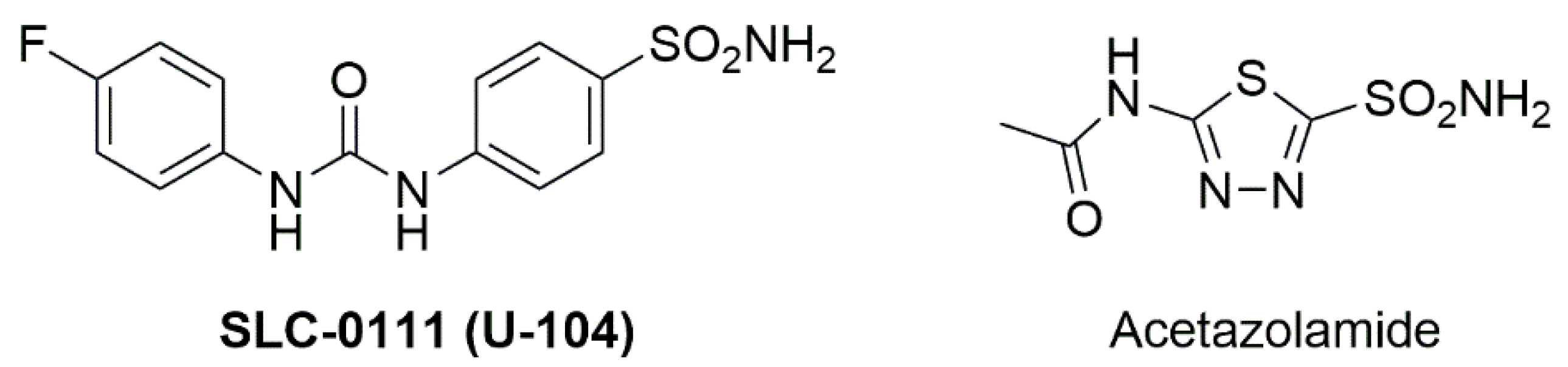
1. Introduction
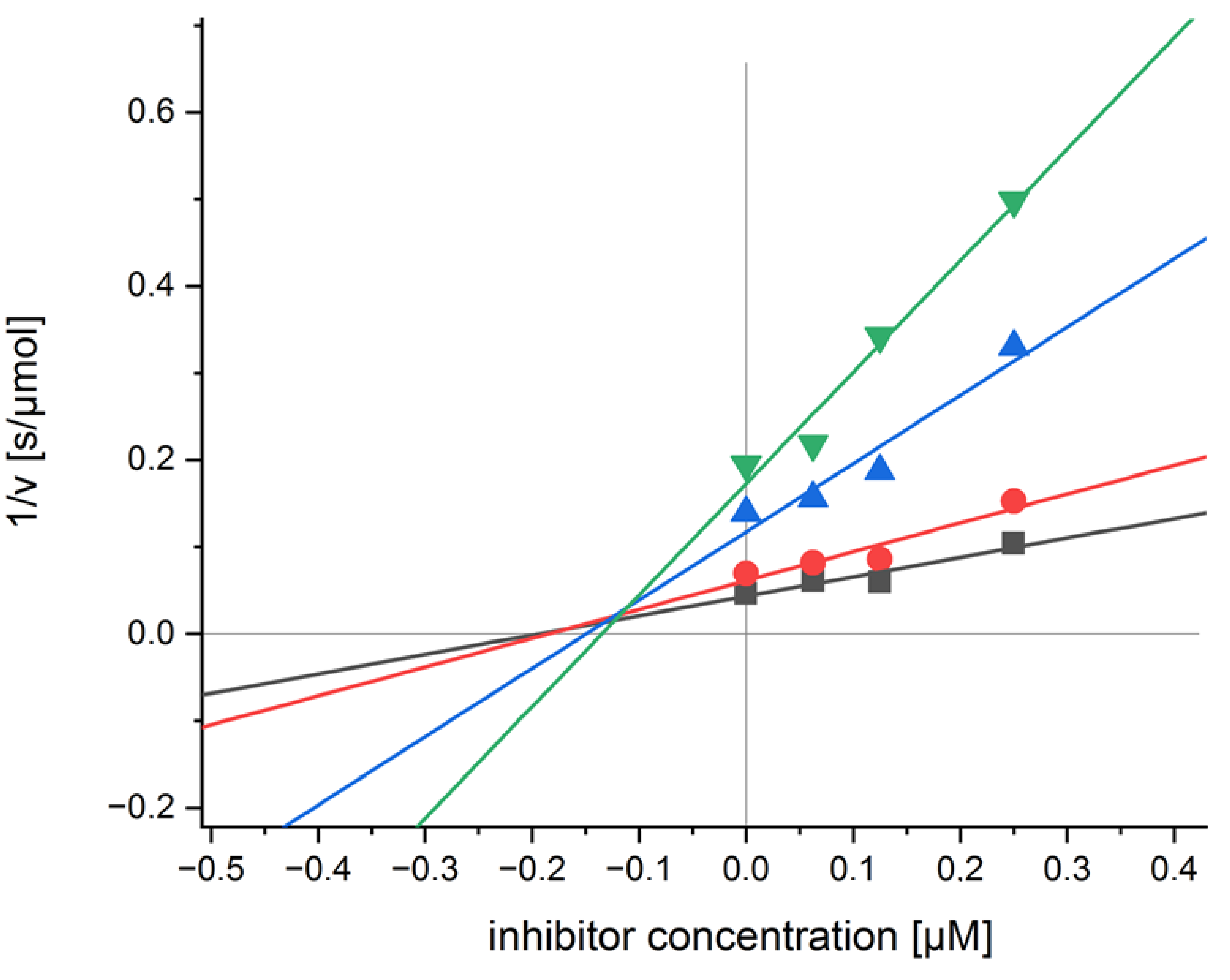
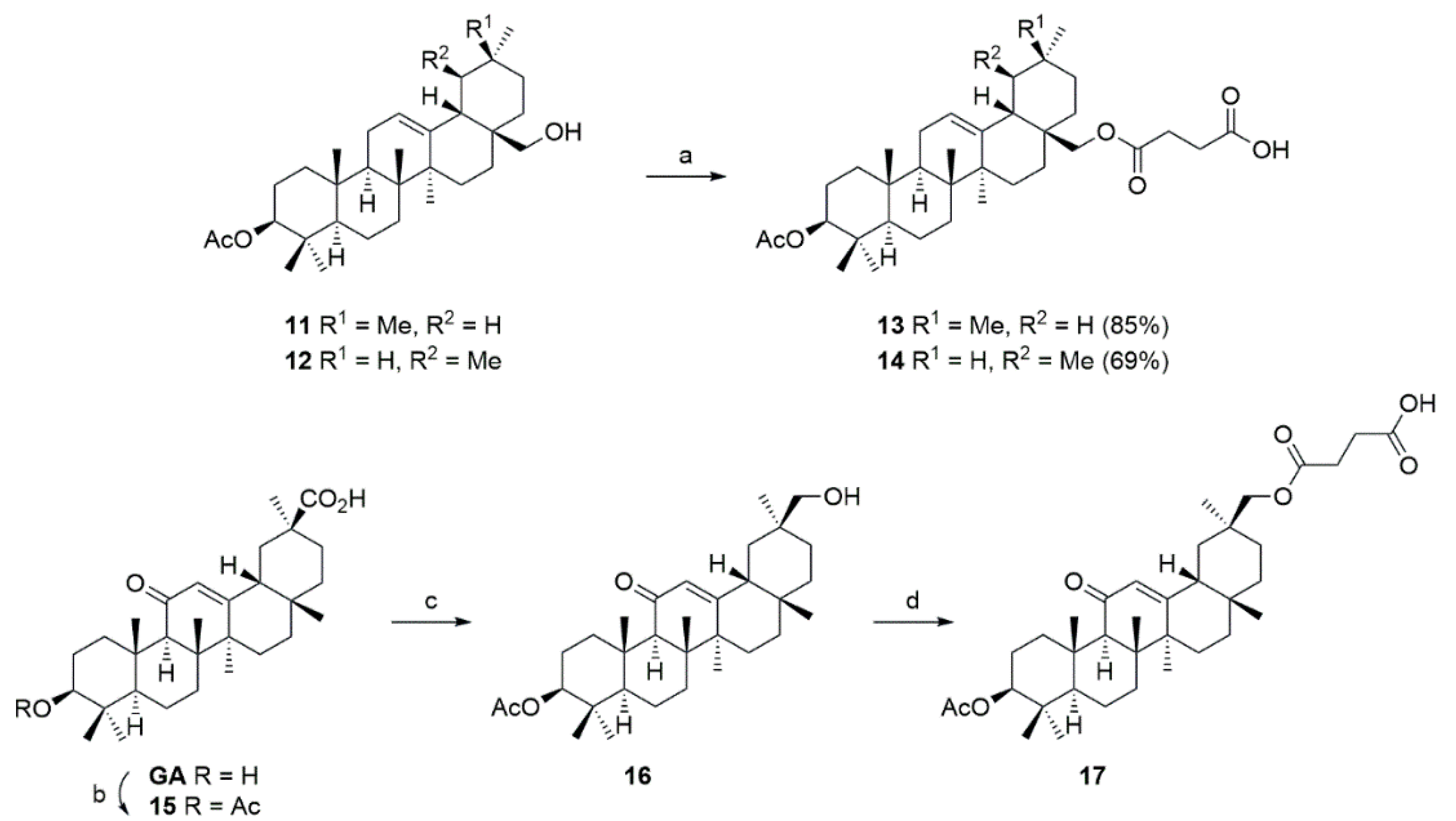
2. Results
3. Conclusions
4. Experimental
4.1. Di-O-Acetyl-betulin
4.2. 3-O-Acetyl-betulin
4.3. 4-{[(3β)-3-(Acetyloxy)lup-20(29)-en-28-yl]oxy}-4-oxobutanoic Acid
4.4. 5-Amino-1,3,4-thiadiazole-2-sulfonamide
4.5. (3β)-3-(Acetyloxy)lup-20(29)-en-28-yl 4-{[5-(aminosulfonyl)-1,3,4-thiadiazol-2-yl]amino}-4-oxobutanoate
4.6. (3β) Olean-12-en3-3,28-diol (Erythrodiol)
4.7. (3β) Urs-12-ene-3,28-diol (Uvaol, 8)
4.8. (3β)-Olean-12-ene-3,28-diyl Diacetate
4.9. (3β)-Urs-12-ene-3,28-diyl Diacetate
4.10. (3β)-28-Hydroxyolean-12-en-3-yl Acetate
4.11. (3β)-28-Hydroxyolean-12-en-3-yl Acetate
4.12. 4-{[(3β)-3-(Acetyloxy)-olean-12-en-28-yl]oxy}-4-oxobutanoic Acid
4.13. 4-{[(3β)-3-(Acetyloxy)urs-12-en-28-yl]oxy}-4-oxobutanoic Acid
4.14. (3β, 20β)-3-Acetyloxy-11-oxoolean-12-en-29-oic Acid
4.15. (3β, 20β) 3-Acetyloxy-29-hydroxyolean-12-en-11-one
4.16. 4-{[(3β,20β)3-(Acetyloxy)-11-oxoolean-12-en-30-yl]oxy}-4-oxobutanoic Acid
4.17. (3β) 3-(Acetyloxy)olean-12-en-28-yl-4-{[5-(aminosulfonyl)-1,3,4-thiadiazol-2-yl]amino}-4-oxobutanoate
4.18. (3β)-3-(Acetyloxy)urs-12-en-28-yl-4-{[5-(aminosulfonyl)-1,3,4-thiadiazol-2-yl]amino}4-oxobutanoate
4.19. (3β, 20β) 3-(Acetyloxy)-11-oxoolean-12-en-30-yl-4-{[5-(aminosulfonyl)-1,3,4-thiadiazol-2-yl] amino}-4-oxobutanoate
Author Contributions
Funding
Institutional Review Board Statement
Informed Consent Statement
Data Availability Statement
Acknowledgments
Conflicts of Interest
Sample Availability
References
- Supuran, C.T. Carbonic anhydrases—An overview. Curr. Pharm. Des. 2008, 14, 603–614. [Google Scholar] [CrossRef]
- Supuran, C.T. Carbonic anhydrase inhibitors. Bioorg. Med. Chem. Lett. 2010, 20, 3467–3474. [Google Scholar] [CrossRef] [PubMed]
- Angeli, A.; Carta, F.; Nocentini, A.; Winum, J.-Y.; Zalubovskis, R.; Onnis, V.; Eldehna, W.M.; Capasso, C.; Carradori, S.; Donald, W.A.; et al. Response to Perspectives on the Classical Enzyme Carbonic Anhydrase and the Search for Inhibitors. Biophys. J. 2021, 120, 178–181. [Google Scholar] [CrossRef]
- Berrino, E.; Michelet, B.; Martin-Mingot, A.; Carta, F.; Supuran, C.T.; Thibaudeau, S. Modulating the Efficacy of Carbonic Anhydrase Inhibitors through Fluorine Substitution. Angew. Chem. Int. Ed. 2021, 60, 23068–23082. [Google Scholar] [CrossRef] [PubMed]
- Kumar, S.; Rulhania, S.; Jaswal, S.; Monga, V. Recent advances in the medicinal chemistry of carbonic anhydrase inhibitors. Eur. J. Med. Chem. 2021, 209, 112923. [Google Scholar] [CrossRef] [PubMed]
- Supuran, C.T. Emerging role of carbonic anhydrase inhibitors. Clin. Sci. 2021, 135, 1233–1249. [Google Scholar] [CrossRef]
- Amedei, A.; Capasso, C.; Nannini, G.; Supuran, C.T. Microbiota, bacterial carbonic anhydrases, and modulators of their activity: Links to human diseases? Mediat. Inflamm. 2021, 926082. [Google Scholar] [CrossRef]
- Campestre, C.; De Luca, V.; Carradori, S.; Grande, R.; Carginale, V.; Scaloni, A.; Supuran, C.T.; Capasso, C. Carbonic Anhydrases: New Perspectives on Protein Functional Role and Inhibition in Helicobacter pylori. Front. Microbiol. 2021, 12, 629163. [Google Scholar] [CrossRef]
- Hines, K.M.; Chaudhari, V.; Edgeworth, K.N.; Owens, T.G.; Hanson, M.R. Absence of carbonic anhydrase in chloroplasts affects C3 plant development but not photosynthesis. Proc. Natl. Acad. Sci. USA 2021, 118, e2107425118. [Google Scholar] [CrossRef]
- Polishchuk, O.V. Stress-Related Changes in the Expression and Activity of Plant Carbonic Anhydrases. Planta 2021, 253, 58. [Google Scholar] [CrossRef]
- Rudenko, N.N.; Ignatova, L.K.; Nadeeva-Zhurikova, E.M.; Fedorchuk, T.P.; Ivanov, B.N.; Borisova-Mubarakshina, M.M. Advances in understanding the physiological role and locations of carbonic anhydrases in C3 plant cells. Protoplasma 2021, 258, 249–262. [Google Scholar] [CrossRef] [PubMed]
- Weerasooriya, H.N.; DiMario, R.J.; Rosati, V.C.; Rai, A.K.; LaPlace, L.M.; Filloon, V.D.; Longstreth, D.J.; Moroney, J.V. Arabidopsis plastid carbonic anhydrase βCA5 is important for normal plant growth. Plant Physiol. 2022, 190, 2173–2186. [Google Scholar] [CrossRef] [PubMed]
- Buabeng, E.R.; Henary, M. Developments of small molecules as inhibitors for carbonic anhydrase isoforms. Bioorg. Med. Chem. 2021, 39, 116140. [Google Scholar] [CrossRef] [PubMed]
- Elimam, D.M.; Elgazar, A.A.; Bonardi, A.; Abdelfadil, M.; Nocentini, A.; El-Domany, R.A.; Abdel-Aziz, H.A.; Badria, F.A.; Supuran, C.T.; Eldehna, W.M. Natural inspired piperine-based sulfonamides and carboxylic acids as carbonic anhydrase inhibitors: Design, synthesis and biological evaluation. Eur. J. Med. Chem. 2021, 225, 113800. [Google Scholar] [CrossRef] [PubMed]
- Kalinin, S.; Malkova, A.; Sharonova, T.; Sharoyko, V.; Bunev, A.; Supuran, C.T.; Krasavin, M. Carbonic Anhydrase IX Inhibitors as Candidates for Combination Therapy of Solid Tumors. Int. J. Mol. Sci. 2021, 22, 13405. [Google Scholar] [CrossRef]
- Nerella, S.G.; Singh, P.; Arifuddin, M.; Supuran, C.T. Anticancer carbonic anhydrase inhibitors: A patent and literature update 2018–2022. Expert Opin. Ther. Pat. 2022, 32, 833–847. [Google Scholar] [CrossRef]
- Shaldam, M.; Eldehna, W.M.; Nocentini, A.; Elsayed, Z.M.; Ibrahim, T.M.; Salem, R.; El-Domany, R.A.; Capasso, C.; Abdel-Aziz, H.A.; Supuran, C.T. Development of novel benzofuran-based SLC-0111 analogs as selective cancer-associated carbonic anhydrase isoform IX inhibitors. Eur. J. Med. Chem. 2021, 216, 113283. [Google Scholar] [CrossRef]
- Testa, C.; Papini, A.M.; Zeidler, R.; Vullo, D.; Carta, F.; Supuran, C.T.; Rovero, P. First studies on tumor associated carbonic anhydrases IX and XII monoclonal antibodies conjugated to small molecule inhibitors. J. Enzym. Inhib. Med. Chem. 2022, 37, 592–596. [Google Scholar] [CrossRef]
- Mincione, F.; Nocentini, A.; Supuran, C.T. Advances in the discovery of novel agents for the treatment of glaucoma. Expert Opin. Drug Discov. 2021, 16, 1209–1225. [Google Scholar] [CrossRef]
- Ozsoy, H.Z. Anticonvulsant Effects of Carbonic Anhydrase Inhibitors: The Enigmatic Link Between Carbonic Anhydrases and Electrical Activity of the Brain. Neurochem. Res. 2021, 46, 2783–2799. [Google Scholar] [CrossRef]
- Supuran, C.T. Novel carbonic anhydrase inhibitors. Future Med. Chem. 2021, 13, 1935–1937. [Google Scholar] [CrossRef] [PubMed]
- Akgul, O.; Lucarini, E.; Mannelli, L.D.C.; Ghelardini, C.; D’Ambrosio, K.; Buonanno, M.; Monti, S.M.; De Simone, G.; Angeli, A.; Supuran, C.T.; et al. Sultam based Carbonic Anhydrase VII inhibitors for the management of neuropathic pain. Eur. J. Med. Chem. 2022, 227, 113956. [Google Scholar] [CrossRef]
- Kumar, A.; Agarwal, P.; Rathi, E.; Kini, S.G. Computer-aided identification of human carbonic anhydrase isoenzyme VII inhibitors as potential antiepileptic agents. J. Biomol. Struct. Dyn. 2022, 40, 4850–4865. [Google Scholar] [CrossRef] [PubMed]
- Magheru, C.; Magheru, S.; Coltau, M.; Hoza, A.; Moldovan, C.; Sachelarie, L.; Gradinaru, I.; Hurjui, L.L.; Marc, F.; Farcas, D.M. Antiepileptic Drugs and Their Dual Mechanism of Action on Carbonic Anhydrase. J. Clin. Med. 2022, 11, 2614. [Google Scholar] [CrossRef] [PubMed]
- Ozaslan, M.S.; Saglamtas, R.; Demir, Y.; Genc, Y.; Saracoglu, I.; Guelcin, I. Isolation of Some Phenolic Compounds from Plantago subulata L. and Determination of Their Antidiabetic, Anticholinesterase, Antiepileptic and Antioxidant Activity. Chem. Biodivers. 2022, 19, e202200280. [Google Scholar] [CrossRef] [PubMed]
- Shukralla, A.A.; Dolan, E.; Delanty, N. Acetazolamide: Old drug, new evidence? Epilepsia Open 2022, 7, 378–392. [Google Scholar] [CrossRef]
- Das Mahapatra, A.; Queen, A.; Yousuf, M.; Khan, P.; Hussain, A.; Rehman, T.M.; Alajmi, M.F.; Datta, B.; Hassan, I.M. Design and development of 5-(4H)-oxazolones as potential inhibitors of human carbonic anhydrase VA: Towards therapeutic management of diabetes and obesity. J. Biomol. Struct. Dyn. 2022, 40, 3144–3154. [Google Scholar] [CrossRef] [PubMed]
- Mori, M.; Supuran, C.T. Acipimox inhibits human carbonic anhydrases. J. Enzym. Inhib. Med. Chem. 2022, 37, 672–679. [Google Scholar] [CrossRef]
- Supuran, C.T. Anti-obesity carbonic anhydrase inhibitors: Challenges and opportunities. J. Enzym. Inhib. Med. Chem. 2022, 37, 2478–2488. [Google Scholar] [CrossRef]
- Lemon, N.; Canepa, E.; Ilies, M.A.; Fossati, S. Carbonic anhydrases as potential targets against neurovascular unit dysfunction in Alzheimer’s disease and stroke. Front. Aging Neurosci. 2021, 13, 772278. [Google Scholar] [CrossRef]
- Poggetti, V.; Salerno, S.; Baglini, E.; Barresi, E.; Da Settimo, F.; Taliani, S. Carbonic Anhydrase Activators for Neurodegeneration: An Overview. Molecules 2022, 27, 2544. [Google Scholar] [CrossRef] [PubMed]
- Bonardi, A.; Micheli, L.; Di Cesare Mannelli, L.; Ghelardini, C.; Gratteri, P.; Nocentini, A.; Supuran, C.T. Development of Hydrogen Sulfide-Releasing Carbonic Anhydrases IX- and XII-Selective Inhibitors with Enhanced Antihyperalgesic Action in a Rat Model of Arthritis. J. Med. Chem. 2022, 65, 13143–13157. [Google Scholar] [CrossRef] [PubMed]
- Bulli, I.; Dettori, I.; Coppi, E.; Cherchi, F.; Venturini, M.; Mannelli, L.D.C.; Ghelardini, C.; Nocentini, A.; Supuran, C.T.; Pugliese, A.M.; et al. Role of carbonic anhydrase in cerebral ischemia and carbonic anhydrase inhibitors as putative protective agents. Int. J. Mol. Sci. 2021, 22, 5029. [Google Scholar] [CrossRef] [PubMed]
- Dettori, I.; Fusco, I.; Bulli, I.; Gaviano, L.; Coppi, E.; Cherchi, F.; Venturini, M.; Di Cesare Mannelli, L.; Ghelardini, C.; Nocentini, A.; et al. Protective effects of carbonic anhydrase inhibition in brain ischaemia in vitro and in vivo models. J. Enzym. Inhib. Med. Chem. 2021, 36, 964–976. [Google Scholar] [CrossRef]
- D’Agostino, I.; Mathew, G.E.; Angelini, P.; Venanzoni, R.; Angeles Flores, G.; Angeli, A.; Carradori, S.; Marinacci, B.; Menghini, L.; Abdelgawad, M.A.; et al. Biological investigation of N-methyl thiosemicarbazones as antimicrobial agents and bacterial carbonic anhydrases inhibitors. J. Enzym. Inhib. Med. Chem. 2022, 37, 986–993. [Google Scholar] [CrossRef] [PubMed]
- De Luca, V.; Carginale, V.; Supuran, C.T.; Capasso, C. The gram-negative bacterium Escherichia coli as a model for testing the effect of carbonic anhydrase inhibition on bacterial growth. J. Enzym. Inhib. Med. Chem. 2022, 37, 2092–2098. [Google Scholar] [CrossRef] [PubMed]
- Giovannuzzi, S.; Hewitt, C.S.; Nocentini, A.; Capasso, C.; Costantino, G.; Flaherty, D.P.; Supuran, C.T. Inhibition studies of bacterial α-carbonic anhydrases with phenols. J. Enzym. Inhib. Med. Chem. 2022, 37, 666–671. [Google Scholar] [CrossRef]
- Giovannuzzi, S.; Hewitt, C.S.; Nocentini, A.; Capasso, C.; Flaherty, D.P.; Supuran, C.T. Coumarins effectively inhibit bacterial α-carbonic anhydrases. J. Enzym. Inhib. Med. Chem. 2022, 37, 333–338. [Google Scholar] [CrossRef]
- Gueller, P.; Atmaca, U.; Gueller, U.; Calisir, U.; Dursun, F. Antibacterial properties and carbonic anhydrase inhibition profiles of azido sulfonyl carbamate derivatives. Future Med. Chem. 2021, 13, 1285–1299. [Google Scholar] [CrossRef]
- Hewitt, C.S.; Abutaleb, N.S.; Elhassanny, A.E.M.; Nocentini, A.; Cao, X.; Amos, D.P.; Youse, M.S.; Holly, K.J.; Marapaka, A.; An, W.; et al. Structure-Activity Relationship Studies of Acetazolamide-Based Carbonic Anhydrase Inhibitors with Activity against Neisseria gonorrhoeae. ACS Infect. Dis. 2021, 7, 1969–1984. [Google Scholar] [CrossRef]
- Nocentini, A.; Hewitt, C.S.; Mastrolorenzo, M.D.; Flaherty, D.P.; Supuran, C.T. Anion inhibition studies of the α-carbonic anhydrases from Neisseria gonorrhoeae. J. Enzym. Inhib. Med. Chem. 2021, 36, 1061–1066. [Google Scholar] [CrossRef] [PubMed]
- Flaherty, D.P.; Seleem, M.N.; Supuran, C.T. Bacterial carbonic anhydrases: Underexploited antibacterial therapeutic targets. Future Med. Chem. 2021, 13, 1619–1622. [Google Scholar] [CrossRef] [PubMed]
- Artasensi, A.; Angeli, A.; Lammi, C.; Bollati, C.; Gervasoni, S.; Baron, G.; Matucci, R.; Supuran, C.T.; Vistoli, G.; Fumagalli, L. Discovery of a Potent and Highly Selective Dipeptidyl Peptidase IV and Carbonic Anhydrase Inhibitor as “Antidiabesity” Agents Based on Repurposing and Morphing of WB-4101. J. Med. Chem. 2022, 65, 13946–13966. [Google Scholar] [CrossRef] [PubMed]
- Carradori, S.; Fantacuzzi, M.; Ammazzalorso, A.; Angeli, A.; De Filippis, B.; Galati, S.; Petzer, A.; Petzer, J.P.; Poli, G.; Tuccinardi, T.; et al. Resveratrol Analogues as Dual Inhibitors of Monoamine Oxidase B and Carbonic Anhydrase VII: A New Multi-Target Combination for Neurodegenerative Diseases? Molecules 2022, 27, 7816. [Google Scholar] [CrossRef] [PubMed]
- Kalayci, M.; Tuerkes, C.; Arslan, M.; Demir, Y.; Beydemir, S. Novel benzoic acid derivatives: Synthesis and biological evaluation as multitarget acetylcholinesterase and carbonic anhydrase inhibitors. Arch. Pharm. 2021, 354, 2000282. [Google Scholar] [CrossRef] [PubMed]
- Lenci, E.; Angeli, A.; Calugi, L.; Innocenti, R.; Carta, F.; Supuran, C.T.; Trabocchi, A. Multitargeting application of proline-derived peptidomimetics addressing cancer-related human matrix metalloproteinase 9 and carbonic anhydrase II. Eur. J. Med. Chem. 2021, 214, 113260. [Google Scholar] [CrossRef] [PubMed]
- Supuran, C.T. Multitargeting approaches involving carbonic anhydrase inhibitors: Hybrid drugs against a variety of disorders. J. Enzym. Inhib. Med. Chem. 2021, 36, 1702–1714. [Google Scholar] [CrossRef]
- Elbadawi, M.M.; Eldehna, W.M.; Nocentini, A.; Abo-Ashour, M.F.; Elkaeed, E.B.; Abdelgawad, M.A.; Alharbi, K.S.; Abdel-Aziz, H.A.; Supuran, C.T.; Gratteri, P.; et al. Identification of N-phenyl-2-(phenylsulfonyl)acetamides/propanamides as new SLC-0111 analogues: Synthesis and evaluation of the carbonic anhydrase inhibitory activities. Eur. J. Med. Chem. 2021, 218, 113360. [Google Scholar] [CrossRef]
- Huo, Z.; Bilang, R.; Supuran, C.T.; von der Weid, N.; Bruder, E.; Holland-Cunz, S.; Martin, I.; Muraro, M.G.; Gros, S.J. Perfusion-Based Bioreactor Culture and Isothermal Microcalorimetry for Preclinical Drug Testing with the Carbonic Anhydrase Inhibitor SLC-0111 in Patient-Derived Neuroblastoma. Int. J. Mol. Sci. 2022, 23, 3128. [Google Scholar] [CrossRef]
- Kumar, A.; Siwach, K.; Supuran, C.T.; Sharma, P.K. A decade of tail-approach based design of selective as well as potent tumor associated carbonic anhydrase inhibitors. Bioorg. Chem. 2022, 126, 105920. [Google Scholar] [CrossRef]
- Mboge, M.Y.; Combs, J.; Singh, S.; Andring, J.; Wolff, A.; Tu, C.; Zhang, Z.; McKenna, R.; Frost, S.C. Inhibition of Carbonic Anhydrase Using SLC-149: Support for a Noncatalytic Function of CAIX in Breast Cancer. J. Med. Chem. 2021, 64, 1713–1724. [Google Scholar] [CrossRef] [PubMed]
- Mussi, S.; Rezzola, S.; Chiodelli, P.; Nocentini, A.; Supuran, C.T.; Ronca, R. Antiproliferative effects of sulphonamide carbonic anhydrase inhibitors C18, SLC-0111 and acetazolamide on bladder, glioblastoma and pancreatic cancer cell lines. J. Enzym. Inhib. Med. Chem. 2022, 37, 280–286. [Google Scholar] [CrossRef] [PubMed]
- Supuran, C.T. Carbonic anhydrase inhibitors: An update on experimental agents for the treatment and imaging of hypoxic tumors. Expert Opin. Invest. Drugs 2021, 30, 1197–1208. [Google Scholar] [CrossRef]
- McDonald, P.C.; Chia, S.; Bedard, P.L.; Chu, Q.; Lyle, M.; Tang, L.; Singh, M.; Zhang, Z.; Supuran, C.T.; Renouf, D.J.; et al. A Phase 1 Study of SLC-0111, a Novel Inhibitor of Carbonic Anhydrase IX, in Patients With Advanced Solid Tumors. Am. J. Clin. Oncol. 2020, 43, 484–490. [Google Scholar] [CrossRef]
- Amiri, A.; Le, P.U.; Moquin, A.; Machkalyan, G.; Petrecca, K.; Gillard, J.W.; Yoganathan, N.; Maysinger, D. Inhibition of carbonic anhydrase IX in glioblastoma multiforme. Eur. J. Pharm. Biopharm. 2016, 109, 81–92. [Google Scholar] [CrossRef] [PubMed]
- Haapasalo, J.; Hilvo, M.; Nordfors, K.; Haapasalo, H.; Parkkila, S.; Hyrskyluoto, A.; Rantala, I.; Waheed, A.; Sly, W.S.; Pastorekova, S.; et al. Identification of an alternatively spliced isoform of carbonic anhydrase XII in diffusely infiltrating astrocytic gliomas. Neuro Oncol. 2008, 10, 131–138. [Google Scholar] [CrossRef] [PubMed]
- Haapasalo, J.; Nordfors, K.; Jarvela, S.; Bragge, H.; Rantala, I.; Parkkila, A.-K.; Haapasalo, H.; Parkkila, S. Carbonic anhydrase II in the endothelium of glial tumors: A potential target for therapy. Neuro Oncol. 2007, 9, 308–313. [Google Scholar] [CrossRef] [PubMed]
- Ihnatko, R.; Kubes, M.; Takacova, M.; Sedlakova, O.; Sedlak, J.; Pastorek, J.; Kopacek, J.; Pastorekova, S. Extracellular acidosis elevates carbonic anhydrase IX in human glioblastoma cells via transcriptional modulation that does not depend on hypoxia. Int. J. Oncol. 2006, 29, 1025–1033. [Google Scholar] [CrossRef]
- Ivanov, S.; Liao, S.-Y.; Ivanova, A.; Danilkovitch-Miagkova, A.; Tarasova, N.; Weirich, G.; Merrill, M.J.; Proescholdt, M.A.; Oldfield, E.H.; Lee, J.; et al. Expression of hypoxia-inducible cell-surface transmembrane carbonic anhydrases in human cancer. Am. J. Pathol. 2001, 158, 905–919. [Google Scholar] [CrossRef] [PubMed]
- Korkolopoulou, P.; Perdiki, M.; Thymara, I.; Boviatsis, E.; Agrogiannis, G.; Kotsiakis, X.; Angelidakis, D.; Rologis, D.; Diamantopoulou, K.; Thomas-Tsagli, E.; et al. Expression of hypoxia-related tissue factors in astrocytic gliomas. A multivariate survival study with emphasis upon carbonic anhydrase IX. Hum. Pathol. 2007, 38, 629–638. [Google Scholar] [CrossRef] [PubMed]
- Nordfors, K.; Haapasalo, J.; Korja, M.; Niemela, A.; Laine, J.; Parkkila, A.-K.; Pastorekova, S.; Pastorek, J.; Waheed, A.; Sly, W.S.; et al. The tumour-associated carbonic anhydrases CA II, CA IX and CA XII in a group of medulloblastomas and supratentorial primitive neuroectodermal tumours: An association of CA IX with poor prognosis. BMC Cancer 2010, 10, 148. [Google Scholar] [CrossRef] [PubMed]
- Proescholdt, M.A.; Mayer, C.; Kubitza, M.; Schubert, T.; Liao, S.-Y.; Stanbridge, E.J.; Ivanov, S.; Oldfield, E.H.; Brawanski, A.; Merrill, M.J. Expression of hypoxia-inducible carbonic anhydrases in brain tumors. Neuro Oncol. 2005, 7, 465–475. [Google Scholar] [CrossRef] [PubMed]
- Proescholdt, M.A.; Merrill, M.J.; Stoerr, E.-M.; Lohmeier, A.; Pohl, F.; Brawanski, A. Function of carbonic anhydrase IX in glioblastoma multiforme. Neuro Oncol. 2012, 14, 1357–1366. [Google Scholar] [CrossRef] [PubMed]
- Said, H.M.; Supuran, C.T.; Hageman, C.; Staab, A.; Polat, B.; Katzer, A.; Scozzafava, A.; Anacker, J.; Flentje, M.; Vordermark, D. Modulation of carbonic anhydrase 9 (CA9) in human brain cancer. Curr. Pharm. Des. 2010, 16, 3288–3299. [Google Scholar] [CrossRef]
- Boyd, N.H.; Walker, K.; Fried, J.; Hackney, J.R.; McDonald, P.C.; Benavides, G.A.; Spina, R.; Audia, A.; Scott, S.E.; Landis, C.J.; et al. Addition of carbonic anhydrase 9 inhibitor SLC-0111 to temozolomide treatment delays glioblastoma growth in vivo. JCI Insight 2017, 2, e92928. [Google Scholar] [CrossRef]
- Mujumdar, P.; Kopecka, J.; Bua, S.; Supuran, C.T.; Riganti, C.; Poulsen, S.-A. Carbonic Anhydrase XII Inhibitors Overcome Temozolomide Resistance in Glioblastoma. J. Med. Chem. 2019, 62, 4174–4192. [Google Scholar] [CrossRef]
- Salaroglio, I.C.; Mujumdar, P.; Annovazzi, L.; Kopecka, J.; Mellai, M.; Schiffer, D.; Poulsen, S.-A.; Riganti, C. Carbonic anhydrase XII inhibitors overcome P-glycoprotein-mediated resistance to temozolomide in glioblastoma. Mol. Cancer Ther. 2018, 17, 2598–2609. [Google Scholar] [CrossRef]
- Tann, A.C.; Ashley, D.M.; López, G.Y.; Malinzak, M.; Friedman, H.S. Khasraw, M. Mangement of glioblastoma: State of the art andfuture directions. S. CA Cancer J. Clin. 2020, 70, 299–312. [Google Scholar] [CrossRef]
- Zhao, K.; Schaefer, A.; Zhang, Z.; Elsaesser, K.; Culmsee, C.; Zhong, L.; Pagenstecher, A.; Nimsky, C.; Bartsch, J.W. Inhibition of Carbonic Anhydrase 2 Overcomes Temozolomide Resistance in Glioblastoma Cells. Int. J. Mol. Sci. 2022, 23, 157. [Google Scholar] [CrossRef]
- Hannen, R.; Selmansberger, M.; Hauswald, M.; Pagenstecher, A.; Nist, A.; Stiewe, T.; Acker, T.; Carl, B.; Nimsky, C.; Bartsch, J.W. Comparative transcriptomic analysis of temozolomide resistant primary GBM stem-like cells and recurrent GBM identifies upo-regulation of carbonic anhydrase CA2 gene as resistance factor. Cancers 2019, 11, 921. [Google Scholar] [CrossRef]
- Schwarz, S.; Sommerwerk, S.; Lucas, S.D.; Heller, L.; Csuk, R. Sulfamates of methyl triterpenoates are effective and competitive inhibitors of carbonic anhydrase II. Eur. J. Med. Chem. 2014, 86, 95–102. [Google Scholar] [CrossRef] [PubMed]
- Rehman, N.U.; Halim, S.A.; Khan, A.; Khan, M.; Al-Hatmi, S.; Al-Harrasi, A. Commikuanoids A-C: New cycloartane triterpenoids with exploration of carbonic anhydrase-II inhibition from the resins of Commiphora kua by in vitro and in silico molecular docking. Fitoterapia 2022, 157, 105125. [Google Scholar] [CrossRef] [PubMed]
- Silva, V.A.O.; Rosa, M.N.; Miranda-Goncalves, V.; Costa, A.M.; Tansini, A.; Evangelista, A.F.; Martinho, O.; Carloni, A.C.; Jones, C.; Lima, J.P.; et al. Euphol, a tetracyclic triterpene, from Euphorbia tirucalli induces autophagy and sensitizes temozolomide cytotoxicity on glioblastoma cells. Invest. New Drugs 2019, 37, 223–237. [Google Scholar] [CrossRef] [PubMed]
- Avula, S.K.; Rehman, N.U.; Khan, M.; Halim, S.A.; Khan, A.; Rafiq, K.; Csuk, R.; Das, B.; Al-Harrasi, A. New synthetic 1H-1,2,3-triazole derivatives of 3-O-acetyl-β-boswellic acid and 3-O-acetyl-11-keto-β-boswellic acid from Boswellia sacra inhibit carbonic anhydrase II in vitro. Med. Chem. Res. 2021, 30, 1185–1198. [Google Scholar] [CrossRef]
- Bulbul, M.; Saracoglu, N.; Irfan Kufrevioglu, O.; Ciftci, M. Bile acid derivatives of 5-amino-1,3,4-thiadiazole-2-sulfonamide as new carbonic anhydrase inhibitors: Synthesis and investigation of inhibition effects. Bioorg. Med. Chem. 2002, 10, 2561–2567. [Google Scholar] [CrossRef] [PubMed]
- Chu, X.; Battle, C.H.; Zhang, N.; Aryal, G.H.; Mottamal, M.; Jayawickramarajah, J. Bile Acid Conjugated DNA Chimera that Conditionally Inhibits Carbonic Anhydrase-II in the Presence of MicroRNA-21. Bioconjugate Chem. 2015, 26, 1606–1612. [Google Scholar] [CrossRef]
- Scozzafava, A.; Supuran, C.T. Carbonic anhydrase inhibitors. Preparation of potent sulfonamides inhibitors incorporating bile acid tails. Bioorg. Med. Chem. Lett. 2002, 12, 1551–1557. [Google Scholar] [CrossRef]
- Trifunovic, J.; Borcic, V.; Mikov, M. Bile acids and their oxo derivatives: Potential inhibitors of carbonic anhydrase I and II, androgen receptor antagonists and CYP3A4 substrates. Biomed. Chromatogr. 2017, 31, 3870. [Google Scholar] [CrossRef]
- Kalyanavenkatapaman, S.; Nanjen, P.; Banerji, A.; Nair, B.C.; Kumar, G.B. Discovery of arjunolic acid as a novel non-zinc binding carbonic anhydrase II inhibitor. Bioorg. Chem. 2016, 66, 72–79. [Google Scholar] [CrossRef]
- Pastorekova, S.; Gillies, R.J.M. The role of carbonic anhydrase IX in cancer development: Links to hypxia, acidosis and beyond. Cancer Metastasis Rev. 2019, 38, 65–77. [Google Scholar] [CrossRef]
- Vanchanagiri, K.; Emmerich, D.; Brusche, M.; Bache, M.; Seifert, F.; Csuk, R.; Vordermark, D.; Paschke, R. Synthesis and biological investigation of new carbonic anhydrase IX (CAIX) inhibitors. Chem. Biol. Interact. 2018, 284, 12–23. [Google Scholar] [CrossRef] [PubMed]
- Annan, D.A.; Maishi, N.; Soga, T.; Bawood, R.; Li, C.; Kikuchi, H.K.; Hajo, T.; Morimoto, M.; Kitamura, T.; Alam, M.T.; et al. Carbonic anhydrase 2 (CA II) supports tumor blood endothelial cell survival under lactic acidosis in the tumor microenvironment. Cell Commun. Signal. 2019, 17, 169. [Google Scholar] [CrossRef]
- Bekku, S.; Mochizuki, H.; Takayama, E.; Shinomiya, N.; Fukamachi, H.; Ichinose, M.; Tadakuma, T.; Yamamoto, T. Carbonic anhydrase I and II as a differntiation marker of human and rat colonic enterocytes. Res. Exp. Med. 1998, 198, 175–185. [Google Scholar] [CrossRef] [PubMed]
- Noor, S.I.; Jamali, S.; Ames, S.; Langer, S.; Deitmer, J.W.; Becker, H.M. A surface proton antenna in carbonic anhydrase II supports lactate transport in cancer cells. eLife 2018, 7, e35176. [Google Scholar] [CrossRef] [PubMed]
- Thibeault, D.; Gauthier, C.; Legault, J.; Bouchard, J.; Dufour, P.; Pichette, A. Synthesis and structure–activity relationship study of cytotoxic germanicane- and lupane-type 3β-O-monodesmosidic saponins starting from betulin. Bioorganic Med. Chem. 2007, 15, 6144–6157. [Google Scholar] [CrossRef] [PubMed]
- Flekhter, O.B.; Karachurina, L.T.; Poroikov, V.V.; Nigmatullina, L.P.; Baltina, L.A.; Zarudii, F.S.; Davydova, V.A.; Spirikhin, L.V.; Baikova, I.P.; Galin, F.Z.; et al. The synthesis and hepatoprotective activity of esters of the lupane group triterpenoids. Russ. J. Bioorg. Chem. 2000, 26, 192–200. [Google Scholar] [CrossRef]
- Flekhter, O.B.; Medvedeva, N.I.; Karachurina, L.T.; Baltina, L.A.; Galin, F.Z.; Zarudii, F.S.; Tolstikov, G.A. Synthesis and Pharmacological Activity of Betulin, Betulinic Acid, and Allobetulin Esters. Pharm. Chem. J. 2005, 39, 401–404. [Google Scholar] [CrossRef]
- Kazakova, O.B.; Smirnova, I.E.; Baltina, L.A.; Boreko, E.I.; Savinova, O.V.; Pokrovskii, A.G. Antiviral Activity of Acyl Derivatives of Betulin and Betulinic and Dihydroquinopimaric Acids. Russ. J. Bioorg. Chem. 2018, 44, 740–744. [Google Scholar] [CrossRef]
- Komissarova, N.G.; Dubovitskii, S.N.; Orlov, A.V.; Shitikova, O.V. New Conjugates of Betulin with 2-Aminoethanesulfonic Acid. Chem. Nat. Compd. 2019, 55, 300–304. [Google Scholar] [CrossRef]
- Pokrovskii, A.G.; Plyasunvoa, O.A.; Il’icheva, T.N.; Borisova, O.A.; Fedyuk, N.V.; Petrenko, N.I.; Petukhova, V.Z.; Shul’ts, E.E.; Tolstikov, G.A. Synthesis of derivatives of plant triterpenes and study of their antiviral and immunostimulating activity. Khim. Interes. Ustoich. Razvit. 2001, 9, 485–491. [Google Scholar]
- Shintyapina, A.B.; Shults, E.E.; Petrenko, N.I.; Uzenkova, N.V.; Tolstikov, G.A.; Pronkina, N.V.; Kozhevnikov, V.S.; Pokrovsky, A.G. Effect of nitrogen-containing derivatives of the plant triterpenes betulin and glycyrrhetic acid on the growth of MT-4, MOLT-4, CEM, and Hep G2 tumor cells. Russ. J. Bioorg. Chem. 2007, 33, 579–583. [Google Scholar] [CrossRef]
- Roblin, R.O., Jr.; Clapp, J.W. The preparation of heterocyclic sulfonamides. J. Am. Chem. Soc. 1950, 72, 4890–4892. [Google Scholar] [CrossRef]
- Kazakova, O.B.; Giniyatullina, G.V.; Tolstikov, G.A.; Baikova, I.P.; Zaprutko, L.; Apryshko, G.N. Synthesis and antitumor activity of aminopropoxy derivatives of betulin, erythrodiol, and uvaol. Russ. J. Bioorg. Chem. 2011, 37, 369–379. [Google Scholar] [CrossRef] [PubMed]
- Zimmermann, J. A monostearic ester of a triterpenediol from the fruits of Erythroxylon novogranatense. Recl. Trav. Chim. Pays-Bas Belg. 1932, 51, 1200–1203. [Google Scholar] [CrossRef]
- Rollinger, J.M.; Kratschmar, D.V.; Schuster, D.; Pfisterer, P.H.; Gumy, C.; Aubry, E.M.; Brandstoetter, S.; Stuppner, H.; Wolber, G.; Odermatt, A. 11β-Hydroxysteroid dehydrogenase 1 inhibiting constituents from Eriobotrya japonica revealed by bioactivity-guided isolation and computational approaches. Bioorg. Med. Chem. 2010, 18, 1507–1515. [Google Scholar] [CrossRef]
- Agarwal, K.P.; Roy, A.C.; Dhar, M.L. Triterpenes from the bark of Myrica esculenta. Indian J. Chem. 1963, 1, 28–30. [Google Scholar]
- Garcia-Granados, A.; Lopez, P.E.; Melguizo, E.; Parra, A.; Simeo, Y. Partial synthesis of C-ring derivatives from oleanolic and maslinic acids. Formation of several triene systems by chemical and photochemical isomerization processes. Tetrahedron 2004, 60, 1491–1503. [Google Scholar] [CrossRef]
- El-Seedi, H.R. Antimicrobial triterpenes from Poulsenia armata Mif. Standl. Nat. Prod. Res. 2005, 19, 197–202. [Google Scholar]
- Kim, M.-R.; Lee, H.-H.; Hahm, K.-S.; Moon, Y.-H.; Woo, E.-R. Pentacyclic triterpenoids and their cytotoxicity from the stem bark of Styrax japonica S. et Z. Arch. Pharmacal Res. 2004, 27, 283–286. [Google Scholar] [CrossRef]
- Alves, H.M.; Arndt, V.H.; Ollis, W.D.; Eyton, W.B.; Gottlieb, O.R.; Magalhaes, M.T. Chemistry of Brazilian leguminosae. VIII. β-Amyrin constituents of Machaerium incorruptibile. An. Acad. Bras. Cienc. 1965, 37, 49–50. [Google Scholar]
- Sengupta, P.; Dey, A.K.; Mukherjee, J.; Ghosh, S.; Das, K.G. Terpenoids and related compounds. XVI. Terpenoids of the bark of Rhododendron falconeri. J. Indian Chem. Soc. 1969, 66, 775–778. [Google Scholar]
- Chen, D.; Xu, F.; Zhang, P.; Deng, J.; Sun, H.; Wen, X.; Liu, J. Practical Synthesis of α-Amyrin, β-Amyrin, and Lupeol: The Potential Natural Inhibitors of Human Oxidosqualene Cyclase. Arch. Pharm. 2017, 350, 1700178. [Google Scholar] [CrossRef] [PubMed]
- Beseda, I.; Czollner, L.; Shah, P.S.; Khunt, R.; Gaware, R.; Kosma, P.; Stanetty, C.; del Ruiz-Ruiz, M.C.; Amer, H.; Mereiter, K.; et al. Synthesis of glycyrrhetinic acid derivatives for the treatment of metabolic diseases. Bioorg. Med. Chem. 2010, 18, 433–454. [Google Scholar] [CrossRef] [PubMed]


| Compound | Inhibition [%] |
|---|---|
| 4 | 89.9 ± 0.6 |
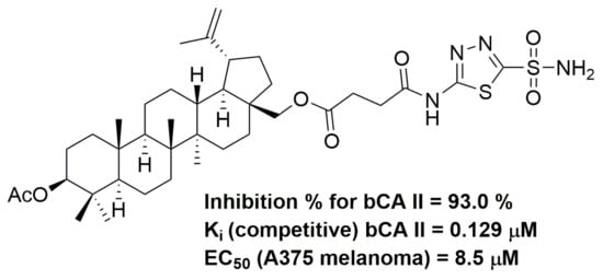
| 6 | 93.0 ± 0.1 |
| 18 | 49.4 ± 0.1 |
| 19 | 70.8 ± 0.2 |
| 20 | 96.8 ± 0.2 |
| Compound | Ki (in μM) |
|---|---|
| 6 | 0.129 ± 0.02 |
| 19 | 0.91 ± 0.17 |
| 20 | 5.22 ± 0.57 |
| A375 | HT29 | MCF-7 | A2780 | NIH 3T3 | |
|---|---|---|---|---|---|
| 4 | >30 | >30 | >30 | >30 | >30 |
| 6 | 8.5 ± 0.7 | 10.2 ± 1.3 | 8.9 ± 0.7 | 9.3 ± 1.2 | 9.5 ± 1.0 |
| 18 | 10.1 ± 0.8 | 14.2 ± 1.4 | 10.6 ± 1.4 | 11.8 ± 1.4 | 14.0 ± 1.5 |
| 19 | 13.7 ± 1.1 | 15.0 ± 0.6 | 12.4 ± 0.8 | 12.5 ± 1.7 | 14.8 ± 1.5 |
| 20 | 9.2 ± 0.5 | 13.0 ± 1.3 | 10.5 ± 1.2 | 9.8 ± 0.8 | 11.9 ± 1.7 |
| DX | n.d. | 0.25 ± 0.02 | 0.1 ± 0.01 | 0.1 ± 0.01 | 0.01 ± 0.001 |
Disclaimer/Publisher’s Note: The statements, opinions and data contained in all publications are solely those of the individual author(s) and contributor(s) and not of MDPI and/or the editor(s). MDPI and/or the editor(s) disclaim responsibility for any injury to people or property resulting from any ideas, methods, instructions or products referred to in the content. |
© 2023 by the authors. Licensee MDPI, Basel, Switzerland. This article is an open access article distributed under the terms and conditions of the Creative Commons Attribution (CC BY) license (https://creativecommons.org/licenses/by/4.0/).
Share and Cite
Denner, T.C.; Heise, N.; Zacharias, J.; Kraft, O.; Hoenke, S.; Csuk, R. Small Structural Differences Govern the Carbonic Anhydrase II Inhibition Activity of Cytotoxic Triterpene Acetazolamide Conjugates. Molecules 2023, 28, 1009. https://doi.org/10.3390/molecules28031009
Denner TC, Heise N, Zacharias J, Kraft O, Hoenke S, Csuk R. Small Structural Differences Govern the Carbonic Anhydrase II Inhibition Activity of Cytotoxic Triterpene Acetazolamide Conjugates. Molecules. 2023; 28(3):1009. https://doi.org/10.3390/molecules28031009
Chicago/Turabian StyleDenner, Toni C., Niels Heise, Julian Zacharias, Oliver Kraft, Sophie Hoenke, and René Csuk. 2023. "Small Structural Differences Govern the Carbonic Anhydrase II Inhibition Activity of Cytotoxic Triterpene Acetazolamide Conjugates" Molecules 28, no. 3: 1009. https://doi.org/10.3390/molecules28031009
APA StyleDenner, T. C., Heise, N., Zacharias, J., Kraft, O., Hoenke, S., & Csuk, R. (2023). Small Structural Differences Govern the Carbonic Anhydrase II Inhibition Activity of Cytotoxic Triterpene Acetazolamide Conjugates. Molecules, 28(3), 1009. https://doi.org/10.3390/molecules28031009