


Naturally Inspired Molecules for Neuropathic Pain Inhibition—Effect of Mirogabalin and Cebranopadol on Mechanical and Thermal Nociceptive Threshold in Mice


Abstract
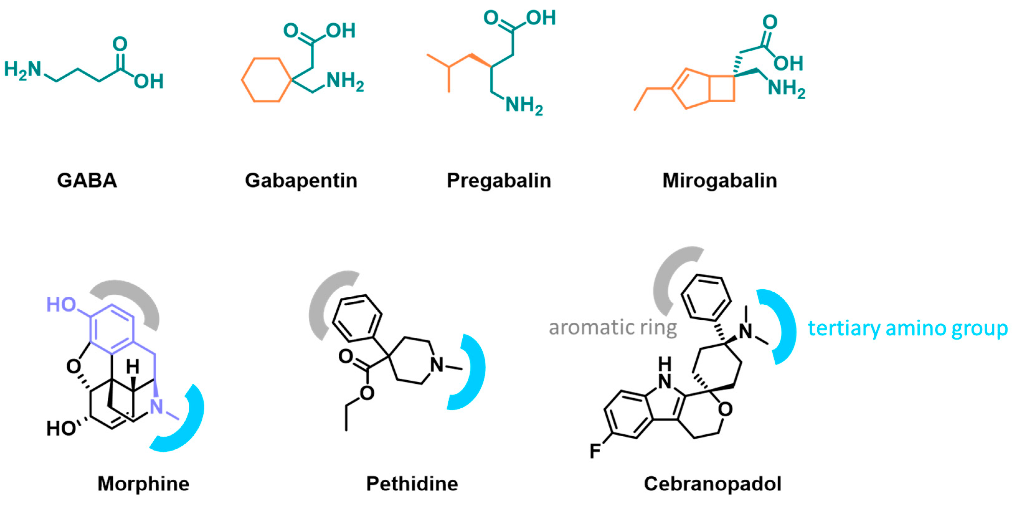
1. Introduction
2. Results
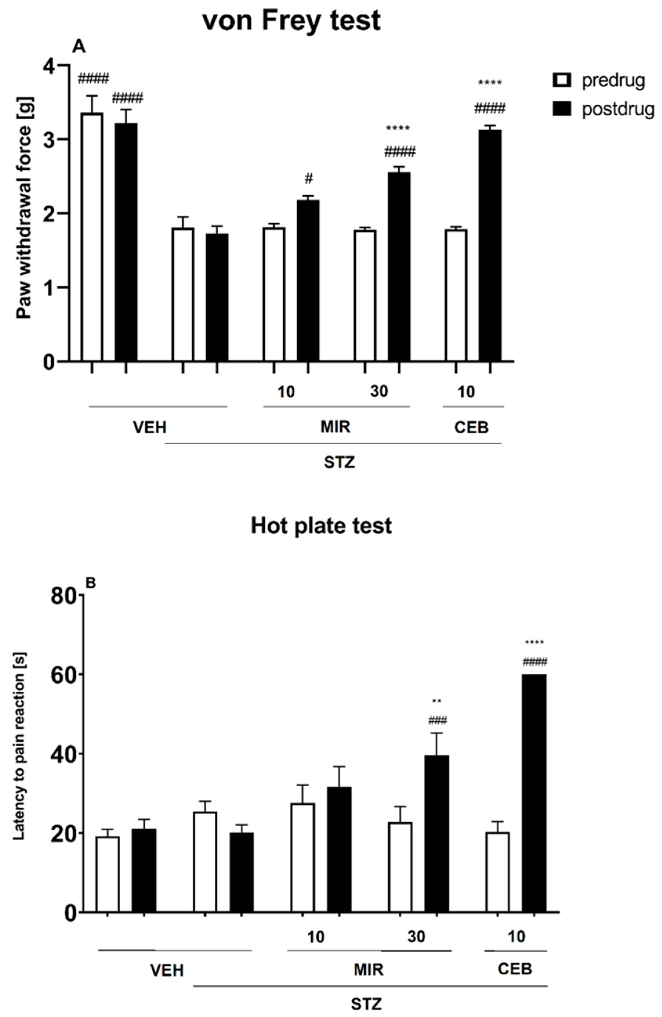
2.1. STZ Model
2.1.1. Effect of Mirogabalin and Cebranopadol on the Mechanical Nociceptive Threshold (von Frey Test)
2.1.2. Effect of Mirogabalin and Cebranopadol on the Heat Nociceptive Threshold (Hot Plate Test)
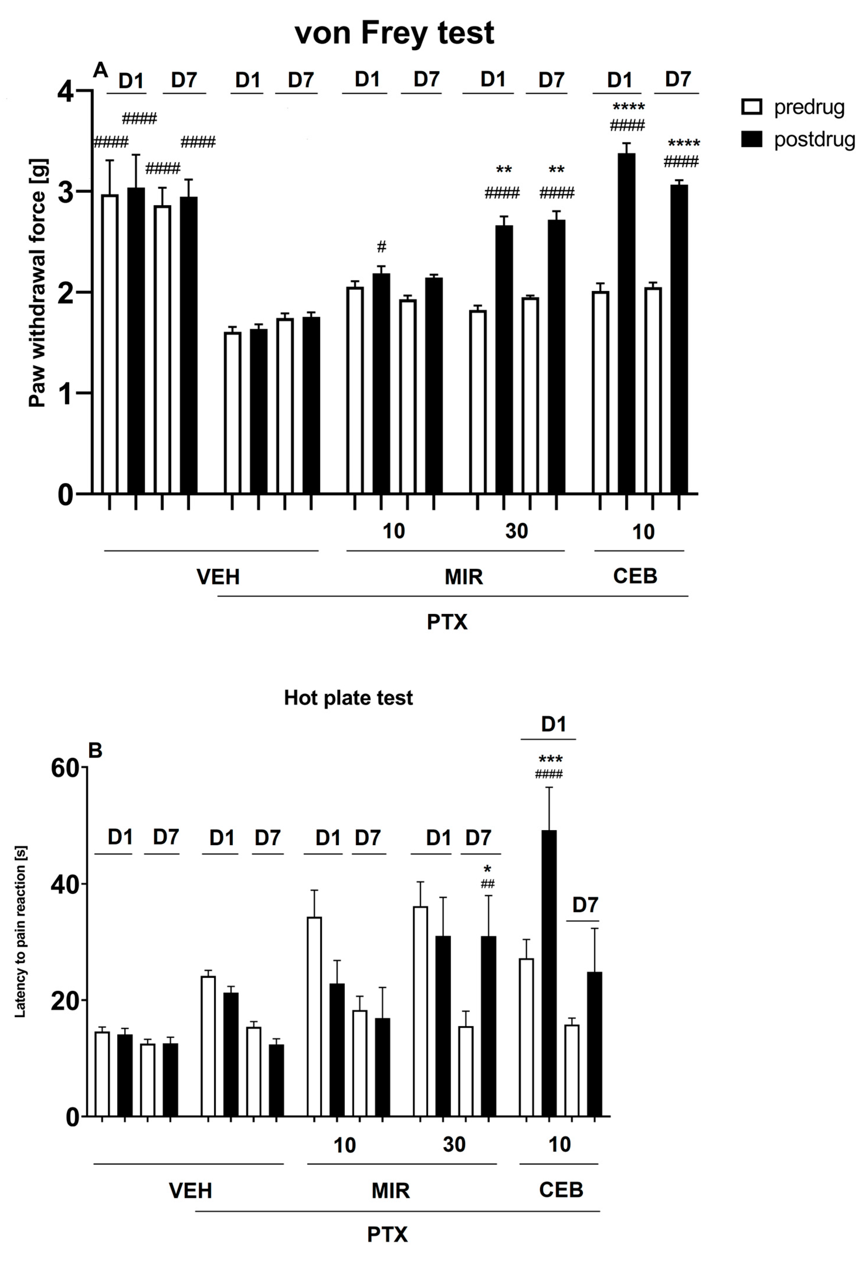
2.2. Paclitaxel Model
2.2.1. Effect of Mirogabalin and Cebranopadol on the Mechanical Nociceptive Threshold (von Frey Test)
2.2.2. Effect of Mirogabalin and Cebranopadol on the Heat Nociceptive Threshold (Hot Plate Test)
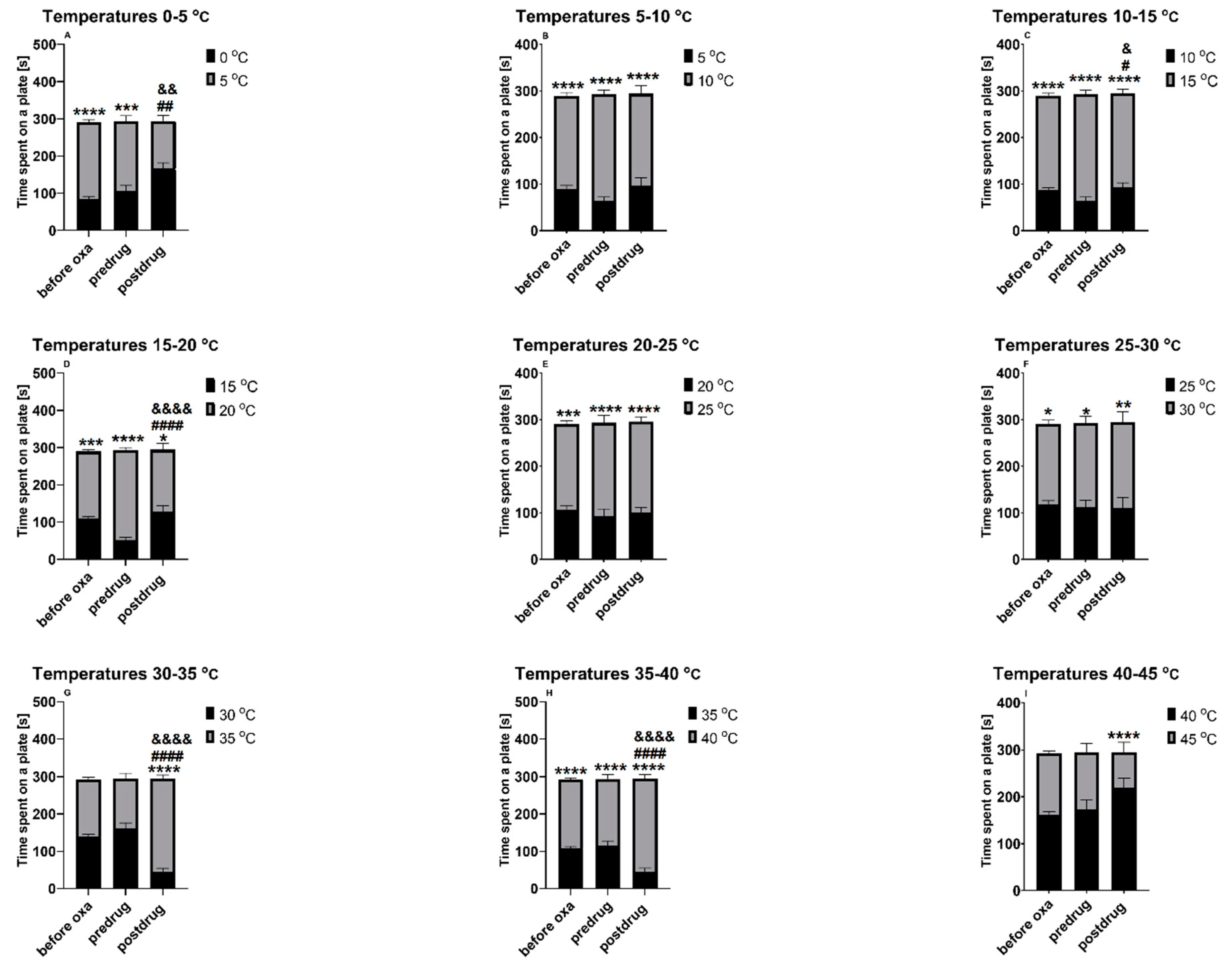
2.3. Oxaliplatin Model
2.3.1. Effect of Mirogabalin on the Thermal Place Preference of Mice (Two-Plate Thermal Place Preference Test)
2.3.2. Effect of Cebranopadol on the Thermal Place Preference of Mice (Two-Plate Thermal Place Preference Test)
3. Discussion
4. Materials and Methods
4.1. Animals and Housing Conditions
4.2. Chemicals
4.3. Behavioral Testing Protocol
4.3.1. Induction of Neuropathy—STZ Model (Diabetic Neuropathy Model)
4.3.2. Induction of Neuropathy—Paclitaxel Model (CIPN Model)
4.3.3. Induction of Neuropathy—Oxaliplatin Model (CIPN Model)
4.3.4. Assessment of Mechanical Nociceptive Threshold (von Frey Test)
4.3.5. Assessment of Heat Nociceptive Threshold (Hot Plate Test)
4.3.6. Assessment of Thermal Nociceptive Threshold (Two-Plate Thermal Place Preference Test)
4.3.7. Data Analysis
5. Conclusions and Future Outlook
Author Contributions
Funding
Institutional Review Board Statement
Informed Consent Statement
Data Availability Statement
Acknowledgments
Conflicts of Interest
Abbreviations
| CEB | Cebranopadol |
| CIPN | Chemotherapy-induced peripheral neuropathy |
| CNS | Central nervous system |
| COX2 | Cyclooxygenase isoform 2 |
| DOR | δ-opioid receptor |
| GABA | γ-aminobutyric acid |
| KOR | κ-opioid receptor |
| MOR | µ-opioid receptor |
| MIR | Mirogabalin |
| NF-κB p50 subunit | Nuclear factor kappa B p50 subunit |
| NOR | Nociceptin/orphanin FQ receptor |
| PGES | Prostaglandin E synthase |
| PTX | Paclitaxel |
| STZ | Streptozotocin |
| SVC | Support vector classification |
| VGCCs | Voltage-gated calcium channels |
References
- Jensen, T.S.; Finnerup, N.B. Allodynia and Hyperalgesia in Neuropathic Pain: Clinical Manifestations and Mechanisms. Lancet Neurol. 2014, 13, 924–935. [Google Scholar] [CrossRef] [PubMed]
- Binder, A.; Baron, R. The Pharmacological Therapy of Chronic Neuropathic Pain. Dtsch. Ärztebl. Int. 2016, 113, 616–625. [Google Scholar] [CrossRef] [PubMed]
- Ziemichod, W.; Kotlinska, J.; Gibula-Tarlowska, E.; Karkoszka, N.; Kedzierska, E. Cebranopadol as a Novel Promising Agent for the Treatment of Pain. Molecules 2022, 27, 3987. [Google Scholar] [CrossRef]
- Kim, J.-Y.; Abdi, S.; Huh, B.; Kim, K.-H. Mirogabalin: Could It Be the next Generation Gabapentin or Pregabalin? Korean J. Pain 2021, 34, 4–18. [Google Scholar] [CrossRef] [PubMed]
- Calandre, E.P.; Rico-Villademoros, F.; Slim, M. Alpha2 Delta Ligands, Gabapentin, Pregabalin and Mirogabalin: A Review of Their Clinical Pharmacology and Therapeutic Use. Expert. Rev. Neurother. 2016, 16, 1263–1277. [Google Scholar] [CrossRef] [PubMed]
- Javed, S.; Alam, U.; Malik, R.A. Mirogabalin and Emerging Therapies for Diabetic Neuropathy. J. Pain Res. 2018, 11, 1559–1566. [Google Scholar] [CrossRef]
- Kozai, D.; Numoto, N.; Nishikawa, K.; Kamegawa, A.; Kawasaki, S.; Hiroaki, Y.; Irie, K.; Oshima, A.; Hanzawa, H.; Shimada, K.; et al. Recognition Mechanism of a Novel Gabapentinoid Drug, Mirogabalin, for Recombinant Human A2δ1, a Voltage-Gated Calcium Channel Subunit. J. Mol. Biol. 2023, 435, 168049. [Google Scholar] [CrossRef]
- Andersen, K.E.; Sørensen, J.L.; Huusfeldt, P.O.; Knutsen, L.J.S.; Lau, J.; Lundt, B.F.; Petersen, H.; Suzdak, P.D.; Swedberg, M.D.B. Synthesis of Novel GABA Uptake Inhibitors. 4. Bioisosteric Transformation and Successive Optimization of Known GABA Uptake Inhibitors Leading to a Series of Potent Anticonvulsant Drug Candidates. J. Med. Chem. 1999, 42, 4281–4291. [Google Scholar] [CrossRef]
- Zhang, Y.; Zheng, Y.; Wu, Q.; Tian, F.; Ma, C.; Xu, H.; Zhan, L.; Gao, Z.; Zhao, G.; Ti, H. Study on Structure-Activity Relationship (SAR) of Simplified Mirogabalin Derivatives as Voltage-Gated Calcium Channel A2δ Ligands for the Treatment of Chronic Neuropathic Pain. Med. Chem. Res. 2023, 32, 288–313. [Google Scholar] [CrossRef]
- Li, Y.; Toyama, K.; Nakatsu, T.; Ishizuka, H.; Wu, H.; Cao, G.; Yu, J.; Wang, Y.; Liu, X.; Guo, B.; et al. Safety, Tolerability and Pharmacokinetics of Single and Multiple Doses of Mirogabalin in Healthy Chinese Participants: A Randomized, Double-Blind, Placebo-Controlled Study. Adv. Ther. 2023, 40, 1628–1643. [Google Scholar] [CrossRef]
- Schunk, S.; Linz, K.; Frormann, S.; Hinze, C.; Oberbörsch, S.; Sundermann, B.; Zemolka, S.; Englberger, W.; Germann, T.; Christoph, T.; et al. Discovery of Spiro[Cyclohexane-Dihydropyrano[3,4-b]Indole]-Amines as Potent NOP and Opioid Receptor Agonists. ACS Med. Chem. Lett. 2014, 5, 851–856. [Google Scholar] [CrossRef] [PubMed]
- Toll, L.; Cippitelli, A.; Ozawa, A. The NOP Receptor System in Neurological and Psychiatric Disorders: Discrepancies, Peculiarities and Clinical Progress in Developing Targeted Therapies. CNS Drugs 2021, 35, 591–607. [Google Scholar] [CrossRef] [PubMed]
- Lednicer, D.; Von Voigtlander, P.F.; Emmert, D.E. 4-Amino-4-Arylcyclohexanones and Their Derivatives, a Novel Class of Analgesics. 1. Modification of the Aryl Ring. J. Med. Chem. 1980, 23, 424–430. [Google Scholar] [CrossRef] [PubMed]
- Lednicer, D.; Von Voigtlander, P.F.; Emmert, D.E. 4-Aryl-4-Aminocyclohexanones and Their Derivatives, a Novel Class of Analgesics. 3. m-Hydroxyphenyl Derivatives. J. Med. Chem. 1981, 24, 341–346. [Google Scholar] [CrossRef]
- Linz, K.; Christoph, T.; Tzschentke, T.M.; Koch, T.; Schiene, K.; Gautrois, M.; Schröder, W.; Kögel, B.Y.; Beier, H.; Englberger, W.; et al. Cebranopadol: A Novel Potent Analgesic Nociceptin/Orphanin FQ Peptide and Opioid Receptor Agonist. J. Pharmacol. Exp. Ther. 2014, 349, 535–548. [Google Scholar] [CrossRef] [PubMed]
- Wachtendorf, D.; Schmidtmann, M.; Christoffers, J. Improved and Flexible Synthetic Access to the Spiroindole Backbone of Cebranopadol. Org. Lett. 2020, 22, 6420–6423. [Google Scholar] [CrossRef] [PubMed]
- Sałat, K.; Furgała, A.; Sałat, R. Evaluation of Cebranopadol, a Dually Acting Nociceptin/Orphanin FQ and Opioid Receptor Agonist in Mouse Models of Acute, Tonic, and Chemotherapy-Induced Neuropathic Pain. Inflammopharmacology 2018, 26, 361–374. [Google Scholar] [CrossRef] [PubMed]
- Schunk, S.; Linz, K.; Hinze, C.; Frormann, S.; Oberbörsch, S.; Sundermann, B.; Zemolka, S.; Englberger, W.; Germann, T.; Christoph, T.; et al. Discovery of a Potent Analgesic NOP and Opioid Receptor Agonist: Cebranopadol. ACS Med. Chem. Lett. 2014, 5, 857–862. [Google Scholar] [CrossRef]
- Manglik, A. Molecular Basis of Opioid Action: From Structures to New Leads. Biol. Psychiatry 2020, 87, 6–14. [Google Scholar] [CrossRef]
- Chi, B.; Chau, B.; Yeo, E.; Ta, P. Virtual Reality for Spinal Cord Injury-Associated Neuropathic Pain: Systematic Review. Ann. Phys. Rehabil. Med. 2019, 62, 49–57. [Google Scholar] [CrossRef]
- Thouaye, M.; Yalcin, I. Neuropathic pain: From actual pharmacological treatments to new therapeutic horizons. Pharmacol. Ther. 2023, 251, 108546. [Google Scholar] [CrossRef]
- Yang, S.; Chang, M.C. Transcranial Direct Current Stimulation for the Management of Neuropathic Pain: A Narrative Review. Pain Physician 2021, 24, E771–E781. [Google Scholar]
- Flatters, S.J.L.; Dougherty, P.M.; Colvin, L.A. Clinical and Preclinical Perspectives on Chemotherapy-Induced Peripheral Neuropathy (CIPN): A Narrative Review. Br. J. Anaesth. 2017, 119, 737–749. [Google Scholar] [CrossRef]
- Temmermand, R.; Barrett, J.E.; Fontana, A.C.K. Glutamatergic Systems in Neuropathic Pain and Emerging Non-Opioid Therapies. Pharmacol. Res. 2022, 185, 106492. [Google Scholar] [CrossRef]
- Tang, H.; Lu, J.; Duan, Y.; Li, D. The Clinical Application and Progress of Mirogabalin on Neuropathic Pain as a Novel Selective Gabapentinoids. Mediat. Inflamm. 2023, 2023, 4893436. [Google Scholar] [CrossRef]
- Baba, M.; Matsui, N.; Kuroha, M.; Wasaki, Y.; Ohwada, S. Mirogabalin for the Treatment of Diabetic Peripheral Neuropathic Pain: A Randomized, Double-blind, Placebo-controlled Phase III Study in Asian Patients. J. Diabetes Investig. 2019, 10, 1299–1306. [Google Scholar] [CrossRef]
- Coluzzi, F.; Rullo, L.; Scerpa, M.S.; Losapio, L.M.; Rocco, M.; Billeci, D.; Candeletti, S.; Romualdi, P. Current and Future Therapeutic Options in Pain Management: Multi-Mechanistic Opioids Involving Both MOR and NOP Receptor Activation. CNS Drugs 2022, 36, 617–632. [Google Scholar] [CrossRef]
- Baba, M.; Takatsuna, H.; Matsui, N.; Ohwada, S. Mirogabalin in Japanese Patients with Renal Impairment and Pain Associated with Diabetic Peripheral Neuropathy or Post-Herpetic Neuralgia: A Phase III, Open-Label, 14-Week Study. J. Pain Res. 2020, 13, 1811–1821. [Google Scholar] [CrossRef]
- Kato, J.; Baba, M.; Kuroha, M.; Kakehi, Y.; Murayama, E.; Wasaki, Y.; Ohwada, S. Safety and Efficacy of Mirogabalin for Peripheral Neuropathic Pain: Pooled Analysis of Two Pivotal Phase III Studies. Clin. Ther. 2021, 43, 822–835.e16. [Google Scholar] [CrossRef] [PubMed]
- Vinik, A.; Rosenstock, J.; Sharma, U.; Feins, K.; Hsu, C.; Merante, D. Efficacy and Safety of Mirogabalin (DS-5565) for the Treatment of Diabetic Peripheral Neuropathic Pain: A Randomized, Double-Blind, Placebo- and Active Comparator–Controlled, Adaptive Proof-of-Concept Phase 2 Study. Diabetes Care 2014, 37, 3253–3261. [Google Scholar] [CrossRef] [PubMed]
- Ushida, T.; Katayama, Y.; Hiasa, Y.; Nishihara, M.; Tajima, F.; Katoh, S.; Tanaka, H.; Maeda, T.; Furusawa, K.; Richardson, M.; et al. Mirogabalin for Central Neuropathic Pain after Spinal Cord Injury: A Randomized, Double-Blind, Placebo-Controlled, Phase 3 Study in Asia. Neurology 2023, 100, e1193–e1206. [Google Scholar] [CrossRef]
- Kato, J.; Matsui, N.; Kakehi, Y.; Murayama, E.; Ohwada, S.; Sugihara, M. Mirogabalin for the Management of Postherpetic Neuralgia: A Randomized, Double-Blind, Placebo-Controlled Phase 3 Study in Asian Patients. Pain 2019, 160, 1175–1185. [Google Scholar] [CrossRef]
- Eerdekens, M.; Kapanadze, S.; Koch, E.D.; Kralidis, G.; Volkers, G.; Ahmedzai, S.H.; Meissner, W. Cancer-related Chronic Pain: Investigation of the Novel Analgesic Drug Candidate Cebranopadol in a Randomized, Double-blind, Noninferiority Trial. Eur. J. Pain 2019, 23, 577–588. [Google Scholar] [CrossRef]
- Koch, E.D.; Kapanadze, S.; Eerdekens, M.-H.; Kralidis, G.; Létal, J.; Sabatschus, I.; Ahmedzai, S.H. Cebranopadol, a Novel First-in-Class Analgesic Drug Candidate: First Experience With Cancer-Related Pain for up to 26 Weeks. J. Pain Symptom Manag. 2019, 58, 390–399. [Google Scholar] [CrossRef]
- Christoph, A.; Eerdekens, M.-H.; Kok, M.; Volkers, G.; Freynhagen, R. Cebranopadol, a Novel First-in-Class Analgesic Drug Candidate: First Experience in Patients with Chronic Low Back Pain in a Randomized Clinical Trial. Pain 2017, 158, 1813–1824. [Google Scholar] [CrossRef]
- Scholz, A. Cebranopadol: A Novel, First-in-Class, StrongAnalgesic: Results from a Randomized Phase IIaClinical Trial in Postoperative Acute Pain. Pain Physician 2018, 1, E193–E205. [Google Scholar] [CrossRef]
- Göhler, K.; Sokolowska, M.; Schoedel, K.A.; Nemeth, R.; Kleideiter, E.; Szeto, I.; Eerdekens, M.-H. Assessment of the Abuse Potential of Cebranopadol in Nondependent Recreational Opioid Users: A Phase 1 Randomized Controlled Study. J. Clin. Psychopharmacol. 2019, 39, 46–56. [Google Scholar] [CrossRef]
- Chen, E.Y.; Beutler, S.S.; Kaye, A.D.; Edinoff, A.N.; Khademi, S.-H.; Stoltz, A.E.; Rueb, N.R.; Cornett, E.M.; Suh, W.J. Mirogabalin as a Novel Gabapentinoid for the Treatment of Chronic Pain Conditions: An Analysis of Current Evidence. Anesthesiol. Pain Med. 2021, 11, e121402. [Google Scholar] [CrossRef]
- Furman, B.L. Streptozotocin-Induced Diabetic Models in Mice and Rats. Curr. Protoc. Pharmacol. 2015, 70. [Google Scholar] [CrossRef] [PubMed]
- Starobova, H.; Vetter, I. Pathophysiology of Chemotherapy-Induced Peripheral Neuropathy. Front. Mol. Neurosci. 2017, 10, 174. [Google Scholar] [CrossRef] [PubMed]
- Sałat, K. Chemotherapy-Induced Peripheral Neuropathy: Part 1—Current State of Knowledge and Perspectives for Pharmacotherapy. Pharmacol. Rep. 2020, 72, 486–507. [Google Scholar] [CrossRef]
- Domon, Y.; Arakawa, N.; Inoue, T.; Matsuda, F.; Takahashi, M.; Yamamura, N.; Kai, K.; Kitano, Y. Binding Characteristics and Analgesic Effects of Mirogabalin, a Novel Ligand for the α2δ Subunit of Voltage-Gated Calcium Channels. J. Pharmacol. Exp. Ther. 2018, 365, 573–582. [Google Scholar] [CrossRef]
- Zajączkowska, R.; Pawlik, K.; Ciapała, K.; Piotrowska, A.; Ciechanowska, A.; Rojewska, E.; Kocot-Kępska, M.; Makuch, W.; Wordliczek, J.; Mika, J. Mirogabalin Decreases Pain-like Behaviors by Inhibiting the Microglial/Macrophage Activation, p38MAPK Signaling, and Pronociceptive CCL2 and CCL5 Release in a Mouse Model of Neuropathic Pain. Pharmaceuticals 2023, 16, 1023. [Google Scholar] [CrossRef]
- Domon, Y.; Kitano, Y.; Makino, M. Analgesic Effects of the Novel A2δ Ligand Mirogabalin in a Rat Model of Spinal Cord Injury. Pharmazie 2018, 11, 659–661. [Google Scholar] [CrossRef]
- Sałat, K.; Gawlik, K.; Witalis, J.; Pawlica-Gosiewska, D.; Filipek, B.; Solnica, B.; Więckowski, K.; Malawska, B. Evaluation of Antinociceptive and Antioxidant Properties of 3-[4-(3-Trifluoromethyl-Phenyl)-Piperazin-1-Yl]-Dihydrofuran-2-One in Mice. Naunyn. Schmiedebergs Arch. Pharmacol. 2013, 386, 493–505. [Google Scholar] [CrossRef]
- Dooley, D.J.; Taylor, C.P.; Donevan, S.; Feltner, D. Ca2+ Channel A2δ Ligands: Novel Modulators of Neurotransmission. Trends Pharmacol. Sci. 2007, 28, 75–82. [Google Scholar] [CrossRef]
- Tzschentke, T.M.; Linz, K.; Frosch, S.; Christoph, T. Antihyperalgesic, Antiallodynic, and Antinociceptive Effects of Cebranopadol, a Novel Potent Nociceptin/Orphanin FQ and Opioid Receptor Agonist, after Peripheral and Central Administration in Rodent Models of Neuropathic Pain. Pain Pract. 2017, 17, 1032–1041. [Google Scholar] [CrossRef]
- Rutten, K.; Tzschentke, T.M.; Koch, T.; Schiene, K.; Christoph, T. Pharmacogenomic Study of the Role of the Nociceptin/Orphanin FQ Receptor and Opioid Receptors in Diabetic Hyperalgesia. Eur. J. Pharmacol. 2014, 741, 264–271. [Google Scholar] [CrossRef]
- Schiene, K.; Tzschentke, T.M.; Schröder, W.; Christoph, T. Mechanical Hyperalgesia in Rats with Diabetic Polyneuropathy Is Selectively Inhibited by Local Peripheral Nociceptin/Orphanin FQ Receptor and µ-Opioid Receptor Agonism. Eur. J. Pharmacol. 2015, 754, 61–65. [Google Scholar] [CrossRef]
- Vogel, H.G.; Vogel, W.H. Drug Discovery and Evaluation; Analgesic, Anti-Inflammatory, and Antipyretic Activity; Springer: Berlin/Heidelberg, Germany, 1997; pp. 360–420. [Google Scholar]
- Mei, C.; Pan, C.; Xu, L.; Miao, M.; Lu, Q.; Yu, Y.; Lin, P.; Wu, W.; Ni, F.; Gao, Y.; et al. Trimethoxyflavanone Relieves Paclitaxel-Induced Neuropathic Pain via Inhibiting Expression and Activation of P2X7 and Production of CGRP in Mice. Neuropharmacology 2023, 236, 109584. [Google Scholar] [CrossRef]
- Sankaranarayanan, I.; Tavares-Ferreira, D.; He, L.; Kume, M.; Mwirigi, J.M.; Madsen, T.M.; Petersen, K.A.; Munro, G.; Price, T.J. Meteorin Alleviates Paclitaxel-Induced Peripheral Neuropathic Pain in Mice. J. Pain 2023, 24, 555–567. [Google Scholar] [CrossRef]
- Meade, J.A.; Alkhlaif, Y.; Contreras, K.M.; Obeng, S.; Toma, W.; Sim-Selley, L.J.; Selley, D.E.; Damaj, M.I. Kappa Opioid Receptors Mediate an Initial Aversive Component of Paclitaxel-Induced Neuropathy. Psychopharmacology 2020, 237, 2777–2793. [Google Scholar] [CrossRef]
- Mori, T.; Kanbara, T.; Harumiya, M.; Iwase, Y.; Masumoto, A.; Komiya, S.; Nakamura, A.; Shibasaki, M.; Kanemasa, T.; Sakaguchi, G.; et al. Establishment of Opioid-Induced Rewarding Effects Under Oxaliplatin- and Paclitaxel-Induced Neuropathy in Rats. J. Pharmacol. Sci. 2014, 126, 47–55. [Google Scholar] [CrossRef]
- Xiao, W.H.; Zheng, H.; Bennett, G.J. Characterization of Oxaliplatin-Induced Chronic Painful Peripheral Neuropathy in the Rat and Comparison with the Neuropathy Induced by Paclitaxel. Neuroscience 2012, 203, 194–206. [Google Scholar] [CrossRef]
- Xu, Y.; Jiang, Z.; Chen, X. Mechanisms underlying paclitaxel-induced neuropathic pain: Channels, inflammation and immune regulations. Eur. J. Pharmacol. 2022, 15, 933:175288. [Google Scholar] [CrossRef]
- Andoh, T.; Sakamoto, A.; Kuraishi, Y. Effects of Xaliproden, a 5-HT1A Agonist, on Mechanical Allodynia Caused by Chemotherapeutic Agents in Mice. Eur. J. Pharmacol. 2013, 721, 231–236. [Google Scholar] [CrossRef]
- Bennett, G.J. Pathophysiology and Animal Models of Cancer-Related Painful Peripheral Neuropathy. Oncologist 2010, 15, 9–12. [Google Scholar] [CrossRef]
- Pascual, D.; Goicoechea, C.; Burgos, E.; Martín, M.I. Antinociceptive Effect of Three Common Analgesic Drugs on Peripheral Neuropathy Induced by Paclitaxel in Rats. Pharmacol. Biochem. Behav. 2010, 95, 331–337. [Google Scholar] [CrossRef]
- Sałat, K.; Furgała-Wojas, A.; Sałat, R. The Microglial Activation Inhibitor Minocycline, Used Alone and in Combination with Duloxetine, Attenuates Pain Caused by Oxaliplatin in Mice. Molecules 2021, 26, 3577. [Google Scholar] [CrossRef]
- Sałat, K.; Cios, A.; Wyska, E.; Sałat, R.; Mogilski, S.; Filipek, B.; Więckowski, K.; Malawska, B. Antiallodynic and Antihyperalgesic Activity of 3-[4-(3-Trifluoromethyl-Phenyl)-Piperazin-1-Yl]-Dihydrofuran-2-One Compared to Pregabalin in Chemotherapy-Induced Neuropathic Pain in Mice. Pharmacol. Biochem. Behav. 2014, 122, 173–181. [Google Scholar] [CrossRef]
- Sałat, K.; Kołaczkowski, M.; Furgała, A.; Rojek, A.; Śniecikowska, J.; Varney, M.A.; Newman-Tancredi, A. Antinociceptive, Antiallodynic and Antihyperalgesic Effects of the 5-HT1A Receptor Selective Agonist, NLX-112 in Mouse Models of Pain. Neuropharmacology 2017, 125, 181–188. [Google Scholar] [CrossRef] [PubMed]
- Sałat, K. Chemotherapy-Induced Peripheral Neuropathy—Part 2: Focus on the Prevention of Oxaliplatin-Induced Neurotoxicity. Pharmacol. Rep. 2020, 72, 508–527. [Google Scholar] [CrossRef] [PubMed]
- Popiolek-Barczyk, K.; Łażewska, D.; Latacz, G.; Olejarz, A.; Makuch, W.; Stark, H.; Kieć-Kononowicz, K.; Mika, J. Antinociceptive Effects of Novel Histamine H3 and H4 Receptor Antagonists and Their Influence on Morphine Analgesia of Neuropathic Pain in the Mouse. Br. J. Pharmacol. 2018, 175, 2897–2910. [Google Scholar] [CrossRef] [PubMed]
- Gordon, C.J. The Mouse Thermoregulatory System: Its Impact on Translating Biomedical Data to Humans. Physiol. Behav. 2017, 179, 55–66. [Google Scholar] [CrossRef] [PubMed]
- Allchorne, A.J.; Broom, D.C.; Woolf, C.J. Detection of Cold Pain, Cold Allodynia and Cold Hyperalgesia in Freely Behaving Rats. Mol. Pain 2005, 1, 36. [Google Scholar] [CrossRef] [PubMed]
- Zajączkowska, R.; Rojewska, E.; Ciechanowska, A.; Pawlik, K.; Ciapała, K.; Kocot-Kępska, M.; Makuch, W.; Wordliczek, J.; Mika, J. Mirogabalin Decreases Pain-like Behaviours and Improves Opioid and Ketamine Antinociception in a Mouse Model of Neuropathic Pain. Pharmaceuticals 2022, 15, 88. [Google Scholar] [CrossRef]
- Tanabe, M.; Murakami, T.; Ono, H. Zonisamide Suppresses Pain Symptoms of Formalin-Induced Inflammatory and Streptozotocin-Induced Diabetic Neuropathy. J. Pharmacol. Sci. 2008, 107, 213–220. [Google Scholar] [CrossRef]
- Sałat, K.; Filipek, B. Antinociceptive Activity of Transient Receptor Potential Channel TRPV1, TRPA1, and TRPM8 Antagonists in Neurogenic and Neuropathic Pain Models in Mice. J. Zhejiang Univ.-Sci. B 2015, 16, 167–178. [Google Scholar] [CrossRef]
- Sałat, K.; Furgała-Wojas, A.; Awtoniuk, M.; Sałat, R. Wide-Range Measurement of Thermal Preference—A Novel Method for Detecting Analgesics Reducing Thermally-Evoked Pain in Mice. Molecules 2021, 26, 612. [Google Scholar] [CrossRef]
- Sałat, K.; Furgała, A.; Malikowska-Racia, N. Searching for analgesic drug candidates alleviating oxaliplatin-induced cold hypersensitivity in mice. Chem. Biol. Drug Des. 2019, 93, 1061–1072. [Google Scholar] [CrossRef]
- Reis, A.S.; Paltian, J.J.; Domingues, W.B.; Novo, D.L.R.; Costa, G.P.; Alves, D.; Campos, V.F.; Mesko, M.F.; Luchese, C.; Wilhelm, E.A. Advances in the Understanding of Oxaliplatin-Induced Peripheral Neuropathy in Mice: 7-Chloro-4-(Phenylselanyl) Quinoline as a Promising Therapeutic Agent. Mol. Neurobiol. 2020, 57, 5219–5234. [Google Scholar] [CrossRef] [PubMed]
- Mathis, M.W.; Mathis, A. Deep Learning Tools for the Measurement of Animal Behavior in Neuroscience. Curr. Opin. Neurobiol. 2020, 60, 1–11. [Google Scholar] [CrossRef] [PubMed]
- Salat, R.; Awtoniuk, M. Black Box Modeling of PIDs Implemented in PLCs without Structural Information: A Support Vector Regression Approach. Neural Comput. Appl. 2015, 26, 723–734. [Google Scholar] [CrossRef] [PubMed][Green Version]
- Shen, D.; Wu, G.; Suk, H.-I. Deep Learning in Medical Image Analysis. Annu. Rev. Biomed. Eng. 2017, 19, 221–248. [Google Scholar] [CrossRef] [PubMed]
- Yamamura, N.; Mikkaichi, T.; Itokawa, K.I.; Hoshi, M.; Damme, K.; Geigner, S.; Baumhauer, C. Mirogabalin, a novel α2δ ligand, is not a substrate of LAT1, but of PEPT1, PEPT2, OAT1, OAT3, OCT2, MATE1 and MATE2-K. Xenobiotica 2022, 52, 997–1009. [Google Scholar] [CrossRef]
- Komatsu, S.; Nakamura, S.; Nonaka, T.; Yamada, T.; Yamamoto, T. Analgesic characteristics of a newly developed α2δ ligand, mirogabalin, on inflammatory pain. Mol. Pain 2021, 17, 17448069211052167. [Google Scholar] [CrossRef]
- Domon, Y.; Kobayashi, N.; Kubota, K.; Kitano, Y.; Ueki, H.; Shimojo, Y.; Ishikawa, K.; Ofune, Y. The Novel Gabapentinoid Mirogabalin Prevents Upregulation of α2δ-1 Subunit of Voltage-Gated Calcium Channels in Spinal Dorsal Horn in a Rat Model of Spinal Nerve Ligation. Drug Res. 2023, 73, 54–60. [Google Scholar] [CrossRef]
- Xie, A.X.; Taves, S.; McCarthy, K. Nuclear Factor κB-COX2 Pathway Activation in Non-myelinating Schwann Cells Is Necessary for the Maintenance of Neuropathic Pain in vivo. Front. Cell Neurosci. 2022, 15, 782275. [Google Scholar] [CrossRef]
- Ma, W.; Quirion, R. Does COX2-dependent PGE2 play a role in neuropathic pain? Neurosci. Lett. 2008, 437, 165–169. [Google Scholar] [CrossRef]
- Kanda, H.; Kobayashi, K.; Yamanaka, H.; Okubo, M.; Noguchi, K. Microglial TNFα Induces COX2 and PGI2 Synthase Expression in Spinal Endothelial Cells during Neuropathic Pain. eNeuro 2017, 4, 1–18. [Google Scholar] [CrossRef]
- Yang, H.Y.; Wu, J.; Lu, H.; Cheng, M.L.; Wang, B.H.; Zhu, H.L.; Liu, L.; Xie, M. Emodin suppresses oxaliplatin-induced neuropathic pain by inhibiting COX2/NF-κB mediated spinal inflammation. J. Biochem. Mol. Toxicol. 2023, 37, e23229. [Google Scholar] [CrossRef] [PubMed]
- Nikaido, T.; Takatsuna, H.; Tabata, S.; Shiosakai, K.; Nakatani, T.; Konno, S.I. Efficacy and Safety of Add-on Mirogabalin to NSAIDs in Lumbar Spinal Stenosis with Peripheral Neuropathic Pain: A Randomized, Open-Label Study. Pain Ther. 2022, 11, 1195–1214. [Google Scholar] [CrossRef] [PubMed]
- Sałat, K.; Gdula-Argasińska, J.; Malikowska, N.; Podkowa, A.; Lipkowska, A.; Librowski, T. Effect of pregabalin on contextual memory deficits and inflammatory state-related protein expression in STZ-induced diabetic mice. Naunyn Schmiedebergs Arch. Pharmacol. 2016, 389, 613–623. [Google Scholar] [CrossRef] [PubMed]


Disclaimer/Publisher’s Note: The statements, opinions and data contained in all publications are solely those of the individual author(s) and contributor(s) and not of MDPI and/or the editor(s). MDPI and/or the editor(s) disclaim responsibility for any injury to people or property resulting from any ideas, methods, instructions or products referred to in the content. |
© 2023 by the authors. Licensee MDPI, Basel, Switzerland. This article is an open access article distributed under the terms and conditions of the Creative Commons Attribution (CC BY) license (https://creativecommons.org/licenses/by/4.0/).
Share and Cite
Sałat, K.; Zaręba, P.; Awtoniuk, M.; Sałat, R. Naturally Inspired Molecules for Neuropathic Pain Inhibition—Effect of Mirogabalin and Cebranopadol on Mechanical and Thermal Nociceptive Threshold in Mice. Molecules 2023, 28, 7862. https://doi.org/10.3390/molecules28237862
Sałat K, Zaręba P, Awtoniuk M, Sałat R. Naturally Inspired Molecules for Neuropathic Pain Inhibition—Effect of Mirogabalin and Cebranopadol on Mechanical and Thermal Nociceptive Threshold in Mice. Molecules. 2023; 28(23):7862. https://doi.org/10.3390/molecules28237862
Chicago/Turabian StyleSałat, Kinga, Paula Zaręba, Michał Awtoniuk, and Robert Sałat. 2023. "Naturally Inspired Molecules for Neuropathic Pain Inhibition—Effect of Mirogabalin and Cebranopadol on Mechanical and Thermal Nociceptive Threshold in Mice" Molecules 28, no. 23: 7862. https://doi.org/10.3390/molecules28237862
APA StyleSałat, K., Zaręba, P., Awtoniuk, M., & Sałat, R. (2023). Naturally Inspired Molecules for Neuropathic Pain Inhibition—Effect of Mirogabalin and Cebranopadol on Mechanical and Thermal Nociceptive Threshold in Mice. Molecules, 28(23), 7862. https://doi.org/10.3390/molecules28237862