Article

Anti-Inflammatory Ergosteroid Derivatives from the Coral-Associated Fungi Penicillium oxalicum HL-44

1
School of Pharmaceutical Sciences, Zhejiang Chinese Medical University, Gao-Ke Rd., Hangzhou 311402, China
2
School of Medicine, Tongji University, 1238 Gonghexin Rd., Shanghai 200070, China
3
Institute of Translational Medicine, Shanghai University, 99 Shangda Rd., Shanghai 200444, China
*
Authors to whom correspondence should be addressed.
These authors contributed equally to this work.
Molecules 2023, 28(23), 7784; https://doi.org/10.3390/molecules28237784
Submission received: 21 August 2023 / Revised: 24 October 2023 / Accepted: 20 November 2023 / Published: 26 November 2023
(This article belongs to the Section Natural Products Chemistry)

Abstract

:
To obtain the optimal fermentation condition for more abundant secondary metabolites, Potato Dextrose Agar (PDA) medium was chosen for the scale-up fermentation of the fungus Penicillium oxalicum HL-44 associated with the soft coral Sinularia gaweli. The EtOAc extract of the fungi HL-44 was subjected to repeated column chromatography (CC) on silica gel and Sephadex LH-20 and semipreparative RP-HPLC to afford a new ergostane-type sterol ester (1) together with fifteen derivatives (216). Their structures were determined with spectroscopic analyses and comparisons with reported data. The anti-inflammatory activity of the tested isolates was assessed by evaluating the expression of pro-inflammatory factors Tnfα and Ifnb1 in Raw264.7 cells stimulated with LPS or DMXAA. Compounds 2, 9, and 14 exhibited significant inhibition of Ifnb1 expression, while compounds 2, 4, and 5 showed strong inhibition of Tnfα expression in LPS-stimulated cells. In DMXAA-stimulated cells, compounds 1, 5, and 7 effectively suppressed Ifnb1 expression, whereas compounds 7, 8, and 11 demonstrated the most potent inhibition of Tnfα expression. These findings suggest that the tested compounds may exert their anti-inflammatory effects by modulating the cGAS-STING pathway. This study provides valuable insight into the chemical diversity of ergosteroid derivatives and their potential as anti-inflammatory agents.

Graphical Abstract

1. Introduction

Marine organisms are a rich source of steroids with potent anti-inflammatory activity. They perform functions by attenuating the activity of the immune system and suppressing inflammation [1]. The ergosteroids are the vast majority of these steroids and construct the main steroid of fungi [2].
Penicillium oxalicum is a frequently isolated fungus exhibiting a wide spectrum of physiological activities that are of relevance in agriculture, biotechnology, food quality assessments, and medicine [3]. Previous chemical investigations of P. oxalicum led to the isolation of alkaloids [4,5], polyketides [6,7], meroterpenoids [8,9], and steroids [10,11], exhibiting bioactivities of brine shrimp lethality and anti-Rhizoctonia Solani, anti-neuroinflammatory, antipancreatic tumor, anti-HAB (harmful algal bloom), antiviral, and antibacterial properties. P. oxalicum is known for its ability in biotransformation. The fungi were found to be able to act as hyperproducers of chitin deacetylase for converting chitin to chitosan, transforming protopanaxadiol-type saponins to ginsenoside compound K, and promoting the biotransformation of ethinylestradiol 1 [12,13].
Research conducted in our group rests on the chemical and pharmacological investigation of secondary metabolites from marine invertebrates and associated fungi. Recently, we focused on the molecules having immunomodulatory and neuronal modulatory activities. A drimane meroterpenoid characterized by a thioglycerate moiety [14] and a drimane meroterpenoid with a unique borate ring system [15] were obtained from the fungi Alternaria sp. ZH-15 associated with the soft coral Lobophytum crassum collected from the Dongsha Atoll in the South China Sea. These compounds displayed potential as novel anti-epileptic agents due to their significant inhibitory activities of spontaneous synchronous Ca2+ oscillations (SCO) and 4-aminopyridine-induced epileptic discharges in the low micromolar concentration range. Sixteen 9,10-secosteroids were isolated from the gorgonian Verrucella umbraculum collected from the Xisha islands in the South China Sea. These compounds exhibited significant suppressive effects on CD4+ T lymphocyte cell differentiation in an in vitro bioassay, representing the first report of 9,10-secosteroids to exhibit immunomodulation activity [16]. As part of ongoing screening for bioactive metabolites from China marine sources, a fungus strain of P. oxalicum HL-44 was isolated from the soft coral Sinularia gaweli collected from the Xisha area of the South China Sea. Chemical investigation of this fungi led to the isolation of a new ergostane-type sterol ester (1) and fifteen known derivatives (216). Their structures were determined with extensive spectroscopic analyses and comparisons with reported data. We investigated the anti-inflammatory activities of these isolates in vitro by examining their effects on the expression of proinflammatory cytokines Tnfα and Ifnb1 in Raw264.7 cells stimulated with LPS or DMXAA. In the present study, we describe the isolation, structure elucidation, and anti-inflammatory activities of these compounds.

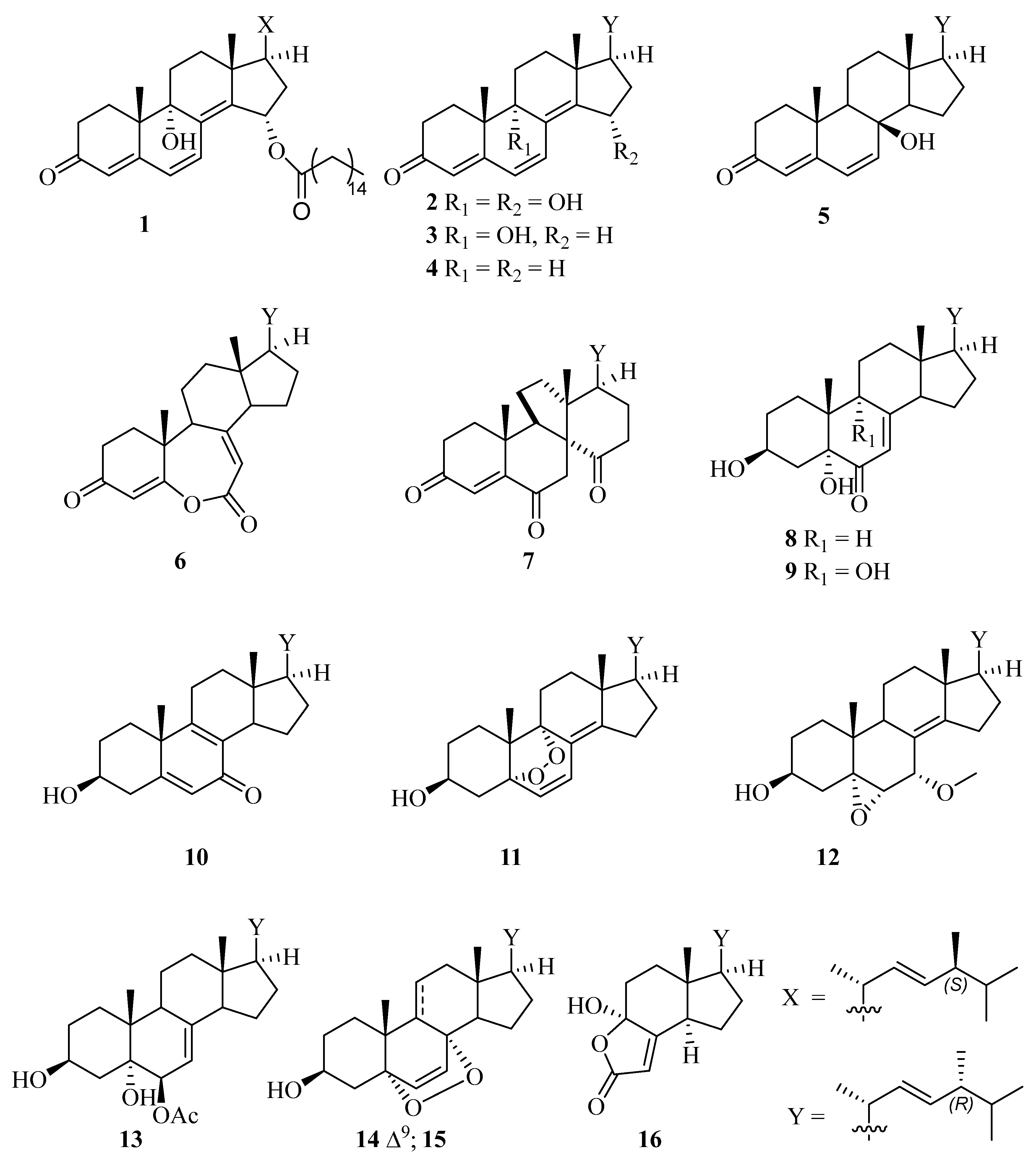
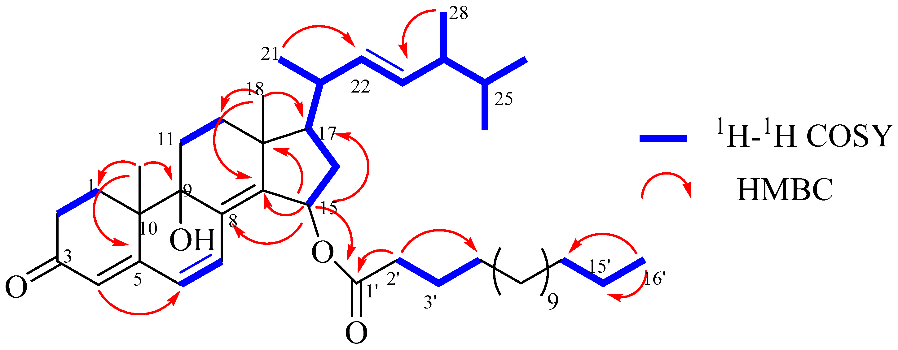
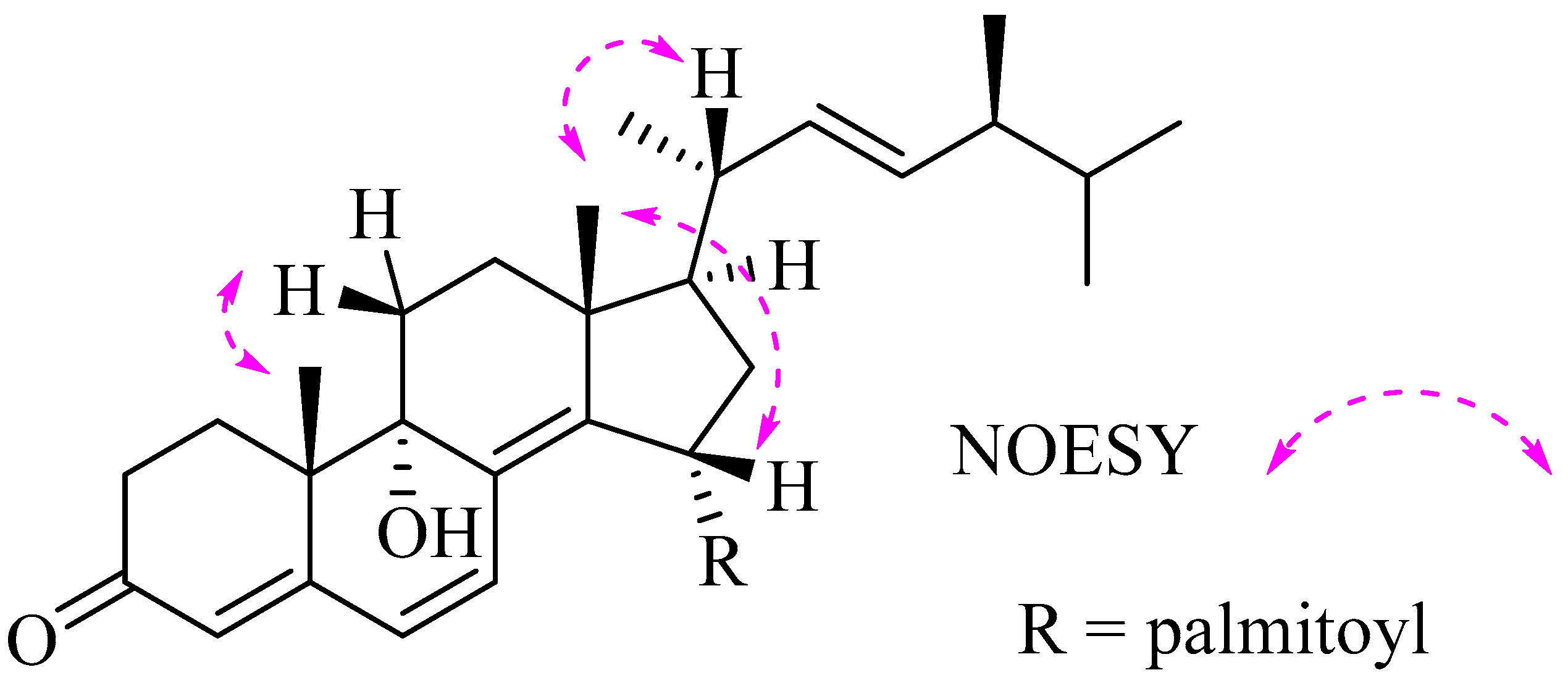
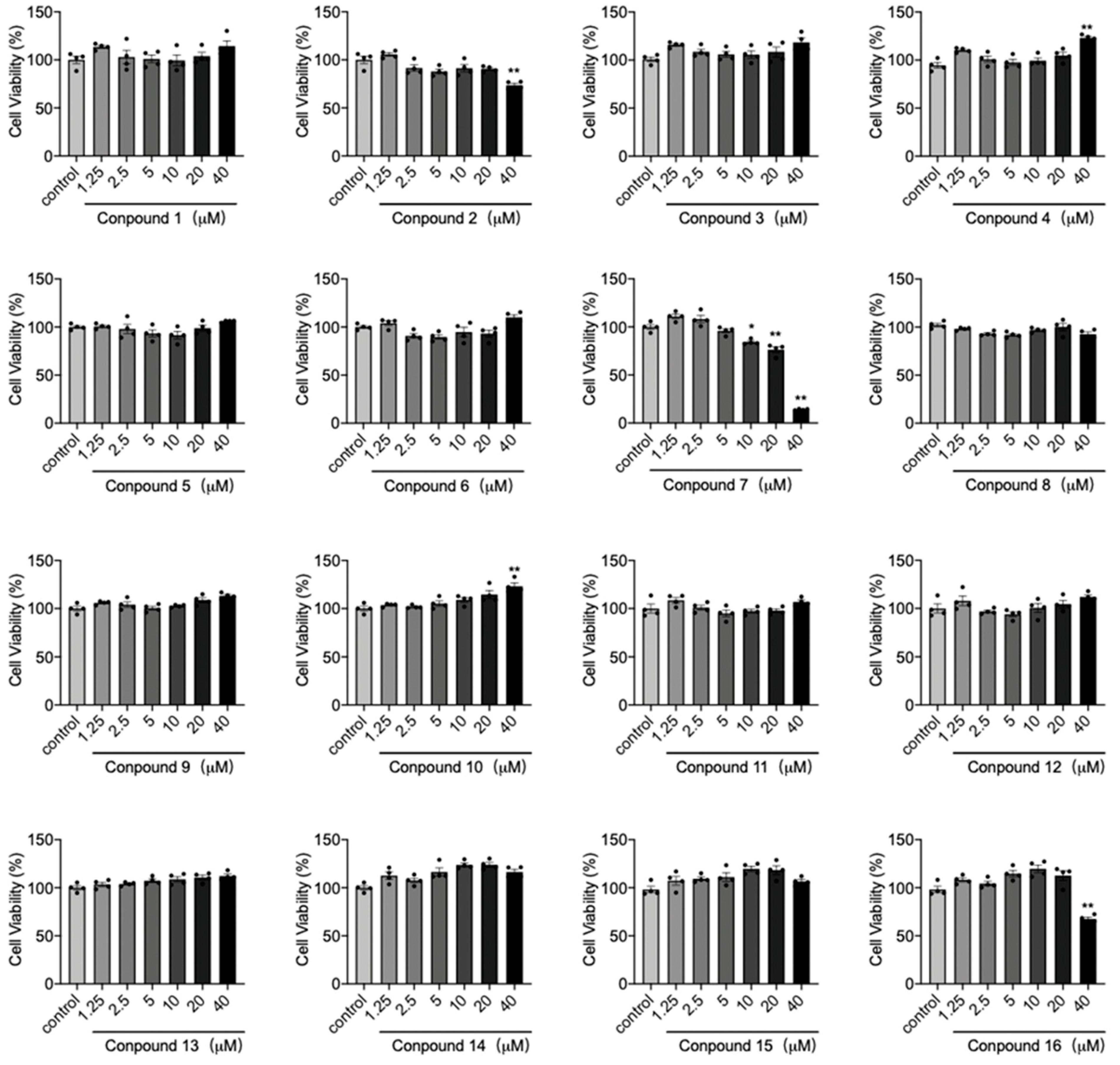
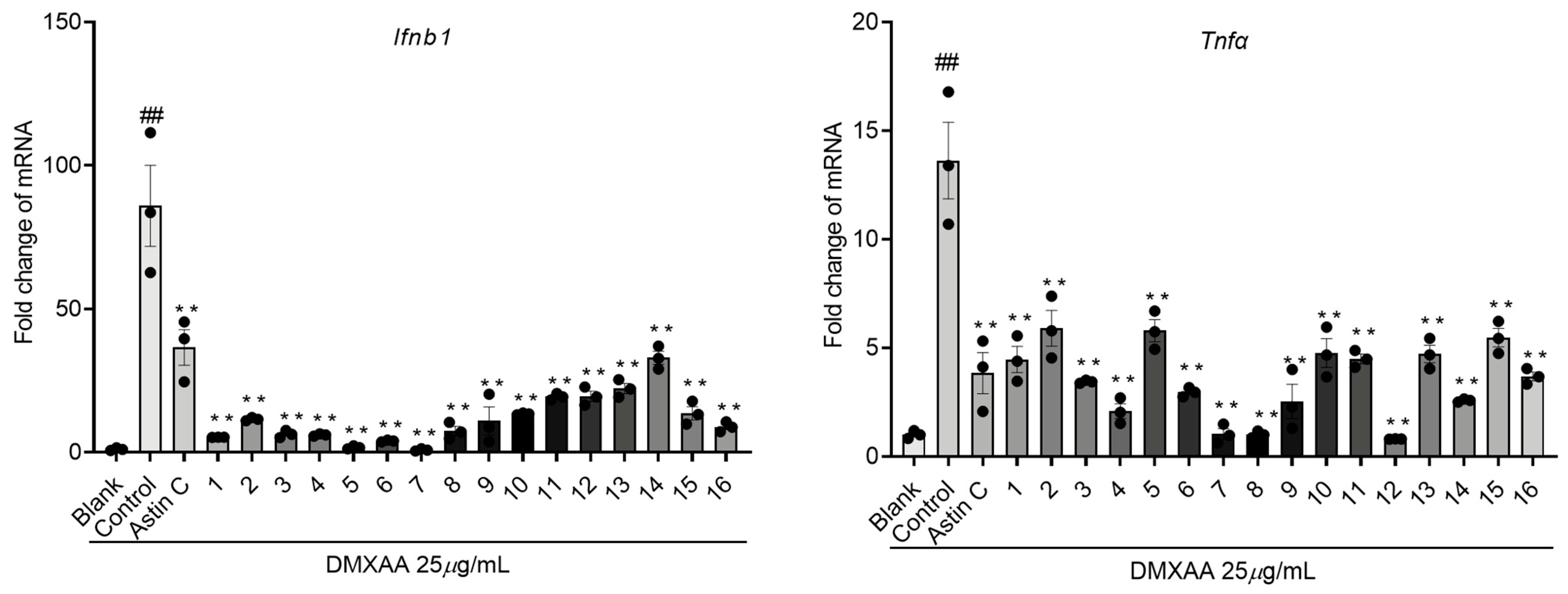
2. Results and Discussion

To obtain the optimal fermentation condition, UPLC-MS was employed for the chemical analysis of fungal metabolites produced with strain HL-44 in five candidates of media, including Czapek Dox Agar (CZA) medium, Glucose Peptone Yeast (GPY) medium, PDA medium, Rose Bengal Medium (RBM), and Rice medium. The Total Ion Chromatography (TIC) of the PDA medium showed more abundant metabolites with ion peaks (m/z) in a mass spectrum ranging from 408 to 688. In particular, the ion peak at m/z 663.534 was fairly interesting. Thus, the PDA medium was chosen for the scale-up fermentation of the fungi HL-44.
The fungal strain was then cultivated on the PDA medium for scale fermentation at 28 °C for 28 days and was then extracted ultrasonically with EtOAc to afford a residue after removal of the solvent under reduced pressure [14,15]. The crude extract was subjected to column chromatography (CC) silica gel, Sephadex LH-20, and reversed phase HPLC to afford compounds 116 (Figure 1). On the basis of spectroscopic techniques (1H-NMR, 13C-NMR) and comparison with data recorded in the references, compounds 216 were determined as (22E,24R)-9α,15α-dihydroxyergosta-4,6,8(14),22-tetraen-3-one (2) [17], ganodermaside D (3) [18], (22E,24R)-ergosta-4,6,8(14),22-tetraen-3-one (4) [19], isocyathisterol (5) [20], herbarulide (6) [21], dankasterones A (7) [22], (22E,24R)-ergosta-7,22-dien-3β,5α-diol-6-one (8) [23], (22E,24R)-ergosta-7,22-dien-3β,5α,9α-trihydroxy-6-one (9) [24], (22E,24R)-3β-hydroxyergosta-5,8,22-trien-7-one (10) [25], (22E,24R)-5α,9α-epidioxyergosta-6,8(14),22-triene-3β-ol (11) [26], (22E, 24R)-7α-methoxy-5α,6α-epoxyergosta-8(14),22-dien-3β-ol (12) [27], (22E,24R)-6-acetoxy-ergosta-7,22-dien-3β,5α,6β-triol (13) [28], (22E,24R)-5α,8α-epidioxyergosta-6,9(11),22-trien-3β-ol (14) [29], (22E,24R)-5α,8α-epidioxyergosta-6,22-dien-3β-ol (15) [30], and demethylincisterol A3 (16) [31].
Compound 1 was obtained as an optically yellowish oil. The HRESIMS gave a molecular formula as C44H70O4 on the basis of its pseudomolecular ion peak at m/z 663.53448 ([M + H]+), indicating 10 degrees of unsaturation. The IR spectrum revealed the presence of a hydroxy group (3344 cm−1) and carbonyl (1733 cm−1) functionalities. The characteristic IR absorptions at 1664 and 1595 cm−1 and the strong UV absorptions at 265 and 330 nm indicated the presence of a large conjugated carbonyl system in this molecule. These observations were in agreement with the NMR data for an oxygenated methine (δH 5.81; δC 71.8, CH), a tertiary oxygenated carbon atom (δC 72.8, C), four pairs of double bonds, one ester carbonyl atom (δC 173.6), and one ketone carbonyl atom (δC 199.3), taking into account six degrees of unsaturation. The remaining four degrees of unsaturation were due to the ring system in the molecule. Compound 1 resembled 2 in the NMR data (Table 1) except for signals for a palmitoyl moiety, which was supported by the 2D NMR analyses, as shown in Figure 2. The palmitoyl moiety was assigned at C-15 by the distinct HMBC effects of H-15 with C-8, C-13, C-17, and the ester carbonyl atom (C-1’). Compound 1 displayed the same relative configuration as that of 2 in the core structure, as shown in Figure 3. The β-configuration of H-15 was indicated by its NOE correlation with H3-18. The configuration at C-9 was suggested by comparing its shift value (δC 72.8, C) to those reported in the literature (δC 72.7/72.8, C) [17,32], indicating that the hydroxy group at C-9 was in the α-configuration. The absolute configuration of C-24 was assigned as S in 1 vs R in 2 based on the 13C NMR shift values of C-28. The NMR data for C-28 are reported at δ 17.6 ± 0.1 ppm for 24R-isomers and δ 18.0 ± 0.1 ppm for 24S-isomers [16,33]. The presence of a palmitic acid residue was proven with an MS analysis and comparison of the spectroscopic data with those reported data [34]. The existence of the fatty acyl moiety was indicated by the characteristic 13C signals for the ester carbonyl carbon (δC 173.6) and 1H signals for triplet methylene at δH 2.30, multiplet methylenes at δH 1.23–1.30, and a terminal methyl group at δH 0.88. The structure of 1 was thus determined as (22E,24S)-9α,15α-dihydroxyergosta-4,6,8(14),22-tetraen-3-one 15-palmitate, showing a 24S configuration vs. 24R in 2. The compound is also characterized by a 15-palmitoyl moiety with respect to a hydroxyl group in 2.
Derivatives of 15α-hydroxy steroids serve as key intermediates in the production of contraceptives [35,36]. A P450 enzyme, which is composed of the cytochrome P450 hydroxylase and the NADPH-cytochrome P450 reductase (CPR), has been reported to be associated with the 15α-hydroxylation reaction in P. raistrickii [37]. P. raistrickii-mediated 15α-hydroxylation of D-ethylgonendione is a key step for the production of gestodene [38]. The 9,15-hydroxylated ergosteroid 2 was only reported in Omphalia lapidescens [32] and Ganoderma resinaceum [17]. The isolation of 1 and 2 suggests that P. oxalicum strains have a potential in fungal transformation to afford 15α-hydroxyergosteroid. Compounds 3 and 4 have a chemical feature of conjugated 4,6,8-trien-3-one and have never been reported in P. oxalicum. Compound 6 is a ketodivinyllactone steroid with an unprecedented homo-6-oxaergostane skeleton isolated from the endophytic fungus Pleospora herbarum [39] with a structure revision at C24 with a chemical synthesis [21]. Compound 7 was an unprecedented steroid possessing a 13(14→8)abeo-8-ergostane skeleton first found in the Halichondria sponge-derived fungus Gymnacella dankaliensis [22]. Compound 11, which features an unusual 1,2-dioxolane moiety, was only reported in G. capense [26] and G. lingzhi [40]. Although 5α,8α-epidioxysterols with variations in the side chains were most commonly reported from a number of sources, rare 5α,9α-epidioxy steroids were mainly isolated from different edible mushrooms [41,42]. The compound showed weak anti-HIV activity and remarkable cytotoxicity against A549 and MCF-7 tumor cell lines. Compound 16 is a highly degraded sterol, and its basic skeleton derives from a dramatic oxidative degradation of the sterol nucleus with the loss of all six carbon atoms of the A ring and the 19-methyl group [43].
Compounds 116 were assessed for their anti-inflammatory activity on the expression of pro-inflammatory cytokine factors Tnfα and Ifnb1 in LPS- or DMXAA-induced Raw264.7 cells. Prior to exploring their anti-inflammatory properties, the potential toxicity of these compounds in Raw264.7 cells was evaluated to ensure that their effects were not confounded with cytotoxicity. The results demonstrated that most compounds had no significant impact on the viability of Raw264.7 cells, except for compounds 2, 7, and 16, which exhibited some reduction in cell viability at high concentrations up to 40 μM (Figure 4).
Cell apoptosis in macrophages is intricately associated with macrophage polarization, allowing macrophages to alter their phenotype and carry out diverse functions in response to changes in the microenvironment. The two main polarization states are classically activated macrophages (M1) and alternatively activated macrophages (M2). M1 macrophages exhibit characteristics such as pro-inflammatory mediator production, promotion of cell cytotoxicity, and antimicrobial abilities. Conversely, M2 macrophages possess anti-inflammatory properties, participate in tissue repair, regulate the immune response, and demonstrate anti-inflammatory capabilities. M1 macrophages induce cell death by releasing toxic molecules such as toxins, oxidants, and proteases, thereby augmenting their antimicrobial and antitumor effects. Nonetheless, excessive activation of M1 macrophages can result in tissue damage and inflammatory responses [44]. However, excessive activation of M1 macrophages can lead to tissue damage and inflammatory responses. The experimental findings revealed that compound 7 exerted a more pronounced inhibitory effect on the survival of Raw264.7 cells at a concentration of 40 µM compared to other compounds. This suggests that compound 7 may promote the polarization of M1 macrophages while inhibiting the expression of growth-related proteins such as P38, Akt, and Wnt and promoting the expression of apoptosis-related genes such as Bcl-2, Bcl-xL, and Mcl-1. Consequently, this inhibits macrophage proliferation and promotes macrophage apoptosis. Moreover, the inhibitory effect of compound 7 on the viability of Raw264.7 cells at a concentration of 40 µM also suggests its potential to inhibit the proliferation and differentiation of tumor cells.
The expression of inflammatory factors in Raw264.7 cells, such as Tnfα and Ifnb1, which were stimulated by LPS, was suppressed by the administration of compounds 116 (Figure 5).
The above results demonstrate that the compounds exhibit good inhibitory activity against type I interferon, and the cGAS-STING pathway in the innate immune response is an important mechanism for regulating type I interferon responses [45,46,47]. The expression levels of Tnfα and Ifnb1 in the STING pathway have practical importance in immune regulation, inflammatory diseases, and therapeutic interventions, serving as key indicators of immune activation, effectiveness against pathogens, and the extent of inflammation [48,49]. Therefore, we further used the specific activator DMXAA of the cGAS-STING pathway to establish an activation model and evaluate the activity of these compounds. Using astin C (20 nM) as a positive control and a natural inhibitor of the STING pathway [50], the results indicate that all compounds significantly inhibit the expression levels of Tnfα and Ifnb1 (Figure 6). In the stimulation of Raw264.7 cells with LPS, compounds 2, 9, and 14 showed significant inhibition of the inflammatory cytokine Ifnb1 expression, while compounds 2, 4, and 5 exhibited strong inhibition of the inflammatory cytokine Tnfα expression. When Raw264.7 cells were stimulated with DMXAA, compounds 1, 5, and 7 effectively suppressed the inflammatory cytokine Ifnb1 expression, whereas compounds 7, 8, and 11 demonstrated the best inhibition of the inflammatory cytokine Tnfα expression. Notably, newly isolated compounds 2 and 11 demonstrated potent anti-inflammatory activity. Hence, it is probable that compounds 116 exert their anti-inflammatory effects through the modulation of the cGAS-STING pathway.

3. Materials and Methods

3.1. General Experimental Procedures

Optical rotations were determined with a Autopol VI polarimeter (Rudolph, Wood County, WI, USA). UV spectra and ECD spectra were taken in MeOH on a Jasco-715 spectropolarimeter. Infrared spectra were recorded on a Nicolet iN10 (micro) spectrometer (Thermo Fisher Scientific, Waltham, MA, USA). The NMR spectra were recorded on a Bruker DRX-600 (Bruker, Germany) spectrometer with chemical shifts reported relative to the residual CDCl3 (δH 7.26 ppm, δC 77.0 ppm) and CD3OD (δH 3.31 ppm; δC 49.0 ppm). The HRESIMS analyses were performed on a Q Exactive Plus Orbitrap (Thermo Fisher Scientific, Waltham, MA, USA) mass spectrometer. The UPLC-MS analysis of total ion chromatogram (TIC) was obtained using Agilent 1290 Infinity-6538 UHD (Agilent, Santa Clara, CA, USA) and Accurate-Mass QTOF/MS (Agilent, Santa Clara, CA, USA). Semipreparative HPLC was performed on a Waters 1525 (Waters, Milford, MA, USA) with a YMC Pack ODS-A column (250 × 10 mm, 5 μM; YMC, Kyoto, Japan). Sephadex LH-20 gel (GE Healthcare Bio-Sciences AB, Uppsala, Sweden) and silica gel (200–300 mesh, 300–400 mesh; Yantai Chemical Engineering Institure, Yantai, China) were used for column chromatography. Precoated silica gel plates (HSGF-254; Yantai Chemical Engineering Institure, Yantai, China) were used for thin-layer chromatography (TLC). Spots were detected on TLC under UV or by heating after spraying with an anisaldehyde sulfuric acid reagent.

3.2. Fungal Material

P. oxalicum HL-44 was isolated from the soft coral S. gaweli that was collected from the Xisha area of the South China Sea at a depth of 15 m in Aug 2018 and identified as P. oxalicum using 18sRNA sequence (GenBank accession number MG585101.1). A voucher strain of this fungus (internal strain No. HL-44) was deposited at Tongji University, Shanghai, China.

3.3. Screening Culture Medium and Cultivation

Five media, including Czapek Dox Agar (CZA) medium, Glucose Peptone Yeast (GPY) medium, Potato Dextrose Agar (PDA) medium, Rose Bengal Medium (RBM), and Rice medium, were used in the fermentation to compare the chemical diversity of fungal HL-44. UPLC-MS analysis of five extracts indicated that the PDA medium allowed the strain to produce metabolites with large molecular weights and more chemical diversity (m/z range from 408 to 688, tR from 10.5 to 14.5 min, Figure S2). The PDA medium was then selected for scale-up fermentation.

3.4. Extraction and Isolation

The culture medium containing mycelia was cut into small pieces and extracted five times ultrasonically with EtOAc to afford 43.5 g of residue after removal of the solvent under reduced pressure. The crude extract was separated into ten fractions (Fr.1–10) with silica gel CC (80 mm × 150 mm, 810 g, 200–300 mesh), eluting with a gradient CH2Cl2/MeOH (100:0 v/v 5 L, 80:1 v/v 3 L, 60:1 v/v 5 L, 40:1 v/v 3 L, 20:1 v/v 5 L, 5:1 v/v 5 L). Fr. 1 (3.3 g) was subjected to a Sephadex LH-20 CC (4 cm × 120 cm) using CH2Cl2/MeOH (v/v 2:1) as eluent to obtain eight subfractions (Fr.1a-h). Fr.1c (411.6 mg) was separated with silica gel CC (10 mm × 150 mm, 14 g, 300–400 mesh) using gradient petroleum (PE) in Me2CO (39:1 v/v 40 mL, 24:1 v/v 50 mL, 19:1 v/v 40 mL, 1:1 v/v 20 mL), then split with HPLC to yield compound 1 (0.9 mg, MeOH/H2O 98:2, 2 mL/min, tR 64 min). Fr.1e (112.8 mg) was separated with silica gel CC (15 mm × 200 mm, 38 g, 300–400 mesh) using PE/Me2CO (39:1 v/v 120 mL, 29:1 v/v 120 mL, 19:1 v/v 160 mL, 9:1 v/v 100 mL, 4:1 v/v 50 mL, 2:1 v/v 30 mL), then split with HPLC to yield compound 11 (1.6 mg, MeOH/H2O 90:10, 2 mL/min, tR 51 min) and compound 6 (12.8 mg, MeOH/H2O 87:13, 2 mL/min, tR 55 min). Fr.1f (788.1 mg) was separated with silica gel CC (20 mm × 150 mm, 55 g, 300–400 mesh) using PE/Me2CO (39:1 v/v 100 mL, 19:1 v/v 100 mL, 14:1 v/v 120 mL, 9:1 v/v 100 mL, 1:1 v/v 50 mL), then split with HPLC to yield compound 14 (1.5 mg, MeOH/H2O 88:12, 2 mL/min, tR 74 min), compound 12 (0.6 mg, MeOH/H2O 88:12, 2 mL/min, tR 81 min) and compound 15 (8.4 mg, MeOH/H2O 88:12, 2 mL/min, tR 95 min). Fr. 2 (794.5 mg) was fractionated using Sephadex LH-20 CC (CH2Cl2/MeOH v/v 2:1, 4 cm × 120 cm) and then purified with silica gel CC (15 mm × 150 mm, 30 g, 300–400 mesh) using a gradient PE in Me2CO (39:1 v/v 80 mL, 19:1 v/v 100 mL, 9:1 v/v 100 mL, 1:1 v/v 40 mL), and HPLC to afford compound 4 (2.8 mg, MeOH/H2O 97:3, 2 mL/min, tR 52 min) and compound 7 (4.4 mg, MeOH/H2O 95:5, 2 mL/min, tR 41 min). Fr. 4 (2.2 g) was fractionated using Sephadex LH-20 CC (CH2Cl2/MeOH v/v 2:1, 4 cm × 120 cm) and then purified with silica gel CC (20 mm × 150 mm, 55 g, 300–400 mesh) using a gradient PE in Me2CO (29:1 v/v 90 mL, 19:1 v/v 100 mL, 14:1 v/v 90 mL, 9:1 v/v 100 mL, 4:1 v/v 50 mL) and HPLC to give compound 5 (4.4 mg, MeOH/H2O 80:20, 2 mL/min, tR 65 min), compound 3 (0.5 mg, CH3CN/H2O 70:30, 2 mL/min, tR 185 min), and compound 16 (1.9 mg, MeOH/H2O 80:20, 2 mL/min, tR 73 min). Fr. 5 (755.9 mg) was fractionated using Sephadex LH-20 CC (CH2Cl2/MeOH v/v 2:1, 4 cm × 120 cm) and then purified with silica gel CC (15 mm × 150 mm, 30 g, 300–400 mesh) using a gradient PE in Me2CO (29:1 v/v 90 mL, 14:1 v/v 90 mL, 9:1 v/v 120 mL, 4:1 v/v 50 mL) and HPLC to give compound 10 (1.8 mg, MeOH/H2O 88:12, 2 mL/min, tR 69 min). Fr. 7 (740.6 mg) was fractionated using Sephadex LH-20 CC (CH2Cl2/MeOH v/v 2:1, 4 cm × 120 cm) and then purified with silica gel CC (15 mm × 150 mm, 30 g, 300–400 mesh) using PE/Me2CO (8:1 v/v 90 mL and 4:1 v/v 50 mL) to give compound 13 (2.0 mg). Fr. 9 (2.4 g) was fractionated using Sephadex LH-20 CC (CH2Cl2/MeOH v/v 2:1, 4 cm × 120 cm) and then purified with silica gel CC (10 mm × 150 mm, 14 g, 300–400 mesh) using PE/ Me2CO (8:1 v/v 90 mL, 6:1 v/v 70 mL, 4:1 v/v 30 mL) and HPLC to give compound 2 (1.3 mg, MeOH/H2O 87:13, 2 mL/min, tR 27 min). Fr. 10 (1.7 g) was fractionated using Sephadex LH-20 CC (CH2Cl2/MeOH v/v 2:1, 4 cm × 120 cm) and then purified with silica gel CC (15 mm × 150 mm, 30 g, 300–400 mesh) using PE/Me2CO (19:1 v/v 80 mL, 9:1 v/v 120 mL, 7:1 v/v 80 mL, 5:1 v/v 120 mL, 2:1 v/v 90 mL) and HPLC to give compound 9 (1.4 mg, MeOH/H2O 90:10, 2 mL/min, tR 35 min) and compound 8 (0.9 mg, MeOH/H2O 90:10, 2 mL/min, tR 47 min).
Characterization data of compound 1: (22E,24S)-9α,15α-dihydroxyergosta-4,6,8(14),22-tetraen-3-one 15-palmitate (1): yellowish oil, [ α ] D 25 +43.40 (c 0.10, CH3OH); UV (CH3OH) λmax (log ε) 330 (2.49), 265 (2.02) nm; ECD (CH3OH, c 1.5 × 10⁻⁴) λmax (∆ ε) 241 (+1.80), 320 (-1.43), 361 (+2.76) nm; IR (micro) νmax 3443, 2929, 2854, 1733, 1664, 1595, 1460, 1260, 1094, 1033, 970, 801 cm−1; 1H and 13C NMR data see Table 1; HRESIMS m/z 663.53448 [M + H]+ (calcd for C44H71O4, 663.53469).

3.5. Cell Viability Detection

The viability of Raw264.7 cells was evaluated using the CCK-8 assay. Initially, the cells were seeded at a density of 3 × 10⁴ cells/well in 96-well plates and allowed to incubate overnight. The cells were treated with the tested compounds at concentrations ranging from 1.25 to 40 μM for 24 h. After removing the culture medium, a CCK-8 solution diluted in Dulbecco’s modified Eagle medium was added to each well. Following a 1-h incubation, the absorbance was measured at 450 nm using a multifunctional microplate spectrophotometer. Cell viability was then calculated as a percentage relative to the blank group [51].

3.6. Quantitative Real-Time PCR (qPCR)

Raw264.7 cells were pretreated with tested compounds at a concentration of 20 μM, along with astin C [50] at a concentration of 20 nM, for 2 h. Subsequently, the cells were stimulated with LPS at a concentration of 100 ng/mL and DMXAA at a concentration of 25 μg/mL for 6 h [52]. Total RNA was extracted from Raw264.7 cells using a triazole reagent supplied by Thermo Fisher Scientific. cDNA synthesis was performed using SuperScript III reverse transcriptase obtained from Invitrogen. Real-time PCR analysis was conducted using a PrimeScript RT reagent kit from Takara. The relative expression levels of the target genes were quantitatively normalized to the expression level of Gapdh using the ΔΔct method. Primer sequences are Ifnb1, forward 5′-GCACTGGGTGGAATGAGACT-3′ and reverse 5′-AGTGGAGAGCAGTTGAGGACA-3′; Tnfα, forward 5′-GTCCCCAAAGGGATGAGAAGTT-3′ and reverse 5′-GTTTGCTACGACGTGGGCTACA-3′ [52].

3.7. Statistical Analysis

The statistical analyses were conducted using GraphPad Prism 8.0. A one-way analysis of variance (ANOVA) was applied to the data, followed by Tukey’s test for comparisons against the blank and control groups. A significance level of p < 0.05 was considered statistically significant.

4. Conclusions

With chemical analysis of the PDA medium extract of the fungus P. oxalicum HL-44 associated with the soft coral S. gaweli, a new ergostane-type sterol ester (1) was isolated together with fifteen derivatives (216). Their structures were determined using spectroscopic analyses and comparisons with reported data. This is the first report of compounds 2 and 11 from the family of Eurotiaceae. The isolation of ergosteroid derivatives with varying degrees of oxidation revealed a remarkable range of chemical diversity, expanding the ergosteroid family associated with this fungus. In an in vitro biotest, these compounds demonstrated potent anti-inflammatory activities at a concentration of 20 μM, effectively suppressing the expression of inflammatory factors such as Tnfα and Ifnb1 in Raw264.7 cells induced with LPS or DMXAA. Among DMXAA-induced Raw264.7 cells, compounds 7 and 8 exhibited the highest level of inhibition in terms of Tnfα and Ifnb1 expression. Conversely, in LPS-induced Raw264.7 cells, compounds 2 and 4 displayed the most pronounced inhibitory effects on Tnfα and Ifnb1 expression. This study provides valuable insight into the chemical diversity of ergosteroid derivatives and their potential as anti-inflammatory agents.

Supplementary Materials

The following supporting information can be downloaded at https://www.mdpi.com/article/10.3390/molecules28237784/s1. Figure S1. HL-44 cultured on PDA medium (A), CZA medium (B), GPY medium (C), RBM, (D) Rice medium, and (E); Figure S2. Total Ion Chromatography (TIC) of 5 candidate media extracts: PDA medium (A), CZA medium (B), GPY medium (C), RBM (D), and Rice medium (E); Figures S3–S13. 1H-NMR, 13C-NMR, DEPT, 1H-1H COSY, HSQC, HMBC, NOESY, UV, IR, HRESIMS, and CD spectra of compound 1; Figure S14. OR data of compound 1 in CH3OH; Figure S15. The 1H-NMR spectrum of previously reported compounds 216.

Author Contributions

C.P. performed the experiments. Y.-H.C. and H.-H.B. contributed to the bioassay. J.-P.Z. was responsible for the fungal fermentation. W.Z., H.H. and L.S. conceived and designed the experiments. All authors have read and agreed to the published version of the manuscript.

Funding

This research was funded by the National Natural Science Foundation of China (Nos. 81820108030, 82173735, and 82273937) and STCSM (21430711400).

Institutional Review Board Statement

Not applicable.

Informed Consent Statement

Not applicable.

Data Availability Statement

All data are available in the main text or the Supplementary Materials.

Conflicts of Interest

The authors declare no conflict of interest.

References

  1. Cheung, R.C.F.; Ng, T.B.; Wong, J.H.; Chen, Y.; Chan, W.Y. Marine natural products with anti-inflammatory activity. Appl. Microbiol. Biotechnol. 2016, 100, 1645–1666. [Google Scholar] [CrossRef]
  2. Zhabinskii, V.N.; Drasar, P.; Khripach, V.A. Structure and Biological Activity of Ergostane-Type Steroids from Fungi. Molecules 2022, 27, 2103. [Google Scholar] [CrossRef] [PubMed]
  3. Kubatova, A.; Hujslova, M.; Frisvad, J.C.; Chudickova, M.; Kolarik, M. Taxonomic revision of the biotechnologically important species Penicillium oxalicum with the description of two new species from acidic and saline soils. Mycol. Prog. 2019, 18, 215–228. [Google Scholar] [CrossRef]
  4. Zhang, P.; Li, X.-M.; Liu, H.; Li, X.; Wang, B.-G. Two new alkaloids from Penicillium oxalicum EN-201, an endophytic fungus derived from the marine mangrove plant Rhizophora stylosa. Phytochem. Lett. 2015, 13, 160–164. [Google Scholar] [CrossRef]
  5. Zhang, R.; Ma, Y.; Xu, M.-M.; Wei, X.; Yang, C.-B.; Zeng, F.; Duan, J.-A.; Che, C.-T.; Zhou, J.; Zhao, M. Oxalactam A, a Novel Macrolactam with Potent Anti-Rhizoctonia solani Activity from the Endophytic Fungus Penicillium oxalicum. Molecules 2022, 27, 8811. [Google Scholar] [CrossRef] [PubMed]
  6. Ren, Y.; Chao, L.-H.; Sun, J.; Chen, X.-N.; Yao, H.-N.; Zhu, Z.-X.; Dong, D.; Liu, T.; Tu, P.-F.; Li, J. Two new polyketides from the fungus Penicillium oxalicum MHZ153. Nat. Prod. Res. 2019, 33, 347–353. [Google Scholar] [CrossRef] [PubMed]
  7. Weng, W.; Li, R.; Zhang, Y.; Pan, X.; Jiang, S.; Sun, C.; Zhang, C.; Lu, X. Polyketides isolated from an endophyte Penicillium oxalicum 2021CDF-3 inhibit pancreatic tumor growth. Front. Microbiol. 2022, 13, 1033823. [Google Scholar] [CrossRef] [PubMed]
  8. Li, X.; Li, X.-M.; Zhang, P.; Wang, B.-G. A new phenolic enamide and a new meroterpenoid from marine alga-derived endophytic fungus Penicillium oxalicum EN-290. J. Asian Nat. Prod. Res. 2015, 17, 1204–1212. [Google Scholar] [CrossRef]
  9. Zhang, Y.-H.; Li, L.; Li, Y.-Q.; Luo, J.-H.; Li, W.; Li, L.-F.; Zheng, C.-J.; Cao, F. Oxalierpenes A and B, unusual indole-diterpenoid derivatives with antiviral activity from a marine-derived strain of the fungus Penicillium oxalicum. J. Nat. Prod. 2022, 85, 1880–1885. [Google Scholar] [CrossRef]
  10. Wang, Y.; Chen, W.; Xu, Z.; Bai, Q.; Zhou, X.; Zheng, C.; Bai, M.; Chen, G. Biological secondary metabolites from the Lumnitzera littorea-derived fungus Penicillium oxalicum HLLG-13. Mar. Drugs 2023, 21, 22. [Google Scholar] [CrossRef]
  11. He, Z.-H.; Xie, C.-L.; Hao, Y.-J.; Xu, L.; Wang, C.-F.; Hu, M.-Y.; Li, S.-J.; Zhong, T.-H.; Yang, X.-W. Solitumergosterol A, a unique 6/6/6/6/5 steroid from the deep-sea-derived Penicillium solitum MCCC 3A00215. Org. Biomol. Chem. 2021, 19, 9369–9372. [Google Scholar] [CrossRef] [PubMed]
  12. Tian, H.; Li, X.P.; Zhao, J.; Gao, H.W.; Xu, Q.M.; Wang, J.W. Biotransformation of artemisinic acid to bioactive derivatives by endophytic Penicillium oxalicum B4 from Artemisia annua L. Phytochemistry 2021, 185, 112682. [Google Scholar] [CrossRef] [PubMed]
  13. de Queiroz, T.M.; Ellena, J.; Porto, A.L.M. Biotransformation of Ethinylestradiol by Whole Cells of Brazilian Marine-Derived Fungus Penicillium oxalicum CBMAI 1996. Mar. Biotechnol. 2020, 22, 673–682. [Google Scholar] [CrossRef] [PubMed]
  14. Wang, H.L.; Li, R.; Li, J.; He, J.; Cao, Z.Y.; Kurtán, T.; Mándi, A.; Zheng, G.L.; Zhang, W. Alternarin A, a drimane meroterpenoid, suppresses neuronal excitability from the coral-associated fungi Alternaria sp. ZH-15. Org. Lett. 2020, 22, 2995–2998. [Google Scholar] [CrossRef]
  15. Wang, H.L.; Li, R.; Zhao, M.; Wang, Z.Y.; Tang, H.; Cao, Z.Y.; Zheng, G.L.; Zhang, W. A drimane meroterpenoid borate as a synchronous Ca plus oscillation inhibitor from the coral-associated fungus Alternaria sp. ZH-15. J. Nat. Prod. 2023, 86, 429–433. [Google Scholar] [CrossRef]
  16. Li, J.; Sun, Y.L.; Tang, H.; Su, L.; Zheng, G.L.; Zhang, W. Immunosuppressive 9,10-secosteroids from the gorgonian Verrucella umbraculum collected in the South China Sea. J. Nat. Prod. 2021, 84, 1671–1675. [Google Scholar] [CrossRef]
  17. Shi, Q.; Huang, Y.; Su, H.; Gao, Y.; Peng, X.; Zhou, L.; Li, X.; Qiu, M. C(28) steroids from the fruiting bodies of Ganoderma resinaceum with potential anti-inflammatory activity. Phytochemistry 2019, 168, 112109. [Google Scholar] [CrossRef]
  18. Weng, Y.F.; Lu, J.; Xiang, L.; Matsuura, A.; Zhang, Y.; Huang, Q.M.; Qi, J.H. Ganodermasides C and D, two new anti-aging ergosterols from spores of the medicinal mushroom Ganoderma lucidum. Biosci. Biotechnol. Biochem. 2011, 75, 800–803. [Google Scholar] [CrossRef]
  19. Kwon, H.C.; Zee, S.D.; Cho, S.Y.; Choi, S.U.; Lee, K.R. Cytotoxic ergosterols from Paecilomyces sp. J300. Arch. Pharmacal Res. 2002, 25, 851–855. [Google Scholar] [CrossRef]
  20. Liu, X.-H.; Miao, F.-P.; Liang, X.-R.; Ji, N.-Y. Ergosteroid derivatives from an algicolous strain of Aspergillus ustus. Nat. Prod. Res. 2014, 28, 1182–1186. [Google Scholar] [CrossRef]
  21. Duecker, F.L.; Heinze, R.C.; Mueller, M.; Zhang, S.D.; Heretsch, P. Synthesis of the alleged structures of fortisterol and herbarulide and structural revision of herbarulide. Org. Lett. 2020, 22, 1585–1588. [Google Scholar] [CrossRef] [PubMed]
  22. Amagata, T.; Tanaka, M.; Yamada, T.; Doi, M.; Minoura, K.; Ohishi, H.; Yamori, T.; Numata, A. Variation in cytostatic constituents of a sponge-derived Gymnascella dankaliensis by manipulating the carbon source. J. Nat. Prod. 2007, 70, 1731–1740. [Google Scholar] [CrossRef] [PubMed]
  23. Fangkrathok, N.; Sripanidkulchai, B.; Umehara, K.; Noguchi, H. Bioactive ergostanoids and a new polyhydroxyoctane from Lentinus polychrous mycelia and their inhibitory effects on E2-enhanced cell proliferation of T47D cells. Nat. Prod. Res. 2013, 27, 1611–1619. [Google Scholar] [CrossRef] [PubMed]
  24. Hoque, N.; Afroz, F.; Khatun, F.; Rony, S.R.; Hasan, C.M.; Rana, M.S.; Sohrab, M.H. Physicochemical, pharmacokinetic and cytotoxicity of the compounds Isolated from an endophyte Fusarium oxysporum: In vitro and in silico approaches. Toxins 2022, 14, 159. [Google Scholar] [CrossRef] [PubMed]
  25. Wang, F.; Fang, Y.; Zhang, M.; Lin, A.; Zhu, A.; Gu, Q.; Zhu, W. Six new ergosterols from the marine-derived fungus Rhizopus sp. Steroids 2008, 73, 19–26. [Google Scholar] [CrossRef] [PubMed]
  26. Tan, Z.; Zhao, J.-L.; Liu, J.-M.; Zhang, M.; Chen, R.-D.; Xie, K.-B.; Chen, D.-W.; Dai, J.-G. Lanostane triterpenoids and ergostane-type steroids from the cultured mycelia of Ganoderma capense. J. Asian Nat. Prod. Res. 2018, 20, 844–851. [Google Scholar] [CrossRef] [PubMed]
  27. Gao, H.; Hong, K.; Chen, G.-D.; Wang, C.-X.; Tang, J.-S.; Yu, Y.; Jiang, M.-M.; Li, M.-M.; Wang, N.-L.; Yao, X.-S. New oxidized sterols from Aspergillus awamori and the endo-boat conformation adopted by the cyclohexene oxide system. Magn. Reson. Chem. 2010, 48, 38–43. [Google Scholar] [CrossRef]
  28. Zhang, W.; Draeger, S.; Schulz, B.; Krohn, K. Ring B aromatic steroids from an endophytic fungus, Colletotrichum sp. Nat. Prod. Commun. 2009, 4, 1449–1454. [Google Scholar] [CrossRef]
  29. Kim, K.H.; Choi, S.U.; Park, K.M.; Seok, S.J.; Lee, K.R. Cytotoxic constituents of Amanita subjunquillea. Arch. Pharmacal Res. 2008, 31, 579–586. [Google Scholar] [CrossRef]
  30. Du, Z.-Z.; Shen, Y.-M. A rare new cleistanthane diterpene from the pericarp of Trewia nudiflora. Helv. Chim. Acta 2006, 89, 2841–2845. [Google Scholar] [CrossRef]
  31. Yajima, A.; Kagohara, Y.; Shikai, K.; Katsuta, R.; Nukada, T. Synthesis of two osteoclast-forming suppressors, demethylincisterol A(3) and chaxine A. Tetrahedron 2012, 68, 1729–1735. [Google Scholar] [CrossRef]
  32. Wang, Y.; Dai, O.; Peng, C.; Su, H.G.; Miao, L.L.; Liu, L.S.; Xiong, L. Polyoxygenated ergosteroids from the macrofungus Omphalia lapidescens and the structure-cytotoxicity relationship in a human gastric cancer cell line. Phytochem. Lett. 2018, 25, 99–104. [Google Scholar] [CrossRef]
  33. Wright, J.L.C.; McInnes, A.G.; Shimizu, S.; Smith, D.G.; Walter, J.A.; Idler, D.; Khalil, W. Identification of C-24 alkyl epimers of marine sterols by C-13 nuclear magnetic-resonance spectroscopy. Can. J. Chem. 1978, 56, 1898–1903. [Google Scholar] [CrossRef]
  34. Gao, H.; Hong, K.; Zhang, X.; Liu, H.-W.; Wang, N.-L.; Zhuang, L.; Yao, X.-S. New steryl esters of fatty acids from the mangrove fungus Aspergillus awamori. Helv. Chim. Acta 2007, 90, 1165–1178. [Google Scholar] [CrossRef]
  35. Mahato, S.B.; Banerjee, S.; Podder, S. Steroid transformations by microorganisms-III. Phytochemistry 1989, 28, 7–40. [Google Scholar] [CrossRef]
  36. Schlosser, D.; Schmauder, H.P. 15-alpha-hydroxylation of 13-ethyl-gon-4-ene-3,17-dione using a hyphal fungus immobilized in calcium alginate gel beads. J. Basic Microbiol. 1991, 31, 385–390. [Google Scholar] [CrossRef]
  37. Irrgang, S.; Schlosser, D.; Fritsche, W. Involvement of cytochrome P-450 in the 15 alpha-hydroxylation of 13-ethyl-gon-4-ene-3,17-dione by Penicillium raistrickii. J. Steroid Biochem. Mol. Biol. 1997, 60, 339–346. [Google Scholar] [CrossRef] [PubMed]
  38. Mao, S.H.; Wang, X.R.; Zhang, Z.H.; Wang, S.; Li, K.; Lu, F.P.; Qin, H.M. 15 alpha-hydroxylation of D-ethylgonendione by Penicillium raistrickii in deep eutectic solvents DESs containing system. Biochem. Eng. J. 2020, 164, 7781–7789. [Google Scholar] [CrossRef]
  39. Krohn, K.; Biele, C.; Aust, H.J.; Draeger, S.; Schulz, B. Herbarulide, a ketodivinyllactone steroid with an unprecedented homo-6-oxaergostane skeleton from the endophytic fungus Pleospora herbarum. J. Nat. Prod. 1999, 62, 629–630. [Google Scholar] [CrossRef]
  40. Yu, J.-H.; Yu, S.-J.; Liu, K.-L.; Wang, C.; Liu, C.; Sun, J.-y.; Zhang, H. Cytotoxic ergostane-type steroids from Ganoderma lingzhi. Steroids 2021, 165, 8767–8773. [Google Scholar] [CrossRef]
  41. Liu, D.; Liu, J. Peroxy natural products. Nat. Prod. Bioprospect. 2013, 3, 161–206. [Google Scholar] [CrossRef]
  42. Liu, D.; Li, X.-M.; Li, C.-S.; Wang, B.-G. Nigerasterols A and B, antiproliferative sterols from the mangrove-derived endophytic fungus Aspergillus niger MA-132. Helv. Chim. Acta 2013, 96, 1055–1061. [Google Scholar] [CrossRef]
  43. Dericcardis, F.; Spinella, A.; Izzo, I.; Giordano, A.; Sodano, G. Synthesis of (17R)-17-methylincisterol, a highly degraded marine steroid. Tetrahedron Lett. 1995, 36, 4303–4306. [Google Scholar] [CrossRef]
  44. Mills, C.D.; Lenz, L.L.; Harris, R.A. A breakthrough: Macrophage-directed cancer immunotherapy. Cancer Res. 2016, 76, 513–516. [Google Scholar] [CrossRef] [PubMed]
  45. Wu, J.J.; Dobbs, N.; Yang, K.; Yan, N. Interferon-independent activities of mammalian STING mediate antiviral response and tumor immune evasion. Immunity 2020, 53, 115–126. [Google Scholar] [CrossRef] [PubMed]
  46. Li, S.R.; Mirlekar, B.; Johnson, B.M.; Brickey, W.J.; Wrobel, J.A.; Yang, N.; Song, D.K.; Entwistle, S.; Tan, X.M.; Deng, M.; et al. STING-induced regulatory B cells compromise NK function in cancer immunity. Nature 2022, 610, 373–380. [Google Scholar] [CrossRef]
  47. Haag, S.M.; Gulen, M.F.; Reymond, L.; Gibelin, A.; Abrami, L.; Decout, A.; Heymann, M.; van der Goot, F.G.; Turcatti, G.; Behrendt, R.; et al. Targeting STING with covalent small-molecule inhibitors. Nature 2018, 559, 269–273. [Google Scholar] [CrossRef]
  48. Garland, K.M.; Sheehy, T.L.; Wilson, J.T. Chemical and biomolecular strategies for STING pathway activation in cancer immunotherapy. Chem. Rev. 2022, 122, 5977–6039. [Google Scholar] [CrossRef]
  49. Wu, J.X.; Chen, Z.J. Innate immune sensing and signaling of cytosolic nucleic acids. Annu. Rev. Immunol. 2014, 32, 461–488. [Google Scholar] [CrossRef]
  50. Li, S.L.; Hong, Z.; Wang, Z.; Li, F.; Mei, J.H.; Huang, L.L.; Lou, X.W.; Zhao, S.M.; Song, L.H.; Chen, W.; et al. The cyclopeptide Astin C specifically inhibits the innate immune CDN sensor STING. Cell Rep. 2018, 25, 3405–3421. [Google Scholar] [CrossRef]
  51. Service, R.F. SYNTHETIC BIOLOGY Fluorine-adding bacteria may transform natural product medicines. Science 2013, 341, 1052–1053. [Google Scholar] [PubMed]
  52. Chen, Y.; Bian, H.; Lv, J.; Song, W.; Xing, C.; Hui, C.; Zhang, D.; Zhang, C.; Zhao, L.; Li, Y.; et al. Gelsevirine is a novel STING-specific inhibitor and mitigates STING-related inflammation in sepsis. Front. Immunol. 2023, 14, 1190707. [Google Scholar] [CrossRef] [PubMed]
Figure 1. Structures of compounds 116 isolated from the fungi P. oxalicum HL-44, having a corresponding β-methyl in X or α-methyl in Y.
Molecules 28 07784 g001
Figure 2. Key 1H-1H COSY and HMBC correlations of compound 1.
Molecules 28 07784 g002
Figure 3. Key NOESY correlations of compound 1.
Molecules 28 07784 g003
Figure 4. Raw264.7 cells were treated with compounds 116 at concentrations of 1.25, 2.5, 5, 10, 20, and 40 μM for 24 h. Cell viability was assessed using a cytotoxicity assay with CCK-8 reagent. * p < 0.05, ** p < 0.01 vs. control group, n = 4.
Molecules 28 07784 g004
Figure 5. Raw264.7 cells were pretreated with compounds 116 at a concentration of 20 μM for 2 h. Subsequently, the cells were stimulated with LPS (100 ng/mL) for 6 h. Total RNA was then extracted from the cells and subjected to a quantitative real-time polymerase chain reaction (qRT-PCR) to analyze the expression of a panel of genes associated with the innate immune response. ** p < 0.01 vs. Control group; ## p < 0.01 vs. Blank group, n = 3.
Molecules 28 07784 g005
Figure 6. Raw264.7 cells were preincubated with compounds 116 (20 μM) and Astin C (20 nM) for a duration of 2 h, followed by stimulation with DMXAA (25 μg/mL) for 6 h. Total RNA was extracted from the cells and subjected to qRT-PCR analysis to evaluate the expression of a panel of genes associated with innate immune-responsive genes. ** p < 0.01 vs. Control group; ## p < 0.01 vs. Blank group, n = 3.
Molecules 28 07784 g006
Table 1. 1H and 13C NMR spectral data of compound 1 in CDCl3. (500 MHz for 1H and 125 MHz for 13C, δ in ppm, J in Hz).
PositionδCδH (J in Hz)PositionδCδH (J in Hz)
127.52.53 m, 1.80 m1921.01.14 s
233.92.53 m2038.42.12 m
3199.3-2121.31.09 d (6.6)
4127.35.91 s22134.45.19 dd (15.3, 8.2)
5160.4-23133.55.26 dd (15.3, 7.6)
6126.56.13 d (9.8)2443.11.85 m
7129.96.41 d (9.8)2533.11.47 m
8132.0-2620.10.83 d (6.7)
972.8-2719.70.81 d (6.8)
1042.4-2817.90.92 d (6.8)
1125.62.02 m, 1.75 m1′173.6-
1232.12.00 m, 1.72 m2′34.72.30 t (7.2)
1344.8-3′25.21.61 m
14154.0-4′–13′29.8–29.31.30–1.23 m
1571.85.81 d (7.1)14′32.01.25 m
1637.61.94 m, 1.72 m15′22.81.31 m, 1.24 m
1753.11.63 m16′14.20.88, t (6.6)
1819.30.95 s
s = singlet; d = doublet; t = triplet; m = multiplet; dd = doublet of doublet.
Disclaimer/Publisher’s Note: The statements, opinions and data contained in all publications are solely those of the individual author(s) and contributor(s) and not of MDPI and/or the editor(s). MDPI and/or the editor(s) disclaim responsibility for any injury to people or property resulting from any ideas, methods, instructions or products referred to in the content.

