Abstract
Our previous study found that 2-phenyl-4-quinolone (2-PQ) derivatives are antimitotic agents, and we adopted the drug design concept of scaffold hopping to replace the 2-aromatic ring of 2-PQs with a 4-aromatic ring, representing 4-phenyl-2-quinolones (4-PQs). The 4-PQ compounds, whose structural backbones also mimic analogs of podophyllotoxin (PPT), maybe a new class of anticancer drugs with simplified PPT structures. In addition, 4-PQs are a new generation of anticancer lead compounds as apoptosis stimulators. On the other hand, previous studies showed that 4-arylcoumarin derivatives with 5-, 6-, and 7-methoxy substitutions displayed remarkable anticancer activities. Therefore, we further synthesized a series of 5-, 6-, and 7-methoxy-substituted 4-PQ derivatives (19–32) by Knorr quinoline cyclization, and examined their anticancer effectiveness. Among these 4-PQs, compound 22 demonstrated excellent antiproliferative activities against the COLO205 cell line (50% inhibitory concentration (IC50) = 0.32 μM) and H460 cell line (IC50 = 0.89 μM). Furthermore, we utilized molecular docking studies to explain the possible anticancer mechanisms of these 4-PQs by the docking mode in the colchicine-binding pocket of the tubulin receptor. Consequently, we selected the candidate compounds 19, 20, 21, 22, 25, 27, and 28 to predict their absorption, distribution, metabolism, excretion, and toxicity (ADMET) profiles. Pharmacokinetics (PKs) indicated that these 4-PQs displayed good drug-likeness and bioavailability, and had no cardiotoxic side effects or carcinogenicity, but we detected risks of drug–drug interactions and AMES toxicity (mutagenic). However, structural modifications of these 4-PQs could improve their PK properties and reduce their side effects, and their promising anticancer activities attracted our attention for further studies.
1. Introduction
Chemotherapy is the most common remedy for localized and metastatic cancers, which uses a single drug or a combination of drugs to kill rapidly growing cancer cells [1]. Microtubules are an important chemotherapeutic target, and the anticancer activities afforded by tubulin inhibition from various small-molecular drugs were assessed [2,3]. Based on our previous studies of 4-phenyl-2-quinolone (4-PQ) analogs as microtubule inhibitors [4,5], we continued to focus on discovering novel 4-PQ derivatives in our current work.
Microtubules are composed of α- and β-tubulin heterodimers [2,3,6,7], and their main functions are to form the skeleton of cells, maintain cell morphology, fix and support the position of organelles, help the transport of intracellular substances, and assist in the movement of organelles, such as the separation of chromosomes, which are moved by microtubule traction. Anticancer drugs in nature against microtubules can be divided into two categories according to their mechanism of action [2,3,6,7]. One mechanism of antimitotic agents is to inhibit microtubule polymerization (assembly), known as destabilizing binders [2,3,6,7], such as colchicine, steganacin, podophyllin-lignan analogs (podophyllotoxin; PPT), Vinca alkaloids (vinblastine and vincristine), and combretastatins (combretastatin A-4; CA-4, and CA-2). The other antimitotic agent mechanism is to promote microtubule polymerization and inhibit microtubule depolymerization, which is referred to as stabilizing binders [2,3,6,7], such as taxanes (paclitaxel and docetaxel), laulimalide, and epothilone.
As shown inFigure 1, the methylenedioxy moiety is commonly found in various antimitotic agents, such as steganacin, cornigerine, combretastatin A-2, and PPT. In previous studies of PPT’s structure–activity relationships (SARs), the structural complexity of its four asymmetric carbon centers (chiral centers) increased the difficulty of its chemical synthesis [8]. Previous research reported that PPT is metabolized in vivo, and the tras-lactone on the D ring is converted to inactive cis-lactone by epimerization [9]; therefore, many simplified PPT analogs have been developed in recent years, such as 4-aza-PPT [10,11,12,13] and podophyllic aldehyde [14,15]. Importantly, these simplified PPT analogs still have excellent anticancer activities (IC50 < 1 μM) at the nanomolar scale.
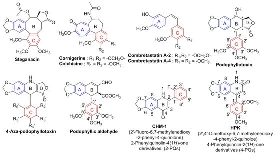
Figure 1.
Structures of antimitotic agents, * = Chiral carbon centers.
Since 1993, Professor Kuo and coworkers published compounds with 2-phenyl-4-quinolone (2-PQ) skeletons as effective antimitotic agents [16,17,18]. Among synthesized 2-PQ derivatives, compound CHM-1, 2′-fluoro-6,7-methylenedioxy-2-phenyl-4-quinolone (Figure 1), showed good anticancer activity (log GI50 < −7.0) against numerous cancer cell lines by panel screening of NCI-60 human cancer cell lines [17]. CHM-1 also demonstrated inhibition of the polymerization of microtubules [19,20]. Therefore, we continued to investigate 2-PQ analogs and applied a scaffold hopping strategy to transfer the phenyl ring at the 2-position of 2-PQ to the 4-position of the quinoline core structure, thereby obtaining a geometric isomer of 2-PQ, representing 4-phenyl-2-quinolones (4-PQs) (Figure 1) [4]. Consequently, a class of 6,7-methylenedioxy-4-substituted phenyl-2-quinolone derivatives was designed and synthesized, and their cytotoxicity against cancer cells was evaluated. These 4-PQ compounds mimic the structure of PPT analogs and serve as one of the simplified asymmetric carbon center structures of PPT [4]. Among these 4-PQ derivatives, HPK (2′,4′-dimethoxy-6,7-methylenedioxy-4-phenyl-2-quinolone) displayed significant antiproliferative activity in HL-60, Hep3B, and H460 cancer cells, and its IC50 values ranged 0.4~1.0 μM. Moreover, HPK treatment induced apoptosis and resulted in G2/M arrest through downregulating cyclin B1 and cyclin-dependent kinase 1 (CDK1) [4,21].
Because the skeleton of 4-PQs shows remarkable anticancer activities [4,21], we attempted to further develop 4-PQ derivatives. As shown in Figure 2, previous reports demonstrated that several synthetic 4-arylcoumarins (neoflavonoids, 4-phenylcoumarin compounds A–D) could inhibit the polymerization of microtubules and induce cancer cell apoptosis [22,23,24,25]. In particular, 4-arylcoumarin (C) was isolated from Exostema species (of the family Rubiaceae) and displayed cytotoxic activity against various cancer cell lines [26]. These neoflavones are abundant components that widely exist naturally in plants and have anticancer properties [27,28]. Studies reported that microtubule inhibition by 4-arylcoumarin derivatives was similar to that of combretastatin A-4, both of which bind to the colchicine-binding site [29,30]. Previous studies showed that these 4-arylcoumarin derivatives have 5-, 6-, and 7-methoxy substitutions on the coumarin core which exhibited good anticancer activity. The 2-quinolone core is the bio-isosteric structure of coumarin (Figure 2). Consequently, these results encouraged us to replace the oxygen atom (O) on the coumarin nucleus of 4-arylcoumarin with a nitrogen atom (N) by applying the concept of bio-isosterism drug design [31]. Since our previously synthesized 6,7-methylenedioxy-4-phenylquinolin-2(1H)-one (4-PQ) derivatives used 2-quinolone as the parent nucleus and displayed excellent anticancer activities [4], we attempted to synthesize further modified molecules of 4-PQs. According to the example of these 4-arylcoumarins having methoxy groups at positions 5, 6, and 7 with potential anticancer activities, a series of 4-PQ derivatives with 5-, 6-, and 7-methoxy groups were optimized and synthesized. Collectively, we established more-complete structure–activity relationships (SARs) of 4-PQ derivatives. In addition, we conducted in-silico studies to assess these 4-PQs by molecular modeling of αβ-tubulin inhibition and pharmacokinetic (PK) predictions.
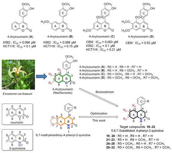
Figure 2.
Rational design of 4-phenylquinolin-2(1H)-one (4-PQ) derivatives as anticancer drugs using the bio-isosteric concept.
2. Results and Discussion
2.1. Chemistry
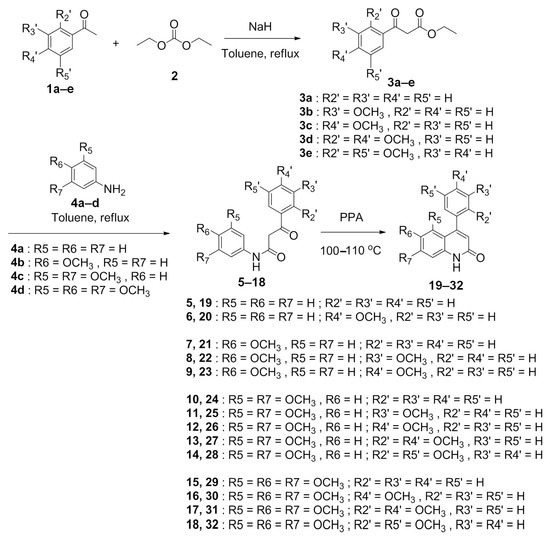
The synthetic route of the target compound 4-PQ derivatives 19–32 was based on our previous methods [4] and illustrated in Scheme 1. Initially, various substituted acetophenones were used as starting materials, and those acetophenones 1a–e were reacted with a diethyl carbonate (2) to obtain corresponding benzoylacetates 3a–e. Compounds 3a–e were then condensed with a substituted aniline 4a–d to generate corresponding benzoylacetanilide intermediates 5–18. Eventually, the target compound 4-phenylquinolin-2(1H)-one derivatives 19–32 were obtained through Knorr quinoline cyclization (acid catalysis) [4,32,33]. This synthesis method utilized intermediate benzoylacetanilides 5–18 and heat with polyphosphoric acid (PPA) at 100–110 °C to obtain the corresponding 4-PQ compounds 19–32 by intramolecular cyclization. Synthetic 4-PQs were characterized by infrared (IR), and 1H nuclear magnetic resonance (NMR) and 13C NMR, and mass spectrometry, and the results of the spectrum are shown in Supplementary Information (Figures S1–S87).
Scheme 1.
The synthesis route of 4-phenylquinolin-2(1H)-one (4-PQ) derivatives.
The 1H NMR spectra of target compounds 19–32 exhibited a characteristic broad peak in the range of 11.56–11.90 ppm which can be attributed to the NH of 2-quinolone and a single peak in the region of 5.86–6.39 ppm which can be attributed to H-3 of the ethylene proton in the 2-quinolone core structure. For instance, the 1H NMR spectrum of compound 22 was characterized by the appearance of single peaks at 3.84 and 3.86 ppm, which were attributable to the methoxy groups of the 2-quinolone moiety and 4-phenyl ring. The characteristic H-3 signal of 2-quinolone appeared at 6.38 ppm. The formation of 4-phenyl-2-quinolone was also confirmed by 13C NMR studies. N-C=O signals in the 2-quinolone core structure of 19–32 were detected within 159.94–161.92 ppm.
The detailed 2D-NMR spectral data of target compounds are provided in Supplementary Information sections. For example, the full 2D assigned correlations of compound 22 are shown in Table 1. The heteronuclear single quantum correlation (HSQC) experiment is frequently used in NMR spectroscopy to determine proton–carbon single-bond correlations. The 1H singlet at δH = 6.38 ppm gave an HSQC correlation with its attached carbon at δC = 106.52 ppm, assigned as H-3 and C-3. The 1H doublets at δH = 7.51 ppm gave an HSQC correlation with its attached carbons at δC = 104.27 ppm, assigned as H-5 and C-5 of the 2-quinolone nucleus. The heteronuclear multiple bond correlation (HMBC) experiments give correlations between carbons and protons separated by two, three, and sometimes, in conjugated systems, four bonds. The C-5 (δC = 104.27 ppm) gave an HMBC correlation with a 1H double doublet at δH = 7.32 ppm, assigned as H-7 of the 2-quinolone nucleus. The H-7 (δH = 7.32 ppm) showed an HSQC correlation at δC = 122.63 ppm, assigned as C-7. Correlated spectroscopy (COSY) is used to determine which signals arise from neighboring protons and the cross-peaks presented by coupled protons. COSY data revealed correlations between H-7 (δH = 7.32 ppm) and δH = 7.76 ppm, assigned as H-8. The 1H doublet at δH = 7.76 ppm gave an HSQC correlation with its attached carbon at δC = 121.18 ppm, assigned as H-8 and C-8. Long-range correlations in the HMBC experiment were observed between methoxy groups (H-9, δH = 3.84 ppm) and C-6 (δC = 156.08 ppm), and HMBC correlations of H-5 (δH = 7.51 ppm) with C-6 (δC = 156.08 ppm) and C-7 (δC = 122.63 ppm) and C-8a (δC = 156.08 ppm), of H-7 (δH = 7.32 ppm) with C-5 (δC = 104.27 ppm), C-6 (δC = 156.08 ppm), and C-8a (δC = 156.08 ppm), and of H-8 (δH = 7.76 ppm) with C-6 (δC = 156.08 ppm), C-8a (δC = 156.08 ppm), and C-4a (δC = 126.20 ppm). The 4-phenyl ring assignments were as follows: the 1H triplet at δH = 7.48 ppm gave an HSQC correlation with its attached carbon at δC = 130.57 ppm, assigned as H-5′ and C-5′. The COSY data revealed that the H-5′ triplet at δH = 7.48 ppm revealed correlations between δH = 7.36–7.39 ppm and δH = 7.12 ppm, assigned as H-4′ and H-6′, respectively. Moreover, the δH = 7.36–7.39 ppm contained another proton signal assigned as the remaining proton (H-2′). The H-6′ signal δH = 7.12 ppm has an HSQC correlation with δC = 116.31 ppm, assigned as C-6′. The 2H multiplet at δH = 7.36–7.39 ppm gave HSQC correlations of H-2′ and H-4′ with its attached carbon at δC = 113.11 and 119.92 ppm, respectively, assigned as C-2′ and C-4′, and was further confirmed by the HMBC spectrum. The HMBC experiment revealed correlations of the methoxy groups (H-10, δH = 3.86 ppm) with C-3′ (δC = 159.94 ppm), as well as HMBC correlations of H-4′ (δH = 7.36–7.39 ppm) with C-2′ (δC = 113.11 ppm) and C-6′ (δC = 116.31 ppm), of H-5′ (δH = 7.48 ppm) with C-3′ (δC = 159.94 ppm) and C-1′ (δC = 136.16 ppm), of H-6′ (δH = 7.12 ppm) with C-2′ (δC = 113.11 ppm) and C-4′ (δC = 119.92 ppm), and of H-2′ (δH = 7.36–7.39 ppm) with C-4′ (δC = 119.92 ppm), C-6′ (δC = 116.31 ppm), and C-4 (δC = 149.47 ppm).

Table 1.
1H and 13C NMR spectroscopic (δ/ppm), COSY, and HMBC data of compound 6-methoxy-4-(3-methoxyphenyl)quinolin-2(1H)-one (22) in DMSO-d6 (400 MHz).
2.2. Anticancer Activities and SAR Studies of New 4-PQ Derivatives
We first screened the synthesized 4-PQ 19–32 to identify the pharmacology of the anticancer activities against four human cancer cell lines, including the COLO205 (colorectal adenocarcinoma), A498 (renal cell carcinoma), H460 (non-small-cell lung cancer), and Hep 3B (liver cancer) cell lines. The cell viability of cancer cells was evaluated by a 3-(4,5-dimethylthiazol-2-yl)-2,5-diphenyltetrazoliun bromide (MTT) assay to determine cell proliferation inhibition. Results are presented as the 50% inhibition of cancer cell growth based on the drug concentration (IC50), and the preliminary results of antiproliferation are shown in Table 2.
Table 2.
Antiproliferative activities of 4-PQ derivatives.
Almost all compounds of the class, 5,6,7-methoxy-substituted 4-phenylquinolin-2(1H)-one (19–32), had IC50 anticancer activities of more than 50 μM. However, these 4-PQ derivatives showed potential anti-proliferation against the COLO205 and H460 cell lines with IC50 values ranging 0.32–47.1 μM. In particular, 6-methoxy-4-(3′-methoxy phenyl)quinolin-2(1H)-one (22) showed excellent anticancer activity, which was highly active against the COLO205 (IC50 = 0.32 μM) and H460 cell lines (IC50 = 0.89 μM). In addition, 4-(2′,4′-dimethoxyphenyl)-5,7-dimethoxy quinolin-2(1H)-one (27) showed moderate anticancer activity against the COLO205 cell line (IC50 = 7.85 μM). Interestingly, a previous study indicated that the 3,4,5-trimethoxyphenyl moiety is commonly employed in antimitotic agents [34]; however, our results showed that a tri-methoxy substitution on 4-PQs (29–32) seemed to have no anticancer effects (IC50 > 50 μM). Among all of the synthesized compounds, compound 22 was more effective than the parent compound, HPK (IC50 = 7.4 μM), with an IC50 of 0.32 μM against COLO205 cells. Therefore, the mechanism of the anticancer activity of compound 22 deserves further investigation. On the other hand, out of the 4-PQ compounds exhibiting anticancer activities (with IC50 values of <50 μM), compounds 19, 20, 21, 22, 25, 27, and 28, were subjected to further in silico molecular docking studies and pharmacokinetic (PK) predictions.
The preliminary results of anti-proliferation indicated that the active compounds 19, 20, 21, 22, 25, 27, and 28 only demonstrated anticancer activities in COLO205 and H460 cells (other cell lines were shown IC50 > 50 μM). As shown in Table 3, we further investigated the selectivity index (SI) to analyze the sensitivities of each compound against COLO205 and H460, respectively. Compound 22 exhibited the highest sensitivity (SI = 66.04) toward COLO205 cells, while compounds 27 and HPK showed moderate selected ratios (SI are 2.69 and 2.86). Other compounds (19, 20, 25, and 28) displayed no sensitivities (SI < 1) compared with 22, 27 as well as HPK in COLO205. Similar results were shown in H460 cells, compound 22 had the highest selected ratio (SI = 33.64). Notably, compound HPK also displayed high sensitivity (SI = 33.27) against H460 cells. However, compounds 19, 20, 21, 25, and 28 showed no selectivity (SI < 1) in H460 cells. Overall, compound 22 is the most active and sensitive within these 4-PQ derivatives against both COLO205 and H460 cell lines.
Table 3.
Antiproliferative sensitivities of 4-PQ derivatives were analyzed by selectivity index (SI).
2.3. Molecular Modeling of 4-Phenyl-2-quinolone (4-PQ) Derivatives Proposes Their Docking into the Colchicine-Binding Site
As previously reported, the cytotoxic activity of 4-phenyl-2-quinolone (4-PQ) [35,36] and its 4-arylcoumarin analog [24,25,29,30] are well-known as antimitotic agents associated with their ability to inhibit tubulin formation via binding of compounds to the colchicine-binding site. In this study, molecular docking was simulated to investigate the binding modalities of 4-PQ derivatives against tubulin using AutoDock4 [37] and AutoDock Vina software [38]. In 2004, the co-crystal structure of αβ-tubulin complexed with N-deacetyl-N-(2-mercaptoacetyl)colchicine (DAMA-colchicine, an analog of colchicine) was discovered by X-ray analysis (PDB ID: 1SA0) [39]. At the same time, the X-ray structure of the tubulin/PPT complex also revealed that PPT inhibits tubulin polymerization by blocking the colchicine-binding site (PDB ID: 1SA1) [39] and presents a similar orientation as that of DAMA-colchicine. Accordingly, as shown in Figure 3A, molecular docking experiments were performed on the 4-PQ series and their virtual binding interactions with α- and β-tubulin (PDB ID: 1SA1), and results were compared to PPT and colchicine as reference drugs. To validate docking protocols, the co-crystallized ligand PPT and colchicine were re-docked into the colchicine-binding site of isolated α- and β-tubulin proteins. As shown in Figure 3B,C, co-crystallized poses of the reference drugs were superimposed on the re-docking pose. PPT and colchicine could be re-docked to their respective target proteins with root mean squared deviation lower bounds (RMSD/lbs) of 0.017 (Figure 3B) and 1.879 Å (Figure 3C) via AutoDock Vina. Additionally, the re-docked conformations of PPT and colchicine had similar orientations to their respective co-crystallized ligands. The docked poses of the 4-PQs are superimposed with PPT and colchicines in Figure 3D. Results showed that the methoxy groups on the 4-phenyl ring (C ring) of 4-PQ were located in the tri-methoxy groups’ position of PPT and colchicines (as indicated by yellow arrows in Figure 3D). Additionally, the methoxy groups on the 2-quinolone core (A ring) of 4-PQ were located in the methylenedioxy moiety of PPT and the ketone group of colchicines (as indicated by red arrows).
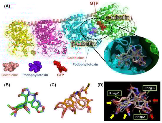
Figure 3.
4-Phenyl-2-quinolone (4-PQ) compounds, HPK, podophyllotoxin, and colchicine docked into αβ-tubulin (PDB ID: 1SA1) via AutoDock Vina. (A) 4-PQs (white lines), HPK (yellow lines), podophyllotoxin (blue spheres), and colchicine (pink spheres) bound to β-tubulin (cyan) at the interface with α-tubulin (green). (B) Podophyllotoxin’s co-crystallized pose (green) and docking pose (blue). (C) The co-crystallized pose (orange) of N-deacetyl-N-(2-mercaptoacetyl)-colchicine (DAMA-colchicine) and docking pose (pink) of colchicine. (D) All docked compounds were superimposed on each other. The white lines represent compounds 19, 20, 21, 22, 25, 27, and 28. The yellow lines represent HPK. The blue sticks represent podophyllotoxin, and the pink sticks represent colchicine.
We selected some potential 4-PQ compounds, representing 19, 20, 21, 22, 25, 27, and 28, which had IC50 values of <50 μM by the MTT assay to predict their binding modalities into the colchicine site. Results suggested that these 4-PQ derivatives could snugly occupy the colchicine-binding site of αβ-tubulin (Figure 3A). On the other hand, a previous report on deciding which program to use in docking studies indicated that Vina is much faster and adopts more accurate binding poses but Autodock4 forms better binding affinity [40]. Therefore, we executed both docking programs to estimate the binding energies, and the possible docking poses were predicted via the AutoDock Vina. The binding affinity of each 4-PQ attached to αβ-tubulin is discussed based on the estimated ΔG value, and results are given in Table 4. The binding energies of these compounds were in the range of −7.46 to −9.91 (kcal/mol) via AutoDock4 calculation, while the range of Vina estimation is −5.2 to −8.1 (kcal/mol). The estimated ΔG of PPT was lower (−9.91 kcal/mol in Autodock4; −9.0 kcal/mol in Vina) than the other 4-PQs and the HPK parent compound (−7.90 kcal/mol in Autodock4; −8.3 kcal/mol in Vina), which means that the inhibitory potency of these 4-PQs was less than that of the control drug. However, compound 22 (−7.97 kcal/mol in Autodock4; −8.1 kcal/mol in Vina) had a very close ΔG value compared to HPK (−7.90 kcal/mol in Autodock4; −8.3 kcal/mol in Vina).
Table 4.
The free binding energies (kcal/mol) of the synthesized 4-PQ compounds and reference drug against the colchicine-binding site of the tubulin receptor.
All docked molecules interacted with amino acid residues of αβ-tubulin within the colchicine-binding active site (Figure 4, Figure 5 and Figure 6); furthermore, the results were analyzed by Discovery Studio Visualizer software and summarized in Table 5. As shown in Figure 4A, the proposed binding mode of PPT formed five carbon–hydrogen bonds with ALA250, VAL315, ALA317, LYS352, and ASN350 residues. The methylenedioxy pharmacophore formed two hydrogen bonds with VAL315 and ASN350. The carbonyl group of the lactone ring formed one hydrogen bond with ALA250, and the methoxy group of PPT’s phenyl ring (C-ring) formed two hydrogen bonds with ALA317 and LYS352. The phenyl ring (ring A) showed one pi-sulfur bond with MET259. In addition, the A-ring, C-ring, methylenedioxy moiety, and tri-methoxy groups of PPT formed several hydrophobic interactions with VAL181, CYS241, LEU242, ALA250, LEU255, ALA316, VAL318, LYS352, ALA354, and ILE378. Similar results were also seen for colchicine (Figure 4B); tri-methoxy groups, the A-ring, and C-ring of colchicines formed hydrophobic interactions with CYS241, LEU242, ALA250, LEU255, ALA316, VAL318, LYS352, ALA354, and ILE378, while the carbonyl group and methoxy group of the A-ring moiety formed four carbon-hydrogen bonds with ALA180, ASN258, VAL315, and ASN350. In addition, one methoxy group of the C-ring of colchicine showed one carbon–hydrogen bond with VAL238. Taken together, these results revealed that the methoxy groups or some hydrogen bond acceptors (containing oxygen, such as carbonyl) on the A- and C-rings of the ligand played pivotal roles with the αβ-tubulin receptor in the colchicine-binding site. Interactions with the A-ring site were delineated by the following amino acid residues: ALA180, VAL181, ASN258, MET259, VAL315, ALA316, ASN350, and LYS352. On the other hand, the ligand’s moiety in the C-ring site interacted with amino acid residues of αβ-tubulin, including residues CYS241, LEU242, VAL238, ALA250, LEU255, ALA316, ALA317, VAL318, LYS352, ALA354, and ILE378.
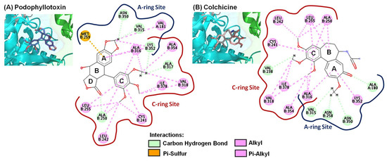
Figure 4.
Close-up of (A) podophyllotoxin and (B) colchicine within the colchicine-binding site of the αβ-tubulin shown along with their corresponding 2D interaction plots.
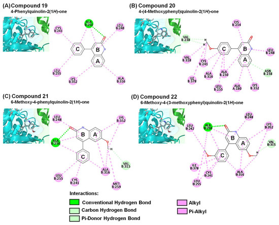
Figure 5.
Close-up of (A) 19, (B) 20, (C) 21, and (D) 22 within the colchicine-binding site of the αβ-tubulin shown along with their corresponding 2D interaction plots.
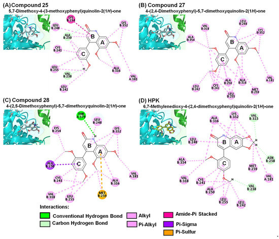
Figure 6.
Close-up of (A) 25, (B) 27, (C) 28, and (D) HPK within the colchicine-binding site of the αβ-tubulin shown along with their corresponding 2D interaction plots.
Table 5.
The protein–ligand interaction analysis of the synthesized 4-PQ compounds and reference drugs are shown in the colchicine-binding site of the tubulin receptor.
In the current study, we identified 4-PQ compounds with extensive interactions with amino acids of the colchicine-binding site of αβ-tubulin. The predicted binding poses of compounds 19, 20, 21, 22, 25, 27, 28, and HPK showed respective affinity values of −7.5, −7.4, −8.1, −8.1, −6.1, −5.9, −5.2, and −8.3 kcal/mol via Vina estimation. The amide groups (carbonyl of 2-quinolone core) of compounds 19, 21, and 22 formed an important conventional hydrogen bond with ALA250 (Figure 5), which is similar to the carbonyl group in the γ-lactone ring (D-ring) of PPT displayed carbon hydrogen bond with β-tubulin. The A- and B-rings of the 2-quinolone moiety displayed several hydrophobic interactions with LEU248, ALA316, and LYS352 (Figure 6A–D). In addition, the amide groups (NH) of compound 28 formed a conventional hydrogen bond with THR179 (Figure 6C). These findings implied that the 2-quinolone core structure plays an important role in the colchicine-binding domain, which is consistent with our previous study of 4-benzyloxy-2-quinolone derivatives on tubulin inhibition [5]. Each of their A- and C-rings revealed similar hydrophobic interactions with αβ-tubulin, which were just like those of PPT and colchicine.
2.4. In Silico Prediction of Drug-Likeness Studies
Drug-likeness is considered an important tool for predicting whether a molecule can be a potential drug candidate. Therefore, we employed SwissADME [41] to forecast the bioavailability and drug-likeness properties, and the results are shown in Table 6. Several rules were used to assess the drug-likeness, the most famous of these descriptions is Lipinski’s rule of five [42]. According to Lipinski’s rule, a compound is labeled as drug-like when it meets the following criteria: molecular weight < 500 Da, H-bond donors (HBDs; the sum of OHs and NHs) ≤ 5, H-bond acceptors (HBAs; the sum of Ns and Os) ≤ 10, and Log P ≤ 5. The Log P values are measured as the partition coefficient between n-octanol and water and represented the molecular hydrophobicity or lipophilicity. High Log P values indicate poor absorption or low permeability, while low Log P values indicate increased absorption and permeability. Log P values of all predicted compounds were <5 (ranging from 2.25–3.05), which is within an acceptable range. The molecular polar surface area (PSA) is a very useful parameter for predicting drug transport properties. The PSA is defined as the sum of surface polar fragments (usually oxygens, nitrogens, and attached hydrogens) in a molecule. The topological PSA (TPSA) provides results of practically the same quality as the classical three-dimensional PSA; the calculations, however, are two to three orders of magnitude faster [43]. The 4-PQ compounds had TPSA values ranging from 32.86–69.78 Å2, which is a good range for oral bioavailability since most therapeutic molecules have TPSA values of <140 Å2 for good passive membrane permeability (in a non-polar environment) [44].
Table 6.
Drug-likeness predictions of tested compounds.
Moreover, none of the 4-PQs were predicted as pan assay interference compounds (PAINS) [45], which suggests the absence of potential pan-assay interferences or being frequent hitters of promiscuous compounds. On the other hand, we found no violations of Lipinski’s [42], Veber’s [44], Ghose’s [46], Egan’s [47], or Mugge’s [48] rules for any of the synthesized 4-PQ compounds based on the obtained data.
2.5. In Silico Analysis of Potential PK (ADMET) and Toxicological Properties (T)
It is clear that in addition to pharmacological properties, ADMET (absorption, distribution, metabolism, elimination, and toxicity) research plays a crucial role in the success of drug candidates [49,50]. Because of the impact on eventual success, such studies now occur early in the drug discovery process [51]. The PK characteristics of 4-PQ compounds (19, 20, 21, 22, 25, 27, and 28), PPT, and colchicine were assessed using online SwissADME software [41] and admetSAR [52]. As shown in Table 7, all compounds were predicted to have high gastrointestinal (GI) absorption (human intestinal absorption; HIA). In addition, all of the 4-PQ derivatives were predicted to have the ability to pass through the blood–brain barrier (BBB), except for PPT and colchicine. P-glycoprotein (P-gp), a member of ATP-binding cassette (ABC) transporters, is an efflux transporter that can pump drugs out of cells to reduce the biological effects of anticancer drugs [53], which may cause a failed treatment. If a drug under investigation is identified as a P-gp substrate, this means that its concentration levels in cells may be clinically affected by a P-gp inhibitor. On the other hand, P-gp plays a key role in multidrug resistance (MDR) in cancer [54]. Most of these were not P-gp substrates, as only HPK and colchicine were predicted to have potential interference with P-gp.
Table 7.
ADMET analysis of 4-PQ compounds, podophyllotoxin (PPT), and colchicine was evaluated by SwissADME [41].
Cytochrome P450 (CYP450) enzymes are pivotal mediators in the metabolism of many medicines. The CYP450 expression level determines a drug’s metabolic rate. Unanticipated drug–drug interactions (DDIs) and drug metabolism problems based on CYP450 enzymes are also common causes of adverse drug events (ADEs) [55]. In the metabolic profiles, all of these compounds served as different kinds of CYP450 enzyme inhibitors (Table 7), suggesting that they may compete with other drugs, which means that they can block other drugs’ metabolic activity to extend the half-life of therapeutic drugs in patients’ serum levels. Further PKs were investigated in the future for any potential drug–drug interactions. In particular, compounds 22, 25, 27, 28, and HPK inhibited more than three types of CYP450 enzymes, including CYP1A2, CYP2C9, CYP2D6, and CYP3A4, which reveals that they would likely interfere with the metabolism of other drugs using the same CYP450 pathway. These findings implied that using these 4-PQs should be done while being aware of the risks of CYP450 enzyme-based unanticipated DDIs and metabolism problems. On the other hand, several computational approaches can help predict the potential toxicity of new compounds in the initial stages of drug discovery, primarily considering human ether-à-go-go-related gene (hERG) inhibition, AMES toxicity, and carcinogenicity. hERG potassium channels are essential for regulating electrical activity in the human heart [56]. Inhibition of hERG potassium channels may lead to long QT syndrome (LQTS), which is known as a fatal ventricular tachyarrhythmia called torsades de pointes in clinical studies [57,58]. As shown in Table 8, the toxicity profile predicted by hERG indicated that 4-PQs were non-hERG inhibitors. Moreover, the carcinogenicity assessment showed that 4-PQ compounds were non-carcinogens. However, AMES toxicity showed that most of the 4-PQ compounds were found to be AMES toxic (mutagens), including compounds 20, 21, 22, 25, and HPK. These results revealed that when using these 4-PQs, one should be aware of potential DDI side effects and risks of mutagenicity. The U.S. Environmental Protection Agency (EPA) established four toxicity categories for acute hazards: class I is high toxicity and severely irritating, class II is moderate toxicity, class III is slightly toxic and low toxicity, and class IV is non-toxic [59]. All of the predicted compounds were found to have 50% lethal dose (LD50) values that ranged from 2.1994–3.0013 mol/kg and were classified as class III. These results implied that these 4-PQs have low acute toxicity.
Table 8.
Toxicity analysis of 4-PQ compounds, podophyllotoxin (PPT), and colchicine was evaluated by admetSAR [52].
The SwissADME software briefly describes six essential properties of oral bioavailability to create a bioavailability radar and boiled egg plots for analysis. These properties and suitable values are lipophilicity (LIPO): Log P (XLOGP3) value from −0.7 to 5.0; SIZE, molecular weight in the 150–500 g/mol range; polarity (POLAR), TPSA values from 20 to 130 Å2; insolubility (INSOLU), solubility with Log S (ESOL) from −6 to 0; insaturation (INSATU), saturation fraction of carbons with sp3 hybridization (fraction Csp3) from 0.25 to 1; and flexibility (FLEX), the number of the rotatable bonds from 0 to 9 [41]. Results are shown in Figure 7A−J with bioavailability radar for 4-PQ compounds, and the pink area in the radar is a range of optimal values. Bioavailability radars for all 4-PQ molecules exhibited suitable parameters in terms of lipophilicity, size, polarity, insolubility, and flexibility, but were not good at insaturation (INSATU). The purpose of the boiled egg plot is to correctly predict HIA and BBB properties of substances that are important for therapeutic research [60]. The blue dots represent P-gp substrates (PGP+) and the red dots indicate P-gp non-substrates (PGP−). The boiled egg forecast of 4PQs is shown in Figure 7K, the outer gray area is for compounds with lower GI absorption and limited BBB penetration. Furthermore, the white area represents the physicochemical space of the molecule with the highest probability of passive HIA, while the yellow area represents the physicochemical space of the molecule with the highest probability of penetrating the BBB. All of the 4-PQs exhibited predicted high BBB penetration, as demonstrated by their location inside the yellow ellipse (yolk) defining BBB absorption (Figure 7K). Essentially, all of the 4-PQs that were predicted to enter the brain also displayed a predicted high HIA (Table 7). Only colchicine and PPT were predicted to display low brain permeation but high intestinal absorption.
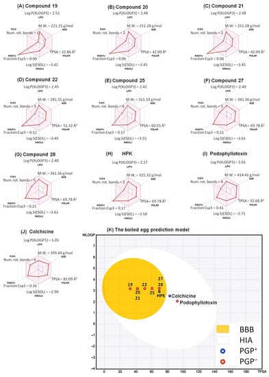
Figure 7.
Bioavailability radar related to physicochemical properties of 4-PQ compounds (A) 19, (B) 20, (C) 21, (D) 22, (E) 25, (F) 27, (G) 28, (H) HPK, (I) podophyllotoxin, and (J) colchicine. The bioavailability radar indicated LIPO (lipophilicity, Log P(XLOGP3)), SIZE (molecular weight), POLAR (polarity, topological polar surface area; TPSA), INSOLU (insolubility, Log S(ESOL)), INSATU (insaturation, fraction Csp3), and FLEX (flexibility, the number of rotatable bonds). (K) Graphical distribution of absorbed compounds based on bioavailability using a boiled egg prediction model.
We further employed the site of metabolism (SOM) prediction (SOMP) [61], a web service for in silico prediction of the SOM, to gain insights into the biotransformed positions of the most effective compound 22 (the original data can be found in Supplementary Information Tables S1–S5). The predicted results for compound 22 are illustrated in Figure 8A. In addition, possible metabolites of compound 22 were presented by BioTransformer 3.0 [62], a web server that accurately predicts metabolic transformation products. As shown in Figure 8B, several metabolites of 4-PQ compound 22 were formed by aromatic hydroxylation on its aromatic A- and C-rings via phase I metabolism of CYP450. Typical metabolism of the methoxy group, O-dealkylation, was also found on compound 22’s 6-methoxy of the 2-quinolone core and 3′-methoxy of the 4-phenyl ring. Of note, the epoxidation of an alkene was catalyzed by CYP450 to form an epoxide on ring B of compound 22. Further phase II metabolism takes place to form OH-glucuronidation and glutathione (GSH)-conjugation of epoxide by UDP-glucuronosyltransferase and glutathione transferase, respectively. These hydrophilic conjugates and their metabolites can be excreted in phase III of their metabolism.
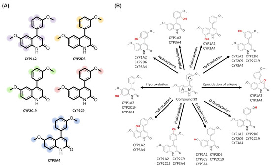
Figure 8.
Applying a metabolism predictor to anticipate sites of metabolism (SOMs) and the metabolite structures of compound 22. (A) SOM prediction (SOMP) predicted metabolic positions of compound 22 by CYP450 isoforms, including CYP1A2, CYP2D6, CYP2C19, CYP2C9, and CYP3A4. (B) The biotransformation of compound 22 was predicted by phase I (CYP450) transformation.
3. Materials and Methods
3.1. Materials and Physical Measurements
Human hepatoma (Hep 3B; HB-8064-ATCC), lung cancer cells (H460; HTB-177-ATCC), and normal skin cells (Detroit 551; CCL-110-ATCC) were purchased from American Type Culture Collection (Manassas, VA, USA) (https://www.atcc.org/; accessed on 3 December 2022). Human colon carcinoma (COLO205; CCL-222-ATCC) and renal cancer cells (A498; HTB-44-ATCC) were commissioned by the Food Industry Research and Development Institute (Hsinchu, Taiwan) to purchase. All solvents and reagents were purchased commercially and applied without additional purification. The progress of all reactions was monitored by thin layer chromatography (TLC) on 2 × 6-cm pre-coated silica gel 60 F254 plates of 0.25 mm thick (Merck, Darmstadt, Germany). Chromatograms were visualized under UV light at 254–366 nm. Silica gel 60 adsorbent (Merck, particle size 0.063–0.200 mm) was used for column chromatography. Melting points (MPs) were determined with a Yanaco MP-500D melting point apparatus and are uncorrected. Infrared (IR) spectra were recorded on a Shimadzu IR-Prestige-21 spectrophotometer (Kyoto, Japan) as KBr pellets. Nuclear magnetic resonance (NMR) spectra were obtained on a Bruker Avance DPX-200 (200 MHz) and DPX-400 (400 MHz) (Billerica, MA, USA) FT-NMR spectrometer at room temperature, and chemical shifts are reported in ppm (δ). The following abbreviations were used: s, singlet; d, doublet; t, triplet; dd, double doublet; and m, multiplet. The MPs, IR, and NMR were tested by the instruments of China Medical University Precision Instrument Center, Taichung, Taiwan. The mass was examined by Finnigan MAT 95S (Instrumentation Center, National Taiwan University, Taipei, Taiwan) to obtain spectra of high-resolution electrospray ionization (HRESI).
3.2. Chemistry
3.2.1. General Procedure for the Synthesis of Benzoylacetates (3a–e)
Compound ethyl 3-oxo-3-phenyl propanoate (3a) was purchased commercially and used without further purification. Compounds 3b–e were prepared according to our previously reported procedures [4]. A mixture of diethyl carbonate (2; 1 equivalent) and appropriate acetophenone (1b–e; 1 equivalent) was dissolved into 50–100 mL toluene, and then sodium hydride (NaH) (2 equivalents, 60% dispersed in mineral oil) was added. The mixture was stirred and heated to reflux for 30 min; the reaction was monitored by thin layer chromatography (TLC; with n-hexane/ethyl acetate = 10/1 as the eluent solution). After the reaction was complete, the mixture was poured into 200 mL of ice water and acidified with glacial acetic acid to pH 4–5. The mixture was extracted with ethyl acetate, dried, filtered with anhydrous magnesium sulfate (MgSO4), concentrated under reduced pressure, and separated and purified by column chromatography (n-hexane/ethyl acetate = 10/1) to obtain the corresponding benzoylacetates (3b–e). All synthetic compounds were in agreement with 1H NMR and 13C NMR spectroscopic data.
3.2.2. General Procedure for the Synthesis of Benzoylacetanilides (5–18)
A mixture of the benzoylacetates (3a-e, 1 equivalent) and substituted aniline (4a–d, 1 equivalent) was stirred and heated to reflux in 50–100 mL toluene overnight. Reactions were monitored by TLC. After the reaction was complete, the mixture was cooled down to room temperature, and if a solid had formed, it was filtered by suction. If no solid had formed, then it was extracted with 10% NaOH, the aqueous layer with glacial acetic acid was acidified to pH 4–5, and the solid was separated out. The resulting precipitate was isolated by suction filtration. If no solid was found after acidification, the residue was extracted with ethyl acetate, and the organic layer was dried with anhydrous magnesium sulfate and filtered. The filtrate was concentrated under reduced pressure and purified by column chromatography to obtain the corresponding benzoylacetanilides (5–18).
3.2.3. General Procedure for the Synthesis of 5-, 6-, 7-Methoxy-substituted 4-Phenyl-2-quinolones (19–32)
A 5–6 weight of polyphosphoric acid (PPA) was added to benzoylacetanilide (5–18), and the mixture was heated at 100–110 °C for 1~2 h. The mixture was cooled after TLC monitoring (n-hexane/ethyl acetate = 1/1 or ethyl acetate) indicated that the reaction was complete. The reaction solution was cooled to room temperature and poured into 200 mL of ice water to terminate the reaction. If a solid had precipitated, the residues were filtered with suction and recrystallized by ethanol or ethyl acetate; if no solid had precipitated, the residues were extracted with ethyl acetate, and the organic layer was dried with anhydrous MgSO4. The filtrate was concentrated under reduced pressure and then separated and purified by column chromatography (silica gel) to obtain the corresponding pure 4-phenylquinolin-2(1H)-one (19–32).
3.3. MTT Assay for Antiproliferative Activity
Human tumor cell lines of a cancer screening panel were maintained in RPMI-1640 medium supplemented with 10% fetal bovine serum (FBS; GIBCO/BRL), penicillin (100 U/mL)/streptomycin (100 g/mL) (GIBCO/BRL), and 1% L-glutamine (GIBCO/BRL) at 37 °C in a humidified atmosphere containing 5% CO2. Human hepatoma Hep 3B and normal skin Detroit 551 cells were maintained in Dulbecco’s modified Eagle medium (DMEM) supplemented with 10% FBS (GIBCO/BRL), penicillin (100 U/mL)/streptomycin (100 g/mL) (GIBCO/BRL), and 1% L-glutamine (GIBCO/BRL) at 37 °C in a humidified atmosphere containing 5% CO2. Logarithmically growing cancer cells were used for all experiments. Human tumor cell lines were treated with vehicle or test compounds for 48 h. According to a previous report [5], the cell growth rate was determined by an MTT (3-(4,5-dimethylthiazol-2-yl)-2,5-diphenyltetrazoliun bromide) reduction assay.
The tested compounds were initially prepared in the stock solution with a concentration of 100 μM and diluted to evaluate until found the IC50 values. The IC50 values of more than 50 μM are regarded as having no anti-proliferation effect. After 48 h of treatment, the cell growth rate was measured on an enzyme-linked immunosorbent assay (ELISA) reader at a wavelength of 570 nm, and IC50 values of the test compounds were calculated.
3.4. Molecular Docking
The crystal structure of αβ-tubulin with the co-crystallized inhibitors, N-deacetyl-N-(2-mercaptoacetyl)colchicine (DAMA-colchicine) and podophyllotoxin (PPT), was obtained from the Protein Data Bank under respective PDB codes of 1SA0 and 1SA1 [39]. The chemical structures of 4-phenyl-2-quinolone (4-PQ) derivatives were drawn, and the energy of the 3D structure was minimized using the MM2 force field [63] in ChemBio3D ultra 12.0. Molecules were docked into the colchicine-binding pocket using AutoDock4 [37] and AutoDock Vina [38]. The binding abilities were calculated as the binding free energy (ΔG, kcal/mol) in both AutoDock4 and Vina. The grid map with autogrid (for grid calculation) within MGLTools (version 1.5.6) was carried out to define interactions of the protein and ligand in the colchicine-binding site. The grid box size was built of 40 × 40 × 40 points in the x, y and z directions, and the centers of x, y and z dimensions are 117, 89, and 7.5 in AutoDock4. The Vian’s parameters were set as follows: a grid box size was 20 × 20 × 20, and the grid center was located at x = 117, y = 89, and z = 7.5. The molecular 3D-docking results were prepared and visualized using PyMOL [64] (https://pymol.org/2/; accessed on 6 May 2022). 2D-interaction maps of docked complexes were visualized with the BIOVIA Discovery Studio Visualizer (http://accelrys.com; accessed on 6 May 2022) to show interactions of compounds with amino acid residues of the target protein.
3.5. In Silico Predictions of Physicochemical Properties
Absorption, distribution, metabolism, excretion/elimination, and toxicity (ADMET) of drug-like PKs were calculated utilizing SwissADME [41] and admetSAR [52]. The drug-like potentials of the chosen ligands (based on the potential for in vitro antiproliferation) were estimated by observing their PK and pharmacodynamics features. SwissADME provided the parameters, including physicochemical properties (molecular weight, fraction Csp3, numbers of rotatable bonds, numbers of hydrogen bond acceptors, and numbers of hydrogen bond donors), lipophilicity (Log Po/w for iLOGP, XLOGP3, WLOGP, MLOGP, and SILICOS-IT), water solubility (Log S for ESOL, Ali, and SILICOSIT), PKs (gastrointestinal (GI) absorption, blood–brain barrier (BBB) permeant, P-glycoprotein (P-gp) substrate, CYP1A2 inhibitor, CYP2C19 inhibitor, CYP2C9 inhibitor, CYP2D6 inhibitor, and CYP3A4 inhibitor), drug-likeness rules (Lipinski, Veber, Ghose, Egan, and Muegge), and pan assay interference compounds (PAINS) methods. The admetSAR online server provided predictions of toxicity properties such as human hERG inhibition, AMES toxicity, and carcinogenicity. The sites of metabolism (SOMs) of compound 22 were predicted by the SOM prediction (SOMP) via submitting SMILES codes [61]. Possible structures of metabolites were accessed using BioTransformer 3.0 [62].
4. Conclusions
Previously, 2-quinolone derivatives were identified as anti-tubulin agents [5,36]. To discover novel 2-quinolone-bearing molecules of 4-phenyl-2-quinolone (4-PQ) derivatives with anticancer activities, we attempted to design and synthesize a series of methoxy-substituted 4-PQ compounds. In the current study, we investigated the structure–activity relationships of the methoxy groups at positions 5, 6, and 7 on the 2-quinolone core, and evaluated the anticancer effects of the methoxy substitutions on their 4-phenyl ring. Preliminary antiproliferative effects of compounds 19–32 indicated that most of the 4-PQ derivatives had IC50 values of >50 μM. However, some of the 4-PQs, such as compounds 19, 20, 21, 22, 25, 27, and 28, showed potential anticancer effects against COLO205 and H460 cells (with IC50 values of <50 μM). In particular, the compound 6-methoxy-4-(3-methoxyphenyl)quinolin-2(1H)-one (22) exhibited excellent anticancer activities against COLO205 and H460 cancer cells with respective IC50 values of 0.32 and 0.89 μM. Computer modeling indicated that the colchicine-binding site of αβ-tubulin could deeply dock to these 4-PQs, which displayed similar orientations as those with podophyllotoxin and colchicine. The A- and C- rings of 4-PQ overlapped with those of colchicine and podophyllotoxin. In the drug discovery process, weak bioavailability and pharmacokinetics always lead to drug development failure. Of note, these 4-PQs showed suitable drug-likeness, adequate bioavailability, and non-cardiotoxicity; however, toxicity predictions indicated the risk of drug–drug interactions and carcinogenic side effects, so these 4-PQs must be used with caution. Overall, LD50 values of these 4-PQs were classified in class III, indicating that these 4-PQs have low acute toxicity. Therefore, further structure optimization of 4-PQs is worth investigating to convert them into clinically useful anticancer agents in the future. Altogether, our findings suggest that 4-PQ derivatives with methoxy substituents may provide new insights into these compounds in antimitotic drug development, particularly through αβ-tubulin inhibition.
Supplementary Materials
The following supporting information can be downloaded at: https://www.mdpi.com/article/10.3390/molecules28020555/s1, compound characterization data of benzoylacetates (3b–e), benzoylacetanilides (5–18) and 5-, 6-, 7-methoxy-substituted 4-phenyl-2-quinolones (19–32). Meta-Pred Web Server prediction results of site of metabolism for compound 22 (Tables S1–S5). Copies of 1H NMR, 13C NMR, COSY, HSQC, HMBC, IR and Mass spectra of the 4-PQ compounds 19–32 (Figure S1–S87).
Author Contributions
Conceptualization, Y.-F.C., L.-J.H. and S.-C.K.; methodology, Y.-F.C., L.-J.H. and M.R.S.; design, synthesis, docking and in silico experimentation, Y.-F.C.; formal analysis, Y.-F.C., L.-J.H. and S.-C.K.; investigation, Y.-F.C.; data curation, Y.-F.C., M.R.S. and N.M.; writing—original draft preparation, Y.-F.C. and B.L.; writing—review and editing, Y.-F.C., B.L. and H.-S.H.; visualization, Y.-F.C.; supervision, L.-J.H., H.-S.H. and H.-Y.L.; project administration, L.-J.H. and S.-C.K.; funding acquisition, L.-J.H., H.-S.H. and H.-Y.L. All authors have read and agreed to the published version of the manuscript.
Funding
This research was funded by the National Science Council of Taiwan, grant number NSC 101-2320-B-039-009-MY3 and NSC 95-2320-B-039-011-MY3 awarded to L.-J. Huang; the National Science and Technology Council, Taiwan, grant number NSTC111-2314-B-038-017 and the Ministry of Science and Technology, Taiwan, grant number MOST111-2314-B-038-122 awarded to H.-S. Huang. TMU Research Center of Cancer Translational Medicine from The Featured Areas Research Center Program within the framework of the Higher Education Sprout Project by the Ministry of Education (MOE) in Taiwan, by grants from the Ministry of Science and Technology, Taiwan (MOST111-2314-B-038-123 and NSTC111-2314-B-038-015 to H.-Y. Lin). The authors also acknowledge the funding support from Researchers Supporting Project Number (NSTC111-2811-B-038-025) from the National Science and Technology Council, Taiwan.
Institutional Review Board Statement
Not applicable.
Informed Consent Statement
Not applicable.
Data Availability Statement
Not applicable.
Conflicts of Interest
The authors declare no conflict of interest.
Sample Availability
Samples of the compounds are available from the authors.
References
- Anand, U.; Dey, A.; Chandel, A.K.S.; Sanyal, R.; Mishra, A.; Pandey, D.K.; De Falco, V.; Upadhyay, A.; Kandimalla, R.; Chaudhary, A.; et al. Cancer chemotherapy and beyond: Current status, drug candidates, associated risks and progress in targeted therapeutics. Genes Dis. 2022, in press. [Google Scholar] [CrossRef]
- Lu, Y.; Chen, J.; Xiao, M.; Li, W.; Miller, D.D. An overview of tubulin inhibitors that interact with the colchicine binding site. Pharm. Res. 2012, 29, 2943–2971. [Google Scholar] [CrossRef] [PubMed]
- Wang, J.; Miller, D.D.; Li, W. Molecular interactions at the colchicine binding site in tubulin: An X-ray crystallography perspective. Drug Discov. Today 2022, 27, 759–776. [Google Scholar] [CrossRef]
- Chen, Y.F.; Lin, Y.C.; Huang, P.K.; Chan, H.C.; Kuo, S.C.; Lee, K.H.; Huang, L.J. Design and synthesis of 6,7-methylenedioxy-4-substituted phenylquinolin-2(1H)-one derivatives as novel anticancer agents that induce apoptosis with cell cycle arrest at G2/M phase. Bioorganic Med. Chem. 2013, 21, 5064–5075. [Google Scholar] [CrossRef]
- Chen, Y.F.; Lin, Y.C.; Morris-Natschke, S.L.; Wei, C.F.; Shen, T.C.; Lin, H.Y.; Hsu, M.H.; Chou, L.C.; Zhao, Y.; Kuo, S.C.; et al. Synthesis and SAR studies of novel 6,7,8-substituted 4-substituted benzyloxyquinolin-2(1H)-one derivatives for anticancer activity. Br. J. Pharmacol. 2015, 172, 1195–1221. [Google Scholar] [CrossRef] [PubMed]
- Hamel, E. Antimitotic natural products and their interactions with tubulin. Med. Res. Rev. 1996, 16, 207–231. [Google Scholar] [CrossRef]
- Stanton, R.A.; Gernert, K.M.; Nettles, J.H.; Aneja, R. Drugs that target dynamic microtubules: A new molecular perspective. Med. Res. Rev. 2011, 31, 443–481. [Google Scholar] [CrossRef]
- Castro, M.A.; Miguel del Corral, J.M.; Gordaliza, M.; Gómez-Zurita, M.A.; García, P.A.; San Feliciano, A. Chemoinduction of cytotoxic selectivity in Podophyllotoxin-related lignans. Phytochem. Rev. 2003, 2, 219–233. [Google Scholar] [CrossRef]
- Gensler, W.J.; Murthy, C.D.; Trammell, M.H. Nonenolizable podophyllotoxin derivatives. J. Med. Chem. 1977, 20, 635–644. [Google Scholar] [CrossRef]
- Magedov, I.V.; Manpadi, M.; Rozhkova, E.; Przheval’skii, N.M.; Rogelj, S.; Shors, S.T.; Steelant, W.F.; Van Slambrouck, S.; Kornienko, A. Structural simplification of bioactive natural products with multicomponent synthesis: Dihydropyridopyrazole analogues of podophyllotoxin. Bioorganic Med. Chem. Lett. 2007, 17, 1381–1385. [Google Scholar] [CrossRef]
- Magedov, I.V.; Manpadi, M.; Slambrouck, S.V.; Steelant, W.F.; Rozhkova, E.; Przheval’skii, N.M.; Rogelj, S.; Kornienko, A. Discovery and investigation of antiproliferative and apoptosis-inducing properties of new heterocyclic podophyllotoxin analogues accessible by a one-step multicomponent synthesis. J. Med. Chem. 2007, 50, 5183–5192. [Google Scholar] [CrossRef] [PubMed]
- Kamal, A.; Suresh, P.; Mallareddy, A.; Kumar, B.A.; Reddy, P.V.; Raju, P.; Tamboli, J.R.; Shaik, T.B.; Jain, N.; Kalivendi, S.V. Synthesis of a new 4-aza-2,3-didehydropodophyllotoxin analogues as potent cytotoxic and antimitotic agents. Bioorganic Med. Chem. 2011, 19, 2349–2358. [Google Scholar] [CrossRef]
- Semenova, M.N.; Kiselyov, A.S.; Tsyganov, D.V.; Konyushkin, L.D.; Firgang, S.I.; Semenov, R.V.; Malyshev, O.R.; Raihstat, M.M.; Fuchs, F.; Stielow, A.; et al. Polyalkoxybenzenes from plants. 5. Parsley seed extract in synthesis of azapodophyllotoxins featuring strong tubulin destabilizing activity in the sea urchin embryo and cell culture assays. J. Med. Chem. 2011, 54, 7138–7149. [Google Scholar] [CrossRef]
- Castro, M.A.; Miguel del Corral, J.M.; Gordaliza, M.; García, P.A.; Gómez-Zurita, M.A.; García-Grávalos, M.D.; de la Iglesia-Vicente, J.; Gajate, C.; An, F.; Mollinedo, F.; et al. Synthesis and biological evaluation of new selective cytotoxic cyclolignans derived from podophyllotoxin. J. Med. Chem. 2004, 47, 1214–1222. [Google Scholar] [CrossRef] [PubMed]
- Castro, M.A.; del Corral, J.M.; García, P.A.; Rojo, M.V.; de la Iglesia-Vicente, J.; Mollinedo, F.; Cuevas, C.; San Feliciano, A. Synthesis and biological evaluation of new podophyllic aldehyde derivatives with cytotoxic and apoptosis-inducing activities. J. Med. Chem. 2010, 53, 983–993. [Google Scholar] [CrossRef] [PubMed]
- Kuo, S.C.; Lee, H.Z.; Juang, J.P.; Lin, Y.T.; Wu, T.S.; Chang, J.J.; Lednicer, D.; Paull, K.D.; Lin, C.M.; Hamel, E.; et al. Synthesis and cytotoxicity of 1,6,7,8-substituted 2-(4′-substituted phenyl)-4-quinolones and related compounds: Identification as antimitotic agents interacting with tubulin. J. Med. Chem. 1993, 36, 1146–1156. [Google Scholar] [CrossRef]
- Li, L.; Wang, H.K.; Kuo, S.C.; Wu, T.S.; Lednicer, D.; Lin, C.M.; Hamel, E.; Lee, K.H. Antitumor agents. 150. 2’,3’,4’,5’,5,6,7-substituted 2-phenyl-4-quinolones and related compounds: Their synthesis, cytotoxicity, and inhibition of tubulin polymerization. J. Med. Chem. 1994, 37, 1126–1135. [Google Scholar] [CrossRef]
- Li, L.; Wang, H.K.; Kuo, S.C.; Wu, T.S.; Mauger, A.; Lin, C.M.; Hamel, E.; Lee, K.H. Antitumor agents. 155. Synthesis and biological evaluation of 3’,6,7-substituted 2-phenyl-4-quinolones as antimicrotubule agents. J. Med. Chem. 1994, 37, 3400–3407. [Google Scholar] [CrossRef]
- Wang, S.W.; Pan, S.L.; Huang, Y.C.; Guh, J.H.; Chiang, P.C.; Huang, D.Y.; Kuo, S.C.; Lee, K.H.; Teng, C.M. CHM-1, a novel synthetic quinolone with potent and selective antimitotic antitumor activity against human hepatocellular carcinoma in vitro and in vivo. Mol. Cancer Ther. 2008, 7, 350–360. [Google Scholar] [CrossRef]
- Liu, C.W.; Lin, Y.C.; Hung, C.M.; Liu, B.L.; Kuo, S.C.; Ho, C.T.; Way, T.D.; Hung, C.H. CHM-1, a novel microtubule-destabilizing agent exhibits antitumor activity via inducing the expression of SIRT2 in human breast cancer cells. Chem. Biol. Interact. 2018, 289, 98–108. [Google Scholar] [CrossRef]
- Chan, H.C.; Liao, Y.H.; Chang, H.W.; Liu, C.H. The mechanism of anticancer activity of the new synthesized compound—6,7-Methylenedioxy-4-(2,4-dimethoxyphenyl)quinolin -2(1H)-one(12e) in human ovarian cancer cell lines. Taiwan. J. Obstet. Gynecol. 2021, 60, 266–272. [Google Scholar] [CrossRef]
- Bailly, C.; Bal, C.; Barbier, P.; Combes, S.; Finet, J.P.; Hildebrand, M.P.; Peyrot, V.; Wattez, N. Synthesis and biological evaluation of 4-arylcoumarin analogues of combretastatins. J. Med. Chem. 2003, 46, 5437–5444. [Google Scholar] [CrossRef] [PubMed]
- Billard, C.; Menasria, F.; Quiney, C.; Faussat, A.M.; Finet, J.P.; Combes, S.; Kolb, J.P. 4-arylcoumarin analogues of combretastatins stimulate apoptosis of leukemic cells from chronic lymphocytic leukemia patients. Exp. Hematol. 2008, 36, 1625–1633. [Google Scholar] [CrossRef] [PubMed]
- Ganina, O.G.; Daras, E.; Bourgarel-Rey, V.; Peyrot, V.; Andresyuk, A.N.; Finet, J.P.; Fedorov, A.Y.; Beletskaya, I.P.; Combes, S. Synthesis and biological evaluation of polymethoxylated 4-heteroarylcoumarins as tubulin assembly inhibitor. Bioorganic Med. Chem. 2008, 16, 8806–8812. [Google Scholar] [CrossRef]
- Combes, S.; Barbier, P.; Douillard, S.; McLeer-Florin, A.; Bourgarel-Rey, V.; Pierson, J.T.; Fedorov, A.Y.; Finet, J.P.; Boutonnat, J.; Peyrot, V. Synthesis and biological evaluation of 4-arylcoumarin analogues of combretastatins. Part 2. J. Med. Chem. 2011, 54, 3153–3162. [Google Scholar] [CrossRef] [PubMed]
- Ito, A.; Chai, H.-B.; Shin, Y.G.; García, R.; Mejía, M.; Gao, Q.; Fairchild, C.R.; Lane, K.E.; Menendez, A.T.; Farnsworth, N.R.; et al. Cytotoxic Constituents of the Roots of Exostema acuminatum. Tetrahedron 2000, 56, 6401–6405. [Google Scholar] [CrossRef]
- Rivero-Cruz, I.; Cristians, S.; Ovalle-Magallanes, B.; Mata, R. Mexican copalchis of the Rubiaceae family: More than a century of pharmacological and chemical investigations. Phytochem. Rev. 2019, 18, 1435–1455. [Google Scholar] [CrossRef]
- Garazd, M.M.; Garazd, Y.L.; Khilya, V.P. Neoflavones. 1. Natural Distribution and Spectral and Biological Properties. Chem. Nat. Compd. 2003, 39, 54–121. [Google Scholar] [CrossRef]
- Rappl, C.; Barbier, P.; Bourgarel-Rey, V.; Grégoire, C.; Gilli, R.; Carre, M.; Combes, S.; Finet, J.P.; Peyrot, V. Interaction of 4-arylcoumarin analogues of combretastatins with microtubule network of HBL100 cells and binding to tubulin. Biochemistry 2006, 45, 9210–9218. [Google Scholar] [CrossRef]
- Mutai, P.; Breuzard, G.; Pagano, A.; Allegro, D.; Peyrot, V.; Chibale, K. Synthesis and biological evaluation of 4 arylcoumarin analogues as tubulin-targeting antitumor agents. Bioorganic Med. Chem. 2017, 25, 1652–1665. [Google Scholar] [CrossRef]
- Lima, L.M.; Barreiro, E.J. Bioisosterism: A useful strategy for molecular modification and drug design. Curr. Med. Chem. 2005, 12, 23–49. [Google Scholar] [CrossRef] [PubMed]
- Sai, K.K.; Gilbert, T.M.; Klumpp, D.A. Knorr cyclizations and distonic superelectrophiles. J. Org. Chem. 2007, 72, 9761–9764. [Google Scholar] [CrossRef] [PubMed]
- Wlodarczyk, N.; Simenel, C.; Delepierre, M.; Barale, J.-C.; Janin, Y.L. On the Knorr Synthesis of 6-Bromo-4-methylquinolin-2(1H)-one. Synthesis 2011, 2011, 934–942. [Google Scholar] [CrossRef]
- Lu, Y.; Chen, J.; Wang, J.; Li, C.-M.; Ahn, S.; Barrett, C.M.; Dalton, J.T.; Li, W.; Miller, D.D. Design, Synthesis, and Biological Evaluation of Stable Colchicine Binding Site Tubulin Inhibitors as Potential Anticancer Agents. J. Med. Chem. 2014, 57, 7355–7366. [Google Scholar] [CrossRef]
- Álvarez, R.; Aramburu, L.; Puebla, P.; Caballero, E.; González, M.; Vicente, A.; Medarde, M.; Peláez, R. Pyridine Based Antitumour Compounds Acting at the Colchicine Site. Curr. Med. Chem. 2016, 23, 1100–1130. [Google Scholar] [CrossRef] [PubMed]
- Yan, J.; Pang, Y.; Chen, J.; Sheng, J.; Hu, J.; Huang, L.; Li, X. Synthesis, biological evaluation and mechanism study of a class of cyclic combretastatin A-4 analogues as novel antitumour agents. RSC Adv. 2015, 5, 98527–98537. [Google Scholar] [CrossRef]
- Morris, G.M.; Huey, R.; Lindstrom, W.; Sanner, M.F.; Belew, R.K.; Goodsell, D.S.; Olson, A.J. AutoDock4 and AutoDockTools4: Automated docking with selective receptor flexibility. J. Comput. Chem. 2009, 30, 2785–2791. [Google Scholar] [CrossRef]
- Trott, O.; Olson, A.J. AutoDock Vina: Improving the speed and accuracy of docking with a new scoring function, efficient optimization, and multithreading. J. Comput. Chem. 2010, 31, 455–461. [Google Scholar] [CrossRef]
- Ravelli, R.B.G.; Gigant, B.; Curmi, P.A.; Jourdain, I.; Lachkar, S.; Sobel, A.; Knossow, M. Insight into tubulin regulation from a complex with colchicine and a stathmin-like domain. Nature 2004, 428, 198–202. [Google Scholar] [CrossRef]
- Nguyen, N.T.; Nguyen, T.H.; Pham, T.N.H.; Huy, N.T.; Bay, M.V.; Pham, M.Q.; Nam, P.C.; Vu, V.V.; Ngo, S.T. Autodock Vina Adopts More Accurate Binding Poses but Autodock4 Forms Better Binding Affinity. J. Chem. Inf. Model. 2020, 60, 204–211. [Google Scholar] [CrossRef]
- Daina, A.; Michielin, O.; Zoete, V. SwissADME: A free web tool to evaluate pharmacokinetics, drug-likeness and medicinal chemistry friendliness of small molecules. Sci. Rep. 2017, 7, 42717. [Google Scholar] [CrossRef]
- Lipinski, C.A.; Lombardo, F.; Dominy, B.W.; Feeney, P.J. Experimental and computational approaches to estimate solubility and permeability in drug discovery and development settings1PII of original article: S0169-409X(96)00423-1. The article was originally published in Advanced Drug Delivery Reviews 23 (1997) 3–25.1. Adv. Drug Deliv. Rev. 2001, 46, 3–26. [Google Scholar] [CrossRef] [PubMed]
- Ertl, P.; Rohde, B.; Selzer, P. Fast calculation of molecular polar surface area as a sum of fragment-based contributions and its application to the prediction of drug transport properties. J. Med. Chem. 2000, 43, 3714–3717. [Google Scholar] [CrossRef] [PubMed]
- Veber, D.F.; Johnson, S.R.; Cheng, H.Y.; Smith, B.R.; Ward, K.W.; Kopple, K.D. Molecular properties that influence the oral bioavailability of drug candidates. J. Med. Chem. 2002, 45, 2615–2623. [Google Scholar] [CrossRef] [PubMed]
- Baell, J.B.; Holloway, G.A. New substructure filters for removal of pan assay interference compounds (PAINS) from screening libraries and for their exclusion in bioassays. J. Med. Chem. 2010, 53, 2719–2740. [Google Scholar] [CrossRef]
- Ghose, A.K.; Viswanadhan, V.N.; Wendoloski, J.J. A Knowledge-Based Approach in Designing Combinatorial or Medicinal Chemistry Libraries for Drug Discovery. 1. A Qualitative and Quantitative Characterization of Known Drug Databases. J. Comb. Chem. 1999, 1, 55–68. [Google Scholar] [CrossRef] [PubMed]
- Egan, W.J.; Merz, K.M.; Baldwin, J.J. Prediction of Drug Absorption Using Multivariate Statistics. J. Med. Chem. 2000, 43, 3867–3877. [Google Scholar] [CrossRef]
- Muegge, I.; Heald, S.L.; Brittelli, D. Simple Selection Criteria for Drug-like Chemical Matter. J. Med. Chem. 2001, 44, 1841–1846. [Google Scholar] [CrossRef]
- Eddershaw, P.J.; Beresford, A.P.; Bayliss, M.K. ADME/PK as part of a rational approach to drug discovery. Drug Discov. Today 2000, 5, 409–414. [Google Scholar] [CrossRef]
- Yu, H.; Adedoyin, A. ADME-Tox in drug discovery: Integration of experimental and computational technologies. Drug Discov. Today 2003, 8, 852–861. [Google Scholar] [CrossRef]
- Wan, H. What ADME tests should be conducted for preclinical studies? ADMET DMPK 2013, 1, 19–28. [Google Scholar] [CrossRef]
- Cheng, F.; Li, W.; Zhou, Y.; Shen, J.; Wu, Z.; Liu, G.; Lee, P.W.; Tang, Y. admetSAR: A comprehensive source and free tool for assessment of chemical ADMET properties. J. Chem. Inf. Model. 2012, 52, 3099–3105. [Google Scholar] [CrossRef] [PubMed]
- Huang, S.M.; Strong, J.M.; Zhang, L.; Reynolds, K.S.; Nallani, S.; Temple, R.; Abraham, S.; Habet, S.A.; Baweja, R.K.; Burckart, G.J.; et al. New era in drug interaction evaluation: US Food and Drug Administration update on CYP enzymes, transporters, and the guidance process. J. Clin. Pharmacol. 2008, 48, 662–670. [Google Scholar] [CrossRef]
- Waghray, D.; Zhang, Q. Inhibit or Evade Multidrug Resistance P-Glycoprotein in Cancer Treatment. J. Med. Chem. 2018, 61, 5108–5121. [Google Scholar] [CrossRef]
- Lynch, T.; Price, A. The effect of cytochrome P450 metabolism on drug response, interactions, and adverse effects. Am. Fam. Physician 2007, 76, 391–396. [Google Scholar] [PubMed]
- Sanguinetti, M.C.; Tristani-Firouzi, M. hERG potassium channels and cardiac arrhythmia. Nature 2006, 440, 463–469. [Google Scholar] [CrossRef]
- Zhou, P.-z.; Babcock, J.; Liu, L.-q.; Li, M.; Gao, Z.-b. Activation of human ether-a-go-go related gene (hERG) potassium channels by small molecules. Acta Pharmacol. Sin. 2011, 32, 781–788. [Google Scholar] [CrossRef]
- Danker, T.; Möller, C. Early identification of hERG liability in drug discovery programs by automated patch clamp. Front. Pharmacol. 2014, 5, 203. [Google Scholar] [CrossRef]
- Environmental Protection Agency U.S. Label Review Manual Chapter 7: Precautionary Statements. 2018. Available online: https://www.epa.gov/pesticide-registration/label-review-manual (accessed on 23 December 2022).
- Daina, A.; Zoete, V. A BOILED-Egg To Predict Gastrointestinal Absorption and Brain Penetration of Small Molecules. ChemMedChem 2016, 11, 1117–1121. [Google Scholar] [CrossRef]
- Rudik, A.; Dmitriev, A.; Lagunin, A.; Filimonov, D.; Poroikov, V. SOMP: Web server for in silico prediction of sites of metabolism for drug-like compounds. Bioinformatics 2015, 31, 2046–2048. [Google Scholar] [CrossRef]
- Wishart, D.S.; Tian, S.; Allen, D.; Oler, E.; Peters, H.; Lui, V.W.; Gautam, V.; Djoumbou-Feunang, Y.; Greiner, R.; Metz, T.O. BioTransformer 3.0—A web server for accurately predicting metabolic transformation products. Nucleic Acids Res. 2022, 50, W115–W123. [Google Scholar] [CrossRef] [PubMed]
- Allinger, N.L.; Yuh, Y.H.; Lii, J.H. Molecular mechanics. The MM3 force field for hydrocarbons. 1. J. Am. Chem. Soc. 1989, 111, 8551–8566. [Google Scholar] [CrossRef]
- Seeliger, D.; de Groot, B.L. Ligand docking and binding site analysis with PyMOL and Autodock/Vina. J. Comput. Aided Mol. Des. 2010, 24, 417–422. [Google Scholar] [CrossRef] [PubMed]


Disclaimer/Publisher’s Note: The statements, opinions and data contained in all publications are solely those of the individual author(s) and contributor(s) and not of MDPI and/or the editor(s). MDPI and/or the editor(s) disclaim responsibility for any injury to people or property resulting from any ideas, methods, instructions or products referred to in the content. |
© 2023 by the authors. Licensee MDPI, Basel, Switzerland. This article is an open access article distributed under the terms and conditions of the Creative Commons Attribution (CC BY) license (https://creativecommons.org/licenses/by/4.0/).