Article

Nanoemulsion of Lavandula angustifolia Essential Oil/Gold Nanoparticles: Antibacterial Effect against Multidrug-Resistant Wound-Causing Bacteria

1
Department of Botany & Microbiology, Faculty of Science, Alexandria University, Alexandria 21568, Egypt
2
Department of Medical Laboratory Technology, Faculty of Applied Health Sciences Technology, Pharos University in Alexandria, Alexandria 21526, Egypt
3
Department of Pharmaceutical Chemistry, Faculty of Pharmacy, Alexandria University, Alexandria 21521, Egypt
*
Author to whom correspondence should be addressed.
Molecules 2023, 28(19), 6988; https://doi.org/10.3390/molecules28196988
Submission received: 12 September 2023 / Revised: 4 October 2023 / Accepted: 4 October 2023 / Published: 9 October 2023

Abstract

:
Hospitalized patients are severely impacted by delayed wound healing. Recently, there has been a growing focus on enhancing wound healing using suitable dressings. Lavandula angustifolia essential oil (LEO) showed potential antibacterial, anti-inflammatory, and wound healing properties. However, the prepared gold nanoparticles possessed multifunctional properties. Consequently, the present investigation aimed to synthesize a novel nanosystem consisting of nano-Lavandula angustifolia essential oil and gold nanoparticles prepared through ultrasonic nanoemulsifying techniques in order to promote wound healing and combat bacterial infection. LEO showed potent antibacterial activity against Klebsiella pneumoniae, MRSA and Staphylococcus aureus with minimum inhibitory concentration (MIC) values of 32, 16 and 16 µg/mL, respectively, while exhibiting low activity against Proteus mirabilis. Interestingly, the newly formulated nano-gold/nano-Lavandula angustifolia penetrated the preformed P. mirabilis biofilm with a full eradication of the microbial cells, with MIC and MBEC (minimal biofilm eradication concentration) values reaching 8 and 16 µg/mL, respectively. The cytotoxic effect of the novel nanoformula was also assessed against WI-38 fibroblasts vero (normal) cells (IC50 = 0.089 mg/mL) while nano-gold and nano-Lavandula angustifolia showed higher results (IC50 = 0.529, and 0.209 mg/mL, respectively). Nano-gold/nano-Lavandula angustifolia formula possessed a powerful wound healing efficacy with a 96.78% wound closure. These findings revealed that nano-gold/nano-Lavandula angustifolia nanoemulsion can inhibit bacterial growth and accelerate the wound healing rate.

Graphical Abstract

1. Introduction

The ability of the human body to protect itself from the external environment is compromised when the epithelium and connective tissues are damaged. The incidence of an infection, particularly from chronic wounds, is the prevailing and unavoidable obstacle to the wound healing process [1]. Bacteria are frequently present in both the normal skin microbiota and in wounds. However, the significant accumulation of bacteria with the subsequent development of a biofilm might hinder the process of wound healing [2]. Despite recent advancements in wound treatment, bacterial and fungal infections are still recognized as highly prevalent and distressing conditions that have a considerable impact on mortality and morbidity rates [3]. There is a high need for effective and tailored wound treatments due to the unique biological characteristics of non-sterile wound environments and the complex nature of the wound healing process for both chronic and acute wound infections [4]. Noble metal nanoparticles (NPs), such as gold (Au), are among the most significant well-known infected-wound treatments.
Gold nanoparticles (AuNPs) have received a lot of interest due to their versatile biological activity. Because of their superior properties in treatments, detection as well as imaging, gold nanoparticles have been employed in several applications such as material sciences, physics, medical and biological sciences [5].
On the other hand, essential oils (EOs) are a class of low-molecular-weight secondary metabolites derived from plants, encompassing compounds such as monoterpenes and sesquiterpenes. Due to their sensory attributes, including flavor, aroma and antimicrobial properties, these substances have been involved in many fields such as agriculture [6], medicine [7], wound healing [8], cosmetics [9], pharmaceuticals, tissue engineering for the development of skin scaffolds [10,11] and the food packaging industry [12]. Lavender essential oil (LEO) is derived from the blossoms of Lavandula angustifolia and is recognized for its potential to alleviate anxiety, provide pain relief, exhibit antioxidant properties and demonstrate anticancer activity [13,14]. Tragically, the utilization of lavender essential oils in pharmaceutical applications is significantly constrained due to their suboptimal physicochemical characteristics, such as their limited solubility in water. Consequently, the development of an appropriate formulation is necessary to address the inadequate biopharmaceutical properties of these oils and fully capitalize on their therapeutic capabilities [15]. Aqueous nanoemulsions have been a topic of growing attention in the field of formulation techniques due to their advantageous properties, such as simple fabrication and handling, as well as relatively low production costs. These characteristics make them a promising option for the efficient delivery of essential oils. Ultrafine isotropic dispersed systems are characterized by the presence of two non-miscible liquids. Typically, these systems consist of an oily phase dispersed within an aqueous phase, forming nanometer-sized droplets [16]. The stability of these droplets is maintained by a surfactant-based interfacial coating. The majority of the physical and pharmacological characteristics of nanoemulsions may be attributed to the diminutive dimensions of the scattered globules. The colloidal dispersion’s significant specific surface area facilitates the solubility and penetration of active chemicals supplied, leading to enhanced bioavailability and pharmacological effectiveness [16]. The kinetic stability of this colloidal dispersion in comparison to traditional emulsion systems may be attributed to the nanoscale size of the dispersed globules. Nanoemulsions are able to prevent coalescence for extended periods of storage due to the Brownian movements of the nanometric droplets, which counteract gravitational separation forces [17]. In this context, Hajiali and colleagues [18] have developed a formulation of lavender essential oil (LEO) in alginate (SA)/PEO nanofiber to assess the antibacterial and anti-inflammatory properties of LEO, as well as its potential to promote the healing of burn wounds. Interestingly, the findings of this study demonstrated that the nanofibers had inhibitory effects on Staphylococcus aureus and effectively regulated cytokine levels with no observed erythema on the wounded skin [18].
Hence, the present study aimed to evaluate the antimicrobial and wound healing efficacy of the newly synthesized Lavandula angustifolia essential oil nanoemulsion, combined with gold nanoparticles.

2. Results and Discussions

2.1. Antibacterial Activity of Lavandula angustifolia Essential Oil

Lavandula angustifolia essential oil showed the highest antibacterial activity against K. pneumoniae, MRSA and S. aureus with inhibition zone (IZ) diameters of 20, 25 and 28 mm, respectively, while MIC values were recorded as 32, 16 and 16 µg/mL, respectively (Table 1). On the other hand, P. mirabilis was the most resistant strain; hence, it was chosen for further analyses.
Jianu et al. [19] mentioned that the Lavandula angustifolia essential oil showed antimicrobial activity against Shigella flexneri, Staphylococcus aureus, E. coli and Salmonella typhimurium, with significant bactericidal effects, while Streptococcus pyogenes was resistant. On the other hand, Ciocarlan et al. [20] revealed that L. angustifolia essential oil reported high antimicrobial activity against Aspergillus niger, Alternaria alternata, Penicillium chrysogenum, Bacillus sp. and Pseudomonas aeroginosa strains at 0.75–6.0 μg/mL, 0.08–0.125 μg/mL and 0.05–4.0 μg/mL, respectively.

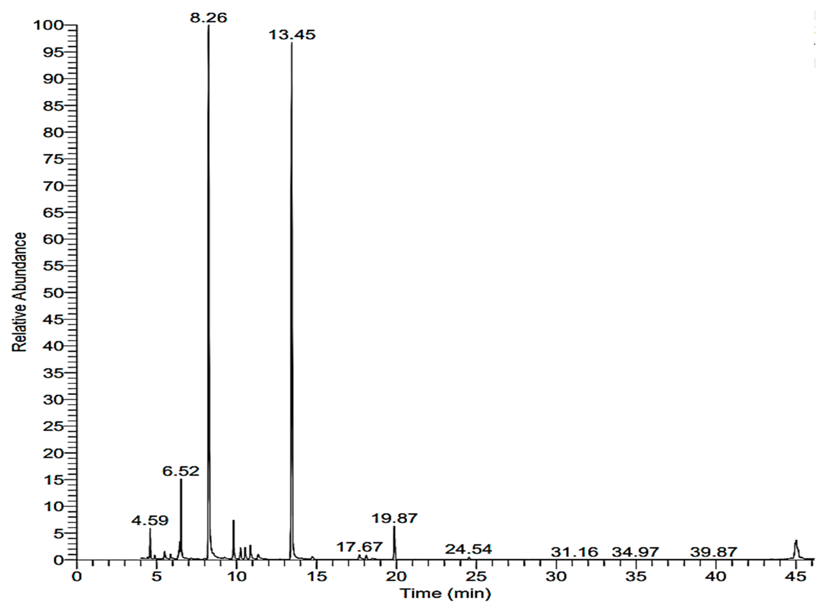
2.2. Chemical Analysis of Lavandula angustifolia

Data in Figure 1 show that cyclohexanol, 2-methyl-5-(1-methylethenyl) (carvomenthol), 1,6-octadien-3-ol, 3,7-dimethyl (linalool) and 1,8-cineol (eucalyptol) had been identified by referring to the corresponding acquisition time with area percentages of 11.4, 60.2 and 38.5, respectively (Table 2). Jianu et al. [19] reported that the essential oil of L. angustifolia included caryophyllene (24.1%), beta-phellandrene (16%) and eucalyptol (15.6%) as its primary constituents. In a study conducted by Ciocarlan et al. [20], it was revealed that the most important constituents of L. angustifolia EO were monoterpenes (84.08–92.55%), followed by sesquiterpenes (3.30–13.45%) and some aliphatic compounds (1.42–3.90%). Moreover, they reported that the main constituents of L. angustifolia essential oil were linalool, linalyl acetate, 1,8-cineole, borneol, camphor, lavandulyl acetate, β-caryophyllene, β-ocimene, α-fenchone, terpinen-4-ol, caryophyllene oxide, limonene, pinenes, geranyl acetate, β-farnesene, santalene, lavandulol, camphene, geraniol and α-terpineol. The content of oxygenated monoterpenes prevails in L. angustifolia essential oil and varies between 36.33 and 92.90% [21]. In another study, 1,4-cyclohexadiene, 1-methyl-4-(1-methylethyl)- was detected in essential oil extracted from the aerial parts of lavender at their full flowering stage [22].

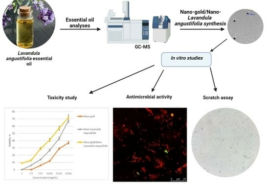
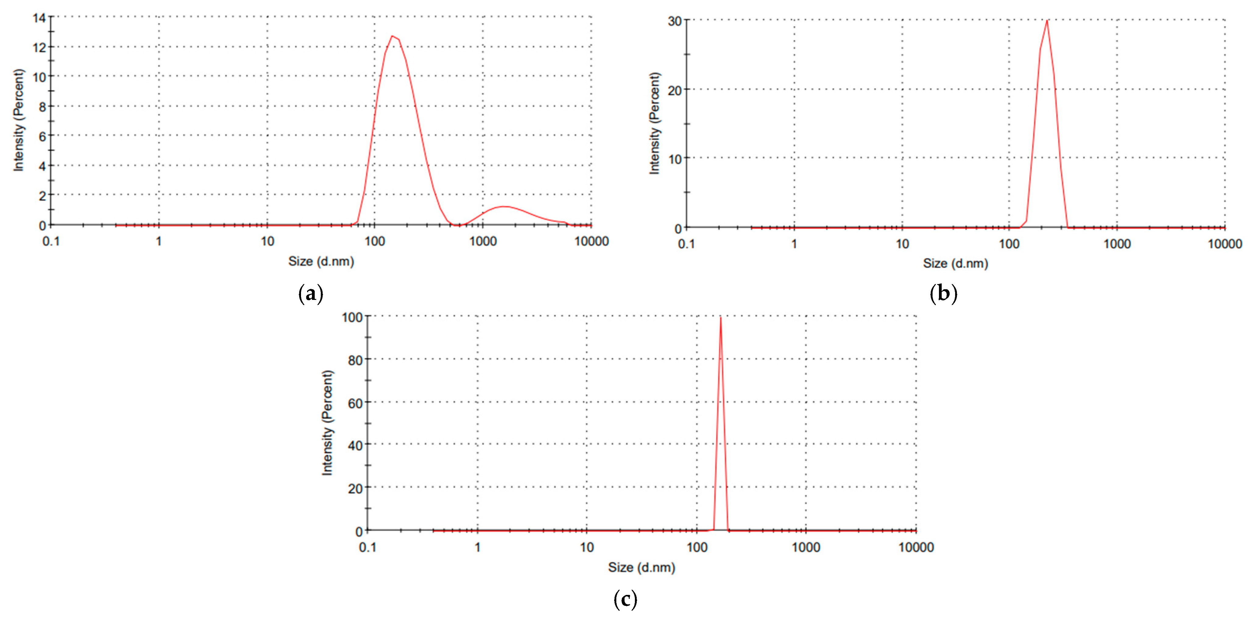
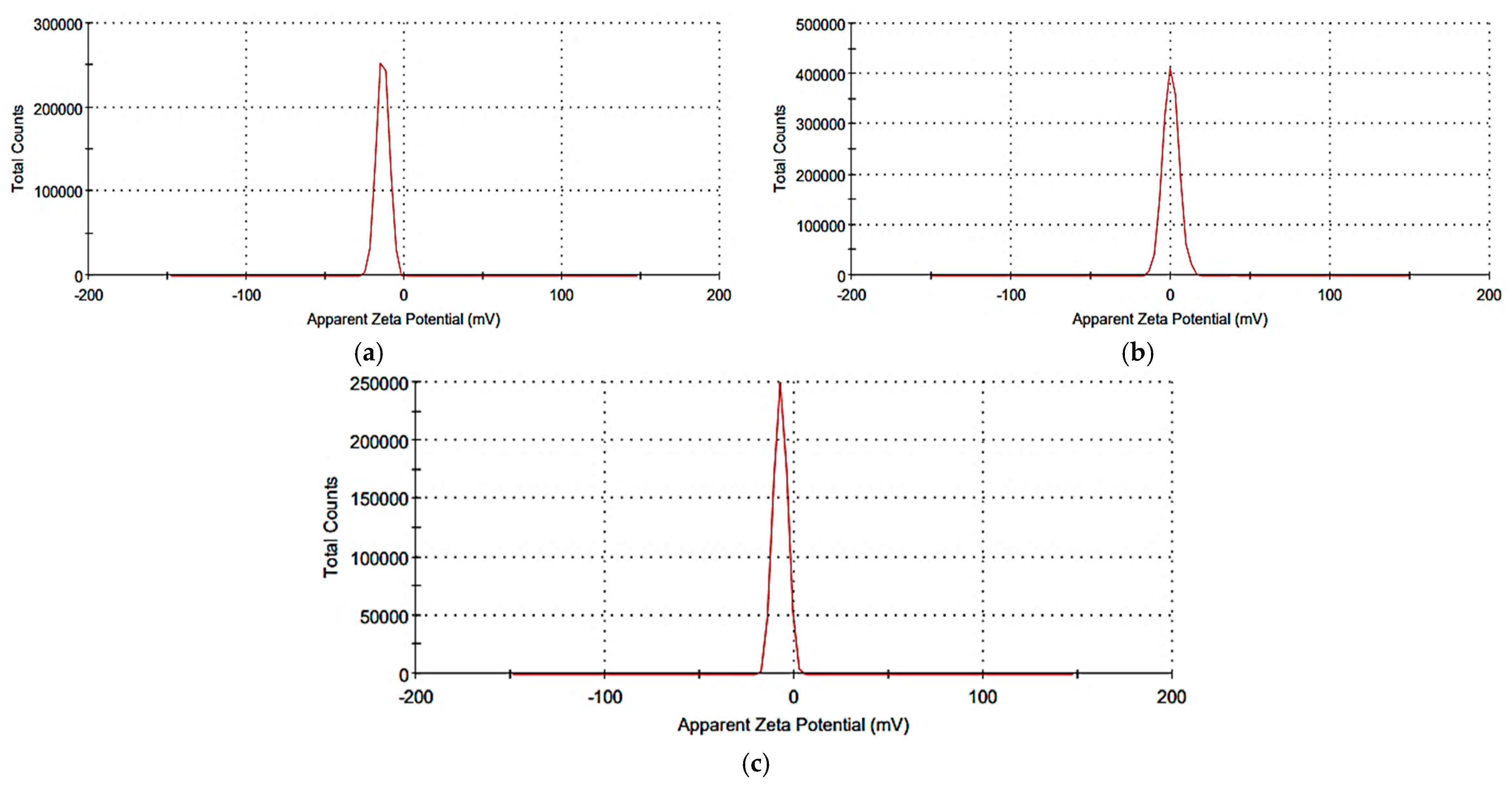
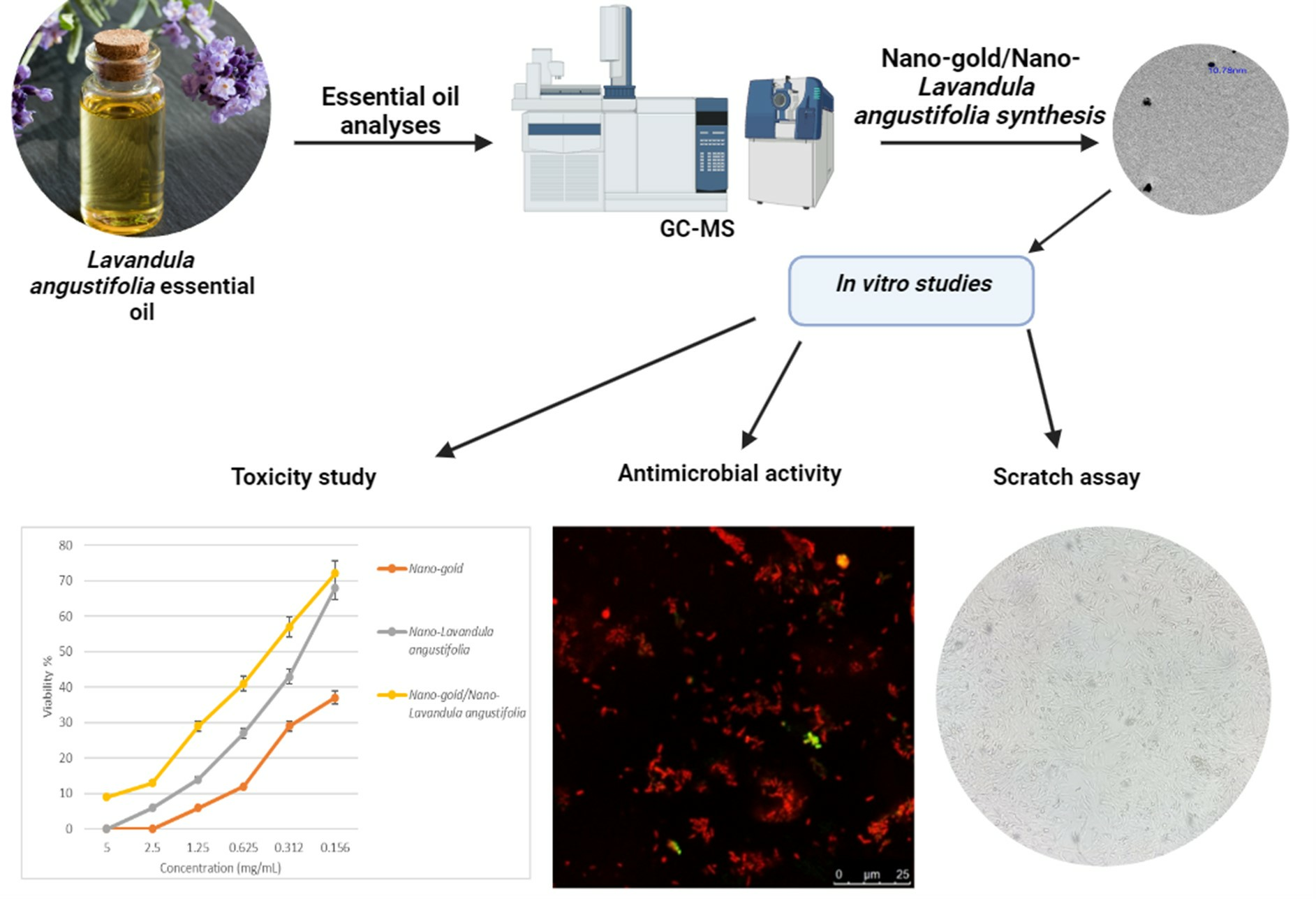
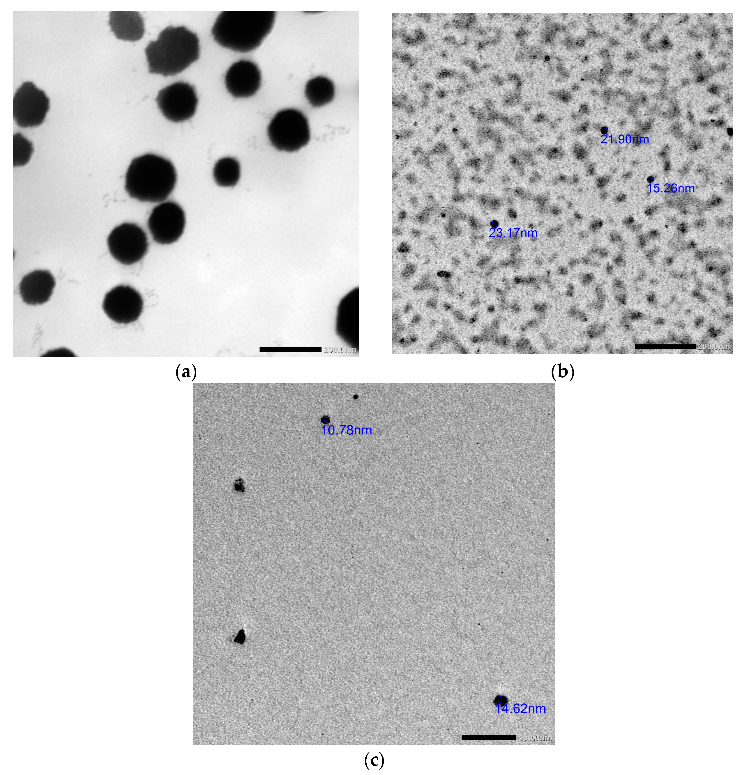
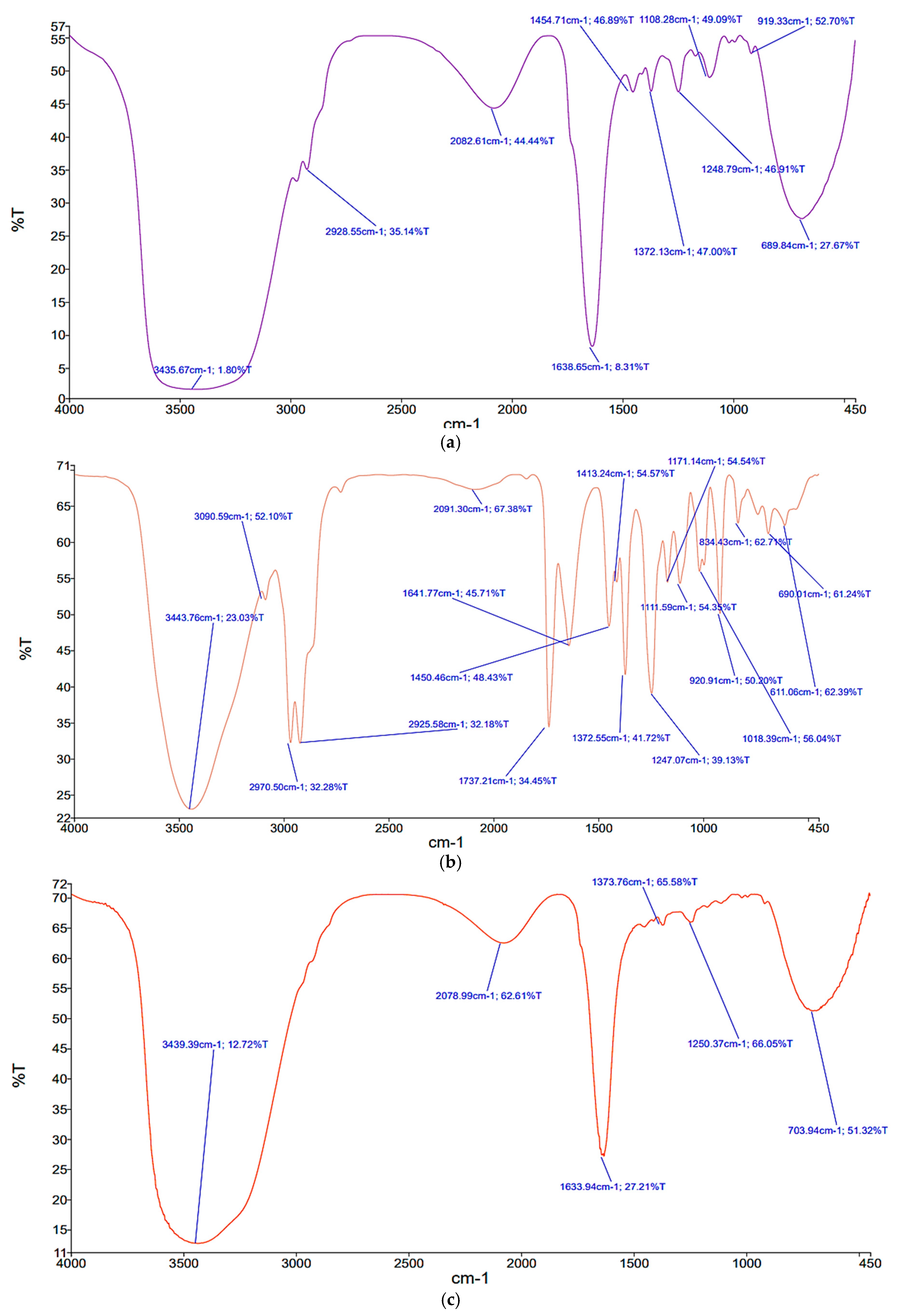
2.3. Synthesis and Characterization of the Prepared Nanosystems

All the synthesized nanosystems were characterized through Zetasizer, Fourier transform infrared spectroscopy (FTIR) and transmission electron microscopy (TEM) analyses. Light scattering (DLS) of the synthesized nanoparticles revealed that the nanoparticles’ size, polydispersity index and zeta potential of nano-Lavandula angustifolia were 199.5 nm, 0.404 and −13.6 mV; of nano-gold were 164.8 nm, 0.25 and +0.088 mV; and of nano-gold/nano-Lavandula angustifolia were 176.4 nm, 0.429 and −7.38 mV, respectively (Figure 2), while TEM micrographs revealed that the average diameter ranges of nano-Lavandula angustifolia, nano-gold and nano-Lavandula angustifolia/nano-gold were 96.8, 19.2 and 12.7 nm, respectively (Figure 3).
The presence of a spherical morphology in the synthesized nanoparticles suggests that they possess a low surface energy and exhibit significant thermodynamic stability. This observation further supports the notion that the synthesized gold nanoparticles have a substantial zeta potential. Bhat et al. [23] provided evidence that the extract derived from the edible fungus P. florida has the potential to be used in the biosynthesis of spherical gold nanoparticles (AuNPs), with 10 to 50 nm particle size. In another study, Rastogi and Arunachalam [24] used an aqueous extract of garlic (Allium sativum) for the manufacture of gold nanoparticles (AuNPs) by the application of microwave irradiation. The nanoparticles that were generated had an average diameter of 23.2 nm and exhibited mostly spherical morphology.
The findings of Chong et al.’s study [25] indicated that the use of mixed surfactants resulted in a superior performance in terms of the storage stability of nanoemulsions compared to pure surfactants with a hydrophilic–lipophilic balance (HLB) value of 15, which was in agreement with previous studies [26,27]. Previous research has shown that the synergistic effects of mixed surfactants may be influenced by variations in headgroup sizes [28]. The presence of greater disparities in headgroup sizes had a significant role in the amplification of synergistic effects. This may be attributed to the favorable interfacial packing between small molecule surfactants and larger surfactants at the interface separating the oil and water phases. The observed significant difference in headgroup size between Tween 80 and Span 80 contributes to the synergistic action of these two compounds, resulting in an improved stability of nanoemulsion systems [29]. Additionally, the stability of nanoemulsion was improved by the use of mixed surfactants, which strengthened the interfacial coating of the nanoemulsion [30]. This phenomenon might be attributed to the inherent hydrophilic and lipophilic properties of mixed surfactants, which promote the adsorption between the oil and water phases. Mixed surfactants exhibit enhanced dispersity and solubility in the continuous phase, facilitating effective mixing with both water and oil [27,31].
Another feature that was assessed in the study was surface charge density, which was determined by the measurement of zeta potential. This parameter helps to identify the presence of repulsive forces that counteract instability events. As an example, the acquired data for the zeta potential (expressed in absolute values) exhibited values around 11. The acquisition of a negative charge may be attributed to the presence of Tween 80 and Span 80 molecules and/or the formation of hydrogen bonds between their oxyethylene groups and hydroxyl ions [32]. Furthermore, it was previously reported that the magnitude of the surface charge density was subject to variations based on the specific kind of oily phase. Marinova et al. [33] observed that the zeta potential is significantly affected by the pH of the aqueous phase. This suggests that the presence of hydroxyl ions at the oil/water interface leads to a negative surface charge, which could explain the relatively higher absolute value of zeta potential observed in the nano-Lavandula angustifolia. This is due to the fact that pH has a substantial impact on the electrical charge of the constituent groups within these systems [34].
FTIR spectral analysis of the nanoemulsion (Figure 4a) revealed a characteristic broad band from 3200 to 3600 cm−1, corresponding to O–H stretching in addition to the common C=O stretching band of around 1700 cm−1. These absorption bands could be assigned to the functional groups of Span 80 and Tween 80 surfactants attached to the nanoparticles [35]. The FTIR (Figure 5b) showed the functional groups responsible for gold nanoparticles synthesis, stabilizing and capping using LEO. It demonstrated the common absorption bands at 3423, 2871, 1636 and 796 cm−1. These observed bands could be attributed to O–H stretching (3423 cm−1), CH stretching (2871 cm−1), C=C stretching (1636 cm−1) and vibrations of the Au–O bond (1057 cm−1), which agrees with the literature values [36]. FTIR indicated the importance of Lavandula angustifolia essential oil ingredients in stabilizing and capping the produced nano-Lavandula angustifolia/nano-gold within the matrix (Figure 5c). Cossetin et al. [37] reported that L. dentata (5%) essential oil-loaded nanoemulsion (NE-LO) had a droplet size of 64.99 nm and a polydispersity index (PDI) of 0.26. The formulation of NE-LO enabled a good protection for α-pinene, sabinene 1.8-cineole, fenchone and camphor at room temperature [38]. The primary components of Lavandula dentata essential oil were found to be 1.8 cineol (50–52%), fenchone (17–16%) and camphor (15–13%), while in the present study, the chemical composition of the tested essential oil differed from the one reported by Cossetin et al. [37], which may be attributed to the observed differences. The reason behind such an observation could be the increased lipophilicity with the increase in the length of the carbon chain, which would require a greater energy or Smix concentration to lower the interfacial tension and generate a stable nanoemulsion system [39].

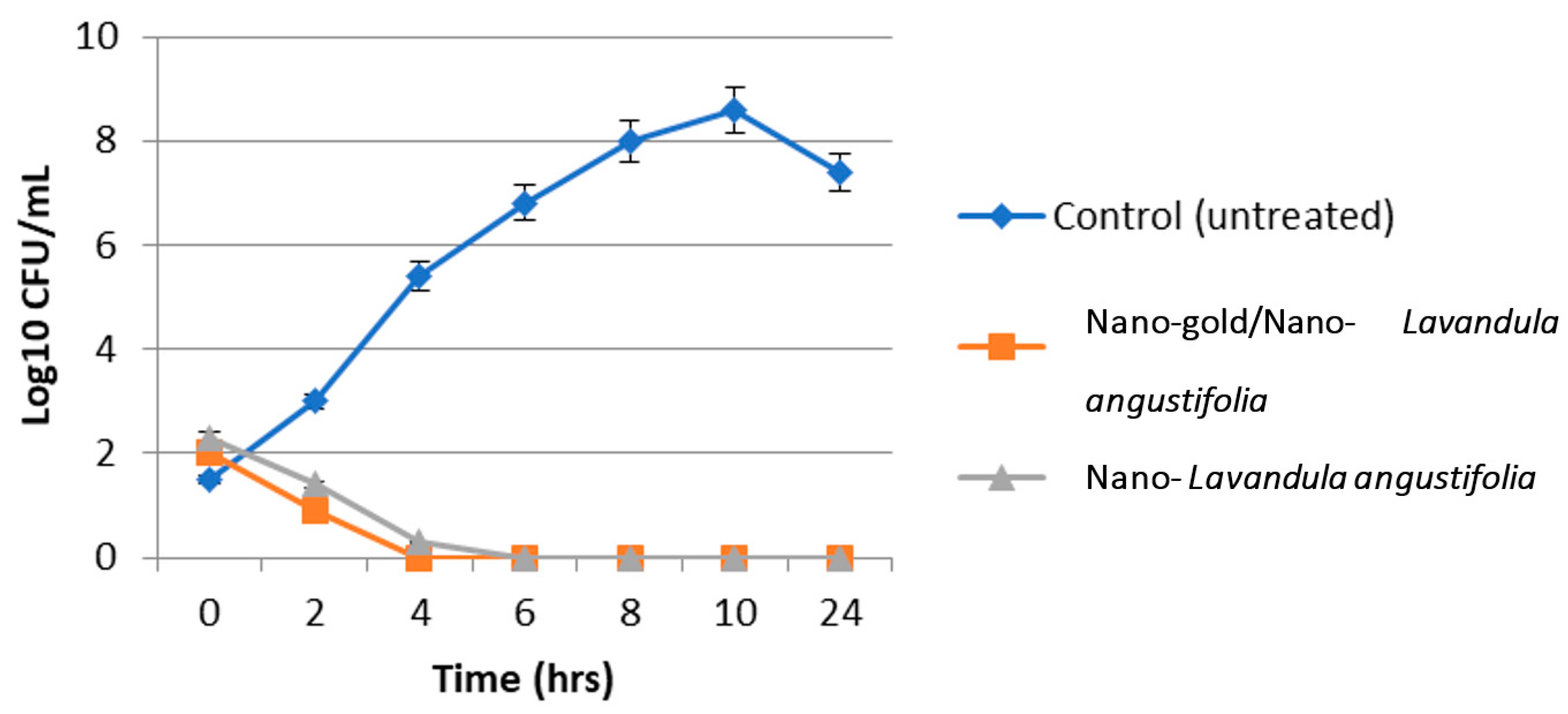
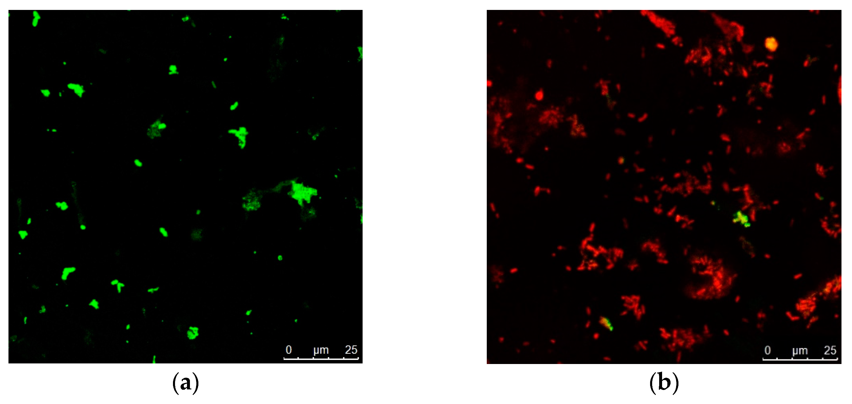
2.4. Antibacterial and Antibiofilm Activity of the Synthesized Nanosystems

Data in Figure 5 show that P. mirabilis bacterial growth reached 0 after 4 and 6 h incubation with nano-gold/nano-Lavandula angustifolia and Lavandula angustifolia nanoemulsion, respectively. P. mirabilis culture grown without nano-gold/nano-Lavandula angustifolia exhibited normal morphology, and the microbial cells were arranged while under the presence of nano-gold/nano-Lavandula angustifolia (16 µg/mL); P. mirabilis showed dramatically restricted bacterial growth inhibition (Figure 6). The observed outcomes may be explained by the inherent capacity of AuNPs to induce cell death via penetration of the bacterial cell wall. Indeed, gold nanoparticles (AuNPs) have the ability to engage with the negatively charged groups present in the constituents of the cell wall, resulting in alterations to the structural composition of the cell surface. A significant quantity of gold nanoparticles (AuNPs) has the ability to form robust bonds with polycyclic aromatic hydrocarbons present on the bacterial cell wall, resulting in the formation of polyelectrolyte complexes. These complexes have the potential to impede the transportation of vital solutes into the cell, as shown by previous studies [40,41,42].
It was proven that nano-gold/nano-Lavandula angustifolia inhibited the bacterial biofilm (Table 3). Biofilm formation plays an essential role in invading the host immune defenses and in increasing the antibiotic resistance, which helps in the persistence of microbial infections [43]. Physicochemical features of nanoparticles, including surface charge, PEGylation (the surface modification of particles with polyethylene glycol to enhance stability) and hydrophobicity, also have a role in influencing the nonspecific absorption and probable destruction of particles in macrophage cells. Consequently, the present approach of combining two nanosystems would synergistically combine the benefits of both active and passive targeting, thereby facilitating the specific targeting of cells within the body [44]. Hence, inhibiting the bacterial biofilm by using the newly prepared nano-gold/nano-Lavandula angustifolia may pave the way to more reliable and effective methods against persistent and virulent microbial infections.

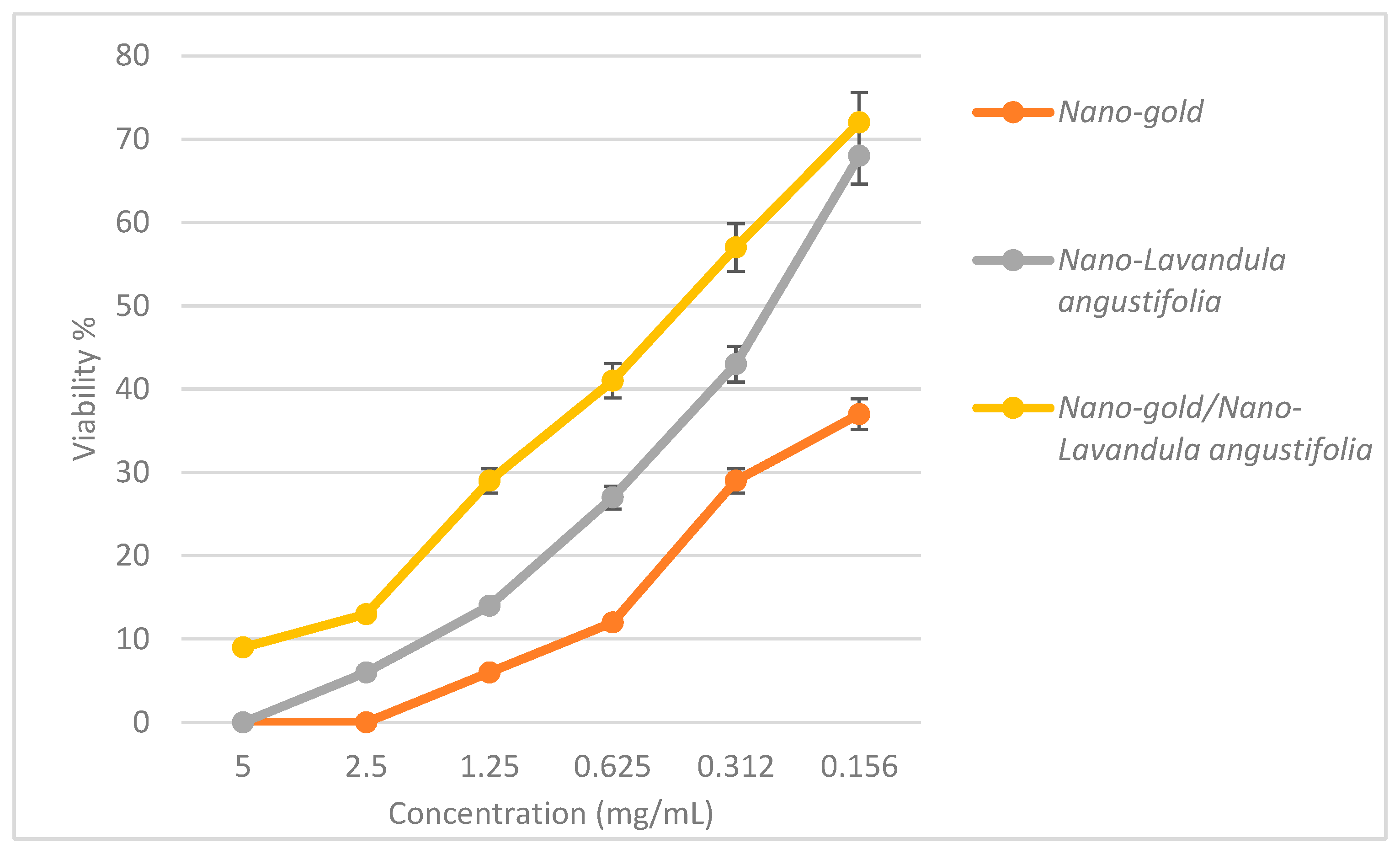
2.5. Cytotoxic Effect of Nano-Gold/Nano-Lavandula angustifolia

The IC50 values of the prepared nanosystems were 0.209, 0.529 and 0.089 mg/mL for nano-Lavandula angustifolia, nano-gold, nano-Lavandula angustifolia/nano-gold, respectively (Figure 7). Chianese et al. [22] tested the cytotoxic effect of L. angustifolia EO and reported no observed cytotoxic effects after 2 and 24 h incubation. Ovidi et al. [16] reported that lavandin EO nanoemulsions showed asserted cytotoxic effects on human lymphoblastic leukemia (CCRF-CEM), human neuroblastoma (SHSY5Y), breast cancer (MCF7) and human colorectal adenocarcinoma (Caco-2) cell lines. EC50 values showed dose-dependent and not time-dependent effects, ranging from 1.30 × 10⁻² ± 0.09 × 10⁻²% after 24 h, to 1.25 × 10⁻² ± 0.08 × 10⁻²% after 72 h in CCRF-CEM; from 1.42 × 10⁻² ± 0.08 × 10⁻²% after 24 h to 1.97 × 10⁻² ± 0.10 × 10⁻²% after 72 h in SHSY5Y cells; and from 13.52 × 10⁻² ± 1.97 × 10⁻²% after 24 h to 12.45 × 10⁻² ± 0.99 × 10⁻²% after 72 h for Caco-2 cells. In contrast, the two breast cells lines have shown a slight time-dependent effect, ranging from 3.62 × 10⁻² ± 0.76 × 10⁻²% after 24 h to 4.53 × 10⁻² ± 0.68 × 10⁻²% after 72 h of treatment for the breast cancer cell line (MCF7) and from 6.73 × 10⁻² ± 1.04 × 10⁻²% after 24 h to 3.47 × 10⁻² ± 0.06 × 10⁻²% after 72 h of treatment for the normal breast epithelial cell (MCF10A). In a study of Enterococcus-derived gold nanoparticles (AuNPs), the findings indicated that the application of AuNPs resulted in the stimulation of reactive oxygen species (ROS) and caspase-3 expressions, while concurrently diminishing the mitochondrial membrane potential. The observation of morphology changes associated with apoptosis after treatment with AuNPs in HT-29 cells suggested the potential use of AuNPs as a pro-apoptotic drug in the treatment of colon cancer [45]. Vilela et al. [46] reported that there was no statistically significant difference seen in the vitality of adipose tissue-derived stem cells (ASCs) and myometrial cells after treatment with lavender oil (LO). Moreover, the application of lavender oil and its derivative, niosomal lavender oil (LON), demonstrated cell viability comparable to that of the control culture, indicating their safety for use in cell culture. The only anomaly seen was a viability decrease in myometrial cells, specifically in the case of 0.063% LO. Despite a notable drop in viability at the highest dosage, it remained over 70% in both LO and LON treatments.

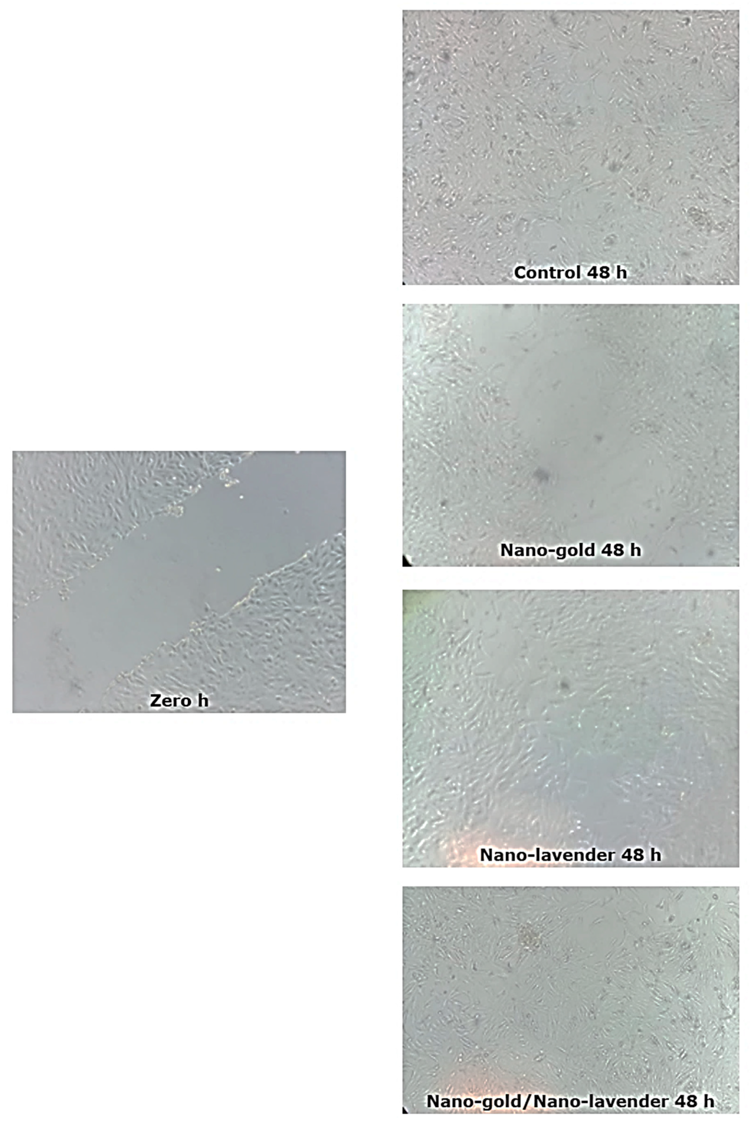
2.6. In Vitro Wound Healing Activity of Nano-Gold/Nano-Lavandula angustifolia

The newly synthesized nano-gold/nano-Lavandula angustifolia showed a powerful wound healing efficacy with a 96.78% wound closing relevant to the control (non-treated wounds) with 70.74% wound closure (Table 4, Figure 8 and Figure 9). It is worth mentioning that Lavandula angustifolia oil accelerates the healing rate and can increase the collagen expression, which preserves the skin elasticity and improves the activity of rebuilding tissue proteins [47].

3. Materials and Methods

3.1. Chemicals

Lavender oil (Lavandula angustifolia) (CAS number 8000-28-0), chloroauric acid (HAuCl4), polyethylene glycol sorbitan monooleate (Tween 80) and sorbitan monooleate (Span 80) were purchased from Sigma-Aldrich (St. Louis, MO, USA).

3.2. Microorganisms

Five bacterial strains, namely P. mirabilis, K. pneumoniae, MRSA, E. coli, S. aureus and A. baumannii (Table S1), were isolated, identified using Vitek 2 automated system (bioMerieux, Marcy l’Etoile, France) and were kindly provided by Microbiology Routine Laboratory, Main University Hospital, Alexandria University, Egypt. All the tested bacterial strains were maintained on brain–heart infusion glycerol broth with monthly transfer in fresh media.

3.3. Antibacterial Activity of the Tested Essential Oil (EO)

Bacterial sensitivity was assessed using the agar disc diffusion technique, which is extensively used to assess antibacterial activity. Petri dishes containing Muller Hinton agar (MHA) were inoculated with 0.1 mL freshly prepared bacterial suspension (0.5 McFarland). Sterile discs (6 mm) saturated with 15 μL Lavandula angustifolia (EO) were placed on the surface of the inoculated MHA plates. The inhibition zone diameters were measured in mm after incubation at 37 °C for 24 h. All the obtained results are the mean of three trials [48]. Furthermore, minimal inhibitory concentration (MIC) and minimal bactericidal concentration (MBC) assessments were performed in a 96-well microplate assay. A total volume of 100 μL of the tested EOs solution were serially diluted, and 50 μL of bacterial suspension was inoculated into each well. Finally, the microplate was incubated for 24 h at 37 °C; after the incubation, the first dilutions showed that no bacterial growth were reported as MICs [49].

3.4. Chemical Characterization of Lavandula angustifolia Essential Oil

The chemical composition of Lavandula angustifolia EO was investigated using gas chromatography TQ8040 NX (Shimadzu, Tokyo, Japan) coupled with a triple quadrupole, tandem mass spectrometer (GC-MSMS). The EO chemical composition was identified using a polar, capillary RTxi-5 Sil MS column (30.00 m long, 00.25 mm inside diameter, and a film thickness of 0.25). The composition of EO was presented as a proportion of the overall peak areas. EO phytochemicals were identified by comparing the resulted retention indices referring to those of the literature database [50].

3.5. Essential Oil Nanoemulsion Formation

Oil/water emulsion (O/W) was prepared at room temperature; the prepared Nanoemulsion consisted of 15 wt % Lavandula angustifolia essential oil/water and 5 wt % each surfactant (Span® 80 and Tween® 80). Nanoemulsions were prepared using the ultrasonic method [51]. The hydrophilic–lipophilic balance (HLB) method is a commonly employed approach for the selection of surfactants as emulsifying agents. HLB number of mixed surfactant system was calculated using the following equation:
HBL = ( m A × HLB A ) + m B × HBL B m A + m B
where mA and mB are the surfactants A and B mass, respectively. HLBA and HLBB are the HLB number of surfactants A and B, respectively. Tween® 80 with a HLB value of 15.0 and Span® 80 with a HLB value of 4.3, were selected for preparation of nanoemulsions. HLB value of 9 (Span® 80, mA = 56% and Tween® 80, mB = 44%) was defined based on Equation (1) with concentration of mixed surfactants in an emulsion system as determined by Shahavi et al. [51].

3.6. Synthesis of Gold Nanoparticles

Lavandula angustifolia essential oil was used as a reducing and stabilizing agent. Two (2) milliliters of Lavandula angustifolia essential oil were mixed with 30 mL of HAuCl4 solution (1 mmol/mL) followed by incubation at room temperature for 24 h. The reduction in Au+3 to Au0 was initially confirmed by visual inspection of color change from pale yellow to ruby red. The mixture was sonicated (Sonicator, Hielscher GmbH, Teltow, Germany) and centrifuged at 15,000 rpm for 11 min [52].

3.7. Synthesis of Gold–EO Conjugated Nanoparticles

The freshly synthesized AuNPs were added to the freshly prepared nanoemulsion (1:1) and the mixture was ultrasonicated (10 min) to allow for the conjugation between the gold nanoparticles and the nanoemulsions. The mixture was then incubated at room temperature for 24 h followed by centrifugation at 15,000 rpm for 11 min [53].

3.8. Characterization of the Prepared Nanosystems

The synthesized nanosystems were characterized using Fourier transform infrared spectroscopy (FTIR), transmission electron microscope and Zetasizer (Malvern Zetasizer Nano ZS, Malvern, UK) to determine the nanoparticles’ size and PDI according to Dorgham et al. [54].

3.9. Antibacterial and Antibiofilm Activity of the Prepared Nanosystems

The antibacterial activity was further evaluated via disc diffusion method, MIC, MBC and bacterial lethality curve [55]. Moreover, bacterial cultures (108 CFU/mL) were grown overnight in liquid broth, then diluted (1:100) into fresh medium for minimal biofilm eradication concentration (MBEC) assay in accordance with Dorgham et al. [54].

3.10. Cytotoxic Effect

In the present study, WI-38 fibroblasts vero (normal) cells (ATCC: CCL-25) were used, containing 3000 cell/well (plated for 24 h). After incubation, 100 µL of different concentrations of extract in RPMI medium without fetal bovine serum was added, and then the plate was re-incubated for an additional 24 h in a CO2 incubator (37 °C, 5% CO2 and 90% relative humidity). After 24 h of incubation, 20 μL of MTT (3-(4,5-dimethylthiazol-2-yl)-2,5-diphenyltetrazolium bromide) solution was added/well and the plates were incubated for 3 h in a CO2 incubator (MTT reaction time). The formazan crystals (MTT byproduct) were re-suspended after centrifugation in 100 μL DMSO. Readings were measured at 570 nm. The % viability was calculated as follows:
(AT − Ab/AC − Ab) × 100
  • AT = Mean absorbances of cells treated with different concentrations of each plant extract;
  • AC = Mean absorbances of control untreated cells with culture medium only;
  • Ab = Mean absorbances of cells treated with vehicle of plant extract (RPMI media without fetal bovine serum).
The cytotoxicity assay of the compound was expressed as IC50 using the % viability calculated from the serial dilutions of each trial [56].

3.11. In Vitro Scratch Assay

Ten thousand (10,000) WI-38 cells/well (ATCC: CCL-25) were plated and incubated for 24 h in CO2 incubator (37 °C, 5% CO2, and 90% relative humidity). After incubation period, the culture medium was replaced with serum-free EMEM (Eagle’s Minimum Essential Medium) to wash the cell monolayer and scratched (wound mimicking) using a sterile 200 µL pipette tip. Afterward, 1.5 mL of complete medium or contained IC50 of the tested nanosystems was added and the plate was incubated again for another 24 h. The migrating cells in the denuded zone were observed and photographed using phase contrast microscopy. The relative wound size was quantitated by using the Image J version 1.49o software [57].

4. Conclusions

Wound healing is a complex process affected by many potential factors, which can delay healing. There is a high need for an effective treatment to protect the wound from contamination and promote such healing. The results of the present investigation revealed that Lavandula angustifolia essential oil possesses antibacterial and antifungal activities, in addition to the anti-inflammatory properties. A novel nanosystem of Lavandula angustifolia essential oil and gold nanoparticles was formulated, which promotes wound healing and combats bacterial infection. Dynamic light scattering (DLS) of the synthesized nanoparticles revealed that the nanoparticles’ size, polydispersity index and zeta potential of nano-Lavandula angustifolia were 199.5 nm, 0.404 and −13.6 mV; of nano-gold were 164.8 nm, 0.25 and +0.088 mV; and of nano-gold/nano-Lavandula angustifolia were 176.4 nm, 0.429 and −7.38 mV, respectively. In contrast, TEM micrographs revealed that the average diameter ranges of nano-Lavandula angustifolia, nano-gold, nano-Lavandula angustifolia/nano-gold were 96.8, 19.2 and 12.7 nm, respectively. The newly prepared nano-gold/nano-Lavandula angustifolia showed promising antibacterial activity as well as powerful wound healing efficacy. Moreover, the newly prepared nano-gold/nano-Lavandula angustifolia showed a cytotoxic effect against WI-38 fibroblasts vero (normal) cells (IC50 value = 0.089 mg/mL). Finally, the results showed that Lavandula angustifolia oil can be used as a candidate for speedy wound healing and bacterial contamination inhibition.

Supplementary Materials

The following supporting information can be downloaded at: https://www.mdpi.com/article/10.3390/molecules28196988/s1, Table S1: Microbial strains and their collection numbers.

Author Contributions

Conceptualization, B.H.E. and Z.A.O.; methodology, B.A.F.; software, E.E.F.; validation, M.M.S.; formal analysis, B.H.E.; investigation, B.A.F.; resources, Z.A.O.; data curation, B.H.E., Z.A.O. and M.M.S.; writing—original draft preparation, B.H.E., Z.A.O. and M.M.S.; writing—review and editing, B.H.E. and Z.A.O.; supervision, B.H.E., E.E.F., M.M.S. and Z.A.O. All authors have read and agreed to the published version of the manuscript.

Funding

This research received no external funding.

Institutional Review Board Statement

The present work was approved by the ethical committee at Faculty of Science, Alexandria University, approval number 04/23/03/28/207.

Informed Consent Statement

Not applicable.

Data Availability Statement

Not applicable.

Conflicts of Interest

The authors declare no conflict of interest.

Sample Availability

Not available.

References

  1. Negut, I.; Grumezescu, V.; Grumezescu, A.M. Treatment Strategies for Infected Wounds. Molecules 2018, 23, 2392. [Google Scholar] [CrossRef] [PubMed]
  2. Bowler, P.G.; Duerden, B.I.; Armstrong, D.G. Wound Microbiology and Associated Approaches to Wound Management. Clin. Microbiol. Rev. 2001, 14, 244–269. [Google Scholar] [CrossRef] [PubMed]
  3. World Health Organization. Who Publishes List of Bacteria for Which New Antibiotics Are Urgently Needed. Available online: https://tinyurl.com/kmva5da (accessed on 26 May 2018).
  4. Vyas, K.S.; Vasconez, H.C. Wound Healing: Biologics, Skin Substitutes, Biomembranes and Scaffolds. Healthcare 2014, 2, 356–400. [Google Scholar] [CrossRef] [PubMed]
  5. Hammami, I.; Alabdallah, N.M.; Al Jomaa, A.; Kamoun, M. Gold nanoparticles: Synthesis properties and applications. J. King Saud Univ. Sci. 2021, 33, 101560. [Google Scholar] [CrossRef]
  6. Moeini, A.; Masi, M.; Zonno, M.C.; Boari, A.; Cimmino, A.; Tarallo, O.; Vurro, M.; Evidente, A. Encapsulation of inuloxin A, a plant germacrane sesquiterpene with potential herbicidal activity, in β-cyclodextrins. Org. Biomol. Chem. 2019, 17, 2508–2515. [Google Scholar] [CrossRef]
  7. Amrati, F.E.-Z.; Bourhia, M.; Slighoua, M.; Ibnemoussa, S.; Bari, A.; Ullah, R.; Amaghnouje, A.; Di Cristo, F.; El Mzibri, M.; Calarco, A.; et al. Phytochemical Study on Antioxidant and Antiproliferative Activities of Moroccan Caralluma europaea Extract and Its Bioactive Compound Classes. Evid.-Based Complement. Altern. Med. 2020, 2020, 8409718. [Google Scholar] [CrossRef]
  8. Moeini, A.; Pedram, P.; Makvandi, P.; Malinconico, M.; Gomez d’Ayala, G. Wound healing and antimicrobial effect of active secondary metabolites in chitosan-based wound dressings: A review. Carbohydr. Polym. 2020, 233, 115839. [Google Scholar] [CrossRef]
  9. Kusumawati, I.; Indrayanto, G. Natural antioxidants in cosmetics. Stud. Nat. Prod. Chem. 2013, 40, 485–505. [Google Scholar]
  10. Amaghnouje, A.; Mechchate, H.; Es-Safi, I.; Alotaibi, A.A.; Noman, O.M.; Nasr, F.A.; Fahd, A.; Al-Zharani, M.; Cerruti, P.; Calarco, A.; et al. Anxiolytic, antidepressant-like proprieties and impact on the memory of the hydro-ethanolic extract of Origanum majorana L. On mice. Appl. Sci. 2020, 10, 8420. [Google Scholar] [CrossRef]
  11. Amaghnouje, A.; Mechchate, H.; Es-Safi, I.; Boukhira, S.S.; Aliqahtani, A.M.; Noman, O.; Nasr, A.; Conte, R.; Calarco, A.; Bousta, D. Subacute assessment of the toxicity and antidepressant-like effects of Origanum majorana L. polyphenols in Swiss albino mice. Molecules 2020, 25, 5653. [Google Scholar] [CrossRef]
  12. Marturano, V.; Bizzarro, V.; Ambrogi, V.; Cutignano, A.; Tommonaro, G.; Abbamondi, G.R.; Giamberini, M.; Tylkowski, B.; Carfagna, C.; Cerruti, P. Light-Responsive Nanocapsule-Coated Polymer Films for Antimicrobial Active Packaging. Polymers 2019, 11, 68. [Google Scholar] [CrossRef] [PubMed]
  13. Cuttle, L.; Pearn, J.; McMillan, J.R.; Kimble, R.M. A review of first aid treatments for burn injuries. Burns 2009, 35, 768–775. [Google Scholar] [CrossRef] [PubMed]
  14. Cavanagh, H.M.A.; Wilkinson, J.M. Biological activities of lavender essential oil. Phytother. Res. 2002, 16, 301–308. [Google Scholar] [CrossRef]
  15. Baptista-Silva, S.; Borges, S.; Ramos, O.L.; Pintado, M.; Sarmento, B. The progress of essential oils as potential therapeutic agents: A review. J. Essent. Oil Res. 2020, 32, 279–295. [Google Scholar] [CrossRef]
  16. Ovidi, E.; Masci, V.L.; Taddei, A.R.; Paolicelli, P.; Petralito, S.; Trilli, J.; Mastrogiovanni, F.; Tiezzi, A.; Casadei, M.A.; Giacomello, P.; et al. Chemical Investigation and Screening of Anti-Proliferative Activity on Human Cell Lines of Pure and Nano-Formulated Lavandin Essential Oil. Pharmaceuticals 2020, 13, 352. [Google Scholar] [CrossRef]
  17. Pavoni, L.; Perinelli, D.R.; Bonacucina, G.; Cespi, M.; Palmieri, G.F. An overview of micro-and nanoemulsions as vehicles for essential oils: Formulation, preparation and stability. Nanomaterials 2020, 10, 135. [Google Scholar] [CrossRef] [PubMed]
  18. Hajiali, H.; Summa, M.; Russo, D.; Armirotti, A.; Brunetti, V.; Bertorelli, R.; Athanassiou, A.; Mele, E. Alginate–lavender nanofibers with antibacterial and anti-inflammatory activity to effectively promote burn healing. J. Mater. Chem. B 2016, 4, 1686–1695. [Google Scholar] [CrossRef]
  19. Jianu, C.; Pop, G.; Gruia, A.T.; Horhat, F.G. Chemical composition and antimicrobial activity of essential oils of lavender (Lavandula angustifolia) and lavandin (Lavandula x intermedia) grown in Western Romania. Int. J. Agric. Biol. 2013, 15, 772–776. [Google Scholar]
  20. Ciocarlan, A.; Lupascu, L.; Aricu, A.; Dragalin, I.; Popescu, V.; Geana, E.-I.; Ionete, R.E.; Vornicu, N.; Duliu, O.G.; Hristozova, G.; et al. Chemical Composition and Assessment of Antimicrobial Activity of Lavender Essential Oil and Some By-Products. Plants 2021, 10, 1829. [Google Scholar] [CrossRef]
  21. Aprotosoaie, A.C.; Gille, E.; Trifan, A.; Luca, V.S.; Miron, A. Essential oils of Lavandula genus: A systematic review of their chemistry. Phytochem. Rev. 2017, 16, 761–799. [Google Scholar] [CrossRef]
  22. Hui, L.; He, L.; Huan, L.; XiaoLan, L.; AiGuo, Z. Chemical composition of lavender essential oil and its antioxidant activity and inhibition against rhinitis-related bacteria. Afr. J. Microbiol. Res. 2010, 4, 309–313. [Google Scholar]
  23. Bhat, R.; Sharanabasava, V.; Deshpande, R.; Shetti, U.; Sanjeev, G.; Venkataraman, A. Photo-bio-synthesis of irregular shaped functionalized gold nanoparticles using edible mushroom Pleurotus florida and its anticancer evaluation. J. Photochem. Photobiol. B: Biol. 2013, 125, 63–69. [Google Scholar] [CrossRef] [PubMed]
  24. Rastogi, L.; Arunachalam, J. Microwave-Assisted Green Synthesis of Small Gold Nanoparticles Using Aqueous Garlic (Allium sativum) Extract: Their Application as Antibiotic Carriers. Int. J. Green Nanotechnol. 2012, 4, 163–173. [Google Scholar] [CrossRef]
  25. Chong, W.-T.; Tan, C.-P.; Cheah, Y.-K.; Lajis, A.F.B.; Dian, N.L.H.M.; Kanagaratnam, S.; Lai, O.-M. Optimization of process parameters in preparation of tocotrienol-rich red palm oil-based nanoemulsion stabilized by Tween80-Span 80 using response surface methodology. PLoS ONE 2018, 13, e0202771. [Google Scholar] [CrossRef]
  26. Pey, C.M.; Maestro, A.; Solé, I.; González, C.; Solans, C.; Gutierrez, J.M. Optimization of nano-emulsions prepared by low-energy emulsification methods at constant temperature using a factorial design study. Colloids Surf. Physicochem. Eng. Asp. 2006, 288, 144–150. [Google Scholar] [CrossRef]
  27. Scamehorn, J.F.; Comstock, M.J.; Osborne-Lee, I.W.; Schechter, R.S.; Kamrath, R.F.; Franses, E.I.; Meguro, K.; Muto, Y.; Sakurai, F.; Esumi, K.; et al. Phenomena in Mixed Surfactant Systems. Anal. Chem. 1986, 58, 1250A. [Google Scholar] [CrossRef]
  28. Bergström, M.; Eriksson, J.C. A Theoretical Analysis of Synergistic Effects in Mixed Surfactant Systems. Langmuir 2000, 16, 7173–7181. [Google Scholar] [CrossRef]
  29. Fu, Z.; Liu, M.; Xu, J.; Wang, Q.; Fan, Z. Stabilization of water-in-octane nano-emulsion. Part I: Stabilized by mixed surfactant systems. Fuel 2010, 89, 2838–2843. [Google Scholar] [CrossRef]
  30. Izquierdo, P.; Esquena, J.; Tadros, T.F.; Dederen, J.C.; Feng, J.; Garcia-Celma, M.J.; Azemar, N.; Solans, C. Phase Behavior and Nano-emulsion Formation by the Phase Inversion Temperature Method. Langmuir 2004, 20, 6594–6598. [Google Scholar] [CrossRef]
  31. Morales, D.; Gutie´rrez, J.M.; Garcı´a-Celma, M.J.; Solans, Y.C. A Study of the Relation between Bicontinuous Microemulsions and Oil/Water Nano-emulsion Formation. Langmuir 2003, 19, 7196–7200. [Google Scholar] [CrossRef]
  32. Liu, W.; Sun, D.; Li, C.; Liu, Q.; Xu, J. Formation and stability of paraffin oil-in-water nano-emulsions prepared by the emulsion inversion point method. J. Colloid Interface Sci. 2006, 303, 557–563. [Google Scholar] [CrossRef] [PubMed]
  33. Marinova, K.G.; Alargova, R.G.; Denkov, N.D.; Velev, O.D.; Petsev, D.N.; Ivanov, I.B.; Borwankar, R.P. Charging of Oil−Water Interfaces Due to Spontaneous Adsorption of Hydroxyl Ions. Langmuir 1996, 12, 2045–2051. [Google Scholar] [CrossRef]
  34. Azmi, N.A.N.; Elgharbawy, A.A.M.; Motlagh, S.R.; Samsudin, N.; Salleh, H.M. Nanoemulsions: Factory for Food, Pharmaceutical and Cosmetics. Processes 2019, 7, 617. [Google Scholar] [CrossRef]
  35. Choudhury, S.R.; Mandal, A.; Chakravorty, D.; Gopal, M.; Goswami, A. Evaluation of physicochemical properties, and antimicrobial efficacy of monoclinic sulfur-nanocolloid. J. Nanoparticle Res. 2013, 15, 1491. [Google Scholar] [CrossRef]
  36. Samfira, S.I.; Rodino, P.; Petrache, R.; Cristina, M.; Butu, M. Butnariu, Characterization and identity confirmation of essential oils by mid infrared absorption spectrophotometry. Dig. J. Nanomater. Biostructures 2015, 10, 557–566. [Google Scholar]
  37. Cossetin, L.F.; Garlet, Q.I.; Velho, M.C.; Gündel, S.; Ourique, A.F.; Heinzmann, B.M.; Monteiro, S.G. Development of nanoemulsions containing Lavandula dentata or Myristica fragrans essential oils: Influence of temperature and storage period on physical-chemical properties and chemical stability. Ind. Crops Prod. 2021, 160, 113115. [Google Scholar] [CrossRef]
  38. Palanisamy, N.K.; Ferina, N.; Amirulhusni, A.N.; Mohd-Zain, Z.; Hussaini, J.; Ping, L.J.; Durairaj, R. Antibiofilm properties of chemically synthesized silver nanoparticles found against Pseudomonas aeruginosa. J. Nanobiotechnol. 2014, 12, 1–7. [Google Scholar] [CrossRef]
  39. Warisnoicharoen, W.; Lansley, A.; Lawrence, M. Nonionic oil-in-water microemulsions: The effect of oil type on phase behaviour. Int. J. Pharm. 2000, 198, 7–27. [Google Scholar] [CrossRef]
  40. Palza, H. Antimicrobial Polymers with Metal Nanoparticles. Int. J. Mol. Sci. 2015, 16, 2099–2116. [Google Scholar] [CrossRef]
  41. Zhou, Y.; Kong, Y.; Kundu, S.; Cirillo, J.D.; Liang, H. Antibacterial activities of gold and silver nanoparticles against Escherichia coli and bacillus Calmette-Guérin. J. Nanobiotechnol. 2012, 10, 1–9. [Google Scholar] [CrossRef]
  42. Eskandari-Nojehdehi, M.; Jafarizadeh-Malmiri, H.; Rahbar-Shahrouzi, J. Optimization of processing parameters in green synthesis of gold nanoparticles using microwave and edible mushroom (Agaricus bisporus) extract and evaluation of their antibacterial activity. Nanotechnol. Rev. 2016, 5, 537–548. [Google Scholar] [CrossRef]
  43. Chianese, A.; Gravina, C.; Morone, M.V.; Ambrosino, A.; Formato, M.; Palma, F.; Foglia, F.; Nastri, B.M.; Zannella, C.; Esposito, A.; et al. Lavandula austroapennina: Assessment of the Antiviral Activity of Lipophilic Extracts from Its Organs. Viruses 2023, 15, 1648. [Google Scholar] [CrossRef] [PubMed]
  44. Bergen, J.M.; von Recum, H.A.; Goodman, T.T.; Massey, A.P.; Pun, S.H. Gold Nanoparticles as a Versatile Platform for Optimizing Physicochemical Parameters for Targeted Drug Delivery. Macromol. Biosci. 2006, 6, 506–516. [Google Scholar] [CrossRef] [PubMed]
  45. Vairavel, M.; Devaraj, E.; Shanmugam, R. An eco-friendly synthesis of Enterococcus sp.–mediated gold nanoparticle induces cytotoxicity in human colorectal cancer cells. Environ. Sci. Pollut. Res. 2020, 27, 8166–8175. [Google Scholar] [CrossRef] [PubMed]
  46. Vilela, J.d.M.V.; Moghassemi, S.; Dadashzadeh, A.; Dolmans, M.-M.; Azevedo, R.B.; Amorim, C.A. Safety of Lavender Oil-Loaded Niosomes for In Vitro Culture and Biomedical Applications. Nanomaterials 2022, 12, 1999. [Google Scholar] [CrossRef]
  47. Bratovcic, A. Green Synthesis of Various Nanostructures Containing Essential Oil and Silver Nanoparticles: Nanocomposites, Nanoemulsions and Nanoencapsules. Int. J. Eng. Res. Appl. 2023, 13, 234–242. [Google Scholar]
  48. El Atki, Y.; Aouam, I.; El Kamari, F.; Taroq, A.; Lyoussi, B.; Oumokhtar, B.; Abdellaoui, A. Phytochemistry, antioxidant and antibacterial activities of two Moroccan Teucrium polium L. subspecies: Preventive approach against nosocomial infections. Arab. J. Chem. 2020, 13, 3866–3874. [Google Scholar] [CrossRef]
  49. El Abdali, Y.; Beniaich, G.; Mahraz, A.M.; El Moussaoui, A.; Bin Jardan, Y.A.; Akhazzane, M.; Chebaibi, M.; Nafidi, H.-A.; Eloutassi, N.; Bourhia, M.; et al. Antibacterial, Antioxidant, and in silico NADPH Oxidase Inhibition Studies of Essential Oils of Lavandula dentata against Foodborne Pathogens. Evid.-Based Complement. Altern. Med. 2023, 2023, 9766002. [Google Scholar] [CrossRef]
  50. Elnahas, R.A.; Elwakil, B.H.; Elshewemi, S.S.; Olama, Z.A. Egyptian Olea europaea leaves bioactive extract: Antibacterial and wound healing activity in normal and diabetic rats. J. Tradit. Complement. Med. 2021, 11, 427–434. [Google Scholar] [CrossRef]
  51. Shahavi, M.H.; Hosseini, M.; Jahanshahi, M.; Meyer, R.L.; Darzi, G.N. Evaluation of critical parameters for preparation of stable clove oil nanoemulsion. Arab. J. Chem. 2019, 12, 3225–3230. [Google Scholar] [CrossRef]
  52. Hosseinzadeh, N.; Shomali, T.; Hosseinzadeh, S.; Raouf Fard, F.; Pourmontaseri, M.; Fazeli, M. Green synthesis of gold nanoparticles by using Ferula persica Willd. gum essential oil: Production, characterization and in vitro anti-cancer effects. J. Pharm. Pharmacol. 2020, 72, 1013–1025. [Google Scholar] [CrossRef]
  53. Dzimitrowicz, A.; Berent, S.; Motyka, A.; Jamroz, P.; Kurcbach, K.; Sledz, W.; Pohl, P. Comparison of the characteristics of gold nanoparticles synthesized using aqueous plant extracts and natural plant essential oils of Eucalyptus globulus and Rosmarinus officinalis. Arab. J. Chem. 2019, 12, 4795–4805. [Google Scholar] [CrossRef]
  54. Dorgham, R.A.; Abd Al Moaty, M.N.; Chong, K.P.; Elwakil, B.H. Molasses-Silver Nanoparticles: Synthesis, Optimization, Characterization, and Antibiofilm Activity. Int. J. Mol. Sci. 2022, 23, 10243. [Google Scholar] [CrossRef] [PubMed]
  55. Elnaggar, Y.S.; Elwakil, B.H.; Elshewemi, S.S.; El-Naggar, M.Y.; Bekhit, A.A.; Olama, Z.A. Novel Siwa propolis and colistin-integrated chitosan nanoparticles: Elaboration; in vitro and in vivo appraisal. Nanomedicine 2020, 15, 1269–1284. [Google Scholar] [CrossRef]
  56. El-Attar, A.A.; El-Wakil, H.B.; Hassanin, A.H.; Bakr, B.A.; Almutairi, T.M.; Hagar, M.; Elwakil, B.H.; Olama, Z.A. Silver/Snail Mucous PVA Nanofibers: Electrospun Synthesis and Antibacterial and Wound Healing Activities. Membranes 2022, 12, 536. [Google Scholar] [CrossRef] [PubMed]
  57. Felice, F.; Zambito, Y.; Belardinelli, E.; Fabiano, A.; Santoni, T.; Di Stefano, R. Effect of different chitosan derivatives on in vitro scratch wound assay: A comparative study. Int. J. Biol. Macromol. 2015, 76, 236–241. [Google Scholar] [CrossRef]
Figure 1. GC–MS chromatogram of Lavandula angustifolia essential oil.
Molecules 28 06988 g001
Figure 2. Zeta size distribution of Lavandula angustifolia nanoemulsion (a), gold nanoparticles (b) and nano-gold/nano-Lavandula angustifolia (c).
Molecules 28 06988 g002
Figure 3. Zeta potential of Lavandula angustifolia nanoemulsion (a), gold nanoparticles (b) and nano-gold/nano-Lavandula angustifolia (c).
Molecules 28 06988 g003
Figure 4. TEM images of Lavandula angustifolia nanoemulsion (a), gold nanoparticles (b) and nano-gold/nano-Lavandula angustifolia (c).
Molecules 28 06988 g004
Figure 5. FTIR spectroscope graphs of Lavandula angustifolia nanoemulsion (a), gold nanoparticles (b) and nano-gold/nano-Lavandula angustifolia (c).
Molecules 28 06988 g005
Figure 6. Microbial lethality curve against P. mirabilis.
Molecules 28 06988 g006
Figure 7. Confocal microscopic examination of P. mirabilis ((a) control untreated cells, (b) nano-gold/nano-Lavandula angustifolia-treated cells).
Molecules 28 06988 g007
Figure 8. Cytotoxic effect of the prepared nanosystems.
Molecules 28 06988 g008
Figure 9. Wound closure effect of the prepared nanosystems.
Molecules 28 06988 g009
Table 1. Antibacterial activity of the tested essential oil.
Tested PathogensLavandula angustifolia EO
IZ (mm)MIC (µg/mL)MBC (µg/mL)
P. mirabilis16.0 ± 1.064.0256.0
K. pneumoniae20.0 ± 2.032.0128.0
MRSA25.0 ± 2.016.0128.0
E. coli17.0 ± 5.064.0256.0
S. aureus28.0 ± 3.016.0128.0
A. baumannii17.0 ± 1.064.0256.0
Table 2. GC–MS analysis of Lavandula angustifolia essential oil.
RT (min)Area %Compound
6.5211.4Cyclohexanol, 2-methyl-5-(1-methylethenyl)
8.2660.2Linalool
13.4538.51,8-cineol
Table 3. Antibacterial and antibiofilm activity of the prepared nanosystems.
Nanosystems under TestMeasured ParametersP. mirabilis
Lavandula angustifolia nanoemulsionIZ (mm)30.0 ± 2.0
MIC (µg/mL)128.0
MBC (µg/mL)512.0
MBEC (µg/mL)128.0
Gold nanoparticlesIZ (mm)20.0 ± 1.0
MIC (µg/mL)256.0
MBC (µg/mL)512.0
MBEC (µg/mL)256.0
Nano-gold/nano-Lavandula angustifoliaIZ (mm)45.0 ± 3.0
MIC (µg/mL)8.0
MBC (µg/mL)256.0
MBEC (µg/mL)16.0
Table 4. Wound healing activity of the prepared nanosystems.
SampleGap Width (µm)Closure %
0 h48 h
Control124.03 ± 30.036.29 ± 6.070.74
Nano-Lavandula angustifolia163.12 ± 12.019.65 ± 2.087.95
Nano-gold155.63 ± 25.0110.63 ± 2.028.91
Nano-Lavandula angustifolia/Nano-gold164.12 ± 10.05.28 ± 0.596.78
Disclaimer/Publisher’s Note: The statements, opinions and data contained in all publications are solely those of the individual author(s) and contributor(s) and not of MDPI and/or the editor(s). MDPI and/or the editor(s) disclaim responsibility for any injury to people or property resulting from any ideas, methods, instructions or products referred to in the content.

