Article

Protective Effects and Mechanisms of Pectolinarin against H2O2-Induced Oxidative Stress in SH-SY5Y Neuronal Cells

1
Department of Food Science and Nutrition & Kimchi Research Institute, Pusan National University, Busan 46241, Republic of Korea
2
Department of Food Science and Nutrition, Gyeongsang National University, Jinju 52725, Republic of Korea
*
Authors to whom correspondence should be addressed.
Molecules 2023, 28(15), 5826; https://doi.org/10.3390/molecules28155826
Submission received: 22 June 2023 / Revised: 24 July 2023 / Accepted: 31 July 2023 / Published: 2 August 2023

Abstract

:
This study aims to investigate the protective effects and mechanisms of pectolinarin against oxidative stress-induced cell damage in SH-SY5Y cells. Neurodegenerative diseases—such as Alzheimer’s disease—are potentially associated with oxidative stress, which causes excessive production of reactive oxygen species (ROS) that damage DNA and proteins in neuronal cells. The results of this study demonstrate that pectolinarin can scavenge hydroxyl and nitric oxide radicals in a concentration-dependent manner. Moreover, pectolinarin significantly increased cell viability while reducing ROS production and LDH release in the hydrogen peroxide (H2O2)-induced control group. Additionally, Pectolinarin recovered protein expression from H2O2-altered levels back to close-to-normal SH-SY5Y cell levels for components of the oxidative stress, inflammation, and apoptosis pathways—such as nuclear factor erythroid 2-related factor 2 (Nrf2), kelch-like ECH-associated protein (Keap1), anti-heme oxygenase 1 (HO-1), inducible nitric oxide synthase (iNOS), cyclooxygenase-2 (COX-2), interleukin-1β (IL-1β), B-cell lympho-ma-2 (Bcl-2) protein, and Bcl-2-associated X protein (Bax). These findings suggest that pectolinarin has the potential to be used as a plant material for functional foods to be applied in the treatment of neurodegenerative diseases, such as Alzheimer’s disease, by mitigating oxidative stress-induced damage to neuronal cells.

Graphical Abstract

1. Introduction

The oxidative and antioxidant systems in human bodies are in dynamic equilibrium [1]. The imbalance between pro-oxidation and anti-oxidation overproduces reactive oxygen species (ROS), which induce oxidative stress [2]. ROS is a general term for substances that are more unstable than oxygen and mainly includes superoxide (O2−), hydroxyl (·OH), and hydrogen peroxide (H2O2) [3]. ROS combine with purines and pyrimidines in deoxyribonucleic acid (DNA), causing damage to the DNA double helix structure and causing oxidative stress, aging, and cancer [4,5]. Oxidative stress can also cause neuronal cell death and neuroinflammation in the brain, resulting in cognitive impairment and memory problems as well as neurodegenerative diseases, including Alzheimer’s disease (AD) and Parkinson’s disease [6,7].
With increases in the oxidative–redox system. it eventually loses its equilibrium—with age and the influence of irregular diets resulting in an excess of ROS production and oxidative stress in our bodies [8,9]. Besides this, neurodegenerative diseases such as AD have been shown to be oxidative stress-related diseases [10,11]. H2O2 stimulates astrocytes in the cortex and hippocampus, which significantly increases iNOS, and iNOS catalyzes nitric oxide (NO) production in the brain [12]. In addition, oxidative stress is also associated with the expression of Nrf2. Nrf2 is an antioxidant response regulator that is released from Keap1, translocates to the nucleus, activates protective gene transcription, and promotes their expression [13]. Studies have shown that inadequate Nrf2 expression has been found in the brains of human AD patients [14,15]. Therefore, the interaction of Keap1 and Nrf2 appears to be related to their antioxidant capacity, which protects neurons from oxidative stress [16].
In human cells, apoptosis is divided into internal pathways and external pathways, with the internal pathway undergoing molecular changes such as the activation of Bcl-2 and the inactivation of the pro-apoptotic effector Bax [17,18]. Apoptosis can help in the removal of damaged or dysfunctional brain cells, such as neurons that have accumulated toxic proteins [19]. This process is important for maintaining the overall health and function of the brain. However, excessive or abnormal apoptosis may contribute to the progression of neurodegenerative diseases [20]. Therefore, studying the apoptosis mechanisms of SH-SY5Y neuronal cells after treatment with the neurotoxic fragment Aβ25–35 is important.
SH-SY5Y cells, which are human-origin neuronal cells, are commonly used in the study of neurodegenerative diseases [21,22,23]. Many studies have reported the use of SH-SY5Y cells for studying oxidative stress-induced inflammation and apoptosis [24,25,26]. H2O2-induced oxidative stress in the SH-SY5Y cell model has been used to confirm the protective effects of active compounds [27,28]. Meanwhile, pectolinarin is a flavonoid that has been identified as an active compound in a number of plants, including Cirsium japonicum var. ussuriense, Lippia rubella, and Cirsium nipponicum (Maxim.) [29,30,31]. Anti-inflammatory, anti-apoptosis, anti-bacterial, and anti-tumor properties have been reported in pectolinarin [32,33,34]. Moreover, some previous studies have demonstrated the pharmacokinetic profile and intestinal absorption of pectolinarin to verify its physiochemical properties. According to Mehta and Dhapte [35], pectolinarin in rat plasma—following the oral administration of Cirsium japonicum DC. extract—reached the maximum possible concentration quickly. Chen et al. [36] reported that the absorption of pectolinarin in the jejunum was the highest among the parts of the small intestine, using the everted rat intestinal sac method. In this study, we investigated the ·OH and NO radical scavenging ability of pectolinarin in vitro and the mechanisms of pectolinarin against H2O2-induced oxidative stress in SH-SY5Y neuronal cells.

2. Results

2.1. Hydroxyl Radical (OH) Scavenging Activity of Pectolinarin

The results of the ·OH radical scavenging activity of pectolinarin are shown in Table 1. Pectolinarin (1, 2.5, 5, and 10 μg/mL) scavenged 63.09%, 80.43%, 83.60%, and 84.77% of the ·OH radicals, respectively. Furthermore, the scavenging activity at concentrations of 2.5, 5, and 10 μg/mL was greater than 80%. These results suggest that pectolinarin has antioxidant properties that make it capable of scavenging ·OH radicals.

2.2. NO Radical Scavenging Activity of Pectolinarin

The results for the NO radical scavenging activity of pectolinarin are shown in Table 2. Pectolinarin (1, 2.5, 5, and 10 μg/mL) showed dose-dependent increases in NO radical scavenging activity—at 23.21%, 24.50%, 30.40%, and 35.19%, respectively. These results suggest that pectolinarin possesses antioxidant properties that make it capable of scavenging NO radicals.

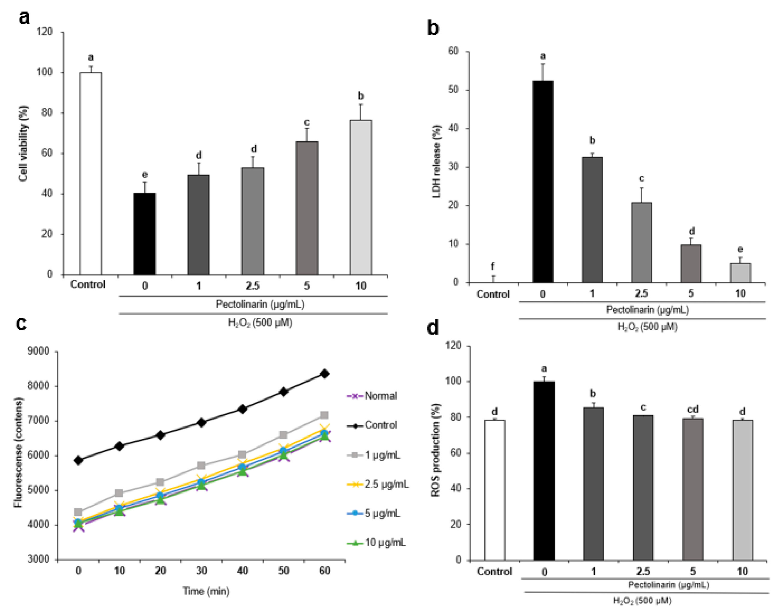
2.3. Effects of Pectolinarin against H2O2-Induced Cell Viability in SH-SY5Y Cells

The 3-(4,5-dimethyl-2-thiazolyl)-2,5-diphenyl-2H-tetrazolium bromide (MTT) assay was used to confirm the cell viability recovery effect of pectolinarin in SH-SY5Y cells after treatment with H2O2 (500 μM; Figure 1a). The cell viability of the control group was 40.26%, whereas the cell viability of the normal group was 100%—confirming that H2O2 was toxic to the SH-SY5Y cells. Pectolinarin (1, 2.5, 5, and 10 μg/mL) was used to treat the SH-SY5Y cells, and cell viability improved dependent on these increasing concentrations—reaching up to 49.20%, 52.72%, 65.82%, and 76.47%, respectively. These results show that pectolinarin protects SH-SY5Y cells from H2O2-induced cell toxicity.

2.4. Effects of Pectolinarin against H2O2-Induced Lactate Dehydrogenase Release in SH-SY5Y Cells

The effects of pectolinarin on H2O2-induced LDH release in SH-SY5Y cells are shown in Figure 1b. In comparison with the normal group—in which 0% of the LDH was released—in the control group, this significantly increased to 52.30%. Moreover, treatment with different concentrations (1, 2.5, 5, and 10 μg/mL) of pectolinarin in H2O2-induced SH-SY5Y cells dose-dependently decreased the level of LDH release to 32.59%, 20.83%, 9.87%, and 5.10%, respectively. These results show that pectolinarin inhibits H2O2-induced LDH release in SH-SY5Y cells.

2.5. Effects of Pectolinarin against H2O2-Induced ROS Formation in SH-SY5Y Cells

The dichlorofluorescein diacetate (DCF-DA) assay was used to confirm the inhibitory effects of pectolinarin against H2O2-induced ROS formation, and the results are presented in Figure 2. ROS production was significantly higher in the H2O2-induced control group (Figure 1c) when compared with the normal group, confirming that H2O2 caused oxidative stress in SH-SY5Y cells. As shown in Figure 2b, when the ROS production in the control group was 100%, the ROS production in the normal group was 78.28%—indicating that when the SH-SY5Y cells were exposed to H2O2 (500 μM) for 24 h and then reacted with DCF-DA, the ROS production in the control group increased by 21.72% compared with the normal group. The intensity of ROS production at 60 min (Figure 1d) showed that treatment with different concentrations (1, 2.5, 5, and 10 μg/mL) of pectolinarin dose-dependently decreased the levels of ROS to 85.65%, 80.92%, 79.32%, and 78.37% when the ROS production of the control group was set as 100%. These results show that pectolinarin inhibits H2O2-induced ROS formation in SH-SY5Y cells.

2.6. Effects of Pectolinarin on Oxidative Stress-Related Protein Expression in H2O2-Induced SH-SY5Y Cells

The action mechanisms of oxidative stress-related proteins such as HO-1, Nrf2, and Keap1 were measured using a Western blot to investigate the protective effects of pectolinarin against H2O2-induced oxidative stress in neural cells. The protein expression of HO-1 and Nrf2 was significantly lower in the H2O2-induced control group compared with the normal group, as shown in Figure 2. However, treatment with pectolinarin (1, 2.5, 5, and 10 μg/mL) in H2O2-induced SH-SY5Y cells increased Nrf2 and HO-1 protein expression. In addition, Keap1 protein expression was significantly higher in the H2O2-induced control group than in the normal group. On the other hand, treatment with pectolinarin (1, 2.5, 5, and 10 μg/mL) dramatically reduced Keap1 protein expression. These results suggested that pectolinarin has antioxidant properties via the regulation of oxidative stress pathway proteins in H2O2-induced SH-SY5Y cells.

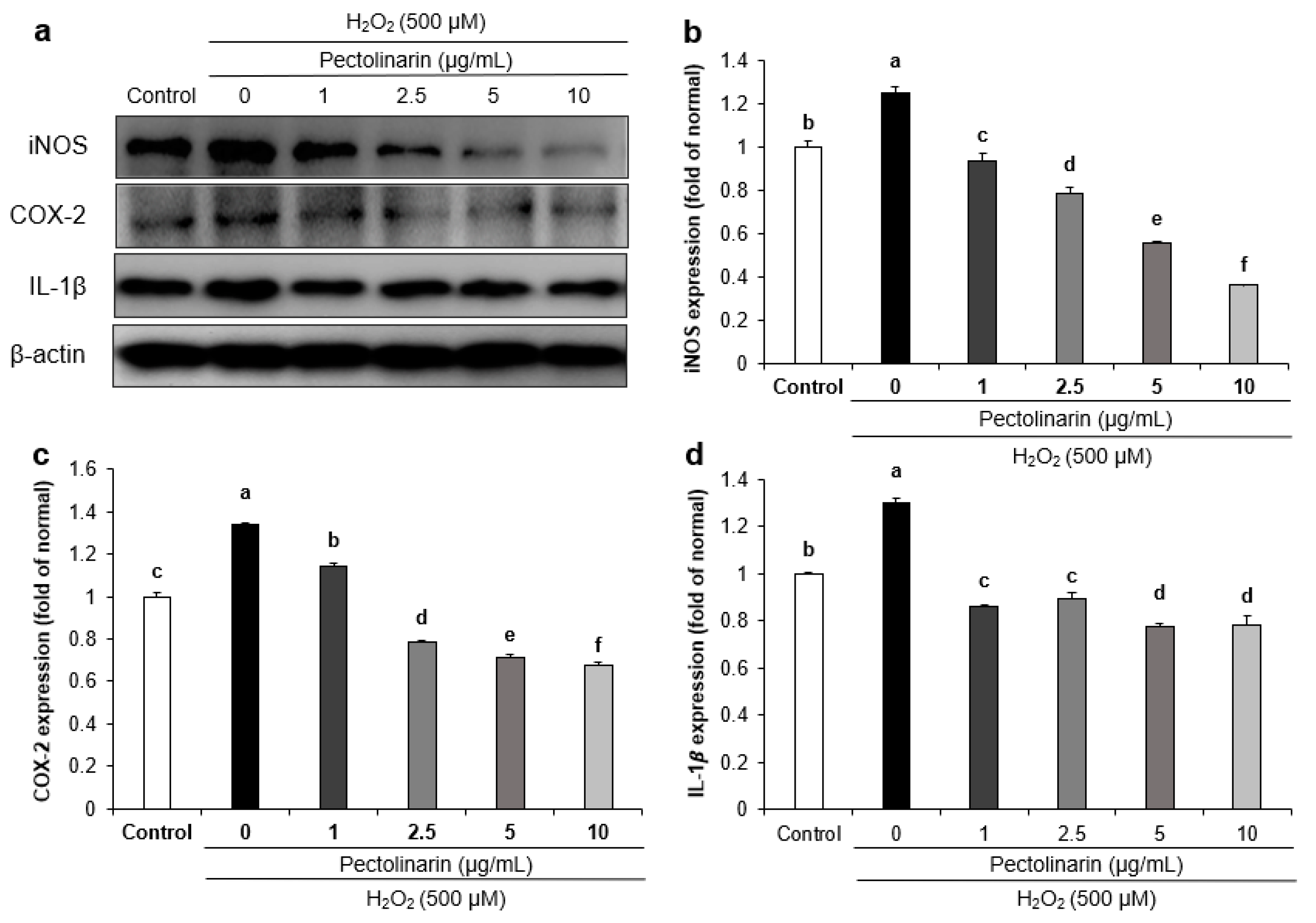
2.7. Effects of Pectolinarin on Inflammation-Related Protein Expression in H2O2-Induced SH-SY5Y Cells

Inflammation-related protein expression in H2O2-induced SH-SY5Y cells was assessed to confirm the mechanisms of pectolinarin. Figure 3 showed that H2O2-induced cells had significantly increased expression of IL-1β, iNOS, and COX-2 proteins compared with the untreated normal SH-SY5Y cells. Furthermore, pectolinarin (1, 2.5, 5, and 10 μg/mL) significantly reduced the expression of the iNOS, COX-2, and IL-1β proteins. These results show that pectolinarin inhibits inflammation in H2O2-induced SH-SY5Y cells by downregulating the inflammation pathway-related proteins iNOS, COX-2, and IL-1β.

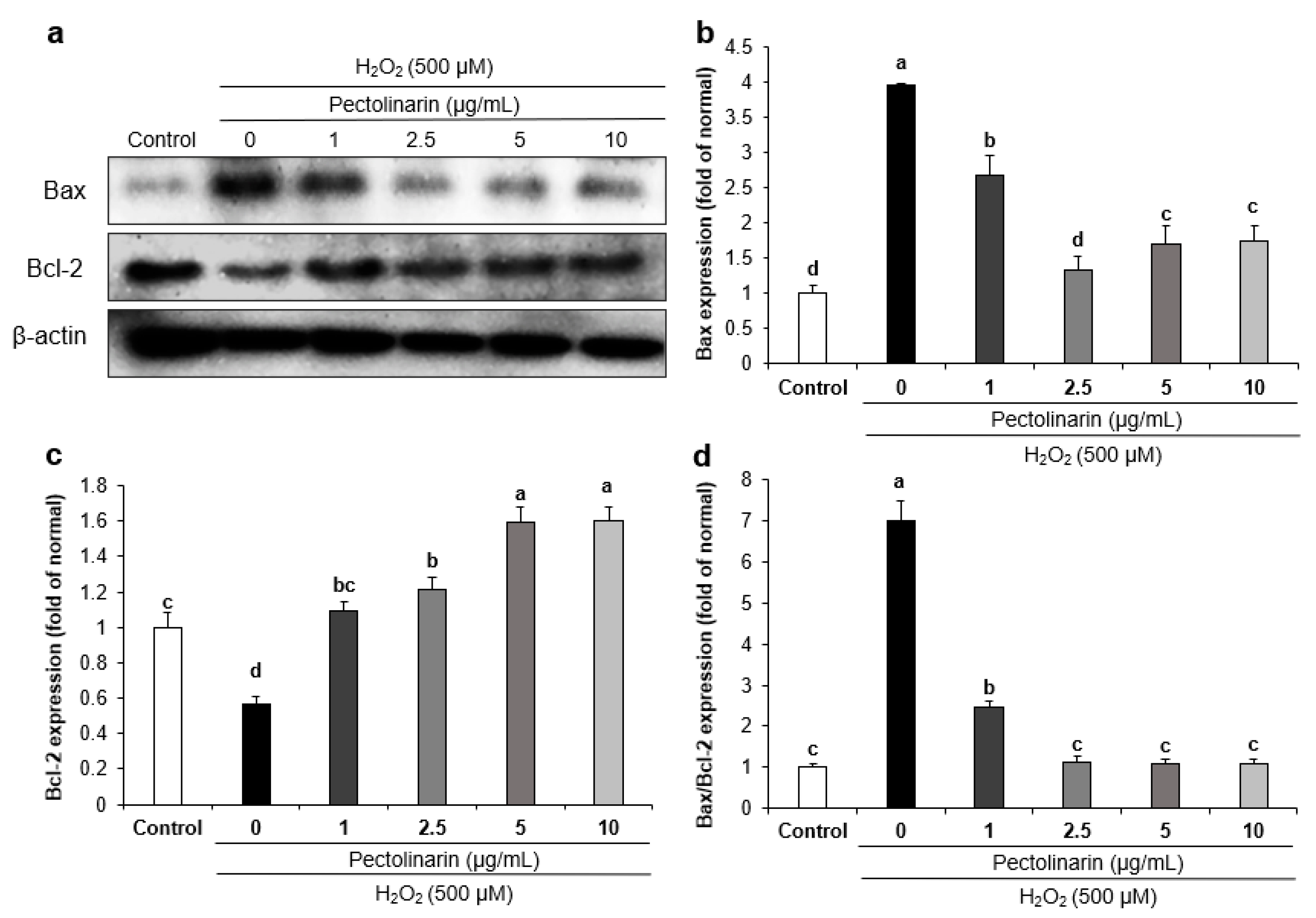
2.8. Effects of Pectolinarin on Apoptosis-Related Protein Expression in H2O2-Induced SH-SY5Y Cells
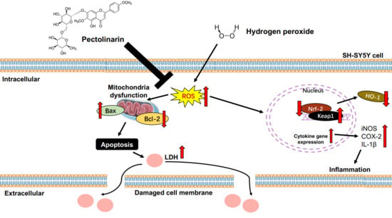
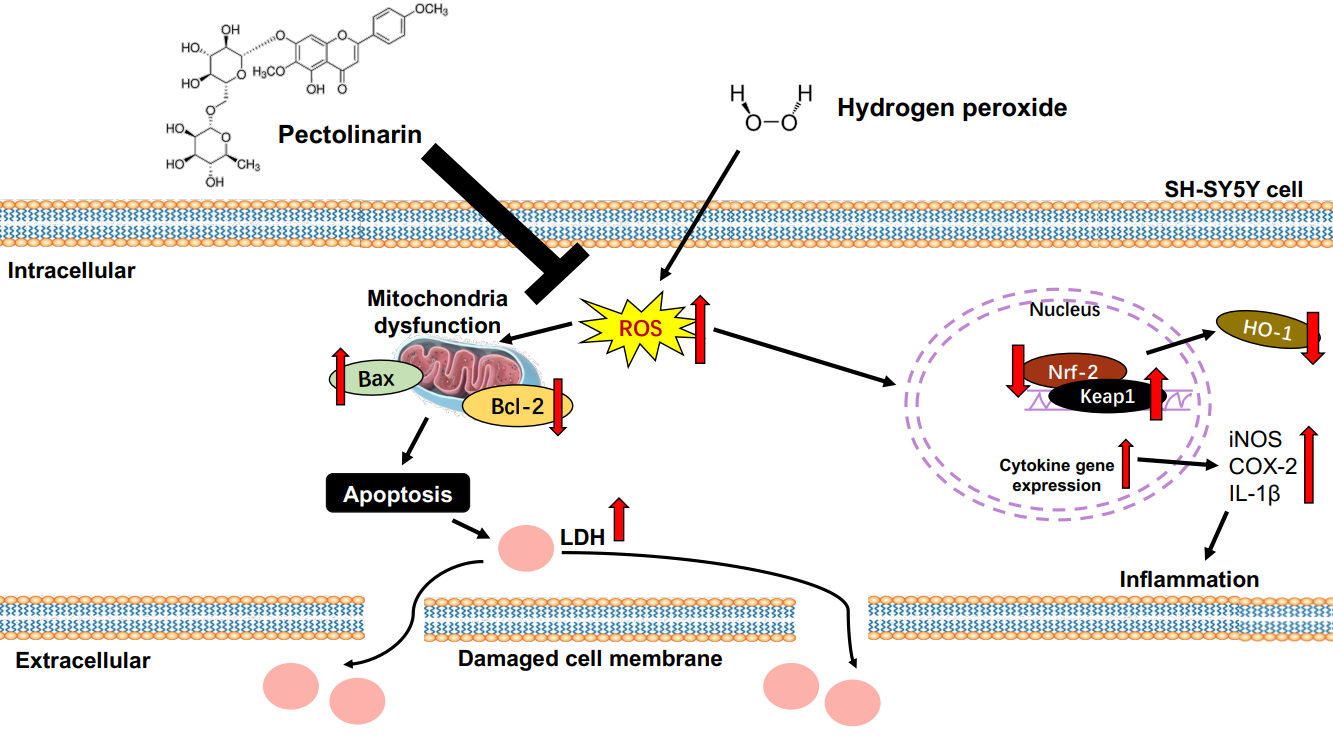
The mechanisms of apoptosis-related proteins were studied using Bax and Bcl-2 protein expression. As shown in Figure 4, H2O2-induced SH-SY5Y cells had significantly higher Bax protein expression than normal SH-SY5Y cells, but treatment with pectolinarin (1, 2.5, 5, and 10 μg/mL) significantly decreased the protein expression of Bax. On the other hand, treatment with H2O2 decreased Bcl-2 protein expression in SH-SY5Y cells compared with normal cells, and treatment with pectolinarin (1, 2.5, 5, and 10 μg/mL) significantly increased the protein expression of Bcl-2. Furthermore, the Bax/Bcl-2 protein expression was significantly higher in the H2O2-induced control group. These findings show that treatment with pectolinarin protects SH-SY5Y cells from H2O2-induced damage by modulating the expression of Bax and Bcl-2, which are proteins associated with apoptosis.

3. Discussion

Under physiological conditions, the production and clearance of ROS in organisms are normal physiological functions [37]. Excessive ROS production in the body can readily lead to oxidative stress [38]. ROS consists of free radicals such as ·OH, O2− and other non-radicals such as H2O2 [39]. Although H2O2 is a toxic by-product, it is involved in many physiological processes such as cell proliferation, differentiation, and immune regulation [40]. However, high concentrations of H2O2 can promote cell apoptosis and oxidative stress in the brain, leading to neurodegenerative diseases such as AD [41,42].
The OH radical, as a by-product of ROS, has high reactivity and a strong oxidation capacity, which can damage cells or tissues in a short period of time [43]. Excess NO production in the brain can cause neuroinflammation and neurodegenerative diseases [44]. The present results showed that the ·OH and NO radical scavenging activity of pectolinarin increased dependent on its concentration, with its ·OH scavenging activity exceeding 80% at concentrations of 2.5, 5, and 10 μg/mL. These results suggest that pectolinarin has antioxidant properties for the scavenging of ·OH and NO radicals.
Several studies have found that throughout the natural aging process, various food intakes or physiological activities cause an increase in the excessive production of ROS, which promotes the deposition of Aβ and accelerates the formation of senile plaques [45,46]. The MTT assay was used in our present study to confirm the protective effects of pectolinarin against H2O2-induced oxidative stress in SH-SY5Y cells. The MTT assay is a standard approach for evaluating mitochondrial damage by measuring cell viability, and importantly, is well-known for assessing cell metabolism—in contrast with the sulforhodamine B assay, which assesses the protein contents [47]. The DCF-DA assay was used to confirm the inhibitory effects of pectolinarin against H2O2-induced ROS formation in SH-SY5Y cells. Dichlorofluorescein (DCF) is a highly fluorescent chemical formed by the oxidation of 2′,7′-dichlorodihydrofluorescein (DCFH) by ROS [48]. Meanwhile, we used the LDH cytotoxicity kit to confirm any inhibitory effects on LDH release. After being treated with pectolinarin, the SH-SY5Y cells showed an antioxidant effect by increasing cell viability and reducing ROS production. Besides this, treatment with pectolinarin lowered LDH secretion. These results suggest that pectolinarin can protect against H2O2-induced cell death, ROS production, and LDH increase.
Nrf2 is a transcription factor that regulates gene expression in antioxidant defense, detoxification, and anti-inflammatory pathways [49]. HO-1 is an enzyme that catalyzes heme breakdown to produce biliverdin, carbon monoxide, and iron. Biliverdin is then metabolized further to bilirubin, which has powerful antioxidant and anti-inflammatory properties [50]. Under normal conditions, Keap1 binds to Nrf2 and regulates Nrf2 activity by promoting ubiquitin-mediated degradation [51]. Studies have shown that activation of the Nrf2/HO-1 system can protect neurons against oxidative stress and prevent the development of neurodegenerative diseases [52,53]. In our present results, treatment with pectolinarin (1, 2.5, 5, and 10 μg/mL) in H2O2-induced SH-SY5Y cells showed a significantly increase in HO-1 and Nrf2 protein expression. In addition, treatment with pectolinarin (1, 2.5, 5, and 10 μg/mL) significantly decreased the expression of Keap1 protein. AD is closely related to the overproduction of ROS and oxidative damage in the brain, and research has shown that regulating the Nrf2/HO-1 pathway can also reduce ROS production, which is consistent with the results of this experiment [54]. These results suggest that pectolinarin has neuroprotective effects against oxidative stress induced by H2O2 through the regulation of antioxidant-related proteins in SH-SY5Y cells.
Inflammation is an immunological response to oxidative stress caused by an excess of ROS. NO is produced by the deamination of arginine under the action of iNOS and enters the adjacent cell through the cell membrane to cause inflammation [55]. COX-2 is an enzyme involved in the production of prostaglandins, as well as the promotion of inflammation and oxidative stress. Inhibiting COX-2 expression may have anti-aging effects by reducing inflammation and oxidative stress [56]. IL-1β is a biologically active small molecule protein that engages in cell signal transmission during the inflammatory response and produces additional pro-inflammatory cytokines [57]. This study found that H2O2-induced SH-SY5Y cells exhibited considerably higher levels of iNOS, COX-2, and IL-1β expression than H2O2-untreated SH-SY5Y cells. Furthermore, treatment with pectolinarin significantly decreased the expression of iNOS, COX-2, and IL-1β proteins. During the pathogenesis of AD, the inflammatory response causes neuronal damage and cell death. These proteins are involved in cell signaling in this pathogenesis process, which promotes the inflammatory response [58]; therefore, iNOS, COX-2, and IL-1β may be key players in the etiology of AD. These results suggest that pectolinarin protects H2O2-induced SH-SY5Y cells through regulating inflammation-related proteins.
Apoptosis is an active process that is regulated by a variety of genes [59]. H2O2 is the source of ·OH that can cross the cell membrane of the brain and cause DNA injury, lipid peroxidation, and oxidative stress—ultimately accelerating apoptosis in the brain [60]. Previous research has investigated apoptosis induction using various methods, such as by TUNEL assay, flow cytometry, and Western blots. Zhao et al. [61] confirmed that H2O2-treatment significantly induced apoptosis in SH-SY5Y cells using the TUNEL assay and flow cytometry with an Annexin V-FITC/PI kit. Moreover, Tian et al. [62] reported that H2O2-induced SH-SY5Y cells increased the expression ratio of Bax/Bcl-2, which is a valuable predictor of apoptosis. In the present study, the effect of pectolinarin on H2O2-induced apoptosis was studied using Western blots for the Bax/Bcl-2 ratio. The Bcl-2 protein family is classified into two categories: the pro-apoptotic protein Bax and the anti-apoptotic protein Bcl-2. The main mechanism of these two proteins is that Bax improves the permeability of the mitochondrial membrane and promotes cytochrome C protein release, ultimately leading to neuronal cell death [63,64]. On the other hand, Bcl-2 protects neuronal cells against death, apoptosis, and oxidative stress stimulation [65]. Our study showed that Bax/Bcl-2 protein expression was significantly increased in the H2O2-induced control group, but treatment with pectolinarin significantly reduced the Bax/Bcl-2 protein expression. These results suggest that pectolinarin protects H2O2-induced SH-SY5Y cells by regulating apoptotic pathway proteins. These results show that pectolinarin could regulate the expression of Bax and Bcl-2 proteins, reducing H2O2-induced oxidative stress and apoptosis in SH-SY5Y cells. This suggests that pectolinarin, as a potential natural compound in functional foods for use against AD, exhibits anti-oxidative stress and cytoprotective effects.

4. Materials and Methods

4.1. Reagents

Thiobarbituric acid (TBA) was acquired from Lancaster Synthesis (Ward Hill, MA, USA), and trichloroacetic acid (TCA) was acquired from Kanto Chemical Co., Inc. (Tokyo, Japan). Dulbecco’s modified eagle medium (DMEM), fetal bovine serum (FBS), penicillin-streptomycin, and trypsin–ethylenediaminetetraacetic acid (EDTA) solution were obtained from Welgene (Daegu, Korea). The LDH cytotoxicity detection kit was purchased from Takara Bio (Siga, Japan). Dimethyl sulfoxide (DMSO) was purchased from Daejung (Siheung-si, Gyeonggi-do, Korea). The MTT was purchased from Bio Pure (Kitchener, ON, Canada). Pectolinarin and DCF-DA were purchased from Sigma-Aldrich (St. Louis, MO, USA). Sodium pentacyanonitrosylferrate (III) dihydrate (SNP) and H2O2 were purchased from Junsei (Tokyo, Japan). The protease inhibitor cocktail was obtained from Calbiochem (Cambridge, MA, USA) and the enhanced chemiluminescence (ECL) substrate solution from Bio-Rad Laboratories (Hercules, CA, USA). The radioimmunoprecipitation (RIPA) buffer was purchased from Elpis Biotech. (Daejeon, Korea) and the polyvinylidene fluoride (PVDF) membrane was obtained from Millipore Co. (Billerica, MA, USA). Primary and secondary antibodies were purchased from Bioss Inc. (Beijing, China), Calbiochem Co. (San Diego, CA, USA), Cell Signaling Tech. (Beverly, CA, USA), and Santa Cruz Biotech. (Santa Cruz, CA, USA). Petolinarin (5,7-Dihydroxy 4′,6-dimethoxyflavone 7-rutinoside) was purchased from Sigma-Aldrich (≥98%, HPLC). Pectolinarin was dissolved in DMSO and stored at 4 °C for use, as shown in Figure 5.

4.2. OH Scavenging Assay

The method of Gutteridge was used to test pectolinarin’s ·OH scavenging abilities [66]. After mixing 10 mM FeSO4·H2O-EDTA, 10 mM 2-deoxyribose solution, and the pectolinarin for each concentration, 10 mM H2O2 was added. The mixture was incubated at 37 °C for 4 h before adding 2.8% TCA and 1.0% TBA solution, boiling at 100 °C for 20 min, ice cooling for 10 min, and measuring absorbance at 490 nm. The formula was as follows:
OH scavenging activity (%) = (Absc − Abss)/Absc × 100
Absc: Absorbance of control, Abss: Absorbance of sample.

4.3. NO Radical Scavenging Assay

The method of Marcocci [67] was used to assess pectolinarin’s NO radical scavenging ability. Each pectolinarin concentration was dissolved in methanol, and a 5 mM sodium nitroprusside solution was added and incubated at room temperature for 150 min. After the reaction, the mixed solution was placed in a 96-well plate at a ratio of 1:1 with Griess reagent and reacted at room temperature with light protection for 30 min, and the absorbance was measured at 540 nm. The formula was as follows:
NO radical scavenging activity (%) = (Absc − Abss)/Absc × 100
Absc: Absorbance of control, Abss: Absorbance of sample.

4.4. Cell Culture

SH-SY5Y human neuronal cells were cultured in DMEM medium containing 10% FBS and 100 units/mL penicillin–streptomycin in an incubator at 37 °C and 5% CO2 humidity. Cells were cultured in a T75 flask, rinsed with phosphate-buffered saline (PBS, PH = 7.4) until they reached a growth state of more than 80% differentiation, and then 2 mL of trypsin-EDTA mixture was added to separate the attached cells before centrifugaton at 1000 rpm for 3 min.

4.5. Cell Viability

The MTT technique was used to assess cell viability [68]. When the cells were more than 80% differentiated, 2.5 × 10⁵ cells/well were seeded in a 96-well plate and incubated at 37 °C for 24 h. After 24 h, pectolinarin in different concentrations was added to the plate for each well and cultured for 4 h before being treated with H2O2 (500 μM) and cultured for another 24 h. After 24 h incubation, the medium was removed from each well, 200 μL of 5 mg/mL MTT solution added, and the plate incubated for 4 h at 37 °C. After that, the MTT solution was removed and DMSO added for 30 min. The absorbance was measured at 540 nm (Thermo Fisher Scientific, Waltham, MA, USA).

4.6. Measurement of ROS Production

The DCF-DA assay was used to measure ROS production according to Wang and Zhu’s method [69]. When the cells had been differentiated by more than 80%, they were seeded at 2.5 × 10⁵ cells/well in a black 96-well plate and cultured at 37 °C for 24 h. After 24 h, pectolinarin at different concentrations was added to the plate for each well and cultured for 4 h before being treated with H2O2 (500 μM) and cultured for 24 h. After 24 h, cells were treated with 80 μM of DCF-DA solution after removing the medium from each well, incubating for 30 min at 37 °C, and then measuring the fluorescence at an excitation of 480 nm and emission of 535 nm using a FLUO starOPTIMA (BMG labtech, Ortenberg, Germany).

4.7. Measurement of LDH Release Activity

The LDH release was assessed using an LDH cytotoxicity detection kit, following the manufacturer’s protocol (Takara Bio Inc., Kusatsu, Japan). The cell supernatant was combined with the LDH reaction mixture (catalyst: dye solution = 1:45) and incubated at room temperature for 30 min. The absorbance was measured at 490 nm (Thermo Fisher Scientific, Waltham, MA, USA).

4.8. Western Blot Analysis

SH-SY5Y cells were seeded at 1 × 10⁶ cells/mL in a 100 mm cell culture dish, cultured at 37 °C for 24 h, and then treated with pectolinarin (1, 2.5, 5, and 10 μg/mL) and incubated for 4 h before being treated with H2O2 (500 μM) and cultured for 24 h. After 24 h, the cells were collected and centrifuged at 4 °C for 30 min at 12,000 rpm in RIPA buffer. The supernatant was then transferred to a new e-tube. Each sample was separated by a 10% or 13% sodium dodecyl sulfate-polyacrylamide gel (SDS-PAGE) and transferred to the PVDF membrane by electrophoresis. Blocking was carried out using 5% skimmed milk for 1 h at room temperature, diluting the primary antibody in PBST to a concentration of 1:1000 or 1:200, and reacting overnight at 4 °C. Each of the following primary antibodies was used: β-actin (1:1000; Cell Signaling Tech.); COX-2 (1:500; Calbiochem Co.); iNOS (1:1000; Calbiochem Co.); IL-1β (1:1000; Cell Signaling Tech.); Bax (1:1000; Cell Signaling Tech.); Bcl-2 (1:1000; Abcam, Cambridge, UK); HO-1 (1:2000; Abcam); Nrf2 (1:1000; Cell Signaling Tech.); and Keap1 (1:1000; Cell Signaling Tech.). After washing the membrane, it was reacted with a 1:1000 dilution of the secondary antibody at room temperature for 1 h. The protein expression was confirmed using a Chemiliuminescence image system (Davinch-ChemiTM) after washing with 1 × PBST and reacting with ECL solution.

4.9. Statistical Analysis

All results are expressed as mean ± standard deviation. Statistical significance was determined using one-way analysis of variance followed by Duncan’s multiple test on the SPSS statistics program (version 25, SPSS Inc., Chicago, IL, USA). Statistical significance was considered as p < 0.05.

5. Conclusions

In conclusion, pectolinarin had in vitro antioxidant capabilities for ·OH and NO radicals scavenging, increased cell viability, and decreased ROS formation in H2O2-induced SH-SY5Y cells. In addition, pectolinarin inhibited LDH release in H2O2-induced SH-SY5Y cells. Furthermore, pectolinarin regulated oxidative stress, inflammation, and apoptosis-related protein expression and provided neuroprotection against H2O2-induced oxidative stress in SH-SY5Y cells. Therefore, pectolinarin has the potential to be used as a plant material for functional foods, to be applied for the treatment of oxidative stress-related neurodegenerative diseases. In the future, we will investigate the protective mechanisms of pectolinarin against inflammation and oxidative damage in aging mouse brain models, in order to provide a theoretical basis for future research and the development of AD functional foods.

Author Contributions

Conceptualization, Q.Q.P. and E.J.C.; methodology, Q.Q.P.; validation, Q.Q.P., J.-H.K. and E.J.C.; investigation, J.H.K. and H.Y.K.; resources, H.Y.K.; data curation, Q.Q.P.; writing—original draft preparation, Q.Q.P.; writing—review and editing, J.H.K., J.-H.K. and E.J.C.; visualization, E.J.C.; supervision, E.J.C.; project administration, J.-H.K. and E.J.C.; funding acquisition, H.Y.K. and E.J.C. All authors have read and agreed to the published version of the manuscript.

Funding

This research was supported by the Basic Science Research Program through the National Research Foundation of Korea (NRF), funded by the Ministry of Education (Grant 2018R1D1A1B07043784).

Institutional Review Board Statement

Not applicable.

Informed Consent Statement

Not applicable.

Data Availability Statement

The data presented in this study are available on request from the corresponding author.

Conflicts of Interest

The authors declare no conflict of interest.

Sample Availabilty

Not available.

References

  1. Chen, Z.; Zhong, C. Oxidative stress in Alzheimer’s disease. Neurosci. Bull. 2014, 30, 271–281. [Google Scholar] [CrossRef] [Green Version]
  2. Wang, Y.; Li, S.; Li, C. Perspectives of new advances in the pathogenesis of vitiligo: From oxidative stress to autoimmunity. Med. Sci. Monit. 2019, 25, 1017–1023. [Google Scholar] [CrossRef]
  3. Kudryavtseva, A.V.; Krasnov, G.S.; Dmitriev, A.A.; Alekseev, B.Y.; Kardymon, O.L.; Sadritdinova, A.F.; Fedorova, M.S.; Pokrovsky, A.V.; Melnikova, N.V.; Kaprin, A.D.; et al. Mitochondrial dysfunction and oxidative stress in aging and cancer. Oncotarget 2016, 7, 44879–44905. [Google Scholar] [CrossRef] [Green Version]
  4. Wojtczyk-Miaskowska, A.; Schlichtholz, B. DNA damage and oxidative stress in long-lived aquatic organisms. DNA Repair 2018, 69, 14–23. [Google Scholar] [CrossRef]
  5. Cooke, M.S.; Evans, M.D.; Dizdaroglu, M.; Lunec, J. Oxidative DNA damage: Mechanisms, mutation, and disease. FASEB J. 2003, 17, 1195–1214. [Google Scholar] [CrossRef] [Green Version]
  6. Kamat, P.K.; Kalani, A.; Rai, S.; Swarnkar, S.; Tota, S.; Nath, C.; Tyagi, N. Mechanism of oxidative stress and synapse dysfunction in the pathogenesis of Alzheimer’s disease: Understanding the therapeutics strategies. Mol. Neurobiol. 2016, 53, 648–661. [Google Scholar] [CrossRef] [Green Version]
  7. Wang, Q.; Liu, Y.; Zhou, J. Neuroinflammation in Parkinson’s disease and its potential as therapeutic target. Transl. Neurodegener. 2015, 4, 19. [Google Scholar] [CrossRef] [Green Version]
  8. Singh, P.K.; Baxi, D.; Diwedi, R.; Ramachandran, A.V. Prior cadmium exposure improves glucoregulation in diabetic rats but exacerbates effects on metabolic dysregulation, oxidative stress, and hepatic and renal toxicity. Drug Chem. Toxicol. 2012, 35, 167–177. [Google Scholar] [CrossRef]
  9. Olajide, O.J.; Yawson, E.O.; Gbadamosi, I.T.; Arogundade, T.T.; Lambe, E.; Obasi, K.; Lawal, I.T.; Ibrahim, A.; Ogunrinola, K.Y. Ascorbic acid ameliorates behavioural deficits and neuropathological alterations in rat model of Alzheimer’s disease. Environ. Toxicol. Pharmacol. 2017, 50, 200–211. [Google Scholar] [CrossRef]
  10. Tang, K.S. The potential role of Nanoyttria in alleviating oxidative stress biomarkers: Implications for Alzheimer’s disease therapy. Life Sci. 2020, 259, 118287. [Google Scholar] [CrossRef]
  11. Tarafdar, A.; Pula, G. The role of NADPH oxidases and oxidative stress in neurodegenerative disorders. Int. J. Mol. Sci. 2018, 19, 3824. [Google Scholar] [CrossRef] [Green Version]
  12. Serou, M.J.; DeCoster, M.A.; Bazan, N.G. Interleukin-1 beta activates expression of cyclooxygenase-2 and inducible nitric oxide synthase in primary hippocampal neuronal culture: Platelet-activating factor as a preferential mediator of cyclooxygenase-2 expression. J. Neurosci. Res. 1999, 58, 593–598. [Google Scholar] [CrossRef]
  13. Li, C.Q.; Kim, M.Y.; Godoy, L.C.; Thiantanawat, A.; Trudel, L.J.; Wogan, G.N. Nitric oxide activation of Keap1/Nrf2 signaling in human colon carcinoma cells. Proc. Natl. Acad. Sci. USA 2009, 106, 14547–14551. [Google Scholar] [CrossRef]
  14. Dinkova-Kostova, A.T.; Kostov, R.V.; Kazantsev, A.G. The role of Nrf2 signaling in counteracting neurodegenerative diseases. FEBS J. 2018, 285, 3576–3590. [Google Scholar] [CrossRef] [Green Version]
  15. Rojo, A.I.; Pajares, M.; Rada, P.; Nuñez, A.; Nevado-Holgado, A.J.; Killik, R.; Van Leuven, F.; Ribe, E.; Lovestone, S.; Yamamoto, M.; et al. NRF2 deficiency replicates transcriptomic changes in Alzheimer’s patients and worsens APP and TAU pathology. Redox Biol. 2017, 13, 444–451. [Google Scholar] [CrossRef]
  16. Deshmukh, P.; Unni, S.; Krishnappa, G.; Padmanabhan, B. The Keap1–Nrf2 pathway: Promising therapeutic target to counteract ROS-mediated damage in cancers and neurodegenerative diseases. Biophys. Rev. 2017, 9, 41–56. [Google Scholar] [CrossRef] [Green Version]
  17. Zawia, N.H.; Lahiri, D.K.; Cardozo-Pelaez, F. Epigenetics, oxidative stress, and Alzheimer disease. Free Radic. Biol. Med. 2009, 46, 1241–1249. [Google Scholar] [CrossRef] [Green Version]
  18. Alamdary, S.Z.; Khodagholi, F.; Shaerzadeh, F.; Ansari, N.; Sonboli, A.; Tusi, S.K. S. choloroleuca, S. mirzayanii and S. santolinifolia protect PC12 cells from H2O2-induced apoptosis by blocking the intrinsic pathway. Cytotechnology 2012, 64, 403–419. [Google Scholar] [CrossRef] [Green Version]
  19. Singh, R.; Letai, A.; Sarosiek, K. Regulation of apoptosis in health and disease: The balancing act of BCL-2 family proteins. Nat. Rev. Mol. Cell Biol. 2019, 20, 175–193. [Google Scholar] [CrossRef]
  20. Bayazid, A.B.; Lim, B.O. Quercetin is an active agent in berries against neurodegenerative diseases progression through modulation of Nrf2/HO1. Nutrients 2022, 14, 5132. [Google Scholar] [CrossRef]
  21. Hu, L.; Zhang, R.; Yuan, Q.; Gao, Y.; Yang, M.Q.; Zhang, C.; Huang, J.; Sun, Y.; Yang, W.; Yang, J.Y.; et al. The emerging role of MicroRNA-4487/6845-3p in Alzheimer’s disease pathologies is induced by Aβ25–35 triggered in SH-SY5Y cell. BMC Syst. Biol. 2018, 12 (Suppl. S7), 119. [Google Scholar] [CrossRef] [Green Version]
  22. Omar, S.H.; Scott, C.J.; Hamlin, A.S.; Obied, H.K. Olive biophenols reduces Alzheimer’s pathology in SH-SY5Y cells and APPswe mice. Int. J. Mol. Sci. 2018, 20, 125. [Google Scholar] [CrossRef] [Green Version]
  23. Zhao, Z.Y.; Zhang, Y.Q.; Zhang, Y.H.; Wei, X.Z.; Wang, H.; Zhang, M.; Yang, Z.J.; Zhang, C.H. The protective underlying mechanisms of schisandrin on SH-SY5Y cell model of Alzheimer’s disease. J. Toxicol. Environ. Health A 2019, 82, 1019–1026. [Google Scholar] [CrossRef]
  24. Akki, R.; Siracusa, R.; Morabito, R.; Remigante, A.; Campolo, M.; Errami, M.; La Spada, G.; Cuzzocrea, S.; Marino, A. Neuronal-like differentiated SH-SY5Y cells adaptation to a mild and transient H2O2 -Induced oxidative stress. Cell Biochem. Funct. 2018, 36, 56–64. [Google Scholar] [CrossRef]
  25. Akyuva, Y.; Nazıroğlu, M. Resveratrol attenuates hypoxia-induced neuronal cell death, inflammation and mitochondrial oxidative stress by modulation of TRPM2 channel. Sci. Rep. 2020, 10, 6449. [Google Scholar] [CrossRef] [Green Version]
  26. Islam, M.d.I.; Shanta, M.A.; Mondal, M.; Hoque, N.; Majumder, S.; Ahmed, T.; Rana, M.S. Protective effect of chloroform extract of Stereospermum Chelonoides Bark against amyloid Beta42 induced cell death in SH-SY5Y cells and against inflammation in Swiss albino mice. J. Basic Clin. Physiol. Pharmacol. 2018, 29, 621–630. [Google Scholar] [CrossRef]
  27. Tian, X.; Gao, L.; An, L.; Jiang, X.; Bai, J.; Huang, J.; Meng, W.; Zhao, Q. Pretreatment of MQA, a caffeoylquinic acid derivative compound, protects against H2O2-induced oxidative stress in SH-SY5Y cells. Neurol. Res. 2016, 38, 1079–1087. [Google Scholar] [CrossRef]
  28. Uğuz, A.C.; Öz, A.; Nazıroğlu, M. Curcumin inhibits apoptosis by regulating intracellular calcium release, reactive oxygen species and mitochondrial depolarization levels in SH-SY5Y neuronal cells. J. Recept. Signal Transduct. Res. 2016, 36, 395–401. [Google Scholar] [CrossRef]
  29. Martins, G.R.; da Fonseca, T.S.; Martínez-Fructuoso, L.; Simas, R.C.; Silva, F.T.; Salimena, F.R.G.; Alviano, D.S.; Alviano, C.S.; Leitão, G.G.; Pereda-Miranda, R.; et al. Antifungal phenylpropanoid glycosides from Lippia rubella. J. Nat. Prod. 2019, 82, 566–572. [Google Scholar] [CrossRef]
  30. Thao, N.T.P.; Cuong, T.D.; Hung, T.M.; Lee, J.H.; Na, M.; Son, J.K.; Jung, H.J.; Fang, Z.; Woo, M.H.; Choi, J.S.; et al. Simultaneous determination of bioactive flavonoids in some selected Korean thistles by high-performance liquid chromatography. Arch. Pharm. Res. 2011, 34, 455–461. [Google Scholar] [CrossRef]
  31. Cheriet, T.; Ben-Bachir, B.; Thamri, O.; Seghiri, R.; Mancini, I. Isolation and biological properties of the natural flavonoids Pectolinarin and Pectolinarigenin—A review. Antibiotics 2020, 9, 417. [Google Scholar] [CrossRef]
  32. Ahmed-Chaouch, M.; Cheriet, T.; Beretta, G.; Sarri, D.; Bensouici, C.; Ouelbani, R.; Mancini, I.; Sekhara, I.; Seghiri, R. Chemical composition, in vitro antioxidant, anticholinesterase and antibacterial activities of Linaria scariosa Desf. Nat. Prod. Res. 2021, 35, 1722–1726. [Google Scholar] [CrossRef]
  33. Lim, H.; Son, K.H.; Chang, H.W.; Bae, K.; Kang, S.S.; Kim, H.P. Anti-inflammatory activity of Pectolinarigenin and Pectolinarin isolated from Cirsium chanroenicum. Biol. Pharm. Bull. 2008, 31, 2063–2067. [Google Scholar] [CrossRef] [Green Version]
  34. Liu, S.; Luo, X.; Li, D.; Zhang, J.; Qiu, D.; Liu, W.; She, L.; Yang, Z. Tumor inhibition and improved immunity in mice treated with flavone from Cirsium japonicum DC. Int. Immunopharmacol. 2006, 6, 1387–1393. [Google Scholar] [CrossRef]
  35. Mehta, P.; Dhapte, V. A comprehensive review on pharmacokinetic profile of some traditional Chinese medicines. New J. Sci. 2016, 2016, 7830367. [Google Scholar] [CrossRef] [Green Version]
  36. Chen, Q.; Gong, F.; Liu, Z.; Zhong, L.; Gong, Q. Comparison on intestinal absorption of pectolinarin and pectolinarigenin by everted rat intestinal sac method. World J. Tradit. Chin. Med. 2014, 12, 352–357. [Google Scholar]
  37. Zorov, D.B.; Juhaszova, M.; Sollott, S.J. Mitochondrial reactive oxygen species (ROS) and ROS-induced ROS release. Physiol. Rev. 2014, 94, 909–950. [Google Scholar] [CrossRef] [Green Version]
  38. Li, L.; Tan, J.; Miao, Y.; Lei, P.; Zhang, Q. ROS and autophagy: Interactions and molecular regulatory mechanisms. Cell. Mol. Neurobiol. 2015, 35, 615–621. [Google Scholar] [CrossRef]
  39. del Río, L.A. ROS and RNS in plant physiology: An overview. J. Exp. Bot. 2015, 66, 2827–2837. [Google Scholar] [CrossRef] [Green Version]
  40. Dickinson, B.C.; Peltier, J.; Stone, D.; Schaffer, D.V.; Chang, C.J. Nox2 redox signaling maintains essential cell populations in the brain. Nat. Chem. Biol. 2011, 7, 106–112. [Google Scholar] [CrossRef]
  41. Lee, Y.M.; He, W.; Liou, Y.-C. The Redox Language in Neurodegenerative Diseases: Oxidative Post-Translational Modifications by Hydrogen Peroxide. Cell Death Dis. 2021, 12, 58. [Google Scholar] [CrossRef]
  42. Yang, H.; Xie, Y.; Yang, D.; Ren, D. Oxidative stress-induced apoptosis in granulosa cells involves JNK, P53 and puma. Oncotarget 2017, 8, 25310–25322. [Google Scholar] [CrossRef] [Green Version]
  43. Valko, M.; Rhodes, C.J.; Moncol, J.; Izakovic, M.; Mazur, M. Free radicals, metals and antioxidants in oxidative stress-induced cancer. Chem. Biol. Interact. 2006, 160, 1–40. [Google Scholar] [CrossRef]
  44. Liochev, S.I. Reactive oxygen species and the free radical theory of aging. Free Radic. Biol. Med. 2013, 60, 1–4. [Google Scholar] [CrossRef]
  45. Ratnayaka, J.A.; Serpell, L.C.; Lotery, A.J. Dementia of the Eye: The role of amyloid beta in retinal degeneration. Eye 2015, 29, 1013–1026. [Google Scholar] [CrossRef]
  46. Ahmad, W.; Ijaz, B.; Shabbiri, K.; Ahmed, F.; Rehman, S. Oxidative toxicity in diabetes and Alzheimer’s disease: Mechanisms behind ROS/RNS generation. J. Biomed. Sci. 2017, 24, 76. [Google Scholar] [CrossRef]
  47. Collier, A.C.; Pritsos, C.A. The mitochondrial uncoupler dicumarol disrupts the MTT assay. Biochem. Pharmacol. 2003, 66, 281–287. [Google Scholar] [CrossRef]
  48. Rastogi, R.P.; Singh, S.P.; Häder, D.P.; Sinha, R.P. Detection of reactive oxygen species (ROS) by the oxidant-sensing probe 2′,7′-dichlorodihydrofluorescein diacetate in the cyanobacterium Anabaena variabilis PCC 7937. Biochem. Biophys. Res. Commun. 2010, 397, 603–607. [Google Scholar] [CrossRef]
  49. Esgalhado, M.; Stenvinkel, P.; Mafra, D. Nonpharmacologic strategies to modulate nuclear factor erythroid 2–related Factor 2 pathway in chronic kidney disease. J. Ren. Nutr. 2017, 27, 282–291. [Google Scholar] [CrossRef]
  50. Loboda, A.; Damulewicz, M.; Pyza, E.; Jozkowicz, A.; Dulak, J. Role of Nrf2/HO-1 system in development, oxidative stress response and diseases: An evolutionarily conserved mechanism. Cell. Mol. Life Sci. 2016, 73, 3221–3247. [Google Scholar] [CrossRef] [Green Version]
  51. Villeneuve, N.F.; Lau, A.; Zhang, D.D. Regulation of the Nrf2–Keap1 antioxidant response by the ubiquitin proteasome system: An insight into Cullin-Ring ubiquitin ligases. Antioxid. Redox Signal. 2010, 13, 1699–1712. [Google Scholar] [CrossRef] [Green Version]
  52. Kwon, S.H.; Lee, S.R.; Park, Y.J.; Ra, M.; Lee, Y.; Pang, C.; Kim, K.H. Suppression of 6-hydroxydopamine-induced oxidative stress by hyperoside via activation of Nrf2/HO-1 signaling in dopaminergic neurons. Int. J. Mol. Sci. 2019, 20, 5832. [Google Scholar] [CrossRef] [Green Version]
  53. Wen, C.; Huang, C.; Yang, M.; Fan, C.; Li, Q.; Zhao, J.; Gan, D.; Li, A.; Zhu, L.; Lu, D. The secretion from bone marrow mesenchymal stem cells pretreated with berberine rescues neurons with oxidative damage through activation of the Keap1-Nrf2-HO-1 signaling pathway. Neurotox. Res. 2020, 38, 59–73. [Google Scholar] [CrossRef]
  54. Xia, Q.; Gao, S.; Rapael Gnanamuthu, S.R.; Zhuang, K.; Song, Z.; Zhang, Y.; Wang, X.; Tu, P.; Li, J.; Liu, K. Involvement of Nrf2-HO-1/JNK-Erk signaling pathways in aconitine-induced developmental toxicity, oxidative stress, and ROS-mitochondrial apoptosis in zebrafish embryos. Front. Pharmacol. 2021, 12, 642480. [Google Scholar] [CrossRef]
  55. Guzik, T.J.; Korbut, R.; Adamek-Guzik, T. Nitric oxide and superoxide in inflammation and immune regulation. J. Physiol. Pharmacol. 2003, 54, 469–487. [Google Scholar]
  56. Luo, C.; Urgard, E.; Vooder, T.; Metspalu, A. The role of COX-2 and Nrf2/ARE in anti-inflammation and antioxidative stress: Aging and anti-aging. Med. Hypotheses 2011, 77, 174–178. [Google Scholar] [CrossRef]
  57. Green, H.F.; Nolan, Y.M. GSK-3 mediates the release of IL-1β, TNF-α and IL-10 from cortical glia. Neurochem. Int. 2012, 61, 666–671. [Google Scholar] [CrossRef]
  58. Webers, A.; Heneka, M.T.; Gleeson, P.A. The role of innate immune responses and neuroinflammation in amyloid accumulation and progression of Alzheimer’s disease. Immunol. Cell Biol. 2020, 98, 28–41. [Google Scholar] [CrossRef]
  59. Pistritto, G.; Trisciuoglio, D.; Ceci, C.; Garufi, A.; D’Orazi, G. Apoptosis as anticancer mechanism: Function and dysfunction of its modulators and targeted therapeutic strategies. Aging 2016, 8, 603–619. [Google Scholar] [CrossRef] [Green Version]
  60. Tripathi, R.; Gupta, R.; Sahu, M.; Srivastava, D.; Das, A.; Ambasta, R.K.; Kumar, P. Free radical biology in neurological manifestations: Mechanisms to therapeutics interventions. Environ. Sci. Pollut. Res. Int. 2022, 29, 62160–62207. [Google Scholar] [CrossRef]
  61. Zhao, S.; Fang, J.; Li, S.; Gaur, U.; Xing, X.; Wang, H.; Zheng, W. Artemisinin attenuated hydrogen peroxide (H2O2)-induced oxidative injury in SH-SY5Y and hippocampal neurons via the activation of AMPK pathway. Int. J. Mol. Sci. 2019, 20, 2680. [Google Scholar] [CrossRef] [Green Version]
  62. Tian, W.; Zhao, J.; Lee, J.-H.; Akanda, M.R.; Cho, J.-H.; Kim, S.-K.; Choi, Y.-J.; Park, B.-Y. Neuroprotective effects of Cornus officinalis on stress-induced hippocampal deficits in rats and H2O2-induced neurotoxicity in SH-SY5Y neuroblastoma cells. Antioxidants 2019, 9, 27. [Google Scholar] [CrossRef] [Green Version]
  63. Aminzadeh, A.; Salarinejad, A. Citicoline protects against lead-induced oxidative injury in neuronal PC12 Cells. Biochem. Cell Biol. 2019, 97, 715–721. [Google Scholar] [CrossRef]
  64. Li, Z.; Xiao, G.; Wang, H.; He, S.; Zhu, Y. A preparation of Ginkgo biloba L. leaves extract inhibits the apoptosis of hippocampal neurons in post-stroke mice via regulating the expression of Bax/Bcl-2 and Caspase-3. J. Ethnopharmacol. 2021, 280, 114481. [Google Scholar] [CrossRef]
  65. Li, Y.; Wang, B.; Liu, C.; Zhu, X.; Zhang, P.; Yu, H.; Li, Y.; Li, Z.; Li, M. Inhibiting c-Jun N-terminal kinase (JNK)-mediated apoptotic signaling pathway in PC12 cells by a polysaccharide (CCP) from Coptis chinensis against Amyloid-β (Aβ)-induced neurotoxicity. Int. J. Biol. Macromol. 2019, 134, 565–574. [Google Scholar] [CrossRef]
  66. Gutteridge, J.M.C. Ferrous-salt-promoted damage to deoxyribose and benzoate. The increased effectiveness of hydroxyl-radical scavengers in the presence of EDTA. Biochem. J. 1987, 243, 709–714. [Google Scholar] [CrossRef]
  67. Marcocci, L.; Maguire, J.J.; Droy-Lefaix, M.T.; Packer, L. The nitric oxide-scavenging properties of Ginkgo biloba extract EGb 761. Biochem. Biophys. Res. Commun. 1994, 201, 748–755. [Google Scholar] [CrossRef]
  68. Mosmann, T. Rapid colorimetric assay for cellular growth and survival: Application to proliferation and cytotoxicity assays. J. Immunol. Methods 1983, 65, 55–63. [Google Scholar] [CrossRef]
  69. Wang, R.G.; Zhu, X.Z. Subtoxic concentration of manganese synergistically potentiates 1-methyl-4-phenylpyridinium-induced neurotoxicity in PC12 cells. Brain Res. 2003, 961, 131–138. [Google Scholar] [CrossRef]
Figure 1. Protective effects of pectolinarin against H2O2-induced cell viability (a), LDH release (b), time course of change in intensity of DCF fluorescence during 60 min (c) and the intensity of ROS production at 60 min (d) in SH-SY5Y cells. Values are mean ± SD. The different letters (a–f) between groups represent significant differences as determined by the Tukey HSD multiple range test (p < 0.05).
Molecules 28 05826 g001
Figure 2. Effects of pectolinarin on oxidative stress-related protein expression in H2O2-induced SH-SY5Y cells. (a) Bands of HO-1, Nrf2, and Keap1 proteins expression level. (b) HO-1 protein expression level. (c) Nrf2 protein expression level. (d) Keap1 protein expression level. Values are mean ± SD. The different letters (a–e) among groups represent significant differences as determined by the Tukey HSD multiple range test (p < 0.05).
Molecules 28 05826 g002
Figure 3. Effects of pectolinarin on inflammation-related protein expression in H2O2-induced SH-SY5Y cells. (a) Bands of iNOS, COX-2, and IL-1β proteins expression level. (b) iNOS protein expression level. (c) COX-2 protein expression level. (d) IL-1β protein expression level. Values are mean ± SD. The different letters (a–f) between groups represent significant differences as determined by the Tukey HSD multiple range test (p < 0.05).
Molecules 28 05826 g003
Figure 4. The effects of pectolinarin on apoptosis-related protein expression in H2O2-induced SH-SY5Y cells. (a) Bands of Bax and Bcl-2 proteins expression level. (b) Bax protein expression level. (c) Bcl-2 protein expression level. (d) Ratio of Bax/Bcl-2 proteins expression level. Values are mean ± SD. The different letters (a–d) between groups represent significant differences as determined by the Tukey HSD multiple range test (p < 0.05).
Molecules 28 05826 g004
Figure 5. Structure of pectolinarin.
Molecules 28 05826 g005
Table 1. OH scavenging activity of pectolinarin.
Treatment (μg/mL)Scavenging Activity (%)
163.09 ± 1.97 c
2.580.43 ± 1.37 b
583.60 ± 0.75 a
1084.77 ± 0.95 a
Note. Values are mean ± SD. The different letters (a–c) show significantly differences (p < 0.05) by the Tukey HSD multiple range test.
Table 2. NO radical scavenging activity of pectolinarin.
Treatment (μg/mL)Scavenging Activity (%)
123.21 ± 1.09 d
2.524.50 ± 1.37 c
530.40 ± 0.59 b
1035.19 ± 0.32 a
Note. Values are mean ± SD. The different letters (a–d) show significant differences (p < 0.05) by the Tukey HSD multiple range test.
Disclaimer/Publisher’s Note: The statements, opinions and data contained in all publications are solely those of the individual author(s) and contributor(s) and not of MDPI and/or the editor(s). MDPI and/or the editor(s) disclaim responsibility for any injury to people or property resulting from any ideas, methods, instructions or products referred to in the content.

