Review

Potential Application of the Plant-Derived Essential Oils for Atherosclerosis Treatment: Molecular Mechanisms and Therapeutic Potential

by
Siarhei A. Dabravolski
1,*,
Vasily N. Sukhorukov
2,
Alexandra A. Melnichenko
2,
Victoria A. Khotina
2 and
Alexander N. Orekhov
2
1
Department of Biotechnology Engineering, Braude Academic College of Engineering, Snunit 51, P.O. Box 78, Karmiel 2161002, Israel
2
Institute of General Pathology and Pathophysiology, 8 Baltiyskaya Street, 125315 Moscow, Russia
*
Author to whom correspondence should be addressed.
Molecules 2023, 28(15), 5673; https://doi.org/10.3390/molecules28155673
Submission received: 21 June 2023 / Revised: 22 July 2023 / Accepted: 25 July 2023 / Published: 26 July 2023

Abstract

:
Essential oils (EOs) are complex secondary metabolites identified in many plant species. Plant-derived EOs have been widely used in traditional medicine for centuries for their health-beneficial effects. Some EOs and their active ingredients have been reported to improve the cardiovascular system, in particular to provide an anti-atherosclerotic effect. The objective of this review is to highlight the recent research investigating the anti-inflammatory, anti-oxidative and lipid-lowering properties of plant-derived EOs and discuss their mechanisms of action. Also, recent clinical trials exploring anti-inflammatory and anti-oxidative activities of EOs are discussed. Future research on EOs has the potential to identify new bioactive compounds and invent new effective agents for treatment of atherosclerosis and related diseases such as diabetes, metabolic syndrome and obesity.

1. Introduction

Atherosclerosis is an advanced chronic inflammatory disease, characterised by a pathological remodelling of the arterial wall, lipid accumulation and build-up of atheromatous plaque. Advanced atherosclerosis is the common cause of many other cardiovascular diseases, which in total accounts for 17.9 million deaths or 32% of all deaths per year worldwide [1]. For convenience, the pathogenesis of atherosclerosis is divided into three “stages”: disease initiation, progression and advanced (with further complications). Atherosclerosis is a complex pathological process which includes many risk factors such as dyslipidaemia, diabetes mellitus, hyperglycaemia, obesity, hypertension, unbalanced nutrition, sedentariness, stress, environmental factors and others [2]. Lipid accumulation, inflammation and oxidative damage are considered as key processes in atherosclerosis initiation. The disease progression stage is characterised by further plaque development, accumulation of foam cells, calcification and formation of a necrotic core. In the advanced stage, the growing atherosclerotic plaque forms a blood flow-limiting lesion, which is prone to rupture and trigger acute thrombosis of coronary arteries and causes myocardial infarction.
LDL (low-density lipoprotein) particles are spheroidal packets of cholesterol-rich lipids (LDL-C or “bad cholesterol”), coated with ApoB (Apolipoprotein B) protein, surrounded by a shell of phospholipids and unesterified cholesterol, whose function is to transport fats around the body through the blood. Among other groups of lipoproteins (chylomicrons or ULDL (ultra-low-density lipoproteins), VLDL (very-low-density lipoprotein), IDL (intermediate-density lipoprotein), LDL and HDL (high-density lipoprotein), high LDL is considered as the main cause of atherosclerosis and associated with increased risk of development of other CVDs (cardiovascular diseases) [3,4]. The primary role of LDL-C in disease initiation and progression was supported by data from patients with familial hypercholesterolaemia, who developed atherosclerosis prematurely, and from individuals with variations in PCSK9 (Proprotein Convertase Subtilisin/Kexin Type 9) gene, which is a crucial regulator of plasma cholesterol homeostasis, associated with lifelong low LDL-C levels [5,6].
Lipid accumulation. The initial stage of lesion formation is characterised by accumulation of LDL particles in the intima, where plasma antioxidants cannot reach, but they can undergo various modifications (desialylation, oxidisation and other modifications)—mmLDL (multiple modified low-density lipoproteins), facilitate foam cell formation, induce inflammation and stimulate humoral and adaptive immunity (Figure 1) [7,8]. Despite significant efforts, available experimental and clinical data could not provide rigorous proof of the causal role of specifically oxidised LDL particles in atherosclerosis initiation [9,10], suggesting, however, other potential targets (such as desialylated LDL) for therapeutic intervention [11]. While the relevance of the exact type (or types) of LDL particle modification to atherosclerosis is an open question, the causative role of LDL-C was indubitably established and confirmed by pharmacological, epidemiological and therapeutic studies (reviewed in [12]).
HDL cholesterol (HDL-C, or “good cholesterol”) levels are often associated with reduced risk of atherosclerotic events; however, the HDL-C increasing therapies did not improve cardiovascular outcomes, thus not supporting the athero-protective role of HDL-C [13]. On the contrary, the causative role of triglyceride-rich lipoproteins and LPA (Lp(A) or Apolipoprotein(A)) in atherosclerosis was supported by substantial human genetic evidence [14,15].
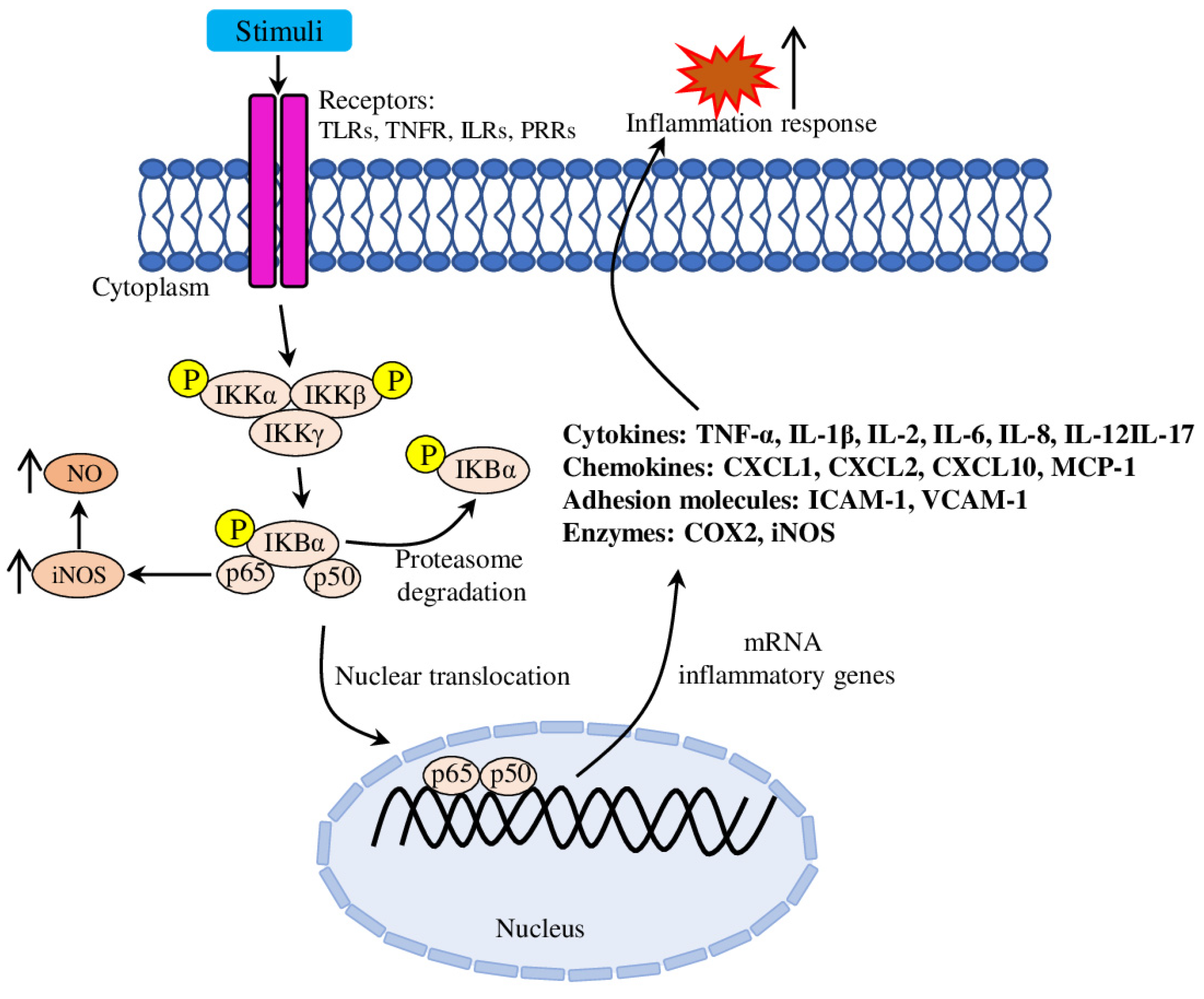
Inflammation. Lipid accumulation and the inflammatory immune response are closely linked to atherosclerosis initiation and development. Modified LDLs form aggregates in the blood vessels’ intima, causing EC (endothelial cell) dysfunction, inducing expression of adhesion molecules (such as VCAM-1 (Vascular Cell Adhesion Molecule), ICAM-1 (Intercellular Adhesion Molecule), P- and E-selectins), thus increasing leukocyte adhesiveness (Figure 2). ECs, surrounding smooth muscle cells and recruited leukocytes release various inflammatory mediators, which promote inflammation, stimulate immunity and facilitate further disease progression [16]. Because of the crucial role of inflammation, various inflammatory molecules (such as TNF-α (tumour necrosis factor-alpha), IL-1β (Interleukin), IL-6, IFN-γ (Interferon gamma), CRP (C-reactive protein) and others) serve as important therapeutic targets [17,18], biomarkers of inflammation and cardiovascular risk factors [19].
Furthermore, a rich experimental basis helped to establish a strong link between inflammation and immunity in atherosclerosis development. For example, some subtypes of T cells have an anti-atherogenic role (such as Treg (regulatory T cells)), others pro-atherogenic (such as TH1 (T helper 1)), while the role of many other subsets (CD8+T, γδ T, CD28nullT, TH17, TH22 and many others) are much less understood [20]. Similarly, targeting of innate and adaptive immunity responses was effective in modulating experimental atherosclerosis, suggesting its high potential in clinical use [21].
Oxidative damage. In addition to inflammation, the oxidative stress is considered as a key player in the initiation and progression of endothelial dysfunction and atherosclerosis [22]. Dysregulation of the antioxidant system and enhanced production of ROS (reactive oxygen species) promote endothelial injury, exacerbate inflammatory stress and accelerate lipid deposition in the pathogenesis of atherosclerosis [23]. ECs are a layer of cells bordering the bloodstream and vascular smooth muscle and regulating vascular permeability and secretion of bioactive substances, which are further associated with anticoagulant, haemostatic and antithrombotic functions. The endothelial monolayer separates the blood flow and the arterial intima and occurs as the site of plaque (atheroma) initiation during early atherogenesis [24]. Initial vascular remodelling of the innermost vascular layer is driven by lipid accumulation, on-site macrophage recruitment and uptake of mmLDL, which is further transformed into foam cells in the atherosclerotic plaque [25]. Further, release of inflammatory cytokines and pro-apoptotic regulators promotes a process of epithelial-to-mesenchymal transition and fibroblast-to-myofibroblast differentiation, thus reinforcing endothelial dysfunction and progression of the atherosclerotic lesion. In the later stages, the lipid core could necrotise and become covered by a thin and often calcified fibrous cap, which is prone to rupture and could lead to a thrombus and vessel occlusion [26].

Initiation of Atherosclerosis

The exposure of ECs to atherogenic risk factors interferes with normal production of endogenous vasodilators (specifically, the most important one, NO (nitric oxide)) [27]. The normal vascular blood flow is mechanotransducting, thus effecting EC function and homeostasis via shear and stretch stresses and wall hydrostatic pressure. Among these mechanical effects, shear stress is considered as the most important factor in atherosclerosis development through the activation of eNOS (endothelial NO synthase) via mechanosensitive transcription factor (such as KLF2 (Kruppel-like factor 2)) [28]. However, pathological stimuli reduced eNOS levels in ECs and promoted production of the vasoconstrictor ET-1 (endothelin-1), PAI-1 (plasminogen activator inhibitor-1) and transcription regulator HIF-1 (hypoxia-inducible factor 1), thus increasing platelet aggregation, endothelial procoagulant effect and smooth muscle cell proliferation and migration, which, in total, resulted in increased risk of thrombosis and atherosclerosis [29].
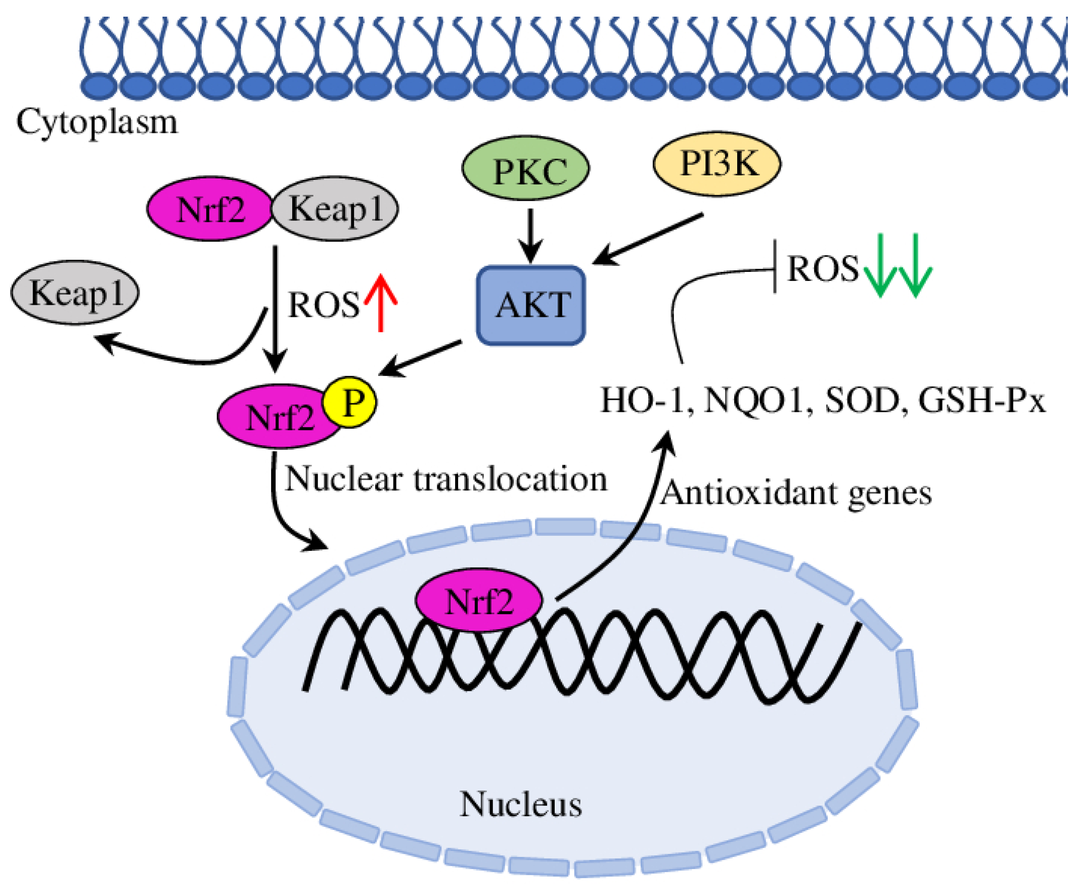
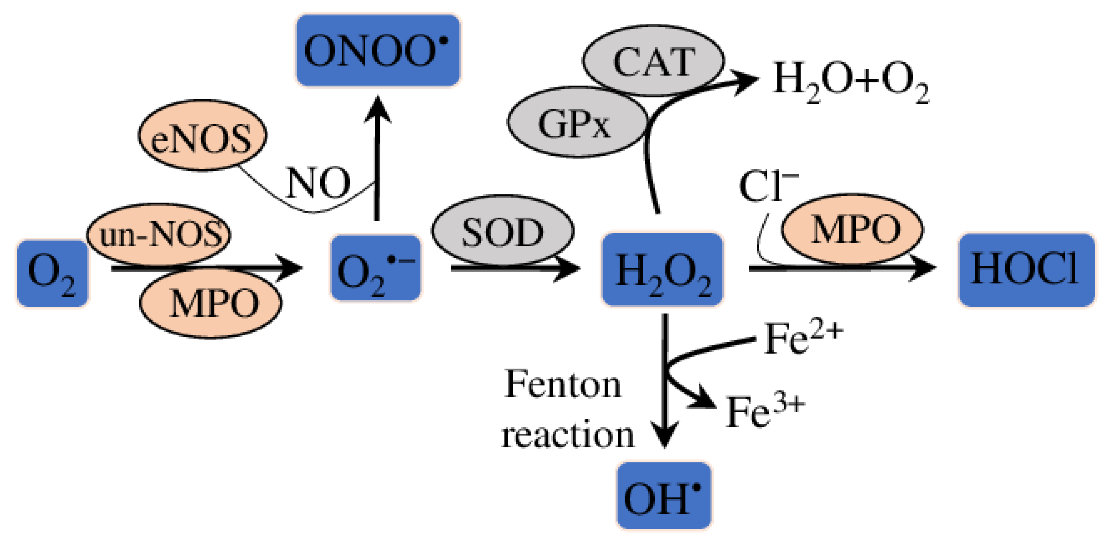
Nrf2 (Nuclear Factor (Erythroid-Derived 2)-Like 2) is a crucial transcription factor regulating a complex regulatory network, including also oxidative stress response in cells (Figure 3). Nrf2-mediated transcription regulates the expression of downstream antioxidant gene (such as SOD (superoxide dismutase), NQO1 (NAD(P)H: Quinone Oxidoreductase 1), HO-1 (Heme Oxygenase 1) or GSH-Px (glutathione peroxidase)), which remove ROS and protect cells from oxidative stress (Figure 4) [30]. Additionally, other proteins, such as PKC (protein kinase C), KEAP1 (Kelch-like ECH Associated Protein 1), AKT (protein kinase B) and PI3K (Phosphoinositide 3-Kinase Alpha), regulate Nrf2 transcription and induce phosphorylation [31]. Among others, the Nrf2/HO-1 pathway is the best studied for its antioxidative potential to protect ECs from oxidative stress-mediated injury, endothelial dysfunction and, subsequently, atherosclerosis [32,33,34].
Progression of atherosclerosis. As the disease progresses, atherosclerotic plaques continue to accumulate lipid particles and blood cells to give rise to foam cells. Also, metaplasia of smooth muscle cells can contribute to foam cell formation and atherosclerotic plaque growth [35]. Smooth muscle cells and macrophages are subjected to programmed cell death, which, in combination with impaired efferocytosis (dead cells clearance process), leads to the formation of the plaque necrotic core (Figure 1) [36]. Furthermore, dysregulation of calcium homeostasis and impaired clearance in the plaque activates a mineralisation process, similar to bone formation [37]. Such spotty calcification usually increases plaques instability, which promotes a tendency to rupture and provoke thrombosis, while extended calcification can stabilise plaques [38].
Advanced stages and complications of atherosclerosis. Eventually, the growing atherosclerotic plaque starts to invade the arterial lumen, impairing normal blood flow. Such a complication of coronary arterial perfusion can produce symptoms of angina pectoris and ischaemia, especially during physical effort, when the myocardial demand for oxygen increases [39]. “Vulnerable” atherosclerotic plaques are characterised by a large lipid core covered with a thin fibrous cap. Such plaques are more prone to rupture and trigger acute thrombosis of coronary arteries and cause myocardial infarction (Figure 1). “Stable” plaques, on the contrary, have thicker fibrous caps over limited lipid cores [40]. Plaque erosion is an alternative thrombotic mechanism for lesions with a distinct morphology (few inflammatory leukocytes and little lipids in the cores, and a rich extracellular matrix without a thin, friable fibrous cap). In comparison to plaque rupture, the molecular mechanisms of plaque erosion are much less studied [41].
Therefore, beneficial treatment for atherosclerosis usually provides anti-inflammatory, antioxidant and/or lipid-lowering properties. In addition to well-known drugs and substances specifically designed for atherosclerosis treatment [42], many drugs used to treat other diseases (such as diabetes, obesity and different types of cancer) have been repurposed for atherosclerosis. Additionally, many natural bioactive compounds delivered from different plants have proven anti-atherosclerotic activities [28,43].
Essential oils (EOs) are volatile compounds widely presented in different parts of plants (flowers, seeds, roots, leaves and stems) and globally used as food, perfumes, cosmetics and, most importantly, in traditional medicine to treat various diseases [44,45]. EOs are usually divided into three subgroups: acyclic (such as nerol, linalool, geraniol, citronellol, myrcene and others), monocyclic (such as carvone, menthol, α-terpineol, terpinolene, limonene, pulegone, phellandrene and others) and bicyclic (such as fenchone, thujone, camphor, α-pinene and others). Various technologies have been developed for effective extraction of EOs and treatment of hypertension, diabetes and obesity [46]. The health beneficial effects of EOs are mostly associated with terpenes, which are a broad group of natural substances classified into several groups based on the length of their carbon chains. Among all groups, mono-terpenes have the most significant biomedical activities in aromatic plants [47]. Specifically, the role of plant EOs and some of their isolated constituents, such as monoterpenoids, was demonstrated to improve cardiovascular function and reduce cardiovascular diseases risk factors [48,49]. Further in this review, we focus on the functional and mechanical anti-atherosclerotic role of EOs extracted from various plant sources. In particular, anti-inflammatory, anti-oxidative and lipid-lowering properties of EOs investigated in various in vitro and in vivo systems are discussed. Additionally, some relevant clinical trials evaluating the beneficial properties of EOs and known limitations on EO-based studies are highlighted.

2. Anti-Inflammatory Effect of EOs

Inflammation is a part of the innate immune defensive response of the organism against harmful stimuli, such as physical injury, chemicals, pathogens, viruses and others. Inflammation is involved in a variety of pathological processes; its regulation is complex and coordinated by many inflammatory mediators whose surplus production could lead to chronification of the inflammation process and initiate development of various diseases [50]. Inflammation is considered as a major factor implicated in the initiation and progression of atherosclerotic processes [51]. LPS (lipopolysaccharide) is the main component of Gram-negative bacteria, and thus is often used in in vitro experiments to imitate bacterial infection and promote secretion of pro-inflammatory cytokines, nitric oxide and eicosanoids [52]. Mechanically, LPS binds TLR4 (Toll-like receptor 4) and activates both MyoD88-dependent and MyoD88-independent signalling pathways. The MyoD88-dependent pathway activates NF-κB (nuclear factor kappaB) and MAPK/AP-1 (mitogen activated protein kinase/activator protein-1) signalling, and the MyoD88-independent pathway regulates IRF3 (interferon regulatory factor 3), while both MyoD88 pathways trigger pro-inflammatory cytokine transcription [53]. Further in this section, we discuss recent papers elucidating the molecular mechanisms of the anti-inflammatory activities of plant-derived EOs.

2.1. In Vitro Raw 264.7 Macrophage Test System

Research over the last decades attempted to identify and characterise EOs with the most pronounced anti-inflammatory effect and to decipher the underlying molecular mechanisms (Table 1). A desert medical and aromatic plant Artemisia judaica L., widely used in traditional medicine as an anti-bacterial and anti-fungal agent, was recently characterised by anti-inflammatory activity. As was shown in in vitro culture of LPS-treated macrophages, EO extracted from A. judaica inhibits NO production with low toxicity to macrophages. Bioactive components were mostly represented by mono-terpenes with piperitone, camphor and ethyl cinnamate as the main compounds [54]. Similarly, NO-inhibiting activity was determined in the EO derived from the Chinese traditional herb Waldheimia glabra (Decne), where α-bisabolol, valeranone and chamazulene were dominating compounds [55]. Anti-inflammatory activity also has been determined in EO derived from Hibiscus sabdariffa L., with n-Hexadecanoic acid as the most abundant volatile component. Treatment of LPS-stimulated macrophages with H. sabdariffa EO greatly inhibited the activation of JNK (C-Jun N-Terminal Kinase 1) and ERK1/2 (extracellular signal-regulated protein kinase 1 and 2) signalling pathways and decreases iNOS (inducible nitric oxide synthase) expression, thus reducing the production of NO and pro-inflammatory cytokines (IL-1, IL-6, TNF-α and COX-2) [56]. EO derived from the blossoms of Citrus aurantium L. has shown similar anti-inflammation activities: reduced levels of NO, IL-1β, IL-6, TNF-α and COX-2, and decreased expression levels of their genes. Additionally, C. aurantium EO suppressed JNK and p38 phosphorylation, inhibited NF-κB and IκB kinase activation, and IκBα phosphorylation and degradation in LPS-treated macrophages. Active components responsible for observed anti-inflammation activity were characterised as linalool, α-terpineol and (R)-limonene [57]. Similar properties were shown for related species L. luisieri (Rozeira) Rivas Mart. and L. pedunculata (Mill.) Cav. with 1,8-cineole and fenchone as prevalent compounds. L. luisieri EO effectively impaired nuclear translocation of NF-κB/p65, thus reducing iNOS levels. Interestingly, the expression of pro-IL-1β gene was increased, while the expression of COX-2 was not changed. On the contrary, L. pedunculata-derived EO was less effective in NO inhibition. Further investigation suggested that necrodane derivatives (trans-α-necrodol and trans-α-necrodyl acetate), the minor component of L. luisieri EO, are the most potent inhibitors of NO production [58].
Recently, anti-inflammatory properties have been characterised in traditional Chinese herbs Siegesbeckia pubescens (Makino) and Siegesbeckia orientalis L., whose EOs reduced NO production and IL-6 release in LPS-stimulated macrophages, respectively. The main components of the EO were β-caryophyllene oxide, trans-longipinocarveol and dehydrosaussurea lactone in S. pubescens and β-caryophyllene, spathulenol and β-caryophyllene oxide in S. orientalis [59]. Similar properties were found in EO derived from Thymus camphoratus (Hoffmanns. and Link) and T. carnosus (Boiss.), which inhibited NO production and reduced expression of iNOS and COX-2 genes. Borneol and camphene were defined as the active components of T. carnosus EO, while T. camphoratus EO was rich in borneol and 1,8-cineole [60]. EO from Santolina rosmarinifolia L., composed mainly of β-pinene, borneol, myrcene and limonene, decreased NO and pro-IL-1β release and expression of iNOS in LPS-stimulated macrophages [61]. Interestingly, EO derived from Cirsium japonicum (Maxim.) demonstrated anti-inflammatory activity (inhibiting NO production in LPS-treated macrophages) combined with lipid-lowering activity (decreased lipid accumulation in ox-LDL-induced (oxidised) macrophages). However, as the active components were not defined in this particular study, further research is required to elucidate the molecular mechanisms responsible for these activities [62].
Anti-inflammatory properties have been demonstrated in the popular spice oregano (Origanum vulgare L.) EO. The results obtained showed that treatment of LPS-treated macrophages with oregano oil effectively inhibited the expression and secretion of IL-1β, IL-6 and TNF-α. A suggested molecular mechanism of O. vulgare oil’s action was defined through the NADPH (nicotinamide adenine dinucleotide phosphate) oxidase pathway [63]. The active components of the O. vulgare oil were identified by other researchers as carvacrol, thymol, γ-terpinene and ρ-cymene [64,65].
Table 1. Plant essential oils with potential anti-atherosclerotic effects.
SpeciesMajor
Component
EffectExperiment SetupReferences
Artemisia
judaica
piperitone, camphor, ethyl cinnamateinhibits NO productionLPS treated Raw 264.7 macrophage[54]
Hibiscus sabdariffan-Hexadecanoic acidinhibits NO, IL-1, IL-6, TNF-α and COX-2 production; JNK and ERK1/2 pathways[56]
Waldheimia glabraα-bisabolol, valeranone and chamazuleneinhibits NO production[55]
Citrus
aurantium
linalool, α-terpineol and (R)-limoneneinhibits NO production; reduces IL-1β, IL-6, TNF-α and COX-2 protein levels and gene expression; inhibits NF-κB/65 and IκB activation[57]
Siegesbeckia pubescensβ-caryophyllene oxide, trans-longipinocarveol and dehydrosaussurea lactoneinhibits NO production[59]
Siegesbeckia orientalisβ-caryophyllene, spathulenol and β-caryophyllene oxideinhibits IL-6 release
Origanum
vulgare
carvacrol, thymol, γ-terpinene and ρ-cymeneinhibits IL-1β, IL-6 and TNF-α expression and secretionLPS treated Raw 264.7 macrophage[63]
[64,65]
Thymus
camphoratus
1,8-cineole and borneolinhibits NO production; reduces expression of iNOS and COX-2 genes[60]
Thymus
carnosus
borneol and camphene
Santolina
rosmarinifolia
β-pinene, borneol, myrcene and limonenedecreases NO and pro-IL-1β release and expression of iNOS[61]
Rosa rugosacitronellol and geraniolreduces production of IL-1β, IL-6 and TNF-α; increases CAT and SOD activities; inhibits MDA section; normalises iNOS, NO and ROS levels[66]
Cirsium
japonicum
-inhibit NO production[62]
decreases lipid accumulationox-LDL-induced Raw 264.7 macrophages
Lavandula
pedunculata
1,8-cineole and fenchoneinhibits NO productionLPS treated Raw 264.7 macrophage[58]
Lavandula luisieri1,8-cineole and fenchone;
trans-α-necrodol and trans-α-necrodyl acetate
reduces iNOS and increases pro-IL-1β expression; impairs nuclear translocation of NF-κB/p65
5-Methylene-2,3,4,4-
tetramethylcyclopent-2-
enone; 1,8-cineole
reduces TNF-α and inhibits CCL2 releaseLPS-stimulated THP-1 cells [67]
Thymbra
capitata
carvacrolreduces TNF-α release
Cymbopogon commutatusgeraniolinhibits IL-1β, IL-6 and TNF-α; down-regulates ICAM-1 mRNA level; suppresses IkBα phosphorylation; NF-κB/p65 activation and nuclear translocation; increases HO-1 expression ox-LDL-induced HUVECs[68]
Lavandula
angustifolia
linalool, linalyl acetate and terpinen-4-oldecreases IL-1β, IL-6, IL-8 and TNF-α mRNA and protein levelsLPS-treated THP-1 macrophages[69]
linalool, linalyl acetatesuppresses TNF-α-induced expression of E-selectin, P-selectin, VCAM-1, ICAM-1 and phosphorylated NF-κB/p65 in the nucleus translocation; inhibits TNF-α-induced increase in E-selectin mRNA levels in HUVECsTNF-α-stimulated bEnd.3 and HUVEC[70]
Rosmarinus
officinalis
1,8-cineole, camphor, limonene and α-pinenereduces total cholesterol, LDL, triglycerides, abdominal fat gain triton and coconut fat-induced rat model of dyslipidaemia[71]
α-pinene, camphor,
and 1,8cineole
increases IL-10 levelLPS treated THP-1 macrophage[72]
[73]
camphor, borneol, α-pinene, 1,8-cineolereduces glucose, triglyceride, total cholesterol; increases HDL; improves serum enzymes (AST, ALT and ALP); ameliorates lipid deposition, macrophage infiltration and stenosis of hepatic veinrats on HFD[74]
Zingiber
officinale
1,8-cineole, terpineol, linalool, borneol
Monarda
didyma
carvacrol, p-cymene and thymol decreases IL-6 and increases miR-146a expressionLPS treated U937 lymphoma cell line[75]
Platycladus orientalis-increases IL-10 and reduces IL-1β and TNF-α content in the serum; reduces the p65 and IκB phosphorylation levelmice and rats’ models[76]
Salvia
officinalis
1,8-Cineole, α-caryophyllene reduces body weight gain, liver and kidney weight; prevents lipid accumulation and focal necrosis in the liver; attenuates haemorrhage foci, reduces Bowman’s space and necrotic epithelial cells lining the tubules in the kidney; normalises antioxidant enzymes (SOD, CAT and Gpx) activity, the lipid profile (total cholesterol, triglyceride, LDL, VLDL and total lipids) and blood biochemical parameters (GGT, pancreatic lipase, AST, ALT, LDH, ALP and CPK; reduces the levels of TBARS and protein carbonyls; increases cholesterol and triglyceride faecal excretionHFD mice model[77]
Psidium
guineense
spathulenolin vitro antioxidant activities (MDA, ABTS and DPPH); reduces inflammation in carrageenan-induced paw oedema and pleurisy models in vitro test systems and mice[78]
Alpinia
zerumbet
β-pinene, α-cadinol and camphordecreases ICAM-1 and VCAM-1 expression, NF-κB/p65 phosphorylation and nuclear translocation, IKKα/β phosphorylation and increases IκBα protein levels; reduces lactate dehydrogenase release and caspase-3 activationLPS-induced HAEC and mice [79]
[80]
inhibits p65 subunit nuclear translocation, secretion of IL-8, TNF-α, ICAM-1 and VCAM-1high-glucose treated HUVEC[81]
[80]
Eucalyptus globulus1,8-cineolereduces levels of TNF-α, NO, IL-1α and IL-1β; reduces phosphorylation of NF-κB/p65 and p38, and increases of ERK1/2 and JNK1/2; down-regulates TREM-1, NLRP3 and MKP-1LPS-treated alveolar macrophage cell line MH-S[82]
-1,8-cineole (purified)reduces levels of IL-1 and IL-6; reduces phosphorylation of JNK1/2, and increases of NF-κB/p65; down-regulates MKP-1
-1,8-cineole (purified)reduces IL-6 and IL-8 secretion; normalises NO levels; improves iNOS expression and eNOS protein levels; decreases p65 phosphorylation and nuclear translocationLPS-induced HUVEC[83]
- 1,8-cineole and α-pinene (purified)decreases TNF-α, IL-1β, IL-6 and eNOS mRNA levels[84]
- 1,8-cineole (purified)inhibits IL-1β, IL-6 and IL-8, and promotes IL-10 release; reduces NF-κB/p65 and VCAM-1, and increases PPAR-γ expressionLPS-treated mice[85]
-bornyl acetatereduces E-selectin, ICAM-1 and VCAM-1, IL-1β and TNF-α expression; ameliorates reduction in cell viabilityox-LDL-induced HUVECs[86]
- β-Elemenereduces aortic root lesion sizes and necrotic core areas; increases the plaque stability score; increases expression of eNOS, CAT, Gpx and GSH; reduces levels of p22phox, IL-1β, TNF-α, INF-γ, MCP-1 and ICAM-1ApoE−/− mice[87]
-citronellalreduces atherosclerotic plaque size; alleviates arterial stenosis; reduces production of IL-1, IL-6, IACM-1 and VACM-1; normalises levels of NO, MDA and SOD activityrats on HFD[88]
-β-caryophylleneinhibits VCAM-1; reduces total cholesterol and triglycerides in serumTNF-α-stimulated HUVECs and mice[89]
-geraniolincreases Gpx, GST, mtSOD, GSH and NQO1; reduces TBARS; enhances Nrf2 and HO-1 expression; decreases IL-1β, IL-18 and TNF-α levels, NF-κB/65 activation and nuclear translocation doxorubicin-treated rats[90]
HUVEC—human umbilical vein endothelial cell; HAEC—human aortic endothelial cells; HFD—high fat diet.

2.2. Other In Vitro Test Systems

Anti-inflammatory properties have also been described in several lavender species. Thus, EO extracted from Lavandula angustifolia Mill., with linalool, linalyl acetate and terpinen-4-ol as the main bioactive components, inhibited the synthesis of four pro-inflammatory cytokines (IL-1β, IL-6, IL-8 and TNF-α) in LPS-stimulated THP-1 cells [69]. Another study has also supported the effect of L. luisieri EO on LPS-stimulated THP-1 cells by reducing TNF-α and inhibiting CCL2 (C-C motif chemokine ligand 2, or MCP-1 (Monocyte Chemoattractant Protein-1)) release. Accordingly, Thymbra capitata (L.) Hoffmanns. and Link) EO was effective only in reducing TNF-α release. The main components for L. luisieri and T. capitata were 5-Methylene-2,3,4,4-tetramethylcyclopent-2-enone, 1,8-cineole and carvacrol, respectively [67]. Similarly, the EO derived from Rosmarinus officinalis (Spenn.) and tested in a LPS-treated THP-1 macrophage model, increased levels of the anti-inflammatory IL-10, while the levels of pro-inflammatory cytokines IL-6 and TNF-α were not changed [72]. The active components of the R. officinalis EO were defined as α-pinene, camphor and 1,8-cineole in another research [73].
Interesting results were obtained with the application of Eucalyptus globulus (Labill.) EO on LPS-induced murine lung alveolar macrophage cells. The levels of pro-inflammatory mediators (TNF-α, NO, IL-1α and IL-1β) were reduced by E. globulus EO treatment, while the purified major component 1,8-cineole reduced only levels of IL-1 and IL-6. Also, the expression of TREM-1 (Triggering Receptor Expressed On Myeloid Cells 1) and PRR (pattern recognition receptor) NLRP3 (NLR Family Pyrin Domain Containing 3) of the inflammasome were downregulated by E. globulus EO treatment. Interestingly, the phosphorylation of NF-κB/p65 and p38 was attenuated, while ERK1/2 and JNK1/2 increased. On the contrary, the effect of 1,8-cineole on NF-κB and JNK1/2 was opposite, while both (E. globulus EO and pure 1,8-cineole) treatments downregulated MKP-1 (Dual Specificity Phosphatase 1) phosphatase, a negative regulator of MAPKs [82]. The EO extracted from the flowering parts of Monarda didyma L. have affected Toll-like receptor-4 signalling pathway by decreasing IL-6 and increasing miR-146a expression in LPS-treated U937 cells (a pro-monocytic, human histiocytic lymphoma cell line). Among 20 defined bioactive compounds, carvacrol, p-cymene and thymol were the most abundant [75].
Pure 1,8-cineole was demonstrated to ameliorate dysfunction of LPS-induced HUVEC (human umbilical vein endothelial cells). The defined molecular mechanism suggested that 1,8-cineole reduced IL-6 and IL-8 secretion and normalised NO levels. Also, iNOS expression and eNOS protein levels were improved, alongside decreased p65 phosphorylation and nuclear translocation [83]. Similarly, application of pure 1,8-cineole and α-pinene decreased TNF-α, IL-1β, IL-6 and eNOS mRNA levels in LPS-induced HUVEC cells [84].

2.3. EO Effect on Adhesion Molecules and Leukocytes Recruitment In Vitro and In Vivo

The recruitment of the leukocytes to the sight of inflammation is regulated by the NF-κB transcription factor and usually include such cell adhesion molecules as E-selectin, P-selectin, VCAM-1 (vascular cell adhesion molecule-1) and ICAM-1 (intercellular adhesion molecule-1) [91]. Recently, EO extracted from a dry fruit from Alpinia zerumbet (Pers.) B.L.Burtt and R.M.Sm. decreased expression of ICAM-1 and VCAM-1 and NF-κB p65 phosphorylation and nuclear translocation in both LPS-induced HAEC (human aortic endothelial cells) and in vivo mice models. Also, LPS-mediated increase in IKKα/β phosphorylation and reduction in IκBα protein levels were prevented in HAEC by A. zerumbet oil treatment. Interestingly, A. zerumbet oil demonstrated also an anti-apoptotic effect on LPS-induced HAEC cells, which was evaluated by reduced lactate dehydrogenase release and caspase-3 activation. While the active components of the A. zerumbet oil were not defined in this work [79], other research suggested that β-pinene, α-cadinol and camphor were the major components responsible for the anti-inflammatory effect [80]. These results were confirmed by another study, where HUVECs injury was achieved with high glucose treatment. A. zerumbet oil treatment inhibited cytokines (IL-8, TNF-α) and adhesion molecules (ICAM-1 and VCAM-1) secretion and p65 subunit nuclear translocation, inhibiting NF-κB signalling pathway activation and reducing HUVEC injury [81].
The effect of Lavandula angustifolia EO and its main components (pure solutions of linalyl acetate and linalool) on the expression of adhesion molecules was explored in TNF-α-stimulated murine brain endothelial bEnd.3 cells and HUVECs. Native L. angustifolia EO and pure linalyl acetate suppressed TNF-α-induced E-selectin, P-selectin, VCAM-1, ICAM-1 expression and phosphorylated-NF-κB p65 translocation to the nucleus in bEnd.3 cells, while linalool had no effect on P-selectin or phosphorylated-NF-κB p65 translocation. Furthermore, in HUVEC native L. angustifolia EO inhibited TNF-α-induced increase in E-selectin mRNA levels [70].
Geraniol, an active component of lemongrass (Cymbopogon commutatus (Steud.) Stapf.) EO, was characterised as an effective anti-inflammatory agent. In the experimental system of ox-LDL-induced HUVECs inflammation, application of geraniol inhibited production of pro-inflammatory cytokines (IL-1β, IL-6 and TNF-α), down-regulated ICAM-1 mRNA level, increased HO-1 expression and suppressed activation and nuclear translocation of NF-κB p65 and phosphorylation of IκBα [68]. In addition to native EOs, extracted from different plants, the effect on adhesion molecules and leukocyte recruitment was shown also for their purified components. For example, bornyl acetate reduced the expression of E-selectin, ICAM-1 and VCAM-1 in ox-LDL-treated HUVECs, thus inhibiting THP-1 monocyte attachment. Also, bornyl acetate reduced expression of the pro-inflammatory cytokines IL-1β and TNF-α, and ameliorated reduction in cell viability of ox-LDL-treated HUVECs [86].
Similarly, target delivery of 1,8-cineole with a self-microemulsifying system ameliorated LPS-induced endothelial injury in mice by inhibiting pro-inflammatory cytokines (IL-1β, IL-6 and IL-8) and promoting a simultaneous increase in anti-inflammatory IL-10 cytokine release. Also, the expression levels of NF-κB p65 and VCAM-1 were reduced, while levels of PPAR-γ (peroxisome proliferator-activated receptor γ) (NF-κB activation suppressor) was increased [85]. The anti-inflammatory effect of EO from Platycladus orientalis L. leaves was investigated on rat and mouse models in vivo. As a result of EO application, paw and granuloma swellings in rats were reduced, and intraperitoneal capillary permeability and auricular swelling in mice were inhibited. Also, the pathological damage of lung tissue was alleviated. Mechanically, EO treatment increased IL-10 and reduced IL-1β and TNF-α content in the serum and reduced the p65 and IκB phosphorylation level in the NF-κB pathway. However, the active components of the Platycladus orientalis EO were not defined in this research [76]. Finally, pure β-caryophyllene effectively inhibited VCAM-1 induction through the JAK2/STAT1/IRF-1 pathway in both in vitro and in vivo model systems (TNF-α-stimulated HUVECs and mice, respectively). In particular, β-caryophyllene is an agonist of CB2R (cannabinoid receptor 2), which, among many other genes, regulates IRF-1 and VCAM-1 expression. Additionally, β-caryophyllene prevented attachment of THP-1 macrophages to HUVECs and reduced total levels of cholesterol and triglycerides in serum [89].
In total, the results of multiple in vitro and in vivo studies suggested that the NF-κB pathway is the major target for EO-mediated anti-inflammatory effects (Table 1). The disruption of the normal NF-κB pathway functioning (subunits phosphorylation and nuclear translocation) is the primary event responsible for subsequent reduction in levels of pro-inflammatory cytokines, chemokines and adhesion molecules.

3. Anti-Oxidative Effect of EOs

ROS and alterations of antioxidant system are crucial triggers in initiation and progression of endothelial dysfunction and atherosclerosis. Recently, the anti-atherosclerotic activities of the β-elemene (the main component of the Curcuma Wenyujin (Y.H. Chen and C. Ling) EO), have been investigated in vivo in ApoE−/− mice. β-elemene effectively reduced the aortic root lesion sizes and necrotic core areas, increased the plaque stability score and increased eNOS expression. Also, the expression of antioxidant enzymes (CAT (catalase), Gpx (glutathione peroxidase) and GSH (Glutathione)) in the aorta was up-regulated by β-elemene, while the protein level of p22phox (a ROS-producing enzyme) was decreased. Furthermore, the levels of cytokines (IL-1β, TNF-α and INF-γ) and adhesion molecules (MCP-1 and ICAM-1) in the arteries were reduced by β-elemene treatment [87]. These data suggest that β-elemene combines both anti-inflammation and anti-oxidative stress activities, thus making it an effective anti-atherosclerotic natural agent.
Recently, combined anti-oxidative and anti-inflammatory properties were shown for EO extracted from Psidium guineense Sw. leaves, with spathulenol as a major bioactive component. Application of both (P. guineense EO and spathulenol) agents demonstrated high antioxidant activities in in vitro systems (MDA (Malondialdehyde), ABTS (ethylbenzothiazoline-6-sulfonic acid) and DPPH (2,2-diphenyl-1-picrylhydrazyl)), while anti-inflammatory properties were demonstrated with oral administration in vivo in mice carrageenan-induced paw oedema and pleurisy models [78]. Similarly, the EO of Rosa rugosa Thunb., where citronellol and geraniol were major bioactive components, demonstrated anti-inflammatory and anti-oxidative activities in LPS-treated RAW 264.7 macrophages, which were evaluated through reduced cytokines production (IL-1β, IL-6 and TNF-α), increased CAT and SOD activities, inhibited MDA section and normalised iNOS, NO and ROS levels [66].
Furthermore, diverse anti-atherosclerotic activities of pure citronellal were studied in vivo in a rat model with HFD-induced atherosclerosis. Citronellal administration effectively reduced the size of atherosclerotic plaque in carotid arteries and alleviated the stenosis of carotid and retinal arteries in atherosclerotic rats, while the levels of total cholesterol, triglycerides, LDL, HDL and blood glucose were not affected. The anti-inflammatory effect of citronellal was mediated via reduced production of cytokines (IL-1 and IL-6) and adhesion molecules (IACM-1 and VACM-1) in blood, and the anti-oxidative effect via normalised levels of NO, MDA and SOD activity [88]. Similarly, geraniol, isolated and purified from lemon grass, demonstrated beneficial cardioprotective properties in the doxorubicin-induced cardiac damage rat model. In particular, orally delivered geraniol alleviated the state of mitochondrial oxidative stress (via increased Gpx, GST, mtSOD, GSH and NQO1 (NAD(P)H: quinone oxidoreductase 1)), mitochondrial lipid peroxidation (via reduced TBARS (thiobarbituric acid reactive substance)) and enhanced Nrf2 and HO-1 expression. Furthermore, application of geraniol reduced doxorubicin-induced inflammation by decreasing levels of cytokines (IL-1β, IL-18 and TNF-α) and NF-κB p65 activation and nuclear translocation [90].
To sum up, in addition to anti-inflammatory activities, plant-derived EOs demonstrate anti-oxidative properties in both in vitro and in vivo conditions (Table 1). The Nrf2 pathway is the major ROS-activated system, responsible for up-regulated expression and production of various antioxidants (GST, SOD, CAT, NQO1 and Gpx), which reduces oxidative stress and damage to ECs and provides cardioprotective effect.

4. Lipid-Lowering Properties of EOs

Dyslipidaemia, an alteration of the lipid metabolism, usually manifested by increased LDL, cholesterol and triglycerides, and decreased HDL, is the major risk factor for the initiation and development of non-alcoholic fatty liver disease, diabetes, metabolic syndrome, atherosclerosis and other cardiovascular diseases [92]. Multiple research evidence suggested that plant-derived EOs could be beneficial for lipid metabolism and alleviate hypercholesterolaemia-associated damage. For example, EOs from rosemary and ginger (Zingiber officinale Rosc.) effectively reduced levels of glucose, triglyceride and total cholesterol, and increased levels of HDL and improved serum enzymes (AST (aspartate aminotransferase), ALT (alanine aminotransferase) and ALP (alkaline phosphatase)) in rats on HFD. Also, application of the mixture of oils ameliorates HFD-induced lipid deposition, macrophage infiltration and stenosis of the hepatic vein. The major bioactive components of the rosemary and ginger oils were camphor, borneol, α-pinene, 1,8-cineole and eucalyptol, terpineol, linalool, borneol, respectively [74]. Similarly, a wide beneficial effect of avocado (Persea americana Mill.) oil on metabolism was demonstrated using an NMR-based urinary metabolomics approach on HFD-fed rats. The main oil components (β-sitosterol, Δ5-avenasterol, stigmasterol and campesterol) greatly affect lipid, energy, amino acid and gut microbiota metabolism, thus ameliorating hypercholesterolaemia-induced damage [93].
In addition to the earlier described hypocholesterolaemic effect of rosemary oil, another recent study has confirmed and expand these results. Recent experiments on rats with Triton and coconut fat-induced dyslipidaemia demonstrated anti-dyslipidaemic and anti-atherogenic activities of both native rosemary oil and applied in the form of nanoemulsion. In particular, both oil forms reduced total cholesterol, LDL, triglycerides and abdominal fat gain. Also, EO-treated rats were characterised with absence of atherogenesis in the vascular endothelium. The oil composition was similar to results reported in other papers and included 1,8-cineole, camphor, limonene and α-pinene [71].
Recently, the effect of Salvia officinalis L. EO on HFD-fed mice was characterised in detail. The main active components were defined as eucalyptol and α-caryophyllene. Thus, application of S. officinalis oil reduced entire body weight gain and the weight of the liver and kidneys, prevented lipid accumulation and focal necrosis in the liver, attenuated haemorrhage foci and reduced Bowman’s space and necrotic epithelial cells lining the tubules in the kidney. Furthermore, the activity of antioxidant enzymes (SOD, CAT and Gpx), the lipid profile (total cholesterol, triglyceride, LDL, VLDL and total lipids) and blood biochemical parameters (GGT gamma-glutamyltranspeptidase), pancreatic lipase, AST, ALT, LDH (lactate dehydrogenase), ALP and CPK (creatine phosphokinase)) were normalised and the levels of TBARS and protein carbonyls were reduced. At the same time, the levels of faecal excretion of cholesterol and triglyceride were significantly increased, therefore suggesting S. officinalis EO as an effective anti-hyperlipidaemic and anti-oxidative agent, with additional hepatoprotective and nephroprotective effects [77].
In total, the beneficial effect of EO on lipid profile, glucose level and blood biochemical parameters was confirmed in several recent studies (Table 1). These data suggest that the described EOs are promising therapeutic agents for the treatment of atherosclerosis and other dyslipidaemia-, hypercholesterolaemia- and hyperglycaemia-associated diseases such as diabetes, metabolic syndrome and obesity. However, the molecular mechanism of these important therapeutic effects is not known and further research is required to elucidate it.

5. Clinical Trials Confirming Anti-Inflammatory and Anti-Oxidative Properties of EOs

Essential oils, similar to other plant extracts, have attracted wide interest as sources of natural medicinal products. They have been proven to exert diverse bioactive properties (anti-inflammatory, anti-apoptotic, antioxidant, antibacterial, antiviral and antifungal), even if the molecular mechanism of action has not been completely elucidated. To the best of our knowledge, there were no (or currently ongoing) clinical trials to evaluate the anti-atherosclerotic properties of EOs. However, there were several relevant trials investigating anti-inflammatory and anti-oxidative properties of EOs for treatment of oral diseases and oral care (gingivitis, dental plaque and gingival bleeding) and metabolic syndrome.
The pilot examination of the anti-bacterial properties of commercially available EO-based alcohol-free mouthwash to improve oral hygiene in seniors was not successful [94]. A later randomised controlled clinical trial investigated the anti-inflammatory potential of sage (Salvia officinalis) EO-containing mouthwash in elderly subjects. Similarly, the inflammatory signs and evaluated parameters (Sulcus Bleeding Index, Plaque Index, tooth staining, xerostomia and degree of stomatitis) were not significantly different from the placebo group [95]. However, recently, a randomized controlled trial comparing the efficiency of EO-containing mouth rinse and flossing efficacy on dental plaque accumulation and gingivitis in interproximal areas has reported some positive outcomes. While after two weeks of treatment there were no significant differences in case of interproximal gingival inflammation and bleeding, the EO mouth rinse was more effective in reducing interproximal dental plaque accumulation in comparison to dental flossing [96]. Similarly, application of Carica papaya L. leaf extract and an alcohol-based EO mouthwash (eucaliptol, menthol, methyl salicylate and thymol) effectively reduced gingival bleeding [97].
Further investigation of long-term application of EO-containing mouthwash (eucaliptol, menthol, methyl salicylate and thymol) confirmed reduction in gingivitis and plaque cases for both alcohol-based and alcohol-free EO-containing mouthwash in comparison to standard mechanical oral hygiene procedures [98]. Another EO-based mouthwash composition (menthol, thymol and eucalyptus) effectively reduced the gingival index, bleeding index and oral hygiene index as a stand-alone treatment and in combination with other substances (such as chlorhexidine, hydrogen peroxide and prebiotic) [99]. Most importantly, a recent randomised controlled clinical trial with diabetes patients reported a significant reduction in the plaque and gingival indices and lower levels of bacteria and volatile sulphur compounds after three months’ use of commercially available EO-based rinse [100].
Oxidative stress and inflammation significantly contribute to the pathogenesis of MetS (metabolic syndrome)—a cluster of interconnected serious disorders (such as dyslipidaemia, hyperglycaemia, central obesity and hypertension), which increase risk of diabetes, atherosclerosis and other cardiovascular diseases [101]. As it was defined in a recent randomized clinical trial, Cuminum cyminum L. EO effectively increased SOD, TAC (total antioxidant capacity) and decreased MDA in patients with MetS. C. cyminum EO, composed of γ-terpinene, β-pinene, cuminaldehyde and p-cymene, was received by patients in form of a soft gel for 8 weeks [102]. However, further investigation showed that important anthropometric and biochemical parameters (such as body weight, glycaemic status, insulin levels, lipid profile and others) were not affected by C. cyminum EO supplementation, except for DBP (diastolic blood pressure), which was significantly lower in the EO-treated group [103].
In sum, despite the fact that the anti-atherosclerotic properties of the EO-based supplementations were not directly confirmed in any type of clinical trials, existing evidence suggested their high potential. In particular, anti-inflammatory and anti-oxidative properties were demonstrated in oral diseases and metabolic syndrome. Likely, to fully reveal anti-atherosclerotic potential and affect biochemical blood parameters, a longer intervention time and different EO doses are required. However, while currently EOs are counted as herbal-based nutraceuticals and their safety, quality and efficacy are less strictly regulated than standard pharmaceuticals, many poor-quality products may be present on the market; therefore, these products might be ineffective.

6. Conclusions

Many studies highlight the important role of plant-derived EOs and some of their isolated constituents in the improvement of cardiovascular diseases. Some EOs have anti-inflammatory, anti-oxidative and/or lipid-lowering properties, which are able to promote and protect the cardiovascular system, and regress the atherosclerosis process. EOs and secondary metabolites derived from leaves, flowers, seeds, fruits and other plant organs demonstrate an anti-inflammatory effect on the NF-κB signalling pathway and can ameliorate oxidative stress in the Nrf2/HO-1 signalling pathway. Additionally, some EOs showed the ability to normalise blood lipid profile, glucose and biochemical blood parameters, thus suggesting promising anti-obesity and anti-diabetic properties. Thus, plant-derived EOs favourably interact with three main atherosclerosis risk factors and can contribute to the prevention and treatment of atherosclerosis.
However, the main EO compounds responsible for anti-atherosclerosis effect are not well understood. Natural EOs are multicomponent system, and all components could be successfully identified and purified. However, the biological effect of the native EO application often differs significantly from the application of one purified component, thus suggesting that minor components are crucial for EO activities. Currently, the bioactive properties have been demonstrated only for some of the most abundant components (such as carvacrol, eucalyptol (1,8-cineole), geraniol, thymol and some others), while the majority of EO components are still uncharacterised. Also, taking into account the extremely low abundance of these components, it would require significant efforts to purify them from natural plant sources. Another point to consider is the availability of plant materials (flowers, for example), which could be especially pressing in case of endemic or rare plant species.
Another limitation for EO application is the absence of data regarding their dosage, toxicity, kinetics and duration of use. The majority of recent research was conducted in vitro, on various macrophages, ECs and cancer cell lines, while experiments in vivo are scarce. Therefore, nontoxic concentrations defined for a specific EO or a composition of EOs on a particular cell line cannot be extrapolated to an in vivo model and have to be defined again. Also, EOs are volatile compounds which are expected to enter the organisms through the lungs. Thus, theoretically, EOs should be delivered through inhalation therapy via an ultrasonic nebuliser. Another method of EO delivery is via skin penetration. However, in both these delivery routes (inhalation and skin penetration), the control of the daily dose is too complicated, which leaves the oral administration method as the most reliable.
In the recent clinical trials evaluating anti-inflammatory properties of EO-based oral care products, some promising outcomes were achieved with external application of the active agent (mouth wash). Very limited in vivo data suggested the efficiency of nanoemulsion or soft gel delivery systems to obtain a therapeutic effect on the organism-wide level. However, the detailed investigation of EO-mediated anti-atherosclerotic effects in in vivo model systems and in patients is a substantial but necessary challenge for future studies.

Author Contributions

S.A.D. and A.N.O. conceptualized the manuscript; S.A.D. wrote the manuscript text; V.N.S., A.A.M., V.A.K. and A.N.O. reviewed the text; V.N.S. and A.A.M. methodology; A.A.M. and V.A.K. formal analysis; V.N.S. and A.N.O. obtained funding and supervised. All authors have read and agreed to the published version of the manuscript.

Funding

The work was supported by the Ministry of Science and Higher Education of the Russian Federation (Project # FGFU-2022-00008).

Institutional Review Board Statement

Not applicable.

Informed Consent Statement

Not applicable.

Data Availability Statement

Not applicable.

Conflicts of Interest

The authors declare no conflict of interest. The funder had no role in the design of the study; in the collection, analyses, or interpretation of data; in the writing of the manuscript, or in the decision to publish the results.

Sample Availability

Not applicable.

References

  1. WHO CVDs Fact Sheets, R Cardiovascular Diseases (CVDs). Available online: https://www.who.int/health-topics/cardiovascular-diseases#tab=tab_1 (accessed on 11 June 2023).
  2. Fan, J.; Watanabe, T. Atherosclerosis: Known and Unknown. Pathol. Int. 2022, 72, 151–160. [Google Scholar] [CrossRef] [PubMed]
  3. Arnesen, E.K.; Retterstøl, K. Secular Trends in Serum Lipid Profiles in Young Adults in Norway, 2001–2019. Atheroscler. Plus 2022, 48, 60–67. [Google Scholar] [CrossRef] [PubMed]
  4. Georgoulis, M.; Chrysohoou, C.; Georgousopoulou, E.; Damigou, E.; Skoumas, I.; Pitsavos, C.; Panagiotakos, D. Long-Term Prognostic Value of LDL-C, HDL-C, Lp(a) and TG Levels on Cardiovascular Disease Incidence, by Body Weight Status, Dietary Habits and Lipid-Lowering Treatment: The ATTICA Epidemiological Cohort Study (2002–2012). Lipids Health Dis. 2022, 21, 141. [Google Scholar] [CrossRef] [PubMed]
  5. Blom, D.J.; Almahmeed, W.; Al-Rasadi, K.; Azuri, J.; Daclin, V.; Kayikcioglu, M.; Mercier, F.; Ruiz, A.J.; Santos, R.D. Low-Density Lipoprotein Cholesterol Goal Achievement in Patients with Familial Hypercholesterolemia in Countries Outside Western Europe: The International ChoLesterol Management Practice Study. J. Clin. Lipidol. 2019, 13, 594–600. [Google Scholar] [CrossRef] [PubMed] [Green Version]
  6. Hopewell, J.C.; Malik, R.; Valdés-Márquez, E.; Worrall, B.B.; Collins, R. METASTROKE Collaboration of the ISGC Differential Effects of PCSK9 Variants on Risk of Coronary Disease and Ischaemic Stroke. Eur. Heart J. 2018, 39, 354–359. [Google Scholar] [CrossRef] [Green Version]
  7. Mezentsev, A.; Bezsonov, E.; Kashirskikh, D.; Baig, M.S.; Eid, A.H.; Orekhov, A. Proatherogenic Sialidases and Desialylated Lipoproteins: 35 Years of Research and Current State from Bench to Bedside. Biomedicines 2021, 9, 600. [Google Scholar] [CrossRef]
  8. Gisterå, A.; Hansson, G.K. The Immunology of Atherosclerosis. Nat. Rev. Nephrol. 2017, 13, 368–380. [Google Scholar] [CrossRef]
  9. Tardif, J.-C.; McMurray, J.J.; Klug, E.; Small, R.; Schumi, J.; Choi, J.; Cooper, J.; Scott, R.; Lewis, E.F.; L’Allier, P.L.; et al. Effects of Succinobucol (AGI-1067) after an Acute Coronary Syndrome: A Randomised, Double-Blind, Placebo-Controlled Trial. Lancet 2008, 371, 1761–1768. [Google Scholar] [CrossRef]
  10. Toledo-Ibelles, P.; Mas-Oliva, J. Antioxidants in the Fight Against Atherosclerosis: Is This a Dead End? Curr. Atheroscler. Rep. 2018, 20, 36. [Google Scholar] [CrossRef] [Green Version]
  11. Poznyak, A.V.; Nikiforov, N.G.; Markin, A.M.; Kashirskikh, D.A.; Myasoedova, V.A.; Gerasimova, E.V.; Orekhov, A.N. Overview of OxLDL and Its Impact on Cardiovascular Health: Focus on Atherosclerosis. Front. Pharmacol. 2021, 11, 613780. [Google Scholar] [CrossRef]
  12. Ference, B.A.; Ginsberg, H.N.; Graham, I.; Ray, K.K.; Packard, C.J.; Bruckert, E.; Hegele, R.A.; Krauss, R.M.; Raal, F.J.; Schunkert, H.; et al. Low-Density Lipoproteins Cause Atherosclerotic Cardiovascular Disease. 1. Evidence from Genetic, Epidemiologic, and Clinical Studies. A Consensus Statement from the European Atherosclerosis Society Consensus Panel. Eur. Heart J. 2017, 38, 2459–2472. [Google Scholar] [CrossRef] [Green Version]
  13. Musunuru, K.; Kathiresan, S. Surprises From Genetic Analyses of Lipid Risk Factors for Atherosclerosis. Circ. Res. 2016, 118, 579–585. [Google Scholar] [CrossRef] [PubMed]
  14. Burgess, S.; Ference, B.A.; Staley, J.R.; Freitag, D.F.; Mason, A.M.; Nielsen, S.F.; Willeit, P.; Young, R.; Surendran, P.; Karthikeyan, S.; et al. Association of LPA Variants With Risk of Coronary Disease and the Implications for Lipoprotein(a)-Lowering Therapies: A Mendelian Randomization Analysis. JAMA Cardiol. 2018, 3, 619. [Google Scholar] [CrossRef] [PubMed] [Green Version]
  15. Nordestgaard, B.G. Triglyceride-Rich Lipoproteins and Atherosclerotic Cardiovascular Disease: New Insights From Epidemiology, Genetics, and Biology. Circ. Res. 2016, 118, 547–563. [Google Scholar] [CrossRef]
  16. Libby, P.; Loscalzo, J.; Ridker, P.M.; Farkouh, M.E.; Hsue, P.Y.; Fuster, V.; Hasan, A.A.; Amar, S. Inflammation, Immunity, and Infection in Atherothrombosis. J. Am. Coll. Cardiol. 2018, 72, 2071–2081. [Google Scholar] [CrossRef]
  17. Biros, E.; Reznik, J.E.; Moran, C.S. Role of Inflammatory Cytokines in Genesis and Treatment of Atherosclerosis. Trends Cardiovasc. Med. 2022, 32, 138–142. [Google Scholar] [CrossRef]
  18. Poznyak, A.V.; Bharadwaj, D.; Prasad, G.; Grechko, A.V.; Sazonova, M.A.; Orekhov, A.N. Anti-Inflammatory Therapy for Atherosclerosis: Focusing on Cytokines. IJMS 2021, 22, 7061. [Google Scholar] [CrossRef] [PubMed]
  19. Castro, A.R.; Silva, S.O.; Soares, S.C. The Use of High Sensitivity C-Reactive Protein in Cardiovascular Disease Detection. J. Pharm. Pharm. Sci. 2018, 21, 496–503. [Google Scholar] [CrossRef] [Green Version]
  20. Saigusa, R.; Winkels, H.; Ley, K. T Cell Subsets and Functions in Atherosclerosis. Nat. Rev. Cardiol. 2020, 17, 387–401. [Google Scholar] [CrossRef]
  21. Nus, M.; Mallat, Z. Immune-Mediated Mechanisms of Atherosclerosis and Implications for the Clinic. Expert Rev. Clin. Immunol. 2016, 12, 1217–1237. [Google Scholar] [CrossRef] [Green Version]
  22. Batty, M.; Bennett, M.R.; Yu, E. The Role of Oxidative Stress in Atherosclerosis. Cells 2022, 11, 3843. [Google Scholar] [CrossRef] [PubMed]
  23. Petrucci, G.; Rizzi, A.; Hatem, D.; Tosti, G.; Rocca, B.; Pitocco, D. Role of Oxidative Stress in the Pathogenesis of Atherothrombotic Diseases. Antioxidants 2022, 11, 1408. [Google Scholar] [CrossRef] [PubMed]
  24. Xu, S.; Ilyas, I.; Little, P.J.; Li, H.; Kamato, D.; Zheng, X.; Luo, S.; Li, Z.; Liu, P.; Han, J.; et al. Endothelial Dysfunction in Atherosclerotic Cardiovascular Diseases and Beyond: From Mechanism to Pharmacotherapies. Pharmacol. Rev. 2021, 73, 924–967. [Google Scholar] [CrossRef] [PubMed]
  25. Glanz, V.; Bezsonov, E.E.; Soldatov, V.; Orekhov, A.N. Thirty-Five-Year History of Desialylated Lipoproteins Discovered by Vladimir Tertov. Biomedicines 2022, 10, 1174. [Google Scholar] [CrossRef]
  26. Canet-Soulas, E.; Bessueille, L.; Mechtouff, L.; Magne, D. The Elusive Origin of Atherosclerotic Plaque Calcification. Front. Cell Dev. Biol. 2021, 9, 622736. [Google Scholar] [CrossRef]
  27. Gliozzi, M.; Scicchitano, M.; Bosco, F.; Musolino, V.; Carresi, C.; Scarano, F.; Maiuolo, J.; Nucera, S.; Maretta, A.; Paone, S.; et al. Modulation of Nitric Oxide Synthases by Oxidized LDLs: Role in Vascular Inflammation and Atherosclerosis Development. Int. J. Mol. Sci. 2019, 20, 3294. [Google Scholar] [CrossRef] [Green Version]
  28. Dabravolski, S.A.; Sukhorukov, V.N.; Kalmykov, V.A.; Grechko, A.V.; Shakhpazyan, N.K.; Orekhov, A.N. The Role of KLF2 in the Regulation of Atherosclerosis Development and Potential Use of KLF2-Targeted Therapy. Biomedicines 2022, 10, 254. [Google Scholar] [CrossRef]
  29. Loscalzo, J. Nitric Oxide Signaling and Atherothrombosis Redux: Evidence From Experiments of Nature and Implications for Therapy. Circulation 2018, 137, 233–236. [Google Scholar] [CrossRef]
  30. Alonso-Piñeiro, J.A.; Gonzalez-Rovira, A.; Sánchez-Gomar, I.; Moreno, J.A.; Durán-Ruiz, M.C. Nrf2 and Heme Oxygenase-1 Involvement in Atherosclerosis Related Oxidative Stress. Antioxidants 2021, 10, 1463. [Google Scholar] [CrossRef]
  31. Cui, W.; Leng, B.; Wang, G. Klotho Protein Inhibits H2O2-Induced Oxidative Injury in Endothelial Cells via Regulation of PI3K/AKT/Nrf2/HO-1 Pathways. Can. J. Physiol. Pharmacol. 2019, 97, 370–376. [Google Scholar] [CrossRef]
  32. Kishimoto, Y.; Kondo, K.; Momiyama, Y. The Protective Role of Heme Oxygenase-1 in Atherosclerotic Diseases. IJMS 2019, 20, 3628. [Google Scholar] [CrossRef] [PubMed] [Green Version]
  33. Fiorelli, S.; Porro, B.; Cosentino, N.; Di Minno, A.; Manega, C.M.; Fabbiocchi, F.; Niccoli, G.; Fracassi, F.; Barbieri, S.; Marenzi, G.; et al. Activation of Nrf2/HO-1 Pathway and Human Atherosclerotic Plaque Vulnerability: An In Vitro and In Vivo Study. Cells 2019, 8, 356. [Google Scholar] [CrossRef] [PubMed] [Green Version]
  34. Zhang, J.; Cai, W.; Fan, Z.; Yang, C.; Wang, W.; Xiong, M.; Ma, C.; Yang, J. MicroRNA-24 Inhibits the Oxidative Stress Induced by Vascular Injury by Activating the Nrf2/Ho-1 Signaling Pathway. Atherosclerosis 2019, 290, 9–18. [Google Scholar] [CrossRef] [PubMed]
  35. Basatemur, G.L.; Jørgensen, H.F.; Clarke, M.C.H.; Bennett, M.R.; Mallat, Z. Vascular Smooth Muscle Cells in Atherosclerosis. Nat. Rev. Cardiol. 2019, 16, 727–744. [Google Scholar] [CrossRef]
  36. Yurdagul, A.; Doran, A.C.; Cai, B.; Fredman, G.; Tabas, I.A. Mechanisms and Consequences of Defective Efferocytosis in Atherosclerosis. Front. Cardiovasc. Med. 2018, 4, 86. [Google Scholar] [CrossRef] [Green Version]
  37. Leszczynska, A.; O’Doherty, A.; Farrell, E.; Pindjakova, J.; O’Brien, F.J.; O’Brien, T.; Barry, F.; Murphy, M. Differentiation of Vascular Stem Cells Contributes to Ectopic Calcification of Atherosclerotic Plaque. Stem Cells 2016, 34, 913–923. [Google Scholar] [CrossRef] [Green Version]
  38. Hutcheson, J.D.; Goettsch, C.; Bertazzo, S.; Maldonado, N.; Ruiz, J.L.; Goh, W.; Yabusaki, K.; Faits, T.; Bouten, C.; Franck, G.; et al. Genesis and Growth of Extracellular-Vesicle-Derived Microcalcification in Atherosclerotic Plaques. Nat. Mater. 2016, 15, 335–343. [Google Scholar] [CrossRef] [Green Version]
  39. Russo, M.; Fracassi, F.; Kurihara, O.; Kim, H.O.; Thondapu, V.; Araki, M.; Shinohara, H.; Sugiyama, T.; Yamamoto, E.; Lee, H.; et al. Healed Plaques in Patients With Stable Angina Pectoris. ATVB 2020, 40, 1587–1597. [Google Scholar] [CrossRef]
  40. Pasterkamp, G.; den Ruijter, H.M.; Libby, P. Temporal Shifts in Clinical Presentation and Underlying Mechanisms of Atherosclerotic Disease. Nat. Rev. Cardiol. 2017, 14, 21–29. [Google Scholar] [CrossRef] [Green Version]
  41. Franck, G. Role of Mechanical Stress and Neutrophils in the Pathogenesis of Plaque Erosion. Atherosclerosis 2021, 318, 60–69. [Google Scholar] [CrossRef]
  42. Libby, P. The Changing Landscape of Atherosclerosis. Nature 2021, 592, 524–533. [Google Scholar] [CrossRef]
  43. Sharif, H.; Akash, M.S.H.; Rehman, K.; Irshad, K.; Imran, I. Pathophysiology of Atherosclerosis: Association of Risk Factors and Treatment Strategies Using Plant-Based Bioactive Compounds. J. Food Biochem. 2020, 44, e13449. [Google Scholar] [CrossRef] [PubMed]
  44. Lunz, K.; Stappen, I. Back to the Roots-An Overview of the Chemical Composition and Bioactivity of Selected Root-Essential Oils. Molecules 2021, 26, 3155. [Google Scholar] [CrossRef]
  45. Edris, A.E. Pharmaceutical and Therapeutic Potentials of Essential Oils and Their Individual Volatile Constituents: A Review. Phytother. Res. 2007, 21, 308–323. [Google Scholar] [CrossRef] [PubMed]
  46. Ferrentino, G.; Morozova, K.; Horn, C.; Scampicchio, M. Extraction of Essential Oils from Medicinal Plants and Their Utilization as Food Antioxidants. CPD 2020, 26, 519–541. [Google Scholar] [CrossRef] [PubMed]
  47. Koyama, S.; Heinbockel, T. The Effects of Essential Oils and Terpenes in Relation to Their Routes of Intake and Application. IJMS 2020, 21, 1558. [Google Scholar] [CrossRef] [Green Version]
  48. Silva, E.A.P.; Santos, D.M.; de Carvalho, F.O.; Menezes, I.A.C.; Barreto, A.S.; Souza, D.S.; Quintans-Júnior, L.J.; Santos, M.R.V. Monoterpenes and Their Derivatives as Agents for Cardiovascular Disease Management: A Systematic Review and Meta-Analysis. Phytomedicine 2021, 88, 153451. [Google Scholar] [CrossRef]
  49. Saljoughian, S.; Roohinejad, S.; Bekhit, A.E.-D.A.; Greiner, R.; Omidizadeh, A.; Nikmaram, N.; Mousavi Khaneghah, A. The Effects of Food Essential Oils on Cardiovascular Diseases: A Review. Crit. Rev. Food Sci. Nutr. 2018, 58, 1688–1705. [Google Scholar] [CrossRef]
  50. Roe, K. An Inflammation Classification System Using Cytokine Parameters. Scand. J. Immunol. 2021, 93, e12970. [Google Scholar] [CrossRef]
  51. García, R.D.; Asensio, J.A.; Perdicaro, D.J.; de los Ángeles Peral, M. The Role of Inflammation as a Preponderant Risk Factor in Cardiovascular Diseases. CVP 2022, 20, 244–259. [Google Scholar] [CrossRef]
  52. Poltorak, A.; He, X.; Smirnova, I.; Liu, M.-Y.; Huffel, C.V.; Du, X.; Birdwell, D.; Alejos, E.; Silva, M.; Galanos, C.; et al. Defective LPS Signaling in C3H/HeJ and C57BL/10ScCr Mice: Mutations in Tlr4 Gene. Science 1998, 282, 2085–2088. [Google Scholar] [CrossRef] [PubMed] [Green Version]
  53. Kawasaki, T.; Kawai, T. Toll-Like Receptor Signaling Pathways. Front. Immunol. 2014, 5, 461. [Google Scholar] [CrossRef] [PubMed] [Green Version]
  54. Abu-Darwish, M.S.; Cabral, C.; Gonçalves, M.J.; Cavaleiro, C.; Cruz, M.T.; Zulfiqar, A.; Khan, I.A.; Efferth, T.; Salgueiro, L. Chemical Composition and Biological Activities of Artemisia judaica Essential Oil from Southern Desert of Jordan. J. Ethnopharmacol. 2016, 191, 161–168. [Google Scholar] [CrossRef]
  55. De, J.; Lu, Y.; Ling, L.; Peng, N.; Zhong, Y. Essential Oil Composition and Bioactivities of Waldheimia glabra (Asteraceae) from Qinghai-Tibet Plateau. Molecules 2017, 22, 460. [Google Scholar] [CrossRef] [Green Version]
  56. Shen, C.-Y.; Zhang, T.-T.; Zhang, W.-L.; Jiang, J.-G. Anti-Inflammatory Activities of Essential Oil Isolated from the Calyx of Hibiscus sabdariffa L. Food Funct. 2016, 7, 4451–4459. [Google Scholar] [CrossRef] [PubMed]
  57. Shen, C.-Y.; Jiang, J.-G.; Zhu, W.; Ou-Yang, Q. Anti-Inflammatory Effect of Essential Oil from Citrus aurantium L. Var. Amara Engl. J. Agric. Food Chem. 2017, 65, 8586–8594. [Google Scholar] [CrossRef]
  58. Zuzarte, M.; Sousa, C.; Cavaleiro, C.; Cruz, M.T.; Salgueiro, L. The Anti-Inflammatory Response of Lavandula luisieri and Lavandula pedunculata Essential Oils. Plants 2022, 11, 370. [Google Scholar] [CrossRef]
  59. Gao, X.; Wei, J.; Hong, L.; Fan, S.; Hu, G.; Jia, J. Comparative Analysis of Chemical Composition, Anti-Inflammatory Activity and Antitumor Activity in Essential Oils from Siegesbeckia orientalis, S. Glabrescens and S. Pubescens with an ITS Sequence Analysis. Molecules 2018, 23, 2185. [Google Scholar] [CrossRef] [Green Version]
  60. Zuzarte, M.; Alves-Silva, J.M.; Alves, M.; Cavaleiro, C.; Salgueiro, L.; Cruz, M.T. New Insights on the Anti-Inflammatory Potential and Safety Profile of Thymus carnosus and Thymus camphoratus Essential Oils and Their Main Compounds. J. Ethnopharmacol. 2018, 225, 10–17. [Google Scholar] [CrossRef]
  61. Alves-Silva, J.M.; Gonçalves, M.J.; Silva, A.; Cavaleiro, C.; Cruz, M.T.; Salgueiro, L. Chemical Profile, Anti-Microbial and Anti-Inflammaging Activities of Santolina rosmarinifolia L. Essential Oil from Portugal. Antibiotics 2023, 12, 179. [Google Scholar] [CrossRef]
  62. Ma, Q.; Jiang, J.-G.; Yuan, X.; Qiu, K.; Zhu, W. Comparative Antitumor and Anti-Inflammatory Effects of Flavonoids, Saponins, Polysaccharides, Essential Oil, Coumarin and Alkaloids from Cirsium japonicum DC. Food Chem. Toxicol. 2019, 125, 422–429. [Google Scholar] [CrossRef]
  63. Cheng, C.; Zou, Y.; Peng, J. Oregano Essential Oil Attenuates RAW264.7 Cells from Lipopolysaccharide-Induced Inflammatory Response through Regulating NADPH Oxidase Activation-Driven Oxidative Stress. Molecules 2018, 23, 1857. [Google Scholar] [CrossRef] [PubMed] [Green Version]
  64. Foti, M.C.; Ingold, K.U. Mechanism of Inhibition of Lipid Peroxidation by γ-Terpinene, an Unusual and Potentially Useful Hydrocarbon Antioxidant. J. Agric. Food Chem. 2003, 51, 2758–2765. [Google Scholar] [CrossRef] [PubMed] [Green Version]
  65. Bakkali, F.; Averbeck, S.; Averbeck, D.; Idaomar, M. Biological Effects of Essential Oils—A Review. Food Chem. Toxicol. 2008, 46, 446–475. [Google Scholar] [CrossRef]
  66. Raka, R.N.; Zhiqian, D.; Yue, Y.; Luchang, Q.; Suyeon, P.; Junsong, X.; Hua, W. Pingyin Rose Essential Oil Alleviates LPS-Induced Inflammation in RAW 264.7 Cells via the NF-ΚB Pathway: An Integrated in Vitro and Network Pharmacology Analysis. BMC Complement. Med. Ther. 2022, 22, 272. [Google Scholar] [CrossRef] [PubMed]
  67. Miguel, M.G.; da Silva, C.I.; Farah, L.; Castro Braga, F.; Figueiredo, A.C. Effect of Essential Oils on the Release of TNF-α and CCL2 by LPS-Stimulated THP-1 Cells. Plants 2020, 10, 50. [Google Scholar] [CrossRef] [PubMed]
  68. Ammar, R.B.; Mohamed, M.E.; Alfwuaires, M.; Abdulaziz Alamer, S.; Bani Ismail, M.; Veeraraghavan, V.P.; Sekar, A.K.; Ksouri, R.; Rajendran, P. Anti-Inflammatory Activity of Geraniol Isolated from Lemon Grass on Ox-LDL-Stimulated Endothelial Cells by Upregulation of Heme Oxygenase-1 via PI3K/Akt and Nrf-2 Signaling Pathways. Nutrients 2022, 14, 4817. [Google Scholar] [CrossRef] [PubMed]
  69. Pandur, E.; Balatinácz, A.; Micalizzi, G.; Mondello, L.; Horváth, A.; Sipos, K.; Horváth, G. Anti-Inflammatory Effect of Lavender (Lavandula angustifolia Mill.) Essential Oil Prepared during Different Plant Phenophases on THP-1 Macrophages. BMC Complement. Med. Ther. 2021, 21, 287. [Google Scholar] [CrossRef]
  70. Aoe, M.; Ueno-Iio, T.; Shibakura, M.; Shinohata, R.; Usui, S.; Arao, Y.; Ikeda, S.; Miyahara, N.; Tanimoto, M.; Kataoka, M. Lavender Essential Oil and Its Main Constituents Inhibit the Expression of TNF-α-Induced Cell Adhesion Molecules in Endothelial Cells. Acta Med. Okayama 2017, 71, 493–503. [Google Scholar]
  71. Santos Rodrigues, A.P.; Faria e Souza, B.S.; Alves Barros, A.S.; de Oliveira Carvalho, H.; Lobato Duarte, J.; Letícia Elizandra Boettger, M.; Barbosa, R.; Maciel Ferreira, A.; Maciel Ferreira, I.; Fernandes, C.P.; et al. The Effects of Rosmarinus officinalis L. Essential Oil and Its Nanoemulsion on Dyslipidemic Wistar Rats. J. Appl. Biomed. 2020, 18, 126–135. [Google Scholar] [CrossRef]
  72. Lorenzo-Leal, A.C.; Palou, E.; López-Malo, A.; Bach, H. Antimicrobial, Cytotoxic, and Anti-Inflammatory Activities of Pimenta dioica and Rosmarinus officinalis Essential Oils. BioMed Res. Int. 2019, 2019, 1639726. [Google Scholar] [CrossRef] [Green Version]
  73. Lorenzo-Leal, A.C.; Palou, E.; López-Malo, A. Evaluation of the Efficiency of Allspice, Thyme and Rosemary Essential Oils on Two Foodborne Pathogens in in-Vitro and on Alfalfa Seeds, and Their Effect on Sensory Characteristics of the Sprouts. Int. J. Food Microbiol. 2019, 295, 19–24. [Google Scholar] [CrossRef] [PubMed]
  74. Eissa, F.A.; Choudhry, H.; Abdulaal, W.H.; Baothman, O.A.; Zeyadi, M.; Moselhy, S.S.; Zamzami, M.A. Possible Hypocholesterolemic Effect of Ginger and Rosemary Oils in Rats. AJTCAM 2017, 14, 188–200. [Google Scholar] [CrossRef] [Green Version]
  75. Fraternale, D.; Dufat, H.; Albertini, M.C.; Bouzidi, C.; D’Adderio, R.; Coppari, S.; Di Giacomo, B.; Melandri, D.; Ramakrishna, S.; Colomba, M. Chemical Composition, Antioxidant and Anti-Inflammatory Properties of Monarda didyma L. Essential Oil. PeerJ 2022, 10, e14433. [Google Scholar] [CrossRef] [PubMed]
  76. Gan, D.; Yao, Y.; Su, H.; Huang, Y.; Shi, J.; Liu, X.; Xiang, M. Volatile Oil of Platycladus orientalis (L.) Franco Leaves Exerts Strong Anti-Inflammatory Effects via Inhibiting the IκB/NF-ΚB Pathway. Curr. Med. Sci. 2021, 41, 180–186. [Google Scholar] [CrossRef] [PubMed]
  77. Koubaa-Ghorbel, F.; Chaâbane, M.; Turki, M.; Makni-Ayadi, F.; El Feki, A. The Protective Effects of Salvia officinalis Essential Oil Compared to Simvastatin against Hyperlipidemia, Liver, and Kidney Injuries in Mice Submitted to a High-fat Diet. J. Food Biochem. 2020, 44, e13160. [Google Scholar] [CrossRef]
  78. Do Nascimento, K.F.; Moreira, F.M.F.; Alencar Santos, J.; Kassuya, C.A.L.; Croda, J.H.R.; Cardoso, C.A.L.; Vieira, M.d.C.; Góis Ruiz, A.L.T.; Ann Foglio, M.; de Carvalho, J.E.; et al. Antioxidant, Anti-Inflammatory, Antiproliferative and Antimycobacterial Activities of the Essential Oil of Psidium guineense Sw. and Spathulenol. J. Ethnopharmacol. 2018, 210, 351–358. [Google Scholar] [CrossRef]
  79. Ji, Y.; Shi, T.; Zhang, Y.; Lin, D.; Linghu, K.; Xu, Y.; Tao, L.; Lu, Q.; Shen, X. Essential Oil from Fructus Alpinia zerumbet (Fruit of Alpinia zerumbet (Pers.) Burtt.et Smith) Protected against Aortic Endothelial Cell Injury and Inflammation In Vitro and In Vivo. J. Ethnopharmacol. 2019, 237, 149–158. [Google Scholar] [CrossRef]
  80. Xiao, R.-Y.; Wu, L.-J.; Hong, X.-X.; Tao, L.; Luo, P.; Shen, X.-C. Screening of Analgesic and Anti-Inflammatory Active Component in Fructus Alpiniae zerumbet Based on Spectrum-Effect Relationship and GC-MS. Biomed. Chromatogr. 2018, 32, e4112. [Google Scholar] [CrossRef]
  81. Huang, N.; Xu, Y.; Zhou, H.; Lin, D.; Zhang, B.; Zhang, Y.; Pan, D.; Tao, L.; Liu, X.; Shen, X. Essential Oil from Fructus Alpiniae zerumbet Protects Human Umbilical Vein Endothelial Cells In Vitro from Injury Induced by High Glucose Levels by Suppressing Nuclear Transcription Factor-Kappa B Signaling. Med. Sci. Monit. 2017, 23, 4760–4767. [Google Scholar] [CrossRef] [Green Version]
  82. Yadav, N.; Chandra, H. Suppression of Inflammatory and Infection Responses in Lung Macrophages by Eucalyptus Oil and Its Constituent 1,8-Cineole: Role of Pattern Recognition Receptors TREM-1 and NLRP3, the MAP Kinase Regulator MKP-1, and NFκB. PLoS ONE 2017, 12, e0188232. [Google Scholar] [CrossRef] [PubMed] [Green Version]
  83. Linghu, K.; Lin, D.; Yang, H.; Xu, Y.; Zhang, Y.; Tao, L.; Chen, Y.; Shen, X. Ameliorating Effects of 1,8-Cineole on LPS-Induced Human Umbilical Vein Endothelial Cell Injury by Suppressing NF-ΚB Signaling in Vitro. Eur. J. Pharmacol. 2016, 789, 195–201. [Google Scholar] [CrossRef] [PubMed]
  84. Kutlu, Z.; Gulaboglu, M.; Halıcı, Z.; Cınar, İ.; Dıyarbakır, B. Biochemical Research of the Effects of Essential Oil Obtained from the Fruit of Myrtus communis L. on Cell Damage Associated with Lipopolysaccharide-Induced Endotoxemia in a Human Umbilical Cord Vein Endothelial Cells. Biochem. Genet. 2021, 59, 315–334. [Google Scholar] [CrossRef]
  85. Jiang, F.; Wu, G.; Li, W.; Yang, J.; Yan, J.; Wang, Y.; Yao, W.; Zhou, X.; He, Z.; Wu, L.; et al. Preparation and Protective Effects of 1,8-Cineole-Loaded Self-Microemulsifying Drug Delivery System on Lipopolysaccharide-Induced Endothelial Injury in Mice. Eur. J. Pharm. Sci. 2019, 127, 14–23. [Google Scholar] [CrossRef] [PubMed]
  86. Yang, L.; Liu, J.; Li, Y.; Qi, G. Bornyl Acetate Suppresses Ox-LDL-Induced Attachment of THP-1 Monocytes to Endothelial Cells. Biomed. Pharmacother. 2018, 103, 234–239. [Google Scholar] [CrossRef]
  87. Liu, M.; Chen, X.; Ma, J.; Hassan, W.; Wu, H.; Ling, J.; Shang, J. β-Elemene Attenuates Atherosclerosis in Apolipoprotein E-Deficient Mice via Restoring NO Levels and Alleviating Oxidative Stress. Biomed. Pharmacother. 2017, 95, 1789–1798. [Google Scholar] [CrossRef]
  88. Lu, J.; Guo, C.; Ou, W.; Jing, Y.; Niu, H.; Song, P.; Li, Q.; Liu, Z.; Xu, J.; Li, P.; et al. Citronellal Prevents Endothelial Dysfunction and Atherosclerosis in Rats. J. Cell. Biochem. 2019, 120, 3790–3800. [Google Scholar] [CrossRef]
  89. Zhang, Z.; Yang, C.; Dai, X.; Ao, Y.; Li, Y. Inhibitory Effect of Trans-Caryophyllene (TC) on Leukocyte-Endothelial Attachment. Toxicol. Appl. Pharmacol. 2017, 329, 326–333. [Google Scholar] [CrossRef]
  90. Younis, N.S.; Elsewedy, H.S.; Soliman, W.E.; Shehata, T.M.; Mohamed, M.E. Geraniol Isolated from Lemon Grass to Mitigate Doxorubicin-Induced Cardiotoxicity through Nrf2 and NF-ΚB Signaling. Chem.-Biol. Interact. 2021, 347, 109599. [Google Scholar] [CrossRef]
  91. Alharbi, K.S.; Fuloria, N.K.; Fuloria, S.; Rahman, S.B.; Al-Malki, W.H.; Javed Shaikh, M.A.; Thangavelu, L.; Singh, S.K.; Rama Raju Allam, V.S.; Jha, N.K.; et al. Nuclear Factor-Kappa B and Its Role in Inflammatory Lung Disease. Chem.-Biol. Interact. 2021, 345, 109568. [Google Scholar] [CrossRef]
  92. Lin, C.-F.; Chang, Y.-H.; Chien, S.-C.; Lin, Y.-H.; Yeh, H.-Y. Epidemiology of Dyslipidemia in the Asia Pacific Region. Int. J. Gerontol. 2018, 12, 2–6. [Google Scholar] [CrossRef]
  93. Tan, C.X.; Chong, G.H.; Hamzah, H.; Ghazali, H.M. Effect of Virgin Avocado Oil on Diet-Induced Hypercholesterolemia in Rats via 1H NMR-Based Metabolomics Approach: Hypocholesterolemic Effect of Virgin Avocado Oil. Phytother. Res. 2018, 32, 2264–2274. [Google Scholar] [CrossRef] [PubMed]
  94. Chalhoub, E.; Emami, E.; Freijé, M.; Kandelman, D.; Campese, M.; St-Georges, A.; Voyer, R.; Rompré, P.; Barbeau, J.; Leduc, A.; et al. Effectiveness of an Alcohol-Free Essential Oil-Containing Mouthwash in Institutionalised Elders Receiving Long-Term Care: A Feasibility Study. Gerodontology 2016, 33, 69–78. [Google Scholar] [CrossRef] [PubMed]
  95. Jünger, H.; Jaun-Ventrice, A.; Guldener, K.; Ramseier, C.A.; Reissmann, D.R.; Schimmel, M. Anti-Inflammatory Potential of an Essential Oil-Containing Mouthwash in Elderly Subjects Enrolled in Supportive Periodontal Therapy: A 6-Week Randomised Controlled Clinical Trial. Clin. Oral Investig. 2020, 24, 3203–3211. [Google Scholar] [CrossRef] [PubMed] [Green Version]
  96. Luís, H.; Luís, L.; Bernardo, M.; dos Santos, N. Randomized Controlled Trial on Mouth Rinse and Flossing Efficacy on Interproximal Gingivitis and Dental Plaque. Int. J. Dent. Hyg. 2018, 16, e73–e78. [Google Scholar] [CrossRef] [PubMed]
  97. Saliasi, I.; Llodra, J.; Bravo, M.; Tramini, P.; Dussart, C.; Viennot, S.; Carrouel, F. Effect of a Toothpaste/Mouthwash Containing Carica Papaya Leaf Extract on Interdental Gingival Bleeding: A Randomized Controlled Trial. IJERPH 2018, 15, 2660. [Google Scholar] [CrossRef] [Green Version]
  98. Lynch, M.C.; Cortelli, S.C.; McGuire, J.A.; Zhang, J.; Ricci-Nittel, D.; Mordas, C.J.; Aquino, D.R.; Cortelli, J.R. The Effects of Essential Oil Mouthrinses with or without Alcohol on Plaque and Gingivitis: A Randomized Controlled Clinical Study. BMC Oral Health 2018, 18, 6. [Google Scholar] [CrossRef] [Green Version]
  99. Yaneva, B.K.; Dermendzhieva, Y.B.; Mutafchieva, M.Z.; Stamenov, N.V.; Kavlakova, L.B.; Tanev, M.Z.; Karaslavova, E.; Tomov, G.T. Randomised Controlled Trial Comparing the Clinical Effectiveness of Mouthwashes Based on Essential Oils, Chlorhexidine, Hydrogen Peroxide and Prebiotic in Gingivitis Treatment. FM 2022, 64, 588–595. [Google Scholar] [CrossRef]
  100. Cortelli, S.C.; Costa, F.O.; Gargioni-Filho, A.; Aquino, D.R.; Cota, L.O.M.; Scherma, A.P.; Miranda, T.B.; Cortelli, J.R. Impact of Gingivitis Treatment for Diabetic Patients on Quality of Life Related to Periodontal Objective Parameters: A Randomized Controlled Clinical Trial. Arch. Oral Biol. 2018, 86, 80–86. [Google Scholar] [CrossRef]
  101. Aboonabi, A.; Meyer, R.R.; Singh, I. The Association between Metabolic Syndrome Components and the Development of Atherosclerosis. J. Hum. Hypertens. 2019, 33, 844–855. [Google Scholar] [CrossRef]
  102. Morovati, A.; Pourghassem Gargari, B.; Sarbakhsh, P.; Azari, H.; Lotfi-Dizaji, L. The Effect of Cumin Supplementation on Metabolic Profiles in Patients with Metabolic Syndrome: A Randomized, Triple Blind, Placebo-controlled Clinical Trial. Phytother. Res. 2019, 33, 1182–1190. [Google Scholar] [CrossRef] [PubMed]
  103. Morovati, A.; Pourghassem Gargari, B.; Sarbakhsh, P. Effects of Cumin (Cuminum cyminum L.) Essential Oil Supplementation on Metabolic Syndrome Components: A Randomized, Triple-blind, Placebo-controlled Clinical Trial. Phytother. Res. 2019, 33, 3261–3269. [Google Scholar] [CrossRef] [PubMed]
Figure 1. Initiation and progression of atherosclerosis. In the early stage of lesion initiation, different factors (hyperglycaemia and oxidative stress) promote the initiation of atherosclerosis. LDL (low-density lipoprotein) particles accumulate in the intima, where they undergo various modifications that can provide them with pro-inflammatory and immunogenic properties. Activated ECs (endothelial cells) express adhesion molecules which bind monocytes, and chemokines further promote migration of the bound monocytes into the bloodstream. Monocytes maturate into macrophages, bind LDL particles and become foam cells. Less abundant T cells also enter the intima and regulate functions of ECs, SMCs (smooth muscle cells) and innate immune cells. Further migration and proliferation of SMCs towards the injured area generate an atherosclerotic plaque structure, whose growth limits the blood flow and nutrient supply to surrounding tissues. Apoptosis and suppressed efferocytosis inside the lipid core (triple arrows over the foam cell inside the lipid core) lead to secondary inflammation and necrosis, which results in necrotic core formation. Plaque rupture is the common complication of atherosclerosis in advanced stages; it activates thrombotic events, thus completely blocking the blood flow and might cause brain stroke or myocardial infarctions.
Molecules 28 05673 g001
Figure 2. Regulation of the canonical NF-κB (nuclear factor kappaB) pathway. Various stimuli (such as TLR ligands, cytokines and antigens) through a wide variety of signalling adaptors to engage, phosphorylate and degrade the inhibitory protein IκB of the IKK complex. The release from IκB activates NF-κB dimer (p65/p50), allows it (p65/p50) to translocate to the nucleus and promote transcription of the NF-κB-dependent genes (such as cytokines, chemokines, adhesion molecules and others).
Molecules 28 05673 g002
Figure 3. The regulation of Nrf2/HO-1 pathway. The high ROS level releases Nrf2 from Keap1 protein, which allows it to be translocated to the nucleus and to activate antioxidant genes (such as HO-1, SOD and NQO1).
Molecules 28 05673 g003
Figure 4. Schematic outline of the interrelationships between reactive oxygen species (ROS) and antioxidative enzymes. O2•− (superoxide) is produced from O2 by various enzymes (such as MPO (myeloperoxidase), un-eNOS (uncoupled endothelial nitric oxide synthase)) from different metabolic pathways. Without cofactor, eNOS shifts to a monomeric form (uncoupled) and produces superoxide anions instead of synthesising NO. Also, superoxide can react with NO (nitric oxide) to form ONOO. SOD (superoxide dismutases) can convert superoxide to H2O2 (hydrogen peroxide). In the presence of Fe2+ (Fenton reaction) H2O2 can spontaneously convert to OH (hydroxyl radical). Various antioxidants (CAT, Gpx) can detoxify H2O2 to H2O and O2. MPO (myeloperoxidase) can oxidize chloride with H2O2 to form the strong-oxidizing agent HOCl (hypochlorous acid).
Molecules 28 05673 g004
Disclaimer/Publisher’s Note: The statements, opinions and data contained in all publications are solely those of the individual author(s) and contributor(s) and not of MDPI and/or the editor(s). MDPI and/or the editor(s) disclaim responsibility for any injury to people or property resulting from any ideas, methods, instructions or products referred to in the content.