Abstract
Phosphatidylcholine-specific phospholipase C (PC-PLC) is an enzyme that catalyzes the formation of the important secondary messengers phosphocholine and diacylglycerol (DAG) from phosphatidylcholine. Although PC-PLC has been linked to the progression of many pathological conditions, including cancer, atherosclerosis, inflammation and neuronal cell death, studies of PC-PLC on the protein level have been somewhat neglected with relatively scarce data. To date, the human gene expressing PC-PLC has not yet been found, and the only protein structure of PC-PLC that has been solved was from Bacillus cereus (PC-PLCBc). Nonetheless, there is evidence for PC-PLC activity as a human functional equivalent of its prokaryotic counterpart. Additionally, inhibitors of PC-PLCBc have been developed as potential therapeutic agents. The most notable classes include 2-aminohydroxamic acids, xanthates, N,N′-hydroxyureas, phospholipid analogues, 1,4-oxazepines, pyrido[3,4-b]indoles, morpholinobenzoic acids and univalent ions. However, many medicinal chemistry studies lack evidence for their cellular and in vivo effects, which hampers the progression of the inhibitors towards the clinic. This review outlines the pathological implications of PC-PLC and highlights current progress and future challenges in the development of PC-PLC inhibitors from the literature.
1. Introduction
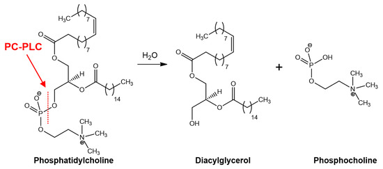
Phosphatidylcholine-specific phospholipase C (PC-PLC) is an enzyme that catalyzes the cleavage of phosphatidylcholine phospholipids to generate diacylglycerol (DAG) and phosphocholine as secondary messengers (Figure 1) [1]. DAG and phosphocholine mediate an array of cellular signaling proteins that maintain normal physiological functions in cells. DAG is well known for its role in the activation of protein kinase C (PKC), important for the activation of downstream proteins in several signal transduction cascades involved with, e.g., immune responses, cell growth and memory formation. The role of phosphocholine is not entirely clear, other than in synthesis of essential lipids in signal transduction [2]. Investigations into the pathophysiology of PC-PLC have revealed that the enzyme is pivotal to the development of several key pathological conditions. Therefore, PC-PLC has emerged as a potential biomedical target leading to the discovery of bespoke PC-PLC inhibitors. Details of the reported PC-PLC inhibitors and their pharmacological actions are outlined in this review.

Figure 1.
PC-PLC hydrolysis of phosphatidylcholine to diacylglycerol (DAG) and phosphocholine.
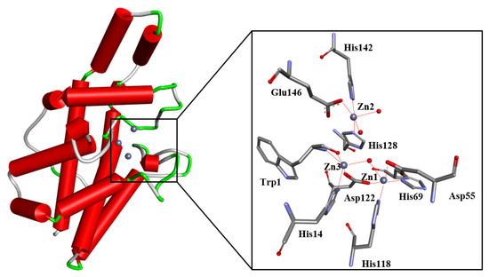
Generally, studies into the biochemistry and pathophysiology of PC-PLC are relatively scant and less developed when compared to other phospholipase C subtypes such as phosphatidylinositol-specific phospholipase C (PI-PLC). Most notably, the genetic sequence of the mammalian PC-PLC has not yet been unambiguously identified. Recently, sphingomyelin synthases 1 and 2 were shown to exhibit PC-PLC-like activity [3], and are required for sphingomyelin homeostasis and growth in human HeLa cancer cells [4]. Thus, it is plausible that other enzymes could mimic PC-PLC activity (moonlighting function). Regardless, progress has been made towards understanding the mechanism of prokaryotic PC-PLC. A body of evidence suggests PC-PLC activity as a human functional equivalent of prokaryotic PC-PLC. The prokaryotic PC-PLC enzyme has been isolated and purified from Bacillus cereus (PC-PLCBc), and its structure has been characterized (Figure 2) [5]. PC-PLCBc was used as a model of its mammalian counterpart, e.g., they share antigenic similarities [6] and PC-PLCBc is able to emulate similar responses in enhancing prostaglandin biosynthesis in mammalian cells [7]. The X-ray crystal structure is a monomeric zinc metalloproteinase with 245 amino acid residues [5]. The enzyme has thirteen α-helices with three Zn2+ ions in the active site. The Zn2+ ions are coordinated with water molecules and amino acid residues within the binding site; as shown in Figure 2, Zn1 is coordinated to a water molecule, Asp122, His69, Asp55 and His118; Zn2 is coordinated to two water molecules, His142, Glu146 and His128; and finally, Zn3 is coordinated to a water molecule along with Trp1 and Asp122.

Figure 2.
The structure of the wild-type PC-PLCBc enzyme (PDB ID: 1AH7) and its catalytic site. The protein α-helixes are shown as red tubes. The trinuclear metal center consists of catalytic Zn2+ ions shown as grey spheres, whereas the water molecules are shown as red spheres. Amino acid residues coordinating to the Zn2+ ions are depicted and labelled. Figure edited from Eurtivong et al. [8].
2. Pathological Implications of PC-PLC
In this section, we summarize the implications of PC-PLC in pathophysiology based on what has been reported in the literature, specifically detailing the developments of pathological diseases and conditions, which include various types of cancers, atherosclerosis progression, induction of inflammation and neuronal cell death.
2.1. Cancer
The contributions of cancers to the global health burden are substantial: the World Health Organization has estimated approximately 10 million deaths in 2020 alone were caused by cancers; this figure is predicted to continue to grow [9]. In the past, significant progress has been made in cancer therapeutics. One of the major problems in cancer therapy is the continuous emergence of mutant proteins, which leads to significant mitigation of drug effectiveness. Thus, there is a strong drive towards the identification of alternative novel targets. One of these new anticancer targets that has gained some traction recently is PC-PLC. Some studies have identified PC-PLC activity to be involved in mediating intramolecular signals that lead to the induction of cancer-like properties. To little surprise, there have been studies suggesting choline metabolites and enzymes relevant to phospholipid homeostasis as biomarkers in monitoring tumor progression and response to therapeutic treatments, with some studies implying resistance to anticancer chemotherapy [10,11,12].
The liver is an organ with a major role in phospholipid metabolism that regulates turnovers of phospholipids via PC-PLC signaling [13]. Calcium-dependent activities of PC-PLC were reported to have been induced by a proliferative promoter, N-nitrosodiethylamine, in hepatocarcinogenic rats where PC-PLC activity peaked during tumor formation [14]. Likewise, using another proliferative promoter, phorbol 12-myristate 13-acetate, induced PC-PLC activity via PKC calcium-dependent pathway in CBRH-7919 rat hepatoma cells [14].
Breast cancer progressions are associated with aberrant signal transduction in cell proliferation. It is well-established that the PLC family, in particular PI-PLCs, mediates the signal transduction pathway by regulating several key membrane oncogenic receptor proteins, e.g., PLC-γ1 have roles in regulating EGFR activities [15], and PLC-δ4 is upregulated and enhances expression of HER2 and EGFR in MCF-7 breast cancer cells [16]. Similarly, a study showed that modulation of PC-PLC activities was able to regulate oncogenic HER2 and EGFR receptors: inhibition of PC-PLC reduced expression of HER2, induced HER2 internalization and reduced receptor recycling in SKBr3 cells and reduced membrane expression of EGFR and HER3 in SKBr3 cells, suggesting a strong relationship between PC-PLC signaling and its tumorous effects in breast cancer progression [17]. Additionally, in recent studies, the activation of PC-PLC has been implicated in the production of a significant portion (20–50%) of intracellular phosphocholine (PCho) within various subtypes of ovarian and breast cancer cells, suggesting PC-PLC as an active contributor to the progression of these cancers [18].
Ovarian cancer was responsible for ~300,000 deaths in 2020 [19]. The most common type of ovarian cancer is epithelial ovarian cancer (EOC), which is distinctively characterized by its ability to invade the abdominal cavity in the later stages. Resistance to platinum-based drugs is a common and unsolved problem. Recently, aberrant choline metabolism has been associated with EOC developments, with evidence of PC-PLC in sustaining the abnormalities of intracellular signaling in ovarian cancer cells [20]. Metabolic functions of PC-PLC were investigated in EOC cells, i.e., phosphocholine levels were detected to be 40–50% lower when PC-PLC was inhibited [18]. A similar protocol was used to show that elevated phosphocholine levels in HER2-overexpressed SKOV3 cells increase PC-PLC activity [21]. Additionally, PC-PLC activity was measured using the Amplex Red fluorescence assay in whole-cell lysates from a set of EOC cell lines with two-to-four-times higher activities in comparison to their non-tumoral counterparts [22].
Squamous cell carcinoma (SCC) is the second most prevalent form of skin cancer, accounting for approximately 23% of all skin cancers [23]. Tumor growth and proliferation of SCCs have been associated with PLC activities, e.g., PLC-γ was shown to be required for EGFR-induced mitogenesis and is overexpressed in human SCC cell lines [24], and PLC-γ has been implicated as a potential prognostic marker for oral SCC patients [25]. Specifically, PC-PLC activities were shown to drive oncogenic processes in human SCC cell lines, i.e., ×2.5 higher PC-PLC activities in an A431 human squamous cell line compared to non-tumoral keratinocytes were seen, and upon inhibition of PC-PLC, Western blot analysis revealed substantial decrease in EGFR phosphorylation activities [26].
Glioblastoma is an aggressive fast-forming type of brain cancer that originates in brain astrocytes. It represents the majority of brain cancers, and occurs in approximately 3 in 100,000 people [27]. Only 5% of patients are expected to live more than five years after diagnosis [27]. Existing treatments are considered ineffective, as the disease tends to recur at the same location [28]. Different forms of PLC have been associated with the developments of glioblastomas, including PLC-γ1 [29] and PLC-β1 [30]. CXCR4 is a G-protein coupled receptor that binds to chemokine ligands, which triggers a cascade in cancer-related signaling pathways that induce tumor growth and metastasis [31]. Neural stem cells have been noted to express high levels of CXCR4 receptors and can differentiate and develop into glioblastoma cells [32]. A recent report suggested that decreased expression of CXCR4 receptors following inhibition of PC-PLC in U87MG glioma cell models results in decreased proliferative and invasive activities, and suppresses EGFR and AKT kinase activities, suggesting modulation of PC-PLC can inhibit CXCR4 signaling and suppress glioblastoma growth [33].
2.2. Atherosclerosis
Atherosclerosis is a health condition resulting from the narrowing of the arteries due to accumulation of atherosclerotic plaque (atheroma) leading to increased risk of cardiovascular disease. It has been estimated that around 30% of deaths globally are associated with atherosclerotic–cardiovascular diseases [34].
A body of evidence suggests that PC-PLC signaling sustains the progression of atherosclerosis, suggesting PC-PLC as a potential drug target to treat cardiovascular diseases. It has been reported that PC-PLC signaling is induced by apolipoprotein C, a regulator of lipoprotein metabolism, which activates PKC and NF-κB, causing adhesion of monocytes to endothelial cells contributing to inflammatory responses in atherogenesis [35]. Inhibition of annexin A7, a phospholipid-binding protein, was shown to suppress PC-PLC activity in vascular endothelial cells and reduce atherosclerosis in apoE −/− mice [36]. Inhibition of PC-PLC in apoE −/− mice revealed suppression of LOX-1 receptor expression; LOX-1 is a key receptor of oxidized LDLs [37]. PC-PLC is a key mediator for NF-κB activation; in the study, it was also shown that NF-κB signaling is closely associated with expression of LOX-1 [37]. Reduced PC-PLC activities and apoptosis in vascular endothelial cells were reported during autophagy induced by sphingosylphosphorylcholine, a cardiovascular mediator with artheroprotective properties [38]. Additionally, it was reported that PC-PLC activities are mediated by PEBP1, a protein inhibitor of protein kinases, during atherosclerosis development, i.e., enhanced PC-PLC activities during elevated PEBP1 levels in atherosclerotic mice were reported, whereas PEBP1 downregulation was seen during PC-PLC inhibition [39].
2.3. Inflammation
Inflammation is a protective response of body tissues from harmful stimuli, and is regulated by a balanced combination of pro- and anti-inflammatory mediators [40]. However, an interference to this equilibrium can result in an excessive inflammatory response resulting in tissue damage [40].
The PLC family also mediate inflammatory responses given that DAG is a pro-inflammatory mediator. DAG activates PKC, which mediates various inflammatory responses in NF-κB and MAPK signal transduction pathways via extracellular signals including interleukins and TNFs [41,42,43]. Isotopic labeling of phosphatidylcholine indicated that hydrolysis via PC-PLC catalysis was enhanced by TNF stimulation, generating DAG mediators in monocytes and T cells [44]. Another report showed that IL-1 stimulation was able to enhance formations of DAG and phosphocholine products in T-lymphocytes in the absence of phosphatidylinositols, suggesting PC-PLC catalysis could be responsible for the generation of DAG secondary messengers via IL-1-mediated signal transduction [45].
2.4. Neuronal Cell Death
The central nervous system is responsible for the integration of sensory information and instructs body responses accordingly. Neuronal cell death is the essence of neurodegenerative diseases and injury, the second leading cause of mortality, responsible for an estimated nine million deaths per year [46].
There is some evidence indicating that suppressing PC-PLC activities may increase the growth and differentiation of neuronal cells, which suggests that PC-PLC could be a potential neurotherapeutic target. The glutamate/cystine antiporters are highly expressed in astrocytes that indirectly regulate intracellular levels of the antioxidant glutathione, through uptake of cystine in exchange for glutamate expulsion [47]. It was reported that high levels of glutamate are able to interfere with the antiporter mechanism via PC-PLC signaling that induces nerve cell death, possibly as a result of glutathione depletion, whereas inhibition of PC-PLC increases cell viability of the cortical cells [47]. Investigation into the effects of PC-PLC inhibition on vascular endothelial cells and marrow stromal cells revealed that the inhibition of PC-PLC signaling induced differentiation into neuron-like morphologies [48]; mechanistic studies have suggested that NADPH oxidation and an increase in ROS levels contribute to PC-PLC-mediated bone marrow stromal cell differentiation signal transduction pathways [49].
3. Discovery and Development of PC-PLC Inhibitors
In the past, a plethora of PI-PLC inhibitors have been discovered and developed, ranging from steroids [50,51], small molecules [52,53], natural products [54] and lipid analogues [55]. The lipid analogue edelfosine, a platelet-activating factor found in small concentrations in the human body that predominantly inhibit cytosolic fibroblast’s PLC-γ, was shown in clinical studies to treat various cancer types [55,56,57]. In contrast, there is a paucity of studies to develop PC-PLC inhibitors. Nevertheless, there have been recent discoveries of PC-PLC inhibitors that demonstrate glimpses of its therapeutic potential. In this section, details of different classes of the reported PC-PLC inhibitors are outlined. The most prominent PC-PLC inhibitors are given in Table 1.
3.1. 2-Aminohydroxamic Acids
The concept of using 2-aminohydroxamic acids as PC-PLCBc inhibitors was mainly driven by Llebaria and coworkers [58,59]. These compounds were developed based on structural knowledge of PC-PLCBc and aminopeptidases in Streptomyces griseus and Aeromonas proteolytica strains, e.g., triplet Zn2+ ion cofactors, as these enzymes may share similar active site architecture with human PC-PLC [59]. Derivatives of aminopeptidase inhibitors were designed and synthesized, including α-aminohydroxamic acids, and the most potent inhibitors are shown in Table 1. This was followed by experimental validation of PC-PLCBc inhibition, with positive results. Pursuing from this success, modifications were introduced to the 2-aminohydroxamic scaffold to achieve stronger potencies, e.g., installation of choline linked to the hydroxamides with the prospect of stronger chelation to Zn2+ ions achieving IC50 values in the submicromolar range. Unexpectedly, the potencies tend to associate with amino acid binding rather than Zn2+ ion chelation. In the design of the series, enantioselectivity was not given priority, as it has been reported that the enzyme lacks enantiomeric selectivity.
3.2. Phospholipid Analogues
Substrate mimics have long been used in drug development, as they are able to imitate transition state conformations. This concept was implemented for the design and synthesis of phospholipid analogues as inhibitors of PC-PLC, and was pioneered by Martin and coworkers [60,61] (see Table 1). It was seen that modifications at the P-O phosphodiester was detrimental to activity. However, substitution of the glyceryl oxygen and oxygens at P=O and P-OH were tolerated. Modifications of side acyl chains favor six to eight carbon atoms, possibly mimicking the length of most PC-PLCBc phospholipid substrates. Modifications at the acyl terminus seemed tolerable, with polar groups such as hydroxyl increasing solubility in aqueous environments.
3.3. Xanthates
PC-PLC inhibitors from the xanthate family primarily revolve around tricyclodecan-9-yl-xanthogenate (D609) with a Ki value of 6.4 μM for PC-PLCBc [62]. The chemical structure of D609 consists of a xanthate group linked to a merged tricyclic cyclodecane ring (see Table 1). The compound was initially discovered to have antiviral properties against DNA and RNA viruses [63]. Following this, D609 was discovered in other pharmacological applications, including having anticancer, anti-inflammatory, anti-atherosclerotic and anti-apoptotic properties. Interestingly, given D609’s pharmacological benefits, it received little interest in developing its structural activity relationship (SAR), most likely due to the non-drug likeness of the compound. Nonetheless, a study revealed that PC-PLCBc lacked diasteromeric control towards the xanthates; eight pure D609 diastereomers were synthesized and evaluated in vitro with comparable potencies with IC50 values between 10 and 17 μM [64]. It was shown that the tricyclic system of D609 is not essential, i.e., replacement with n-decyl displayed a comparable Ki value of 10 μM (see Table 1). Moreover, it was reported that shortening the length of alkyl chains and substituting with more bulky aromatic rings diminished PC-PLCBc inhibition activities.
3.4. Pyrido[3,4-b]indoles
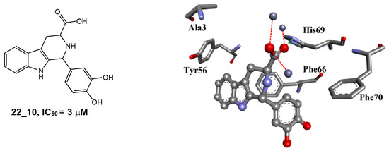
The pyrido[2,3-b]indole is a prominent core scaffold in medicinal chemistry exhibiting a range of biological activities and mechanisms of action, e.g., anticancer [65], antileishmanial [66], NMDA antagonism [67], anti-depression [68] and estrogen antagonists [69]. Reynisson and coworkers [8] were the first to report the series as inhibitors of PC-PLC in a combined virtual screening SAR study. Molecular modelling studies revealed that the derivatives were able to chelate with the Zn2+ ions at the active site with the carboxylate functional group and interact with the imidazole ring in His69 (Figure 3). Only racemic mixtures of the pyrido[2,3-b]indoles were reported. Although molecular modelling suggests the (1R,3S)-diastereomer as the best inhibitor, there is as of yet no in vitro validation to confirm this finding. The chemical structure of a pyrido[3,4-b]indole derivative and the predicted binding interactions to the enzyme are shown in Figure 3.

Figure 3.
The chemical structure and potency of pyrido[3,4-b]indole derivative 22_10 as well as the predicted binding mode of (1R,3S)-22_10 derivative in the PC-PLC binding site. Figure edited from Eurtivong et al. [8].
3.5. Morpholinobenzoic Acids
The morpholinobenzoic acids were first reported to show PC-PLCBc inhibition by Reynisson and coworkers [8]. A selection of inhibitors from the series are shown in Table 1. In that study, computational modelling was initially used to elucidate its binding mode to PC-PLCBc, which demonstrated that the series chelates to the Zn2+ ions with the carboxylate functionality. Thus, it was initially assumed that the strong potencies observed were heavily reliant on this key interaction. However, good activities were maintained after derivatization of the carboxylic acid group to ester and hydroxamic acid, whilst complete removal of carboxylic acid improved activity for some derivatives [70,71,72]. In vitro tests for their antiproliferative effects revealed the carboxylate and ester derivatives displayed poor antiproliferative activities [71]. Nonetheless, the hydroxamic acid derivatives displayed promising antiproliferative activities against two cancer cell lines, e.g., 2-morpholinobenzene hydroxamic acid 9i (see structure in Table 1) had an IC50 value of 3.25 μM against MDA-MB-231 (epithelial human breast cancer), and IC50 = 5.8 μM against HCT-116 (human colorectal carcinoma) [71].
3.6. 1,4-Oxazepines
Recently, Zhao and coworkers [73] were the first to synthesize two chiral compounds, (R)-7-amino-2,3,4,5-tetrahydrobenzo[b][1,4]oxazepin-3-ol (R-7ABO) and (S)-7-amino-2,3,4,5-tetrahydrobenzo[b][1,4]oxazepin-3-ol (S-7ABO), based on the 1,4-oxazepine scaffold, and verified their PC-PLCBc inhibition activities (see Table 1); the inhibition activities for the compounds were dose-dependent. Derivatives of 1,4-oxazepines have been implicated to an array of pharmacological activities, e.g., anti-convulsion [74], anti-diabetics [75], carbonic anhydrase inhibition [76], PI3K inhibition [77], and progesterone agonists [78].
3.7. N,N′-Dihydroxyureas
A selection of N,N′-dihydroxyureas were tested for PC-PLC activity due to successful coordination of polyhydroxy tropolones to bimetallic sites in enzymes. In a study conducted by Martin et al. [79], a bidentate ligand, 2,7-dihydroxytropolone (Table 1) was identified as a PC-PLCBc inhibitor. Given this success, and knowing that hydroxamic acids are excellent Zn2+ chelators, derivatives of N,N′-dihydroxyureas were synthesized (as an example see derivative in Table 1). In the report, N-OH groups were incorporated into the rings for strong bimetallic Zn2+ chelation. Interestingly, PC-PLC inhibition experiments are pH-dependent, i.e., a high pH favors PC-PLC inhibition by N,N′-dihydroxyurea derivatives.

Table 1.
Some PC-PLC inhibitors from literature.
Table 1.
Some PC-PLC inhibitors from literature.
| Compound | Chemical Structure | IC50/Ki | Assay | Reference |
|---|---|---|---|---|
| α-aminohydroxamic acid 6 |  | IC50 = 4 μM | Chromogenic-based assay | [59] |
| 2-aminohydroxamic acid 18 |  | IC50 = 2 μM | Chromogenic-based assay | [58] |
| Phospholipid analogue 7 |  | Ki = 7 μM | pH-based assay | [60] |
| Dihydroxy phospholipid 7 |  | Ki = 5.4 μM | Chromogenic-based assay | [61] |
| D609 |  | Ki = 6.4 μM | Radiometric enzyme assay | [62] |
| Potassium O-decyl xanthate |  | Ki = 10 μM | Chromogenic-based assay | [64] |
| 1H-pyrido[3,4-b]indole 22_10 |  | IC50 = 3.1 μM | Amplex Red assay | [8] |
| 2-morpholinobenzoic acid 84 |  | IC50 = 3.7 μM | Amplex Red assay | [8] |
| Morpholinobenzene 10k |  | IC50 = 1.1 μM | Amplex Red assay | [70] |
| 2-morpholinobenzene hydroxamic acid 9i |  | Unknown | Amplex Red assay | [71] |
| R-7ABO |  | Unknown | Amplex Red assay | [73] |
| S-7ABO |  | Unknown | Amplex Red assay | [73] |
| 2,7-dihydroxytropolone |  | Ki(pH = 7.3) = 16 μMKi(pH = 9.5) = 23 μM | Chromogenic-based assay | [79] |
| N,N′-dihydroxyurea 10 |  | Ki(pH = 7.3) = 388 μMKi(pH = 9.5) = 53 μM | Chromogenic-based assay | [79] |
3.8. Univalent Anions
The PC-PLCBc enzyme was reported to be inhibited by several univalent anions by coordinative interactions with the trinuclear Zn2+ ions. It was seen that univalent anions were most effective at inhibiting PC-PLCBc activities as reported by Aakre and Little [80]. From the same study, phospholipid hydrolysis was substantially decreased in the presence of iodide, cyanate, chloride, nitrate and bicarbonate solutions. The univalent anions interact electrostatically with the trinuclear Zn2+ ions, which was confirmed by X-ray crystallographic data, e.g., complexation of iodide to Zn2+ ions in the PC-PLCBc active site [81].
4. PC-PLC Enzymatic Assays
The ability to monitor enzyme activity accurately is a prerequisite for the development of enzyme inhibitors in modern medicinal chemistry. A reliable assay allows the efficacy of potential inhibitor molecules to be measured and quantified so that SAR can be established. In the last 30 years, a number of PC-PLC assays have been reported in the literature. Some of the earliest assays were low-throughput and/or required specialist reagents and instrumentations. For example, in 1994, Martin et al. conducted one of the first SAR studies on PC-PLCBc by monitoring pH alterations during phosphatidylcholine hydrolysis [60]. Two years later, in 1996, Amtmann applied a radiometric assay that measured the rate of phosphorylcholine production by using 3H-labelled phosphatidylcholine substrates [62]. Although these methods yielded satisfactory results, their widespread application was hindered by their inherent limitations.
The first assay that was applied widely by the medicinal chemistry community was described by Hergenrother and Martin [82]. The assay allows PC-PLC activity to be measured in three indirect steps. The first one is enzyme-coupled, in which inorganic phosphate was released from the phosphomonoester product such as phosphocholine by the use of alkaline phosphatases. In the second step, the resulting inorganic phosphate ion is reacted with ammonium molybdate. The phosphate–ammonium molybdate complex is then reduced chemically using ascorbic acid in the last step. This results in a blue molybdenum chromogen, which can be observed through ultraviolet/visible (UV/vis) spectrophotometry with a maximum absorbance at 700 nm. As this assay can be conducted on a plate, it gained traction in the biochemical community and it has been applied in a number of inhibitor discovery studies for PC-PLCBc [58,59,61,64,79]. However, as this assay is indirect and relies on both the activity of alkaline phosphatase and chemical reactions via ammonium molybdate and ascorbate, it is tedious and potentially prone to errors. In 2000, Flieger et al. developed a second UV/vis-based assay that allowed direct monitoring of PC-PLC activity [83]. By using α-naphthylphosphorylcholine (α-NPPC) as a (non-native) substrate, para-nitrophenol is generated as a product, which can be monitored spectrophotometrically with a maximum absorbance at 410 nm [83]. However, as this assay requires the use of non-native substrates, the results may not be directly translatable to the native system.
One of the most widely used assays to monitor PC-PLC activity is the so-called “Amplex Red assay” [84,85,86]. This assay utilizes a fluorogenic probe called 10-acetyl-3,7-dihydroxyphenoxazine (Amplex Red) to identify the presence of hydrogen peroxide. The hydrogen peroxide is generated through the conversion of phosphocholine (a product of PC-PLC-catalyzed reaction) to choline by alkaline phosphatase, which is followed by the oxidation of choline by choline oxidase. When hydrogen peroxide is present, it reacts with Amplex Red in the presence of horseradish peroxidase, resulting in the production of a highly fluorescent substance called resorufin. However, the Amplex Red assay has its own limitations, since the detection of PC-PLC activity is indirect and relies on multiple enzymatic and chemical conversion steps. For example, Sharma et al. found that the activity of horseradish peroxidase used in the assay may be inhibited by potential PC-PLC inhibitors, which may give false positive results [72].
Finally, several assays that allow direct detection of the turnover of native PC-PLC substrate have also been developed in the last decade. In 2017, Murakami et al. reported a liquid chromatography-mass spectrometry (LC-MS) assay to separate and quantify different PC-PLC substrates and products [87]. In 2021, Sharma et al. described a matrix-assisted laser desorption ionization time-of-flight (MALDI-TOF) mass spectrometry-based assay to monitor the formation of the diacylglycerol products [72]. In principle, these methods hold the potential for precise measurement and quantification of PC-PLC activity. However, as these assays were only published in the last few years, their novelty means that their uptake and adoption by the biochemical and medicinal chemistry communities are yet to be observed.
5. Conclusions and Future Perspective
PC-PLC has the potential to be a novel pharmacological target. It is apparent that PC-PLC is associated with developments of several pathological conditions and diseases, as outlined in this review. There is evidence at the molecular level for PC-PLC signaling being involved in the expression of oncogenic proteins. In addition, chemokine signaling has been linked to PC-PLC activity, which led to the progression of many cancers. As a consequence, this led to influences over various cellular processes such the regulation of cell cycles, signaling and proliferation. These observations are often supported by quantification of phosphocholine generation, and modulation of PC-PLC activities using small molecule PC-PLC inhibitors such as D609. Given DAG generation is a product of PC-PLC catalysis, it can be inferred that cancer is progressed via PKC signaling as the main route. There is evidence to suggest that PC-PLC is mediated by several signaling proteins and is involved in the development of atherosclerosis, albeit explanations to most of the underlying mechanisms remain unclear. Moreover, PC-PLC is suggested to activate inflammatory mediators via DAG and PKC signaling, and PC-PLC has been responsive to interleukin ligands. Lastly, reports of PC-PLC signaling associated with induction of nerve cell deaths via physiological mechanisms regulating glutathione levels, NADPH and ROS activities.
Despite some evidence indicating PC-PLC as a potential pharmacological drug target, it has so far received limited interest from the drug discovery community. Most of the findings were based on running biochemical assays to investigate PC-PLC activity and PC-PLC protein expression (detected by polyclonal antibodies against bacterial PC-PLC) in different cell-based models. Recent inhibitors that have been developed include drug-like inhibitors, indicating that PC-PLC has partly gained traction. Most of the inhibitors were conceptualized to chelate, or form electrostatic interactions, with the Zn2+ ions in the PC-PLC binding pocket as an important key feature. However, this concept was shown to be somewhat obscured, e.g., potencies of the 2-aminohydroxamic acids were reported with the tendency to correlate more with amino acid interactions rather than Zn2+ chelation. Furthermore, the removal of the carboxylic acid functionality from the 2-morpholinobenozic acid scaffold, or substitution with ester groups, were tolerated strongly, suggesting that the metal chelation hypothesis was invalid. Regardless, the PC-PLCBc is commonly used as a model to study mammalian PC-PLC. The gene coding for the mammalian PC-PLC has not yet been identified, and as a result, the mammalian structure is unavailable. Given the lack of mammalian PC-PLC structural data, it remains questionable whether the compounds can actually inhibit the mammalian form. Therefore, the location of the corresponding human gene and characterization of the human PC-PLC structure is essential to verify the pharmacological applications of the current inhibitors, and future developments of PC-PLC inhibitors. Without the gene, the possibility remains that other enzymes serve as surrogates (moonlighters) in human physiology. The elucidation of the human PC-PLC gene is paramount in progressing the further development of PC-PLC as potential therapeutic target. Nevertheless, a body of biological data supports the case for the existence of PC-PLC in human physiology and several drug-like inhibitors have been proposed.
Author Contributions
Conceptualization, writing—original draft preparation, and funding, C.E.; writing—review and editing, C.E., E.L., N.S., I.K.H.L. and J.R.; supervision, E.L. and J.R. All authors have read and agreed to the published version of the manuscript.
Funding
This research received no external funding.
Institutional Review Board Statement
Not applicable.
Informed Consent Statement
Not applicable.
Data Availability Statement
Not applicable.
Acknowledgments
C.E. would like to thank Mahidol University for the facilities and support given in writing up this review article.
Conflicts of Interest
The authors declare no conflict of interest.
References
- Vines, C.M. Phospholipase C. In Calcium Signaling; Islam, M.S., Ed.; Springer: Dordrecht, The Netherlands, 2012; pp. 235–254. [Google Scholar]
- Ridgway, N.D. The role of phosphatidylcholine and choline metabolites to cell proliferation and survival. Crit. Rev. Biochem. Mol. Biol. 2013, 48, 20–38. [Google Scholar] [CrossRef]
- Chiang, Y.P.; Li, Z.; Chen, Y.; Cao, Y.; Jiang, X.C. Sphingomyelin synthases 1 and 2 exhibit phosphatidylcholine phospholipase C activity. J. Biol. Chem. 2021, 297, 101398. [Google Scholar] [CrossRef]
- Tafesse, F.G.; Huitema, K.; Hermansson, M.; van der Poel, S.; van den Dikkenberg, J.; Uphoff, A.; Somerharju, P.; Holthuis, J.C.M. Both Sphingomyelin Synthases SMS1 and SMS2 Are Required for Sphingomyelin Homeostasis and Growth in Human HeLa Cells. J. Biol. Chem. 2007, 282, 17537–17547. [Google Scholar] [CrossRef]
- Hough, E.; Hansen, L.K.; Birknes, B.; Jynge, K.; Hansen, S.; Hordvik, A.; Little, C.; Dodson, E.; Derewenda, Z. High-resolution (1.5 Å) crystal structure of phospholipase C from Bacillus cereus. Nature 1989, 338, 357–360. [Google Scholar] [CrossRef]
- Clark, M.A.; Shorr, R.G.L.; Bomalaski, J.S. Antibodies prepared to Bacillus cereus phospholipase C crossreact with a phosphatidylcholine preferring phosoholipase C in mammalian cells. Biochem. Biophys. Res. Commun. 1986, 140, 114–119. [Google Scholar] [CrossRef]
- Levine, L.; Xiao, D.M.; Little, C. Increased arachidonic acid metabolites from cells in culture after treatment with the phosphatidylcholine-hydrolyzing phospholipase C from Bacillus cereus. Prostaglandins 1987, 34, 633–642. [Google Scholar] [CrossRef]
- Eurtivong, C.; Pilkington, L.I.; van Rensburg, M.; White, R.M.; Brar, H.K.; Rees, S.; Paulin, E.K.; Xu, C.S.; Sharma, N.; Leung, I.K.H.; et al. Discovery of novel phosphatidylcholine-specific phospholipase C drug-like inhibitors as potential anticancer agents. Eur. J. Med. Chem. 2020, 187, 111919. [Google Scholar] [CrossRef]
- World Health Organization. Cancer Key Facts. Available online: http://www.who.int/news-room/fact-sheets/detail/cancer (accessed on 22 March 2023).
- Meisamy, S.; Bolan, P.J.; Baker, E.H.; Bliss, R.L.; Gulbahce, E.; Everson, L.I.; Nelson, M.T.; Emory, T.H.; Tuttle, T.M.; Yee, D.; et al. Neoadjuvant chemotherapy of locally advanced breast cancer: Predicting response with in vivo 1H MR spectroscopy-a pilot study at 4 T. Radiology 2004, 233, 424–431. [Google Scholar] [CrossRef]
- Sola-Leyva, A.; López-Cara, L.C.; Ríos-Marco, P.; Ríos, A.; Marco, C.; Carrasco-Jiménez, M.P. Choline kinase inhibitors EB-3D and EB-3P interferes with lipid homeostasis in HepG2 cells. Sci. Rep. 2019, 9, 5109. [Google Scholar] [CrossRef]
- Shah, T.; Wildes, F.; Penet, M.F.; Winnard, P.T., Jr.; Glunde, K.; Artemov, D.; Ackerstaff, E.; Gimi, B.; Kakkad, S.; Raman, V.; et al. Choline kinase overexpression increases invasiveness and drug resistance of human breast cancer cells. NMR Biomed. 2010, 23, 633–642. [Google Scholar] [CrossRef]
- Reo, N.V.; Goecke, C.M.; Narayanan, L.; Jarnot, B.M. Effects of perfluoro-n-octanoic acid, perfluoro-n-decanoic acid, and clofibrate on hepatic phosphorus metabolism in rats and guinea pigs in vivo. Toxicol. Appl. Pharmacol. 1994, 124, 165–173. [Google Scholar] [CrossRef]
- Xingzhong, W.U.; Lu, H.; Zhou, L.A.N.; Huang, Y.; Chen, H. Changes of phosphatidylcholine-specific phospholipase C in hepatocarcinogenesis and in the proliferation and differentiation of rat liver cancer cells. Cell Biol. Int. 1997, 21, 375–381. [Google Scholar] [CrossRef]
- Piccolo, E.; Innominato, P.F.; Mariggio, M.A.; Maffucci, T.; Iacobelli, S.; Falasca, M. The mechanism involved in the regulation of phospholipase Cgamma1 activity in cell migration. Oncogene 2002, 21, 6520–6529. [Google Scholar] [CrossRef][Green Version]
- Leung, D.W.; Tompkins, C.; Brewer, J.; Ball, A.; Coon, M.; Morris, V.; Waggoner, D.; Singer, J.W. Phospholipase C δ-4 overexpression upregulates ErbB1/2 expression, Erk signaling pathway, and proliferation in MCF-7 cells. Mol. Cancer 2004, 3, 15. [Google Scholar] [CrossRef]
- Paris, L.; Cecchetti, S.; Spadaro, F.; Abalsamo, L.; Lugini, L.; Pisanu, M.E.; Iorio, E.; Natali, P.G.; Ramoni, C.; Podo, F. Inhibition of phosphatidylcholine-specific phospholipase C downregulates HER2 overexpression on plasma membrane of breast cancer cells. Breast Cancer Res. 2010, 12, R27. [Google Scholar] [CrossRef]
- Podo, F.; Paris, L.; Cecchetti, S.; Spadaro, F.; Abalsamo, L.; Ramoni, C.; Ricci, A.; Pisanu, M.E.; Sardanelli, F.; Canese, R.; et al. Activation of Phosphatidylcholine-Specific Phospholipase C in Breast and Ovarian Cancer: Impact on MRS-Detected Choline Metabolic Profile and Perspectives for Targeted Therapy. Front. Oncol. 2016, 6, 171. [Google Scholar] [CrossRef]
- Sung, H.; Ferlay, J.; Siegel, R.L.; Laversanne, M.; Soerjomataram, I.; Jemal, A.; Bray, F. Global Cancer Statistics 2020: GLOBOCAN Estimates of Incidence and Mortality Worldwide for 36 Cancers in 185 Countries. CA Cancer J. Clin. 2021, 71, 209–249. [Google Scholar] [CrossRef]
- Bagnoli, M.; Granata, A.; Nicoletti, R.; Krishnamachary, B.; Bhujwalla, Z.M.; Canese, R.; Podo, F.; Canevari, S.; Iorio, E.; Mezzanzanica, D. Choline Metabolism Alteration: A Focus on Ovarian Cancer. Front. Oncol. 2016, 6, 153. [Google Scholar] [CrossRef]
- Pisanu, M.E.; Ricci, A.; Paris, L.; Surrentino, E.; Liliac, L.; Bagnoli, M.; Canevari, S.; Mezzanzanica, D.; Podo, F.; Iorio, E.; et al. Monitoring response to cytostatic cisplatin in a HER2(+) ovary cancer model by MRI and in vitro and in vivo MR spectroscopy. Br. J. Cancer 2014, 110, 625–635. [Google Scholar] [CrossRef]
- Spadaro, F.; Ramoni, C.; Mezzanzanica, D.; Miotti, S.; Alberti, P.; Cecchetti, S.; Iorio, E.; Dolo, V.; Canevari, S.; Podo, F. Phosphatidylcholine-specific phospholipase C activation in epithelial ovarian cancer cells. Cancer Res. 2008, 68, 6541–6549. [Google Scholar] [CrossRef]
- Cancer Research UK, Types of Skin Cancer. Available online: https://www.cancerresearchuk.org/about-cancer/skin-cancer/types (accessed on 8 June 2023).
- Xie, Z.; Chen, Y.; Liao, E.Y.; Jiang, Y.; Liu, F.Y.; Pennypacker, S.D. Phospholipase C-gamma1 is required for the epidermal growth factor receptor-induced squamous cell carcinoma cell mitogenesis. Biochem. Biophys. Res. Commun. 2010, 397, 296–300. [Google Scholar] [CrossRef]
- Zhu, D.; Tan, Y.; Yang, X.; Qiao, J.; Yu, C.; Wang, L.; Li, J.; Zhang, Z.; Zhong, L. Phospholipase C gamma 1 is a potential prognostic biomarker for patients with locally advanced and resectable oral squamous cell carcinoma. Int. J. Oral Maxillofac. Surg. 2014, 43, 1418–1426. [Google Scholar] [CrossRef]
- Cecchetti, S.; Bortolomai, I.; Ferri, R.; Mercurio, L.; Canevari, S.; Podo, F.; Miotti, S.; Iorio, E. Inhibition of Phosphatidylcholine-Specific Phospholipase C Interferes with Proliferation and Survival of Tumor Initiating Cells in Squamous Cell Carcinoma. PLoS ONE 2015, 10, e0136120. [Google Scholar] [CrossRef]
- Gallego, O. Nonsurgical treatment of recurrent glioblastoma. Curr. Oncol. 2015, 22, e273–e281. [Google Scholar] [CrossRef]
- Birzu, C.; French, P.; Caccese, M.; Cerretti, G.; Idbaih, A.; Zagonel, V.; Lombardi, G. Recurrent Glioblastoma: From Molecular Landscape to New Treatment Perspectives. Cancers 2020, 13, 47. [Google Scholar] [CrossRef]
- Li, T.; Yang, Z.; Li, H.; Zhu, J.; Wang, Y.; Tang, Q.; Shi, Z. Phospholipase Cγ1 (PLCG1) overexpression is associated with tumor growth and poor survival in IDH wild-type lower-grade gliomas in adult patients. Lab. Investig. 2022, 102, 143–153. [Google Scholar] [CrossRef]
- Ratti, S.; Marvi, M.V.; Mongiorgi, S.; Obeng, E.O.; Rusciano, I.; Ramazzotti, G.; Morandi, L.; Asioli, S.; Zoli, M.; Mazzatenta, D.; et al. Impact of phospholipase C β1 in glioblastoma: A study on the main mechanisms of tumor aggressiveness. Cell. Mol. Life Sci. 2022, 79, 195. [Google Scholar] [CrossRef]
- Bianchi, M.E.; Mezzapelle, R. The Chemokine Receptor CXCR4 in Cell Proliferation and Tissue Regeneration. Front. Immunol. 2020, 11, 2109. [Google Scholar] [CrossRef]
- Chatterjee, S.; Azad, B.B.; Nimmagadda, S. The intricate role of CXCR4 in cancer. Adv. Cancer Res. 2014, 124, 31–82. [Google Scholar]
- Mercurio, L.; Cecchetti, S.; Ricci, A.; Pacella, A.; Cigliana, G.; Bozzuto, G.; Podo, F.; Iorio, E.; Carpinelli, G. Phosphatidylcholine-specific phospholipase C inhibition down- regulates CXCR4 expression and interferes with proliferation, invasion and glycolysis in glioma cells. PLoS ONE 2017, 12, e0176108. [Google Scholar] [CrossRef]
- World Health Organization. Cardiovascular Diseases (CVDs) Key Facts. Available online: http://www.who.int/news-room/fact-sheets/detail/cardiovascular-diseases-(cvds) (accessed on 25 March 2023).
- Kawakami, A.; Aikawa, M.; Nitta, N.; Yoshida, M.; Libby, P.; Sacks, F.M. Apolipoprotein CIII-induced THP-1 cell adhesion to endothelial cells involves pertussis toxin-sensitive G protein- and protein kinase C alpha-mediated nuclear factor-kappaB activation. Arterioscler. Thromb. Vasc. Biol. 2007, 27, 219–225. [Google Scholar] [CrossRef]
- Li, H.; Huang, S.; Wang, S.; Zhao, J.; Su, L.; Zhao, B.; Zhang, Y.; Zhang, S.; Miao, J. Targeting annexin A7 by a small molecule suppressed the activity of phosphatidylcholine-specific phospholipase C in vascular endothelial cells and inhibited atherosclerosis in apolipoprotein E−/− mice. Cell Death Dis. 2013, 4, e806. [Google Scholar] [CrossRef]
- Zhang, L.; Zhao, J.; Su, L.; Huang, B.; Wang, L.; Su, H.; Zhang, Y.; Zhang, S.; Miao, J. D609 inhibits progression of preexisting atheroma and promotes lesion stability in apolipoprotein e−/− mice: A role of phosphatidylcholine-specific phospholipase in atherosclerosis. Arterioscler. Thromb. Vasc. Biol. 2010, 30, 411–418. [Google Scholar] [CrossRef]
- Ge, D.; Jing, Q.; Meng, N.; Su, L.; Zhang, Y.; Zhang, S.; Miao, J.; Zhao, J. Regulation of apoptosis and autophagy by sphingosylphosphorylcholine in vascular endothelial cells. J. Cell. Physiol. 2011, 226, 2827–2833. [Google Scholar] [CrossRef]
- Wang, L.; Li, H.; Zhang, J.; Lu, W.; Zhao, J.; Su, L.; Zhao, B.; Zhang, Y.; Zhang, S.; Miao, J. Phosphatidylethanolamine binding protein 1 in vacular endothelial cell autophagy and atherosclerosis. J. Physiol. 2013, 591, 5005–5015. [Google Scholar] [CrossRef] [PubMed]
- Chen, L.; Deng, H.; Cui, H.; Fang, J.; Zuo, Z.; Deng, J.; Li, Y.; Wang, X.; Zhao, L. Inflammatory responses and inflammation-associated diseases in organs. Oncotarget 2018, 9, 7204–7218. [Google Scholar] [CrossRef] [PubMed]
- Schütze, S.; Machleidt, T.; Krönke, M. The role of diacylglycerol and ceramide in tumor necrosis factor and interleukin-1 signal transduction. J. Leukoc. Biol. 1994, 56, 533–541. [Google Scholar] [CrossRef] [PubMed]
- Zhu, L.; Jones, C.; Zhang, G. The Role of Phospholipase C Signaling in Macrophage-Mediated Inflammatory Response. J. Immunol. Res. 2018, 2018, 5201759. [Google Scholar] [CrossRef]
- Schütze, S.; Potthoff, K.; Machleidt, T.; Berkovic, D.; Wiegmann, K.; Krönke, M. TNF activates NF-kappa B by phosphatidylcholine-specific phospholipase C-induced “acidic” sphingomyelin breakdown. Cell 1992, 71, 765–776. [Google Scholar] [CrossRef] [PubMed]
- Schütze, S.; Berkovic, D.; Tomsing, O.; Unger, C.; Krönke, M. Tumor necrosis factor induces rapid production of 1′2′diacylglycerol by a phosphatidylcholine-specific phospholipase C. J. Exp. Med. 1991, 174, 975–988. [Google Scholar] [CrossRef]
- Rosoff, P.M.; Savage, N.; Dinarello, C.A. Interleukin-1 stimulates diacylglycerol production in T lymphocytes by a novel mechanism. Cell 1988, 54, 73–81. [Google Scholar] [CrossRef] [PubMed]
- GBD 2016 Neurological Disorders Collaborator Group. Global, regional, and national burden of neurological disorders during 1990–2016: A systematic analysis for the Global Burden of Disease Study 2016. Lancet Neurol. 2019, 18, 459–480. [Google Scholar] [CrossRef] [PubMed]
- Li, Y.; Maher, P.; Schubert, D. Phosphatidylcholine-specific phospholipase C regulates glutamate-induced nerve cell death. Proc. Natl. Acad. Sci. USA 1998, 95, 7748–7753. [Google Scholar] [CrossRef]
- Wang, N.; Du, C.Q.; Wang, S.S.; Xie, K.; Zhang, S.L.; Miao, J.Y. D609 induces vascular endothelial cells and marrow stromal cells differentiation into neuron-like cells. Acta Pharmacol. Sin. 2004, 25, 442–446. [Google Scholar]
- Wang, N.; Xie, K.; Huo, S.; Zhao, J.; Zhang, S.; Miao, J. Suppressing phosphatidylcholine-specific phospholipase C and elevating ROS level, NADPH oxidase activity and Rb level induced neuronal differentiation in mesenchymal stem cells. J. Cell. Biochem. 2007, 100, 1548–1557. [Google Scholar] [CrossRef] [PubMed]
- Xie, W.; Peng, H.; Kim, D.-I.; Kunkel, M.; Powis, G.; Zalkow, L.H. Structure–activity relationship of Aza-steroids as PI-PLC inhibitors. Bioorg. Med. Chem. 2001, 9, 1073–1083. [Google Scholar] [CrossRef] [PubMed]
- Xie, W.; Peng, H.; Zalkow, L.H.; Li, Y.H.; Zhu, C.; Powis, G.; Kunkel, M. 3β-Hydroxy-6-aza-cholestane and related analogues as phosphatidylinositol specific phospholipase C (PI-PLC) inhibitors with antitumor activity. Bioorg. Med. Chem. 2000, 8, 699–706. [Google Scholar] [CrossRef] [PubMed]
- Reynisson, J.; Court, W.; O’Neill, C.; Day, J.; Patterson, L.; McDonald, E.; Workman, P.; Katan, M.; Eccles, S.A. The identification of novel PLC-γ inhibitors using virtual high throughput screening. Bioorg. Med. Chem. 2009, 17, 3169–3176. [Google Scholar] [CrossRef]
- Reynisson, J.; Jaiswal, J.K.; Barker, D.; D’mello, S.A.N.; Denny, W.A.; Baguley, B.C.; Leung, E.Y. Evidence that phospholipase C is involved in the antitumour action of NSC768313, a new thieno[2,3-b]pyridine derivative. Cancer Cell Int. 2016, 16, 18. [Google Scholar] [CrossRef]
- Lee, J.S.; Kim, J.; Yu, Y.U.; Kim, Y.C. Inhibition of phospholipase Cγ1 and cancer cell proliferation by lignans and flavans from Machilus thunbergii. Arch. Pharm. Res. 2004, 27, 1043–1047. [Google Scholar] [CrossRef]
- Powis, G.; Seewald, M.J.; Gratas, C.; Melder, D.; Riebow, J.; Modest, E.J. Selective inhibition of phosphatidylinositol phospholipase C by cytotoxic ether lipid analogues. Cancer Res. 1992, 52, 2835–2840. [Google Scholar]
- Drings, P.; Günther, I.; Gatzemeier, U.; Ulbrich, F.; Khanavkar, B.; Schreml, W.; Lorenz, J.; Brugger, W.; Schick, H.D.; von Pawel, J.; et al. Final Evaluation of a Phase II Study on the Effect of Edelfosine (an Ether Lipid) in Advanced Non-Small-Cell Bronchogenic Carcinoma. Oncol. Res. Treat. 1992, 15, 375–382. [Google Scholar] [CrossRef]
- Vogler, W.R.; Berdel, W.E.; Geller, R.B.; Brochstein, J.A.; Beveridge, R.A.; Dalton, W.S.; Miller, K.B.; Lazarus, H.M. A phase II trial of autologous bone marrow transplantation (ABMT) in acute leukemia with edelfosine purged bone marrow. Adv. Exp. Med. Biol. 1996, 416, 389–396. [Google Scholar] [PubMed]
- González-Bulnes, P.; González-Roura, A.; Canals, D.; Delgado, A.; Casas, J.; Llebaria, A. 2-Aminohydroxamic acid derivatives as inhibitors of Bacillus cereus phosphatidylcholine preferred phospholipase C PC-PLCBc. Bioorg. Med. Chem. 2010, 18, 8549–8555. [Google Scholar] [CrossRef]
- González-Roura, A.; Navarro, I.; Delgado, A.; Llebaria, A.; Casas, J. Disclosing new inhibitors by finding similarities in three-dimensional active-site architectures of polynuclear zinc phospholipases and aminopeptidases. Angew. Chem. Int. Ed. Engl. 2004, 43, 862–865. [Google Scholar] [CrossRef]
- Martin, S.F.; Wong, Y.L.; Wagman, A.S. Design, Synthesis, and Evaluation of Phospholipid Analogs as Inhibitors of the Bacterial Phospholipase C from Bacillus cereus. J. Org. Chem. 1994, 59, 4821–4831. [Google Scholar] [CrossRef]
- Franklin, C.L.; Li, H.; Martin, S.F. Design, synthesis, and evaluation of water-soluble phospholipid analogues as inhibitors of phospholipase C from Bacillus cereus. J. Org. Chem. 2003, 68, 7298–7307. [Google Scholar] [CrossRef]
- Amtmann, E. The antiviral, antitumoural xanthate D609 is a competitive inhibitor of phosphatidylcholine-specific phospholipase C. Drugs Exp. Clin. Res. 1996, 22, 287–294. [Google Scholar] [PubMed]
- Sauer, G.; Amtmann, E.; Melber, K.; Knapp, A.; Müller, K.; Hummel, K.; Scherm, A. DNA and RNA virus species are inhibited by xanthates, a class of antiviral compounds with unique properties. Proc. Natl. Acad. Sci. USA 1984, 81, 3263–3267. [Google Scholar] [CrossRef] [PubMed]
- González-Roura, A.; Casas, J.; Llebaria, A. Synthesis and phospholipase C inhibitory activity of D609 diastereomers. Lipids 2002, 37, 401–406. [Google Scholar] [CrossRef]
- Patil, S.A.; Addo, J.K.; Deokar, H.; Sun, S.; Wang, J.; Li, W.; Suttle, D.P.; Wang, W.; Zhang, R.; Buolamwini, J.K. Synthesis, Biological Evaluation and Modeling Studies of New Pyrido[3,4-b]indole Derivatives as Broad-Spectrum Potent Anticancer Agents. Drug Des. 2017, 6, 143. [Google Scholar] [CrossRef] [PubMed]
- Ashok, P.; Chander, S.; Smith, T.K.; Prakash Singh, R.; Jha, P.N.; Sankaranarayanan, M. Biological evaluation and structure activity relationship of 9-methyl-1-phenyl-9H-pyrido[3,4-b]indole derivatives as anti-leishmanial agents. Bioorg. Chem. 2019, 84, 98–105. [Google Scholar] [CrossRef] [PubMed]
- Tamiz, A.P.; Whittemore, E.R.; Woodward, R.M.; Upasani, R.B.; Keana, J.F.W. Structure-activity relationship for a series of 2-substituted 1,2,3,4-tetrahydro-9H-pyrido[3,4-b]indoles: Potent subtype-selective inhibitors of N-methyl-d-aspartate (NMDA) receptors. Bioorg. Med. Chem. Lett. 1999, 9, 1619–1624. [Google Scholar] [CrossRef] [PubMed]
- Saxena, A.K.; Jain, P.C.; Anand, N.; Dua, P.R. Agents acting on the central nervous system. 15. 2-Substituted 1,2,3,4,6,7,12,12a-octahydropyrazino [2′,1′:6,1]pyrido[3,4-b]indoles. New class of central nervous system depressants. J. Med. Chem. 1973, 16, 560–564. [Google Scholar] [CrossRef]
- De Savi, C.; Bradbury, R.H.; Rabow, A.A.; Norman, R.A.; de Almeida, C.; Andrews, D.M.; Balard, P.; Buttar, D.; Callis, R.J.; Currie, G.S.; et al. Optimization of a Novel Binding Motif to (E)-3-(3,5-Difluoro-4-((1R,3R)-2-(2-fluoro-2-methylpropyl)-3-methyl-2,3,4,9-tetrahydro-1H-pyrido[3,4-b]indol-1-yl)phenyl)acrylic Acid (AZD9496), a Potent and Orally Bioavailable Selective Estrogen Receptor Downregulator and Antagonist. J. Med. Chem. 2015, 58, 8128–8140. [Google Scholar]
- Pilkington, L.I.; Sparrow, K.; Rees, S.W.P.; Paulin, E.K.; van Rensburg, M.; Xu, C.S.; Langley, R.J.; Leung, I.K.H.; Reynisson, J.; Leung, E.; et al. Development, synthesis and biological investigation of a novel class of potent PC-PLC inhibitors. Eur. J. Med. Chem. 2020, 191, 112162. [Google Scholar] [CrossRef]
- Rees, S.W.P.; Leung, E.; Reynisson, J.; Barker, D.; Pilkington, L.I. Development of 2-Morpholino-N-hydroxybenzamides as anti-proliferative PC-PLC inhibitors. Bioorg. Chem. 2021, 114, 105152. [Google Scholar] [CrossRef] [PubMed]
- Sharma, N.; Langley, R.J.; Eurtivong, C.; Leung, E.; Dixon, R.J.; Paulin, E.K.; Rees, S.W.P.; Pilkington, L.I.; Barker, D.; Reynisson, J.; et al. An optimised MALDI-TOF assay for phosphatidylcholine-specific phospholipase C. Anal. Methods 2021, 13, 491–496. [Google Scholar] [CrossRef]
- Zhao, Y.; Su, L.; Li, K.; Zhao, B. Discovery of novel PC-PLC activity inhibitors. Chem. Biol. Drug Des. 2020, 95, 380–387. [Google Scholar] [CrossRef]
- Deng, X.Q.; Wei, C.X.; Li, F.N.; Sun, Z.G.; Quan, Z.S. Design and synthesis of 10-alkoxy-5, 6-dihydro-triazolo[4,3-d]benzo[f][1,4]oxazepine derivatives with anticonvulsant activity. Eur. J. Med. Chem. 2010, 45, 3080–3086. [Google Scholar] [CrossRef]
- Wilson, J.E.; Kurukulasuriya, R.; Sinz, C.; Lombardo, M.; Bender, K.; Parker, D.; Sherer, E.C.; Costa, M.; Dingley, K.; Li, X.; et al. Discovery and development of benzo-[1,2,4]-triazolo-[1,4]-oxazepine GPR142 agonists for the treatment of diabetes. Bioorg. Med. Chem. Lett. 2016, 26, 2947–2951. [Google Scholar] [CrossRef]
- Sapegin, A.; Kalinin, S.; Angeli, A.; Supuran, C.T.; Krasavin, M. Unprotected primary sulfonamide group facilitates ring-forming cascade en route to polycyclic [1,4]oxazepine-based carbonic anhydrase inhibitors. Bioorg. Chem. 2018, 76, 140–146. [Google Scholar] [CrossRef]
- Yin, Y.; Zhang, Y.Q.; Jin, B.; Sha, S.; Wu, X.; Sangani, C.B.; Wang, S.F.; Qiao, F.; Lu, A.M.; Lv, P.C.; et al. 6,7-Dihydrobenzo[f]benzo[4,5]imidazo[1,2-d][1,4]oxazepine derivatives as selective inhibitors of PI3Kα. Bioorg. Med. Chem. 2015, 23, 1231–1240. [Google Scholar] [CrossRef] [PubMed]
- Dols, P.P.M.A.; Folmer, B.J.B.; Hamersma, H.; Kuil, C.W.; Lucas, H.; Ollero, L.; Rewinkel, J.B.M.; Hermkens, P.H.H. SAR study of 2,3,4,14b-tetrahydro-1H-dibenzo[b,f]pyrido[1,2-d][1,4]oxazepines as progesterone receptor agonists. Bioorg. Med. Chem. Lett. 2008, 18, 1461–1467. [Google Scholar] [CrossRef]
- Martin, S.F.; Follows, B.C.; Hergenrother, P.J.; Franklin, C.L. A Novel Class of Zinc-Binding Inhibitors for the Phosphatidylcholine-Preferring Phospholipase C from Bacillus cereus. J. Org. Chem. 2000, 65, 4509–4514. [Google Scholar] [CrossRef]
- Aakre, S.E.; Little, C. Inhibition of Bacillus cereus phospholipase C by univalent anions. Biochem. J. 1982, 203, 799–801. [Google Scholar] [CrossRef] [PubMed]
- Aalmo, K.; Hansen, L.; Hough, E.; Jynge, K.; Krane, J.; Little, C.; Storm, C.B. An anion binding site in the active centre of phospholipase C from Bacillus cereus. Biochem. Int. 1984, 8, 27–33. [Google Scholar] [PubMed]
- Hergenrother, P.J.; Martin, S.F. Determination of the Kinetic Parameters for Phospholipase C (Bacillus cereus) on Different Phospholipid Substrates Using a Chromogenic Assay Based on the Quantitation of Inorganic Phosphate. Anal. Biochem. 1997, 251, 45–49. [Google Scholar] [CrossRef]
- Flieger, A.; Gong, S.; Faigle, M.; Neumeister, B. Critical evaluation of p-nitrophenylphosphorylcholine (p-NPPC) as artificial substrate for the detection of phospholipase C. Enzym. Microb. Technol. 2000, 26, 451–458. [Google Scholar] [CrossRef]
- Mingjie, Z.; Cailan, Z.; Richard, P.H. Choline oxidase: A useful tool for high-throughput assays of acetylcholinesterase, phospholipase D, phosphatidylcholine-specific phospholipase C, and sphingomyelinase. In Advances in Nucleic Acid and Protein Analysis, Manipulation, and Sequencing; Limbach, P.A., Owicki, J.C., Raghavachari, R., Tan, W., Eds.; Society of Photo-Optical Instrumentation Engineers (SPIE): San Jose, CA, USA, 2000; Volume 3926, pp. 166–171. [Google Scholar]
- Zhou, M.; Diwu, Z.; Panchuk-Voloshina, N.; Haugland, R.P. A stable nonfluorescent derivative of resorufin for the fluorometric determination of trace hydrogen peroxide: Applications in detecting the activity of phagocyte NADPH oxidase and other oxidases. Anal. Biochem. 1997, 253, 162–168. [Google Scholar] [CrossRef]
- Mohanty, J.G.; Jaffe, J.S.; Schulman, E.S.; Raible, D.G. A highly sensitive fluorescent micro-assay of H2O2 release from activated human leukocytes using a dihydroxyphenoxazine derivative. J. Immunol. Methods 1997, 202, 133–141. [Google Scholar] [CrossRef] [PubMed]
- Murakami, C.; Mizuno, S.; Kado, S.; Sakane, F. Development of a liquid chromatography-mass spectrometry based enzyme activity assay for phosphatidylcholine-specific phospholipase C. Anal. Biochem. 2017, 526, 43–49. [Google Scholar] [CrossRef] [PubMed]
Disclaimer/Publisher’s Note: The statements, opinions and data contained in all publications are solely those of the individual author(s) and contributor(s) and not of MDPI and/or the editor(s). MDPI and/or the editor(s) disclaim responsibility for any injury to people or property resulting from any ideas, methods, instructions or products referred to in the content. |
© 2023 by the authors. Licensee MDPI, Basel, Switzerland. This article is an open access article distributed under the terms and conditions of the Creative Commons Attribution (CC BY) license (https://creativecommons.org/licenses/by/4.0/).