
Smartphone-Enabled Fluorescence and Colorimetric Platform for the On-Site Detection of Hg2+ and Cl− Based on the Au/Cu/Ti3C2 Nanosheets

Abstract
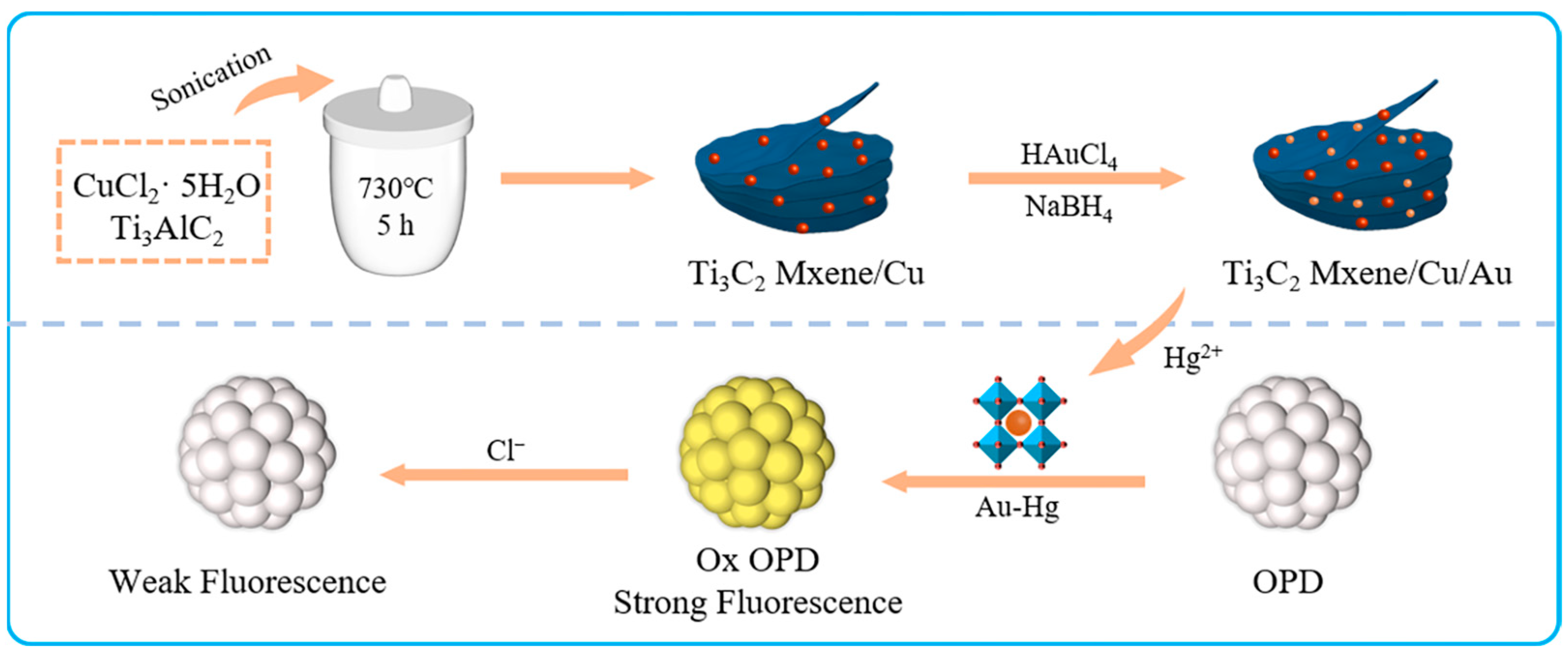
1. Introduction
2. Results and Discussion
2.1. Characterization of Au/Cu/Ti3C2 NSs
2.2. Oxidase-like Activity of Au/Cu/Ti3C2 NSs
2.3. Detection of Hg2+
2.4. Detection of Cl−
2.5. POCT for Hg2+ and Cl−
2.6. Selectivity for Hg2+ and Cl−
2.7. Real Sample Detection
3. Experimental
3.1. Materials
3.2. Instuments
3.3. Preparation of Au/Cu/Ti3C2 NSs
3.4. Fluorescence and Colorimetric Method for the Detection of Hg2+ and Cl−
3.5. Preparation of Test Papers
3.6. Real Sample Analysis
4. Conclusions
Supplementary Materials
Author Contributions
Funding
Institutional Review Board Statement
Informed Consent Statement
Data Availability Statement
Conflicts of Interest
Sample Availability
References
- Dimitrijevs, P.; Arsenyan, P. Cardiolipin in the spotlight: Quantitative analysis and fluorescence-based competitive binding assay. Sens. Actuators B-Chem. 2021, 346, 130537. [Google Scholar] [CrossRef]
- Boyd-Shiwarski, C.R.; Weaver, C.J.; Beacham, R.T.; Shiwarski, D.J.; Connolly, K.A.; Nkashama, L.J.; Mutchler, S.M.; Griffiths, S.E.; Knoell, S.A.; Sebastiani, R.S.; et al. Effects of extreme potassium stress on blood pressure and renal tubular sodium transport. Am. J. Physiol. Renal 2020, 318, 1341–1356. [Google Scholar] [CrossRef] [PubMed]
- Addis, D.R.; Aggarwal, S.; Lazrak, A.; Jilling, T.; Matalon, S. Halogen-induced chemical injury to the mammalian cardiopulmonary systems. Physiology 2021, 36, 272–291. [Google Scholar] [CrossRef] [PubMed]
- Madhesan, T.; Mitra, S.; Deivasigamani, P.; Nagarajan, S.; Brahmmananda Rao, C.V.S.; Mohan, A.M. Probe anchored porous organic polymer monolithic architectures as optical sensor for ultra-trace analysis of Hg2+ in water samples. Micropor. Mesopor. Mat. 2022, 333, 111724. [Google Scholar] [CrossRef]
- Wu, H.B.; Xie, R.Y.; Hao, Y.Q.; Pang, J.Y.; Gao, H.; Qu, F.Y.; Tian, M.M.; Guo, C.H.; Mao, B.D.; Chai, F. Portable smartphone-integrated AuAg nanoclusters electrospun membranes for multivariate fluorescent sensing of Hg2+, Cu2+and l-histidine in water and food samples. Food Chem. 2023, 418, 135961. [Google Scholar] [CrossRef] [PubMed]
- Zhang, H.J.; Wang, D.; Zhang, D.; Zhang, T.T.; Yang, L.K.; Li, Z.P. In situ microfluidic sers chip for ultrasensitive Hg2+ sensing based on I−-functionalized silver aggregates. ACS Appl. Mater. Interfaces 2022, 14, 2211–2218. [Google Scholar] [CrossRef]
- Fang, Y.M.; Zhang, Y.; Cao, L.G.; Yang, J.Z.; Hu, M.H.; Pang, Z.L.; He, J.H. Portable Hg2+ Nanosensor with ppt level sensitivity using nanozyme as the recognition unit, enrichment carrier, and signal amplifier. ACS Appl. Mater. Interfaces 2020, 12, 11761–11768. [Google Scholar] [CrossRef]
- Hou, Y.; Chen, Y.; Guo, X.Y.; Liu, W.; Zhang, L.; Lv, C.C.; Xu, Y.L.; Jin, Y.; Li, B.X. Aggregation-induced chemiluminescence system for sensitive detection of mercury ions. Anal. Bioanal. Chem. 2021, 413, 625–633. [Google Scholar] [CrossRef]
- Qi, Y.Y.; Xiu, F.R.; Yu, G.D.; Huang, L.L.; Li, B.X. Simple and rapid chemiluminescence aptasensor for Hg2+ in contaminated samples: A new signal amplification mechanism. Biosens. Bioelectron. 2017, 87, 439–446. [Google Scholar] [CrossRef]
- Wang, X.B.; Ma, X.Y.; Wen, J.H.; Geng, Z.R.; Wang, Z.L. A novel bimacrocyclic polyamine-based fluorescent probe for sensitive detection of Hg2+ and glutathione in human serum. Talanta 2020, 207, 120311. [Google Scholar] [CrossRef]
- Panthi, G.; Park, M. Synthesis of metal nanoclusters and their application in Hg2+ ions detection: A review. J. Hazard. Mater. 2022, 424, 127565. [Google Scholar] [CrossRef]
- Yin, P.C.; Niu, Q.F.; Liu, J.Q.; Wei, T.; Hu, T.T.; Li, T.D.; Qin, X.Y.; Chen, J.B. A new AIEE-active carbazole based colorimetric/fluorimetric chemosensor for ultra-rapid and nano-level determination of Hg2+ and Al3+ in food/environmental samples and living cells. Sens. Actuators B-Chem. 2021, 331, 129418. [Google Scholar] [CrossRef]
- Ismail, S.; Yusof, N.A.; Abdullah, J.; Rahman, S.F.A. Development of electrochemical sensor based on silica/gold nanoparticles modified electrode for detection of arsenite. IEEE Sens. J. 2019, 20, 1558–1748. [Google Scholar] [CrossRef]
- Cetin, D.; Yavuz, O.; Alcay, Y.; Yildirim, M.S.; Kaplan, M.; Aribuga, H.; Ozdemir, E.; Ertugral, U.; Yilmaz, I. Development of a new near-infrared, spectrophotometric, and colorimetric probe based on phthalocyanine containing mercaptoquinoline unit for discriminative and highly sensitive detection of Ag+, Cu2+, and Hg2+ ions. Spectrochim. Acta A 2023, 297, 122725. [Google Scholar] [CrossRef] [PubMed]
- Michalski, R.; Pecyna-Utylska, P.; Kernert, J. Ion chromatography and related techniques in carboxylic acids analysis. Crit. Rev. Anal. Chem. 2020, 51, 549–564. [Google Scholar] [CrossRef]
- Noviana, E.; McCord, C.P.; Clark, K.M.; Jang, I.; Henry, C.S. Electrochemical paper-based devices: Sensing approaches and progress toward practical applications. Lab Chip 2020, 20, 9–34. [Google Scholar] [CrossRef] [PubMed]
- Chen, Y.C.; Jiang, S.J. Simultaneous speciation of arsenic and mercury in fish by high-performance liquid chromatography inductively coupled plasma mass spectrometry. J. Anal. Atom. Spectrom. 2021, 36, 938–945. [Google Scholar] [CrossRef]
- Xing, Y.Q.; Han, Y.; Pierce, D.T.; Zhao, X.J. Aggregation-based determination of mercury (II) using DNA-modified single gold nanoparticle, T-Hg (II)-T interaction, and single-particle ICP-MS. Microchim. Acta 2020, 187, 56. [Google Scholar] [CrossRef]
- Das, D.; Dutta, R.K. N-doped carbon dots synthesized from ethylene glycol and β-alanine for detection of Cr (VI) and 4-nitrophenol via photoluminescence quenching. ACS Appl. Nano Mater. 2021, 4, 3444–3454. [Google Scholar] [CrossRef]
- Wazuddin, D.A.; Mujawar, L.H.; Abduljabbar, T.N.; El-Shahawi, M.S. In-situ droplet assay on wax-modified paper for rapid and trace determination of Fe3+ in water. Microchem. J. 2021, 170, 106723. [Google Scholar] [CrossRef]
- Li, X.; Gao, L.N.; Chen, Z.B. Highly sensitive colorimetric detection of glucose through glucose oxidase and Cu2+-catalyzed 3,3′,5,5′-tetramethylbenzidine oxidation. Spectrochim. Acta A 2019, 213, 37–41. [Google Scholar] [CrossRef] [PubMed]
- Qin, M.; Li, J.S.; Song, Y.L. Toward high sensitivity: Perspective on colorimetric photonic crystal sensors. Anal. Chem. 2022, 94, 9497–9507. [Google Scholar] [CrossRef]
- Zhou, Y.; Ma, Z.F. Colorimetric detection of Hg2+ by Au nanoparticles formed by H2O2 reduction of HAuCl4 using Au nanoclusters as the catalyst. Sens. Actuators B-Chem. 2017, 241, 1063–1068. [Google Scholar] [CrossRef]
- Logan, N.; McVey, C.; Elliott, C.; Cao, C. Amalgamated gold-nanoalloys with enhanced catalytic activity for the detection of mercury ions (Hg2+) in seawater samples. Nano Res. 2020, 13, 989–998. [Google Scholar] [CrossRef]
- Wang, L.J.; Xu, X.C.; Liu, P.; Wang, M.Z.; Niu, X.H.; Pan, J.M. A single-nanozyme colorimetric array based on target-induced differential surface passivation for quantification and discrimination of Cl−, Br−and I− ions. Anal. Chim. Acta 2021, 1160, 338451. [Google Scholar] [CrossRef]
- Yan, Z.Q.; Xing, L.; Zhao, L.; Zhang, X.Y.; Zhang, Y.F.; Tang, Y.L.; Zhou, X.M.; Hu, L.; Zhu, N.L. β-Cyclodextrin and graphene oxide co-strengthened AgRu bimetal mesoporous nanozyme: An efficient strategy for visual detection and removal of toxic Hg2+ and Cl−. J. Environ. Chem. Eng. 2022, 10, 108242. [Google Scholar] [CrossRef]
- Alwarappan, S.; Nesakumar, N.; Sun, D.; Hu, T.Y.; Li, C.Z. 2D metal carbides and nitrides (MXenes) for sensors and biosensors. Biosens. Bioelectron. 2022, 205, 113943. [Google Scholar] [CrossRef]
- Qin, R.; Shan, G.; Hu, M.; Huang, W. Two-dimensional transition metal carbides and/or nitrides (MXenes) and their applications in sensors. Mater. Today Phys. 2021, 21, 100527. [Google Scholar] [CrossRef]
- Li, Y.J.; Ding, L.; Guo, Y.C.; Liang, Z.Q.; Cui, H.Z.; Tian, J. Boosting the photocatalytic ability of g-C3N4 for hydrogen production by Ti3C2 MXene quantum dots. ACS Appl. Mater. Interfaces 2019, 11, 41400–41407. [Google Scholar] [CrossRef]
- Peng, X.Y.; Zhang, Y.L.; Lu, D.T.; Guo, Y.J.; Guo, S.J. Ultrathin Ti3C2 nanosheets based “off-on” fluorescent nanoprobe for rapid and sensitive detection of HPV infection. Sens. Actuators B-Chem. 2019, 286, 222–229. [Google Scholar] [CrossRef]
- Mai, Y.J.; Li, Y.G.; Li, S.L.; Zhang, L.Y.; Liu, C.S.; Jie, X.H. Self-lubricating Ti3C2 nanosheets/copper composite coatings. J. Alloys Compd. 2019, 770, 1–5. [Google Scholar] [CrossRef]
- Wu, S.S.; Su, Y.M.; Zhu, Y.; Zhang, Y.M.; Zhu, M.S. In-situ growing Bi/BiOCl microspheres on Ti3C2 nanosheets for upgrading visible-light-driven photocatalytic activity. Appl. Surf. Sci. 2020, 520, 146339. [Google Scholar] [CrossRef]
- Chen, D.; Shao, S.B.; Zhang, W.; Zhao, J.B.; Lian, M.L. Nitrogen and sulfur co-doping strategy to trigger the peroxidase-like and electrochemical activity of Ti3C2 nanosheets for sensitive uric acid detection. Anal. Chim. Acta 2022, 1197, 339520. [Google Scholar] [CrossRef]
- Wu, X.J.; Chen, T.M.; Chen, Y.; Yang, G.W. Modified Ti3C2 nanosheets as peroxidase mimetics for use in colorimetric detection and immunoassays. J. Mater Chem. B 2020, 8, 2650–2659. [Google Scholar] [CrossRef]
- Zhang, Z.G.; Lu, X.T.; Xu, J.R.; Luo, H.J. Characterization and tribological properties of graphene/copper composites fabricated by electroless plating and powder metallurgy. Acta Metall. Sin.-Engl. 2020, 33, 903–912. [Google Scholar] [CrossRef]
- Poorshamohammad, C.; Liu, L.G.; Cheng, X.R.; Momtazi-Borojeni, A.A. Green synthesis of plant-stabilized Au nanoparticles for the treatment of gastric carcinoma. Arab. J. Chem. 2023, 16, 104386. [Google Scholar] [CrossRef]
- Yan, L.X.; Chen, Z.P.; Zhang, Z.Y.; Qu, C.L.; Chen, L.X.; Shen, D.Z. Fluorescent sensing of mercury (II) based on formation of catalytic gold nanoparticles. Analyst 2013, 138, 4280–4283. [Google Scholar] [CrossRef]


| Samples | Spiked (µM) | Found (µM) | Recovery (%) | RSD (%) |
|---|---|---|---|---|
| Shahu Lake | 24 | 25.23 | 105.1 | 2.3 |
| 80 | 87.78 | 109.7 | 2.1 | |
| 160 | 149.22 | 93.3 | 2.5 | |
| East Lake | 24 | 23.04 | 96.0 | 2.8 |
| 80 | 82.29 | 102.9 | 5.5 | |
| 160 | 159.1 | 99.4 | 1.4 | |
| Yangtze River | 24 | 22.49 | 93.7 | 2.6 |
| 80 | 83.39 | 104.3 | 3.1 | |
| 160 | 154.71 | 96.7 | 4.5 |
Disclaimer/Publisher’s Note: The statements, opinions and data contained in all publications are solely those of the individual author(s) and contributor(s) and not of MDPI and/or the editor(s). MDPI and/or the editor(s) disclaim responsibility for any injury to people or property resulting from any ideas, methods, instructions or products referred to in the content. |
© 2023 by the authors. Licensee MDPI, Basel, Switzerland. This article is an open access article distributed under the terms and conditions of the Creative Commons Attribution (CC BY) license (https://creativecommons.org/licenses/by/4.0/).
Share and Cite
Chen, K.; Fu, S.; Jin, C.; Guo, F.; He, Y.; Ren, Q.; Wang, X. Smartphone-Enabled Fluorescence and Colorimetric Platform for the On-Site Detection of Hg2+ and Cl− Based on the Au/Cu/Ti3C2 Nanosheets. Molecules 2023, 28, 5355. https://doi.org/10.3390/molecules28145355
Chen K, Fu S, Jin C, Guo F, He Y, Ren Q, Wang X. Smartphone-Enabled Fluorescence and Colorimetric Platform for the On-Site Detection of Hg2+ and Cl− Based on the Au/Cu/Ti3C2 Nanosheets. Molecules. 2023; 28(14):5355. https://doi.org/10.3390/molecules28145355
Chicago/Turabian StyleChen, Keyan, Shiqi Fu, Chenyu Jin, Fan Guo, Yu He, Qi Ren, and Xuesheng Wang. 2023. "Smartphone-Enabled Fluorescence and Colorimetric Platform for the On-Site Detection of Hg2+ and Cl− Based on the Au/Cu/Ti3C2 Nanosheets" Molecules 28, no. 14: 5355. https://doi.org/10.3390/molecules28145355
APA StyleChen, K., Fu, S., Jin, C., Guo, F., He, Y., Ren, Q., & Wang, X. (2023). Smartphone-Enabled Fluorescence and Colorimetric Platform for the On-Site Detection of Hg2+ and Cl− Based on the Au/Cu/Ti3C2 Nanosheets. Molecules, 28(14), 5355. https://doi.org/10.3390/molecules28145355