Article

Nonaqueous Capillary Electrophoretic Separation of Analogs of (24R)-1,24-Dihydroxyvitamin D3 Derivative as Predicted by Quantum Chemical Calculations

by
Błażej Grodner
1,*,†,
Teresa Żołek
2,† and
Andrzej Kutner
3
1
Department of Biochemistry and Pharmacogenomics, Faculty of Pharmacy, Medical University of Warsaw, 1 Banacha, 02-097 Warsaw, Poland
2
Department of Organic and Physical Chemistry, Faculty of Pharmacy, Medical University of Warsaw, 1 Banacha, 02-097 Warsaw, Poland
3
Department of Drug Chemistry, Faculty of Pharmacy, Medical University of Warsaw, 1 Banacha, 02-097 Warsaw, Poland
*
Author to whom correspondence should be addressed.
These authors contributed equally to this work.
Molecules 2023, 28(13), 5055; https://doi.org/10.3390/molecules28135055
Submission received: 18 May 2023 / Revised: 16 June 2023 / Accepted: 25 June 2023 / Published: 28 June 2023

Abstract

:
Nonaqueous capillary electrophoretic (NACE) separation was obtained of analogs of (24R)-1,24-dihydroxyvitamin D3 derivative (calcipotriol) as predicted by quantum chemical calculations supported by the density functional theory (DFT). Among the key electronic properties investigated, absolute values of the dipole polarizability and energy gap between HOMO and LUMO molecular orbitals of the analog molecules differ significantly for particular analogs, and there is a direct relationship with their electrophoretic migration time. These differences and relationships suggest that the structurally related analogs should be separable in the electrostatic field. Indeed, the robust, sensitive, and rapid NACE method was first developed for the identification and determination of the anticancer analog of calcipotriol (coded PRI-2205) and its process-related impurities (coded PRI-2201, PRI-2203, and PRI-2204) in organic and aqueous biological solutions. The direct relation between the calculated electronic properties of the analogs and the experimental electrophoretic migration time could be a promising prospect for theoretically predicting the electrophoretic separations.

1. Introduction

Vitamin D3 is a fat-soluble vitamin that is synthesized in the skin from 5,7-dehydrocholesterol upon exposure to sunlight [1]. The biological effect of vitamin D3 is expressed through its interaction with the nuclear vitamin D receptor (VDR). The VDR is a tetrameric protein complex typical of all nuclear steroid receptors. Activation of the VDR leads to the activation of transcription factors [2,3,4,5,6]. Thus, the mechanism of interaction of the VDR—vitamin D3 complex with nuclear DNA is typical for steroid hormones [4,7]. The interaction of vitamin D3 with the membrane receptor in vitro was also reported. The final biological effect of vitamin D3 is caused by the transduction of the intracellular signal via second messengers [5,8,9]. The major biological function of vitamin D3 is to maintain normal blood levels of calcium and phosphorus. This is why vitamin D3 is necessary for mineral homeostasis and the proper formation of bone [10]. Vitamin D3 is reported to provide protection from and decrease an individual’s risk of developing osteoporosis, hypertension, cancer, and several autoimmune diseases. This is related to the stimulation of the body to produce antibodies and increase the strength of the immune system [11]. Vitamin D3 activates the production of anti-inflammatory factors, such as interleukin-4, and inhibits the formation of proinflammatory factors, such as interleukin 6, tumor necrosis factor, and macrophage colony-stimulating factor [12,13,14,15]. The neuroprotective role of vitamin D3 was also reported, regarding the reduction of the Ca2+ level in the brain [16,17,18,19,20,21] and the inhibition of brain γ-glutamyl transpeptidase [12,16]. The anticancer property of vitamin D has been studied in a wide variety of commonly occurring cancers (both in vitro and in vivo) of which colorectal, breast, and prostate cancers have been most responsive. Epidemiological studies provide strong evidence that the active form of vitamin D3, 1α,25-dihydroxyvitamin D3 [1,25(OH)2D3, calcitriol] (Figure 1), reduces the incidence of common human cancers, including carcinomas of the breast, prostate, and colon [22,23,24,25].
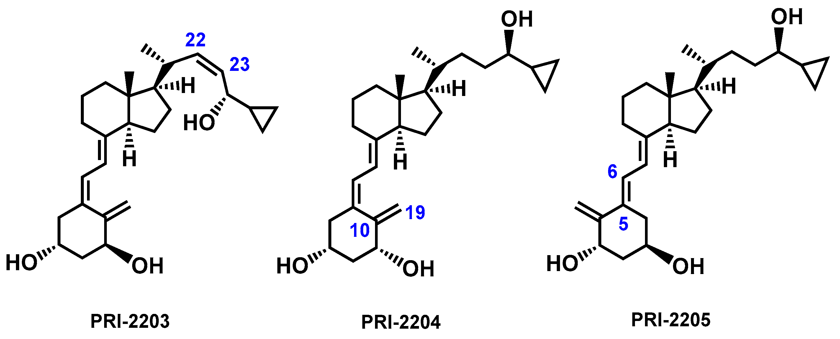
The above information clearly shows that vitamin D3 plays a very important role in our lives, and therefore, research related to the function of vitamin D3 still arouses great interest. There are also high expectations associated with vitamin D3 analogs due to their potential therapeutic effect [26]. Analogs of calcipotriol, PRI-2201 (Figure 1) and PRI-2205 (Figure 2) showed antiproliferative activity in vitro against human cancer cell lines [27].
The analogs were more active than calcitriol and calcipotriol in terms of mouse Lewis lung cancer inhibition. Very advantageously, PRI-2205 revealed no undesired calcemic activity that limits the clinical use of the parent calcitriol. Although PRI-2205 potentiated the antiproliferative effect of cis-platin and doxorubicin against leukemia HL-60 cells and that of tamoxifen against MCF-7 breast cancer cells at higher doses than the reference calcitriol, it significantly increased VDR expression [28]. Additionally, PRI-2205 significantly enhanced the antitumor activity of 5-FU depending on the treatment regimen, decreased tumor growth and metastasis, and prolonged the survival time of research animals, compared to 5-FU alone [29]. The analogs PRI-2203 and PRI-2204 are representative process-related or degradation impurities of PRI-2205 [28].
To assess pharmacokinetic, pharmacological, and bioavailability parameters, there is a need to develop a method to confirm the identity and homogeneity of a given analog as well as one for their qualitative and quantitative determination, especially in biological material. Of great interest is a straightforward method for the separation of an analog from its diastereomers and geometric isomers as potential process-related impurities or degradation products. HPLC has mainly been reported [30,31,32,33,34] for the determination of vitamin D3 and metabolites in biological matrices and food products. However, none of these methods were used for synthetic analogs of calcitriol. Previously, we achieved the separation of vitamin D diastereomers by chiral stationary phase HPLC [35,36]. However, due to the sensitivity of chiral HPLC columns, these separations were limited to a few solvent systems and are not applicable to a broad range of synthetic vitamin D analogs.
At present, the effectiveness of separation is often based on experiments using capillary electrophoresis (CE). CE is a family of related techniques that employ narrow-bore capillaries to perform high efficiency separations of both large and small molecules. These separations are facilitated by the use of high voltages, which may generate electroosmotic and electrophoretic flow of buffer solutions and ionic species, respectively, within the capillaries. Under the influence of an electric field, the ionic species in a sample that is introduced as a zone into an electrolyte at one end of a capillary will be separated into discrete bands when they migrate to the other end of the capillary at different electrophoretic velocities. Capillary electrophoresis comprises a family of techniques that sometimes have extremely different operative and separative characteristics. Among these techniques, the following should be distinguished: capillary zone electrophoresis (CZE), isoelectric focusing, capillary gel electrophoresis, isotachophoresis, and micellar electrokinetic capillary chromatography. Nonaqueous capillary electrophoresis (NACE) is a variant of CZE that uses organic solvents, which are the main components of solutions in which the separation of compounds that are sparingly soluble or insoluble in polar solvents is carried out. The main benefit of using organic solvents instead of water in CE is the improvement of analyte solubility, especially when UV detectors are used. In many cases, the use of organic solvents leads to significantly shorter analysis times and a higher separation efficiency [37]. To the best of our knowledge, the NACE method has not been used to study vitamin D analogs so far.
However, experimental methods are labor-intensive and expensive. For this reason, analog synthesis is being optimized using new methods, such as quantum mechanical calculations [38,39]. In recent years, quantum chemical (QC) methods have emerged as an efficient tool for the accurate prediction of stability and chemical reactivity of molecules [40,41,42]. Among the full methods used in QC, density functional theory (DFT) can mainly be mentioned. Due to its theoretical nature, DFT is considered a “green technique” for optimizing chemical structures. Various QC parameters, such as the dipole moment (μ), dipole polarizability (α), energy difference, highest occupied molecular orbital (EHOMO), and lowest occupied molecular orbital (ELUMO), etc., are used to determine the molecular properties of newly designed compounds. The global molecular properties also justify the reactivity and stability of synthetic calcitriol analogs. The most stable analog structure has the largest HOMO-LUMO energy gap. Therefore, an electron system with a larger HOMO-LUMO energy gap is less reactive than one with a smaller gap. Moreover, according to the principle of minimum polarizability of an electric dipole, the natural direction of the evolution of a given structure is the state of minimum polarizability [43].
In this paper, we present a combination of computational and experimental techniques to investigate the possible relationship between the molecular structures and electronic properties of a series of structurally related vitamin D3 analogs and their electrophoretic separation. Here, we report the method for their simultaneous determination using nonaqueous capillary electrophoresis (NACE) and quantum chemical parameters calculated by the DFT method.

2. Results and Discussion

2.1. Electronic Properties of Vitamin D3 Analogs

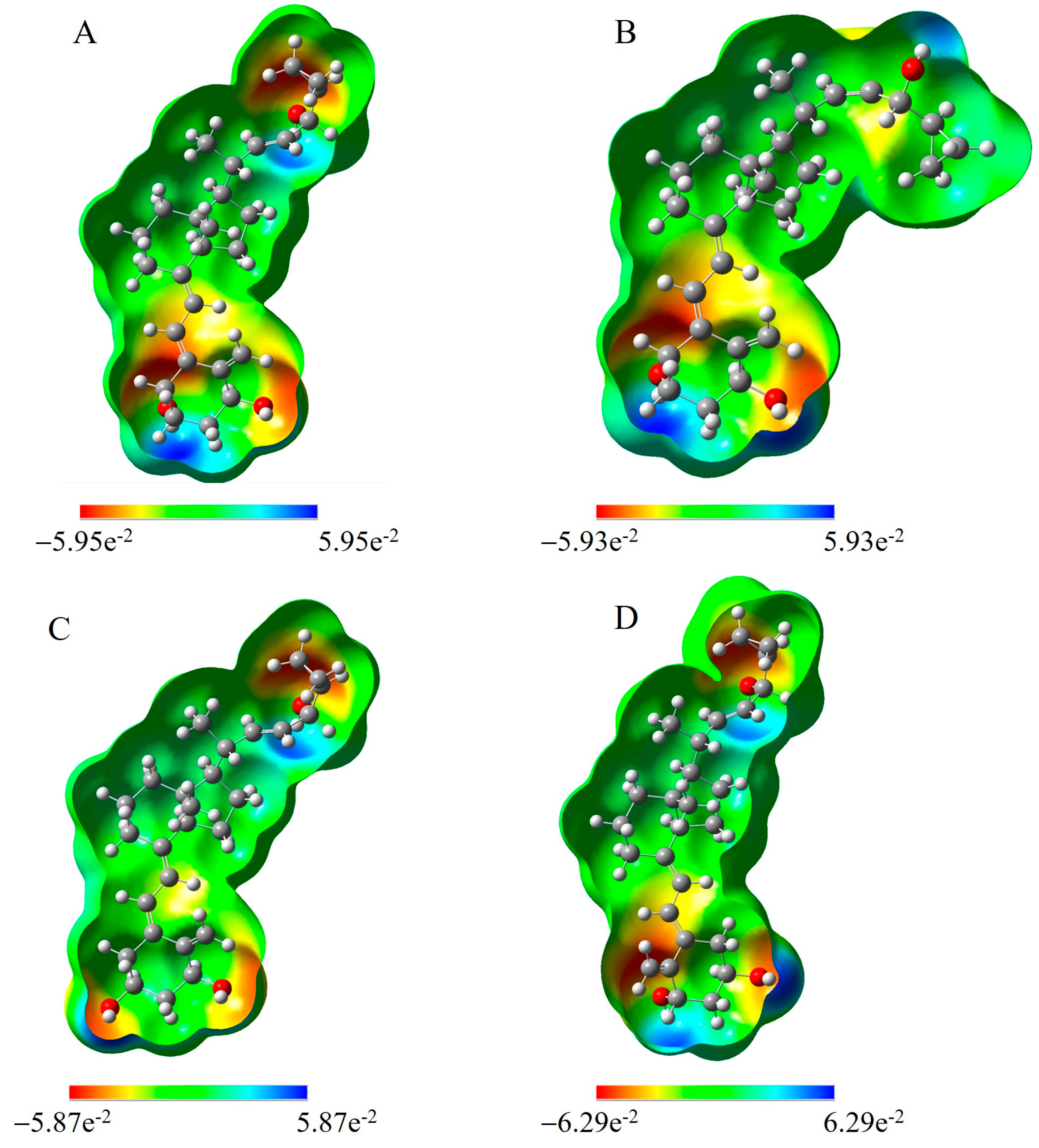
To investigate the effect of the molecular structure on the mobility of analogs PRI-2201, PRI-2203, PRI-2204, and PRI-2205 in the electrostatic field, full-structure optimization was first performed for each molecule. All categories of the electronic properties of the analogs, including the dipole moment, dipole polarizability, the energies of the HOMO and LUMO molecular orbitals, and the molecular electrostatic potential (MEP) were calculated (Table 1, Figure 4 and Figure 5, and Tables S1 and S2 in the Supplementary Materials).
The molecular dipole moment is a basic property of a molecule that is used to investigate intermolecular interactions. An increase in the dipole moment raises the polar nature of a bond in a molecule. The DFT-calculated data indicate that the dipole moment of four vitamin D3 analogs was in the order PRI-2204 < PRI-2203 < PRI-2205 < PRI-2201. The highest dipole moments of PRI-2201 and PRI-2205 signified their strong intermolecular interactions. The dipole moment for PRI-2201 (μ = 4.23 Debye), larger than that of PRI-2205 (μ = 3.49 Debye), may be due to the presence of a strong electron donor–acceptor configuration in the molecule. As we noted earlier, dipole polarizability is a quantity of particular interest for structural chemistry. The molecular polarizability of a molecule characterizes the capability of its electronic system to be distorted by the external field, and it plays an important role in modeling many molecular properties and biological activities [44]. This parameter relates to how the susceptibility of system electrons can be influenced by the approaching charge. This is affected by several factors, such as the compound complexity, the size of the molecular systems, and the number of free electrons. Herein, the order of mean polarizability PRI-2205 > PRI-2204 > PRI-2203 > PRI-2201 suggests that the molecule PRI-2205 (αtotal = 434.66 a.u.) is the most polarizable; regardless, their differences are not very high. In addition, the theoretically determined dipole polarizability values were well correlated with the experimentally determined average electrophoretic migration times for a small set of data with four molecules (R2 = 0.83, Figure 3A), only. The correlation coefficient was high enough, proving the model’s predictive ability. Thus, the model is suitable for predicting the order of migration of vitamin D3 analogs in NACE.
Another key electronic property investigated was the study of the HOMO and LUMO distribution in the molecules to assess their electron delocalization and chemical reactivity. The energy gap (Eg = EHOMO-ELUMO) explains the charge transfer interaction occurring within the molecules. HOMO represents the ability to donate an electron, and LUMO means the ability to obtain an electron. The Eg reveals the chemical reactivity of molecules and proves the occurrence of charge transfer within molecules. The frontier orbital gap helps to characterize the molecules’ chemical reactivity and kinetic stability. A molecule with a small frontier orbital gap is more polarizable. It is generally associated with a high chemical reactivity and low kinetic stability and is also termed a soft molecule [45]. The calculated HOMO and LUMO gaps, the Eg values, are in the narrow range of −4.48 to −4.69 eV (Table 1). The PRI-2205 molecule has the lowest Eg (−4.48 eV), suggesting it has the highest polarizability. The Eg values increase from −4.67 eV for PRI-2204 through to−4.68 eV for PRI-2203 and−4.69 eV for PRI-2201. Similarly, the electrophoretic mobility changes from 1.82 min for PRI-2204 to 1.93 min for PRI-2203 and 2.08 min for PRI-2201. The correlation between the Eg value and the experimental electrophoretic average migration time (R2 = 0.80, Figure 3B)confirms that structural differences affect the migration of analytes.
For all investigated molecules, the isosurfaces of molecular orbitals and electron density differences were delineated (Figure 4).The charge distribution in the HOMOs is localized on the A-ring hydroxyls, 19-methylene at C-10, at the C-5 position in the vitamin D triene system, and on the side chain for PRI-2203, whereas the distribution of LUMOs occurs in 19-methylene at C-10 and in the triene system at C-5.
Moreover, for all analogs, we calculated molecular electrostatic potential (MEP) maps(Figure 5).The MEP is an important descriptor to validate the reactivity related to the molecular system. It is also an important tool to envisage how molecules with different geometries can interact, and how the charge can be transferred. The three-dimensional MEP surface gives an indication of the position, shape, and size of the positive, negative, and neutral electrostatic potentials. The maximum positive region is the preferred site for the nucleophilic attack, which is designated by blue color. Likewise, a maximum negative region is favored for an electrophilic attack, represented by a red surface. The proton-rich region exhibits positive potential, and the repulsion energy between the protons is marked as the green region. For all analogs, high electrostatic potential regions are present in the vicinity of the hydroxyl’s oxygen. A low electropositive potential is mainly found on the hydrogen atoms of hydroxyls present in the A-ring. Further, the variance in the MEP surface shows an increase in positive potential due to the proton environment in the molecule.

2.2. Development of Capillary Electrophoresis

The NACE method was developed for the determination of analogs of the derivative of (24R)-1,24-dihydroxyvitamin D3. The type and composition of the separation electrolyte (background electrolyte—BGE) were studied in different concentrations of sodium acetate and methanol and with different acetonitrile ratios in the separation solution for the quantification of the analogs PRI-2205,PRI-2204, PRI-2203, and PRI-2201.
The effect of the sodium acetate concentration was studied in the range of 50 to 200 mM for the BGE containing methanol only. When the concentration of sodium acetate was 50 mM, the peak shapes were inadequate, and the resolution for all investigated compounds was not obtained (Figure S1A included in the Supplementary Materials). Therefore, by varying the concentration, the best resolution was achieved. The resolution of the investigated compounds increased with an increasing concentration of sodium acetate in BGE. A clear but only partial separation of PRI-2205, PRI-2204, PRI-2203, and PRI-2201 was obtained at a sodium acetate concentration of 100 mM (Figure S1B included in the Supplementary Materials). Determination of the analogs was carried out further by increasing the concentration of sodium acetate. Increasing the sodium acetate concentration to 200 mM negatively affected the resolving power of BGE (Figure S1C included in the Supplementary Materials). Finally, the best primary resolution was obtained with a sodium acetate concentration of 100 mM (Figure S1B included in the Supplementary Materials).
Then, in order to increase the resolution, the quantitative composition of the methanol:acetonitrile mixture in the BGE was modified. The initial separation of the PRI-2205, PRI-2204, PRI-2203, and PRI-2201 analogs was carried out in plain methanol and at a sodium acetate concentration of 100 mM (Figure S2A included in the Supplementary Materials). Improvement of the resolution was obtained after increasing the content of acetonitrile to the BGE. The use of a mixture of methanol:acetonitrile at a ratio of 90:10 significantly improved the resolution (Figure S2B included in the Supplementary Materials). However, the best resolution was obtained using a mixture of methanol:acetonitrile at a ratio of 80:20 and a sodium acetate concentration of 100 mM (Figure S2C included in the Supplementary Materials).
Various analytical conditions were also examined. The voltage, analysis temperature, capillary length, and detection wavelength were optimized in the ranges of 5–20 kV, 20–30 °C, 30–80 cm (total length), and 200–300 nm, respectively. The greatest influences on the detection, separation, and analysis time of PRI-2205, PRI-2204, PRI-2203 and PRI-2201 came from the wavelength, current voltage, and capillary length. The best detection was obtained at a wavelength of 273 nm, providing maximal absorption of 5,6-trans analogs of vitamin D, such as PRI-2205. The best resolution and the shortest analysis time were obtained at a voltage of 10 kV. Further increasing the voltage value accelerated the analysis time but decreased the resolution and stability of the current intensity. The length of the capillary also had significant effects on the resolution and analysis time. The use of the longest capillary resulted in very good separation, but the analysis time was very long. With the use of shorter capillaries, the migration time between individual analogs was shortened, together with the analysis time. However, the use of the shortest capillary maintained appropriate resolutions for all four vitamin D3 analogs, and the separation was obtained in the shortest analysis time. Ultimately, the best separation parameters were obtained using a separation solution of 80:20 (methanol: acetonitrile) containing sodium acetate at a concentration of 100 mM at 273 nm, 20 °C, and 10 kV with a capillary length of 30 cm (Figure S1 is included in the Supplementary Materials).
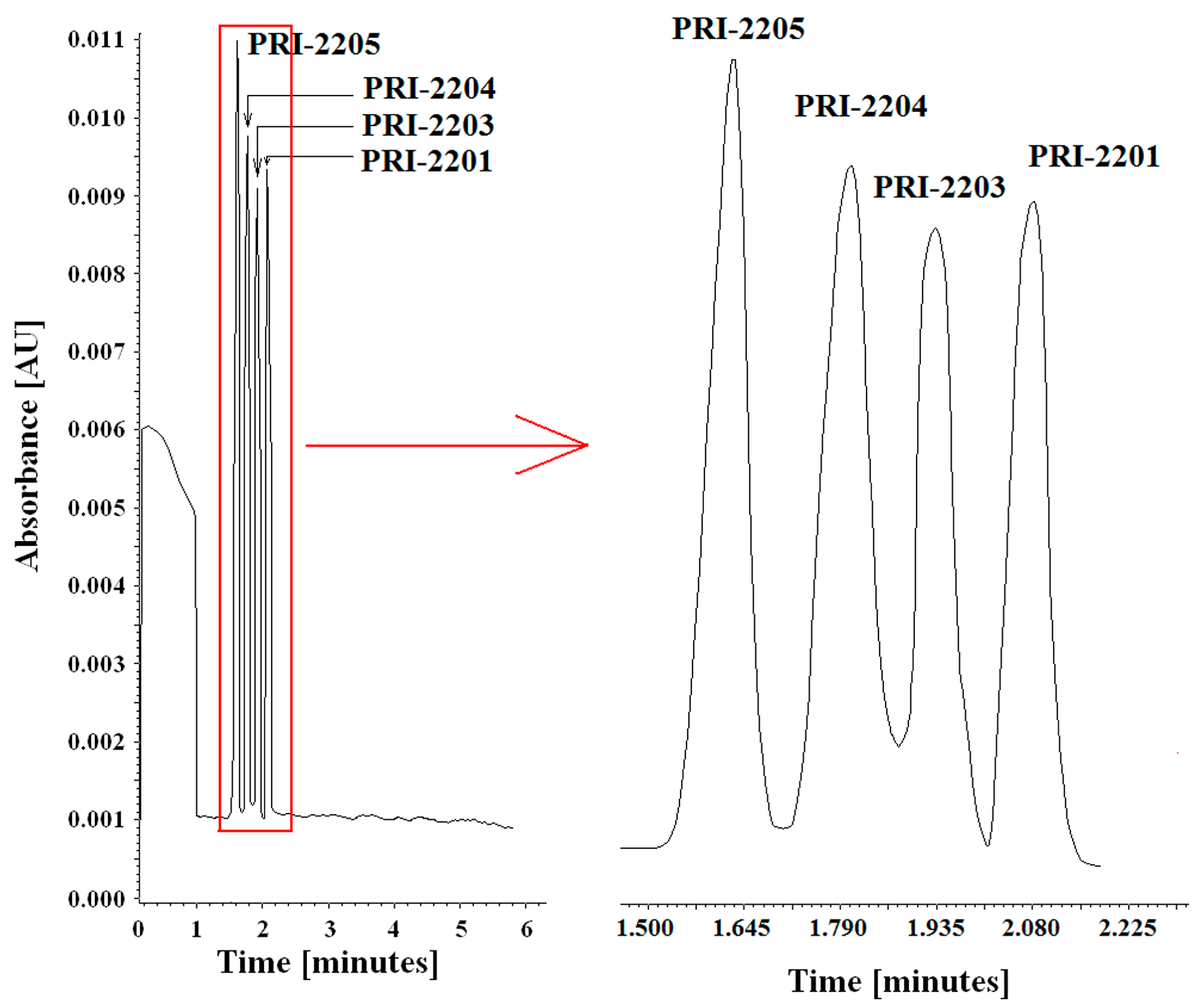
To determine the selectivity of the method, electropherograms of extracts of blank serum samples and serum samples containing PRI-2205, PRI-2204, PRI-2203, and PRI-2201 were analyzed. In the absence of interference caused by the presence of endogenous compounds, the method was defined as selective. The specificity of the method was determined by comparing electropherograms of the blank solution, solutions containing PRI-2205, PRI-2204, PRI-2203, and PRI-2201, blank serum samples, and serum samples containing analogs. In all cases, no interference was observed between the peaks from endogenous substances and the migration times of the tested analogs (Figure 6 and Figure S2 is included in the Supplementary Materials).

3. Materials and Methods

3.1. Quantum Chemical Calculations

Theoretical calculations were performed using the Gaussian 16W suite of software [46]. The starting conformations of PRI-2205, PRI-2204, PRI-2203, and PRI-2201 were constructed based on the solid-state diffraction data of structurally related compounds to eliminate any subjectivity in generating the three-dimensional structure [26]. The molecular structures of the analogs were optimized using density functional theory (DFT) with the Becke-3-parameter–Lee–Yang–Parr (B3LYP) hybrid functional [47] and the 6-311G(d,p) basic set [48]. The solvent effect was considered using the Polarizable Continuum Model (PCM) implemented in the Gaussian 16W. Methanol was used in all calculations considering the experimental conditions. Vibrational frequency calculations ensured that the obtained structures represented the global minima. All calculations were performed using standard gradient techniques and default convergence criteria. The quantum chemical parameters, such as the EHOMO (energy of the highest occupied molecular orbital), ELUMO (energy of the lowest unoccupied molecular orbital), Eg (energy gap), dipole moment (μ), and dipole polarizability (α) were calculated to reveal the compounds’ stability and chemical reactivity. The electrostatic potential (ESP) of the atomic partial charges on the atoms was computed using the Breneman model [49], which reproduces the molecular electrostatic potential.

3.2. Chemicals and Reagents

The synthesis, spectroscopic analysis (NMR and MS), and biological activity of PRI-2205, PRI-2204, PRI-2203, and PRI-2201 were described previously [27,28,29]. The purities of the reference compounds were found by HPLC to be greater than 98%. All other analytes were of a purity of 99%. Acetonitrile, methanol, sodium acetate, and human serum standard were obtained from Sigma-Aldrich (Poznań, Poland).

3.3. Instrumentation

A Capillary Electrophoresis Beckman Coulter P/ACE MDQ system, equipped with an autosampler and a UV/Visible detector, was used. All the parameters of the CE were controlled by Karat software version 32.

3.4. CE Conditions

The measurements were performed using a Beckman capillary electrophoresis system with a UV array detector. An amine permanently coated fused-silica eCAP capillary with an inner diameter of 75 μm, total length of 40 cm, and efficient length of 30 cm was used. An eCAP fused-silica capillary (30 cm total length, 20 cm effective length, 50 μm id, 375 μm od) was also used. The capillaries were kept at 20 °C. Detection was performed at 273 nm. Hydrodynamic sample injection was performed under a pressure of 3 psi in 5 s. Prior to use, the capillary was washed with a 0.1 M NaOH solution for 5 min, then with water for 5 min, and with a background electrolyte solution for 10 min. Between the analyses, it was washed with a background electrolyte solution (100 mM sodium acetate in methanol:acetonitrile 80:20) for 3 min. The separation was carried out at 10 kV.

3.5. Preparation of Stock and Working Standard Solutions

Primary standard stock solutions for the analogs PRI-2201, PRI-2203, PRI-2204, and PRI-2205 were prepared separately by dissolving 100 μg of each analog in 1 mL of methanol. These solutions were further diluted with methanol to obtain working standard solutions of 10.00 μg/mL, 20.00 μg/mL, 40.00 μg/mL, 80.00 μg/mL, 160.00 μg/mL, and 320.00 μg/m.
To prepare the calibration curve, 25 µL of each of the working solutions was taken and supplemented with methanol to a volume of 1 mL to obtain mixtures of all analogs with final concentrations of 0.25, 0.50, 1.00, 2.00, 4.00, and 8.00 μg/mL. The serum solutions were obtained similarily. For this purpose, 25 mL of each of the analog working solutions was added to six test tubes containing 0.975 mL of serum, thus obtaining final concentrations of 0.25, 0.50, 1.00, 2.00, 4.00, and 8.00 μg/mL for each analog. All samples were thoroughly vortexed for 5 min. Then, 3 mL of 80:20 hexane:ethyl acetate was added to each sample and thoroughly vortexed again for 5 min. The mixture was centrifuged (5 min, 1000 g), and the organic layer was transferred to a separate tube. This operation was repeated. Then, the test tubes with the extracted analogs and the hexane:ethyl acetate mixture were evaporated in a stream of nitrogen at 37 °C. The residues were dissolved in 1 mL of methanol and injected into the capillary.

3.6. Method Validation

A description and the validation conditions of the method are included in the “Supplementary Materials” section.

4. Conclusions

Electrophoretic separation of diastereomeric and isomeric vitamin D analogs was first predicted theoretically by calculating the electronic properties of the molecules and then obtained experimentally. The average electrophoretic migration time correlated best with the dipole polarizability. Rapid nonaqueous capillary electrophoresis (NACE) allowed for the identification and accurate, precise, and sensitive quantification of the structurally related analogs PRI-2205 and PRI-2204 and PRI-2203, and PRI-2201, which are associated with anticancer activity in organic solutions and serum. The extraction method used showed good linearity for all tested analogs (R2 > 0.9976). The method was suitable for determining analogs within an LOQ range of 287.1–310.1 ng/mL for the nominal analyte concentration. In addition, the RSD for intra- as well as interday precision was low and did not exceed 5% for all analogs. Further, to examine the scope and limitations of the predictive power of the calculated dipole polarizability for electrophoretic separation, closely related analogs of active forms of not only vitamin D3 but also D2 will be examined.

Supplementary Materials

The following supporting information can be downloaded at: https://www.mdpi.com/article/10.3390/molecules28135055/s1, Table S1.The DFT calculated total electric dipole moments, μ (Debye) and dipole moment components for PRI-2201, PRI-2203, PRI-2204, and PRI-2205.; Table S2. The DFT calculated total dipole polarizability and some selected components of the dipole polarizability for PRI-2201, PRI-2203, PRI-2204, and PRI-2205.; Table S3. Regression equation, limits of detection, and quantification for the PRI-2205, PRI-2204, PRI-2203, and PRI-2201 analogs (n = 6).; Table S4. Recovery data for the PRI-2205, PRI-2204, PRI-2203, and PRI-2201 vitamin D3 analogs from serum samples after the extraction procedure (n = 6). Table S5. Intraday and interday precision of the PRI-2205, PRI-2204, PRI-2203, and PRI-2201 analogs.; Figure S1. Electrophoregrams of the mixture of PRI-2205, PRI-2204, PRI-2203, and PRI-2201 analogs in different separation phase solutions: (A) 50 mM sodium acetate in methanol, (B) 100 mM sodium acetate in methanol, (C) 200 mM mM sodium acetate in methanol.; Figure S2. Electropherograms of the mixture of PRI-2205, PRI-2204, PRI-2203, and PRI-2201 analogs in 50 mM sodium acetate and different proportions of the methanol:acetonitrile mixture: (A) 50 mM sodium acetate in methanol, (B) 50 mM sodium acetate in 90:10 methanol:acetonitrile.

Author Contributions

Conceptualization, B.G. and A.K.; methodology, B.G and T.Ż.; formal analysis, B.G. and T.Ż.; investigation, B.G. and T.Ż.; data curation, B.G. and T.Ż.; writing-original draft preparation, B.G, T.Ż. and A.K.; writing-review and editing, B.G, T.Ż. and A.K.; visualization, T.Ż.; supervision, B.G.; project administration, B.G. All authors have read and agreed to the published version of the manuscript.

Funding

This research received no external funding.

Institutional Review Board Statement

Not applicable.

Informed Consent Statement

Not applicable.

Data Availability Statement

All relevant data are provided in the article.

Acknowledgments

The analytical studies were performed in the Chair and Department of Biochemistry and Pharmacogenomics, calculations at the Department of Organic and Physical Chemistry and manuscript preparation, as well as at the Department of Drug Chemistry, Faculty of Pharmacy of the Medical University of Warsaw (MUW), Poland. Financial support from the statutory funds of MUW is greatly acknowledged.

Conflicts of Interest

The authors declare no conflict of interest.

References

  1. Lehmann, B. The vitamin D3 pathway in human skin and its role for regulation of biological processes. Photochem. Photobiol. 2005, 81, 1246–1251. [Google Scholar] [CrossRef]
  2. Sanchez, B.; Lopez-Martin, E.; Segura, C.; Labandeira-Garcia, J.L.; Perez-Fernandez, R. 1,25-Dihydroxyvitamin D(3) increases striatal GDNF mRNA and protein expression in adult rats. Brain Res. Mol. Brain Res. 2002, 108, 143–146. [Google Scholar] [CrossRef]
  3. Kamei, Y.; Kawada, T.; Fukuwatari, T.; Ono, T.; Kato, S.; Sugimoto, E. Cloning and sequencing of the gene encoding the mouse vitamin D receptor. Gene 1995, 152, 281–282. [Google Scholar] [CrossRef]
  4. Losel, R.; Wehling, M. Nongenomic actions of steroid hormones. Nat. Rev. Mol. Cell Biol. 2003, 4, 46–56. [Google Scholar] [CrossRef]
  5. Norman, A.W.; Ishizuka, S.; Okamura, W.H. Ligands for the vitamin D endocrine system: Different shapes function as agonists and antagonists for genomic and rapid response receptors or as a ligand for the plasma vitamin D binding protein. J. Steroid Biochem. Mol. Biol. 2001, 76, 49–59. [Google Scholar] [CrossRef]
  6. Brown, A.J.; Dusso, A.; Slatopolsky, E. Vitamin D. Am. J. Physiol. 1999, 277, F157–F175. [Google Scholar] [CrossRef]
  7. Losel, R.; Feuring, M.; Wehling, M. Non-genomic aldosterone action: From the cell membrane to human physiology. J. Steroid Biochem. Mol. Biol. 2002, 83, 167–171. [Google Scholar] [CrossRef]
  8. Wali, R.K.; Kong, J.; Sitrin, M.D.; Bissonnette, M.; Li, Y.C. Vitamin D receptor is not required for the rapid actions of 1,25-dihydroxyvitamin D3 to increase intracellular calcium and activate protein kinase C in mouse osteoblasts. J. Cell Biochem. 2003, 88, 794–801. [Google Scholar] [CrossRef]
  9. Boyan, B.D.; Bonewald, L.F.; Sylvia, V.L.; Nemere, I.; Larsson, D.; Norman, A.W.; Rosser, J.; Dean, D.D.; Schwartz, Z. Evidence for distinct membrane receptors for 1 alpha,25-(OH)(2)D(3) and 24R,25-(OH)(2)D(3) in osteoblasts. Steroids 2002, 67, 235–246. [Google Scholar] [CrossRef]
  10. Çaykara, B.; Öztürk, G.; Mutlu, H.H.; Arslan, E. Relationship Between Vitamin D, Calcium, and Phosphorus Levels. J. Acad. Res. Med. 2020, 10, 252–257. [Google Scholar]
  11. Khammissa, R.A.G.; Fourie, J.; Motswaledi, M.H.; Ballyram, R.; Lemmer, J.; Feller, L. The Biological Activities of Vitamin D and Its Receptor in Relation to Calcium and Bone Homeostasis, Cancer, Immune and Cardiovascular Systems, Skin Biology, and Oral Health. Biomed. Res. Int. 2018, 2018, 9276380. [Google Scholar] [CrossRef] [Green Version]
  12. Garcion, E.; Sindji, L.; Nataf, S.; Brachet, P.; Darcy, F.; Montero-Menei, C.N. Treatment of experimental autoimmune encephalomyelitis in rat by 1,25-dihydroxyvitamin D3 leads to early effects within the central nervous system. Acta Neuropathol. 2003, 105, 438–448. [Google Scholar] [CrossRef]
  13. Cantorna, M.T.; Hayes, C.E.; DeLuca, H.F. 1,25-Dihydroxyvitamin D3 reversibly blocks the progression of relapsing encephalomyelitis, a model of multiple sclerosis. Proc. Natl. Acad. Sci. USA 1996, 93, 7861–7864. [Google Scholar] [CrossRef] [Green Version]
  14. Cantorna, M.T.; Woodward, W.D.; Hayes, C.E.; DeLuca, H.F. 1,25-dihydroxyvitamin D3 is a positive regulator for the two anti-encephalitogenic cytokines TGF-beta 1 and IL-4. J. Immunol. 1998, 160, 5314–5319. [Google Scholar] [CrossRef] [PubMed]
  15. Lefebvre d’Hellencourt, C.; Montero-Menei, C.N.; Bernard, R.; Couez, D. Vitamin D3 inhibits proinflammatory cytokines and nitric oxide production by the EOC13 microglial cell line. J.Neurosci. Res. 2003, 71, 575–582. [Google Scholar] [CrossRef]
  16. Garcion, E.; Wion-Barbot, N.; Montero-Menei, C.N.; Berger, F.; Wion, D. New clues about vitamin D functions in the nervous system. Trends Endocrinol. Metab. 2002, 13, 100–105. [Google Scholar] [CrossRef]
  17. Regulska, M.; Leskiewicz, M.; Budziszewska, B.; Kutner, A.; Basta-Kaim, A.; Kubera, M.; Jaworska-Feil, L.; Lason, W. Involvement of PI3-K in neuroprotective effects of the 1,25-dihydroxyvitamin D3 analogue-PRI-2191. Pharmacol. Rep. 2006, 58, 900–907. [Google Scholar] [PubMed]
  18. Regulska, M.; Leskiewicz, M.; Budziszewska, B.; Kutner, A.; Jantas, D.; Basta-Kaim, A.; Kubera, M.; Jaworska-Feil, L.; Lason, W. Inhibitory effects of 1,25-dihydroxyvitamin D3 and its low-calcemic analogues on staurosporine-induced apoptosis. Pharmacol. Rep. 2007, 59, 393–401. [Google Scholar]
  19. Kajta, M.; Makarewicz, D.; Zieminska, E.; Jantas, D.; Domin, H.; Lason, W.; Kutner, A.; Lazarewicz, J.W. Neuroprotection by co-treatment and post-treating with calcitriol following the ischemic and excitotoxic insult in vivo and in vitro. Neurochem. Int. 2009, 55, 265–274. [Google Scholar] [CrossRef]
  20. Brewer, L.D.; Thibault, V.; Chen, K.C.; Langub, M.C.; Landfield, P.W.; Porter, N.M. Vitamin D hormone confers neuroprotection in parallel with downregulation of L-type calcium channel expression in hippocampal neurons. J.Neurosci. 2001, 21, 98–108. [Google Scholar] [CrossRef] [Green Version]
  21. Ibi, M.; Sawada, H.; Nakanishi, M.; Kume, T.; Katsuki, H.; Kaneko, S.; Shimohama, S.; Akaike, A. Protective effects of 1 alpha,25-(OH)(2)D(3) against the neurotoxicity of glutamate and reactive oxygen species in mesencephalic culture. Neuropharmacology 2001, 40, 761–771. [Google Scholar] [CrossRef]
  22. Shin, M.H.; Holmes, M.D.; Hankinson, S.E.; Wu, K.; Colditz, G.A.; Willett, W.C. Intake of dairy products, calcium, and vitamin D and risk of breast cancer. J. Natl. Cancer Inst. 2002, 94, 1301–1311. [Google Scholar] [CrossRef] [Green Version]
  23. Grant, W.B.; Garland, C.F. Evidence supporting the role of vitamin D in reducing the risk of cancer. J. Intern. Med. 2002, 252, 178–179, author reply 179–180. [Google Scholar] [CrossRef] [Green Version]
  24. Guyton, K.Z.; Kensler, T.W.; Posner, G.H. Vitamin D and vitamin D analogs as cancer chemopreventive agents. Nutr. Rev. 2003, 61, 227–238. [Google Scholar] [CrossRef]
  25. Grant, W.B. Ecologic studies of solar UV-B radiation and cancer mortality rates. Recent Results Cancer Res. 2003, 164, 371–377. [Google Scholar] [CrossRef]
  26. Pietraszek, A.; Malinska, M.; Chodynski, M.; Krupa, M.; Krajewski, K.; Cmoch, P.; Wozniak, K.; Kutner, A. Synthesis and crystallographic study of 1,25-dihydroxyergocalciferol analogs. Steroids 2013, 78, 1003–1014. [Google Scholar] [CrossRef]
  27. Wietrzyk, J.; Chodynski, M.; Fitak, H.; Wojdat, E.; Kutner, A.; Opolski, A. Antitumor properties of diastereomeric and geometric analogs of vitamin D3. Anticancer Drugs 2007, 18, 447–457. [Google Scholar] [CrossRef]
  28. Milczarek, M.; Chodynski, M.; Filip-Psurska, B.; Martowicz, A.; Krupa, M.; Krajewski, K.; Kutner, A.; Wietrzyk, J. Synthesis and Biological Activity of Diastereomeric and Geometric Analogs of Calcipotriol, PRI-2202 and PRI-2205, Against Human HL-60 Leukemia and MCF-7 Breast Cancer Cells. Cancers 2013, 5, 1355–1378. [Google Scholar] [CrossRef] [Green Version]
  29. Milczarek, M.; Psurski, M.; Kutner, A.; Wietrzyk, J. Vitamin D analogs enhance the anticancer activity of 5-fluorouracil in an in vivo mouse colon cancer model. BMC Cancer 2013, 13, 294. [Google Scholar] [CrossRef] [Green Version]
  30. Brunetto, M.R.; Obando, M.A.; Gallignani, M.; Alarcon, O.M.; Nieto, E.; Salinas, R.; Burguera, J.L.; Burguera, M. HPLC determination of Vitamin D(3) and its metabolite in human plasma with on-line sample cleanup. Talanta 2004, 64, 1364–1370. [Google Scholar] [CrossRef]
  31. Temova, Z.; Roskar, R. Stability-Indicating HPLC-UV Method for Vitamin D3 Determination in Solutions, Nutritional Supplements and Pharmaceuticals. J. Chromatogr. Sci. 2016, 54, 1180–1186. [Google Scholar] [CrossRef] [Green Version]
  32. Rashidi, L.; Rashidi Nodeh, H.; Shahabuddin, S. Determination of Vitamin D3 in the Fortified Sunflower Oil: Comparison of Two Developed Methods. Food Anal. Methods 2022, 15, 330–337. [Google Scholar] [CrossRef]
  33. Craige Trenerry, V.; Plozza, T.; Caridi, D.; Murphy, S. The determination of vitamin D3 in bovine milk by liquid chromatography mass spectrometry. Food Chem. 2011, 125, 1314–1319. [Google Scholar] [CrossRef]
  34. Pedersen-Bjergaard, S.; Einar Rasmussen, K.; Tilander, T. Separation of fat-soluble vitamins by hydrophobic interaction electrokinetic chromatography with tetradecylammonium ions as pseudostationary phase. J. Chromatogr. A 1998, 807, 285–295. [Google Scholar] [CrossRef]
  35. Odrzywolska, M.; Chodynski, M.; Halkes, S.J.; van de Velde, J.P.; Fitak, H.; Kutner, A. Convergent synthesis, chiral HPLC, and vitamin D receptor affinity of analogs of 1,25-dihydroxycholecalciferol. Chirality 1999, 11, 249–255. [Google Scholar] [CrossRef]
  36. Odrzywolska, M.; Chodynski, M.; Zorgdrager, J.; Van De Velde, J.P.; Kutner, A. Diastereoselective synthesis, binding affinity for vitamin D receptor, and chiral stationary phase chromatography of hydroxy analogs of 1,25-dihydroxycholecalciferol and 25-hydroxycholecalciferol. Chirality 1999, 11, 701–706. [Google Scholar] [CrossRef]
  37. Kenndler, E. A critical overview of non-aqueous capillary electrophoresis. Part I: Mobility and separation selectivity. J. Chromatogr. A 2014, 1335, 16–30. [Google Scholar] [CrossRef]
  38. Zolek, T.; Yasuda, K.; Brown, G.; Sakaki, T.; Kutner, A. In Silico Prediction of the Metabolic Resistance of Vitamin D Analogs against CYP3A4 Metabolizing Enzyme. Int. J. Mol. Sci. 2022, 23, 7845. [Google Scholar] [CrossRef]
  39. Jyoti; Dmitrieva, E.; Zlolek, T.; Maciejewska, D.; Rybakiewicz-Sekita, R.; Kutner, W.; Noworyta, K.R. An insight into the polymerization process of the selected carbazole derivatives—Why does it not always lead to a polymer formation? Electrochim. Acta 2022, 429, 140948. [Google Scholar] [CrossRef]
  40. Karakas, A.; Karakaya, M.; Ceylan, Y.; El Kouari, Y.; Taboukhat, S.; Boughaleb, Y.; Sofiani, Z. Ab-initio and DFT methodologies for computing hyperpolarizabilities and susceptibilities of highly conjugated organic compounds for nonlinear optical applications. Opt. Mater. 2016, 56, 8–17. [Google Scholar] [CrossRef]
  41. Khajehzadeh, M.; Sadeghi, N. Molecular structure, X-ray crystallography, spectroscopic characterization, solvent effect, NLO, NBO, FMO analysis of [Cu(bpabza)] complexe. J. Mol. Liq. 2018, 249, 281–293. [Google Scholar] [CrossRef]
  42. Hinchliffe, A.; Perez, J.J.; Machado, H.J.S. Density functional studies of molecular polarizabilities. Part 4: The C10H8 molecules azulene, fulvalene and naphthalene. Electron. J. Theor. Chem. 1997, 2, 325–336. [Google Scholar] [CrossRef] [Green Version]
  43. Chattaraj, P.K.; Sengupta, S. Popular electronic structure principles in a dynamical context. J. Phys. Chem. 1996, 100, 16126–16130. [Google Scholar] [CrossRef]
  44. Kurtz, H.A.; Dudis, D.S. Quantum Mechanical Methods for Predicting Nonlinear Optical Properties. Rev. Comput. Chem. 1998, 12, 241–280. [Google Scholar]
  45. Fleming, I. Molecular Orbitals and Organic Chemical Reactions; Reference Edition; Wiley: Hoboken, NJ, USA, 2010; pp. 5–27. [Google Scholar]
  46. Frisch, M.J.; Trucks, G.W.; Schlegel, H.B.; Scuseria, G.E.; Robb, M.A.; Cheeseman, J.R.; Scalmani, G.; Barone, V.; Petersson, G.A.; Nakatsuji, H.; et al. Gaussian 16; Gaussian, Inc.: Wallingford, CT, USA, 2016. [Google Scholar]
  47. Becke, A.D. Density-functional theory vs density-functional fits: The best of both. J. Chem. Phys. 2022, 157, 234102. [Google Scholar] [CrossRef]
  48. Pople, J.A.; Gordon, M. Molecular orbital theory of the electronic structure of organic compounds. I. Substituent effects and dipole moments. J. Am. Chem. Soc. 1967, 89, 4253–4261. [Google Scholar] [CrossRef]
  49. Breneman, C.M.; Wiberg, K.B. Determing atom-centered monopoles from molecular electrostatic potentials. The need for high sampling in formamide conformational analysis. J. Comput. Chem. 1990, 11, 361–373. [Google Scholar] [CrossRef]
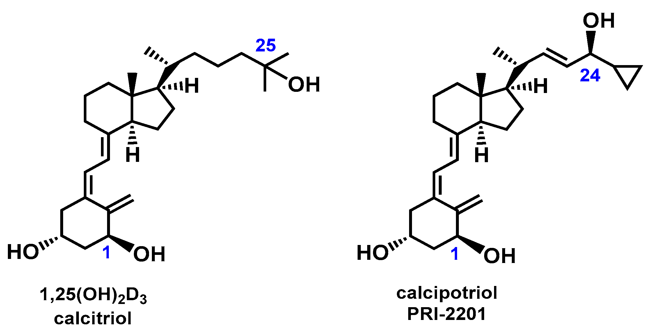
Figure 1. Structural formulas of 1,25-dihydroxyvitamin D3 [1,25(OH)2D3,calcitriol] and calcipotriol (PRI-2201).
Molecules 28 05055 g001
Figure 2. Structural formulas of analogs of calcipotriol, PRI-2203, PRI-2204, and PRI-2205.
Molecules 28 05055 g002
Figure 3. The linear correlation between electrophoretic average migration time vs. dipole polarizability (A) and HOMO-LUMO energy gap (B) of analogs of calcipotriol.
Molecules 28 05055 g003
Figure 4. HOMO-LUMO diagram of PRI-2201 (A), PRI-2203 (B), PRI-2204 (C), and PRI-2205 (D). The positive (red) and negative (green) phase distributions represent the molecular orbital wave functions. HOMO (electron donor regions) determines the ionization potential. LUMO (electron acceptor regions) determines the electron affinity.
Molecules 28 05055 g004
Figure 5. Molecular electrostatic potential (MEP) maps of PRI-2201 (A), PRI-2203 (B), PRI-2204 (C), and PRI-2205 (D). The MEP represents electron-rich (red) and electron-poor (blue) regions.
Molecules 28 05055 g005
Figure 6. Electropherograms of the mixture of analogs, PRI-2205, PRI-2204, PRI-2203, and PRI-2201, in 50 mM of sodium acetate and various ratios of methanol:acetonitrile mixture: 50 mM sodium acetate in 80:20 methanol:acetonitrile mixture.
Molecules 28 05055 g006
Table 1. Calculated values for the dipole moment (μ) and dipole polarizability (α) and the average experimental electrophoretic migration times for PRI-2201, PRI-2203, PRI-2204, and PRI-2205.
AnalogsHybrid Functional B3LYP 6-311 (d,p) in MethanolAverage Electrophoretic Migration Time a
[min]
μtotalαtotal ELUMOEHOMOEg
[Debye][a.u.][eV][eV][eV]
PRI-22014.23429.65−0.97−5.66−4.692.08 ± 0.07
PRI-22033.14430.34−0.97−5.66−4.681.93 ± 0.07
PRI-22043.09430.51−1.04−5.71−4.671.82 ± 0.05
PRI-22053.49434.66−1.19−5.68−4.481.62 ± 0.02
a Average from n = 6 experiments.
Disclaimer/Publisher’s Note: The statements, opinions and data contained in all publications are solely those of the individual author(s) and contributor(s) and not of MDPI and/or the editor(s). MDPI and/or the editor(s) disclaim responsibility for any injury to people or property resulting from any ideas, methods, instructions or products referred to in the content.