

Efficient pH-Responsive Nano-Drug Delivery System Based on Dynamic Boronic Acid/Ester Transformation

Abstract

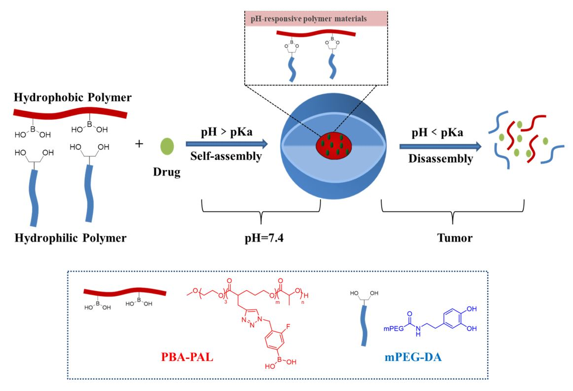
1. Introduction
2. Results
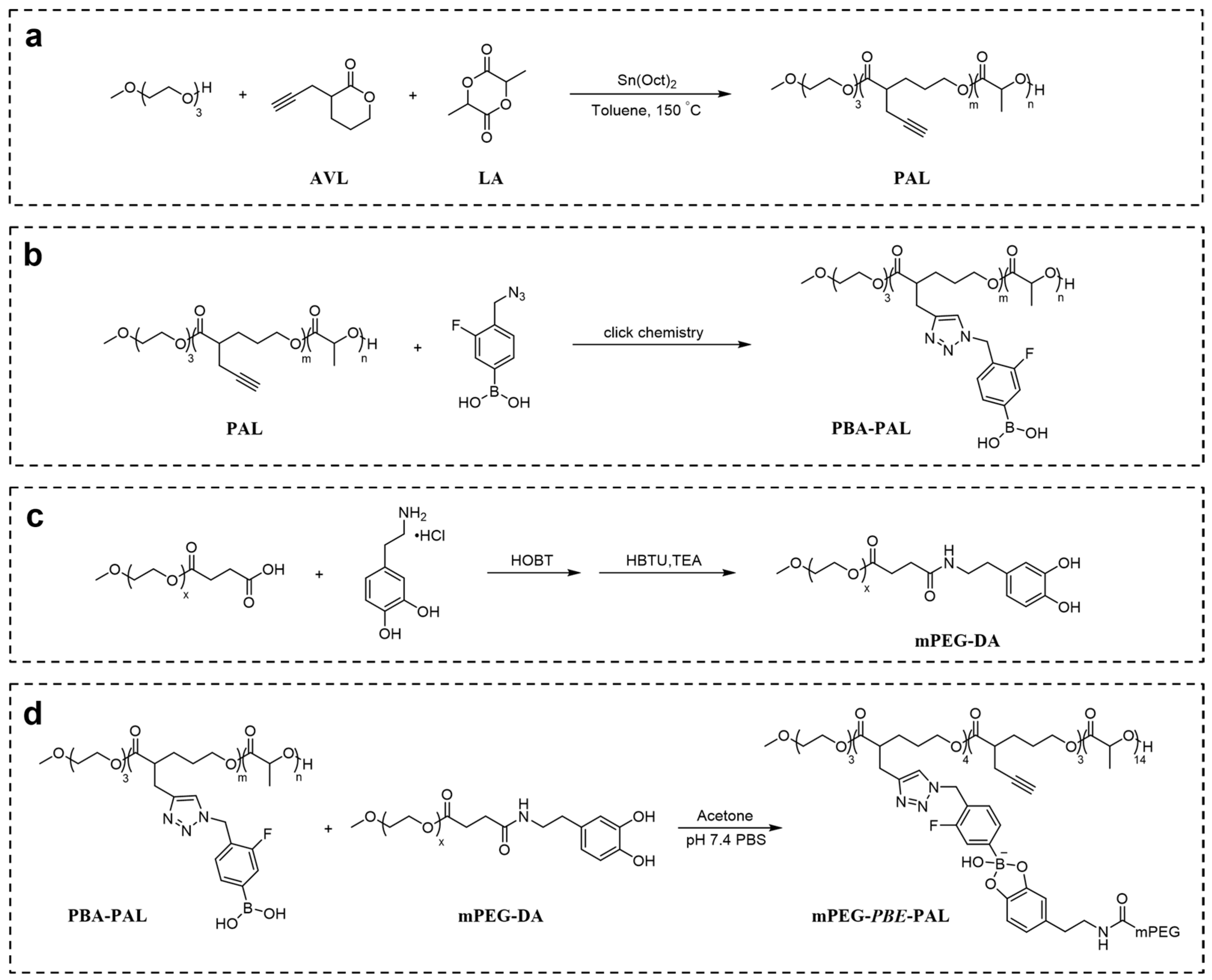
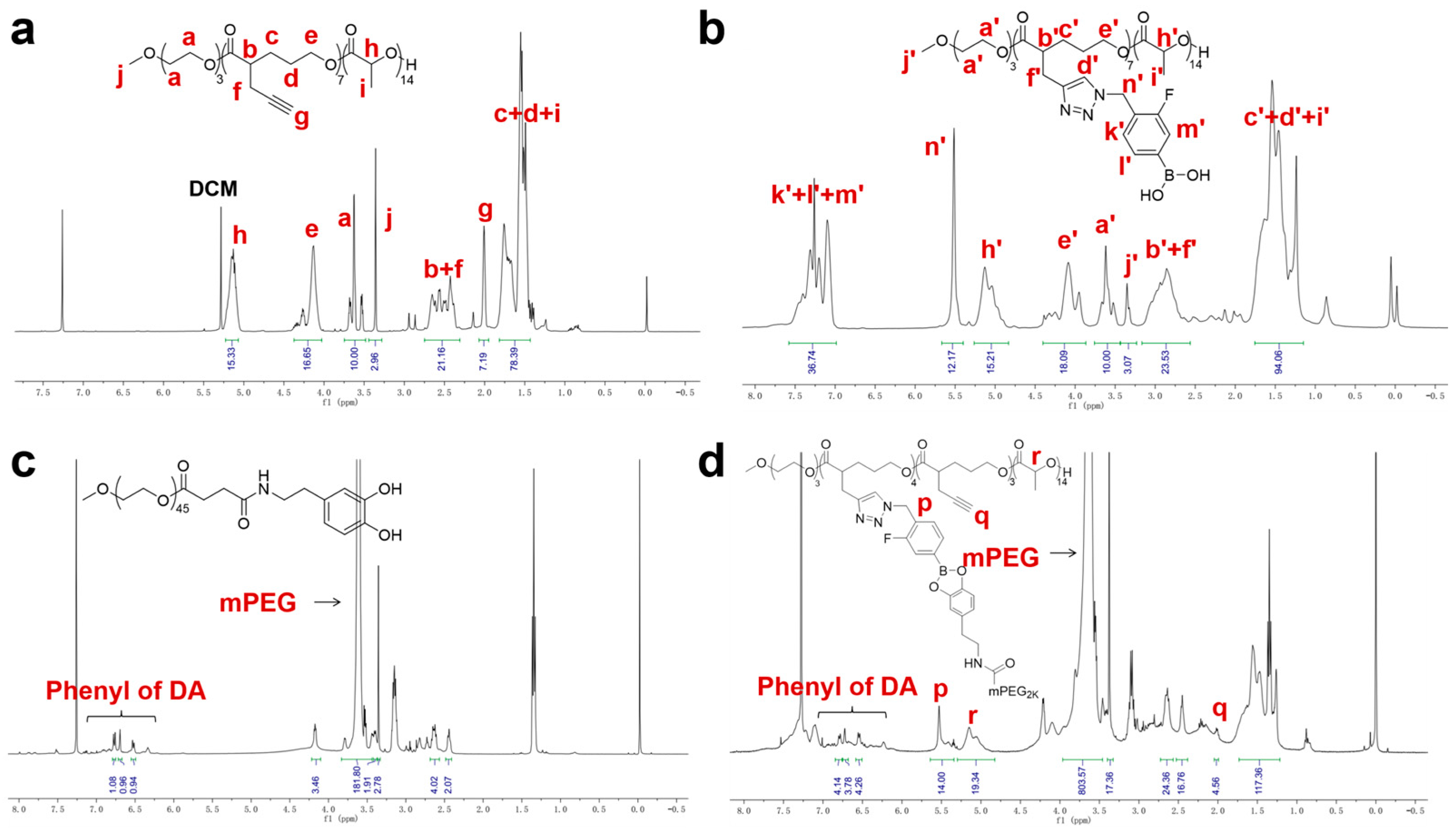
2.1. Synthesis and Characterization of Polymers
2.2. Preparation and Characterization of PTX-Loaded Nanoparticles
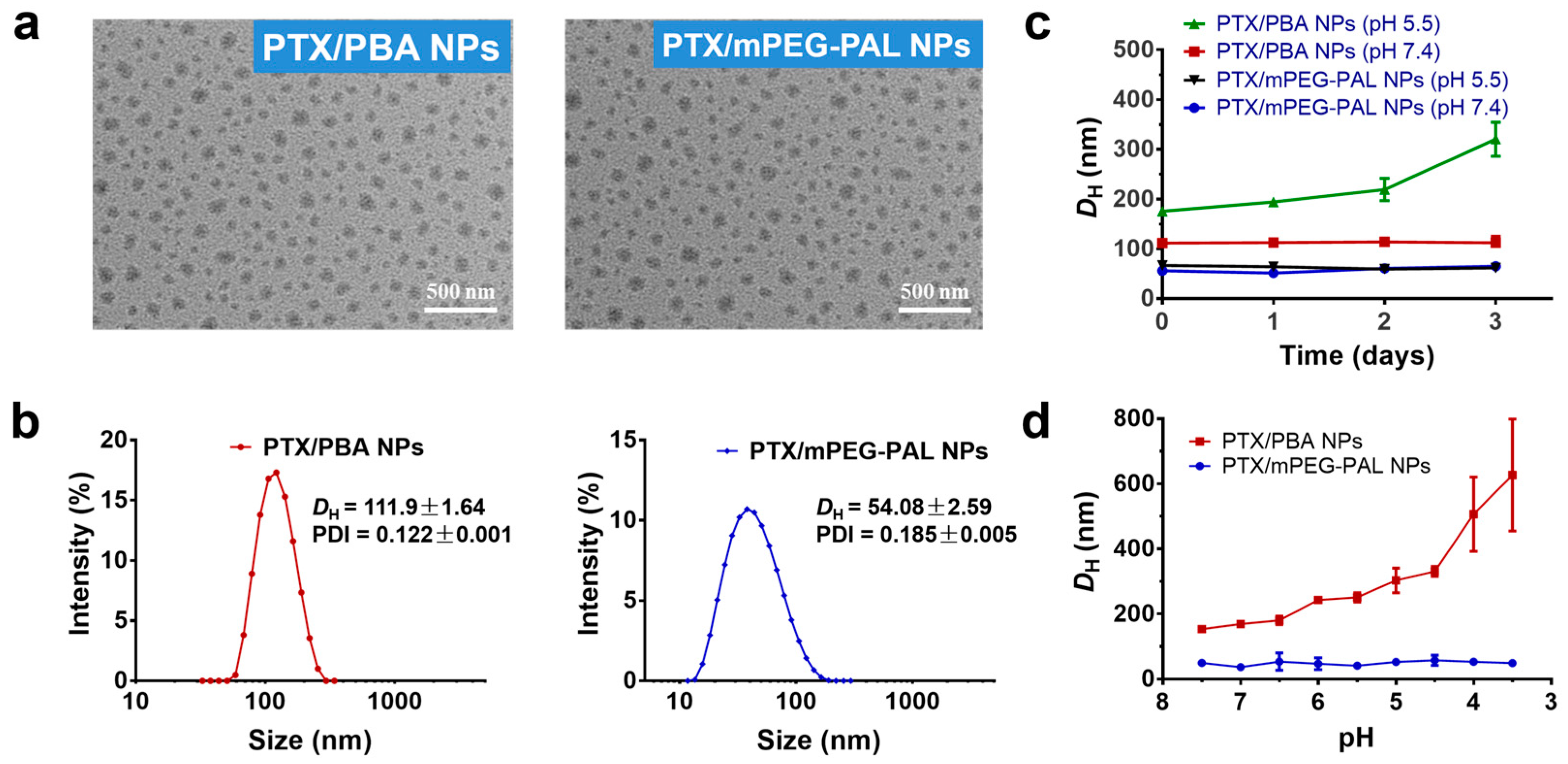
2.3. Morphology, Stability, and pH-Responsiveness of PTX-Loaded Nanoparticles
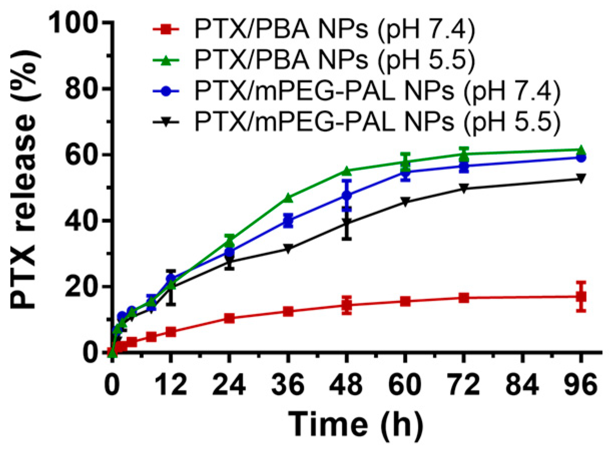
2.4. In Vitro PTX Release Profiles
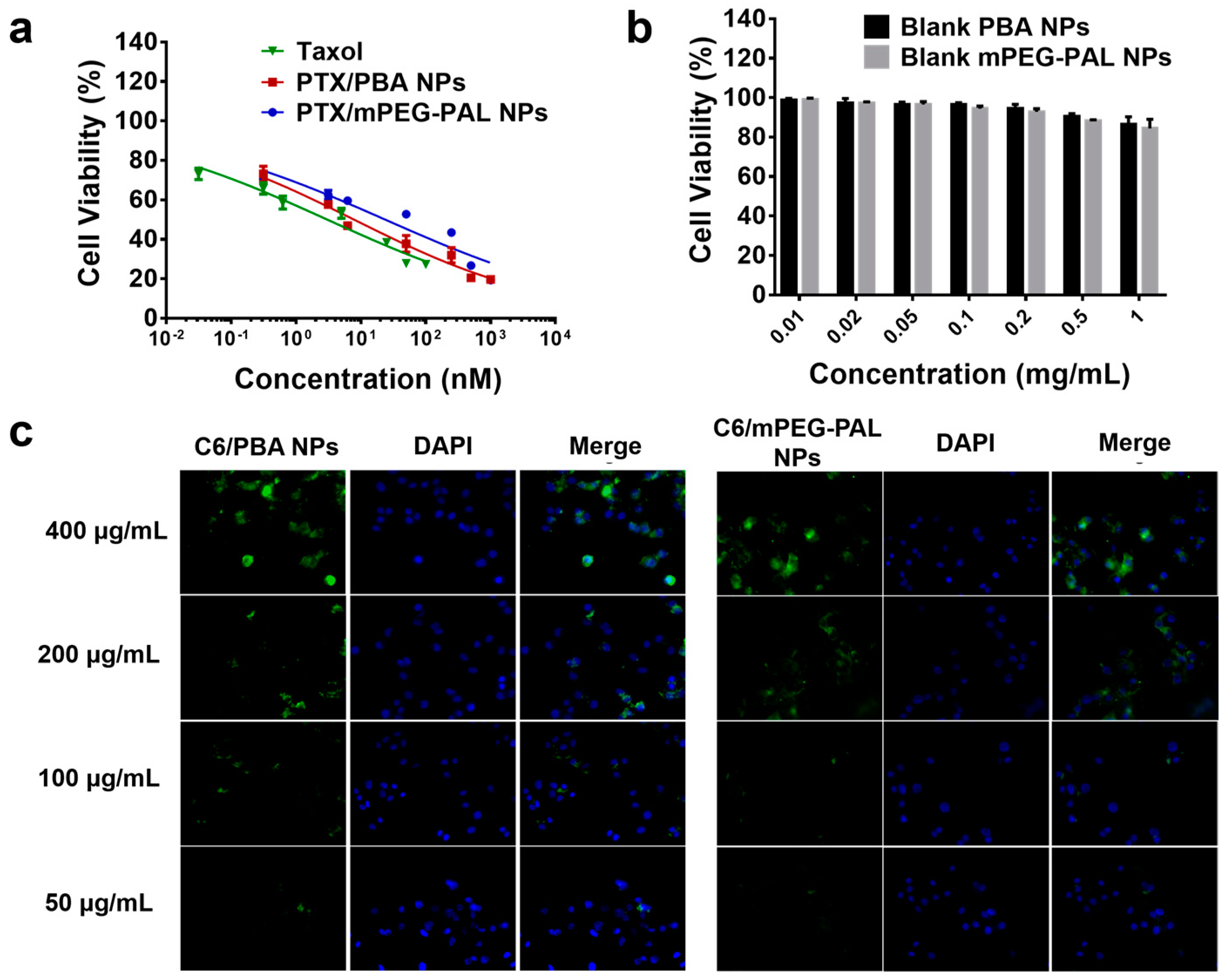
2.5. Assessment of In Vitro Cytotoxicity and Cellular Uptake
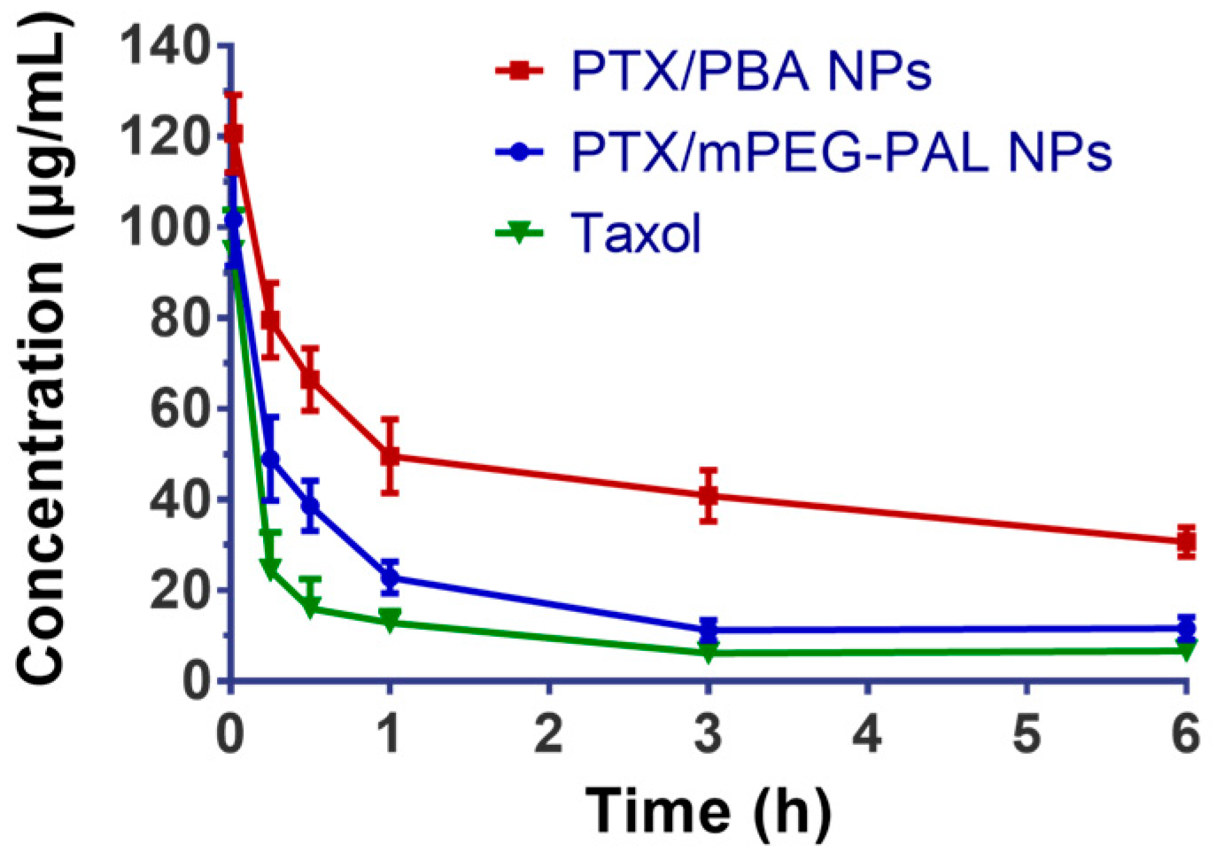
2.6. Pharmacokinetics Studies
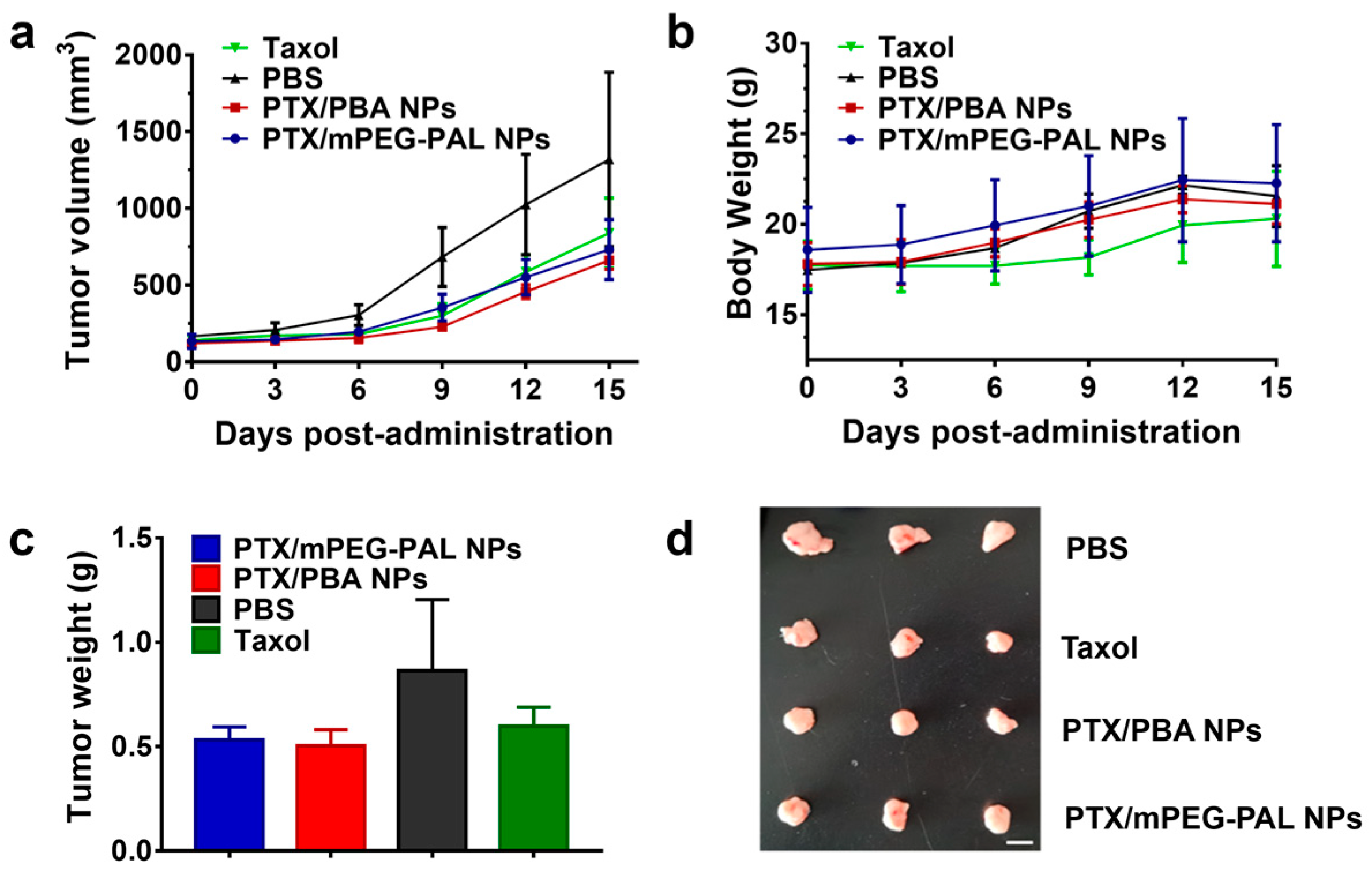
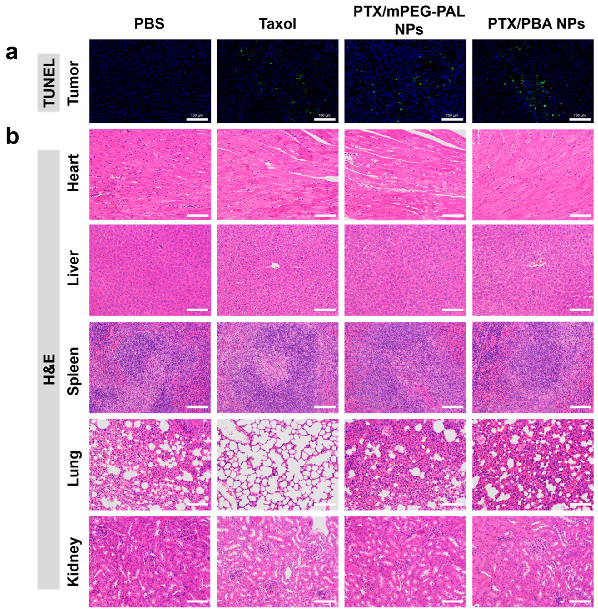
2.7. In Vivo Antitumor Activity
3. Materials and Methods
3.1. Materials
3.2. Instruments
3.3. General Procedures for the Synthesis of Poly (AVL-co-LA) (PAL)
3.4. Synthesis of PBA-PAL Polymer
3.5. General Procedures for the Synthesis of mPEG-DA
3.6. Binding Affinity of PBA-PAL and mPEG-DA
3.7. Preparation and Characterization of PTX/PBA NPs and PTX/mPEG-PAL NPs
3.8. In Vitro Drug Release Assay
3.9. Cell Lines and Cell Culture
3.10. Cytotoxicity Assay
3.11. Cellular Uptake
3.12. Pharmacokinetics Studies
3.13. In Vivo Antitumor Activity
3.14. Histologic Analysis
3.15. Statistical Analysis
4. Conclusions
Supplementary Materials
Author Contributions
Funding
Institutional Review Board Statement
Informed Consent Statement
Data Availability Statement
Acknowledgments
Conflicts of Interest
Sample Availability
References
- Nie, S.M.; Xing, Y.; Kim, G.J.; Simons, J.W. Nanotechnology applications in cancer. Annu. Rev. Biomed. Eng. 2007, 9, 257–288. [Google Scholar] [CrossRef]
- Fioramonti Calixto, G.M.; Bernegossi, J.; de Freitas, L.M.; Fontana, C.R.; Chorilli, M. Nanotechnology-Based Drug Delivery Systems for Photodynamic Therapy of Cancer: A Review. Molecules 2016, 21, 342–359. [Google Scholar]
- Mura, S.; Nicolas, J.; Couvreur, P. Stimuli-responsive nanocarriers for drug delivery. Nat. Mater. 2013, 12, 991–1003. [Google Scholar] [CrossRef]
- Chen, W.; Meng, F.H.; Cheng, R.; Deng, C.; Feijen, J.; Zhong, Z.Y. Advanced drug and gene delivery systems based on functional biodegradable polycarbonates and copolymers. J. Control. Release 2014, 190, 398–414. [Google Scholar] [CrossRef]
- Deng, C.; Jiang, Y.J.; Cheng, R.; Meng, F.H.; Zhong, Z.Y. Biodegradable polymeric micelles for targeted and controlled anticancer drug delivery: Promises, progress and prospects. Nano Today 2012, 7, 467–480. [Google Scholar] [CrossRef]
- Rosler, A.; Vandermeulen, G.W.M.; Klok, H.-A. Advanced drug delivery devices via self-assembly of amphiphilic block copolymers. Adv. Drug Deliv. Rev. 2001, 53, 95–108. [Google Scholar] [CrossRef]
- Ganta, S.; Devalapally, H.; Shahiwala, A.; Amiji, M. A review of stimuli-responsive nanocarriers for drug and gene delivery. J. Control. Release 2008, 126, 187–204. [Google Scholar] [CrossRef]
- Bapat, A.P.; Roy, D.; Ray, J.G.; Savin, D.A.; Sumerlin, B.S. Dynamic-Covalent Macromolecular Stars with Boronic Ester Linkages. J. Am. Chem. Soc. 2011, 133, 19832–19838. [Google Scholar] [CrossRef]
- Li, Y.P.; Xiao, W.W.; Xiao, K.; Berti, L.; Luo, J.T.; Tseng, H.P.; Fung, G.; Lam, K.S. Well-Defined, Reversible Boronate Crosslinked Nanocarriers for Targeted Drug Delivery in Response to Acidic pH Values and cis-Diols. Angew. Chem. Int. Ed. Engl. 2012, 51, 2864–2869. [Google Scholar] [CrossRef]
- Tang, R.P.; Ji, W.H.; Panus, D.; Palumbo, R.N.; Wang, C. Block copolymer micelles with acid-labile ortho ester side-chains: Synthesis, characterization, and enhanced drug delivery to human glioma cells. J. Control. Release 2011, 151, 18–27. [Google Scholar] [CrossRef]
- Yu, Y.; Chen, C.-K.; Law, W.-C.; Sun, H.T.; Prasad, P.N.; Cheng, C. Degradable Brush Polymer-Drug Conjugate for pH-Responsive Release of Doxorubicin. Polym. Chem. 2015, 6, 953–961. [Google Scholar] [CrossRef]
- Huang, L.; Tao, K.X.; Liu, J.; Qi, C.; Xu, L.M.; Chang, P.P.; Gao, J.B.; Shuai, X.M.; Wang, G.B.; Wang, Z.; et al. Design and Fabrication of Multifunctional Sericin Nanoparticles for Tumor Targeting and pH-Responsive Subcellular Delivery of Cancer Chemotherapy Drugs. ACS Appl. Mater. Interfaces 2016, 8, 6577–6585. [Google Scholar] [CrossRef]
- Cheng, R.; Feng, F.; Meng, F.H.; Deng, C.; Feijen, J.; Zhong, Z.Y. Glutathione-responsive nano-vehicles as a promising platform for targeted intracellular drug and gene delivery. J. Control. Release 2011, 152, 2–12. [Google Scholar] [CrossRef]
- Broaders, K.E.; Grandhe, S.; Frechet, J.M.J. A Biocompatible Oxidation-Triggered Carrier Polymer with Potential in Therapeutics. J. Am. Chem. Soc. 2011, 133, 756–758. [Google Scholar] [CrossRef]
- Lee, M.-R.; Baek, K.-H.; Jin, H.J.; Jung, Y.-G.; Shin, I. Targeted Enzyme-Responsive Drug Carriers: Studies on the Delivery of a Combination of Drugs. Angew. Chem. Int. Ed. Engl. 2004, 43, 1675–1678. [Google Scholar] [CrossRef]
- Stubbs, M.; McSheehy, P.M.J.; Griffiths, J.R.; Bashford, C.L. Causes and consequences of tumour acidity and implications for treatment. Mol. Med. Today 2000, 6, 15–19. [Google Scholar] [CrossRef]
- Chang, Y.-T.; Liao, P.-Y.; Sheu, H.-S.; Tseng, Y.-J.; Cheng, F.-Y.; Yeh, C.-S. Near-Infrared Light-Responsive Intracellular Drug and siRNA Release Using Au Nanoensembles with Oligonucleotide-Capped Silica Shell. Adv. Mater. 2012, 24, 3309–3314. [Google Scholar] [CrossRef]
- Gao, W.W.; Chan, J.M.; Farokhzad, O.C. pH-Responsive Nanoparticles for Drug Delivery. Mol. Pharm. 2010, 7, 1913–1920. [Google Scholar] [CrossRef]
- Felber, A.E.; Dufresne, M.-H.; Leroux, J.-C. pH-sensitive vesicles, polymeric micelles, and nanospheres prepared with polycarboxylates. Adv. Drug Deliv. Rev. 2012, 64, 979–992. [Google Scholar] [CrossRef]
- Ulbrich, K.; Subr, V. Polymeric anticancer drugs with pH-controlled activation. Adv. Drug Deliv. Rev. 2004, 56, 1023–1050. [Google Scholar] [CrossRef]
- Fattal, E.; Couvreur, P.; Dubernet, C. “Smart” delivery of antisense oligonucleotides by anionic pH-sensitive liposomes. Adv. Drug Deliv. Rev. 2004, 56, 931–946. [Google Scholar] [CrossRef]
- Zheng, Y.; Shi, S.D.; Liu, Y.R.; Zhao, Y.D.; Sun, Y.Q. Targeted pharmacokinetics of polymeric micelles modified with glycyrrhetinic acid and hydrazone bond in H22 tumor-bearing mice. J. Biomater. Appl. 2019, 34, 141–151. [Google Scholar] [CrossRef]
- Shi, Y.; van der Meel, R.; Theek, B.; Blenke, E.O.; Pieters, E.H.E.; Fens, M.H.A.M.; Ehling, J.; Schiffelers, R.M.; Storm, G.; van Nostrum, C.F.; et al. Complete Regression of Xenograft Tumors upon Targeted Delivery of Paclitaxel via π-π Stacking Stabilized Polymeric Micelles. ACS Nano 2015, 9, 3740–3752. [Google Scholar] [CrossRef]
- Lv, S.X.; Wu, Y.C.; Cai, K.M.; He, H.; Li, Y.J.; Lan, M.; Chen, X.S.; Cheng, J.J.; Yin, L.C. High Drug Loading and Sub-Quantitative Loading Efficiency of Polymeric Micelles Driven by Donor-Receptor Coordination Interactions. J. Am. Chem. Soc. 2018, 140, 1235–1238. [Google Scholar] [CrossRef]
- Che, J.Y.; Tao, A.Q.; Chen, S.; Li, X.M.; Zhao, Y.; Yuan, W.E. Biologically responsive carrier-mediated anti-angiogenesis shRNA delivery for tumor treatment. Sci. Rep. 2016, 6, 35661–35671. [Google Scholar] [CrossRef]
- Gillies, E.R.; Goodwin, A.P.; Frechet, J.M.J. Acetals as pH-Sensitive Linkages for Drug Delivery. Bioconjug. Chem. 2004, 15, 1254–1263. [Google Scholar] [CrossRef]
- Griset, A.P.; Walpole, J.; Liu, R.; Gaffey, A.; Colson, Y.L.; Grinstaff, M.W. Expansile Nanoparticles: Synthesis, Characterization, and in Vivo Efficacy of an Acid-Responsive Polymeric Drug Delivery System. J. Am. Chem. Soc. 2009, 131, 2469–2471. [Google Scholar] [CrossRef]
- Aryal, S.; Hu, C.-M.J.; Zhang, L.F. Polymer-Cisplatin Conjugate Nanoparticles for Acid-Responsive Drug Delivery. ACS Nano 2010, 4, 251–258. [Google Scholar] [CrossRef]
- Wang, B.D.; Xu, C.J.; Xie, J.; Yang, Z.Y.; Sun, S.H. pH Controlled Release of Chromone from Chromone-Fe3O4 Nanoparticles. J. Am. Chem. Soc. 2008, 130, 14436–14437. [Google Scholar] [CrossRef]
- Li, Y.; Lee, P.I. A new bioerodible system for sustained local drug delivery based on hydrolytically activated in situ macromolecular association. Int. J. Pharm. 2010, 383, 45–52. [Google Scholar] [CrossRef]
- Mok, H.; Park, J.W.; Park, T.G. Enhanced Intracellular Delivery of Quantum Dot and Adenovirus Nanoparticles Triggered by Acidic pH via Surface Charge Reversal. Bioconjug. Chem. 2008, 19, 797–801. [Google Scholar] [CrossRef]
- Ali, M.M.; Oishi, M.; Nagatsugi, F.; Mori, K.; Nagasaki, Y.; Kataoka, K.; Sasaki, S. Intracellular Inducible Alkylation System That Exhibits Antisense Effects with Greater Potency and Selectivity than the Natural Oligonucleotide. Angew. Chem. Int. Ed. Engl. 2006, 45, 3136–3140. [Google Scholar] [CrossRef]
- Cao, J.; Gao, X.F.; Cheng, M.B.; Niu, X.Y.; Li, X.M.; Zhang, Y.P.; Liu, Y.; Wang, W.; Yuan, Z. Reversible Shielding between Dual Ligands for Enhanced Tumor Accumulation of ZnPc-Loaded Micelles. Nano Lett. 2019, 19, 1665–1674. [Google Scholar] [CrossRef]
- Kundu, M.; Sadhukhan, P.; Ghosh, N.; Chatterjee, S.; Manna, P.; Das, J.; Sil, P.C. pH-responsive and targeted delivery of curcumin via phenylboronic acid-functionalized ZnO nanoparticles for breast cancer therapy. J. Adv. Res. 2019, 18, 161–172. [Google Scholar] [CrossRef]
- He, L.H.; Fullenkamp, D.E.; Rivera, J.G.; Messersmith, P.B. pH responsive self-healing hydrogels formed by boronate-catechol complexation. Chem. Commun. 2011, 47, 7497–7499. [Google Scholar] [CrossRef]
- Anzai, J.-I. Recent progress in electrochemical biosensors based on phenylboronic acid and derivatives. Mater. Sci. Eng. C. Mater. Biol. Appl. 2016, 67, 737–746. [Google Scholar] [CrossRef]
- Yao, Y.; Zhao, L.Y.; Yang, J.J.; Yang, J. Glucose-Responsive Vehicles Containing Phenylborate Ester for Controlled Insulin Release at Neutral pH. Biomacromolecules 2012, 13, 1837–1844. [Google Scholar] [CrossRef]
- Wang, X.J.; Xia, N.; Liu, L. Boronic Acid-Based Approach for Separation and Immobilization of Glycoproteins and Its Application in Sensing. Int. J. Mol. Sci. 2013, 14, 20890–20912. [Google Scholar] [CrossRef]
- Ren, J.; Zhang, Y.X.; Zhang, J.; Gao, H.J.; Liu, G.; Ma, R.J.; An, Y.L.; Kong, D.L.; Shi, L.Q. pH/Sugar Dual Responsive Core-Cross-Linked PIC Micelles for Enhanced Intracellular Protein Delivery. Biomacromolecules 2013, 14, 3434–3443. [Google Scholar] [CrossRef]
- Su, J.; Chen, F.; Cryns, V.L.; Messersmith, P.B. Catechol Polymers for pH-Responsive, Targeted Drug Delivery to Cancer Cells. J. Am. Chem. Soc. 2011, 133, 11850–11853. [Google Scholar] [CrossRef]
- Elshaarani, T.; Yu, H.J.; Wang, L.; Zain-ul-Abdin; Ullah, R.S.; Haroon, M.; Khan, R.U.; Fahad, S.; Khan, A.; Nazir, A.; et al. Synthesis of hydrogel-bearing phenylboronic acid moieties and their applications in glucose sensing and insulin delivery. J. Mater. Chem. B 2018, 6, 3831–3854. [Google Scholar] [CrossRef]
- Zhao, Z.W.; Yao, X.M.; Zhang, Z.; Chen, L.; He, C.L.; Chen, X.S. Boronic Acid Shell-Crosslinked Dextran-b-PLA Micelles for Acid-Responsive Drug Delivery. Macromol. Biosci. 2014, 14, 1609–1618. [Google Scholar] [CrossRef]
- Liu, S.Q.; Ono, R.J.; Yang, C.; Gao, S.J.; Tan, J.Y.M.; Hedrick, J.L.; Yang, Y.Y. Dual pH-Responsive Shell-Cleavable Polycarbonate Micellar Nanoparticles for in Vivo Anticancer Drug Delivery. ACS Appl. Mater. Interfaces 2018, 10, 19355–19364. [Google Scholar] [CrossRef]
- Aguirre-Chagala, Y.E.; Santos, J.L.; Huang, Y.X.; Herrera-Alonso, M. Phenylboronic Acid-Installed Polycarbonates for the pH-Dependent Release of Diol-Containing Molecules. ACS Macro Lett. 2014, 3, 1249–1253. [Google Scholar] [CrossRef]
- Kasuya, Y.; Lu, Z.-R.; Kopeckova, P.; Minko, T.; Tabibi, S.E.; Kopecek, J. Synthesis and characterization of HPMA copolymer-aminopropylgeldanamycin conjugates. J. Control. Release 2001, 74, 203–211. [Google Scholar] [CrossRef]
- Negri, G.E.; Deming, T.J. Triggered Copolypeptide Hydrogel Degradation Using Photolabile Lysine Protecting Groups. ACS Macro Lett. 2016, 5, 1253–1256. [Google Scholar] [CrossRef]
- Pan, R.H.; Zeng, Y.; Liu, G.Q.; Wei, Y.; Xu, Y.S.; Tao, L. Curcumin-polymer conjugates with dynamic boronic acid ester linkages for selective killing of cancer cells. Polym. Chem. 2020, 11, 1321–1326. [Google Scholar] [CrossRef]
- Zhang, L.Z.; Lin, Y.; Wang, J.J.; Yao, W.; Wu, W.; Jiang, X.Q. A Facile Strategy for Constructing Boron-Rich Polymer Nanoparticles via a Boronic Acid-Related Reaction. Macromol. Rapid Commun. 2011, 32, 534–539. [Google Scholar] [CrossRef]
- Zhao, G.K.; Ge, T.T.; Yan, Y.F.; Shuai, Q.; Su, W.-K. Highly Efficient Modular Construction of Functional Drug Delivery Platform Based on Amphiphilic Biodegradable Polymers via Click Chemistry. Int. J. Mol. Sci. 2021, 22, 10407. [Google Scholar] [CrossRef]
- Ramaswamy, M.; Zhang, X.C.; Burt, H.M.; Wasan, K.M. Human plasma distribution of free paclitaxel and paclitaxel associated with diblock copolymers. J. Pharm. Sci. 1997, 86, 460–464. [Google Scholar] [CrossRef]
- Shuai, Q.; Cai, Y.; Zhao, G.K.; Sun, X.R. Cell-Penetrating Peptide Modified PEG-PLA Micelles for Efficient PTX Delivery. Int. J. Mol. Sci. 2020, 21, 1856. [Google Scholar] [CrossRef]


| Polymers | [AVL]:[LA] (Theoretical Molar Ratio) | [AVL]:[LA] (Actual Molar Ratio) | Mn, NMRa (g/mol) | Mn, GPCb (g/mol) | PDI c | Grafting Rates (%) d |
|---|---|---|---|---|---|---|
| PAL2K | 1:1 | 1.1:1 | 2200 | 2300 | 1.32 | - |
| PAL4K | 1:1 | 1.1:1 | 4300 | 4200 | 1.37 | - |
| PBA-PAL2K | 1:1 | 1.1:1 | 3400 | 3700 | 1.38 | 85.1 |
| PBA-PAL4K | 1:1 | 1.1:1 | 6400 | 7200 | 1.39 | 80.2 |
| mPEG2K-DA | - | - | 2400 | 3100 | 1.04 | |
| mPEG4K-DA | - | - | 4300 | 4700 | 1.02 |
| PTX/PBA NPs | [PBA-PAL2K]:[mPEG2K-DA] Molar Ratio | Size (nm) | PDI | EE (%) | DL (%) |
|---|---|---|---|---|---|
| 1 | 1:7 | 111.9 ± 1.64 | 0.122 ± 0.001 | 96.1 | 2.0 |
| 2 | 1:3.5 | 126.9 ± 2.56 | 0.143 ± 0.004 | 70.0 | 2.3 |
| 3 | 1:1 | 116.4 ± 4.21 | 0.133 ± 0.004 | 60.8 | 3.8 |
| 4 | 1:0.5 | 109.58 ± 2.89 | 0.174 ± 0.014 | 55.0 | 1.1 |
| PTX/PBA NPs | Mn, PBA-PAL-Mn, mPEG-DA (g/mol) | Size (nm) | PDI | EE (%) | DL (%) |
|---|---|---|---|---|---|
| 1 | 2K−2K | 111.9 ± 1.64 | 0.122 ± 0.001 | 96.1 | 2.0 |
| 2 | 2K−4K | 136.9 ± 1.26 | 0.103 ± 0.001 | 49.7 | 0.6 |
| 3 | 4K−4K | 133.5 ± 3.85 | 0.103 ± 0.001 | 43.1 | 0.5 |
| 4 | 4K−2K | 79.88 ± 4.69 | 0.184 ± 0.017 | 23.7 | 0.5 |
| Pharmacokinetic Parameters | PTX/PBA NPs | PTX/mPEG-PAL NPs | Taxol |
|---|---|---|---|
| t1/2α (h) | 0.195 | 0.153 | 0.054 |
| t1/2β (h) | 7.364 | 3.395 | 0.434 |
| AUC (0–t) (μg/L·h) | 270.465 | 114.049 | 67.456 |
| AUC (0–∞) (μg/L·h) | 589.846 | 118.627 | 85.372 |
| Cmax (μg/L) | 120.677 | 101.66 | 94.619 |
Disclaimer/Publisher’s Note: The statements, opinions and data contained in all publications are solely those of the individual author(s) and contributor(s) and not of MDPI and/or the editor(s). MDPI and/or the editor(s) disclaim responsibility for any injury to people or property resulting from any ideas, methods, instructions or products referred to in the content. |
© 2023 by the authors. Licensee MDPI, Basel, Switzerland. This article is an open access article distributed under the terms and conditions of the Creative Commons Attribution (CC BY) license (https://creativecommons.org/licenses/by/4.0/).
Share and Cite
Chen, W.; Xie, W.; Zhao, G.; Shuai, Q. Efficient pH-Responsive Nano-Drug Delivery System Based on Dynamic Boronic Acid/Ester Transformation. Molecules 2023, 28, 4461. https://doi.org/10.3390/molecules28114461
Chen W, Xie W, Zhao G, Shuai Q. Efficient pH-Responsive Nano-Drug Delivery System Based on Dynamic Boronic Acid/Ester Transformation. Molecules. 2023; 28(11):4461. https://doi.org/10.3390/molecules28114461
Chicago/Turabian StyleChen, Weijun, Wanxuan Xie, Guangkuo Zhao, and Qi Shuai. 2023. "Efficient pH-Responsive Nano-Drug Delivery System Based on Dynamic Boronic Acid/Ester Transformation" Molecules 28, no. 11: 4461. https://doi.org/10.3390/molecules28114461
APA StyleChen, W., Xie, W., Zhao, G., & Shuai, Q. (2023). Efficient pH-Responsive Nano-Drug Delivery System Based on Dynamic Boronic Acid/Ester Transformation. Molecules, 28(11), 4461. https://doi.org/10.3390/molecules28114461