

Antitumor Effect of Bleomycin Nanoaerosol in Murine Carcinoma Model
 , , ,
, , , 
 and
and
Abstract
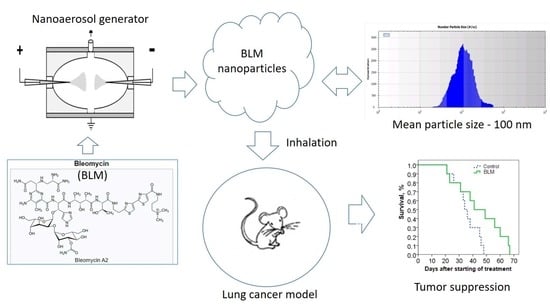
1. Introduction
2. Results
2.1. Dose Calculation
2.2. Antitumor Effect of NAPs on the Murine Melanoma Model
2.3. Comparative Effectiveness of NAPs and Intraperitoneal Injections of BLM
2.4. Antitumor Effect of NAPs on the Murine Lewis Lung Carcinoma Model
2.5. Histological Analysis of the Lungs
2.6. Pharmacokinetics of BLM NAPs
3. Discussion
4. Materials and Methods
4.1. Reagents
4.2. Electrospray-Based Nanoaerosol Generator
4.3. Estimation of BLM NAP Concentration in Aerosol
4.4. Animals and Models of Lung Metastasis
4.4.1. Pulmonary Fibrosis Model
4.4.2. Histology
4.5. Liquid Chromatography and Tandem Mass Spectrometry (LC-MS/MS)
4.5.1. Sample Preparation
4.5.2. LC/MRM-MS
4.6. Data Analysis
5. Conclusions
Supplementary Materials
Author Contributions
Funding
Institutional Review Board Statement
Informed Consent Statement
Data Availability Statement
Acknowledgments
Conflicts of Interest
Sample Availability
References
- Lee, W.-H.; Loo, C.-Y.; Traini, D.; Young, P.M. Inhalation of nanoparticle-based drug for lung cancer treatment: Advantages and challenges. Asian J. Pharm. Sci. 2015, 10, 481–492. [Google Scholar] [CrossRef]
- Rosiere, R.; Amighi, K.; Wauthoz, N. Nanomedicine-Based Inhalation Treatments for Lung Cancer. Nanotechnol.-Based Target. Drug Deliv. Syst. Lung Cancer 2019, 249–268. [Google Scholar] [CrossRef]
- Hong, S.-H.; Park, S.-J.; Lee, S.; Cho, C.S.; Cho, M.-H. Aerosol gene delivery using viral vectors and cationic carriers for in vivo lung cancer therapy. Expert Opin. Drug Deliv. 2014, 12, 977–991. [Google Scholar] [CrossRef] [PubMed]
- Ahmad, J.; Akhter, S.; Rizwanullah, M.; Amin, S.; Rahman, M.; Ahmad, M.Z.; Rizvi, M.A.; Kamal, M.A.; Ahmad, F.J. Nanotechnology-based inhalation treatments for lung cancer: State of the art. Nanotechnol. Sci. Appl. 2015, 8, 55–66. [Google Scholar] [CrossRef]
- Lee, W.-H.; Loo, C.-Y.; Young, P.M.; Traini, D.; Mason, R.S.; Rohanizadeh, R. Recent advances in curcumin nanoformulation for cancer therapy. Expert Opin. Drug Deliv. 2014, 11, 1183–1201. [Google Scholar] [CrossRef]
- El-Maradny, H.; Mneimneh, A.T. A Review on Aerosol Drug Delivery: Fundamentals, Classifications, Particle Size Analysis and the Engagement of Nanoparticulate Systems. Drug Deliv. Lett. 2022, 12, 258–275. [Google Scholar] [CrossRef]
- Ramsey, J.D.; Stewart, I.E.; Madden, E.A.; Lim, C.; Hwang, D.; Heise, M.T.; Hickey, A.J.; Kabanov, A.V. Nanoformulated Remdesivir with Extremely Low Content of Poly(2-oxazoline)-Based Stabilizer for Aerosol Treatment of COVID-19. Macromol. Biosci. 2022, 22, 2200056. [Google Scholar] [CrossRef]
- Vahedifard, F.; Chakravarthy, K. Nanomedicine for COVID-19: The role of nanotechnology in the treatment and diagnosis of COVID-19. Emergent Mater. 2021, 4, 75–99. [Google Scholar] [CrossRef]
- Jansen, E.M.; Frijlink, H.W.; Hinrichs, W.L.; Ruigrok, M.J. Are inhaled mRNA vaccines safe and effective? A review of preclinical studies. Expert Opin. Drug Deliv. 2022, 19, 1471–1485. [Google Scholar] [CrossRef]
- Kataoka, K.; Matsumoto, T.; Yokoyama, M.; Okano, T.; Sakurai, Y.; Fukushima, S.; Okamoto, K.; Kwon, G.S. Doxorubicin-loaded poly(ethylene glycol)-poly(beta-benzyl-l-aspartate) copolymer micelles: Their pharmaceutical characteristics and biological significance. J. Control. Release 2000, 64, 143–153. [Google Scholar] [CrossRef]
- Curtis, J.; Greenberg, M.; Kester, J.; Phillips, S.; Krieger, G. Nanotechnology and nanotoxicology: A primer for clinicians. Toxicol. Sci. 2006, 25, 245–260. [Google Scholar] [CrossRef] [PubMed]
- Hagens, W.I.; Oomen, A.G.; de Jong, W.H.; Cassee, F.R.; Sips, A.J. What do we (need to) know about the kinetic properties of nanoparticles in the body? Regul. Toxicol. Pharmacol. 2007, 49, 217–229. [Google Scholar] [CrossRef] [PubMed]
- Moghimi, S.M.; Hunter, A.C.; Murray, J.C. Nanomedicine: Current status and future prospects. FASEB J. 2005, 19, 311–330. [Google Scholar] [CrossRef] [PubMed]
- Oberdörster, G.; Oberdörster, E.; Oberdörster, J. Nanotoxicology: An emerging discipline evolving from studies of ultrafine particles. Environ. Health Perspect. 2005, 113, 823–839. [Google Scholar] [CrossRef] [PubMed]
- Onischuk, A.A.; Tolstikova, T.G.; Baklanov, A.M.; Khvostov, M.V.; Sorokina, I.V.; Zhukova, N.A.; An’kov, S.V.; Borovkova, O.V.; Dultseva, G.G.; Boldyrev, V.V.; et al. Generation, inhalation delivery and anti-hypertensive effect of nisoldipine nanoaerosol. J. Aerosol Sci. 2014, 78, 41–54. [Google Scholar] [CrossRef]
- Kwon, G.; Naito, M.; Yokoyama, M.; Okano, T.; Sakurai, Y.; Kataoka, K. Block copolymer micelles for drug delivery: Loading and release of doxorubicin. J. Control. Release 1997, 48, 195–201. [Google Scholar] [CrossRef]
- Kalantarian, P.; Najafabadi, A.R.; Haririan, I.; Vatanara, A.; Yamini, Y.; Darabi, M.; Gilani, K. Preparation of 5-fluorouracil nanoparticles by supercritical antisolvents for pulmonary delivery. Int. J. Nanomed. 2010, 5, 763–770. [Google Scholar] [CrossRef]
- Tomoda, K.; Ohkoshia, T.; Hirotaa, K.; Sonavane, G.S.; Nakajima, T.; Terada, H.; Komuro, M.; Kitazato, K.; Makino, K. Preparation and properties of inhalable nanocomposite particles for treatment of lung cancer. Colloids Surf. B Biointerfaces 2009, 71, 177–182. [Google Scholar] [CrossRef]
- Videira, M.A.; Almeida, A.J.; Fabra, A. Preclinical evaluation of a pulmonary delivered paclitaxel loaded lipid nanocarrier antitumor effect. Nanomedicine 2012, 8, 1208–1215. [Google Scholar] [CrossRef]
- Videira, M.A.; Botelho, M.F.; Santos, A.C.; Gouveia, L.F.; DeLima, J.J.; Almeida, A.J. Lymphatic uptake of pulmonary delivered radiolabeled solid lipid nanoparticles. J. Drug Target. 2002, 10, 607–613. [Google Scholar] [CrossRef]
- Hu, L.D.; Jia, Y.; Ding, W. Preparation and characterization of solid lipid nanoparticles loaded with epirubicin for pulmonary delivery. Pharmazie 2010, 65, 585–587. [Google Scholar] [PubMed]
- Jyoti, K.; Kaur, K.; Pandey, R.S.; Jain, U.K.; Chandra, R.; Madan, J. Inhalable nanostructured lipid particles of 9-bromo-noscapine, a tubulin-binding cytotoxic agent: In vitro and in vivo studies. J. Colloid Interface Sci. 2015, 445, 219–230. [Google Scholar] [CrossRef] [PubMed]
- Taratula, O.; Kuzmov, A.; Shah, M.; Garbuzenko, O.B.; Minko, T. Nanostructured lipid carriers as multifunctional nanomedicine platform for pulmonary co-delivery of anticancer drugs and siRNA. J. Control. Release 2013, 171, 349–357. [Google Scholar] [CrossRef]
- Yeo, L.Y.; Friend, J.R.; McIntosh, M.P.; Meeusen, E.N.T.; Morton, D.A.V. Ultrasonic nebulization platforms for pulmonary drug delivery. Expert Opin. Drug Deliv. 2010, 7, 663–679. [Google Scholar] [CrossRef] [PubMed]
- Onischuk, A.A.; Tolstikova, T.G.; An’kov, S.V.; Baklanov, A.M.; Valiulin, S.V.; Khvostov, M.V.; Sorokina, I.V.; Dultseva, G.G.; Zhukova, N.A. Ibuprofen, indomethacin and diclofenac sodium nanoaerosol: Generation, inhalation delivery and biological effects in mice and rats. J. Aerosol Sci. 2016, 100, 164–177. [Google Scholar] [CrossRef]
- Valiulin, S.V.; Onischuk, A.A.; Baklanov, A.M.; Dubtsov, S.N.; An’kov, S.V.; Tolstikova, T.G.; Mazunina, P. Excipient-free isoniazid aerosol administration in mice: Evaporation-nucleation particle generation, pulmonary delivery and body distribution. Int. J. Pharm. 2019, 563, 101–109. [Google Scholar] [CrossRef]
- Morozov, V.N.; Kanev, I.L.; Mikheev, A.Y.; Shlyapnikova, E.A.; Shlyapnikov, Y.M.; Nikitin, M.P.; Nikitin, P.I.; Nwabueze, A.O.; van Hoek, M.L. Generation and delivery of nanoaerosols from biological and biologically active substances. J. Aerosol Sci. 2014, 69, 48–61. [Google Scholar] [CrossRef]
- Kanev, I.L.; Mikheev, A.Y.; Shlyapnikov, Y.M.; Shlyapnikova, E.A.; Morozov, V.N. Are reactive species generated in electrospray at low current? Anal. Chem. 2014, 86, 1511–1517. [Google Scholar] [CrossRef]
- Propst, C.N.; Nwabueze, A.O.; Pepin, R.E.; Morozov, V.N.; van Hoek, M.L.; Gutting, B.W.; Kanev, I.L. Nanoaerosols reduce required effective dose of liposomal levofloxacin against pulmonary murine Francisella tularensis subsp. novicida infection. J. Nanobiotechn. 2016, 14, 29. [Google Scholar] [CrossRef]
- Umezawa, H. Bleomycin and other antitumor antibiotics of high molecular weight. Antimicrob. Agents Chemother. 1965, 5, 1079–1085. [Google Scholar]
- Syed, T.R. Bleomycin: An overview on anticancer drug. Int. J. Recent Adv. Multidiscip. Res. 2018, 5, 3618–3622. [Google Scholar]
- Rodríguez, J.C.; Morales, M.C.J.; Casero, M.A.R. Death by bleomycin pulmonary toxicity in ovarian dysgerminoma with pathologic complete response to chemotherapy. A case report. Respir. Med. Case Rep. 2016, 18, 48–50. [Google Scholar] [CrossRef]
- Brown, R.F.R.; Drawbaugh, R.B.; Marrs, T.C. An investigation of possible models for the production of progressive pulmonary fibrosis in the rat. The effects of repeated intraatracheal instillation of bleomycin. Toxicology 1988, 51, 101–110. [Google Scholar] [CrossRef] [PubMed]
- Singer, I.I.; Kawka, D.W.; McNally, S.M.; Eiermann, G.J.; Metzger, J.M.; Peterson, L.B. Extensive laminin and basement membrane accumulation occurs at the onset of bleomycin-induced rodent pulmonary fibrosis. Am. J. Pathol. 1986, 125, 258–268. [Google Scholar] [PubMed]
- Thrall, R.S.; McCormick, J.R.; Jack, R.M.; McReynolds, R.A.; Ward, P.A. Bleomycin-induced pulmonary toxicity in the rat. Am. J. Pathol. 1979, 95, 117–127. [Google Scholar]
- Ishida, Y.; Kuninaka, Y.; Mukaida, N.; Kondo, T. Immune Mechanisms of Pulmonary Fibrosis with Bleomycin. Int. J. Mol. Sci. 2023, 24, 3149. [Google Scholar] [CrossRef]
- Lindenschmidt, R.C.; Tryka, A.F.; Godfrey, G.A.; Frome, E.L.; Witschi, H. Intratracheal versus intravenous administration of bleomycin in mice: Acute effects. Toxicol. Appl. Pharmacol. 1986, 85, 69–77. [Google Scholar] [CrossRef]
- Shlyapnikova, E.A.; Kanev, I.L.; Novikova, N.N.; Litvinova, E.G.; Shlyapnikov, Y.M.; Morozov, V.N. Exposure to bleomycin nanoaerosol does not induce fibrosis in mice. Eur. J. Nanomed. 2016, 8, 213–223. [Google Scholar] [CrossRef]
- Cutroneo, K.R. TGF-β–induced fibrosis and SMAD signaling: Oligo decoys as natural therapeutics for inhibition of tissue fibrosis and scarring. Wound Repair Regen. 2007, 15 (Suppl. S1), S54–S60. [Google Scholar] [CrossRef]
- Zhou, M.; Chen, D.-L.; Jiang, T.; Feng, Y.-M.; Han, X.-L. Effects of bone marrow-derived mesenchymal stem cells transfected with survivin on pulmonary fibrosis in mice. Exp. Ther. Med. 2015, 10, 1857–1864. [Google Scholar] [CrossRef]
- Ahmad, V. Prospective of extracellular matrix and drug correlations in disease management. Asian J. Pharm. Sci. 2021, 16, 147–160. [Google Scholar] [CrossRef] [PubMed]
- Ju, N.; Hayashi, H.; Shimamura, M.; Baba, S.; Yoshida, S.; Morishita, R.; Rakugi, H.; Nakagami. Prevention of bleomycin-induced pulmonary fibrosis by a RANKL peptide in mice. Sci. Rep. 2022, 12, 12474. [Google Scholar] [CrossRef] [PubMed]
- Davy, M.; Paus, E.; Lehne, G. A pharmacokinetic evaluation of IM administration of bleomycin oil suspension. Cancer Chemother. Pharmacol. 1985, 14, 274–276. [Google Scholar] [CrossRef] [PubMed]
- Anjilvel, S.; Asgharian, B. A multiple-path model of particle deposition in the rat lung. Fundam. Appl. Toxicol. 1995, 28, 41–50. [Google Scholar] [CrossRef] [PubMed]
- Roa, W.H.; Azarmi, S.; Al-Hallak, M.H.D.K.; Finlay, W.H.; Magliocco, A.M.; Lobenberg, R. Inhalable nanoparticles, a non-invasive approach to treat lung cancer in a mouse model. J. Control. Release 2011, 150, 49–55. [Google Scholar] [CrossRef]
- Tseng, C.-L.; Su, W.-Y.; Yen, K.-C.; Yang, K.-C.; Lin, F.-H. The use of biotinylated-EGF-modified gelatin nanoparticle carrier to enhance cisplatin accumulation in cancerous lungs via inhalation. Biomaterials 2009, 30, 3476–3485. [Google Scholar] [CrossRef]
- Rosiere, R.; Van Woensel, M.; Gelbcke, M.; Mathieu, V.; Hecq, J.; Mathivet, T.; Vermeersch, M.; Van Antwerpen, P.G.; Amighi, K.; Wauthoz, N. A new folate-grafted chitosan derivative to improve the delivery of paclitaxel-loaded solid lipid nanoparticles for lung tumour therapy by inhalation. Mol. Pharm. 2018, 15, 899–910. [Google Scholar] [CrossRef]
- Morozov, V.N.; Kanev, I.L. Dry lung as a physical model in studies of aerosol deposition. Lung 2015, 193, 799–804. [Google Scholar] [CrossRef]
- Lazo, J.S.; Pham, E.T. Pulmonary fate of [3H]bleomycin A2 in mice. J. Pharmacol. Exp. Ther. 1984, 228, 13–18. [Google Scholar]
- Harrison, J.H., Jr.; Lazo, J.S. Plasma and pulmonary pharmacokinetics of bleomycin in murine strains that are sensitive and resistant to bleomycin-induced pulmonary fibrosis. J. Pharmacol. Exp. Ther. 1988, 247, 1052–1058. [Google Scholar]
- Morozov, V.N.; Kanev, I.L. Device for Production of Biologically Active Nanoaerosols. Russian. Federation Patent RU2629353C1, 19 May 2016. [Google Scholar]
- Mikheev, A.Y.; Shlyapnikov, Y.M.; Kanev, I.L.; Avseenko, A.V.; Morozov, V.N. Filtering and optical properties of free standing electrospun nanomats from nylon-4,6. Eur. Polym. J. 2016, 75, 317–328. [Google Scholar] [CrossRef]
- Kosjek, T.; Krajnc, A.; Gornik, T.; Zigon, D.; Groselj, A.; Sersa, G.; Cemazar, M. Identification and quantification of bleomycin in serum and tumor tissue by liquid chromatography coupled to high resolution mass spectrometry. Talanta 2016, 160, 164–171. [Google Scholar] [CrossRef] [PubMed]


| Model | Inhalation Time, h/Day | Number of Days | Average Aerosol Concentration, μg/L | Total Dose, μg/Mouse | Total Dose, mg/kg |
|---|---|---|---|---|---|
| B16F10 melanoma (experiment 1) | 5 | 14 | 0.23 ± 0.11 | 8.5 ± 4.2 | 0.35 ± 0.17 |
| B16F10 melanoma (experiment 2) | 5 | 14 | 0.19 ± 0.13 | 7.1 ± 4.7 | 0.27 ± 0.18 |
| B16F10 melanoma (experiment 3) | 2.5 | 14 | 0.31 ± 0.17 | 5.8 ± 3.2 | 0.22 ± 0.12 |
| Lewis lung carcinoma | 5 | 14 | 0.43 ± 0.20 | 15.9 ± 7.4 | 0.60 ± 0.28 |
| Groups | Absolute Weight of Spleen, g |
|---|---|
| Control | 0.226 ± 0.048 |
| BLM NAPs 5 h | 0.214 ± 0.050 |
| BLM NAPs 2.5 h | 0.182 ± 0.007 |
| BLM i.p. 8 mg/kg | 0.107 ± 0.011 * |
| BLM i.p. 4 mg/kg | 0.104 ± 0.008 * |
Disclaimer/Publisher’s Note: The statements, opinions and data contained in all publications are solely those of the individual author(s) and contributor(s) and not of MDPI and/or the editor(s). MDPI and/or the editor(s) disclaim responsibility for any injury to people or property resulting from any ideas, methods, instructions or products referred to in the content. |
© 2023 by the authors. Licensee MDPI, Basel, Switzerland. This article is an open access article distributed under the terms and conditions of the Creative Commons Attribution (CC BY) license (https://creativecommons.org/licenses/by/4.0/).
Share and Cite
Karshieva, S.S.; Babayeva, G.; Pokrovsky, V.S.; Shlyapnikov, Y.M.; Shlyapnikova, E.A.; Bugrova, A.E.; Kononikhin, A.S.; Nikolaev, E.N.; Kanev, I.L. Antitumor Effect of Bleomycin Nanoaerosol in Murine Carcinoma Model. Molecules 2023, 28, 4157. https://doi.org/10.3390/molecules28104157
Karshieva SS, Babayeva G, Pokrovsky VS, Shlyapnikov YM, Shlyapnikova EA, Bugrova AE, Kononikhin AS, Nikolaev EN, Kanev IL. Antitumor Effect of Bleomycin Nanoaerosol in Murine Carcinoma Model. Molecules. 2023; 28(10):4157. https://doi.org/10.3390/molecules28104157
Chicago/Turabian StyleKarshieva, Saida S., Gulalek Babayeva, Vadim S. Pokrovsky, Yuri M. Shlyapnikov, Elena A. Shlyapnikova, Anna E. Bugrova, Alexey S. Kononikhin, Evgeny N. Nikolaev, and Igor L. Kanev. 2023. "Antitumor Effect of Bleomycin Nanoaerosol in Murine Carcinoma Model" Molecules 28, no. 10: 4157. https://doi.org/10.3390/molecules28104157
APA StyleKarshieva, S. S., Babayeva, G., Pokrovsky, V. S., Shlyapnikov, Y. M., Shlyapnikova, E. A., Bugrova, A. E., Kononikhin, A. S., Nikolaev, E. N., & Kanev, I. L. (2023). Antitumor Effect of Bleomycin Nanoaerosol in Murine Carcinoma Model. Molecules, 28(10), 4157. https://doi.org/10.3390/molecules28104157