Article

In Silico Drug Design of Anti-Breast Cancer Agents

by
Kalirajan Rajagopal
1,*,
Anandarajagopal Kalusalingam
2,*,
Anubhav Raj Bharathidasan
1,
Aadarsh Sivaprakash
1,
Krutheesh Shanmugam
1,
Monall Sundaramoorthy
1 and
Gowramma Byran
1
1
Department of Pharmaceutical Chemistry, JSS College of Pharmacy, JSS Academy of Higher Education & Research, The Nilgiris, Ooty 643001, Tamilnadu, India
2
Centre of Excellence for Pharmaceutical Sciences, School of Pharmacy, KPJ Healthcare University College, Nilai 71800, Negeri Sembilan, Malaysia
*
Authors to whom correspondence should be addressed.
Molecules 2023, 28(10), 4175; https://doi.org/10.3390/molecules28104175
Submission received: 14 March 2023 / Revised: 18 April 2023 / Accepted: 12 May 2023 / Published: 18 May 2023
(This article belongs to the Special Issue Artificial Intelligence and Data Science in the Drug Discovery)

Abstract

:
Cancer is a condition marked by abnormal cell proliferation that has the potential to invade or indicate other health issues. Human beings are affected by more than 100 different types of cancer. Some cancer promotes rapid cell proliferation, whereas others cause cells to divide and develop more slowly. Some cancers, such as leukemia, produce visible tumors, while others, such as breast cancer, do not. In this work, in silico investigations were carried out to investigate the binding mechanisms of four major analogs, which are marine sesquiterpene, sesquiterpene lactone, heteroaromatic chalcones, and benzothiophene against the target estrogen receptor-α for targeting breast cancer using Schrödinger suite 2021-4. The Glide module handled the molecular docking experiments, the QikProp module handled the ADMET screening, and the Prime MM-GB/SA module determined the binding energy of the ligands. The benzothiophene analog BT_ER_15f (G-score −15.922 Kcal/mol) showed the best binding activity against the target protein estrogen receptor-α when compared with the standard drug tamoxifen which has a docking score of −13.560 Kcal/mol. TRP383 (tryptophan) has the highest interaction time with the ligand, and hence it could act for a long time. Based on in silico investigations, the benzothiophene analog BT_ER_15f significantly binds with the active site of the target protein estrogen receptor-α. Similar to the outcomes of molecular docking, the target and ligand complex interaction motif established a high affinity of lead candidates in a dynamic system. This study shows that estrogen receptor-α targets inhibitors with better potential and low toxicity when compared to the existing market drugs, which can be made from a benzothiophene derivative. It may result in considerable activity and be applied to more research on breast cancer.

1. Introduction

Breast cancer is defined as a malignant tumor that starts in the cells of the breast. The type of breast cancer is determined by which cells in the breast becomes cancerous [1]. There are numerous locations in the breast where breast cancer can begin. Breasts primarily consist of lobules, ducts, and connective tissue. The milk travels through the ducts, which are tubes, from the breast to the nipple [2]. Connective tissue, which is made up of fibrous and fatty tissue, holds everything together. Usually, ducts or lobules are places where breast cancer develops [3]. Blood and lymphatic vessels are two ways that breast cancer can spread to other body areas. Metastasis refers to the spread of breast cancer to other bodily regions [4]. Cancer is a disorder wherein some body cells proliferate out of control and spread to other body regions [5]. In any one of the billions of cells that make up the human body, cancer can start almost anywhere. With more than 10 million deaths from cancer in the previous year, it is the leading cause of mortality worldwide. In all regions of India, the incidence of breast cancer has been rising by 0.5% to 2% annually in all age groups, but it has been especially high among women over the age of 45 years [6]. In the US, it is the second most common cause of death. By the end of the next five years, cancer incidences in India are expected to increase by 12%, according to the Indian Council of Medical Research (ICMR) [7]. Almost 23% of mortality in cancer patients is due to breast cancer. Many signaling mechanisms, including estrogen receptors (ER-alpha) and HER2 signaling pathways, which regulate stem cell proliferation, cell death, cell differentiation, and cell motility, control the normal breast development and mammary stem cells. ER-α (encoded by ESR1) is a crucial driver in oncogenic proliferation and metastasis, and about 70% of these individuals display it. The estrogen receptor, a nuclear hormone receptor, is divided into two types: estrogen receptor alpha (ER-alpha) and estrogen receptor beta (ER-beta). The estrogen receptor is involved in the development and maintenance of the female reproductive system [8], whereas ER- is mostly expressed in the prostate, bladder, ovary, colon, adipose tissue, and immune system. ER- is found in the mammary gland, uterus, ovary, bone, male reproductive organs (testis, prostate), liver, and adipose tissues [9]. Endocrine treatments, such as tamoxifen (TAM), have long been used to treat breast cancer by blocking estrogen binding to the receptor or preventing estrogen synthesis under aromatase catalysis [10,11]. Selected estrogen receptor degrader (SERD), such as fulvestrant, was discovered as a result of efforts to develop novel ER-α antagonist without this risk [12,13]. Tamoxifen, a selective estrogen receptor modulator (SERM), inhibits the E2-mediated activity of AF2, causing it to become ER-antagonistic while still retaining some partial agonistic effect. Contrary to tamoxifen, fulvestrant induces a change in the ER’s structure that interferes with both the transcriptional activity associated with AF2 and AF1 genes [14].
The ER-alpha receptor has lately received a lot of interest as a possible anti-cancer drug. The nuclear transcription factors estrogen receptors alpha (ER-alpha) and beta (ER-beta) are involved in the control of many complicated physiological processes in humans. The estrogen receptor subtypes alpha (ER-alpha) and beta (ER-beta) significantly influence the physiological effects of estrogenic substances. These proteins regulate the transcription of certain target genes in the cell nucleus by binding to related DNA regulatory regions [15]. Both receptor subtypes are expressed in many cells and tissues in humans, and they control key physiological functions in many organ systems, including the reproductive, skeletal, cardiovascular, and central nervous systems, as well as specific tissues (such as the breast and prostate, and ovary sub-compartments). The mammary gland, uterus, ovary (thecal cells), bone, male reproductive organs (testes and epididymis), prostate (stroma), liver, and adipose tissue are the primary sites of ER-alpha expression [16].
In this work, benzothiophene (BT) analogs, marine sesterterpene (MS) analogs, heteroaromatic chalcones (HC) analogs, and sesquiterpene lactone (SL) analogs have been discussed. These compounds were collected from literature studies that have inhibitory activities (IC50) in micromolar concentrations against breast cancer protein. These above-discussed analogs in our study target ER-alpha as their major target and possess inhibitory action. The majority of the compounds were far more effective against both drug-sensitive and drug-resistant breast cancer cells. The protein (PDB ID: 2IOG) was selected as the target as it possesses ER-alpha and was reported in the Protein Data Bank (PDB).

2. Results and Discussion

The results are summarized in Table 1, Table 2 and Table 3 and Figure 1, Figure 2, Figure 3, Figure 4, Figure 5, Figure 6, Figure 7, Figure 8, Figure 9, Figure 10, Figure 11, Figure 12, Figure 13 and Figure 14. The observation showed that the chemical makeup of the substituents had a significant impact on the compounds’ ability to inhibit breast cancer. The chemical structures of benzothiophene derivatives are given in Figure 1. The marine sesquiterpene analogs, heteroaromatic chalcones analogs, and sesquiterpene lactone analogs which have been tested are given in Figure 2, Figure 3 and Figure 4.

2.1. Molecular Docking Studies

For the purpose of assessing the compounds’ binding affinities at atomic levels, the ligands were docked to the active sites of proteins using the molecular docking program Glide module of Schrodinger suite 2021-4. To ascertain the inhibitory action of the developed analogs, they were docked to the breast cancer target (PDB ID: 2IOG). It is amply established that when compared to the standard drug tamoxifen, the compound BT_ER_15f has the highest Glide G-score (−16.14). BT_ER_15f represents BT-benzothiophene analog; ER is the target, and 15f is the compound code as given in Figure 1. The docking score and Glide G-score are given in Table 1 below which shows the best binding pose of the top 60 compounds. Figure 5 below represents the 2D and 3D docked poses of compound BT_ER_15f. The other 2D and 3D docked poses of the top 10 compounds are given in Figure S1a–j in the Supplementary Data.
Table 1. Molecular docking results for selected compounds against 2IOG.pdb.
S.NoCompound CodeGlide ENERGYDocking ScoreGlide-GscoreXP H-Bond
1BT_ER_15f53.441−15.922−16.14−1.546
2BT_ER_Tf59.574−13.560−13.563−0.9
3BT_ER_21b65.999−12.577−12.964−0.385
4BT_ER_15e51.353−12.155−12.825−1.164
5BT_ER_15b57.5−12.007−12.776−0.688
6BT_ER_23c44.431−12.394−12.404−0.599
7BT_ER_15d60.801−11.524−12.321−0.9
8BT_ER_15c52.788−11.459−12.32−0.7
9BT_ER_23b49.559−11.622−11.632−0.35
10BT_ER_Rf46.16−8.852−11.314−0.627
11BT_ER_21d69.206−10.469−11.035−0.335
12SL_TN_5527.163−10.962−10.9620
13SL_TN_5627.163−10.962−10.9620
14SL_TN_3415.699−10.856−10.8560
15BT_ER_23a45.782−10.773−10.7820
16MS_ER_8b18.796−10.726−10.7260
17SL_TN_5126.449−10.553−10.5530
18SL_TN_6326.449−10.535−10.5350
19BT_ER_21c62.876−9.931−10.519−0.605
20BT_ER_15a48.368−7.952−10.4950
21SL_TN_3213.11−10.492−10.4920
22SL_TN_5328.726−10.462−10.4650
23SL_TN_3817.353−10.447−10.4470
24MS_ER_8a24.947−10.418−10.418−0.027
25HC_TI_CT52.763−10.333−10.333−0.854
26SL_TN_3520.901−10.255−10.2550
27SL_TN_212.415−10.214−10.2140
28SL_TN_3717.418−10.146−10.1460
29BT_ER_21e61.13−9.576−10.101−0.7
30SL_TN_6018.294−10.065−10.0650
31SL_TN_4717.206−10.043−10.0430
32SL_TN_33_DETD_3918.699−10.04−10.040
33BT_ER_21a56.951−9.415−10.027−0.7
34SL_TN_405.994−9.892−9.8920
35SL_TN_5227.449−9.861−9.8610
36MS_ER_6a24.224−9.836−9.8360
37SL_TN_3924.888−9.765−9.7650
38SL_TN_5935.939−9.75−9.750
39SL_TN_5729.053−9.719−9.7190
40SL_TN_5829.053−9.719−9.7190
41BT_ER_25a39.481−7.193−9.6660
42MS_ER_5b19.258−9.627−9.6270
43MS_ER_4b17.706−9.603−9.603−0.178
44SL_TN_4412.471−9.559−9.5590
45SL_TN_4210.596−9.34−9.340
46SL_TN_3111.769−9.329−9.3290
47SL_TN_418.182−9.286−9.2860
48SL_TN_275.437−9.273−9.2730
49SL_TN_169.358−9.256−9.2560
50SL_TN_194.803−9.249−9.2490
51SL_TN_124.923−9.13−9.130
52SL_TN_468.311−8.998−8.9980
53SL_TN_203.842−8.994−8.9940
54BT_ER_25b42.416−8.96−8.9690
55SL_TN_2612.472−8.959−8.9590
56SL_TN_2515.772−8.941−8.9410
57SL_TN_173.063−8.894−8.8940
58SL_TN_5019.536−8.723−8.7230
59HC_TI_1420.434−8.713−8.7130
60SL_TN_183.524−8.71−8.710
Glide energy; Docking score; Glide Gscore; XP H-bond (extra precision hydrogen bonding).
Figure 5. 2D and 3D interaction diagram of BT_ER_15f with protein 2IOG.
Molecules 28 04175 g005
The obtained Glide score is between −16.14 and −8.71, and the top score is for BT_ER_15f.
The amino acids residues binding LEU931, MET388, LEU387, LEU384, TRP383, LEU346, ALA350, LEU354, LEU530, TYR537, LEU536, PRO535, CYS530, MET528, TYR526, LEU525, VAL418, MET421, ILE424, PHE425, PHE404, and LEU428 make hydrophobic interaction with the ligand (BT_ER_15f). The amino acid residues THR347, ASN532, and (histidine) HIS524 make polar regions.
The lipophilic evidence of the aromatic moieties is what mostly causes the Glide scores to increase. The amino acid residues such as ASP351, GLU 380, and GLU419 form a negative charge around the ligand (BT_ER_15f_), and LYS 531 forms a positive charge around the ligand.
The discovered binding modes demonstrated that the ligand (BT_ER_15f) created connections with various residues LEU391 to LEU428 surrounding the active pocket through hydrogen bonds, hydrophobic interactions, and other mechanisms. The 2D and 3D interaction diagram of BT_ER_15f with protein 2IOG is given in Figure 5.
The amino group of the ligand (BT_ER_15f) binds to the active pocket with the amino acid residues TRP383 and ASP351.

2.2. Binding Free Energy Calculation Using MM/GBSA

Additionally, molecular docking was evaluated using MM/GBSA free restricting vitality, which is identified for breast cancer (PDB ID: 2IOG) target using a post-scoring approach, and the results are displayed in Table 2. The free energy of binding for the group of ligands was calculated using the Prime molecular mechanics-generalized Born surface area (MM/GBSA) of Schrödinger 2021-4 suite. The OPLS4 force field was used to minimize energy from the post-docked ligand-receptor complex with generalized-Born/surface area (MM/GBSA).
ΔG(bind) = Ecomplex(minimized) − Eligand(minimized) + Ereceptor(minimized)
Because of the significant negative values produced by all compounds in the MM/GBSA experiment, the energies that showed strong ligand binding in the binding pocket of 2IOG are van der Waals energy (MMGBA dG Bind vdW) and non-polar solvation (MMGBA dG Bind Lipo). Other energies, such as covalent energy (MMGBA dG Bind Covalent) and electrostatic solvation (ΔGSolv), do not favor receptor binding. Moreover, greater negative values of MMGBA dG Bind vdW and MMGBA dG Bind Lipo demonstrate extraordinary hydrophobic interaction with 2IOG and ligands.
According to the findings of the MM/GBSA research, the DG bind values for considerably active compounds were found to be in the range of −15.33 to −84.12 kcal/mol. Additionally, dGvdW values, dG lipophilic values, and the energies are favorably contributing to the total binding energy [17]. BT_ER_15f, which has the highest docking score, exhibited excellent DG bind values of −70.59 kcal/mol.
Table 2. Binding free energy calculation using Prime MM-GBSA approach.
CompoundMMGBA dG BindMMGBSA dG Bind CoulombMMGBA dG Bind CovalentMMGBA dG Bind H-bondMMGBA dG Bind LipoMMGBA dG Bind vdW
BT_ER_15f−70.59−31.3925.770.29−47.46−66.83
BT_ER_Tf−73.77−37.3613.731.85−50.23−51.61
BT_ER_21b−67.846.283.913.72−44.3−58.54
BT_ER_15e−58.72−7.1411.680.65−43.31−50.6
BT_ER_15b−84.122.2312.681.55−49.81−67.59
BT_ER_23c−35.8535.144.562.43−38.46−38.31
BT_ER_15d−69.3515.2316.51.6−50.67−76.44
BT_ER_15c−77.87−8.524.85−0.1−43.55−51.96
BT_ER_23b−42.394.4515.462.18−35.55−43.15
BT_ER_Rf−39.69.745.842.32−40.64−54.61
BT_ER_21d−83.6−23.019.8−0.26−46.56−61.19
SL_TN_55−47.6819.4813.241.19−37.05−63.86
SL_TN_56−47.6819.4813.241.19−37.05−63.86
SL_TN_34−47.892.4617.920.06−34.68−54.25
BT_ER_23a−22.0643.874.153.73−33.83−37.62
MS_ER_8b−51.0723.1616.51.72−38.3−53.5
SL_TN_51−23.0619.2116.720.71−27.94−44.25
SL_TN_63−32.267.7528.360.71−33.29−60.91
BT_ER_21c−62.02−11.8910.67−0.17−41.19−40.9
BT_ER_15a−48.48−28.7420.870.87−40.95−69.26
SL_TN_32−51.7821.5610.522.17−37.46−60.5
SL_TN_53−26.1526.6311.071.24−26.98−42.96
SL_TN_38−43.8236.193.874.46−32.63−52.89
MS_ER_8a−32.2613.048.483.91−35.7−31.91
HC_TI_CT−37.4933.679.554.97−35.4−59.22
SL_TN_35−70.618.9417.370.14−35.16−71.95
SL_TN_21−40.6833.5117.411.79−33.22−58.98
SL_TN_37−44.6210.1616.172.79−35.66−54.67
BT_ER_21e−77.57−12.328.39−0.4−42.39−57.26
SL_TN_60−15.6630.4311.012.91−22.07−33.97
SL_TN_47−14.4536.3414.471.62−26.13−42.54
SL_TN_33−44.77−4.5123.031.05−37.47−54
BT_ER_21a−74.17−25.9919.610.51−42.98−64.65
SL_TN_40−46.783.7214.691.93−34.26−45.35
SL_TN_52−61.6651.176.732−36.49−64.87
MS_ER_6a−80.5145.687.443.79−42.18−69.73
SL_TN_39−32.1727.45.894.14−29.4−38.86
SL_TN_59−23.4616.16−4.632.44−23.08−20.15
SL_TN_57−45.0124.713.933.5−38.92−47.17
SL_TN_58−45.0124.713.933.5−38.92−47.17
BT_ER_25a−20.7320.058.41.86−32.66−40.55
MS_ER_5b−44.691.9727.560.39−37.99−43.9
MS_ER_4b−45.14−4.2326.93−0.77−36.71−43.04
SL_TN_44−51.7429.573.531.71−31.97−53.17
SL_TN_42−25.687.3414.671.59−25.3−45.84
SL_TN_31−59.0926.0834.570.18−44.44−62.36
SL_TN_41−32.3527.428.564.49−31.66−39.33
SL_TN_27−36.1419.288.973.86−31.29−44.2
SL_TN_16−26.8932.3815.443.74−32.99−45.44
SL_TN_19−28.8241.1410.293.07−32.82−41.2
SL_TN_12−36.4616.033.563.35−29.29−42.28
SL_TN_46−17.0531.24141.77−25.1−43.97
SL_TN_20−45.8733.376.754.13−31.83−52.63
BT_ER_25b−63.44−23.5313.990.3−43.08−57.07
SL_TN_26−31.13−2.4117.30.95−28.38−56.03
SL_TN_25−20.6724.889.381.91−21.97−37.14
SL_TN_17−24.2641.87−1.614.43−25.99−34.99
SL_TN_50−40.323.618.681.26−29.99−35.15
HC_TI_14−36.2947.93−4.625.56−29.23−33.83
SL_TN_18−15.3345.873.184.13−25.08−29.85
MMGBA dG Bind (free energy of binding); MMGBSA dG Bind Coulomb (Coulomb energy); MMGBA dG Bind Covalent (covalent energy); MMGBA dG Bind H-bond (hydrogen bonding energy); MMGBA dG Bind Lipo (hydrophobic energy); MMGBA dG Bind vdW (van der Waals energy).

2.3. ADMET Studies

ADMET features were predicted using the Schrödinger suite 2021-4’s Qikprop module. Properties such as molecular weight, dipole, hydrogen bond donor, hydrogen bond acceptor, log P o/w, and Lipinski’s rule of five are identified and mentioned in Table 3 below.
Table 3. In silico ADMET screening results of top 60 molecules using Qikprop module.
CompoundMol MWDipoleDonor HBAccpt HBQP Log Po/wRule of Five
BT_ER_15f561.675.60537.55.9952
BT_ER_Tf355.5220.865026.6821
BT_ER_21b518.6026.426156.8742
BT_ER_15e563.6424.86639.25.0132
BT_ER_15b506.5916.32526.55.8272
BT_ER_23c463.5227.69924.56.3191
BT_ER_15d547.6433.64937.562
BT_ER_15c533.6164.877366.0592
BT_ER_23b463.52210.47624.56.3541
BT_ER_Rf473.5863.21626.254.6860
BT_ER_21d559.6546.407267.162
SL_TN_55488.5366.50208.753.5670
SL_TN_56488.5366.50208.753.5670
SL_TN_34490.4328.923083.9390
BT_ER_23a449.4966.23824.55.9391
MS_ER_8b454.6484.201065.8141
SL_TN_51444.4835.698083.0070
SL_TN_63444.4835.698083.0020
BT_ER_21c545.6278.06224.57.7542
BT_ER_15a492.5647.054365.1271
SL_TN_32440.8796.971083.2910
SL_TN_53445.4717.567092.3050
SL_TN_38450.4877.07908.753.3250
MS_ER_8a480.6867.171066.6281
HC_TI_CT348.3579.98417.751.7420
SL_TN_35472.466.03106.754.8180
SL_TN_21426.5087.617083.5040
SL_TN_37450.4876.5508.753.3230
BT_ER_21e575.6535.01927.76.7952
SL_TN_60450.5056.824082.9340
SL_TN_47420.4186.22408.52.3070
SL_TN_33436.466.73208.752.8480
BT_ER_21a504.5757.87924.56.8822
SL_TN_40396.3967.29508.52.0420
SL_TN_52474.5095.82408.753.0420
MS_ER_6a452.6336.505065.4991
SL_TN_39450.4447.3509.52.2090
SL_TN_59488.5366.65708.753.8150
SL_TN_57474.5096.07109.72.7680
SL_TN_58474.5096.07109.72.7680
BT_ER_25a461.5077.977146.111
MS_ER_5b412.5688.87064.6030
MS_ER_4b398.5413.782064.2680
SL_TN_44410.4236.06308.752.1010
SL_TN_42386.4196.579081.9750
SL_TN_31420.4617.979083.1120
SL_TN_41412.4567.171082.7070
SL_TN_27386.4446.974082.2360
SL_TN_16386.4448.057082.4810
SL_TN_19402.4866.561082.8480
SL_TN_12358.398.016081.7830
SL_TN_46459.2926.371082.6140
SL_TN_20388.466.587082.3820
BT_ER_25b475.5337.097146.2661
SL_TN_26372.4176.793081.8780
SL_TN_25344.3636.336081.1960
SL_TN_17374.4336.114081.8560
SL_TN_50424.4495.36408.752.2150
HC_TI_14302.4058.1203.254.6810
SL_TN_18374.4336.621082.1590
Mol MW (molecular weight of the molecule); Dipole (computed dipole moment); Donor HB (estimated number of hydrogen bonds that would be donated by the solute to water molecules in an aqueous solution); Accpt HB (estimated number of hydrogen bonds that would be accepted by the solute from water molecules in an aqueous solution); QP Log Po/w (predicted octanol/water partition coefficient); Rule of Five (Rule of Five Number of violations of Lipinski’s rule of five).
According to Lipinski’s rule of five, the molecule’s molecular weight should be ≤500, the partition coefficient should be ≤5, and the number of hydrogen bond donors and acceptors should be ≤5 and ≤10, respectively. All of these qualities, together with molecular flexibility, are thought to be important drivers of oral bioavailability. The BT_ER_15f ligand possesses a molecular weight of 561.67, a dipole moment of 5.605, an estimated number of hydrogen bonds that would be donated by the solute to water molecules in an aqueous solution is 3, and an estimated number of hydrogen bonds that would be accepted by the solute from water molecules in an aqueous solution is 7.5. With fewer exceptions, the obtained ADMET attributes are within the suggested ranges.
The number of H-bond donors is in the range of 0–2; the number of H-bond acceptors is in the range of 2–9.7. The number of violations of Lipinski’s rule of five is 0–2.

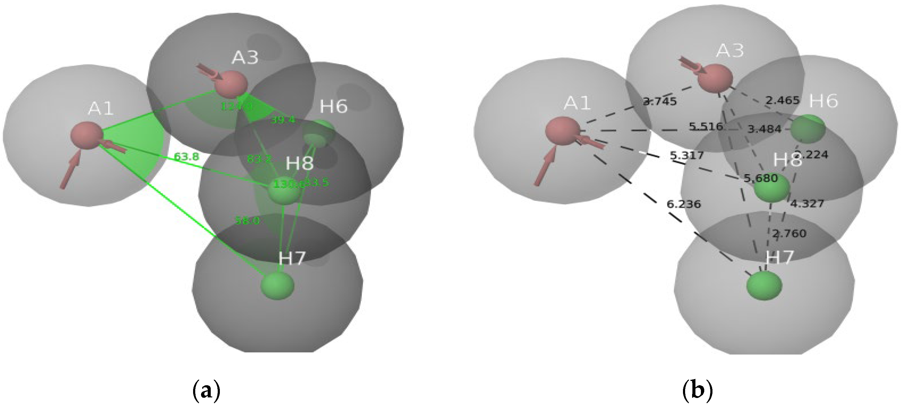
2.4. Pharmacophore Modeling

A pharmacophore model is a theory that explains how a group of compounds that bind to the same biological target exhibit the biological behaviors that have been observed [18]. The electron-withdrawing group, hydrogen bond donor, and hydrophobic top-active compounds are given below in Figure 6. The pharmacophore models were created using the Phase module of the Schrödinger suite 2021-4. The default set of six chemical properties of Phase was used to build pharmacophore sites for these compounds: hydrogen bond acceptor (A), hydrogen bond donor (D), hydrophobic (H) negative ionizable (N), positive ionizable (P), and aromatic ring (R). The distance and angles between different AAHHH.3 sites are shown in Figure 7a,b. AAHHH.3 represents that two hydrogen bond acceptors and three hydrophobic groups are essential for the activity. All ligands had their fitness scores evaluated using the AAHHH.3 model. A scatter plot analysis was also used to uncover discrete vital pharmacophoric requirements at spatial structure areas. The blue cubes around the ligand represented a favorable position for group substitution, whereas the red cubes showed a non-favorable position in Figure 6a–c for the top four ligands of this study.
Figure 6. (a) Electron-withdrawing group favorable positions (blue colour); (b) Hydrogen bond donor group non-favorable positions for BT_ER_15f(red colour); (c) Hydrophobic group favorable (blue) and non-favorable (red) positions for BT_ER_15f.
Molecules 28 04175 g006
Figure 7. (a) Bond angle between the AAHHH.3; (b) Bond distance between the AAHHH.3.
Molecules 28 04175 g007

2.5. 3D-QSAR Results
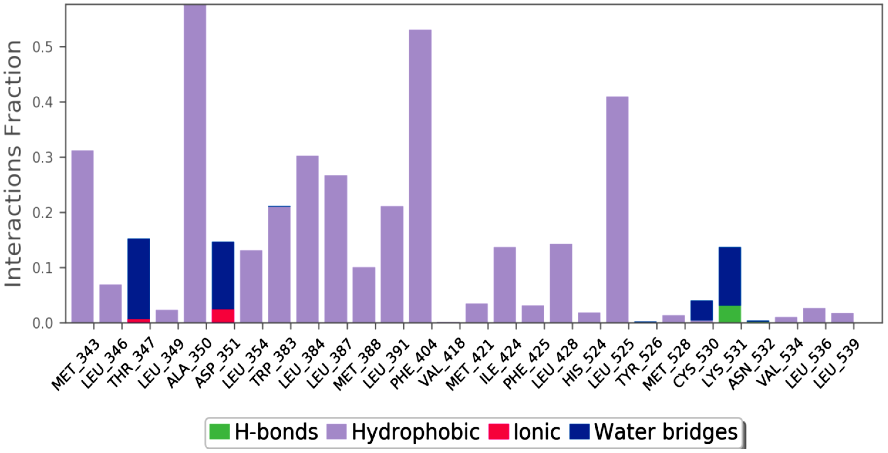
The atom-based QSAR module of Schrodinger suite 2021-4 was used to create the 3D-QSAR models for ER-alpha. Pharmacophore-based alignment of the ligands was taken into consideration in order to produce a statistically meaningful and highly predictive 3D-QSAR model [19]. Both the training and test sets of molecules had their prediction ability examined. Additionally, the default settings were applied, and a maximum of 2000 conformers and 15 conformations per rotatable bond were produced. Using vector, volume, site, survival, and survival in actives scores, the generated hypotheses were graded and ranked. Five places were determined to be common to all compounds in the hypothesis. A 3D-QSAR model was then developed using partial least squares (PLS) regression statistics.
The formula for the test set:
y = 0.58x + 2.29 (R2 = 0.83)
The green dots in Figure 8a,b represent ligands of the test set and training set. The ligands must be near the linear progression curve. The scatter plot with the XY-axis of the actual correlation with the predicted pIC50 is represented in Figure 8a,b for the test and training set compounds.
Figure 8. (a) Scatter plot for the test set; (b) Scatter plot for the training set.
Molecules 28 04175 g008

2.6. MD Simulation

The MD simulation is used to estimate macromolecule mechanics, and it is based on classical mechanics and the application of Newton’s equation of motion to compute the speed and location of each atom in the investigated system. As a result, MD undertakes a more thorough structural investigation than docking, resulting in a more realistic depiction of protein motion [20].
Using a 100 ns MD, the stability of the docked BT ER 15f/2IOG complex was examined. Using the Desmond module of Schrödinger 2021-4, the complex in the explicit solvent system with the OPLS4 force field was investigated. The BT_ER_15f compound interacts with the protein residues as shown in Figure 9. The interaction fraction of each amino acid is given in Figure 10.
Figure 9. Ligand atom interactions with the protein residues.
Molecules 28 04175 g009
Figure 10. Interaction fraction of amino acids of BT_ER_15f.
Molecules 28 04175 g010
The amino acid LYS 531, which is depicted in green in Figure 10, has the highest H-bond and a maximum interaction fraction of 0.5. H-bonds are essential for ligand binding. The donor and acceptor atoms in the donor-acceptor-hydrogen bond (D—H•••A) must be separated by 2.5 Å, the donor-acceptor-hydrogen bond (D—H•••A) must have a donor angle of 120°, and the hydrogen-acceptor-bonded atoms in the acceptor bond (H•••A—X) must have a donor angle of 90°. The following are the geometric requirements for hydrophobic interactions: p-cation, -aromatic, and charged groups that are within 4.5; p-p: two aromatic groups that are stacked face-to-face or face-to-edge; other non-specific hydrophobic side chains that are within 3.6 Å of a ligands’ aromatic or aliphatic carbons. A distance of 2.8 Å between the donor and acceptor atoms (D—H•••A), a donor angle of 110° between the donor-hydrogen-acceptor atoms (D—H•••A), and an acceptor angle of 90° between the hydrogen-acceptor-bonded atoms (H•••A—X) are needed for a H-bond to exist between a protein and water or water-ligand.
MD of standard tamoxifen was also performed, and it was found that the amino acids ALA350 and PHE404 have the highest interaction time. It is represented in Figure 11 and Figure 12. Amino acid residue ALA350 has a continuous interaction time. The RMSD value from the resulting trajectory analysis was in the range of 1.0 to 3.0. Green vertical bars in Figure 13 indicate protein residues that interact with the ligand, and the interactions between residues 100 and 130 showed the largest changes up to 2.4 Å. Through the formation of hydrophobic contacts with TRP383, ALA 350, PHE 404, and LEU 387, the molecule was positioned in the active pocket.
Figure 11. Interaction fraction of amino acids of the standard tamoxifen drug.
Molecules 28 04175 g011
Figure 12. Standard tamoxifen interactions with the protein residues.
Molecules 28 04175 g012
Figure 13. Interaction time of each amino acid.
Molecules 28 04175 g013
At 37 ns, higher ligand RMSD fluctuations (up to 2.7 Å) were noted, given in Figure 14. Stable hydrophobic interactions with ALA 350, LEU 387, and PHE 404 were noted during the simulation. Utilizing measures of root-mean-square fluctuations, the flexibility of residues on ligand bindings was examined. To comprehend the molecular insights involved in the binding of TRP383 in the active pocket of protein target 2IOG, a 100 ns molecular dynamic simulation was conducted.
It could be noted from Figure 15 that the deviation in the displacement of atoms is larger compared to BT_ER_15f. Thus BT_ER_15f, which has the best fit in the binding pocket, is best compared to the market available drug tamoxifen.
Figure 14. PL-RMSD of simulated protein 2IOG in complex with BT_ER_15f during 100 ns MD.
Molecules 28 04175 g014
Figure 15. PL-RMSD of simulated protein 2IOG in complex with Tamoxifen during 100 ns MD.
Molecules 28 04175 g015
The interaction time of each amino acid is given in Figure 13. It could be noted that the interaction times of amino acid TRP383 were greater than all other amino acids. Amino acid ASN 532 interaction was steady for the first 60 nanoseconds (ns), and then interaction was lost. Again, interaction occurs from 80 to 100 ns. Figure 16 provides the ligand (BT_ER_15f) characteristics such as ligand RMSD, radius of gyration (rGyr), intramolecular hydrogen bonds (intraHB), molecular surface area (MolSA), solvent accessible surface area (SASA), and polar surface area (PSA). The ligand and protein root-mean-square fluctuation is shown in Figure 17a,b, and it is important for describing local variations throughout the protein chain. RMSF is a measure of the displacement of a particular atom or group of atoms relative to the reference structure averaged over the number of atoms. RMSD is useful for the analysis of time-dependent motions of the structure.
L-RMSF (local root-mean-square fluctuation) and P-RMSF (protein root-mean-square Fluctuation) are both measures of a protein molecule’s flexibility or mobility. The average deviation or fluctuation in the position of each atom in a protein molecule from its average position in a given simulation or experimental data is measured as L-RMSF. It is calculated for a specific region or residue in a protein rather than the entire protein, and it is commonly used to identify flexible or disordered regions of a protein that are important for its function. P-RMSF, on the other hand, is the average RMSF value calculated for all the atoms in a protein molecule. It is used to quantify the protein’s overall flexibility or mobility and can aid in identifying regions that are relatively stable or flexible. P-RMSF can also be used to compare the flexibility of various proteins or conformations of the same protein. Both L-RMSF and P-RMSF are key techniques in the study of protein structure and function because they give insight into the dynamic features of proteins that are vital for their biological activity.

3. Materials and Method

3.1. Docking Studies

Docking studies were carried out mainly for four analogs which are marine sesquiterpene [21], sesquiterpene lactone analogs [22,23], heteroaromatic chalcones [24,25], and benzothiophene analogs [26] which were obtained from literature studies. The 3D crystal structure of the breast cancer protein 2IOG was previously co-crystallized with the N-[(1R)-3-(4hydroxyphenyl)-1-methylpropyl]-2-[2-phenyl-6-(2-piperidin-1-ylethoxy)-1H-indol-3-yl]acetamide. From the Protein Data Bank, the protein PDB ID 2IOG (resolution 1.6 Å) was retrieved. Arpita Roy published a paper on in silico investigation of agonists for proteins involved in breast cancer using the same target 2IOG [27]. The protein was optimized using the epic module of the Schrödinger suite 2021-4’s protein preparation wizard. By adjusting bond ordering, adding hydrogen atoms, and eliminating water molecules longer than 5 Å, the protein was optimized using the protein preparation wizard. Missing chains were then added using the Prime module of the Schrödinger suite 2021-4. The RMSD of the crystallographic heavy atoms was held at 0.30 for the OPLS4 molecular force field, which was used to minimize the protein. To pinpoint the centroid of the active site, a grid box was created. Using the Glide module of the Schrödinger suite 2021-4, all the compounds were docked into the catalytic pocket of the target protein 2IOG [28]. Significant Glide scores indicate ligands with higher 2IOG binding affinities [29,30]. Hussein highlighted in his work that the compound CH4 (chalcone) exhibited a binding energy of −10.83 kcal/mol against the target protein 2IOG [31], which possesses anti-breast cancer activity.

3.2. MM/GBSA Binding Free Energy Calculation

The precise determination of binding free energy plays a very essential role among the several strategies that may be used to analyze the ligand-receptor interaction [32,33]. Using the Prime molecular mechanics-generalized Born surface area (MM/GBSA) of Schrödinger 2021-4, post-docking energy minimization calculations were carried out to determine the free energy of binding for the collection of ligands in complex with a receptor [34]. The Poisson–Boltzmann surface area (MM/PBSA) and molecular mechanics-generalized Born surface area (MM/GBSA) are arguably very popular methods for binding free energy predictions [35]. Imaobong Etti published that the Artocarpus species has good anti-cancerous properties [36]. He and his co-workers found that Artonin E possesses the best drug-likeness using the Prime module of the Schrodinger software 2021-4 against the target protein 2IOG.

3.3. In Silico Predicted ADMET Properties

By identifying the most promising candidates for development and eliminating those with a low chance of success, early assessment of ADME-Tox characteristics can reduce the time and expense of screening and testing. The regulatory authorities are now very interested in the practical application of in silico methodologies for predicting preclinical toxicological endpoints, clinical side effects, and ADME features of new chemical entities [37]. ChemAxon properties such as molecular weight, total polar surface area (TPSA), hydrogen bond acceptor and donor count, log P, log D, log S, molar volume, and dissociation constant (KD), as well as the number of violations of Lipinski’s rule of five, van der Waals volume, and other properties were used to predict the physically and pharmacokinetically significant descriptors for the top hits by using the Qikprop module of Schrodinger suite 2021-4. Table 3 displays these outcomes.

3.4. Pharmacophore Modeling

An explanation for the pharmacological effects of a collection of substances that bind to the same biological target is known as a pharmacophore model [38]. “An ensemble of steric and electronic features necessary to produce optimal supramolecular interactions with a given biological target” is a pharmacophore model [39]. A pharmacophore model can then be used to query the 3D chemical library to look for potential ligands, which are referred to as “pharmacophore-based virtual screening,” depending on whether the approach was ligand- or structure-based virtual screening (VS) [40]. The pharmacophore model was created using the Phase module of the Schrodinger suite 2021-4. The common pharmacophore AAHHH.3 found from our work can be used for further high-throughput screening to screen a large database [41]. Tien-Yi-Hou and his co-workers performed work on estrogen receptor-α ligand binding through pharmacophore modeling and concluded few pharmacophore models active against breast cancer.

3.5. QSAR-Quantitative Structure Activity Relationship

The application of force field calculations requiring three-dimensional structures of a given collection of small molecules with known activities is referred to as 3D-QSAR. 3D-QSAR is an extension of classical QSAR that uses robust statistical analysis such as PLS, G/PLS, and AN to explain the three-dimensional features of ligands and predict their biological activity [42].
A computational modeling method known as the quantitative structure-activity relationship (QSAR) helps researchers connect the structural characteristics of chemical compounds with their biological functions. Drug development requires QSAR modeling [43].

3.6. Molecular Dynamics

A technique for simulating the physical motions of atoms and molecules is called molecular dynamics (MD) [44]. For a predetermined period of time, the atoms and molecules are allowed to interact, giving insight into the dynamic “evolution” of the system. Molecular dynamics is a method for computing the time evolution of a group of interacting atoms using Newton’s equations of motion. In the thermodynamic process of protein-ligand interaction, a tiny molecule’s solvation free energy acts as a stand-in for the ligand’s desolvation [45]. MD of the highest Glide score compound was performed in this work using the Desmond module of Schrodinger suite 2021-4. From these geometric requirements around the ligand, BT_ER_15f was identified.

4. Conclusions

In the realm of drug design and discovery, integrated methodologies of QSAR and molecular docking-based prediction have been successfully used in a number of statistically supported examples. The current research on benzothiophene analogs, specifically BT_ER_15f, using molecular docking and QSAR demonstrated that it has a sizable anti-cancer effect against the target 2IOG.
From the docking study, the benzothiophene derivative demonstrated better arrangement at the dynamic site. The current investigation aided in identifying the key compounds and their beneficial effects. In subsequent analysis using in vitro and in vivo techniques, it could be optimized as a drug to treat breast cancer. According to the findings, the compound BT_ER_15f, a benzothiophene derivative, exhibits strong anti-breast cancer action and is useful for future research.
The pharmacokinetics and drug-likeness studies revealed that the ligand BT_ER_15f could be the best drug candidate against breast cancer.
In the future, this study will be a reliable resource for achieving further benzothiophene derivatives through innovative structural modifications in benzothiophene derivatives that are being widely researched. These findings provide compelling support for novel studies that involve developing more methodological frameworks to investigate molecular facets of their anti-cancer action.

Supplementary Materials

The following supporting information can be downloaded at: https://www.mdpi.com/article/10.3390/molecules28104175/s1.

Author Contributions

Conceptualization, K.R.; Methodology, K.R., A.R.B., A.S., K.S. and M.S.; Software, K.R. and G.B.; Validation, K.R. and G.B.; Formal analysis, K.R., A.K., A.R.B., A.S., K.S., M.S. and G.B.; Investigation, K.R., A.K. and G.B.; Resources, A.K., A.R.B., A.S., K.S. and M.S.; Data curation, K.R., A.K., A.R.B., A.S., K.S., M.S. and G.B.; Writing—original draft, A.R.B., A.S., K.S. and M.S.; Writing—review & editing, K.R., A.K. and G.B.; Supervision, K.R. All authors have read and agreed to the published version of the manuscript.

Funding

Financial assistance was provided by JSS Academy of Higher Education & Research, Mysore under JSS AHER seed research grant.

Institutional Review Board Statement

Not applicable.

Informed Consent Statement

Not Applicable.

Data Availability Statement

The data supporting reported results can be available with corresponding author Kalirajan Rajagopal which will be shared on request by mail.

Acknowledgments

The authors’ heartfelt gratitude for the financial assistance provided by JSS Academy of Higher Education & Research, Mysore under JSS AHER research grant. S.P. Dhanabal, JSS College of Pharmacy in Ooty, is also acknowledged by the authors for his technical assistance.

Conflicts of Interest

The authors declare no conflict of interest.

Sample Availability

Not Applicable.

References

  1. Feng, Y.; Spezia, M.; Huang, S.; Yuan, C.; Zeng, Z.; Zhang, L.; Ji, X.; Liu, W.; Huang, B.; Luo, W.; et al. Breast cancer development and progression: Risk factors, cancer stem cells, signaling pathways, genomics, and molecular pathogenesis. Genes Dis. 2018, 5, 77–106. [Google Scholar] [CrossRef] [PubMed]
  2. Lemaine, V.; Simmons, P.S. The adolescent female: Breast and reproductive embryology and anatomy. Clin. Anat. 2013, 26, 22–28. [Google Scholar] [CrossRef] [PubMed]
  3. Bisoyi, P. Malignant Tumors—As Cancer. In Understanding Cancer; Elsevier: Amsterdam, The Netherlands, 2022; pp. 21–36. ISBN 978-0-323-99883-3. Available online: https://linkinghub.elsevier.com/retrieve/pii/B9780323998833000111 (accessed on 13 March 2023).
  4. Follain, G.; Herrmann, D.; Harlepp, S.; Hyenne, V.; Osmani, N.; Warren, S.C.; Timpson, P.; Goetz, J.G. Fluids and their mechanics in tumour transit: Shaping metastasis. Nat. Rev. Cancer 2020, 20, 107–124. [Google Scholar] [CrossRef] [PubMed]
  5. Šarenac, T.; Mikov, M. Cervical Cancer, Different Treatments and Importance of Bile Acids as Therapeutic Agents in This Disease. Front. Pharmacol. 2019, 10, 484. [Google Scholar] [CrossRef] [PubMed]
  6. Giaquinto, A.N.; Sung, H.; Miller, K.D.; Kramer, J.L.; Newman, L.A.; Minihan, A.; Jemal, A.; Siegel, R.L. Breast Cancer Statistics, 2022. CA Cancer J. Clin. 2022, 72, 524–541. [Google Scholar] [CrossRef]
  7. Mathur, P.; Sathishkumar, K.; Chaturvedi, M.; Das, P.; Sudarshan, K.L.; Santhappan, S.; Nallasamy, V.; John, A.; Narasimhan, S.; Roselind, F.S.; et al. Cancer Statistics, 2020: Report From National Cancer Registry Programme, India. JCO Glob. Oncol. 2020, 6, 1063–1075. [Google Scholar] [CrossRef]
  8. Kumar, N.; Gulati, H.K.; Sharma, A.; Heer, S.; Jassal, A.K.; Arora, L.; Kaur, S.; Singh, A.; Bhagat, K.; Kaur, A.; et al. Most recent strategies targeting estrogen receptor alpha for the treatment of breast cancer. Mol. Divers. 2021, 25, 603–624. [Google Scholar] [CrossRef]
  9. Nilsson, S.; Gustafsson, J.-Å. Estrogen Receptors: Therapies Targeted to Receptor Subtypes. Clin. Pharmacol. Ther. 2011, 89, 44–55. [Google Scholar] [CrossRef]
  10. Santen, R.J. Inhibition of aromatase: Insights from recent studies. Steroids 2003, 68, 559–567. [Google Scholar] [CrossRef]
  11. Zhang, X.; Wang, Y.; Li, X.; Wu, J.; Zhao, L.; Li, W.; Liu, J. Dynamics-Based Discovery of Novel, Potent Benzoic Acid Derivatives as Orally Bioavailable Selective Estrogen Receptor Degraders for ERα+ Breast Cancer. J. Med. Chem. 2021, 64, 7575–7595. [Google Scholar] [CrossRef]
  12. McDonnell, D.P.; Wardell, S.E.; Norris, J.D. Oral Selective Estrogen Receptor Downregulators (SERDs), a Breakthrough Endocrine Therapy for Breast Cancer. J. Med. Chem. 2015, 58, 4883–4887. [Google Scholar] [CrossRef]
  13. Baselga, J.; Swain, S.M. CLEOPATRA: A Phase III Evaluation of Pertuzumab and Trastuzumab for HER2-Positive Metastatic Breast Cancer. Clin. Breast Cancer 2010, 10, 489–491. [Google Scholar] [CrossRef]
  14. Nathan, M.R.; Schmid, P. A Review of Fulvestrant in Breast Cancer. Oncol. Ther. 2017, 5, 17–29. [Google Scholar] [CrossRef]
  15. Paterni, I.; Granchi, C.; Katzenellenbogen, J.A.; Minutolo, F. Estrogen receptors alpha (ERα) and beta (ERβ): Subtype-selective ligands and clinical potential. Steroids 2014, 90, 13–29. [Google Scholar] [CrossRef]
  16. Dahlman-Wright, K.; Cavailles, V.; Fuqua, S.A.; Jordan, V.C.; Katzenellenbogen, J.A.; Korach, K.S.; Maggi, A.; Muramatsu, M.; Parker, M.G.; Gustafsson, J.-Å. International Union of Pharmacology. LXIV. Estrogen Receptors. Pharmacol. Rev. 2006, 58, 773–781. [Google Scholar] [CrossRef]
  17. Rajagopal, K.; Varakumar, P.; Aparna, B.; Byran, G.; Jupudi, S. Identification of some novel oxazine substituted 9-anilinoacridines as SARS-CoV-2 inhibitors for COVID-19 by molecular docking, free energy calculation and molecular dynamics studies. J. Biomol. Struct. Dyn. 2021, 39, 5551–5562. [Google Scholar] [CrossRef]
  18. Horvath, D. Pharmacophore-Based Virtual Screening. In Chemoinformatics and Computational Chemical Biology; Bajorath, J., Ed.; Methods in Molecular Biology; Humana Press: Totowa, NJ, USA, 2010; Volume 672, pp. 261–298. ISBN 978-1-60761-838-6. [Google Scholar] [CrossRef]
  19. Chaudhaery, S.S.; Roy, K.K.; Saxena, A.K. Consensus Superiority of the Pharmacophore-Based Alignment, Over Maximum Common Substructure (MCS): 3D-QSAR Studies on Carbamates as Acetylcholinesterase Inhibitors. J. Chem. Inf. Model. 2009, 49, 1590–1601. [Google Scholar] [CrossRef]
  20. Alnajjar, R.; Mostafa, A.; Kandeil, A.; Al-Karmalawy, A.A. Molecular docking, molecular dynamics, and in vitro studies reveal the potential of angiotensin II receptor blockers to inhibit the COVID-19 main protease. Heliyon 2020, 6, e05641. [Google Scholar] [CrossRef]
  21. Liang, J.-J.; Yu, W.-L.; Yang, L.; Xie, B.-H.; Qin, K.-M.; Yin, Y.-P.; Yan, J.-J.; Gong, S.; Liu, T.-Y.; Zhou, H.-B.; et al. Design and synthesis of marine sesterterpene analogues as novel estrogen receptor α degraders for breast cancer treatment. Eur. J. Med. Chem. 2022, 229, 114081. [Google Scholar] [CrossRef]
  22. Nakagawa-Goto, K.; Chen, J.-Y.; Cheng, Y.-T.; Lee, W.-L.; Takeya, M.; Saito, Y.; Lee, K.-H.; Shyur, L.-F. Novel sesquiterpene lactone analogues as potent anti-breast cancer agents. Mol. Oncol. 2016, 10, 921–937. [Google Scholar] [CrossRef]
  23. Zhang, S.; Won, Y.-K.; Ong, C.-N.; Shen, H.-M. Anti-Cancer Potential of Sesquiterpene Lactones: Bioactivity and Molecular Mechanisms. Curr. Med. Chem.-Anti-Cancer Agents 2005, 5, 239–249. [Google Scholar] [CrossRef]
  24. Kalirajan, R.; Sivakumar, S.U.; Jubie, S.; Gowramma, B.; Suresh, B. Synthesis and biological evaluation of some heterocyclic derivatives of chalcones. Int. J. Chem. Tech. Res. 2009, 1, 27–34. [Google Scholar]
  25. Jeon, K.-H.; Yu, H.-B.; Kwak, S.Y.; Kwon, Y.; Na, Y. Synthesis and topoisomerases inhibitory activity of heteroaromatic chalcones. Bioorg. Med. Chem. 2016, 24, 5921–5928. [Google Scholar] [CrossRef]
  26. Bai, C.; Wu, S.; Ren, S.; Zhu, M.; Luo, G.; Xiang, H. Benzothiophene derivatives as selective estrogen receptor covalent antagonists: Design, synthesis and anti-ERα activities. Bioorg. Med. Chem. 2021, 47, 116395. [Google Scholar] [CrossRef] [PubMed]
  27. Roy, A.; Anand, A.; Garg, S.; Khan, M.S.; Bhasin, S.; Asghar, M.N.; Emran, T.B. Structure-Based In Silico Investigation of Agonists for Proteins Involved in Breast Cancer. Evid.-Based Complement. Alternat. Med. 2022, 2022, 7278731. [Google Scholar] [CrossRef] [PubMed]
  28. Pattar, S.V.; Adhoni, S.A.; Kamanavalli, C.M.; Kumbar, S.S. In silico molecular docking studies and MM/GBSA analysis of coumarin-carbonodithioate hybrid derivatives divulge the anticancer potential against breast cancer. Beni-Suef Univ. J. Basic Appl. Sci. 2020, 9, 36. [Google Scholar] [CrossRef]
  29. Sousa, S.F.; Fernandes, P.A.; Ramos, M.J. Protein-ligand docking: Current status and future challenges. Proteins Struct. Funct. Bioinforma. 2006, 65, 15–26. [Google Scholar] [CrossRef]
  30. Bissantz, C.; Folkers, G.; Rognan, D. Protein-Based Virtual Screening of Chemical Databases. 1. Evaluation of Different Docking/Scoring Combinations. J. Med. Chem. 2000, 43, 4759–4767. [Google Scholar] [CrossRef]
  31. Hussein, K.; Shihab, N.; Saeed, B. Anti-Cancer, Anti-Osteoporosis, and Molecular Docking Studies of Novel Chalcone and Epoxy Chalcone. Biointerface Res. Appl. Chem. 2021, 12, 6668–6685. [Google Scholar] [CrossRef]
  32. Tuccinardi, T. What is the current value of MM/PBSA and MM/GBSA methods in drug discovery? Expert Opin. Drug Discov. 2021, 16, 1233–1237. [Google Scholar] [CrossRef]
  33. Wang, W.; Kollman, P.A. Free energy calculations on dimer stability of the HIV protease using molecular dynamics and a continuum solvent model. J. Mol. Biol. 2000, 303, 567–582. [Google Scholar] [CrossRef]
  34. Rajagopal, K.; Kannan, R.; Aparna, B.; Varakumar, P.; Pandiselvi, A.; Gowramma, B. COVID-19 Activity of Some 9-Anilinoacridines substituted with Pyrazole against SARS CoV2 Main Protease: An In-silico Approach. Res. J. Pharm. Technol. 2023, 16, 529–534. [Google Scholar] [CrossRef]
  35. Wang, E.; Sun, H.; Wang, J.; Wang, Z.; Liu, H.; Zhang, J.Z.H.; Hou, T. End-Point Binding Free Energy Calculation with MM/PBSA and MM/GBSA: Strategies and Applications in Drug Design. Chem. Rev. 2019, 119, 9478–9508. [Google Scholar] [CrossRef]
  36. Etti, I.; Abdullah, R.; Hashim, N.; Kadir, A.; Abdul, A.; Etti, C.; Malami, I.; Waziri, P.; How, C. Artonin E and Structural Analogs from Artocarpus Species Abrogates Estrogen Receptor Signaling in Breast Cancer. Molecules 2016, 21, 839. [Google Scholar] [CrossRef]
  37. Roy, K.; Kar, S. In Silico Models for Ecotoxicity of Pharmaceuticals. In In Silico Methods for Predicting Drug Toxicity; Benfenati, E., Ed.; Methods in Molecular Biology; Springer: New York, NY, USA, 2016; pp. 237–304. ISBN 978-1-4939-3609-0. [Google Scholar] [CrossRef]
  38. Vuorinen, A.; Odermatt, A.; Schuster, D. In silico methods in the discovery of endocrine disrupting chemicals. J. Steroid Biochem. Mol. Biol. 2013, 137, 18–26. [Google Scholar] [CrossRef]
  39. Yang, S.-Y. Pharmacophore modeling and applications in drug discovery: Challenges and recent advances. Drug Discov. Today 2010, 15, 444–450. [Google Scholar] [CrossRef]
  40. Seidel, T.; Ibis, G.; Bendix, F.; Wolber, G. Strategies for 3D pharmacophore-based virtual screening. Drug Discov. Today Technol. 2010, 7, e221–e228. [Google Scholar] [CrossRef]
  41. Veeramachaneni, G.K.; Raj, K.K.; Chalasani, L.M.; Bondili, J.S.; Talluri, V.R. High-throughput virtual screening with e-pharmacophore and molecular simulations study in the designing of pancreatic lipase inhibitors. Drug Des. Devel. Ther. 2015, 9, 4397–4412. [Google Scholar] [CrossRef]
  42. Patel, H.M.; Noolvi, M.N.; Sharma, P.; Jaiswal, V.; Bansal, S.; Lohan, S.; Kumar, S.S.; Abbot, V.; Dhiman, S.; Bhardwaj, V. Quantitative structure–activity relationship (QSAR) studies as strategic approach in drug discovery. Med. Chem. Res. 2014, 23, 4991–5007. [Google Scholar] [CrossRef]
  43. Kalirajan, R.; Kulshrestha, V.; Sankar, S. Synthesis, Characterization and Antitumour Activity of Some Novel Oxazine Substituted 9-Anilinoacridines and their 3D-QSAR Studies. Indian J. Pharm. Sci. 2018, 80, 921–929. [Google Scholar] [CrossRef]
  44. Adcock, S.A.; McCammon, J.A. Molecular Dynamics: Survey of Methods for Simulating the Activity of Proteins. Chem. Rev. 2006, 106, 1589–1615. [Google Scholar] [CrossRef] [PubMed]
  45. Shivakumar, D.; Williams, J.; Wu, Y.; Damm, W.; Shelley, J.; Sherman, W. Prediction of Absolute Solvation Free Energies using Molecular Dynamics Free Energy Perturbation and the OPLS Force Field. J. Chem. Theory Comput. 2010, 6, 1509–1519. [Google Scholar] [CrossRef] [PubMed]
Figure 1. Structure of benzothiophene (BT) analogs.
Molecules 28 04175 g001aMolecules 28 04175 g001b
Figure 2. Structure of marine sesterterpene (MS) analogs.
Molecules 28 04175 g002
Figure 3. Structure of heteroaromatic chalcones (HC) analogs.
Molecules 28 04175 g003
Figure 4. Structure of sesquiterpene lactone (SL) analogs.
Molecules 28 04175 g004aMolecules 28 04175 g004bMolecules 28 04175 g004c
Figure 16. Ligand properties of BT_ER_15f in complex with 2IOG during 100 ns MD simulation.
Molecules 28 04175 g016
Figure 17. (a) L-RMSF of simulated protein; (b) P-RMSF of simulated protein 2IOG in complex with BT_ER_15f.
Molecules 28 04175 g017
Disclaimer/Publisher’s Note: The statements, opinions and data contained in all publications are solely those of the individual author(s) and contributor(s) and not of MDPI and/or the editor(s). MDPI and/or the editor(s) disclaim responsibility for any injury to people or property resulting from any ideas, methods, instructions or products referred to in the content.

