
The Power of Field-Flow Fractionation in Characterization of Nanoparticles in Drug Delivery
Abstract

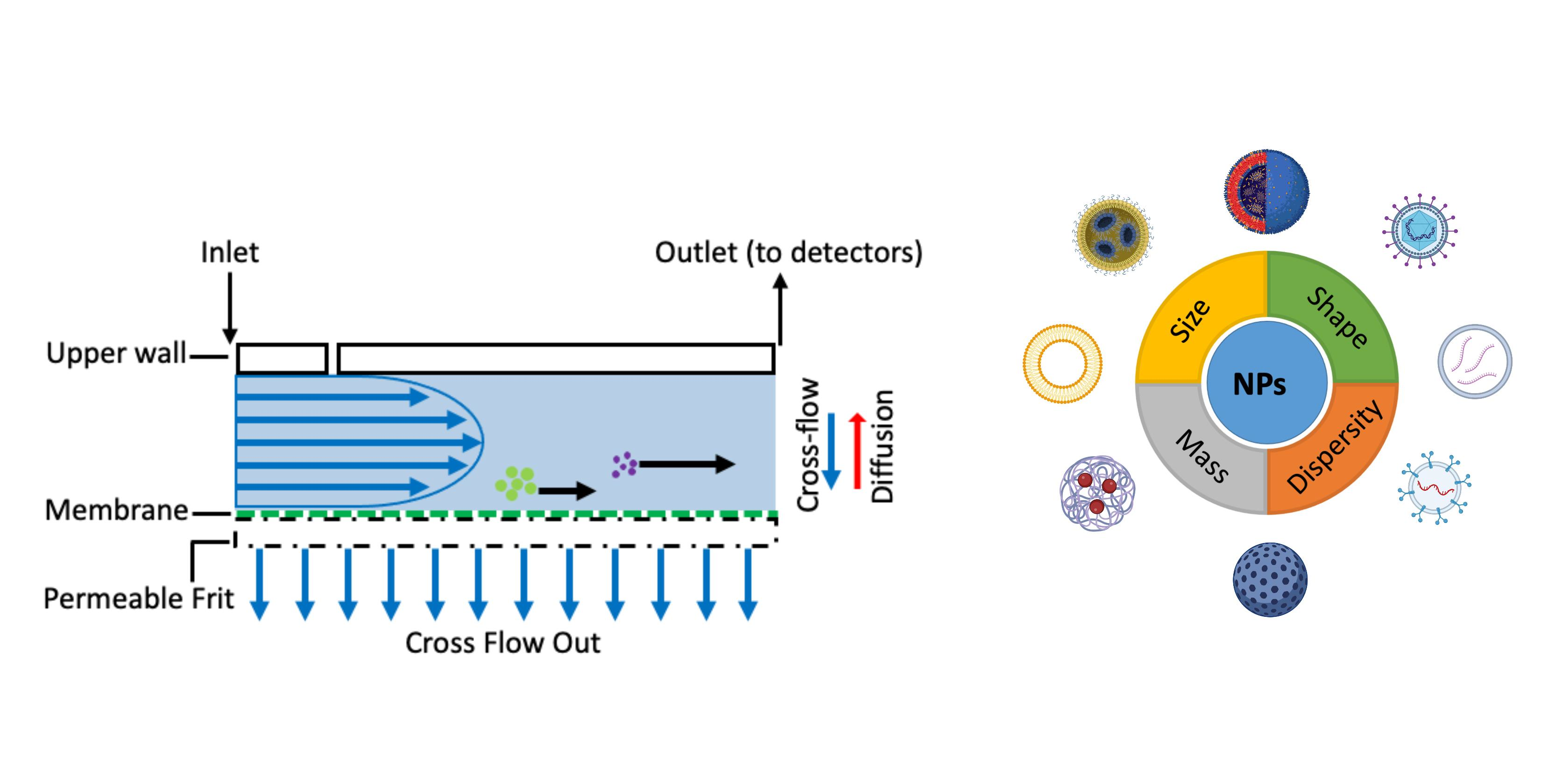
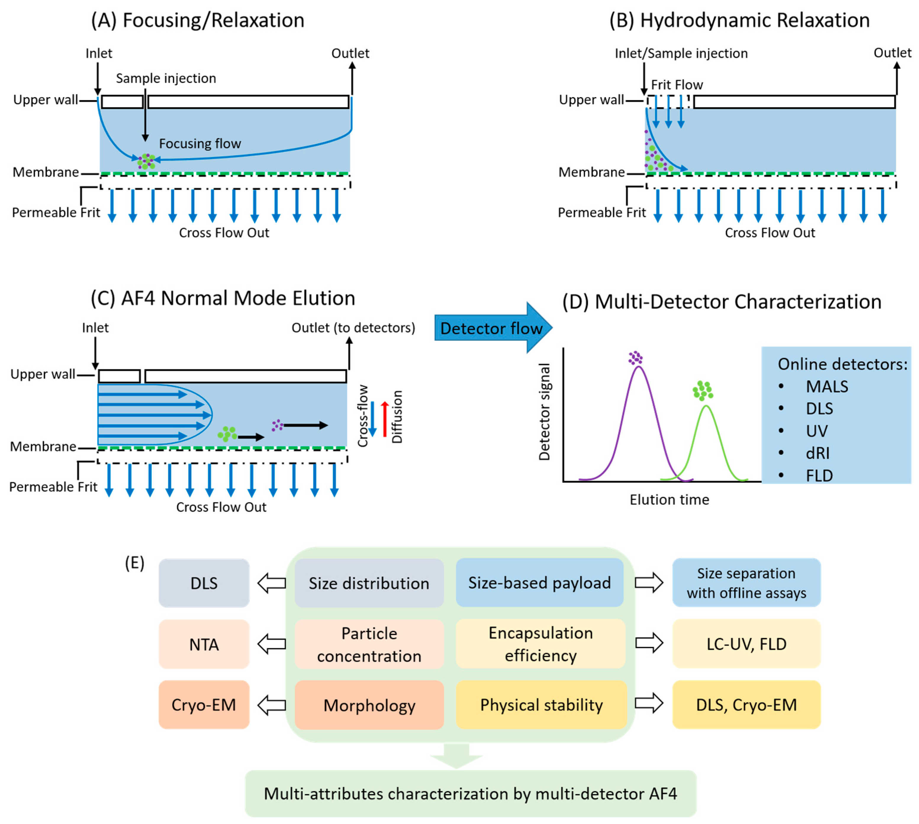
1. Introduction
2. Applications of FFF in Nanoparticle Drug Delivery Systems
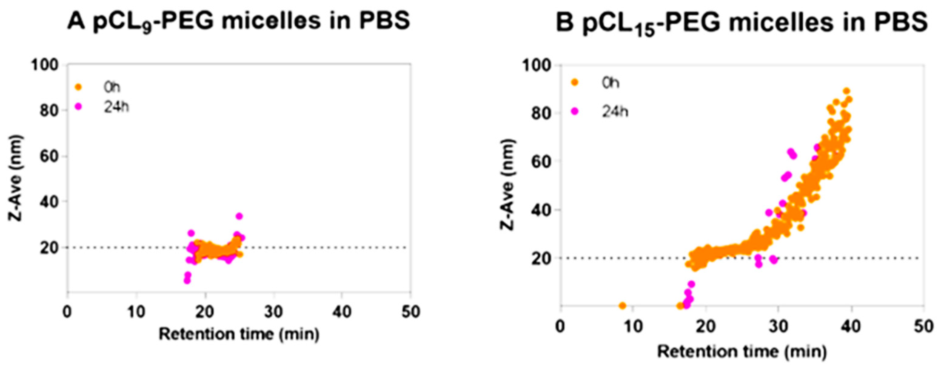
2.1. Lipid-Based Nanoparticles
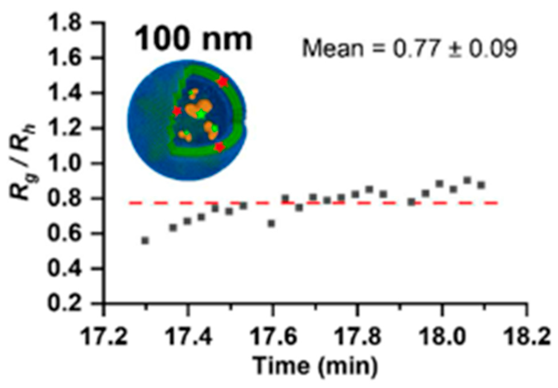
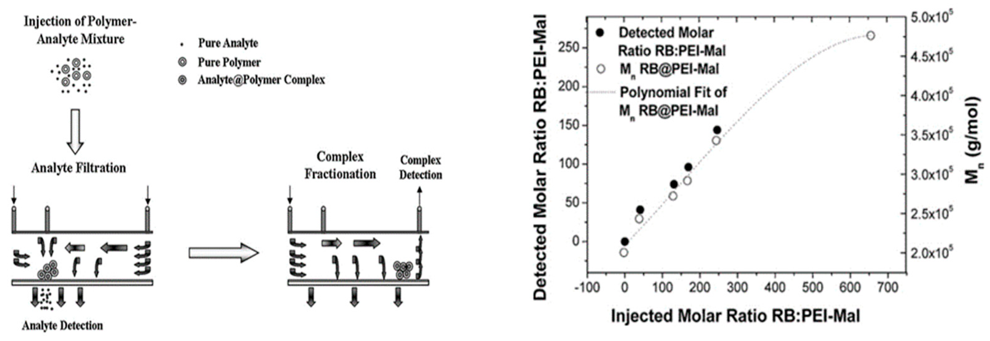
2.2. Polymer-Based Nanoparticles
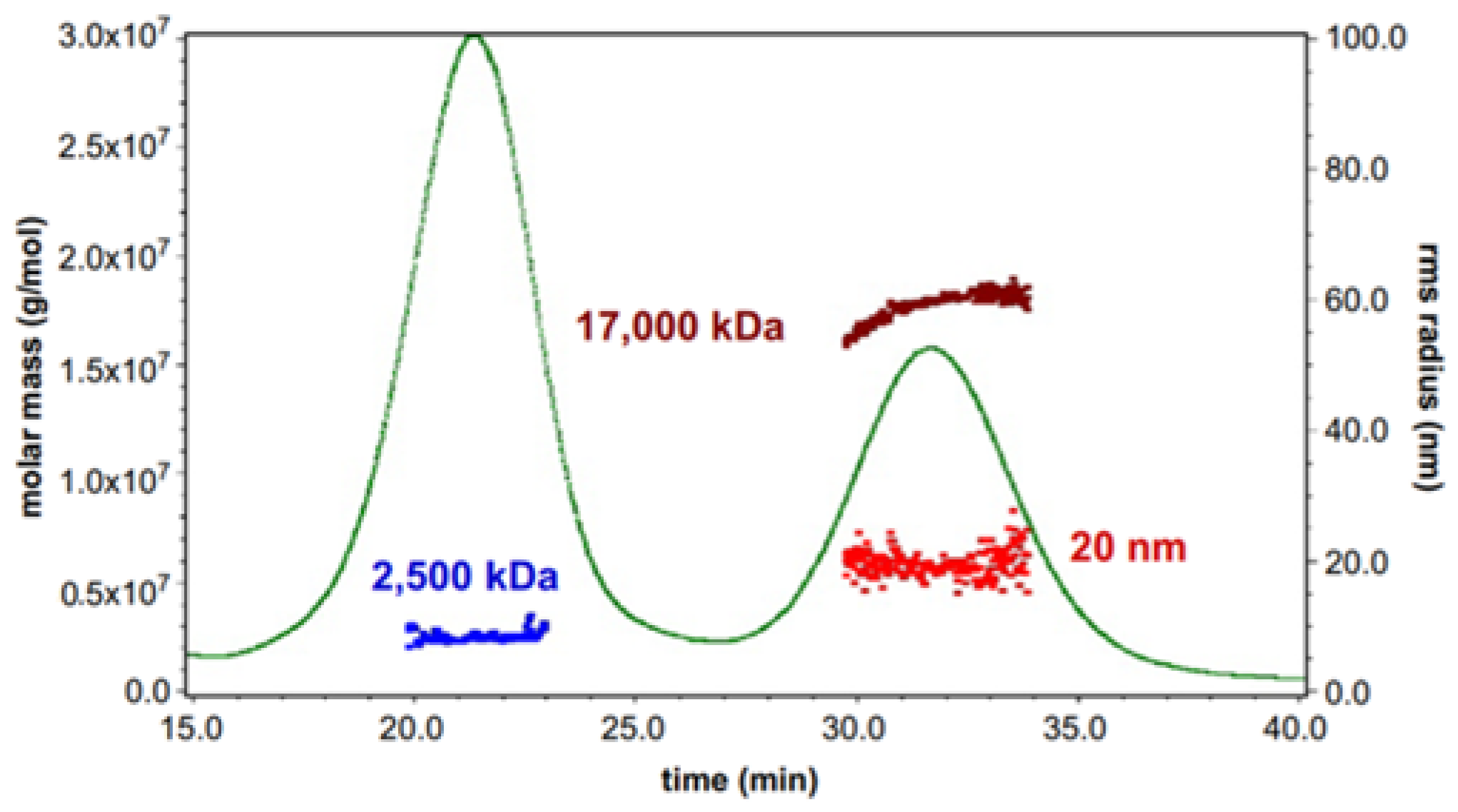
2.3. Viral Vectors and Virus-like Nanoparticles
2.4. Extracellular Vesicles
2.5. Inorganic Nanoparticles
3. Current Challenges and Future Trends
4. Summary
Author Contributions
Funding
Institutional Review Board Statement
Informed Consent Statement
Data Availability Statement
Acknowledgments
Conflicts of Interest
References
- Sharma, K.; Koirala, A.; Nicolopoulos, K.; Chiu, C.; Wood, N.; Britton, P.N. Vaccines for COVID-19: Where do we stand in 2021? Paediatr. Respir. Rev. 2021, 39, 22–31. [Google Scholar] [CrossRef] [PubMed]
- Hussain, A.; Yang, H.; Zhang, M.; Liu, Q.; Alotaibi, G.; Irfan, M.; He, H.; Chang, J.; Liang, X.J.; Weng, Y.; et al. Mrna vaccines for COVID-19 and diverse diseases. J. Control Release 2022, 345, 314–333. [Google Scholar] [CrossRef] [PubMed]
- Rauf, A.; Abu-Izneid, T.; Khalil, A.A.; Hafeez, N.; Olatunde, A.; Rahman, M.; Semwal, P.; Al-Awthan, Y.S.; Bahattab, O.S.; Khan, I.N.; et al. Nanoparticles in clinical trials of COVID-19: An update. Int. J. Surg. 2022, 104, 106818. [Google Scholar] [CrossRef] [PubMed]
- Jahangirian, H.; Lemraski, E.G.; Webster, T.J.; Rafiee-Moghaddam, R.; Abdollahi, Y. A review of drug delivery systems based on nanotechnology and green chemistry: Green nanomedicine. Int. J. Nanomed. 2017, 12, 2957–2978. [Google Scholar] [CrossRef]
- Patra, J.K.; Das, G.; Fraceto, L.F.; Campos, E.V.R.; Rodriguez-Torres, M.D.P.; Acosta-Torres, L.S.; Diaz-Torres, L.A.; Grillo, R.; Swamy, M.K.; Sharma, S.; et al. Nano based drug delivery systems: Recent developments and future prospects. J. Nanobiotechnol. 2018, 16, 71. [Google Scholar] [CrossRef]
- Yao, Y.; Zhou, Y.; Liu, L.; Xu, Y.; Chen, Q.; Wang, Y.; Wu, S.; Deng, Y.; Zhang, J.; Shao, A. Nanoparticle-based drug delivery in cancer therapy and its role in overcoming drug resistance. Front. Mol. Biosci. 2020, 7, 193. [Google Scholar] [CrossRef]
- Dos Santos Rodrigues, B.; Lakkadwala, S.; Kanekiyo, T.; Singh, J. Development and screening of brain-targeted lipid-based nanoparticles with enhanced cell penetration and gene delivery properties. Int. J. Nanomed. 2019, 14, 6497–6517. [Google Scholar] [CrossRef]
- Ickenstein, L.M.; Garidel, P. Lipid-based nanoparticle formulations for small molecules and rna drugs. Expert. Opin. Drug. Deliv. 2019, 16, 1205–1226. [Google Scholar] [CrossRef]
- Thi, T.T.H.; Suys, E.J.A.; Lee, J.S.; Nguyen, D.H.; Park, K.D.; Truong, N.P. Lipid-based nanoparticles in the clinic and clinical trials: From cancer nanomedicine to COVID-19 vaccines. Vaccines 2021, 9, 359. [Google Scholar] [CrossRef]
- Sur, S.; Rathore, A.; Dave, V.; Reddy, K.R.; Chouhan, R.S.; Sadhu, V. Recent developments in functionalized polymer nanoparticles for efficient drug delivery system. Nano-Struct. Nano-Objects 2019, 20, 100397. [Google Scholar] [CrossRef]
- Charelli, L.E.; de Mattos, G.C.; de Jesus Sousa-Batista, A.; Pinto, J.C.; Balbino, T.A. Polymeric nanoparticles as therapeutic agents against coronavirus disease. J. Nanopart Res. 2022, 24, 12. [Google Scholar] [CrossRef] [PubMed]
- Ghezzi, M.; Pescina, S.; Padula, C.; Santi, P.; Del Favero, E.; Cantu, L.; Nicoli, S. Polymeric micelles in drug delivery: An insight of the techniques for their characterization and assessment in biorelevant conditions. J. Control Release 2021, 332, 312–336. [Google Scholar] [CrossRef] [PubMed]
- Jeevanandam, J.; Pal, K.; Danquah, M.K. Virus-like nanoparticles as a novel delivery tool in gene therapy. Biochimie 2019, 157, 38–47. [Google Scholar] [CrossRef]
- Chung, Y.H.; Cai, H.; Steinmetz, N.F. Viral nanoparticles for drug delivery, imaging, immunotherapy, and theranostic applications. Adv. Drug. Deliv. Rev. 2020, 156, 214–235. [Google Scholar] [CrossRef] [PubMed]
- Walker, S.; Busatto, S.; Pham, A.; Tian, M.; Suh, A.; Carson, K.; Quintero, A.; Lafrence, M.; Malik, H.; Santana, M.X.; et al. Extracellular vesicle-based drug delivery systems for cancer treatment. Theranostics 2019, 9, 8001–8017. [Google Scholar] [CrossRef]
- Herrmann, I.K.; Wood, M.J.A.; Fuhrmann, G. Extracellular vesicles as a next-generation drug delivery platform. Nat. Nanotechnol. 2021, 16, 748–759. [Google Scholar] [CrossRef]
- Ghosn, Y.; Kamareddine, M.H.; Tawk, A.; Elia, C.; El Mahmoud, A.; Terro, K.; El Harake, N.; El-Baba, B.; Makdessi, J.; Farhat, S. Inorganic nanoparticles as drug delivery systems and their potential role in the treatment of chronic myelogenous leukaemia. Technol. Cancer Res. Treat. 2019, 18, 1533033819853241. [Google Scholar] [CrossRef]
- Shi, Z.; Zhou, Y.; Fan, T.; Lin, Y.; Zhang, H.; Mei, L. Inorganic nano-carriers based smart drug delivery systems for tumor therapy. Smart Mater. Med. 2020, 1, 32–47. [Google Scholar] [CrossRef]
- Drug Products, Including Biological Products, That Contain Nanomaterials—Guidance for Industry; US Food and Drug Administration: Silver Spring, MD, USA, 2022. Available online: https://www.fda.gov/regulatory-information/search-fda-guidance-documents/drug-products-including-biological-products-contain-nanomaterials-guidance-industry (accessed on 20 April 2022).
- Liposome Drug Products Chemistry, Manufacturing, and Controls; Human Pharmacokinetics and Bioavailability; and Labeling Documentation; US Food and Drug Administration: Silver Spring, MD, USA, 2018. Available online: https://www.fda.gov/regulatory-information/search-fda-guidance-documents/liposome-drug-products-chemistry-manufacturing-and-controls-human-pharmacokinetics-and (accessed on 4 October 2021).
- Reflection Paper on the Data Requirements for Intravenous Liposomal Products Developed with Reference to an Innovator Liposomal Product; European Medicines Agency: London, UK, 2013; Available online: https://www.ema.europa.eu/en/data-requirements-intravenous-liposomal-products-developed-reference-innovator-liposomal-product-0#current-effective-version-section (accessed on 13 March 2013).
- Giddings, J.C. A new separation concept based on a coupling of concentration and flow nonuniformities. Sep. Sci. 1966, 1, 123–125. [Google Scholar] [CrossRef]
- Plavchak, C.L.; Smith, W.C.; Bria, C.R.M.; Williams, S.K.R. New advances and applications in field-flow fractionation. Annu. Rev. Anal. Chem. 2021, 14, 257–279. [Google Scholar] [CrossRef]
- Zattoni, A.; Roda, B.; Borghi, F.; Marassi, V.; Reschiglian, P. Flow field-flow fractionation for the analysis of nanoparticles used in drug delivery. J. Pharm. Biomed. Anal. 2014, 87, 53–61. [Google Scholar] [CrossRef] [PubMed]
- Hovingh, M.E.; Thompson, G.H.; Giddings, J.C. Column parameters in thermal field-flow fractionation. Anal. Chem. 1970, 42, 195–203. [Google Scholar] [CrossRef]
- Liu, G.; Giddings, J.C. Separation of particles in aqueous suspensions by thermal field-flow fractionation—Measurement of thermal-diffusion coefficients. Chromatographia 1992, 34, 483–492. [Google Scholar] [CrossRef]
- Giddings, J.C.; Yang, F.J.F.; Myers, M.N. Sedimentation field-flow fractionation. Anal. Chem. 1974, 46, 1917–1924. [Google Scholar] [CrossRef]
- Chianéa, T.; Assidjo, N.E.; Cardot, P.J.P. Sedimentation field-flow-fractionation: Emergence of a new cell separation methodology. Talanta 2000, 51, 835–847. [Google Scholar] [CrossRef]
- Williams, P.S.; Carpino, F.; Zborowski, M. Magnetic nanoparticle drug carriers and their study by quadrupole magnetic field-flow fractionation. Mol. Pharm. 2009, 6, 1290–1306. [Google Scholar] [CrossRef]
- Caldwell, K.D.; Gao, Y.S. Electrical field-flow fractionation in particle separation. 1. Monodisperse standards. Anal. Chem. 1993, 65, 1764–1772. [Google Scholar] [CrossRef]
- Johann, C.; Elsenberg, S.; Schuch, H.; Rosch, U. Instrument and method to determine the electrophoretic mobility of nanoparticles and proteins by combining electrical and flow field-flow fractionation. Anal. Chem. 2015, 87, 4292–4298. [Google Scholar] [CrossRef]
- Contado, C. Field flow fractionation techniques to explore the “nano-world”. Anal. Bioanal. Chem. 2017, 409, 2501–2518. [Google Scholar] [CrossRef]
- Giddings, J.C.; Yang, F.J.; Myers, M.N. Flow-field-flow fractionation: A versatile new separation method. Science 1976, 193, 1244–1245. [Google Scholar] [CrossRef]
- Wahlund, K.G.; Giddings, J.C. Properties of an asymmetrical flow field-flow fractionation channel having one permeable wall. Anal. Chem. 1987, 59, 1332–1339. [Google Scholar] [CrossRef] [PubMed]
- Caputo, F.; Mehn, D.; Clogston, J.D.; Rosslein, M.; Prina-Mello, A.; Borgos, S.E.; Gioria, S.; Calzolai, L. Asymmetric-flow field-flow fractionation for measuring particle size, drug loading and (in)stability of nanopharmaceuticals. The joint view of european union nanomedicine characterization laboratory and national cancer institute—Nanotechnology characterization laboratory. J. Chromatogr. A 2021, 1635, 461767. [Google Scholar] [PubMed]
- Wahlund, K.G. Flow field-flow fractionation: Critical overview. J. Chromatogr. A 2013, 1287, 97–112. [Google Scholar] [CrossRef]
- Moon, M.H.; Hwang, I. Hydrodynamic vs. Focusing relaxation in asymmetrical flow field-flow fractionation. J. Liq. Chromatogr. Relat. Technol. 2007, 24, 3069–3083. [Google Scholar] [CrossRef]
- Fuentes, C.; Choi, J.; Zielke, C.; Penarrieta, J.M.; Lee, S.; Nilsson, L. Comparison between conventional and frit-inlet channels in separation of biopolymers by asymmetric flow field-flow fractionation. Analyst 2019, 144, 4559–4568. [Google Scholar] [CrossRef] [PubMed]
- Mildner, R.; Hak, S.; Parot, J.; Hyldbakk, A.; Borgos, S.E.; Some, D.; Johann, C.; Caputo, F. Improved multidetector asymmetrical-flow field-flow fractionation method for particle sizing and concentration measurements of lipid-based nanocarriers for rna delivery. Eur. J. Pharm. Biopharm. 2021, 163, 252–265. [Google Scholar] [CrossRef]
- Elvang, P.A.; Stein, P.C.; Bauer-Brandl, A.; Brandl, M. Characterization of co-existing colloidal structures in fasted state simulated fluids fassif: A comparative study using af4/malls, dls and dosy. J. Pharm. Biomed. Anal. 2017, 145, 531–536. [Google Scholar] [CrossRef]
- Champagne, J. Vlp Characterization: The Light Scattering Biophysical Toolbox. Available online: https://www.wyatt.com/library/webinars/vlp-characterization-light-scattering-biophysical-toolbox.html (accessed on 5 February 2014).
- Gobalasingham, N. Expanding the Characterization Toolkit with fff-mals: Developments, Techniques, and Applications. Available online: https://www.wyatt.com/library/webinars/expanding-the-characterization-toolkit-with-fff-mals-developments-techniques-and-applications.html?utm_source=lcgc&utm_medium=digital-ad&utm_campaign=resource-center-08-2021 (accessed on 15 June 2021).
- Gioria, S.; Caputo, F.; Urban, P.; Maguire, C.M.; Bremer-Hoffmann, S.; Prina-Mello, A.; Calzolai, L.; Mehn, D. Are existing standard methods suitable for the evaluation of nanomedicines: Some case studies. Nanomedicine 2018, 13, 539–554. [Google Scholar] [CrossRef]
- Hinna, A.; Steiniger, F.; Hupfeld, S.; Brandl, M.; Kuntsche, J. Asymmetrical flow field-flow fractionation with on-line detection for drug transfer studies: A feasibility study. Anal. Bioanal. Chem. 2014, 406, 7827–7839. [Google Scholar] [CrossRef]
- Citkowicz, A.; Petry, H.; Harkins, R.N.; Ast, O.; Cashion, L.; Goldmann, C.; Bringmann, P.; Plummer, K.; Larsen, B.R. Characterization of virus-like particle assembly for DNA delivery using asymmetrical flow field-flow fractionation and light scattering. Anal. Biochem. 2008, 376, 163–172. [Google Scholar] [CrossRef]
- Janwitayanuchit, W.; Suwanborirux, K.; Patarapanich, C.; Pummangura, S.; Lipipun, V.; Vilaivan, T. Synthesis and anti-herpes simplex viral activity of monoglycosyl diglycerides. Phytochemistry 2003, 64, 1253–1264. [Google Scholar] [CrossRef] [PubMed]
- Nilsson, L. Separation and characterization of food macromolecules using field-flow fractionation: A review. Food Hydrocoll. 2013, 30, 1–11. [Google Scholar] [CrossRef]
- Fraunhofer, W.; Winter, G. The use of asymmetrical flow field-flow fractionation in pharmaceutics and biopharmaceutics. Eur. J. Pharm. Biopharm. 2004, 58, 369–383. [Google Scholar] [CrossRef] [PubMed]
- Wagner, M.; Holzschuh, S.; Traeger, A.; Fahr, A.; Schubert, U.S. Asymmetric flow field-flow fractionation in the field of nanomedicine. Anal. Chem. 2014, 86, 5201–5210. [Google Scholar] [CrossRef]
- Wyatt, P.J. Measurement of special nanoparticle structures by light scattering. Anal. Chem. 2014, 86, 7171–7183. [Google Scholar] [CrossRef]
- Mogensen, K.B.; Kneipp, K. Size-dependent shifts of plasmon resonance in silver nanoparticle films using controlled dissolution: Monitoring the onset of surface screening effects. J. Phys. Chem. C 2014, 118, 28075–28083. [Google Scholar] [CrossRef]
- Thomsen, T.; Ayoub, A.B.; Psaltis, D.; Klok, H.A. Fluorescence-based and fluorescent label-free characterization of polymer nanoparticle decorated t cells. Biomacromolecules 2021, 22, 190–200. [Google Scholar] [CrossRef]
- Ansar, S.M.; Mudalige, T. Characterization of doxorubicin liposomal formulations for size-based distribution of drug and excipients using asymmetric-flow field-flow fractionation (af4) and liquid chromatography-mass spectrometry (lc-ms). Int. J. Pharm. 2019, 574, 118906. [Google Scholar] [CrossRef]
- Iavicoli, P.; Urban, P.; Bella, A.; Ryadnov, M.G.; Rossi, F.; Calzolai, L. Application of asymmetric flow field-flow fractionation hyphenations for liposome-antimicrobial peptide interaction. J. Chromatogr. A 2015, 1422, 260–269. [Google Scholar] [CrossRef]
- Huclier-Markai, S.; Grivaud-Le Du, A.; N’Tsiba, E.; Montavon, G.; Mougin-Degraef, M.; Barbet, J. Coupling a gamma-ray detector with asymmetrical flow field flow fractionation (af4): Application to a drug-delivery system for alpha-therapy. J. Chromatogr. A 2018, 1573, 107–114. [Google Scholar] [CrossRef]
- Hinna, A.H.; Hupfeld, S.; Kuntsche, J.; Bauer-Brandl, A.; Brandl, M. Mechanism and kinetics of the loss of poorly soluble drugs from liposomal carriers studied by a novel flow field-flow fractionation-based drug release-/transfer-assay. J. Control Release 2016, 232, 228–237. [Google Scholar] [CrossRef] [PubMed]
- Bohsen, M.S.; Tychsen, S.T.; Kadhim, A.A.H.; Grohganz, H.; Treusch, A.H.; Brandl, M. Interaction of liposomes with bile salts investigated by asymmetric flow field-flow fractionation (af4): A novel approach for stability assessment of oral drug carriers. Eur. J. Pharm. Sci. 2023, 182, 106384. [Google Scholar] [CrossRef] [PubMed]
- Holzschuh, S.; Kaess, K.; Fahr, A.; Decker, C. Quantitative in vitro assessment of liposome stability and drug transfer employing asymmetrical flow field-flow fractionation (af4). Pharm. Res. 2016, 33, 842–855. [Google Scholar] [CrossRef]
- Shakiba, S.; Astete, C.E.; Cueto, R.; Rodrigues, D.F.; Sabliov, C.M.; Louie, S.M. Asymmetric flow field-flow fractionation (af4) with fluorescence and multi-detector analysis for direct, real-time, size-resolved measurements of drug release from polymeric nanoparticles. J. Control Release 2021, 338, 410–421. [Google Scholar] [CrossRef] [PubMed]
- Deng, J.Z.; Lin, J.; Chen, M.; Lancaster, C.; Zhuang, P. Characterization of high molecular weight pneumococcal conjugate by sec-mals and af4-mals. Polymers 2022, 14, 3769. [Google Scholar] [CrossRef]
- Liu, Y.; Fens, M.; Capomaccio, R.B.; Mehn, D.; Scrivano, L.; Kok, R.J.; Oliveira, S.; Hennink, W.E.; van Nostrum, C.F. Correlation between in vitro stability and pharmacokinetics of poly(epsilon-caprolactone)-based micelles loaded with a photosensitizer. J. Control Release 2020, 328, 942–951. [Google Scholar] [CrossRef]
- Wauters, A.C.; Pijpers, I.A.B.; Mason, A.F.; Williams, D.S.; Tel, J.; Abdelmohsen, L.; van Hest, J.C.M. Development of morphologically discrete peg-pdlla nanotubes for precision nanomedicine. Biomacromolecules 2019, 20, 177–183. [Google Scholar] [CrossRef]
- Lee, S.; Kwen, H.D.; Lee, S.K.; Nehete, S.V. Study on elution behavior of poly(amidoamine) dendrimers and their interaction with bovine serum albumin in asymmetrical flow field-flow fractionation. Anal. Bioanal. Chem. 2010, 396, 1581–1588. [Google Scholar] [CrossRef]
- Boye, S.; Polikarpov, N.; Appelhans, D.; Lederer, A. An alternative route to dye-polymer complexation study using asymmetrical flow field-flow fractionation. J. Chromatogr. A 2010, 1217, 4841–4849. [Google Scholar] [CrossRef]
- Shiri, F.; Petersen, K.E.; Romanov, V.; Zou, Q.; Gale, B.K. Characterization and differential retention of q beta bacteriophage virus-like particles using cyclical electrical field-flow fractionation and asymmetrical flow field-flow fractionation. Anal. Bioanal. Chem. 2020, 412, 1563–1572. [Google Scholar] [CrossRef]
- Oeyen, E.; Van Mol, K.; Baggerman, G.; Willems, H.; Boonen, K.; Rolfo, C.; Pauwels, P.; Jacobs, A.; Schildermans, K.; Cho, W.C.; et al. Ultrafiltration and size exclusion chromatography combined with asymmetrical-flow field-flow fractionation for the isolation and characterisation of extracellular vesicles from urine. J. Extracell. Vesicles 2018, 7, 1490143. [Google Scholar] [CrossRef] [PubMed]
- Sitar, S.; Kejžar, A.; Pahovnik, D.; Kogej, K.; Tušek-Žnidarič, M.; Lenassi, M.; Žagar, E. Size characterization and quantification of exosomes by asymmetrical-flow field-flow fractionation. Anal. Chem. 2015, 87, 9225–9233. [Google Scholar] [CrossRef]
- Zhang, H.; Freitas, D.; Kim, H.S.; Fabijanic, K.; Li, Z.; Chen, H.; Mark, M.T.; Molina, H.; Martin, A.B.; Bojmar, L.; et al. Identification of distinct nanoparticles and subsets of extracellular vesicles by asymmetric flow field-flow fractionation. Nat. Cell Biol. 2018, 20, 332–343. [Google Scholar] [CrossRef] [PubMed]
- Yang, J.S.; Lee, J.C.; Byeon, S.K.; Rha, K.H.; Moon, M.H. Size dependent lipidomic analysis of urinary exosomes from patients with prostate cancer by flow field-flow fractionation and nanoflow liquid chromatography-tandem mass spectrometry. Anal. Chem. 2017, 89, 2488–2496. [Google Scholar] [CrossRef] [PubMed]
- Gao, Z.; Hutchins, Z.; Li, Z.; Zhong, W. Offline coupling of asymmetrical flow field-flow fractionation and capillary electrophoresis for separation of extracellular vesicles. Anal. Chem. 2022, 94, 14083–14091. [Google Scholar] [CrossRef]
- Marassi, V.; Maggio, S.; Battistelli, M.; Stocchi, V.; Zattoni, A.; Reschiglian, P.; Guescini, M.; Roda, B. An ultracentrifugation—Hollow-fiber flow field-flow fractionation orthogonal approach for the purification and mapping of extracellular vesicle subtypes. J. Chromatogr. A 2021, 1638, 461861. [Google Scholar] [CrossRef]
- Drexel, R.; Siupa, A.; Carnell-Morris, P.; Carboni, M.; Sullivan, J.; Meier, F. Fast and purification-free characterization of bio-nanoparticles in biological media by electrical asymmetrical flow field-flow fractionation hyphenated with multi-angle light scattering and nanoparticle tracking analysis detection. Molecules 2020, 25, 4703. [Google Scholar] [CrossRef]
- Schmidt, B.; Loeschner, K.; Hadrup, N.; Mortensen, A.; Sloth, J.J.; Koch, C.B.; Larsen, E.H. Quantitative characterization of gold nanoparticles by field-flow fractionation coupled online with light scattering detection and inductively coupled plasma mass spectrometry. Anal. Chem. 2011, 83, 2461–2468. [Google Scholar] [CrossRef]
- Lee, J.; Goda, E.S.; Choi, J.; Park, J.; Lee, S. Synthesis and characterization of elution behavior of nonspherical gold nanoparticles in asymmetrical flow field-flow fractionation (asflfff). J. Nanoparticle Res. 2020, 22, 256. [Google Scholar] [CrossRef]
- Roda, B.; Marassi, V.; Zattoni, A.; Borghi, F.; Anand, R.; Agostoni, V.; Gref, R.; Reschiglian, P.; Monti, S. Flow field-flow fractionation and multi-angle light scattering as a powerful tool for the characterization and stability evaluation of drug-loaded metal-organic framework nanoparticles. Anal. Bioanal. Chem. 2018, 410, 5245–5253. [Google Scholar] [CrossRef]
- Hupfeld, S.; Moen, H.H.; Ausbacher, D.; Haas, H.; Brandl, M. Liposome fractionation and size analysis by asymmetrical flow field-flow fractionation/multi-angle light scattering: Influence of ionic strength and osmotic pressure of the carrier liquid. Chem. Phys. Lipids 2010, 163, 141–147. [Google Scholar] [CrossRef] [PubMed]
- Akbarzadeh, A.; Rezaei-Sadabady, R.; Davaran, S.; Joo, S.W.; Zarghami, N.; Hanifehpour, Y.; Samiei, M.; Kouhi, M.; Nejati-Koshki, K. Liposome: Classification, preparation, and applications. Nanoscale Res. Lett. 2013, 8, 102. [Google Scholar] [CrossRef]
- Cevc, G. Rational design of new product candidates: The next generation of highly deformable bilayer vesicles for noninvasive, targeted therapy. J. Control Release 2012, 160, 135–146. [Google Scholar] [CrossRef]
- Barenholz, Y. Doxil(r)—The first fda-approved nano-drug: Lessons learned. J. Control Release 2012, 160, 117–134. [Google Scholar] [CrossRef] [PubMed]
- Chang, H.I.; Yeh, M.K. Clinical development of liposome-based drugs: Formulation, characterization, and therapeutic efficacy. Int. J. Nanomed. 2012, 7, 49–60. [Google Scholar]
- Hupfeld, S.; Holsaeter, A.M.; Skar, M.; Frantzen, C.B.; Brandl, M. Liposome size analysis by dynamic/static light scattering upon size exclusion-/field flow-fractionation. J. Nanosci. Nanotechnol. 2006, 6, 3025–3031. [Google Scholar] [CrossRef]
- Yohannes, G.; Pystynen, K.-H.; Riekkola, M.-L.; Wiedmer, S.K. Stability of phospholipid vesicles studied by asymmetrical flow field-flow fractionation and capillary electrophoresis. Anal. Chim. Acta 2006, 560, 50–56. [Google Scholar] [CrossRef]
- Kuntsche, J.; Decker, C.; Fahr, A. Analysis of liposomes using asymmetrical flow field-flow fractionation: Separation conditions and drug/lipid recovery. J. Sep. Sci. 2012, 35, 1993–2001. [Google Scholar] [CrossRef]
- Rades, A.S.T. Solid lipid nanoparticles. In Nanocarrier Technologies: Frontiers of Nanotherapy; Mozafari, M.R., Ed.; Springer: Berlin/Heidelberg, Germany, 2006; pp. 41–50. [Google Scholar]
- Jenning, V.; Thunemann, A.F.; Gohla, S.H. Characterisation of a novel solid lipid nanoparticle carrier system based on binary mixtures of liquid and solid lipids. Int. J. Pharm. 2000, 199, 167–177. [Google Scholar] [CrossRef]
- Kristen, A.V.; Ajroud-Driss, S.; Conceição, I.; Gorevic, P.; Kyriakides, T.; Obici, L. Patisiran, an rnai therapeutic for the treatment of hereditary transthyretin-mediated amyloidosis. Neurodegener. Dis. Manag. 2018, 9, 5–23. [Google Scholar] [CrossRef]
- Baden, L.R.; El Sahly, H.M.; Essink, B.; Kotloff, K.; Frey, S.; Novak, R.; Diemert, D.; Spector, S.A.; Rouphael, N.; Creech, C.B.; et al. Efficacy and safety of the mrna-1273 SARS-CoV-2 vaccine. N. Engl. J. Med. 2021, 384, 403–416. [Google Scholar] [CrossRef] [PubMed]
- Polack, F.P.; Thomas, S.J.; Kitchin, N.; Absalon, J.; Gurtman, A.; Lockhart, S.; Perez, J.L.; Perez Marc, G.; Moreira, E.D.; Zerbini, C.; et al. Safety and efficacy of the bnt162b2 mrna COVID-19 vaccine. N. Engl. J. Med. 2020, 383, 2603–2615. [Google Scholar] [CrossRef] [PubMed]
- Pardi, N.; Hogan, M.J.; Porter, F.W.; Weissman, D. Mrna vaccines—A new era in vaccinology. Nat. Rev. Drug. Discov. 2018, 17, 261–279. [Google Scholar] [CrossRef] [PubMed]
- Parot, J.; Caputo, F.; Mehn, D.; Hackley, V.A.; Calzolai, L. Physical characterization of liposomal drug formulations using multi-detector asymmetrical-flow field flow fractionation. J. Control Release 2020, 320, 495–510. [Google Scholar] [CrossRef]
- Chen, S.; Tam, Y.Y.C.; Lin, P.J.C.; Sung, M.M.H.; Tam, Y.K.; Cullis, P.R. Influence of particle size on the in vivo potency of lipid nanoparticle formulations of sirna. J. Control Release 2016, 235, 236–244. [Google Scholar] [CrossRef]
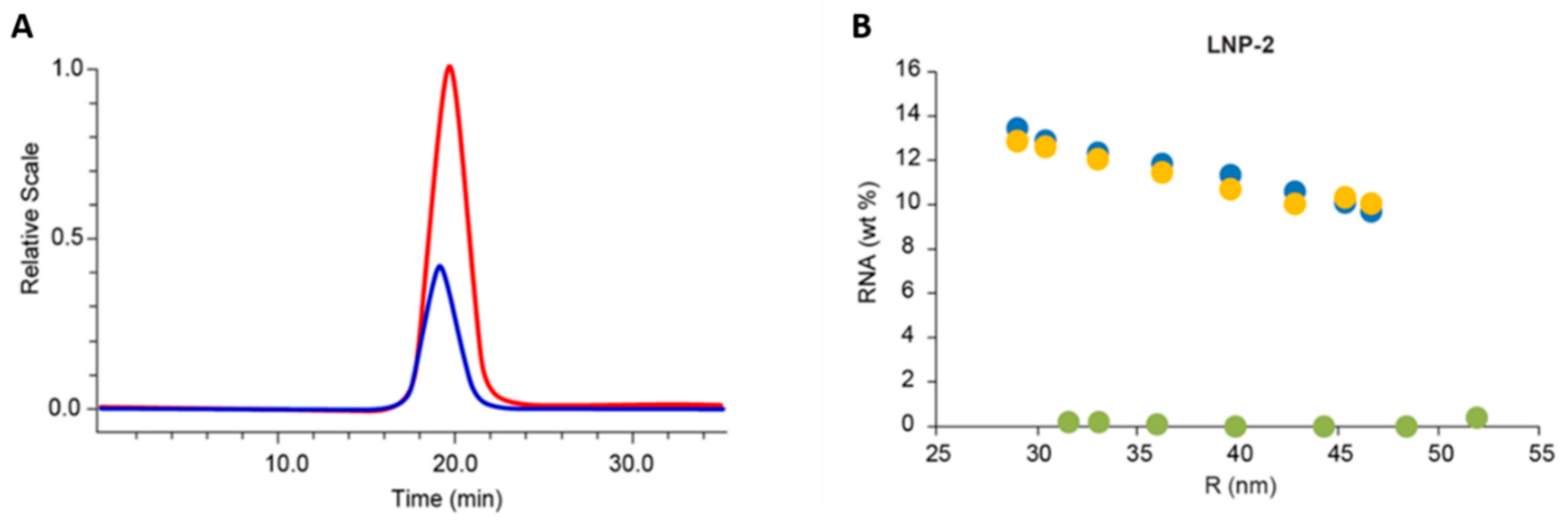
- Jia, X.; Liu, Y.; Wagner, A.M.; Chen, M.; Zhao, Y.; Smith, K.J.; Some, D.; Abend, A.M.; Pennington, J. Enabling online determination of the size-dependent rna content of lipid nanoparticle-based rna formulations. J. Chromatogr. B Anal. Technol. Biomed. Life Sci. 2021, 1186, 123015. [Google Scholar] [CrossRef]
- Quattrini, F.; Berrecoso, G.; Crecente-Campo, J.; Alonso, M.J. Asymmetric flow field-flow fractionation as a multifunctional technique for the characterization of polymeric nanocarriers. Drug. Deliv. Transl. Res. 2021, 11, 373–395. [Google Scholar] [CrossRef]
- Zhao, H.; Lin, Z.Y.; Yildirimer, L.; Dhinakar, A.; Zhao, X.; Wu, J. Polymer-based nanoparticles for protein delivery: Design, strategies and applications. J. Mater. Chem. B 2016, 4, 4060–4071. [Google Scholar] [CrossRef]
- Shakiba, S.; Shariati, S.; Wu, H.; Astete, C.E.; Cueto, R.; Fini, E.H.; Rodrigues, D.F.; Sabliov, C.M.; Louie, S.M. Distinguishing nanoparticle drug release mechanisms by asymmetric flow field–flow fractionation. J. Control Release 2022, 352, 485–496. [Google Scholar] [CrossRef]
- Sadat Tabatabaei Mirakabad, F.; Nejati-Koshki, K.; Akbarzadeh, A.; Yamchi, M.R.; Milani, M.; Zarghami, N.; Zeighamian, V.; Rahimzadeh, A.; Alimohammadi, S.; Hanifehpour, Y.; et al. Plga-based nanoparticles as cancer drug delivery systems. Asian Pac. J. Cancer Prev. 2014, 15, 517–535. [Google Scholar] [CrossRef]
- Alvi, M.; Yaqoob, A.; Rehman, K.; Shoaib, S.M.; Akash, M.S.H. Plga-based nanoparticles for the treatment of cancer: Current strategies and perspectives. AAPS Open 2022, 8, 1–7. [Google Scholar] [CrossRef]
- Yadav, N.; Francis, A.P.; Priya, V.V.; Patil, S.; Mustaq, S.; Khan, S.S.; Alzahrani, K.J.; Banjer, H.J.; Mohan, S.K.; Mony, U.; et al. Polysaccharide-drug conjugates: A tool for enhanced cancer therapy. Polymers 2022, 14, 950. [Google Scholar] [CrossRef] [PubMed]
- Dacoba, T.G.; Omange, R.W.; Li, H.; Crecente-Campo, J.; Luo, M.; Alonso, M.J. Polysaccharide nanoparticles can efficiently modulate the immune response against an hiv peptide antigen. ACS Nano 2019, 13, 4947–4959. [Google Scholar] [CrossRef] [PubMed]
- Klein, M.; Menta, M.; Dacoba, T.G.; Crecente-Campo, J.; Alonso, M.J.; Dupin, D.; Loinaz, I.; Grassl, B.; Séby, F. Advanced nanomedicine characterization by dls and af4-uv-mals: Application to a hiv nanovaccine. J. Pharm. Biomed. Anal. 2020, 179, 113017. [Google Scholar] [CrossRef]
- Biemans, R.; Micoli, F.; Romano, M.R. Glycoconjugate vaccines, production and characterization. In Recent Trends in Carbohydrate Chemistry; Elsevier: Amsterdam, The Netherlands, 2020; pp. 285–313. [Google Scholar]
- Barth, H.G.; Carlin, F.J. A review of polymer shear degradation in size-exclusion chromatography. J. Liq. Chromatogr. 1984, 7, 1717–1738. [Google Scholar] [CrossRef]
- Kuntsche, J.; Horst, J.C.; Bunjes, H. Cryogenic transmission electron microscopy (cryo-tem) for studying the morphology of colloidal drug delivery systems. Int. J. Pharm. 2011, 417, 120–137. [Google Scholar] [CrossRef]
- Truong, N.P.; Whittaker, M.R.; Mak, C.W.; Davis, T.P. The importance of nanoparticle shape in cancer drug delivery. Expert. Opin. Drug. Deliv. 2015, 12, 129–142. [Google Scholar] [CrossRef]
- Wong, C.K.; Stenzel, M.H.; Thordarson, P. Non-spherical polymersomes: Formation and characterization. Chem. Soc. Rev. 2019, 48, 4019–4035. [Google Scholar] [CrossRef]
- Burchard, W.; Schmidt, M.; Stockmayer, W.H. Information on polydispersity and branching from combined quasi-elastic and intergrated scattering. Macromolecules 1980, 13, 1265–1272. [Google Scholar] [CrossRef]
- Kok, C.M.; Rudin, A. Relationship between the hydrodynamic radius and the radius of gyration of a polymer in solution. Die Makromol. Chem. Rapid Commun. 1981, 2, 655–659. [Google Scholar] [CrossRef]
- Tomalia, D.A.; Naylor, A.M.; Goddard, W.A. Starburst dendrimers: Molecular-level control of size, shape, surface chemistry, topology, and flexibility from atoms to macroscopic matter. Angew. Chem. Int. Ed. Engl. 1990, 29, 138–175. [Google Scholar] [CrossRef]
- Wang, J.; Li, B.; Qiu, L.; Qiao, X.; Yang, H. Dendrimer-based drug delivery systems: History, challenges, and latest developments. J. Biol. Eng. 2022, 16, 18. [Google Scholar] [CrossRef] [PubMed]
- Kootstra, N.A.; Verma, I.M. Gene therapy with viral vectors. Annu. Rev. Pharmacol. Toxicol. 2003, 43, 413–439. [Google Scholar] [CrossRef] [PubMed]
- Shah, P.B.; Losordo, D.W. Non-viral vectors for gene therapy: Clinical trials in cardiovascular disease. In Advances in Genetics; Academic Press: Cambridge, MA, USA, 2005; Volume 54, pp. 339–361. [Google Scholar]
- Eisenman, D. The united states’ regulatory environment is evolving to accommodate a coming boom in gene therapy research. Appl. Biosaf. 2019, 24, 147–152. [Google Scholar] [CrossRef] [PubMed]
- Lee, C.S.; Bishop, E.S.; Zhang, R.; Yu, X.; Farina, E.M.; Yan, S.; Zhao, C.; Zheng, Z.; Shu, Y.; Wu, X.; et al. Adenovirus-mediated gene delivery: Potential applications for gene and cell-based therapies in the new era of personalized medicine. Genes. Dis. 2017, 4, 43–63. [Google Scholar] [CrossRef] [PubMed]
- Rodriguez, D.A.; Vader, P. Extracellular vesicle-based hybrid systems for advanced drug delivery. Pharmaceutics 2022, 14, 267. [Google Scholar] [CrossRef]
- Jayasinghe, M.K.; Tan, M.; Peng, B.; Yang, Y.; Sethi, G.; Pirisinu, M.; Le, M.T.N. New approaches in extracellular vesicle engineering for improving the efficacy of anti-cancer therapies. Semin. Cancer Biol. 2021, 74, 62–78. [Google Scholar] [CrossRef]
- Ferreira, D.; Moreira, J.N.; Rodrigues, L.R. New advances in exosome-based targeted drug delivery systems. Crit. Rev. Oncol. Hematol. 2022, 172, 103628. [Google Scholar] [CrossRef]
- Amiri, A.; Bagherifar, R.; Ansari Dezfouli, E.; Kiaie, S.H.; Jafari, R.; Ramezani, R. Exosomes as bio-inspired nanocarriers for rna delivery: Preparation and applications. J. Transl. Med. 2022, 20, 125. [Google Scholar] [CrossRef]
- De Jong, O.G.; Kooijmans, S.A.A.; Murphy, D.E.; Jiang, L.; Evers, M.J.W.; Sluijter, J.P.G.; Vader, P.; Schiffelers, R.M. Drug delivery with extracellular vesicles: From imagination to innovation. Acc. Chem. Res. 2019, 52, 1761–1770. [Google Scholar] [CrossRef]
- Zhang, Q.; Zhang, H.; Ning, T.; Liu, D.; Deng, T.; Liu, R.; Bai, M.; Zhu, K.; Li, J.; Fan, Q.; et al. Exosome-delivered c-met sirna could reverse chemoresistance to cisplatin in gastric cancer. Int. J. Nanomed. 2020, 15, 2323–2335. [Google Scholar] [CrossRef] [PubMed]
- Pascucci, L.; Cocce, V.; Bonomi, A.; Ami, D.; Ceccarelli, P.; Ciusani, E.; Vigano, L.; Locatelli, A.; Sisto, F.; Doglia, S.M.; et al. Paclitaxel is incorporated by mesenchymal stromal cells and released in exosomes that inhibit in vitro tumor growth: A new approach for drug delivery. J. Control Release 2014, 192, 262–270. [Google Scholar] [CrossRef] [PubMed]
- Thery, C.; Witwer, K.W.; Aikawa, E.; Alcaraz, M.J.; Anderson, J.D.; Andriantsitohaina, R.; Antoniou, A.; Arab, T.; Archer, F.; Atkin-Smith, G.K.; et al. Minimal information for studies of extracellular vesicles 2018 (misev2018): A position statement of the international society for extracellular vesicles and update of the misev2014 guidelines. J. Extracell. Vesicles 2018, 7, 1535750. [Google Scholar] [CrossRef]
- Witwer, K.W.; Goberdhan, D.C.; O’Driscoll, L.; Thery, C.; Welsh, J.A.; Blenkiron, C.; Buzas, E.I.; Di Vizio, D.; Erdbrugger, U.; Falcon-Perez, J.M.; et al. Updating misev: Evolving the minimal requirements for studies of extracellular vesicles. J. Extracell. Vesicles 2021, 10, e12182. [Google Scholar] [CrossRef]
- Yang, J.S.; Kim, J.Y.; Lee, J.C.; Moon, M.H. Investigation of lipidomic perturbations in oxidatively stressed subcellular organelles and exosomes by asymmetrical flow field-flow fractionation and nanoflow ultrahigh performance liquid chromatography-tandem mass spectrometry. Anal. Chim. Acta 2019, 1073, 79–89. [Google Scholar] [CrossRef] [PubMed]
- Wu, B.; Chen, X.; Wang, J.; Qing, X.; Wang, Z.; Ding, X.; Xie, Z.; Niu, L.; Guo, X.; Cai, T.; et al. Separation and characterization of extracellular vesicles from human plasma by asymmetrical flow field-flow fractionation. Anal. Chim. Acta 2020, 1127, 234–245. [Google Scholar] [CrossRef]
- Kim, Y.B.; Yang, J.S.; Lee, G.B.; Moon, M.H. Evaluation of exosome separation from human serum by frit-inlet asymmetrical flow field-flow fractionation and multiangle light scattering. Anal. Chim. Acta 2020, 1124, 137–145. [Google Scholar] [CrossRef]
- Li, P.; Kaslan, M.; Lee, S.H.; Yao, J.; Gao, Z. Progress in exosome isolation techniques. Theranostics 2017, 7, 789–804. [Google Scholar] [CrossRef]
- Chia, B.S.; Low, Y.P.; Wang, Q.; Li, P.; Gao, Z. Advances in exosome quantification techniques. TrAC Trends Anal. Chem. 2017, 86, 93–106. [Google Scholar] [CrossRef]
- Anderson, W.; Kozak, D.; Coleman, V.A.; Jamting, A.K.; Trau, M. A comparative study of submicron particle sizing platforms: Accuracy, precision and resolution analysis of polydisperse particle size distributions. J. Colloid. Interface Sci. 2013, 405, 322–330. [Google Scholar] [CrossRef]
- Zhang, H.; Zhang, H.; Lyden, D. A protocol for asymmetric-flow field-flow fractionation (af4) of small extracellular vesicles. Protocol Exchange 2018, 1–9. [Google Scholar] [CrossRef]
- Kim, Y.B.; Lee, G.B.; Moon, M.H. Size separation of exosomes and microvesicles using flow field-flow fractionation/multiangle light scattering and lipidomic comparison. Anal. Chem. 2022, 94, 8958–8965. [Google Scholar] [CrossRef] [PubMed]
- Podzimek, S.; Johann, C. Asymmetric flow field-flow fractionation: Current status, possibilities, analytical limitations and future trends. Chromatographia 2021, 84, 531–534. [Google Scholar] [CrossRef]
- Jia, Y.-P.; Ma, B.-Y.; Wei, X.-W.; Qian, Z.-Y. The in vitro and in vivo toxicity of gold nanoparticles. Chin. Chem. Lett. 2017, 28, 691–702. [Google Scholar] [CrossRef]
- Xu, Z.P.; Zeng, Q.H.; Lu, G.Q.; Yu, A.B. Inorganic nanoparticles as carriers for efficient cellular delivery. Chem. Eng. Sci. 2006, 61, 1027–1040. [Google Scholar] [CrossRef]
- Biener, J.; Wittstock, A.; Baumann, T.; Weissmüller, J.; Bäumer, M.; Hamza, A. Surface chemistry in nanoscale materials. Materials 2009, 2, 2404–2428. [Google Scholar] [CrossRef]
- Kong, F.Y.; Zhang, J.W.; Li, R.F.; Wang, Z.X.; Wang, W.J.; Wang, W. Unique roles of gold nanoparticles in drug delivery, targeting and imaging applications. Molecules 2017, 22, 1445. [Google Scholar] [CrossRef]
- Wang, W.; Wang, J.; Ding, Y. Gold nanoparticle-conjugated nanomedicine: Design, construction, and structure-efficacy relationship studies. J. Mater. Chem. B 2020, 8, 4813–4830. [Google Scholar] [CrossRef]
- Song, M.; Wang, X.; Li, J.; Zhang, R.; Chen, B.; Fu, D. Effect of surface chemistry modification of functional gold nanoparticles on the drug accumulation of cancer cells. J. Biomed. Mater. Res. A 2008, 86, 942–946. [Google Scholar] [CrossRef]
- Zeng, X.; Liu, G.; Tao, W.; Ma, Y.; Zhang, X.; He, F.; Pan, J.; Mei, L.; Pan, G. A drug-self-gated mesoporous antitumor nanoplatform based on ph-sensitive dynamic covalent bond. Adv. Funct. Mater. 2017, 27, 1605985. [Google Scholar] [CrossRef]
- Farjadian, F.; Roointan, A.; Mohammadi-Samani, S.; Hosseini, M. Mesoporous silica nanoparticles: Synthesis, pharmaceutical applications, biodistribution, and biosafety assessment. Chem. Eng. J. 2019, 359, 684–705. [Google Scholar] [CrossRef]
- Anselmo, A.C.; Mitragotri, S. A review of clinical translation of inorganic nanoparticles. AAPS J. 2015, 17, 1041–1054. [Google Scholar] [CrossRef] [PubMed]
- Kong, L.; Qiu, J.; Sun, W.; Yang, J.; Shen, M.; Wang, L.; Shi, X. Multifunctional pei-entrapped gold nanoparticles enable efficient delivery of therapeutic sirna into glioblastoma cells. Biomater. Sci. 2017, 5, 258–266. [Google Scholar] [CrossRef] [PubMed]
- Wang, F.; Wang, Y.C.; Dou, S.; Xiong, M.H.; Sun, T.M.; Wang, J. Doxorubicin-tethered responsive gold nanoparticles facilitate intracellular drug delivery for overcoming multidrug resistance in cancer cells. ACS Nano 2011, 5, 3679–3692. [Google Scholar] [CrossRef] [PubMed]
- Wang, J.; Giordani, S.; Marassi, V.; Roda, B.; Reschiglian, P.; Zattoni, A. Quality control and purification of ready-to-use conjugated gold nanoparticles to ensure effectiveness in biosensing. Front. Sens. 2022, 3, 1087115. [Google Scholar] [CrossRef]
- Marassi, V.; Zanoni, I.; Ortelli, S.; Giordani, S.; Reschiglian, P.; Roda, B.; Zattoni, A.; Ravagli, C.; Cappiello, L.; Baldi, G.; et al. Native study of the behaviour of magnetite nanoparticles for hyperthermia treatment during the initial moments of intravenous administration. Pharmaceutics 2022, 14, 2810. [Google Scholar] [CrossRef]
- Agostoni, V.; Anand, R.; Monti, S.; Hall, S.; Maurin, G.; Horcajada, P.; Serre, C.; Bouchemal, K.; Gref, R. Impact of phosphorylation on the encapsulation of nucleoside analogues within porous iron(iii) metal-organic framework mil-100(fe) nanoparticles. J. Mater. Chem. B 2013, 1, 4231–4242. [Google Scholar] [CrossRef] [PubMed]
- Kowalkowski, T.; Sugajski, M.; Buszewski, B. Impact of ionic strength of carrier liquid on recovery in flow field-flow fractionation. Chromatographia 2018, 81, 1213–1218. [Google Scholar] [CrossRef]
- Mudalige, T.K.; Qu, H.; Sanchez-Pomales, G.; Sisco, P.N.; Linder, S.W. Simple functionalization strategies for enhancing nanoparticle separation and recovery with asymmetric flow field flow fractionation. Anal. Chem. 2015, 87, 1764–1772. [Google Scholar] [CrossRef]
- Gigault, J.; Pettibone, J.M.; Schmitt, C.; Hackley, V.A. Rational strategy for characterization of nanoscale particles by asymmetric-flow field flow fractionation: A tutorial. Anal. Chim. Acta 2014, 809, 9–24. [Google Scholar] [CrossRef]
- Valto, P.; Knuutinen, J.; Alen, R. Evaluation of resin and fatty acid concentration levels by online sample enrichment followed by atmospheric pressure chemical ionization-mass spectrometry (APCI-MS). Environ. Sci. Pollut. Res. Int. 2009, 16, 287–294. [Google Scholar] [CrossRef] [PubMed]
- Multia, E.; Liangsupree, T.; Jussila, M.; Ruiz-Jimenez, J.; Kemell, M.; Riekkola, M.L. Automated on-line isolation and fractionation system for nanosized biomacromolecules from human plasma. Anal. Chem. 2020, 92, 13058–13065. [Google Scholar] [CrossRef] [PubMed]
- Caputo, F.; Clogston, J.; Calzolai, L.; Rosslein, M.; Prina-Mello, A. Measuring particle size distribution of nanoparticle enabled medicinal products, the joint view of euncl and nci-ncl. A step by step approach combining orthogonal measurements with increasing complexity. J. Control Release 2019, 299, 31–43. [Google Scholar] [CrossRef] [PubMed]


| Sub-Techniques of FFF | External Field | Physicochemical Property |
|---|---|---|
| Thermal FFF (ThFFF) | Thermal gradient | Soret coefficient |
| Sedimentation FFF (SeFFF) | Gravity/Centrifugal force | Effective mass (density) |
| Electrical FFF (ElFFF) | Electric field | Electrophoretic mobility |
| Magnetic FFF (MgFFF) | Magnetic field | Magnetic properties |
| Flow FFF (FlFFF) | Cross flow | Diffusion coefficient |
| Nanoparticles | FFF Technique and Applications | Key Results | Ref |
|---|---|---|---|
| Lipid-based nanoparticles | AF4 with offline NTA and LC-MS for doxorubicin liposome formulations | Particle size distribution of the liposome and drug-to-lipid ratios were analyzed and compared across different doxorubicin formulations. | [53] |
| AF4-MALS-DLS for peptide-liposome interaction | Selectivity of the peptide, quantity of the bound peptide, and size distribution and morphology of liposomes were revealed for understanding of structure-activity relationship. | [54] | |
| AF4-MALS-Gamma ray detector for liposome loaded with high energy alpha emitter (212Bi) | Liposome particle size and stability of encapsulation in the serum were studied. | [55] | |
| AF4-MALS for drug transfer assay to quantify retention of lipophilic model compounds | Transfer kinetics of lipophilic model compounds from donor liposomes to acceptor liposomes were elucidated at different lipid mass ratios, and with different vesicle morphology and lamellarity. | [56] | |
| AF4-MALS-RI for stability evaluation of liposomes against the intestinal bile salts in oral delivery application | Different mechanisms of entrapped calcein leakages were revealed. | [57] | |
| AF4-MALS-DLS-dRI-UV for liposome-plasma protein interaction (from albumin HDL and LDL | Liposomes were separated from albumin, and HDL, LHL, and the size were determined. The effect of the biolayer composition on liposome stability was also observed. | [58] | |
| AF4 with frit-inlet channel coupled with MALS to analyze LNP for RNA delivery | Frit-inlet channel enabled size and physical stability of LNP-RNA with great reliability and recovery. | [43] | |
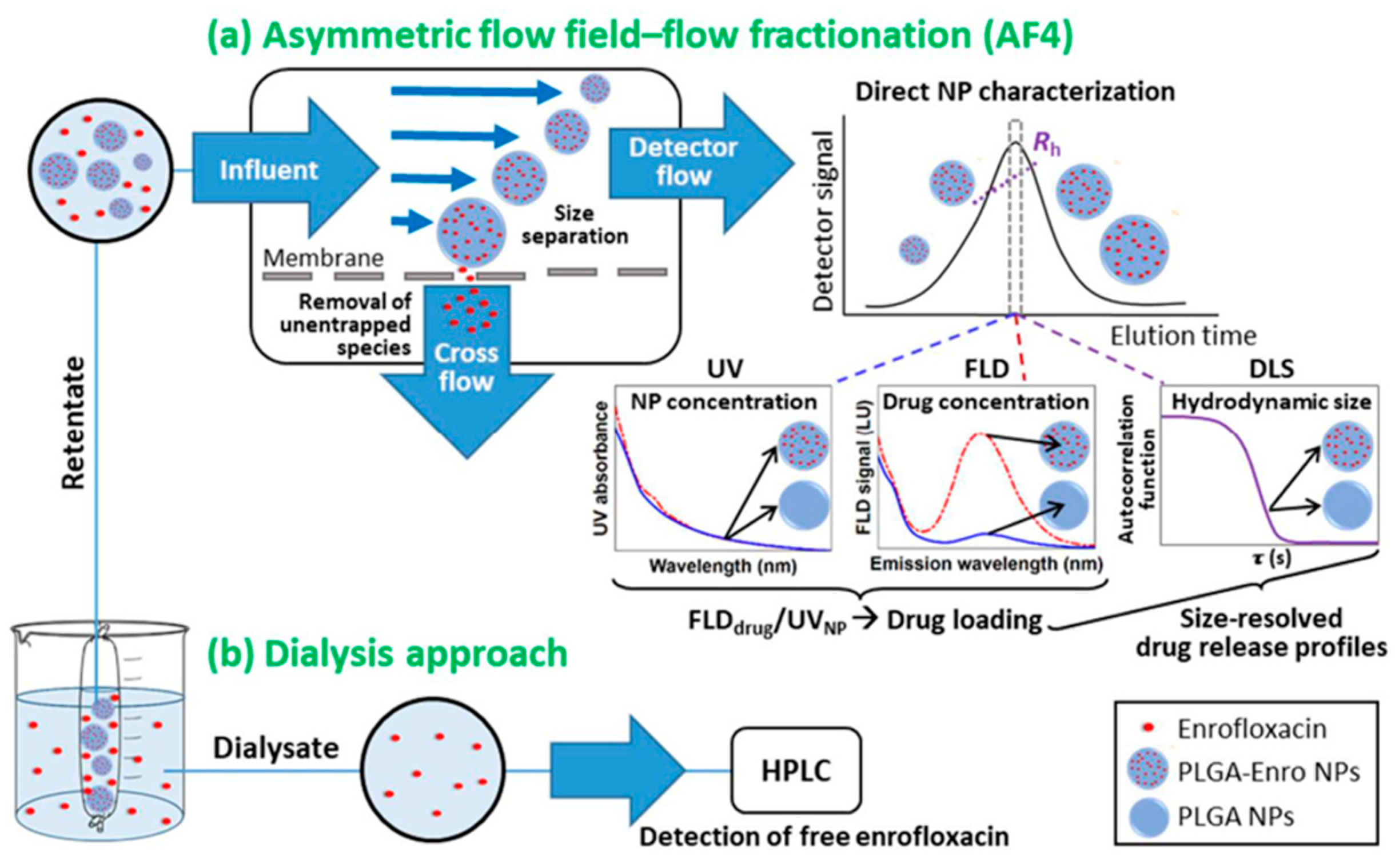
| Polymer-based nanoparticles | AF4-DLS-UV-FLD for enrofloxacin in PLGA nanoparticles | Comprehensive analysis of nanoparticle concentration (via UV), drug concentration (via FLD), and particle size distribution (via DLS). Unentrapped drug was easily removed via crossflow. | [59] |
| AF4-MALS for protein-conjugated polysaccharides | Complementary analysis by SEC-MALS and AF4-MALS revealed heterogeneity in conformation and aggregation of the conjugates from molar mass and size determination. | [60] | |
| AF4-RI-FLD-DLS for polymer micelles in vitro stability | AF4 enabled the separation of polymer micelles from plasma protein and can be used to study the in vitro instability of drug-loaded nanoparticles. | [61] | |
| AF4-MALS-DLS for PEG-PDLLA polymersomes | Insights in size and shape of polymersomes via combination of MALS and DLS, and whether they are empty or loaded. | [62] | |
| AF4-RI for PAMAM dendrimers | Separate impurities (i.e., missing arm) and aggregates from PAMAM main populations and monitor interactions of PAMAM dendrimers with BSA. | [63] | |
| AF4-MALS-RI-UV for PEI-Mal dendrimers | Characterization of crossflow pathway enabled quantification of free, unencapsulated dye in addition to molar mass distributions. | [64] | |
| Viral vectors and Virus-like Nanoparticles | AF4-MALS-DLS-UV-FLD-RI for VLPs derived from human polyoma JC virus | Comprehensive analysis of VLP molar mass and radius (via MALS), hydrodynamic radius (via DLS), concentration (via RI), sample composition and concentration (via UV), and improved small molecule limit of detection (via FLD). | [49] |
| AF4-MALS-UV & ElFFF-MALS-UV of bacteriophage-like VLPs | Complementary analysis by ElFFF and AF4 obtained size and electrophoretic mobility of three VLPs. | [65] | |
| Extracellular vesicles | AF4-UV-MALS for characterization of EVs from urine and comparison with ultrafiltration combined with SEC method | AF4-UV-MALS was demonstrated to be a straightforward and reproducible method for determining size, amount, and purity of isolated urinary EVs. | [66] |
| AF4-UV-MALS combined with batch DLS and NTA for size separation, characterization and quantification of exosomes | Fractionation quality of exosomes was significantly influenced by crossflow conditions and channel thickness where focusing time has less impact. AF4-UV-MALS and DLS both showed the presence of two particle subpopulations. Compared to DLS and AF4-MALS, NTA overestimated the size and number density for the larger exosome population. | [67] | |
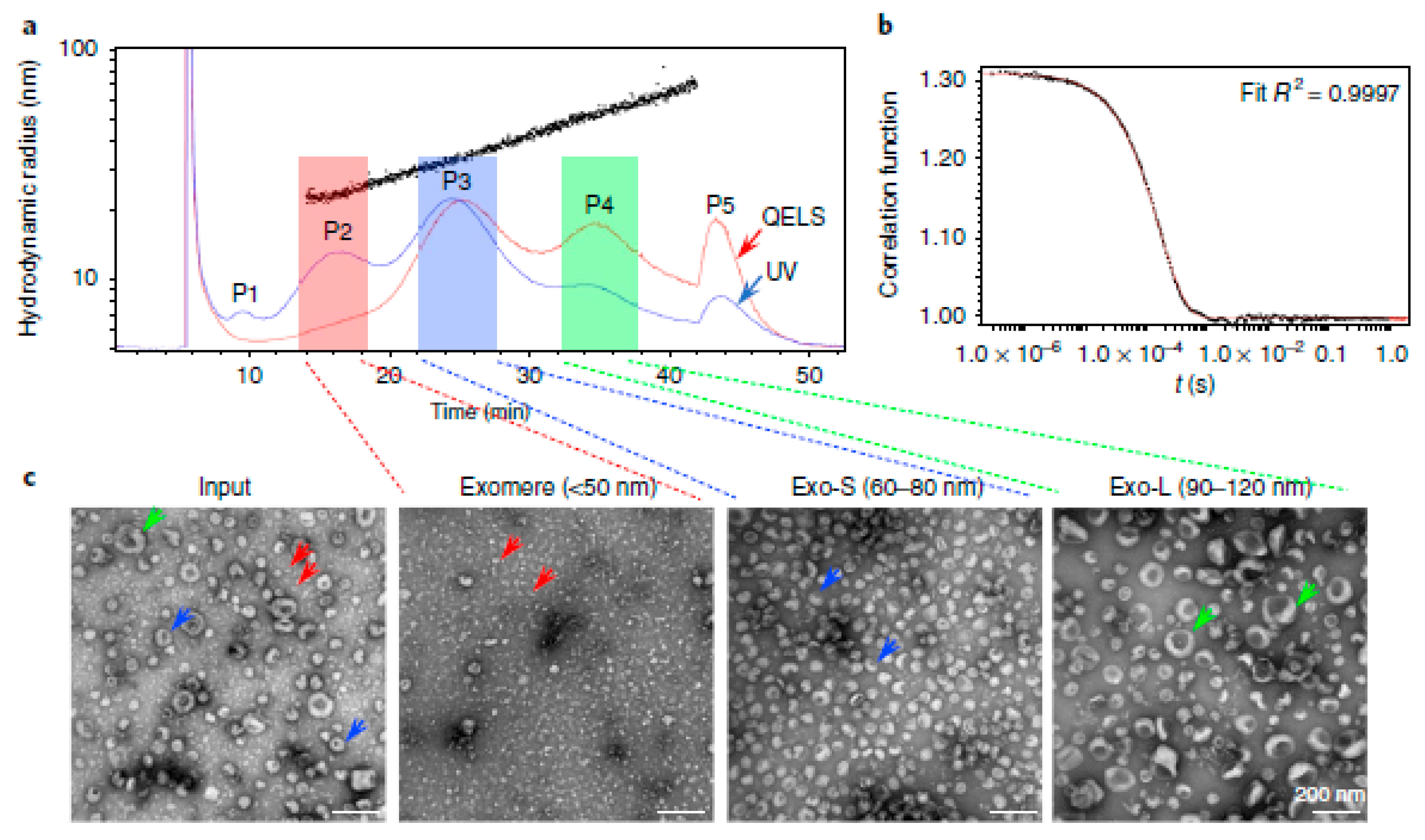
| AF4-UV-DLS with EM imaging for identification of subsets of EVs | Two exosome subpopulations and one non-membrane NPs exomere were discovered and identified | [68] | |
| AF4 and nanoflow-LC-ESI-MS/MS for size dependent lipidomic analysis of urinary exosomes | AF4 enabled the fractionation of exosomes with different sizes that originated from different types of cells. Degree of lipid increase was more significant in the smaller fractions, indicating that AF4 is capable of screening of urinary exosomes in cancer patients. | [69] | |
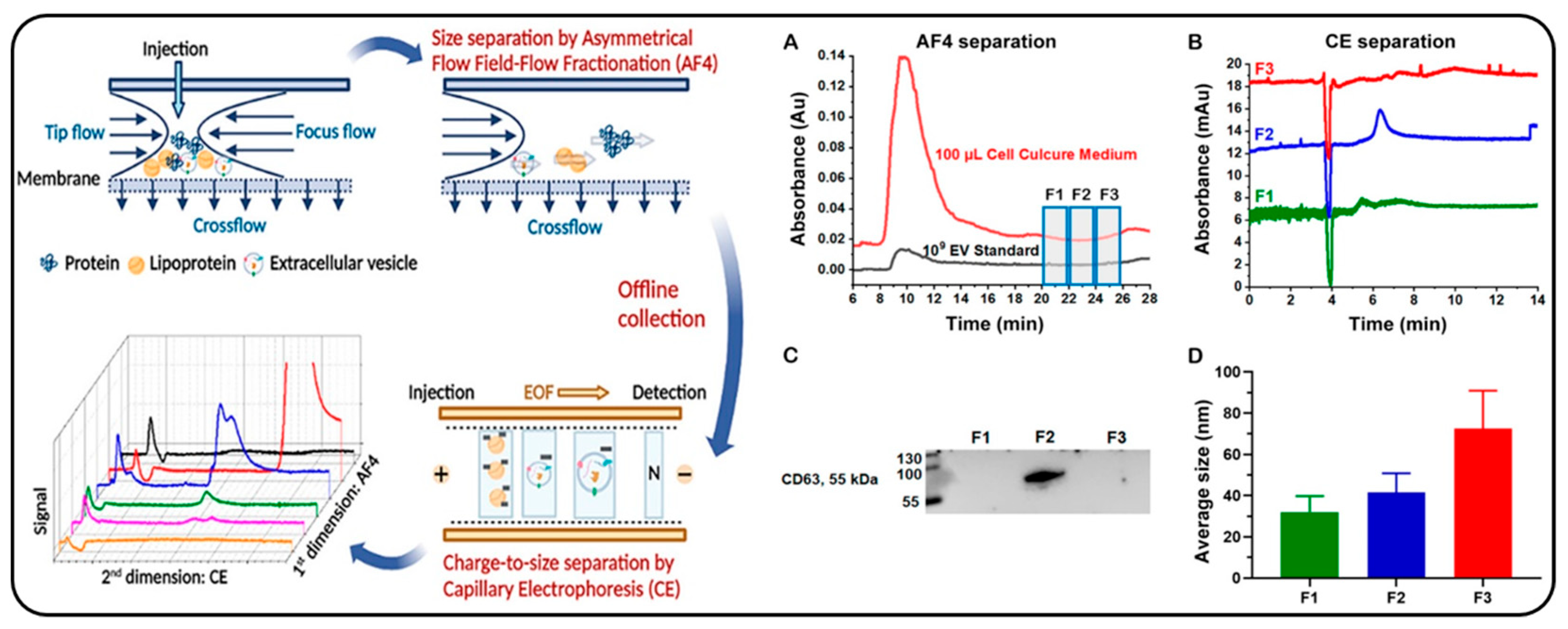
| Offline coupling of AF4 and CE for separation of EVs | EVs could be resolved from free proteins and high-density lipoproteins by AF4 and further separated from the low-density lipoproteins co-eluted in AF4 by offline CE. | [70] | |
| Orthogonal approach of ultracentrifugation and HF5-MALS-UV-FLD for purification and mapping of EV subtypes | Size, abundance, and DNA/protein content of the large and small EVs were characterized by HF5-MALS-UV-FLD as the second dimension, showing potential in sorting particles with different sizes and contents. | [71] | |
| EAF4 hyphenated with MALS and NTA for fast and purification-free characterization of NPs | EAF4 provided online sample purification and simultaneous access to size and Zeta-potential; high resolution size and number concentration was achieved by hyphenation of EAF4 with MALS and NTA. | [72] | |
| Inorganic nanoparticles | AF4-MALS-DLS-ICPMS for quantitative characterization of GNPs | Mixtures of three GNPs were separated by AF4 and then each fraction was quantified by ICPMS. Both geometric diameters and hydrodynamic diameters were determined online by MALS and DLS. | [73] |
| AF4 for characterization of elution behavior of non-spherical GNPs | Elution behavior of the GNPs with three different morphologies was studied by AF4 and particle size was compared with DLS and TEM. | [74] | |
| AF4-MALS-UV-RI for characterization and stability evaluation of drug-loaded metal-organic framework (MOF) NPs | Empty and drug-loaded nanoMOFs were studied in terms of particle size distribution and stability. Detection of aggregate formation and monitoring of nanoMOF morphological changes indicates their interaction with the drug molecules. | [75] |
Disclaimer/Publisher’s Note: The statements, opinions and data contained in all publications are solely those of the individual author(s) and contributor(s) and not of MDPI and/or the editor(s). MDPI and/or the editor(s) disclaim responsibility for any injury to people or property resulting from any ideas, methods, instructions or products referred to in the content. |
© 2023 by the authors. Licensee MDPI, Basel, Switzerland. This article is an open access article distributed under the terms and conditions of the Creative Commons Attribution (CC BY) license (https://creativecommons.org/licenses/by/4.0/).
Share and Cite
Bian, J.; Gobalasingham, N.; Purchel, A.; Lin, J. The Power of Field-Flow Fractionation in Characterization of Nanoparticles in Drug Delivery. Molecules 2023, 28, 4169. https://doi.org/10.3390/molecules28104169
Bian J, Gobalasingham N, Purchel A, Lin J. The Power of Field-Flow Fractionation in Characterization of Nanoparticles in Drug Delivery. Molecules. 2023; 28(10):4169. https://doi.org/10.3390/molecules28104169
Chicago/Turabian StyleBian, Juan, Nemal Gobalasingham, Anatolii Purchel, and Jessica Lin. 2023. "The Power of Field-Flow Fractionation in Characterization of Nanoparticles in Drug Delivery" Molecules 28, no. 10: 4169. https://doi.org/10.3390/molecules28104169
APA StyleBian, J., Gobalasingham, N., Purchel, A., & Lin, J. (2023). The Power of Field-Flow Fractionation in Characterization of Nanoparticles in Drug Delivery. Molecules, 28(10), 4169. https://doi.org/10.3390/molecules28104169