Article

Chemical Constituents from the Leaves of Ligustrum robustum and Their Bioactivities

1
College of Pharmacy, Youjiang Medical University for Nationalities, Baise 533000, China
2
Department of Laboratory Science of Public Health, West China School of Public Health, Sichuan University, Chengdu 610041, China
3
Key Laboratory of Drug Targeting, Ministry of Education, West China School of Pharmacy, Sichuan University, Chengdu 610041, China
4
Nursing School, Youjiang Medical University for Nationalities, Baise 533000, China
*
Authors to whom correspondence should be addressed.
These authors contributed equally to this work.
Molecules 2023, 28(1), 362; https://doi.org/10.3390/molecules28010362
Submission received: 7 December 2022 / Revised: 20 December 2022 / Accepted: 23 December 2022 / Published: 2 January 2023

Abstract

:
The leaves of Ligustrum robustum have been consumed as Ku-Ding-Cha for clearing heat and removing toxins, and they have been used as a folk medicine for curing hypertension, diabetes, and obesity in China. The phytochemical research on the leaves of L. robustum led to the isolation and identification of two new hexenol glycosides, two new butenol glycosides, and five new sugar esters, named ligurobustosides X (1a), X1 (1b), Y (2a), and Y1 (2b) and ligurobustates A (3a), B (3b), C (4b), D (5a), and E (5b), along with seven known compounds (4a and 610). Compounds 110 were tested for their inhibitory effects on fatty acid synthase (FAS), α-glucosidase, and α-amylase, as well as their antioxidant activities. Compound 2 showed strong FAS inhibitory activity (IC50 4.10 ± 0.12 μM) close to that of the positive control orlistat (IC50 4.46 ± 0.13 μM); compounds 7 and 9 revealed moderate α-glucosidase inhibitory activities; compounds 110 showed moderate α-amylase inhibitory activities; and compounds 1 and 10 displayed stronger 2,2′-azino-bis(3-ethylbenzthiazoline-6-sulphonic acid) ammonium salt (ABTS) radical scavenging effects (IC50 3.41 ± 0.08~5.65 ± 0.19 μM) than the positive control l-(+)-ascorbic acid (IC50 10.06 ± 0.19 μM). This study provides a theoretical foundation for the leaves of L. robustum as a functional tea to prevent diabetes and its complications.

Graphical Abstract

1. Introduction

Diabetes, which affects nearly 10.5% of the population worldwide, is a chronic metabolic disease characterized by hyperglycemia caused by insulin resistance, a deficiency in insulin secretion, or both [1]. Its complications, including diabetic neuropathy, nephropathy, and cardiovascular diseases, lead to serious morbidity and mortality [1]. Current drugs, such as insulin, metformin, sulfonylureas, and acarbose, can control hyperglycemia, but their effect on preventing the complications of diabetes is not ideal. Therefore, it is significant to search for new resources for the prevention of diabetes and its complications.
Studies have revealed that long-term obesity might trigger specific metabolic disorders, such as cardiovascular diseases, insulin resistance, and diabetes [2,3]; fatty acid synthase (FAS), which catalyzes the synthesis of saturated long-chain fatty acids, is a potential target to prevent obesity [4]; carbohydrate digestive enzymes, such as α-glucosidase and α-amylase, play a crucial role in promoting hyperglycemia by releasing monosaccharides in the course of digestion [5]; and the contribution of reactive oxygen species generated by oxidative stress induced by chronic hyperglycemia has been linked to the onset and progression of diabetes and its complications [6]. Thus, natural products with inhibitory activities on FAS, α-glucosidase, and α-amylase as well as an antioxidant effect might be a new resource to prevent diabetes and its complications.
Ligustrum robustum (Roxb.) Blume is a plant of Oleaceae, and it is distributed extensively in Southwest China, India, Burma, Vietnam, and Cambodia [4]. The leaves of L. robustum have been used for Ku-Ding-Cha, a tea with functions in clearing heat and removing toxins, in China since the Dong Han Dynasty [7,8]. In addition, L. robustum is believed as a folk medicine for curing hypertension, diabetes, obesity, etc. [8,9]. In the previous studies on L. robustum [4,7,8,9,10,11,12,13,14,15,16,17,18,19], more than 70 chemical ingredients, including monoterpenoid glycosides, iridoid glycosides, phenylethanoid glycosides, phenylmethanoid glycosides, flavonoid glycosides, lignan glycosides, and triterpenoids were reported. The antiobesity, anti-inflammatory, and antioxidative activities of the extract; the inhibitory effects on α-glucosidase, α-amylase, and FAS; and the antioxidant effects of some compositions were also discovered. In order to further determine the active constituents for preventing diabetes and its complications, phytochemical and biological research on the leaves of L. robustum, which was carried out preliminarily [4,15,16], was further performed. As a result, two new hexenol glycosides, two new butenol glycosides, and five new sugar esters, named ligurobustosides X (1a), X1 (1b), Y (2a), and Y1 (2b) and ligurobustates A (3a), B (3b), C (4b), D (5a), and E (5b), along with seven reported compounds (4a and 610) (Figure 1), were isolated and identified from the leaves of L. robustum. This paper reports the isolation and structural identification of compounds 110 and describes their inhibitory activities on FAS, α-glucosidase, and α-amylase and their antioxidant effects.

2. Results and Discussion

2.1. Identification of Compounds 110

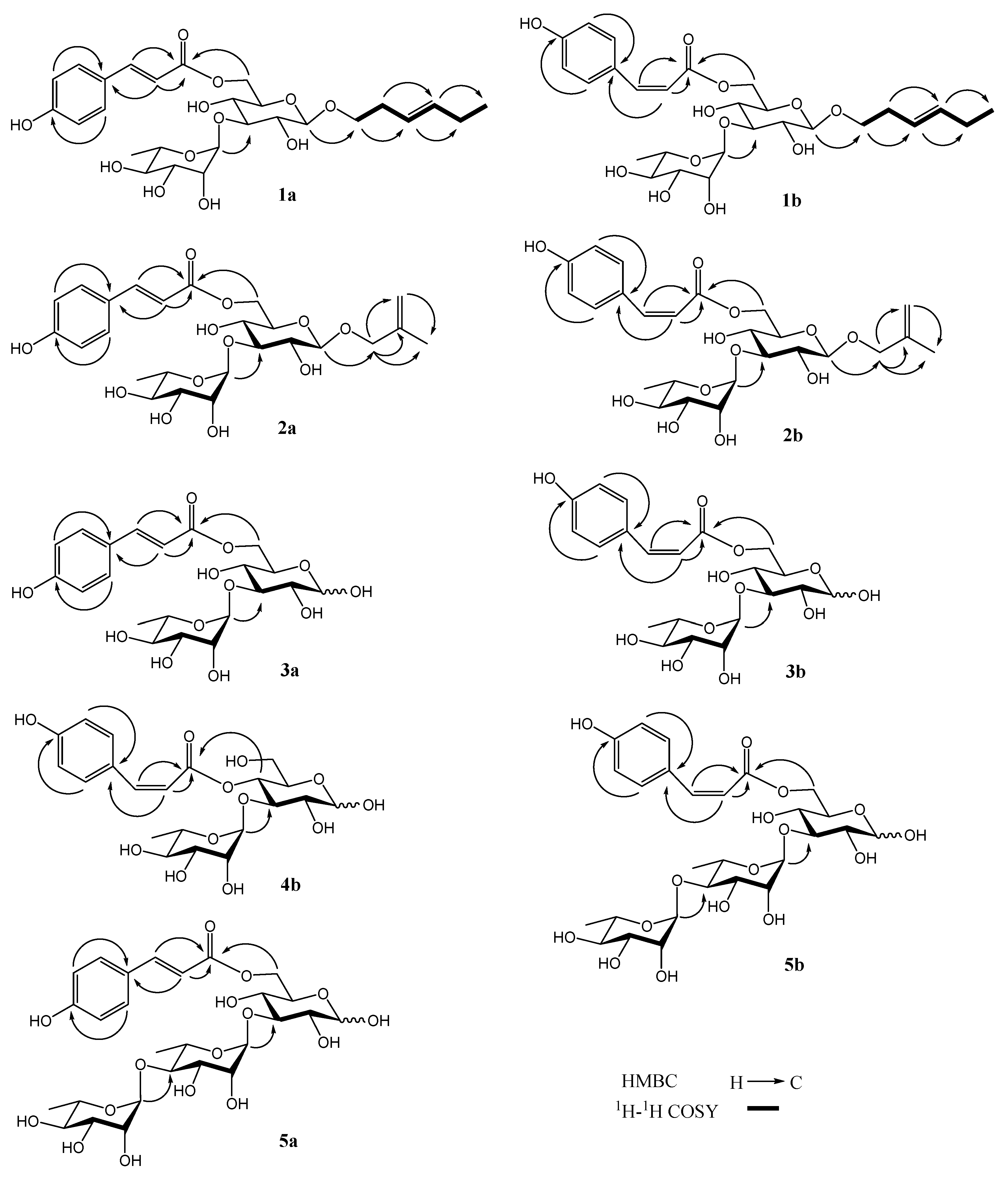
Compound 1 was obtained as a white amorphous powder, and its molecular formula was analyzed as C27H38O12 by HRESIMS (m/z 577.2260 [M + Na]+, calculated 577.2261 for C27H38NaO12). The NMR spectra of 1 showed two stereoisomers: 1a and 1b (5:3). In the 1H NMR spectrum of 1a (Table 1), the following signals were observed: (1) a 4-substituted phenyl at δH 6.77, 7.43 (2H each, d, J = 8.4 Hz); (2) two trans double bonds at δH 6.33, 7.63 (1H each, d, J = 15.6 Hz) and 5.36, 5.42 (1H each, dt, J = 17.4, 6.6 Hz); (3) two anomeric protons at δH 4.31 (1H, d, J = 8.4 Hz) and 5.18 (1H, d, J = 1.8 Hz); (4) a methylene linking with oxygen at δH 3.55, 3.80 (1H each, m), two methylene groups at δH 2.05, 2.37 (2H each, m), and two methyl groups at δH 0.93 (2H, t, J = 7.2 Hz, 6a), 0.97 (1H, t, J = 7.2 Hz, 6b) and 1.25 (3H, d, J = 6.0 Hz). In the 13C NMR spectrum of 1a (Table 2), the following signals were observed: a carbonyl at δC 169.2, a phenyl at δC 117.4–163.0, two double bonds at δC 114.1–147.1, two anomeric carbons at δC 102.7 and 104.4, nine sugar carbons at δC 64.6–84.0, a methylene linking with oxygen at δC 70.8, two methylene groups at δC 21.5 and 28.9, and two methyl groups at δC 14.6 and 17.9. The above 1H and 13C NMR data suggested 1a should be a glycoside, including a trans-p-coumaroyl and two monosaccharide moieties. The 1H-1H COSY experiment of 1a (Figure 2) showed correlations between δH 2.37 (H-2 of aglycone) and δH 3.80 (H-1b of aglycone); 5.36 (H-3 of aglycone) between δH 5.36 (H-3 of aglycone) and δH 5.42 (H-4 of aglycone); between δH 2.05 (H-5 of aglycone) and δH 5.42 (H-4 of aglycone), 0.93 (H-6a of aglycone). Together with the HMBC experiment on 1a (Figure 2), the aglycone of 1a was affirmed as (E)-3-hexen-1-ol. The acid hydrolysis experiment of 1 resulted in d-glucose and l-rhamnose, affirmed by TLC and a comparison of its NMR data with those of ligurobustoside E [12]. The HMBC experiment on 1a (Figure 2) displayed the following long-distance correlations: between δH 4.31 (H-1′ of glucosyl) and δC 70.8 (C-1 of aglycone), between δH 5.18 (H-1″ of rhamnosyl) and δC 84.0 (C-3′ of glucosyl), and between δH 4.35 (H-6′a of glucosyl), 4.48 (H-6′b of glucosyl), and δC 169.2 (carbonyl of coumaroyl). The 1H and 13C NMR signals of 1 were assigned by 1H-1H COSY, HSQC, and HMBC experiments (Figure S1). Based on above evidence, 1a was identified as (E)-3-hexen-1-yl 3-O-(α-l-rhamnopyranosyl)-6-O-(trans-p-coumaroyl)-O-β-d-glucopyranoside. It is a novel hexenol glycoside, named ligurobustoside X.
The NMR data of 1b (Table 1 and Table 2) were similar to those of 1a, except the trans-p-coumaroyl in 1a was replaced by the cis-p-coumaroyl (δH 5.79, 6.88 (1H each, d, J = 13.2 Hz, H-8′″, H-7′″)) in 1b. The HMBC experiment on 1b (Figure 2) displayed long-distance correlations between δH 4.27 (H-1′ of glucosyl) and δC 70.7 (C-1 of aglycone), between δH 5.16 (H-1″ of rhamnosyl) and δC 84.0 (C-3′ of glucosyl), and between δH 4.34 (H-6′a of glucosyl), 4.46 (H-6′b of glucosyl), and δC 168.1 (carbonyl of coumaroyl). Therefore, the structure of compound 1b was identified as (E)-3-hexen-1-yl 3-O-(α-l-rhamnopyranosyl)-6-O-(cis-p-coumaroyl)-O-β-d-glucopyranoside. It is a novel hexenol glycoside, named ligurobustoside X1. In conclusion, compound 1 is a mixture of ligurobustosides X and X1.
Compound 2 was obtained as a white amorphous powder, and its molecular formula was determined as C25H34O12 by HRESIMS (m/z 549.1941 [M + Na]+, calculated 549.1948 for C25H34NaO12). The NMR spectra of 2 showed two stereoisomers: 2a and 2b (2:1). In the 1H NMR spectrum of 2a (Table 1), the following signals were revealed: (1) a 4-substituted phenyl at δH 6.80 and 7.47 (2H each, d, J = 8.4 Hz); (2) a trans double bond at δH 6.37 and 7.65 (1H each, d, J = 16.2 Hz); (3) two olefinic proton signals at δH 4.88 and 5.02 (1H each, br. s); (4) two anomeric protons at δH 4.30 (1H, d, J = 7.2 Hz) and 5.18 (1H, d, J = 1.8 Hz); (5) a methylene linking with oxygen at δH 4.07 and 4.20 (1H each, d, J = 12.6 Hz); and two methyl groups at δH 1.75 (3H, s) and 1.25 (3H, d, J = 6.6 Hz). In the 13C NMR spectrum of 2a (Table 2), the following signals were shown: a carbonyl at δC 169.1, a phenyl at δC 116.9–161.6, two double bonds at δC 113.4–146.9, two anomeric carbons at δC 102.8 and 103.0, nine sugar carbons at δC 64.6–84.0, a methylene linking with oxygen at δC 74.0, and two methyl groups at δC 17.9 and 19.7. The above 1H and 13C NMR data indicated that 2a should be a glycoside, including a trans-p-coumaroyl and two monosaccharide moieties. In the HMBC experiment on 2a (Figure 2), the following long-distance correlations were displayed: between δH 4.07 (H-1a of aglycone) and 4.20 (H-1b of aglycone) and δC 143.1 (C-2 of aglycone), 113.4 (C-3 of aglycone), and 19.7 (C-4 of aglycone); between δH 4.88 (H-3a of aglycone), 5.02 (H-3b of aglycone), and δC 19.7 (C-4 of aglycone). Together with the HSQC experiment on 2a (Figure S2), the aglycone of 2a was affirmed as 2-methyl-2-propen-1-ol. The acid hydrolysis experiment on 2 afforded d-glucose and l-rhamnose, confirmed by TLC and a comparison of its NMR data with those of ligurobustoside E [12]. Furthermore, the HMBC experiment on 2a (Figure 2) displayed the following long-distance correlations: between δH 4.30 (H-1′ of glucosyl) and δC 74.0 (C-1 of aglycone), between δH 5.18 (H-1″ of rhamnosyl) and δC 84.0 (C-3′ of glucosyl), and between δH 4.36 (H-6′a of glucosyl), 4.48 (H-6′b of glucosyl), and δC 169.1 (carbonyl of coumaroyl). The 1H and 13C NMR signals of 2 were assigned by 1H-1H COSY, HSQC, and HMBC experiments (Figure S2). Thus, the structure of 2a was elucidated as 2-methyl-2-propen-1-yl 3-O-(α-l-rhamnopyranosyl)-6-O-(trans-p-coumaroyl)-O-β-d-glucopyranoside. It is a novel butenol glycoside, named ligurobustoside Y.
The NMR data of 2b (Table 1 and Table 2) were similar to those of 2a, except the trans-p-coumaroyl in 2a was replaced by the cis-p-coumaroyl (δH 5.80, 6.89 (1H each, d, J = 12.6 Hz, H-8′″, H-7′″)) in 2b. In the HMBC experiment on 2b (Figure 2), the following long-distance correlations were observed: between δH 4.26 (H-1′ of glucosyl) and δC 73.8 (C-1 of aglycone), between δH 5.16 (H-1″ of rhamnosyl) and δC 84.0 (C-3′ of glucosyl), and between δH 4.36 (H-6′a of glucosyl), 4.46 (H-6′b of glucosyl), and δC 168.1 (carbonyl of coumaroyl). Therefore, the structure of 2b was identified as 2-methyl-2-propen-1-yl 3-O-(α-l-rhamnopyranosyl)-6-O-(cis-p-coumaroyl)-O-β-d-glucopyranoside. It is a novel butenol glycoside, named ligurobustoside Y1. In summary, compound 2 is a mixture of ligurobustosides Y and Y1.
Compound 3 was obtained as a white amorphous powder, and its molecular formula was determined as C21H28O12 by HRESIMS (m/z 495.1474 [M + Na]+, calculated 495.1478 for C25H34NaO12). The NMR spectra of 3 exhibited two stereoisomers: 3a and 3b (4:1). The 1H and 13C NMR spectra of 3a (Table 3 and Table 4) showed a trans-p-coumaroyl (δH 7.63, 6.33 (1H each, d, J = 16.2 Hz, H-7″, H-8″), 7.45 and 6.80 (2H each, d, J = 8.4 Hz, H-2″, H-3″, H-5″, H-6″); δC 126.9 (C-1″), 161.6 (C-4″), 169.2 (CO)], an α-rhamnosyl (δH 5.18 (1H, d, J = 1.8 Hz, H-1′), 1.26 (3H, d, J = 6.0 Hz, H-6′); δC 102.7 (C-1′), 17.9 (C-6′)), and a substituted glucose, which kept balance between the β and α configurations in CD3OD (β-configuration: δH 4.52 (1H, d, J = 7.8 Hz, H-1), δC 98.1 (C-1); α-configuration: δH 5.08 (1H, d, J = 3.6 Hz, H-1), δC 94.0 (C-1)). The acid hydrolysis experiment on 3 offered d-glucose and l-rhamnose confirmed by TLC and a comparison of its NMR data with those of ligurobustoside E [12]. The HMBC experiment on 3a (β, Figure 2) displayed the following long-distance correlations: between δH 5.18 (H-1′ of rhamnosyl) and δC 84.1 (C-3 of glucose) and between δH 4.36 (H-6a of glucose), 4.45 (H-6b of glucose) and δC 169.2 (carbonyl of coumaroyl). The 1H and 13C NMR signals of 3 were assigned by 1H-1H COSY, HSQC and HMBC experiment (Figure S3). Based on the above evidence, the structure of compound 3a was identified to be 3-O-(α-l-rhamnopyranosyl)-6-O-(trans-p-coumaroyl)-d-glucopyranose. It is a new sugar ester, named ligurobustate A.
The NMR data of 3b (Table 3 and Table 4) were close to those of 3a. The main difference was that the trans-p-coumaroyl in 3a was replaced by the cis-p-coumaroyl (δH 6.86, 5.76 (1H each, d, J = 13.2 Hz, H-7″, H-8″)) in 3b. The HMBC experiment on 3b (β, Figure 2) displayed the following long-distance correlations: between δH 5.15 (H-1′ of rhamnosyl) and δC 84.2 (C-3 of glucose) and between δH 4.26 (H-6a of glucose), 4.39 (H-6b of glucose), and δC 168.2 (carbonyl of coumaroyl). Therefore, the structure of compound 3b was identified to be 3-O-(α-l-rhamnopyranosyl)-6-O-(cis-p-coumaroyl)-d-glucopyranose. It is a new sugar ester, named ligurobustate B. In summary, compound 3 is a mixture of ligurobustates A and B.
Compound 4, a white amorphous powder, was determined as C21H28O12 by HRESIMS (m/z 495.1476 [M + Na]+, calculated 495.1478 for C21H28NaO12). The NMR spectra of 4 exhibited two stereoisomers: 4a and 4b (3:1). The 1H and 13C NMR data of 4a (Supplementary Materials Section S2) was in accordance with those of 3-O-(α-l-rhamnopyranosyl)-4-O-(trans-p-coumaroyl)-d-glucopyranose (cistanoside I) [20]. The NMR data of 4b (Table 3 and Table 4) were similar to those of 4a, except the trans-p-coumaroyl (δH 7.67, 6.35 (1H each, d, J = 16.0 Hz, H-7″, H-8″)) in 4a was replaced by the cis-p-coumaroyl (δH 6.94, 5.81 (1H each, d, J = 12.8 Hz, H-7″, H-8″)) in 4b. The acid hydrolysis experiment on 4 resulted in d-glucose and l-rhamnose, confirmed by TLC. The HMBC experiment on 4b (β, Figure 2) showed the following long-distance correlations: between δH 5.12 (H-1′ of rhamnosyl) and δC 81.9 (C-3 of glucose), and between δH 4.85 (H-4 of glucose) and δC 167.0 (carbonyl of coumaroyl). The 1H and 13C NMR signals of 4 were assigned by 1H-1H COSY, HSQC, and HMBC experiments (Figure S4). Thus, 4b was identified as 3-O-(α-l-rhamnopyranosyl)-4-O-(cis-p-coumaroyl)-d-glucopyranose. It is a new sugar ester, named ligurobustate C. To sum up, compound 4 is a mixture of cistanoside I and ligurobustate C.
Compound 5, a white amorphous powder, was analyzed as C27H38O16 by HRESIMS (m/z 641.2057 [M + Na]+, calculated 641.2058 for C27H38NaO16). The NMR spectra of 5 showed two stereoisomers: 5a and 5b (5:1). The NMR data of 5a (Table 3 and Table 4) were close to those of 3a, except for another α-rhamnosyl (δH 5.19 (1H, d, J = 1.6 Hz, H-1′), 1.29 (3H, d, J = 6.0 Hz, H-6′); δC 102.4 (C-1′), 18.6 (C-6′)). The acid hydrolysis experiment on 5 afforded d-glucose and l-rhamnose, affirmed by TLC and a comparison of its NMR data with those of 3. The HMBC experiment on 5a (β, Figure 2) revealed the following long-distance correlations: between δH 5.19 (H-1′ of inner rhamnosyl) and δC 83.6 (C-3 of glucose), between δH 5.20 (H-1″ of outer rhamnosyl) and δC 81.2 (C-4′ of inner rhamnosyl), and between δH 4.33 (H-6a of glucose), 4.45 (H-6b of glucose), and δC 169.2 (carbonyl of coumaroyl). The 1H and 13C NMR signals of 5 were assigned by 1H-1H COSY, HSQC, and HMBC experiment s(Figure S5). Based on the above evidence, 5a was identified to be 3-O-[α-l-rhamnopyranosyl-(1→4)-α-l-rhamnopyranosyl]-6-O-(trans-p-coumaroyl)-d-glucopyranose. It is a new sugar ester, named ligurobustate D.
The NMR data of 5b (Table 3 and Table 4) were close to those of 5a; the main difference was that the trans-p-coumaroyl (δH 7.64, 6.35 (1H each, d, J = 16.0 Hz, H-7″′, H-8″′)) in 5a was replaced by the cis-p-coumaroyl (δH 6.87, 5.79 (1H each, d, J = 12.8 Hz, H-7′″, H-8′″)) in 5b. The HMBC experiment on 5b (β, Figure 2) showed the following long-distance correlations: between δH 5.17 (H-1′ of inner rhamnosyl) and δC 83.6 (C-3 of glucose), between δH 5.20 (H-1″ of outer rhamnosyl) and δC 81.2 (C-4′ of inner rhamnosyl), and between δH 4.33 (H-6a of glucose), 4.45 (H-6b of glucose), and δC 168.2 (carbonyl of coumaroyl). Thus, the structure of 5b was elucidated to be 3-O-[α-l-rhamnopyranosyl-(1→4)-α-l-rhamnopyranosyl]-6-O-(cis-p-coumaroyl)-d-glucopyranose. It is a new sugar ester, named ligurobustate E. In conclusion, compound 5 is a mixture of ligurobustates D and E.
Compounds 610 (1H, 13C NMR data see Supplementary Materials Section S2) were identified as reported 3-O-(α-l-rhamnopyranosyl)-4-O-(trans-caffeoyl)-d-glucopyranose (cistanoside F, 6) [21]; kaempferol 3, 7-diglucoside (peonoside, 7) [22]; (+)-cycloolivil 6-O-β-d-glucopyranoside (8) [23]; (E)-methyl p-hydroxycinnamate (9a) [24]; (Z)-methyl p-hydroxycinnamate (9b) [25]; and 4-hydroxyphenylethanol (10) [26]; by comparison with published NMR data and 2D-NMR experiments (1H-1H COSY, HSQC, and HMBC). Compounds 4a, 6, 7, 8, 9a, 9b, and 10 were isolated from this plant for the first time.

2.2. The Bioactivities of Compounds 110

Compounds 110 isolated from L. robustum were tested for their inhibitory activities on FAS, α-glucosidase, and α-amylase as well as their antioxidant effects. The results of the bioactivity assays are listed in Table 5.
(1) The FAS inhibitory activity of compound 2 (IC50 4.10 ± 0.12 μM) was as strong as the positive control orlistat (IC50 4.46 ± 0.13 μM), while the FAS inhibitory activities of compounds 35 and 79 (IC50 6.25 ± 0.20~15.41 ± 0.42 μM) were weaker than orlistat. (2) The α-glucosidase inhibitory activities of compounds 7 and 9 were moderate and weaker than acarbose, which was used as a positive control. (3) The α-amylase inhibitory activities of compounds 110 were moderate and weaker than the positive control acarbose. (4) The 2,2-diphenyl-1-picrylhydrazyl (DPPH) radical scavenging effect of compound 6 (IC50 46.66 ± 1.58 μM) were weaker than l-(+)-ascorbic acid (IC50 13.66 ± 0.13 μM), which was applied as a positive control. (5) The 2,2′-azino-bis(3-ethylbenzthiazoline-6-sulphonic acid) ammonium salt (ABTS) radical scavenging effects of compounds 1 and 10 (IC50 3.41 ± 0.08~5.65 ± 0.19 μM) were more potent than the positive control l-(+)-ascorbic acid (IC50 10.06 ± 0.19 μM), while the ABTS radical scavenging effects of compounds 3, 4, 7, and 9 (IC50 8.78 ± 0.09~12.04 ± 0.08 μM) were as strong as l-(+)-ascorbic acid.
From the results of the DPPH and ABTS assays, the phenolic hydroxy group in a compound is believed to be a key factor for the antioxidant effect. Because FAS, obesity, and reactive oxygen species play vital roles in the initiation and progression of diabetes and its complications, and α-glucosidase and α-amylase are two important targets for treating diabetes [2,3,4,5,6], antioxidants 110, which have some FAS, α-glucosidase, and α-amylase inhibitory activities, might be a part of the active constituents of L. robustum that prevent diabetes and its complications.

3. Materials and Methods

3.1. General Experimental Procedure

The NMR spectra were collected on a Bruker AscendTM 400 NMR spectrometer (Bruker, Germany) (1H at 400 MHz, 13C at 100 MHz) or an Agilent 600/54 Premium Compact NMR spectrometer (Agilent, Santa Clara, CA, USA) (1H at 600 MHz, 13C at 150 MHz) with CD3OD (6, 7: CD3OD + DMSO-d6) as the solvent at 25 °C. The chemical shifts are expressed in δ (ppm) and tetramethylsilane (TMS) was used as an internal standard, while coupling the constants (J) are expressed in Hz. The UV spectrum was carried out using a UV2700 spectrophotometer (Shimadzu, Kyoto, Japan). The IR absorption spectrum was recorded with a PerkinElmer Spectrum Two FT-IR spectrometer (PerkinElmer, Waltham, MA, USA). High-resolution electrospray ionization mass spectroscopy (HRESIMS) was determined on a Waters Q-TOF Premier mass spectrometer (Waters, Milford, MA, USA). The optical rotation value was tested with an AUTOPOL VI automatic polarimeter (Rudolph, Hackettstown, NJ, USA).
Column chromatography (CC) was executed on silica gel (SiO2: 200–300 mesh, Qingdao Ocean Chemical Industry Co., Shandong, China), polyamide (60–90 mesh, Jiangsu Changfeng Chemical Industry Co., China), and MCI-gel CHP-20P (75–150 μm, Mitsubishi Chemical Co., Tokyo, Japan). The preparative HPLC was executed using a GL3000-300 mL system instrument (Chengdu Gelai Precision Instruments Co., Ltd., Sichuan, China) with a UV-3292 detector (running at 215 nm) and a C-18 column (particle size: 5 μm, 50 × 450 mm), eluting with MeOH-H2O at 30 mL/min. The TLC was carried out on precoated HPTLC Fertigplatten Kieselgel 60 F254 plates (Merck), which were sprayed with 10% sulfuric acid ethanolic solution or α-naphthol-sulfuric acid solution and then baked at 105 °C for 2–5 min. The UV-vis absorbance was measured with a Spark 10M microplate reader (Tecan Trading Co. Ltd., Shanghai, China) or a UV2700 spectrophotometer (Shimadzu, Kyoto, Japan). NADPH and acetyl-coenzyme A (Ac-CoA) were afforded by Zeye Biochemical Co., Ltd. (Shanghai, China). The Methylmalonyl coenzyme A tetralithium salt hydrate (Mal-CoA) was purchased from Sigma-Aldrich (St. Louis, MO, USA). 2,2′-Azino-bis(3-ethylbenzthiazoline-6-sulphonic acid) ammonium salt (ABTS) was acquired from Aladdin Industrial Co., Ltd. (Shanghai, China). 2,2-Diphenyl-1-picrylhydrazyl (DPPH) was obtained from Macklin Biochemical Co., Ltd. (Shanghai, China).

3.2. Plant Material

The fresh leaves of L. robustum were gathered from Yibin City, Sichuan Province, China, in April 2017, and confirmed by Guo-Min Liu (Kudingcha Research Institute, Hainan University, Haikou, China). A voucher sample (No. 201704lsh) was saved at the West China School of Pharmacy, Sichuan University, Chengdu, China.

3.3. Extraction and Isolation

The fresh leaves of L. robustum were turned and heated at 120 °C for 50 min and then crushed. The crushed leaves (7.0 kg) were extracted with 70% ethanol (28 L × 1) under reflux in a multifunction extractor for 2 h [4]. The ethanol extract was filtered and condensed in vacuo to acquire a paste (2.2 kg). The paste was dissolved with 3 L 95% ethanol, and then 3 L of purified water was added to deposit the chlorophyll. After percolation, the filtrate was concentrated in vacuo to obtain a residue (1.0 kg). The residue was separated on a silica gel column (CH2Cl2-MeOH, 10:0–0:10) to offer Fr. I (84 g), Fr. II (145 g), Fr. III (93 g), and Fr. IV (70 g). Fr. II was separated twice on silica gel column (CH2Cl2-MeOH-H2O, 200:10:1–80:20:2; or EtOAc-MeOH-H2O, 100:4:2–100:6:2), isolated by CC with polyamide (EtOH-H2O, 0:10–6:4) and MCI (MeOH-H2O, 0:10–7:3), and then purified by preparative HPLC (MeOH-H2O, 24:76–62:38) to obtain 1 (21.5 mg), 2 (5.1 mg), 8 (53.2 mg), 9 (8.3 mg), and 10 (27.9 mg). Fr. III was separated repeatedly by CC with silica gel (EtOAc-MeOH-H2O, 100:4:2–100:20:10), subjected to a polyamide column (EtOH-H2O, 0:10–6:4) and MCI column (MeOH-H2O, 2:8–6:4), and then purified by preparative HPLC (MeOH-H2O, 20:80–40:60) and a silica gel column (EtOAc-MeOH-H2O, 100:4:2–100:6:3) or recrystallized in methanol to yield 3 (87.8 mg), 4 (32.8 mg), 5 (15.8 mg), 6 (32.6 mg), and 7 (6.1 mg).
Compound 1: white amorphous powder. [α]30D −34.8 (c 0.33, MeOH); UV (MeOH) λmax: (log ε) 213 (4.1), 227 (4.2), 316 (4.4) nm; IR (film) νmax: 3380, 2927, 1692, 1604, 1514, 1446, 1269, 1168, 1089, 1038, 834 cm–1; 1H NMR (CD3OD, 600 MHz) data, see Table 1; 13C NMR (CD3OD, 150 MHz) data, see Table 2; HRESIMS m/z 577.2260 [M + Na]+ (calculated for C27H38NaO12, 577.2261).
Compound 2: white amorphous powder. [α]30D −11.8 (c 0.10, MeOH); UV (MeOH) λmax (log ε): 213 (4.1), 226 (4.2), 317 (4.4) nm; IR (film) νmax: 3360, 2924, 2853, 1692, 1635, 1605, 1515, 1456, 1170, 1040 cm–1; 1H NMR (CD3OD, 600 MHz) data, see Table 1; 13C NMR (CD3OD, 150 MHz) data, see Table 2; HRESIMS m/z 549.1941 [M + Na]+ (calculated for C25H34NaO12, 549.1948).
Compound 3: white amorphous powder. [α]28D −3.1 (c 0.19, MeOH); UV (MeOH) λmax (log ε): 214 (4.1), 228 (4.2), 316 (4.4) nm; IR (film) νmax: 3360, 2988, 2902, 1690, 1632, 1605, 1445, 1263, 1171, 1042, 834 cm–1; 1H NMR (CD3OD, 600 MHz) data, see Table 3; 13C NMR (CD3OD, 100 MHz) data, see Table 4; HRESIMS m/z 495.1474 [M + Na]+ (calculated for C21H28NaO12, 495.1478).
Compound 4: white amorphous powder. [α]28D −26.0 (c 0.66, MeOH); UV (MeOH) λmax (log ε): 213 (4.1), 228 (4.2), 317 (4.4) nm; IR (film) νmax: 3382, 2925, 1694, 1630, 1604, 1515, 1262, 1169, 1037, 834 cm–1; 1H NMR (CD3OD, 400 MHz) data, see Table 3; 13C NMR (CD3OD, 100 MHz) data, see Table 4; HRESIMS m/z 495.1476 [M + Na]+ (calculated for C21H28NaO12, 495.1478).
Compound 5: white amorphous powder. [α]27D −13.2 (c 0.32, MeOH); UV (MeOH) λmax (log ε): 214 (4.1), 227 (4.2), 316 (4.4) nm; IR (film) νmax: 3361, 2922, 1686, 1632, 1604, 1448, 1204, 1171, 1040, 833 cm–1; 1H NMR (CD3OD, 400 MHz) data, see Table 3; 13C NMR (CD3OD, 100 MHz) data, see Table 4; HRESIMS m/z 641.2057 [M + Na]+ (calculated for C27H38NaO16, 641.2058).

3.4. Acid Hydrolysis of Compounds 15

Compounds 15 (2 mg), dissolved with 0.1 mL MeOH, were added into 2 mL H2SO4 aqueous solution (1 M) and kept at 95 °C for 6 h. Then, 2 mL Ba(OH)2 solution (1 M) was injected. The hydrolyzed solution was percolated and condensed. The monosaccharides in the concentrated solution were confirmed by TLC (EtOAc-MeOH-HOAc-H2O, 8:1:1:0.7, 2 developments) with authentic samples [4]. The Rf values of D-glucose and L-rhamnose were 0.43 and 0.73, respectively.

3.5. Determination of Bioactivities

The inhibitory activities on FAS, α-glucosidase, and α-amylase and the DPPH and ABTS radical scavenging effects of compounds 110 were tested by previously published methods [4,15,27,28], while orlistat, acarbose, and l-(+)-ascorbic acid were used as positive controls (Supplementary Materials Section S1).

3.6. Statistical Analyses

The statistical analyses were executed using GraphPad Prism 5.01. Every sample was tested in triplicate. The IC50 value of a compound (the ultimate concentration of a compound needed to inhibit 50% of the enzyme activity or clear away 50% of the free radicals) was obtained by plotting the inhibition or scavenging percentage of every sample of the compound against its concentration. The results are expressed as the mean ± standard deviation (SD). The difference of the means between groups was analyzed by one-way analysis of variance (ANOVA) using the statistical package SPSS 25.0. The difference between groups was considered to be significant when p < 0.05.

4. Conclusions

In summary, nine novel compounds, including two hexenol glycosides (1a and 1b), two butenol glycosides (2a and 2b), and five sugar esters (3a, 3b, 4b, 5a, and 5b), together with seven known compounds (4a and 610), were isolated from the leaves of L. robustum and identified with spectroscopic methods (i.e., 1H, 13C NMR, 1H-1H COSY, HSQC, HMBC, and HRESIMS) and a chemical method. The biological assays showed that the FAS inhibitory activity of compound 2 (IC50 4.10 ± 0.12 μM) was as strong as the positive control orlistat (IC50 4.46 ± 0.13 μM); the α-glucosidase inhibitory activities of compounds 7 and 9 and the α-amylase inhibitory activities of compounds 110 were moderate; the DPPH radical scavenging effects of compound 6 (IC50 46.66 ± 1.58 μM) were weaker than l-(+)-ascorbic acid (IC50 13.66 ± 0.13 μM); the ABTS radical scavenging effects of compounds 1 and 10 (IC50 3.41 ± 0.08~5.65 ± 0.19 μM) were more potent than the positive control l-(+)-ascorbic acid (IC50 10.06 ± 0.19 μM), while the ABTS radical scavenging effects of compounds 3, 4, 7, and 9 (IC50 8.78 ± 0.09~12.04 ± 0.08 μM) were as strong as l-(+)-ascorbic acid. Based on this work and previous studies [4,15,16], phenylethanoid, phenylmethanoid, monoterpenoid, hexenol, and butenol glycosides, together with sugar esters, are considered as the main active constituents of L. robustum for the prevention of diabetes and its complications. This study provides a theoretical foundation for the leaves of L. robustum as a functional tea to prevent diabetes and its complications. It is well known, however, that the effect of a compound in vitro is not necessarily equal to its actual effect in vivo. Therefore, further study should be performed to evaluate the activity of the isolates in vivo in the future.

Supplementary Materials

The following supporting information can be downloaded at: https://www.mdpi.com/article/10.3390/molecules28010362/s1, Figures S1–S5: 1H NMR, 13C NMR, 1H-1H COSY, HSQC, HMBC, HRESIMS, and IR spectra of compounds 1 (Figure S1), 2 (Figure S2), 3 (Figure S3), 4 (Figure S4), and 5 (Figure S5); Section S1: Determination of bioactivities; Section S2: 1H NMR and 13C NMR data of 4a and 610.

Author Contributions

Conceptualization, S.-H.L., J.H., and H.-J.Z.; methodology, S.-H.L.; formal analysis, S.-H.L. and W.-N.L.; investigation, S.-H.L., H.-J.Z., W.-N.L., J.-L.H. and X.-X.L.; data curation, J.H.; writing—original draft preparation, S.-H.L.; writing—review and editing, J.H. and X.-X.L.; supervision, J.H.; funding acquisition, S.-H.L. All authors have read and agreed to the published version of the manuscript.

Funding

This study was funded by the Guangxi Natural Science Foundation Project (grant number: 2020GXNSFAA297129), Guangxi Science and Technology Base and Talents Special Project (grant number: Guike AD21075006), and Youjiang Medical University for Nationalities Science Research Project (grant number: yy2021sk004).

Institutional Review Board Statement

Not applicable.

Informed Consent Statement

Not applicable.

Data Availability Statement

The data presented in this study are available in the Supplementary Materials.

Acknowledgments

The authors are obliged to Fu Su and You Zhou, West China School of Pharmacy, Sichuan University, for measuring the NMR spectra. The authors sincerely thank Ming-Hai Tang, State Key Laboratory of Biotherapy, West China Hospital, Sichuan University, for measuring the HRESIMS.

Conflicts of Interest

The authors declare no conflict of interest. The funders had no role in the design of the study; in the collection, analyses, or interpretation of data; in the writing of the manuscript; or in the decision to publish the results.

Sample Availability

Samples of the compounds are not available from the authors.

Abbreviation

AbbreviationFull Spelling
ABTS2,2′-azino-bis(3-ethylbenzthiazoline-6-sulphonic acid) ammonium salt
Ac-CoAacetyl-coenzyme A
ANOVAone-way analysis of variance
Caffcaffeoyl
CCcolumn chromatography
1H-1H COSY1H-1H homonuclear chemical shift correlation spectroscopy
Coucoumaroyl
DMSOdimethyl sulfoxide
DPPH2,2-diphenyl-1-picrylhydrazyl
EtOAcethyl acetate
FASfatty acid synthase
Glcglucosyl
HMBCheteronuclear multiple bond coherence spectroscopy
HRESIMShigh-resolution electrospray ionization mass spectroscopy
HSQCheteronuclear single quantum coherence spectroscopy
IC50half inhibitory concentration
IRinfrared absorption spectrum
Mal-CoAmethylmalonyl coenzyme A
NMRnuclear magnetic resonance
HPLChigh-performance liquid chromatography
SDstandard deviation
Rharhamnosyl
TLCthin-layer chromatography
UVultraviolet visible absorption spectrum

References

  1. Ansari, P.; Akther, S.; Hannan, J.M.A.; Seidel, V.; Nujat, N.J.; Abdel-Wahab, Y.H.A. Pharmacologically active phytomolecules isolated from traditional antidiabetica plants and their therapeutic role for the management of diabetes mellitus. Molecules 2022, 27, 4278. [Google Scholar] [CrossRef] [PubMed]
  2. Lin, X.-Q.; Chen, W.; Ma, K.; Liu, Z.-Z.; Gao, Y.; Zhang, J.-G.; Wang, T.; Yang, Y.-J. Akkermansia muciniphila suppresses high-fat diet-induced obesity and related metabolic disorders in beagles. Molecules 2022, 27, 6074. [Google Scholar] [CrossRef] [PubMed]
  3. Mika, K.; Szafarz, M.; Zadrozna, M.; Nowak, B.; Bednarski, M.; Szczepa´nska, K.; Pociecha, K.; Kubacka, M.; Nicosia, N.; Juda, I.; et al. KSK-74: Dual histamine H3 and sigma-2 receptor ligand with anti-obesity potential. Int. J. Mol. Sci. 2022, 23, 7011. [Google Scholar] [CrossRef] [PubMed]
  4. Lu, S.-H.; Huang, J.; Zuo, H.-J.; Zhou, Z.-B.; Yang, C.-Y.; Huang, Z.-L. Monoterpenoid glycosides from the leaves of Ligustrum robustum and their bioactivities. Molecules 2022, 27, 3709. [Google Scholar] [CrossRef] [PubMed]
  5. Martiz, R.M.; Patil, S.M.; Thirumalapura Hombegowda, D.; Shbeer, A.M.; Alqadi, T.; Al-Ghorbani, M.; Ramu, R.; Prasad, A. Phyto-computational intervention of diabetes mellitus at multiple stages using isoeugenol from Ocimum tenuiflorum: A combination of pharmacokinetics and molecular modelling approaches. Molecules 2022, 27, 6222. [Google Scholar] [CrossRef]
  6. Akinyede, K.A.; Oyewusi, H.A.; Hughes, G.D.; Ekpo, O.E.; Oguntibeju, O.O. In vitro evaluation of the anti-diabetic potential of aqueous acetone helichrysum petiolare extract (AAHPE) with molecular docking relevance in diabetes mellitus. Molecules 2022, 27, 155. [Google Scholar] [CrossRef]
  7. He, Z.D.; Lau, K.M.; But, P.P.-H.; Jiang, R.W.; Dong, H.; Ma, S.C.; Fung, K.P.; Ye, W.C.; Sun, H.D. Antioxidative glycosides from the leaves of Ligustrum robustum. J. Nat. Prod. 2003, 66, 851–854. [Google Scholar] [CrossRef]
  8. Zhu, F.; Cai, Y.Z.; Sun, M.; Ke, J.X.; Lu, D.Y.; Corke, H. Comparison of major phenolic constituents and in vitro antioxidant activity of diverse kudingcha genotypes from Ilex kudingcha, Ilex cornuta, and Ligustrum robustum. J. Agric. Food Chem. 2009, 57, 6082–6089. [Google Scholar] [CrossRef]
  9. Yang, R.M.; Liu, F.; He, Z.D.; Ji, M.; Chu, X.X.; Kang, Z.Y.; Cai, D.Y.; Gao, N.N. Anti-obesity effect of total phenylpropanoid glycosides from Ligustrum robustum Blume in fatty diet-fed mice via up-regulating leptin. J. Ethnopharmacol. 2015, 169, 459–465. [Google Scholar] [CrossRef]
  10. Li, L.; Peng, Y.; Xu, L.J.; Wu-Lan, T.N.; Shi, R.B.; Xiao, P.G. Chemical constituents from Ligustrum robustum Bl. Biochem. Syst. Ecol. 2010, 38, 398–401. [Google Scholar] [CrossRef]
  11. Li, L.; Peng, Y.; Liu, Y.; Xu, L.J.; Guo, N.; Shi, R.B.; Xiao, P.G. Two new phenethanol glycosides from Ligustrum robustum. Chin. Chem. Lett. 2011, 22, 326–329. [Google Scholar] [CrossRef]
  12. Tian, J.; Zhang, H.J.; Sun, H.D.; Pan, L.T.; Yao, P.; Chen, D.Y. Monoterpenoid glycosides from Ligustrum robustum. Phytochemistry 1998, 48, 1013–1018. [Google Scholar] [CrossRef]
  13. Tian, J.; Sun, H.D. New monoterpenoid glycosides from Ligustrum robustum. Chin. J. Appl. Environ. Biol. 1999, 5, 501–506. [Google Scholar]
  14. Yu, Z.L.; Gao, H.X.; Zhang, Z.; He, Z.; He, Q.; Jia, L.R.; Zeng, W.C. Inhibitory effects of Ligustrum robustum (Roxb.) Blume extract on α-amylase and α-glucosidase. J. Funct. Foods 2015, 19, 204–213. [Google Scholar] [CrossRef]
  15. Lu, S.-H.; Zuo, H.-J.; Shi, J.-X.; Li, C.-R.; Li, Y.-H.; Wang, X.; Li, L.-R.; Huang, J. Two new glycosides from the leaves of Ligustrum robustum and their antioxidant activities and inhibitory effects on α-glucosidase and α-amylase. S. Afr. J. Bot. 2019, 125, 521–526. [Google Scholar] [CrossRef]
  16. Lu, S.-H.; Zuo, H.-J.; Huang, J.; Chen, R.; Pan, J.-P.; Li, X.-X. Phenylethanoid and phenylmethanoid glycosides from the leaves of Ligustrum robustum and their bioactivities. Molecules 2022, 27, 7390. [Google Scholar] [CrossRef]
  17. Ito, H.; Otsuki, A.; Mori, H.; Li, P.; Kinoshita, M.; Kawakami, Y.; Tsuji, H.; Fang, D.Z.; Takahashi, Y. Two new monoterpene glycosides from Qing Shan Lu Shui tea with inhibitory effects on leukocyte-type 12-lipoxygenase activity. Molecules 2013, 18, 4257–4266. [Google Scholar] [CrossRef]
  18. Kawakami, Y.; Otsuki, A.; Mori, Y.; Kanzaki, K.; Suzuki-Yamamoto, T.; Fang, D.Z.; Ito, H.; Takahashi, Y. Involvement of the hydroperoxy group in the irreversible inhibition of leukocyte-type 12-lipoxygenase by monoterpene glycosides contained in the Qing Shan Lu Shui tea. Molecules 2019, 24, 304. [Google Scholar] [CrossRef] [Green Version]
  19. Wu, Y.; Yang, J.; Liu, X.J.; Zhang, Y.; Lei, A.L.; Yi, R.K.; Tan, F.; Zhao, X. Preventive effect of small-leaved Kuding tea (Ligustrum robustum) on high-diet-induced obesity in C57BL/6J mice. Food Sci. Nutr. 2020, 8, 4512–4522. [Google Scholar] [CrossRef]
  20. Karasawa, H.; Kobayashi, H.; Takizawa, N.; Miyase, T.; Fukushima, S. Studies on the constituents of Cistanchis herba. VII. Isolation and structures of citanoside H and I. Yakugaku Zasshi 1986, 106, 562–566. [Google Scholar] [CrossRef] [Green Version]
  21. Kobayashi, H.; Karasawa, H.; Miyase, T.; Fukushima, S. Studies on the constituents of Cistanchis herba. V. Isolation and structures of two phenylpropanoid glycosides, citanoside E and F. Chem. Pharm. Bull. 1985, 33, 1452–1457. [Google Scholar] [CrossRef]
  22. Zheng, Z.-P.; Liang, J.-Y.; Hu, L.-H. Water-soluble constituents of Cudrania tricuspidata (Carr.) Bur. J. Integr. Plant Biol. 2006, 48, 996–1000. [Google Scholar] [CrossRef]
  23. Sugiyama, M.; Nagayama, E.; Kikuchi, M. Lignan and phenylpropanoid glycosides from Osmanthus asiaticus. Phytochemistry 1993, 33, 1215–1219. [Google Scholar] [CrossRef]
  24. Leng, L.-F.; Yi, C.-D.; Zhao, W.-K.; Yin, J.-L.; Zeng, G.-Z. A new lupane-type triterpenoid from Dichroa hirsuta. Zhongguo Zhong Yao Za Zhi 2019, 44, 1829–1835. [Google Scholar] [PubMed]
  25. Kuang, T.-D.; Chen, H.-Q.; Li, W.; Yang, J.-L.; Zhou, L.-M.; Cai, C.-H.; Dong, W.-H.; Mei, W.-L.; Dai, H.-F. A new sesquiterpene from Chinese agarwood induced by artificial holing. Zhongguo Zhong Yao Za Zhi 2017, 42, 4618–4623. [Google Scholar]
  26. Liu, N.-Z.; Zhao, B.-Q.; Qian, Q.-G.; Chen, N.-H.; Zhou, X.-J. Chemical constituents from Scropularia ningpoensis. Chin. Trad. Pat. Med. 2019, 41, 576–579. [Google Scholar]
  27. Fan, H.J.; Wu, D.; Tian, W.X.; Ma, X.F. Inhibitory effects of tannic acid on fatty acid synthase and 3T3-L1 preadipocyte. Biochim. Biophys. Acta 2013, 1831, 1260–1266. [Google Scholar] [CrossRef]
  28. Wu, D.; Ma, X.F.; Tian, W.X. Pomegranate husk extract, punicalagin and ellagic acid inhibit fatty acid synthase and adipogenesis of 3T3-L1 adipocyte. J. Funct. Foods 2013, 5, 633–641. [Google Scholar] [CrossRef]
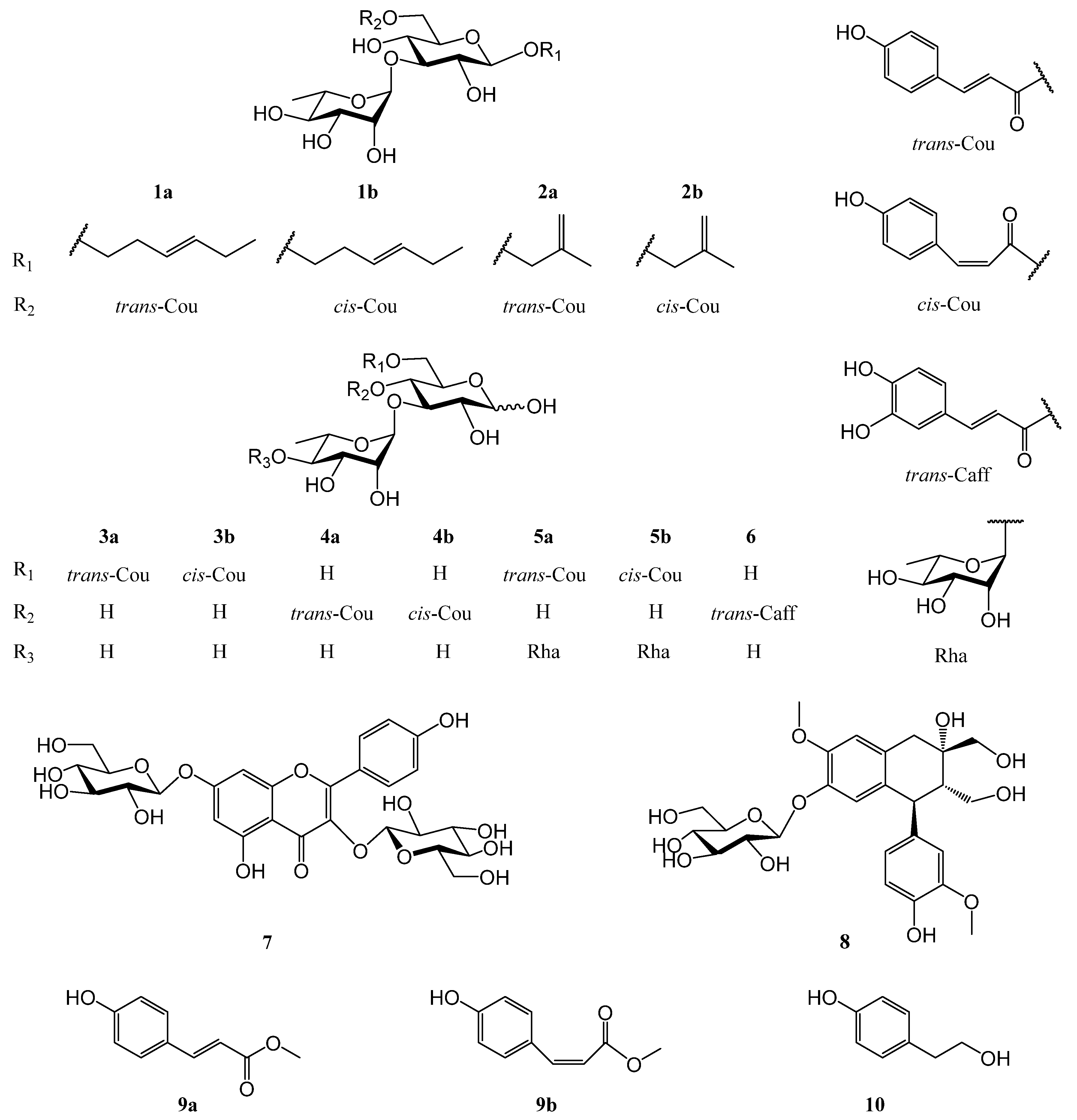
Figure 1. Structures of compounds 110 from the leaves of L. robustum.
Molecules 28 00362 g001
Figure 2. Key HMBC and 1H-1H COSY correlations of compounds 15.
Molecules 28 00362 g002
Table 1. 1H NMR (600 MHz) data of compounds 12 from L. robustum in CD3OD a.
No.1a 1b 2a 2b
1a3.55 m3.55 m4.07 d (12.6)4.10 d (12.6)
1b3.80 m3.80 m4.20 d (12.6)4.15 d (12.6)
22.37 m2.37 m
3a5.36 dt (17.4, 6.6)5.36 dt (17.4, 6.6)4.88 br. s4.88 br. s
3b 5.02 br. s5.02 br. s
45.42 dt (17.4, 6.6)5.42 dt (17.4, 6.6)1.75 s1.73 s
52.05 m2.05 m
6a0.93 t (7.2)0.93 t (7.2)
6b0.97 t (7.2)0.97 t (7.2)
Glc
1′4.31 d (8.4)4.27 d (7.8)4.30 d (7.2)4.26 d (7.8)
2′3.30 m3.30 m3.34 m3.34 m
3′3.51 m3.51 m3.52 m3.52 m
4′3.40 t (9.6)3.40 t (9.6)3.42 br. d (9.0)3.42 br. d (9.0)
5′3.54 m3.54 m3.52 m3.52 m
6′a4.35 dd (12.0, 6.0)4.34 dd (12.0, 6.0)4.36 dd (12.0, 6.0)4.36 dd (12.0, 6.0)
6′b4.48 dd (12.0, 2.4)4.46 dd (12.0, 2.4)4.48 dd (12.0, 1.8)4.46 dd (12.0, 1.8)
Rha
1″5.18 d (1.8)5.16 d (1.8)5.18 d (1.8)5.16 d (1.8)
2″3.94 m3.94 m3.94 dd (3.6, 1.8)3.94 dd (3.6, 1.8)
3″3.71 dd (9.6, 3.6)3.71 dd (9.6, 3.6)3.70 dd (9.6, 3.6)3.70 dd (9.6, 3.6)
4″3.39 t (9.6)3.39 t (9.6)3.40 br. d (9.6)3.40 br. d (9.6)
5″4.00 m4.00 m4.00 m4.00 m
6″1.25 d (6.0)1.24 d (6.0)1.25 d( 6.6)1.25 d( 6.6)
Cou
2′″7.43 d (8.4)7.65 d (8.4)7.47 d (8.4)7.65 d (8.4)
3′″6.77 d (8.4)6.75 d (8.4)6.80 d (8.4)6.76 d (8.4)
5′″6.77 d (8.4)6.75 d (8.4)6.80 d (8.4)6.76 d (8.4)
6′″7.43 d (8.4)7.65 d (8.4)7.47 d (8.4)7.65 d (8.4)
7′″7.63 d (15.6)6.88 d (13.2)7.65 d (16.2)6.89 d (12.6)
8′″6.33 d (15.6)5.79 d (13.2)6.37 d (16.2)5.80 d (12.6)
a Coupling constants (J values in Hz) are shown in parentheses.
Table 2. 13C NMR (150 MHz) data of compounds 12 from L. robustum in CD3OD.
No.1a 1b 2a 2b
170.870.774.073.8
228.928.9143.1143.1
3125.8 125.8 113.4 113.4
4134.6 134.6 19.719.7
521.5 21.5
614.6 14.6
Glc
1′104.4 104.2103.0 103.0
2′75.6 75.6 75.7 75.7
3′84.084.084.084.0
4′70.570.470.470.4
5′75.6 75.3 75.4 75.4
6′64.6 64.5 64.6 64.6
Rha
1″102.7 102.8102.8 102.8
2″72.4 72.4 72.4 72.4
3″72.3 72.3 72.3 72.3
4″74.074.074.074.0
5″70.0 70.0 70.0 70.0
6″17.917.917.917.9
Cou
1′″126.3 127.5126.9 127.5
2′″131.3 133.8 131.2 133.8
3′″117.4 116.0 116.9 115.9
4′″163.0160.4161.6160.2
5′″117.4116.0116.9115.9
6′″131.3133.8131.2133.8
7′″147.1 145.3 146.9 145.3
8′″114.1116.2114.8116.2
CO169.2168.1169.1168.1
Table 3. 1H NMR data of compounds 35 from L. robustum in CD3OD a.
No.3a b3b b4b c
βαβαβ
Glc
14.52 d (7.8)5.08 d (3.6)4.49 d (7.8)5.06 d (4.2)4.52 d (7.6)
23.27 m3.49 dd (9.6, 3.6)3.26 m3.48 dd (9.6, 4.2)3.33 m
33.53 t (9.6)3.81 t (9.6)3.52 t (9.0)3.77 t (9.6)3.75 t (9.2)
43.40 m3.41 m3.39 m3.40 m4.85 t (9.2)
53.58 m4.08 dd (9.6, 3.6)3.57 m4.07 dd (9.6, 3.6)3.55 m
6a4.36 dd (12.0, 6.0)4.32 dd (12.0, 3.6)4.26 dd (12.0, 5.4)4.26 dd (12.0, 3.6)3.52 m
6b4.45 dd (12.0, 1.8)4.49 dd (12.0, 1.8)4.39 dd (12.0, 1.8)4.45 dd (12.0, 1.8)3.58 m
Rha
1′5.18 d (1.8)5.13 d (1.8)5.15 d (1.8)5.10 d (1.8)5.12 d (2.0)
2′3.97 m3.97 m3.96 m3.96 m3.93 m
3′3.72 m3.72 m3.71 m3.71 m3.58 m
4′3.41 m3.41 m3.40 m3.40 m3.32 m
5′4.02 dd (9.6, 6.0)4.02 dd (9.6, 6.0)4.01 dd (9.6, 6.0)4.01 dd (9.6, 6.0)3.63 m
6′1.26 d (6.0)1.26 d (6.0)1.25 d (6.0)1.25 d (6.0)1.17 d (6.0)
Cou
2″7.45 d (8.4)7.45 d (8.4)7.66 d (7.8)7.66 d (7.8)7.72 d (8.8)
3″6.80 d (8.4)6.80 d (8.4)6.75 d (7.8)6.75 d (7.8)6.76 d (8.8)
5″6.80 d (8.4)6.80 d (8.4)6.75 d (7.8)6.75 d (7.8)6.76 d (8.8)
6″7.45 d (8.4)7.45 d (8.4)7.66 d (7.8)7.66 d (7.8)7.72 d (8.8)
7″7.63 d (16.2)7.63 d (16.2)6.86 d (13.2)6.86 d (13.2)6.94 d (12.8)
8″6.33 d (16.2)6.33 d (16.2)5.76 d (13.2)5.76 d (13.2)5.81 d (12.8)
No.4b c5a c5b c
αβαβα
Glc
15.11 d (3.6)4.51 d (8.0)5.07 d (3.6)4.51 d (8.0)5.06 d (3.6)
23.56 m3.26 m3.48 m3.26 m3.48 m
34.06 t (9.2)3.53 m3.81 t (9.2)3.53 m3.81 t (9.2)
44.88 t (9.2)3.40 m3.40 m3.40 m3.40 m
54.01 m3.56 m4.07 m3.56 m4.07 m
6a3.52 m4.33 dd (12.0, 5.6)4.30 dd (12.0, 6.0)4.33 dd (12.0, 5.6)4.30 dd (12.0, 6.0)
6b3.58 m4.45 dd (12.0, 2.0)4.50 dd (12.0, 2.0)4.45 dd (12.0, 2.0)4.50 dd (12.0, 2.0)
Inner-Rha
1′5.17 d (2.0)5.19 d (1.6)5.13 d (1.6)5.17 d (1.6)5.11 d (1.6)
2′3.93 m3.91 m3.91 m3.91 m3.91 m
3′3.58 m3.61 dd (9.6, 3.2)3.85 dd (9.2, 3.2)3.61 dd (9.6, 3.2)3.85 dd (9.2, 3.2)
4′3.32 m3.54 m3.54 m3.54 m3.54 m
5′3.63 m4.12 dd (9.6, 6.0)4.12 dd (9.6, 6.0)4.12 dd (9.6, 6.0)4.12 dd (9.6, 6.0)
6′1.16 d (6.0)1.29 d (6.0)1.29 d (6.0)1.29 d (6.0)1.29 d (6.0)
Outer-Rha
1″ 5.20 d (1.6)5.20 d (1.6)5.20 d (1.6)5.20 d (1.6)
2″ 3.95 dd (3.2, 1.6)3.95 dd (3.2, 1.6)3.95 dd (3.2, 1.6)3.95 dd (3.2, 1.6)
3″ 3.61 dd (9.6, 3.2)3.61 dd (9.6, 3.2)3.61 dd (9.6, 3.2)3.61 dd (9.6, 3.2)
4″ 3.40 m3.40 m3.40 m3.40 m
5″ 3.72 dd (9.2, 6.0)3.72 dd (9.2, 6.0)3.72 dd (9.2, 6.0)3.72 dd (9.2, 6.0)
6″ 1.25 d (6.0)1.25 d (6.0)1.25 d (6.0)1.25 d (6.0)
Cou
2′″7.72 d (8.8)7.46 d (8.4)7.46 d (8.4)7.64 d (8.4)7.63 d (8.4)
3′″6.76 d (8.8)6.81 d (8.4)6.81 d (8.4)6.76 d (8.4)6.75 d (8.4)
5′″6.76 d (8.8)6.81 d (8.4)6.81 d (8.4)6.76 d (8.4)6.75 d (8.4)
6′″7.72 d (8.8)7.46 d (8.4)7.46 d (8.4)7.64 d (8.4)7.63 d (8.4)
7′″6.95 d (12.8)7.64 d (16.0)7.64 d (16.0)6.87 d (12.8)6.87 d (12.8)
8′″5.80 d (12.8)6.35 d (16.0)6.34 d (16.0)5.79 d (12.8)5.78 d (12.8)
a Coupling constants (J values in Hz) are shown in parentheses. b At 600 MHz. c At 400 MHz.
Table 4. 13C NMR (100 MHz) data of compounds 3-5 from L. robustum in CD3OD.
No.3a 3b 4b 5a5b
βαβαβαβαβα
Glc
198.194.098.194.198.294.098.194.198.194.1
276.8 74.2 76.774.277.3 74.6 77.0 74.4 77.0 74.4
384.181.784.281.881.979.483.681.383.681.3
470.670.470.770.570.670.570.670.470.670.4
575.4 70.8 75.370.876.1 71.2 75.5 70.9 75.5 70.9
664.8 64.8 64.664.662.4 62.5 64.9 64.9 64.9 64.9
Inner-Rha
1′102.7 102.8102.9102.9103.1 103.2 102.4 102.6 102.4 102.6
2′72.3 72.3 72.372.372.3 72.3 72.9 72.9 72.9 72.9
3′72.2 72.2 72.272.272.1 72.0 72.9 73.1 72.9 73.1
4′74.074.074.174.073.873.881.281.181.281.1
5′70.0 70.0 70.070.070.4 70.4 68.4 68.4 68.4 68.4
6′17.917.917.917.918.218.218.618.618.618.6
Outer-Rha
1″ 103.2103.2103.2103.2
2″ 72.472.472.472.4
3″ 72.472.472.472.4
4″ 73.973.973.973.9
5″ 70.470.470.470.4
6″ 17.817.817.817.8
Cou
1′″126.9 126.9 127.5127.5127.5 127.5 127.2 127.1 127.5127.5
2′″131.1 131.1 133.7133.7134.3 134.3 131.2 131.2 133.8 133.8
3′″116.9 116.9 115.9115.9115.8 115.9 116.8 116.8 115.9 115.9
4′″161.6161.6160.2160.2160.4160.5161.3161.3160.4160.4
5′″116.9116.9115.9115.9115.8115.9116.8116.8115.9115.9
6′″131.1131.1133.7133.7134.3134.3131.2131.2133.8133.8
7′″146.8 146.8 145.3145.3147.1 147.3 146.7 146.8 145.2 145.2
8′″114.7114.7116.2116.2116.1116.1115.0114.9116.3116.3
CO169.2169.1168.2168.1167.0166.9169.2169.1168.2168.2
Table 5. Results of the bioactivity assays of compounds 110 from L. robustuma.
CompoundFAS IC50 (μM) bα-Glucosidase Inhibition at 0.1 mM (% )α-Amylase Inhibition at 0.1 mM (%)DPPH IC50 (μM) bABTS•+ IC50 (μM) b
1NA cNA27.9 ± 6.4 bc NA5.65 ± 0.19 b
24.10 ± 0.12 aNA24.0 ± 1.5 bcNA103.4 ± 4.00 g
36.25 ± 0.20 b NA29.8 ± 1.8 bc>250 12.04 ± 0.08 d
410.49 ± 0.32 e NA25.6 ± 1.0 bcNA11.21 ± 0.40 cd
59.75 ± 0.24 dNA26.5 ± 4.0 bc>25015.54 ± 0.36 e
6NANA23.0 ± 0.7 c46.66 ± 1.58 b17.01 ± 0.45 e
78.10 ± 0.37 c15.6 ± 0.9 c31.8 ± 0.5 bNA9.34 ± 0.04 cd
88.01 ± 0.26 cNA28.5 ± 2.7 bc>25029.13 ± 1.11 f
915.41 ± 0.42 f 33.8 ± 2.9 b29.5 ± 0.6 bc >2508.78 ± 0.09 c
10NANA16.2 ± 5.0 d NA3.41 ± 0.08 a
Orlistat d4.46 ± 0.13 a
Acarbose d 93.2 ± 0.1 a51.8 ± 2.5 a
l-(+)-ascorbic acid d 13.66 ± 0.13 a10.06 ± 0.19 cd
a Data are expressed as the mean ± SD (n = 3). Means with the same letter are not significantly different (one-way analysis of variance, α = 0.05). b IC50: the ultimate concentration of sample needed to inhibit 50% of the enzyme activity or clear away 50% of the free radicals. cNA: no activity. dPositive control.
Disclaimer/Publisher′s Note: The statements, opinions and data contained in all publications are solely those of the individual author(s) and contributor(s) and not of MDPI and/or the editor(s). MDPI and/or the editor(s) disclaim responsibility for any injury to people or property resulting from any ideas, methods, instructions or products referred to in the content.