Article

Characterization of Bioactive Compounds from Acacia concinna and Citrus limon, Silver Nanoparticles’ Production by A. concinna Extract, and Their Biological Properties

by
Ibrahim Ahmed Shaikh
1,
Uday M. Muddapur
2,*,
Zabin K. Bagewadi
2,
Sneha Chiniwal
2,
Mohammed M. Ghoneim
3,4,
Mater H. Mahnashi
5,
Fahad Alsaikhan
6,
Deepak Yaraguppi
2,
Francois N. Niyonzima
7,
Sunil S. More
8,
Basheerahmed Abdulaziz Mannasaheb
3,
Amer Al Ali
9,
Abdulaziz Asiri
10,
Aejaz Abdullatif Khan
11 and
S. M. Shakeel Iqubal
11
1
Department of Pharmacology, College of Pharmacy, Najran University, Najran 66462, Saudi Arabia
2
Department of Biotechnology, KLE Technological University, BVB Campus, Hubballi 580031, Karnataka, India
3
Department of Pharmacy Practice, College of Pharmacy, AlMaarefa University, Dariyah, Riyadh 13713, Saudi Arabia
4
Pharmacognosy and Medicinal Plants Department, Faculty of Pharmacy (Boys), Al-Azhar University, Cairo 11884, Egypt
5
Department of Pharmaceutical Chemistry, College of Pharmacy, Najran University, Najran 66462, Saudi Arabia
6
Department of Clinical Pharmacy, College of Pharmacy, Prince Sattam Bin Abdulaziz University, Alkharj 11942, Saudi Arabia
7
Department of Math, Science and PE, CE, University of Rwanda, Rwamagana 55, Rwanda
8
School of Basic and Applied Sciences, Dayananda Sagar University, Bangalore 560111, Karnataka, India
9
Department of Clinical Laboratory Sciences, Faculty of Applied Medical Sciences, University of Bisha, 255, Al Nakhil, Bisha 67714, Saudi Arabia
10
Faculty of Applied Medical Sciences, University of Bisha, 255, Al Nakhil, Bisha 67714, Saudi Arabia
11
Department of General Science, Ibn Sina National College for Medical Studies, Jeddah 21418, Saudi Arabia
*
Author to whom correspondence should be addressed.
Molecules 2022, 27(9), 2715; https://doi.org/10.3390/molecules27092715
Submission received: 17 March 2022 / Revised: 16 April 2022 / Accepted: 20 April 2022 / Published: 22 April 2022
(This article belongs to the Special Issue Recent Advances in Nano-Based Drug Delivery System)

Abstract

:
The applications of bioactive compounds from medicinal plants as therapeutic drugs are largely increasing. The present study selected the bioactive compounds from Acacia concinna (A. concinna) and Citrus limon (C. limon) to assess their phytochemicals, proteins, and biological activity. The plant material was collected, and extraction performed as per the standard procedure. Qualitative analysis was undertaken, and identification of functional organic groups was performed by FTIR and HPLC. Antibacterial, anticancer, antioxidant, antihyperglycemic, antihyperlipidemic, and inhibition kinetics studies for enzymes were performed to assess the different biological activities. Flavonoids and phenols were present in a significant amount in both the selected plants. A. concinna showed significant antimicrobial activity against Z. mobilis, E. coli, and S. aureus, with minimum inhibition zones (MIZ) of 24, 22, and 20 mm, respectively. C. limon strongly inhibited all the tested pathogenic bacteria with maximum and minimum MIZ of 32 and 17 mm. A. concinna silver nanoparticles also exhibited potent antimicrobial activity. Both extracts showed substantial antioxidant, antihyperlipidemic, antidiabetic, anticancer (MCF-7), and anti-urease (antiulcer) properties. To conclude, these plants can be used to treat hyperlipidemia, diabetes, cancer, and gastrointestinal ulcers. They can also serve as antimicrobial and antioxidant agents. Thus, the studied plants must be exploited cost-effectively to generate therapeutic drugs for various diseases.

1. Introduction

Medicinal plants are utilized as disease remedials, leading to minor side effects [1]. They are currently used to manage diseases of human beings. They are used by the worldwide population as primary health care [2]. They are thus rich in phyto-substances with positive pharmacological aspects. Some of them are alkaloids, glycosides, flavonoids, phenols, insecticides, polyphenols, vitamins, steroids, terpenoids, saponins, tannins, and coumarins [1,3,4]. For the synthesis of polysaccharides and glycosides, the pentose pathway is followed. However, the shikimate pathway is utilized for aromatic alkaloids, phenols, and tannins. The pathway followed for alkaloids and phenols is acetate–malonate, while the mevalonate route is considered for steroids and terpenes [2,5]. Phytocompounds of medicinal plants can serve as curing agents of various diseases such as diabetes, cancer, blood pressure, etc. [2,6,7].
As per the ethnobotanical literature of phytotherapy of Indian medicinal plants, species such as A. concinna and C. limon are used by the communities for disease treatment as well as in modern medicine [8]. In Ayurveda, the fruits of A. concinna are used for promoting hair growth [9]. Citrus peels reduce coughs, diabetes, phlegm, and are thus used as antithyroidal, antiperoxidative, and hypoglycemic agents. This could be ascribed to the presence of total polyphenols in considerable amounts [10]. The medicinal plants have been extensively used in drug and pharmaceutical industries [4]. A whole medicinal plant or a part of it, such as leaves, seeds, roots, flowers, stems, bark, etc., may be utilized, but the phytochemical contents vary greatly [11]. Cardiovascular diseases and obesity are currently increasing owing to the food intake rich in fat and cholesterol. The pancreatic lipase is inactive in both cases. Thus, the search for phytocompounds that can overcome this issue is necessary [12]. The occurrence of gastrointestinal and urinary tract infections is also increasing due to dysfunction of the urease enzyme, resulting in an increase of pH owing to ammonia production and better growth conditions for Helicobacter pylori. Thus, the inhibition of urease by phyto-substances could be a better option [13].
Infectious diseases are causing morbidity in various countries, especially in underdeveloped countries owing to poverty, augmentation of multiple drug resistance incidence, and increase of undesirable antibiotic side effects [14]. Thus, medicinal plants can be exploited to solve these problems as excellent antimicrobial agents because they possess various phytochemicals [5]. For instance, citrus peels possess essential oils that can prevent the growth of pathogenic bacteria [10]. The phytochemicals from A. concinna and Piper betel leaves are bactericidal against pathogenic bacteria such as E. coli, S. pyogenes, P. vulgaris, and S. aureus [15]. It is thus vital to search for active phytosubstances able to cure pathogenic bacterial infections, since most of these microorganisms are gaining resistance towards available antibiotics [16]. In the present study, A. concinna and C. limon were selected. The study focused on phytochemical screening, characterization, and biological activity evaluation of antibacterial, antidiabetic, antioxidant, hypocholesterolemic, and anticancer activities. In addition, the use of A. concinna leaf extract as a reducing agent for silver nanoparticles’ preparation was also highlighted.

2. Materials and Methods

2.1. Plants, Microorganisms, Cell Lines, and Chemicals

A. concinna and C. limon used in the present investigation were collected from the University of Agriculture Science (Dharwad, India), and brought to the biochemistry laboratory. The bacterial strains used were B. cereus, E. coli, Z. mobilis, B. subtilis, M. luteus, P. aeruginosa, and S. aureus. They were obtained from the Microbial Type Culture Collection (Chandigarh, India). The MCF-7 cell line was bought from the National Centre for Cell Sciences (Pune, India). The chemicals such as DMSO, DPPH, MTT, BSA, p-nitrophenyl palmitate, etc., were bought from Himedia (New Delhi, India) and Sigma-Aldrich Pvt Ltd. (St. Louis, MO, USA), Bangalore, Karnataka. All the reagents utilized in the present study were of analytical grade.

2.2. Extraction and Quantification of the Phytochemicals

The plants were cleaned with tap water, rinsed with distilled water, dried under an aseptic condition for a week at 30 ± 3 °C, and powdered with an electrical stainless steel grinder. Ten grams of the powdered plant sample of A. concinna was taken and it was extracted with 150 mL methanol (95%, v/v) using a Soxhlet apparatus at 60 °C for 6 h. The extract obtained was concentrated using a rotary evaporator so that the solvent evaporates, leaving behind the pure sample. After evaporation, the crude extract stock solution with the concentration of 94.11 mg/mL was stored in amber-colored glass bottle in the refrigerator at 4 °C. The powdered peel of C. limon was treated as above. The profiling of phytochemicals was conducted to confirm the presence of the bioactive secondary metabolites present in the extracts. Various phytochemicals (alkaloids, flavonoids, glycosides, coumarins, diterpenes, steroids, saponins, phlobatannins, tannins, phenols, triterpenoids, catechins, and anthocyanosides) were analyzed for their presence. Quantitative analysis was also carried out for alkaloids, flavonoids, and phenols [17,18]. The plant extracts were kept at 4 °C for future investigations.

2.3. Identification of Functionally Organic Groups in the Phytochemicals

The chemical bonds and organic functional groups were given by FTIR spectroscopy [19] and high performance liquid chromatography (HPLC) according to Sundararajan et al.’s procedure [20].

2.4. Antibacterial Action of A. concinna and C. limon Extracts

2.4.1. Antibacterial Activity Studies and Minimum Inhibitory Concentration (MIC) Investigations

The inoculums were prepared from B. cereus, B. subtilis, M. luteus, P. aeruginosa, Z. mobilis, E. coli, and S. aureus using nutrient broth. The incubation was carried out overnight at 37 °C. For the agar well assay, microbial culture (0.1 mL) was spread on each Muller Hinton agar plate, and then the 6 mm wells were made in agar plates with a borer. Then, 100 µL of A. concinna or C. limon extract was added in each hole and left undisturbed for 60 min for proper extract diffusion. Clear zones were noted in mm after overnight incubation at 37 °C. Gentamicin, an unfilled well, and dimethyl sulphoxide were utilized as positive, neutral, and negative controls, respectively [15,18].
MIC was investigated using the concentration range of 50, 100, and 150 µg/mL. Dimethyl sulphoxide (DMSO) was used as a solvent to dilute plant extracts. Then, 1 mL of A. concinna and C. limon extract was mixed with 5 mL nutrient broth separately, and then it was inoculated with the pathogen, and then incubated at 37 °C. The turbidity was measured at 600 nm after a 24 h incubation period. Later, it was streaked on a nutrient agar plate for examination of growth. When there was no visible growth on the nutrient agar plate, it was considered as the MIC of plant extract on that microorganism [21].

2.4.2. Partial Purification of Bioactive Compounds and Antimicrobial Activity for Protein Fractions

The partial purification of proteins from the plant extracts were carried out with ammonium sulfate followed by dialysis [22]. The protein level was quantified by Lowry et al.’s procedure using BSA as standard [23]. The antimicrobial activity of protein fractions from plant extracts were studied as described earlier by the agar well assay with B. cereus, M. luteus, Z. mobilis, and B. subtilis.

2.4.3. Silver Nanoparticles’ Production by A. concinna Extract

In this study, 100 mL of silver nitrate solutions of 1, 5, and 10 mM were prepared. The A. concinna extract samples of 0.1, 0.5, and 1 mg/mL were added. A positive reaction was indicated by a change from colorless to a brownish color. The spectrum scanning was then carried out between 200 and 800 nm. The synthesized nanoparticles were checked if they could inhibit Z. mobilis as described above.

2.5. Anticancer Activity of A. concinna Extract and C. limon Extracts

The cytotoxic activity of A. concinna and C. limon extracts on the MCF-7 cell line was assessed. The principle of MTT assay was followed [22].

2.6. Antioxidant Activity of A. concinna and C. limon Extracts

The present activity was studied using DPPH as highlighted by Sundararajan et al. [20].

2.7. Enzyme Inhibition Investigations

2.7.1. α-Amylase and α-Glucosidase Inhibitions by A. concinna and C. limon Extracts

α-amylase and α-glucosidase inhibitory activities of A. concinna and C. limon extracts were studied. In this assay, if the plant extract has the ability to inhibit the enzyme, the enzyme cannot utilize the substrate and hence there will be minimum product formation. % inhibition of α-glucosidase/amylase = (Absorbance control − Absorbance sample)/Absorbance control × 100 [24].

2.7.2. In Vitro Lipase Activity Inhibition by A. concinna and C. limon Extracts

The lipase activity of A. concinna and C. limon extracts was studied using in vitro lipase enzyme inhibitory activity method.
% inhibition of lipase = (Absorbance control − Absorbance sample)/Absorbance control × 100 [18].

2.7.3. In Vitro Urease Activity Inhibition by A. concinna and C. limon Extracts

The urease activity of A. concinna and C. limon extracts was studied using in vitro urease inhibitory activity [25].

2.8. Inhibition Kinetics Studies of Amylase, Glycosidase, and Urease

Enzyme inhibition kinetics was evaluated by analyzing the various enzyme activity with A. concinna extract at different concentration ranges of the enzyme substrate. The α-amylase activity was carried out with 2–20 mg/mL starch. For glucosidase inhibition kinetics, different concentration ranges of pNPG (1–10 mM) were considered. Urea in the range of 10 to 100 mM was used for urease inhibition studies. The activity was quantified as above [18]. Km and Vmax were quantified by double reciprocal plot [26].

2.9. Statistical Analysis of Data

All the procedures were repeated 3 times. The experimental results are shown as mean ± standard deviation (SD). They were uploaded in SPSS and analyzed. p < 0.05 was statistically significant after ANOVA and DMRT.

3. Results and Discussion

3.1. Screening of Phytochemicals

Plants have more importance due to the presence of active ingredients or essential organic compounds [11]. In the present investigation, A. concinna and C. limon plants were analyzed. With methanol as the extraction solvent, the phyto-substances observed in both plants were alkaloids, flavonoids, saponins, steroids and triterpenoids, tannins, phenols, glycosides, quinones, coumarins, phlobatannins, and anthocyanosides. However, diterpenes were only present in C. limon, while catechins were absent in both medicinal plants (Table 1). Similarly, the methanol was the solvent of choice to extract polyphenol/flavonoids compared to nonpolar solvents such as hexane and acetone [27]. Thus, A. concinna and C. limon are capable of producing phyto-substances with medical properties and have the ability to synthesize aromatic substances such as phenolics (e.g., phenolic acids, flavonoids, quinones, coumarins, lignans, stilbenes, tannins), nitrogen compounds (alkaloids, amines), vitamins, terpenoids (including carotenoids), and some other endogenous metabolites [2,4]. Quantitative phytochemical analysis was carried and total phenol, alkaloid, and flavonoid contents in the plant extracts are summarized in Table 2. These phytocompounds were present in a significant amount in both plants, except alkaloids, which were found in trace quantity in C. limon.

3.2. Identification of Functionally Organic Groups in the Phytochemicals

3.2.1. FTIRS Analysis of A. concinna and C. limon Extracts

FTIRS was carried out on A. concinna and C. limon extracts. The peaks were as follows: A. concinna (Figure 1A). A broad peak at 723.79, 1000.00, 1100.00, 1230.47, 1383.74, 1461.23, 1620.91, 1700, 2900, 2960.92, 3300 cm−1 corresponds to the alkynes, C–O primary alcoholic group, C–N of amine, C–O alkyl aryl ether and aromatic ester, methyl symmetrical bending, methylene scissoring, C=C of aromatic compounds, ester group, C–H bond in methylene and methyl groups, aldehydic C–H group, and O–H of alcohols and phenols, respectively. The peaks for C. limon are depicted in Figure 1B, and a broad peak for alkynes, C–H bending vibration of substituted compounds, C–O primary alcoholic group, C=O in ester and lactone groups, aldehydic C–H, O–H in alcohols and phenols, C–H of alkanes, was observed at 723.79, 876.86, 1000.00, 1739.90, 2960.92, 3000, and 3010.96 cm−1, respectively. Bagewadi et al. helped to interpret the above results [18]. Therefore, based on the FTIR spectrum, various organic compounds (viz. aromatic compounds, alcohols, lactone, phenols, esters, alkanes, amines, alkynes, terpene, etc.) were observed in the plant extracts and corresponded to the phytochemicals seen.

3.2.2. Determination of Quercetin from A. concinna Extract

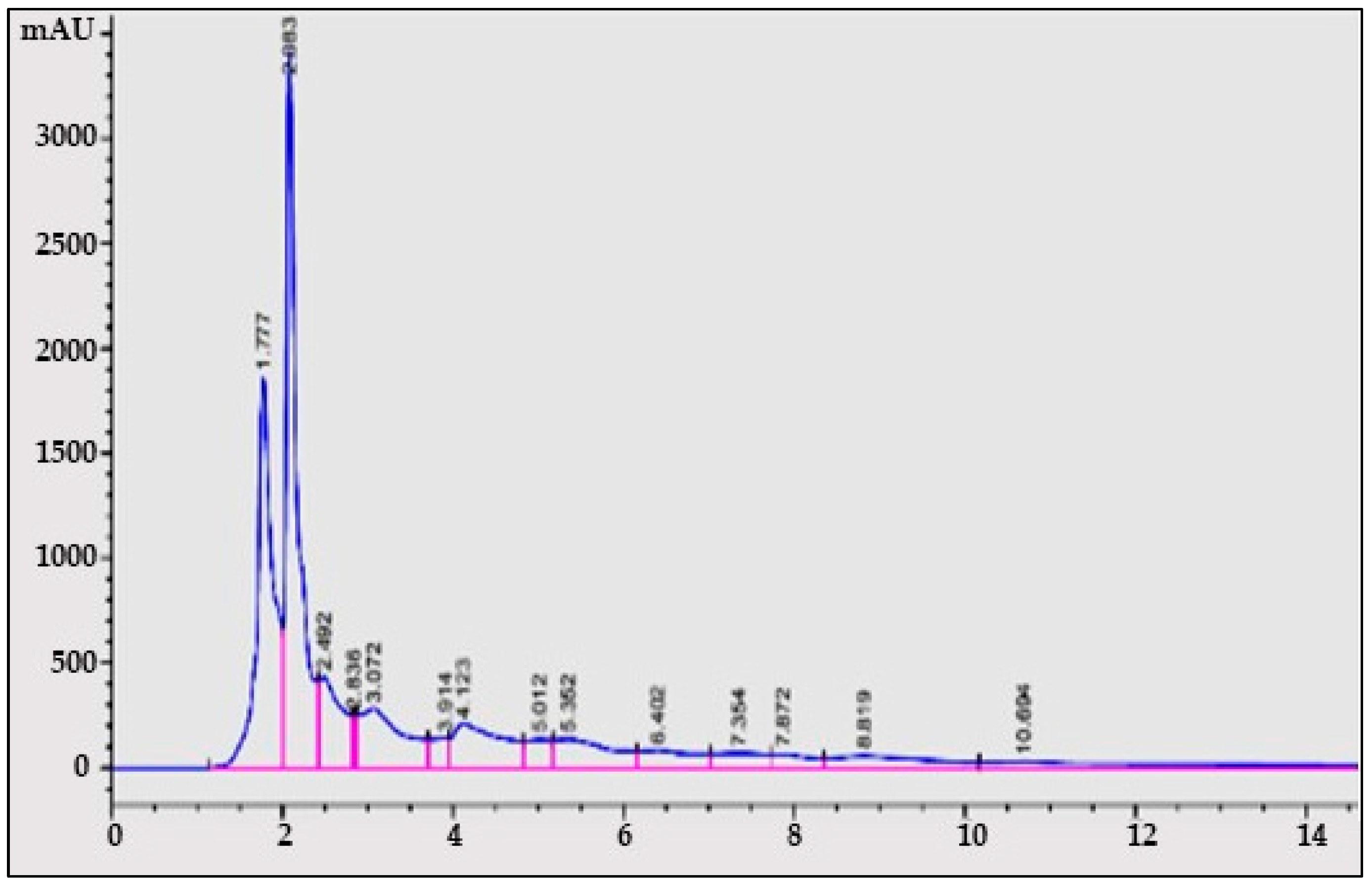
For determination of quercetin, methanol:water (38:62 v/v) was used as the mobile phase with 1 mL/min elution flow rate, and UV wavelength (λ) = 220 nm. A C-18 column was used for the quantification and identification of quercetin acid. With gallic acid as standard, the retention time for quercetin was 1.8 min as shown by the HPLC chromatograms from A. concinna extract (Figure 2).

3.2.3. Determination of Compounds from Plant C. limon Extract

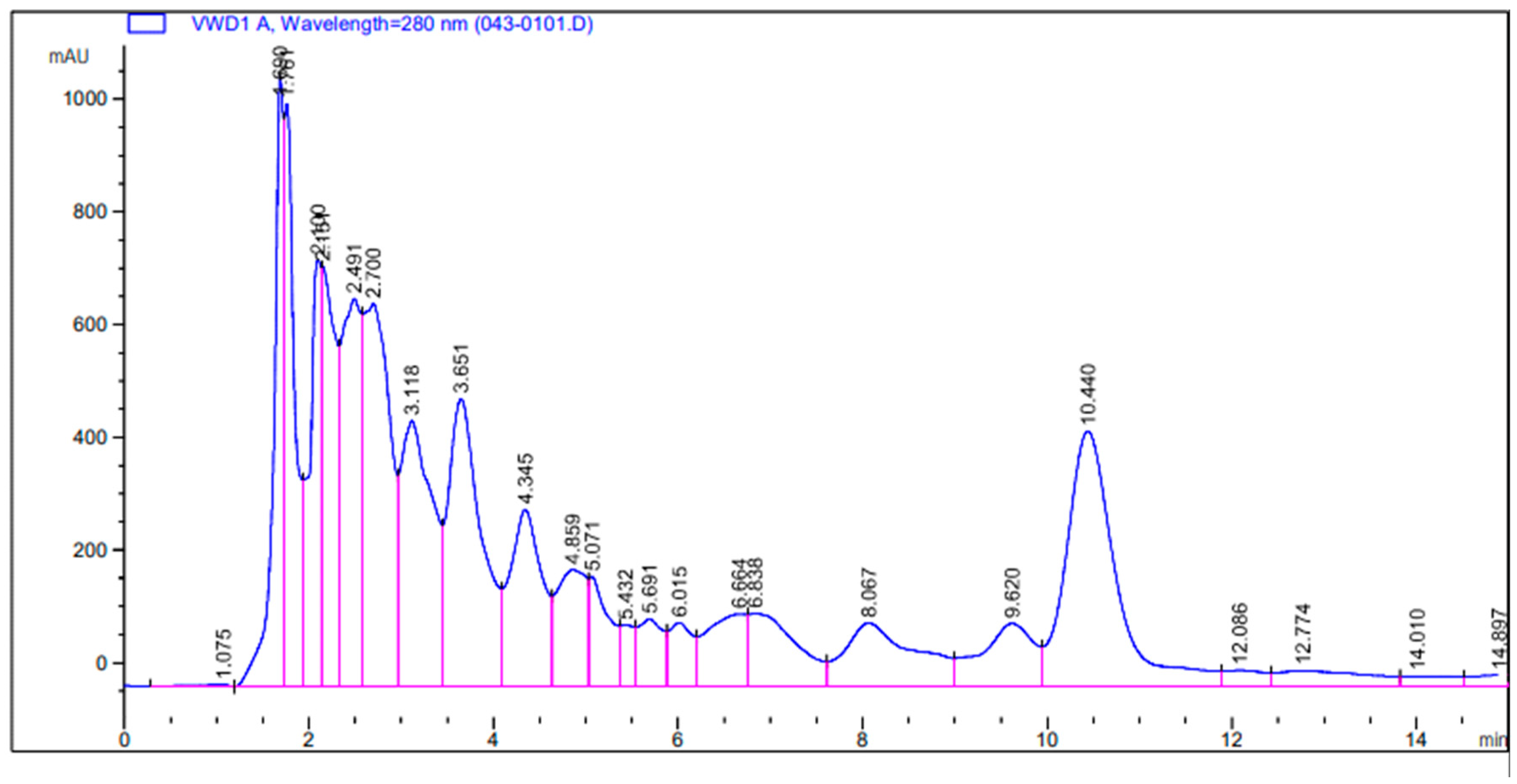
For determination of vanillin, gallic acid, caffeine, and quercetin, methanol:water (10:90, 25:25, 60:40, and 38:62 v/v) was used as the mobile phase with 1, 0.7, 1, and 1 mL/min as elution flow rates, and λ of 220, 270, 272, and 280 nm UV wavelength, respectively. C-18 was used for the quantification and identification of compounds. The compounds determined from C. limon were vanillin, quercetin, gallic acid, and caffeine, with retention time of 2.49, 1.8, 2.3, and 4.8 min, respectively (Figure 3).

3.3. Antibacterial Activity of A. concinna and C. limon Extracts and Determination of Minimum Inhibitory Concentration

Microbial species produce enterotoxins and exotoxins responsible for human diseases. Medicinal plants were reported to possess various compounds able to cure various diseases caused by microorganisms [15]. In the present study, antibacterial activity of methanolic extracts was performed by an agar well diffusion procedure on three Gram-negative (E. coli, Z. mobilis, P. aeruginosa) and four Gram-positive bacteria (B. subtilis, B. cereus, S. aureus, and M. luteus). Table 3 shows the zone of clearance of various extracts. The phytochemicals such as steroids obtained in a huge amount were responsible for antibacterial activity observed. For instance, the sterol acts by pore formation in the bacterial cell wall leading to its death [28]. The antibacterial action may also be due to the occurrence of flavonoids and polyphenols in the extracts present in high concentrations. Similar results have been reported by Chakraborty and Shah [15]. David and Sudarsanam [14] reported that bacteria might be killed by tannins after forming complex soluble substances with bacterial membrane proteins.
C. limon, commonly known as lemon, is bactericidal owing to alkaloids present in a significant amount in different parts of lemon [29]. In the same vein, the lack of antimicrobial effect by plant extracts may be ascribed to the solvent used, as some prefer polar and others non-polar solvents. The plant age, astringent aspects, harvesting time, and extraction procedure may also be responsible for inactive effects of the same phytocompounds [5,30]. These extracts can be used to cure diseases such as pneumonia, skin infections, and meningitis caused by S. aureus, and gastroenteritis, neonatal meningitis, and urinary tract infections caused by E. coli [28]. The bacterial growth prevention by the phytochemicals can be ascribed to inhibition of enzymes and nucleic acid biosynthesis by them [31].
Minimum inhibitory concentration (MIC) investigation of the A. concinna and C. limon extracts was quantified against investigated pathogenic bacteria. The MIC values observed against the assessed pathogenic microorganisms were 50, 100, or 150 µg/mL (Table 4). Similarly, MIC of 50 µg/mL against C. krusei and E. coli, 75 µg/mL against S. aureus, K. pneumoniae, and B. cereus, and 100 µg/mL against C. tropicalis, C. kefyr, and C. albicans were noticed [14].

3.4. Partial Purification of Bioactive Compounds and Antimicrobial Activity for Protein Fractions

Partial purification with 40% ammonium sulfate saturation followed by dialysis was used. The total protein in 2 mL observed after the dialysis step was 0.60, and 1.31 mg for C. limon and A. concinna, respectively.

3.5. Synthesis of Silver Nanoparticles and Antimicrobial Activity of A. concinna

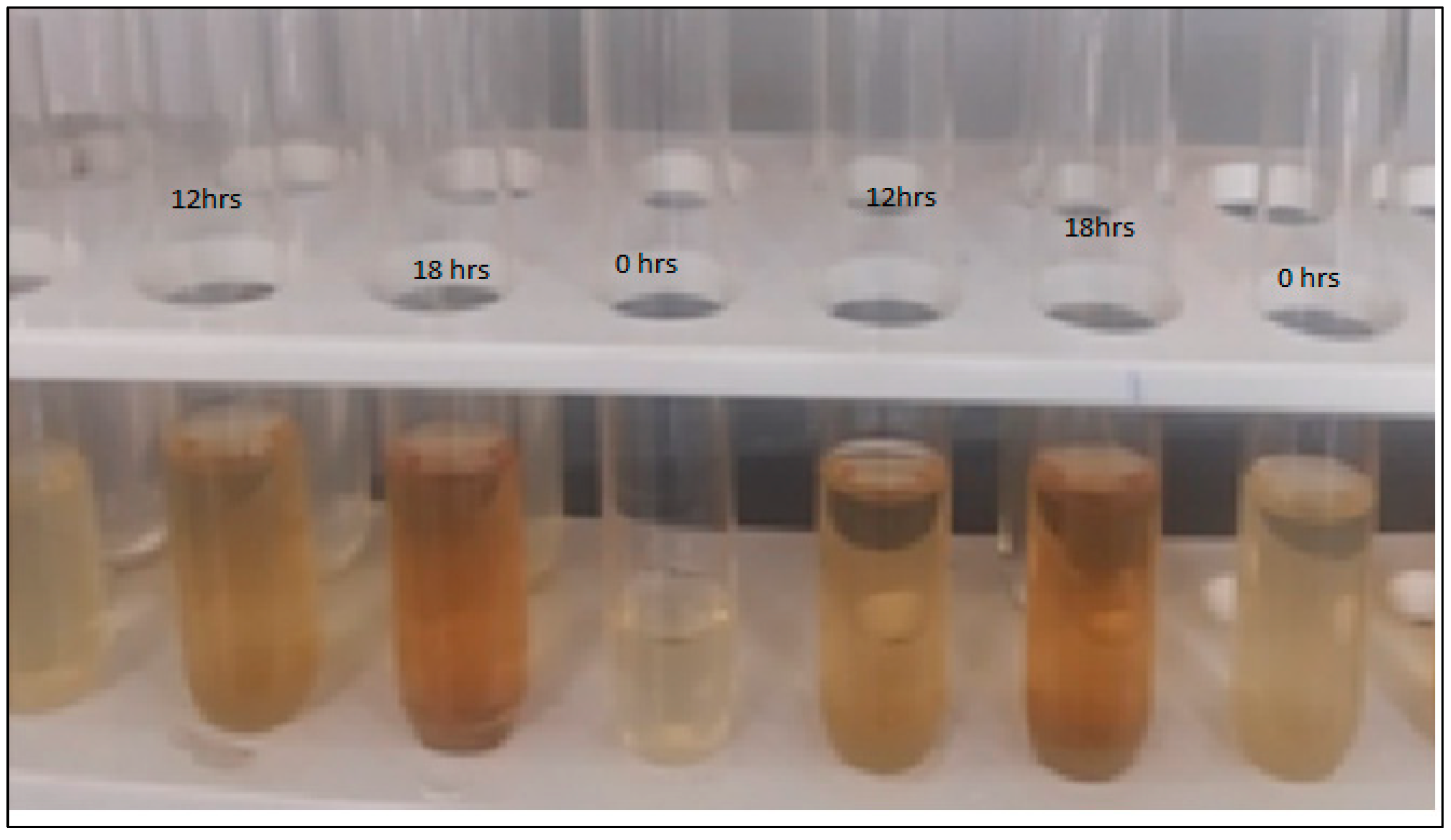
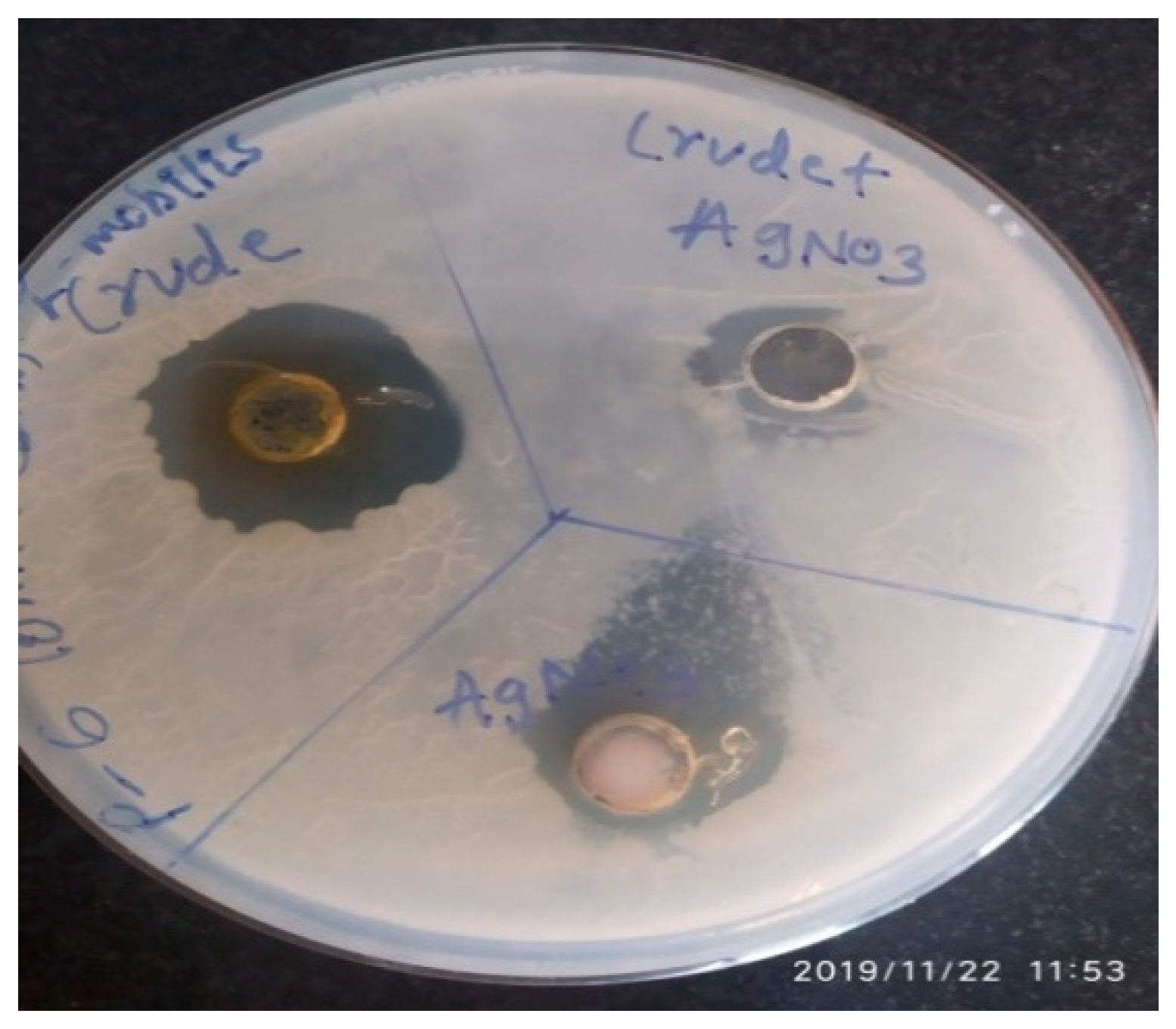
Silver nanoparticles can act as an antibacterial agents [32,33]. Leaf extracts of A. concinna were utilized as reducing agents for silver nanoparticles’ production. After 18 h of incubation, AgNO3 solution, which was incubated with A. concinna extract, changed to brown (Figure 4). The characteristic absorbance was measured at 200–400 nm in the UV–vis spectrum for the confirmation of silver nanoparticles (Figure 5). Silver nanoparticles synthesized using A. concinna extract exhibited antimicrobial activities against Z. mobilis, which is a pathogenic microorganism (Figure 6). Similarly, the Ag-ZnO nanocomposites produced by Verbascum speciosum were able to kill the most infectious pathogenic S. aureus and E. coli [34]. Likewise, a nanocomposite composed of Ag2O3 and ZnO was able to inhibit both Gram-positive and -negative pathogenic bacteria [35]. The bacterial cells are killed by direct interaction between nanoparticles and bacterial cells, leading to disorganization of bacteria caused by positive ion release or by interaction between positive ions and negative bacterial cell wall, resulting in death of microbial cells due to electrostatic interactions [36,37].

3.6. Anticancer Activity of A. concinna and C. limon Extracts

Various phytochemicals were reported to manage and treat effectively cancer diseases [29,38]. The cytotoxicity of the A. concinna extract was assessed on the MCF-7 cell line. At 100–500 µg/mL, the % of cell viability was noticed to decrease with the increase in the concentration of A. concinna extracts and C. limon extracts (Table 5 and Table 6). The IC50 value calculated for A. concinna extract was 171.25 μg/mL and C. limon extracts was 98.61 μg/mL. The plant extract may thus exhibit anticancer activity, and this could be ascribed to the alkaloids present [1,38,39]. Diab [29] reported the strongest antioxidant against HL-60 cells by the lemon peel extract as highlighted by the highest DPPH radical scavenging influence, with EC50 of 42.97 μg extract /mL. The extracts of Pogostemon heyneanus and Plectranthus amboinicus were effective against MCF-7 cells [40]. The leaf extract of D. inoxia showed anticancer properties owing to alkaloids, phenolic compounds, and steroids’ presence [18]. Similar observation of possessing cytotoxic properties by phyto-substances due to steroids and alkaloids were reported with similar cell lines to this study [41]. The phytocompounds may act synergistically with anticancer agents to conquer the resistance of these agents. The utilization of phyto-substances may also lead to the use of a low dosage of anticancer drugs [18].

3.7. Antioxidant Effect of A. concinna and C. limon Extracts

Antioxidants from a plant origin may prevent various severe consequences to humans, such as prevention of cancer and heart diseases [1]. The antioxidant activity of A. concinna and C. limon extracts was assessed with the DPPH procedure. The percentage scavenging activities calculated were 99.9 and 75.1% for A. concinna and C. limon, respectively. This important scavenging action could be ascribed to the phyto-substances present, especially those with OH groups [25,27]. Similarly, the presence of flavonoids and phenolic compounds in D. inoxia were responsible for the antioxidant capacity observed in D. inoxia [27,42]. Hegazy and Ibrahim [27] reported an excellent antioxidant effect of the juice from orange peels. The antioxidant effect was also shown by the extracts of grapefruit and lemon peels during in vitro investigations [29]. Thus, plant antioxidants may prevent various diseases (like cancer disorders) by preventing the free radicals/cell damage action.

3.8. Enzyme Inhibition Activities

3.8.1. α-Amylase and α-Glucosidase Inhibition by A. concinna and C. limon Extracts

Diabetes can be controlled by inhibiting the important enzymes involved in carbohydrate metabolism, such as α-glucosidase and α-amylase [1,39]. In vitro antidiabetic action of A. concinna and C. limon extracts was carried out using alpha amylase and glucosidase enzymes. The inhibition of amylotic activity was observed with A. concinna (100.0%) and C. limon (73.2%). The glucosidase activity was also optimally inhibited by the extract of A. concinna (100%) and C. limon (95.8%). The same enzymes were also inhibited by the extracts obtained from D. inoxia [18] and Sapium ellipticum [24]. This inhibition can be ascribed to the occurrence of OH groups in phenols and flavonoids that hydroxylate and substitute the carbohydrate β-bonds [7]. In addition, the C. limon peel extract with hexane was reported to possess an important activity compared to the commercial Glimepiride, which is used as standard antidiabetic agent [10]. Thus, A. concinna and C. limon plants can be cost-effectively exploited as curative agents of diabetes.

3.8.2. In Vitro Lipase Activity Inhibition by A. concinna and C. limon Extracts

Obesity may arise owing to the dietary fat absorption in a significant amount, leading to the inability of the lipase of the pancreas to digest fat [12]. In vitro fat metabolism activity of A. concinna and C. limon extracts was carried out by analyzing the lipase inhibitory effect. Maximum lipase inhibition of 100% was noticed for the extracts from both plants. The lipid hydrolysis was also inhibited by the extracts from D. inoxia [18]. The juice obtained from the fruit of Cudrania tricuspidata strongly inhibited the pancreatic lipase [12]. The inhibition of lipase can be attributed to flavonoids such as polymethoxyflavones, as suggested by the Zeng et al. [3]. The extracts of A. concinna and C. limon can therefore be useful for the management of obesity.

3.8.3. In Vitro Urease Activity Inhibition by A. concinna and C. limon Extracts

The urease action leads to ammonia secretion (increase of pH), thereby favoring the growth of the pathogenic bacterium Helicobacter pylori. This present pathogen is responsible for various diseases such as gastrointestinal and urinary tract infections [13]. The urease activity was completely inhibited by the extract of C. limon and A. concinna. These findings are in contrast to previous studies, which reported weak inhibition of urease activity by Geranium purpureum [25] and D. inoxia [18] extracts. A vital anti-urease activity was reported by Shah [43] for the phytochemicals extracted from Asparagus gracilis. A. concinna and C. limon extracts can inhibit the urease activity, thereby curing various diseases, such as throat infections, dysuria, common cold, urinary troubles, asthma, ulcer, and cough. Thus, urease inhibition is also known as a better option to prevent H. pylori survival [13].

3.9. Inhibition Kinetics Studies of Amylase, Glycosidase, and Urease

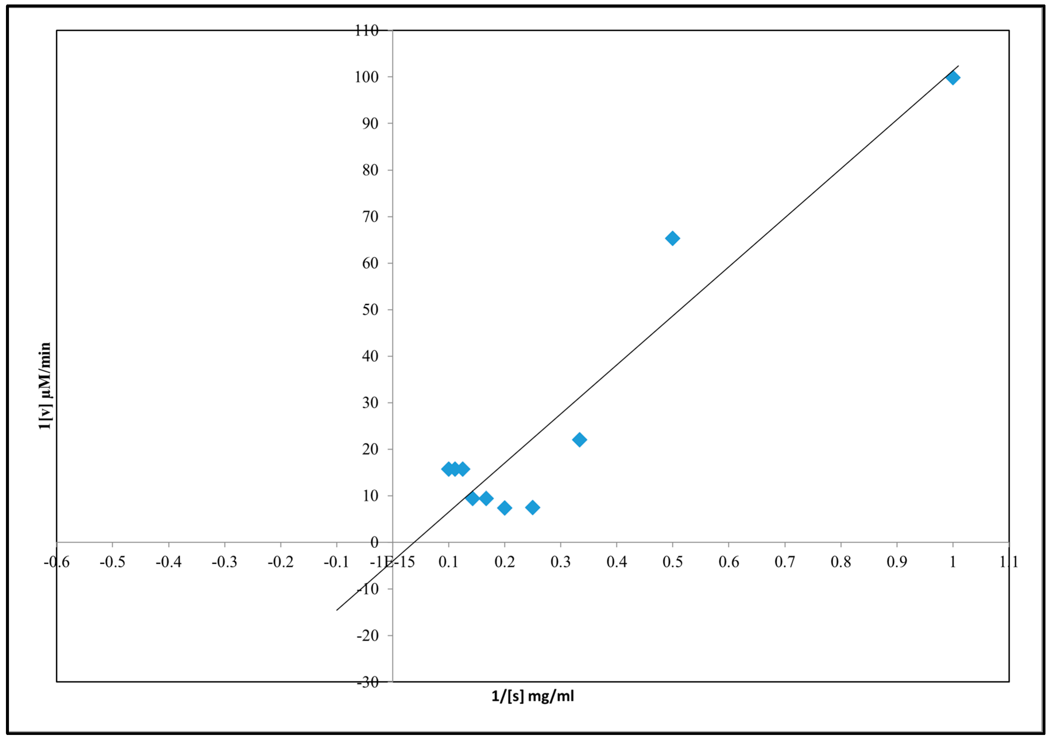
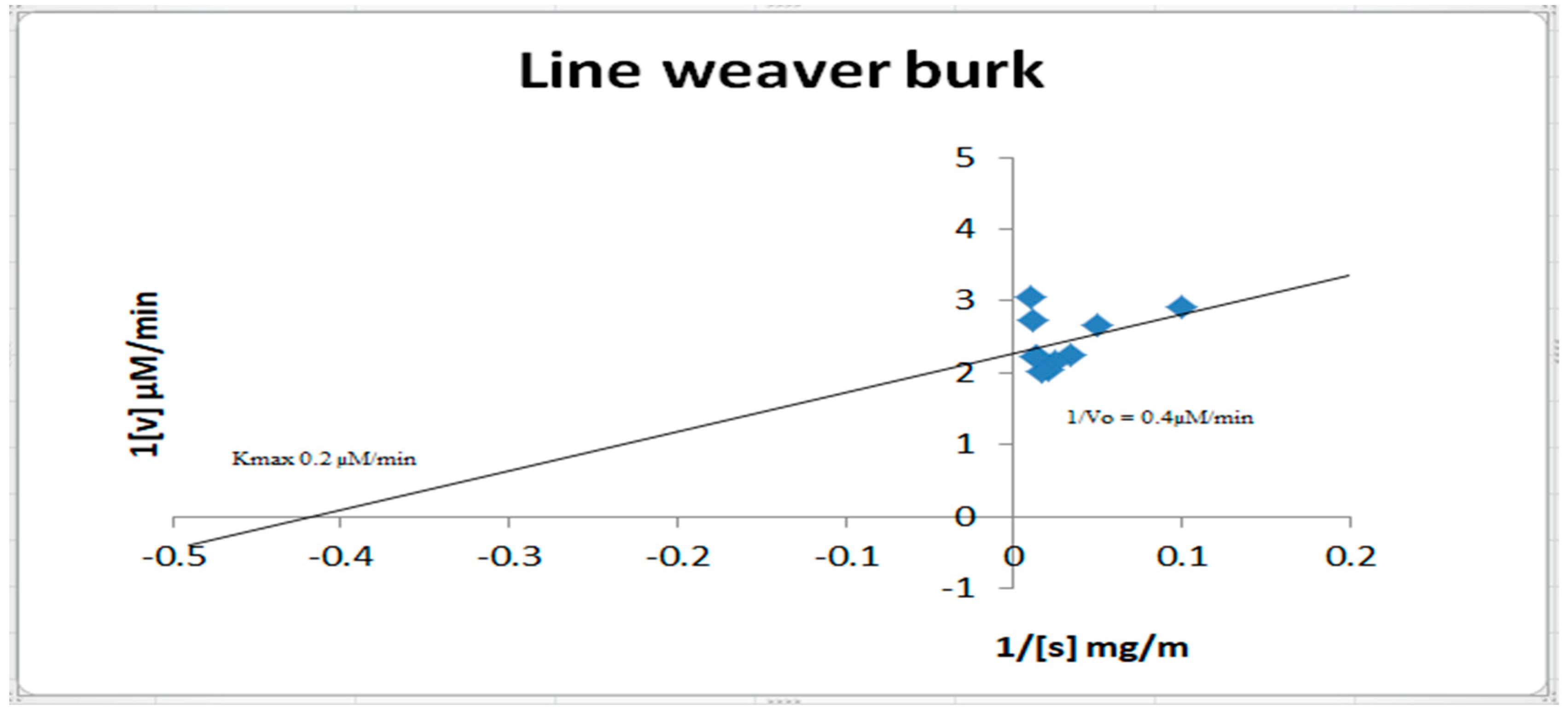
Kinetics inhibition of α-amylase, urease, and α-glucosidase by A. concinna extract were carried out by varying the different substrate concentrations. The substrates used for α-amylase, α-glucosidase, and urease activities were starch (2–20 mg/mL), pNPG (1–10 mM), and urea (1–100 mM), respectively. Km and Vmax were calculated. The results are in Figure 7, Figure 8 and Figure 9. For α-amylase, Vmax was 0.2 µM/min and Km 1 mg/mL (Figure 7). Vmax was 0.05 µM/min and Km was 0.9 mg for α-glucosidase (Figure 8). For urease, the extract was found with a Vmax of 0.2 µM/min and Km of 0.9 mg/mL (Figure 9).

4. Conclusions

In the present investigation, the extraction of the phytocompounds of A. concinna and C. limon was performed. The plant extracts showed significant antimicrobial activities against studied pathogenic bacteria owing to the presence of phytocompounds such as alkaloids, flavonoids, and phenols. The inhibition of α-amylase, α-glucosidase, and urease enzymes showed that the plants could be used in the management of diabetes and acid-peptic disorders. Silver nanoparticles synthesized using A. concinna extract exhibited antimicrobial activities against pathogenic Z. mobilis. The current study demonstrates that the bioactive chemicals found in A. concinna and C. limon can be used in clinical and pharmacological applications to treat a variety of ailments such as cancer, diabetes, gastrointestinal ulcers, and bacterial infections. However, further research is warranted to elucidate the exact mechanism of action of these medicinal plants.

Author Contributions

“Conceptualization, U.M.M., Z.K.B., S.C. and MMG.; methodology, I.A.S., M.H.M., F.A., D.Y. and F.N.N.; software, S.S.M., B.A.M. and A.A.A.; validation, A.A., A.A.K. and S.M.S.I.; formal analysis, I.A.S. and U.M.M.; investigation, Z.K.B. and S.C.; resources, U.M.M.; data curation, A.A.; writing—original draft preparation, U.M.M. and Z.K.B.; writing—review and editing, I.A.S.; visualization, M.H.M.; supervision, S.S.M.; project administration, U.M.M.; funding acquisition, I.A.S. and M.M.G. All authors have read and agreed to the published version of the manuscript.”

Funding

The authors are thankful to the Deanship of Scientific Research, Najran University, Najran, Saudi Arabia, for funding this research through grant research code NU/NRP/MRC/11/2; the authors deeply acknowledge the Researchers Supporting Program (TUMA-Project-2021-6), AlMaarefa University, Riyadh, Saudi Arabia for supporting steps of this work.

Institutional Review Board Statement

Not applicable.

Informed Consent Statement

Not applicable.

Data Availability Statement

Not applicable.

Acknowledgments

The authors are thankful to Najran University, Najran, KSA; and KLE Technological University, BVB Campus, Hubballi, India; and the authors deeply acknowledge the Researchers Supporting Program (TUMA-Project-2021-6), AlMaarefa University, Riyadh, Saudi Arabia for supporting steps of this work.

Conflicts of Interest

The authors declare no conflict of interest.

Sample Availability

Samples of the compounds are available from the authors.

References

  1. Zhang, L.; Tu, Z.C.; Xie, X.; Lu, Y.; Wang, Z.X.; Wang, H.; Sha, X.M. Antihyperglycemic, antioxidant activities of two Acer palmatum cultivars, and identification of phenolics profile by UPLC-QTOFMS/MS: New natural sources of functional constituents. Ind. Crops Prod. 2016, 89, 522–532. [Google Scholar] [CrossRef]
  2. Kabera, J.N.; Semana, E.; Mussa, A.R.; He, X. Plant Secondary Metabolites: Biosynthesis, Classification, Function and Pharmacological Properties. J. Pharm. Pharmacol. 2014, 2, 377–383. [Google Scholar]
  3. Zeng, S.L.; Li, S.Z.; Wei, M.Y.; Chen, B.Z.; Li, P.; Zheng, G.D.; Liu, E.H. Evaluation of anti-lipase activity and bioactive flavonoids in the citri reticulatae pericarpium from different harvest time. Phytomedicine 2018, 43, 103–109. [Google Scholar] [CrossRef] [PubMed]
  4. Jain, C.; Khatana, S. Rekha Vijayvergia Bioactivity of secondary metabolites of various plants: A review. Int. J. Pharm. Sci. Res. 2019, 10, 494–504. [Google Scholar] [CrossRef]
  5. Alo, M.N.; Anyim, C.; Igwe, J.C.; Elom, M.; Uchenna, D.S. Antibacterial activity of water, ethanol and methanol extracts of Ocimumgratissimum, Vernoniaamygdalina and Aframomummelegueta. Adv. Appl. Sci. Res. 2012, 3, 844–848. [Google Scholar]
  6. Khoja, K.K.; Shaf, G.; Hasan, T.N.; Syed, N.A.; Al-Khalifa, A.S.; Al-Assaf, A.H.; Alshatwi, A.A. Fenugreek, a naturally occurring edible spice, kills MCF-7 human breast cancer cells via an apoptotic pathway. Asian Pac. J. Cancer Prev. 2011, 12, 3299–3304. [Google Scholar]
  7. You, Q.; Chen, F.; Wang, X.; Jiang, Y.; Lin, S. Antidiabetic activities of phenolic compounds in muscadine against alpha-glucosidase and pancreatic lipase. Food Sci. Technol. 2012, 46, 164–168. [Google Scholar]
  8. Singh, A.; Nautiyal, M.C.; Kunwar, R.M.; Bussmann, R.W. Ethnomedicinal plants used by local inhabitants of Jakholi block, Rudraprayag district, western Himalaya, India. J. Ethnobiol. Ethnomed. 2017, 13, 49. [Google Scholar] [CrossRef] [Green Version]
  9. Tezuka, Y.; Honda, K.; Banskota, A.H.; Thet, M.M.; Kadota, S. Kinmoonosides A−C, Three New Cytotoxic Saponins from the Fruits of Acacia concinna, a Medicinal Plant Collected in Myanmar. J. Nat. Prod. 2000, 63, 1658–1664. [Google Scholar] [CrossRef]
  10. Naim, M.; Amjad, F.M.; Sultana, S.; Islam, S.N.; Hossain, M.A.; Begum, R.; Rashid, M.A.; Amran, M.S. Comparative Study of Antidiabetic Activity of Hexane-Extract of Lemon Peel (Limon citrus) and Glimepiride in Alloxan-Induced Diabetic Rats. Bangladesh Pharm. J. 2012, 15, 131–134. [Google Scholar] [CrossRef]
  11. Ogbulie, J.N.; Ogueke, C.C.; Nwanebu, F.C. Antibacterial properties of Uvaria chamae, Congronema latifolium, Garcinia kola, Vemonia amygdalina and Aframomium melegueta. Afr. J. Biotechnol. 2007, 6, 1549–1553. [Google Scholar]
  12. Jeong, J.Y.; Jo, Y.H.; Lee, K.Y.; Do, S.G.; Hwang, B.Y.; Lee, M.K. Optimization of pancreatic lipase inhibition by Cudrania tricuspidata fruits using response surface methodology. Bioorganic Med. Chem. Lett. 2014, 24, 2329–2333. [Google Scholar] [CrossRef] [PubMed]
  13. Khan, A.W.; Jan, S.; Parveen, S.; Khan, R.A.; Saeed, A.; Tanveer, A.J.; Shad, A.A. Phytochemical analysis and Enzyme Inhibition Assay of Aerva javanica for Ulcer. Chem. Cent. J. 2012, 6, 76. [Google Scholar] [CrossRef] [Green Version]
  14. David, B.C.; Sudarsanam, G. Antimicrobial activity of Gymnema sylvestre (Asclepiadaceae). J. Acute Dis. 2013, 2, 222–225. [Google Scholar] [CrossRef] [Green Version]
  15. Chakraborty, D.; Shah, B. Antimicrobial, antioxidative and antihemolytic activity of Piper betel leaf extracts. Int. J. Pharm. Pharm. Sci. 2011, 3, 192–199. [Google Scholar]
  16. Muhsinah, A.B.; Maqbul, M.S.; Mahnashi, M.H.; Jalal, M.M.; Altayar, M.A.; Saeedi, N.H.; Alshehri, O.M.; Shaikh, I.A.; Khan, A.A.L.; Shakeel Iqubal, S.M.; et al. Antibacterial Activity of Illicium Verum Essential Oil against MRSA Clinical Isolates and Determination of Its Phyto-Chemical Components. J. King Saud Univ. Sci. 2022, 34, 101800. [Google Scholar] [CrossRef]
  17. Pascale, R.; Bianco, G.; Cataldi, T.R.I.; Kopplin, P.S.; Bosco, F.; Vignola, L.; Uhl, J.; Lucio, M.; Milell, L. Mass spectrometry-based phytochemical screening for hypoglycemic activity of Fagioli di Sarconi beans (Phaseolus vulgaris L.). Food Chem. 2018, 242, 497–504. [Google Scholar] [CrossRef] [Green Version]
  18. Bagewadi, Z.K.; Muddapur, U.M.; Madiwal, S.S.; Mulla, S.I.; Khan, A. Biochemical and enzyme inhibitory attributes of methanolic leaf extract of Datura inoxia Mill. Environ. Sustain. 2019, 2, 75–87. [Google Scholar] [CrossRef]
  19. Bagewadi, Z.K.; Mulla, S.I.; Ninnekar, H.Z. Purification and characterization of endo β-1,4-D-glucanase from Trichoderma harzianum strain HZN11 and its application in production of bioethanol from sweet sorghum bagasse. 3 Biotech 2016, 6, 101. [Google Scholar] [CrossRef] [Green Version]
  20. Sundararajan, M.; Thomas, P.A.; Babyshalini, K.; Geraldine, P. Identification of phytoconstituents and in-vitro evaluation of the putative anticataractogenic effect of an ethanolic root extract of Leucas aspera. Biomed. Pharmacother. 2017, 85, 87–101. [Google Scholar] [CrossRef]
  21. Tandon, C.; Mathur, P.; Sen, M.; Kanojiya, S. Identification of an antibacterial withanolide (dinoxin b) from leaf of Datura inoxia mill. Int. J. Phytomed. 2016, 8, 1–12. [Google Scholar]
  22. Arulvasu, C.; Shiva Shakthi, S.K.; Babu, G.; Radhakrishnan, N. Purification and identification of bioactive protein from leaves of Datura inoxia P.mil. Biomed. Prev. Nutr. 2014, 4, 143–149. [Google Scholar] [CrossRef]
  23. Lowry, O.H.; Rosenberg, W.J.; Farr, A.L.; Randell, R.J. Quantitation of protein using Folin Ciocalteu reagent. J. Biol. Chem. 1951, 193, 265–275. [Google Scholar] [CrossRef]
  24. Ighodaro, O.M.; Akinloye, O.A.; Ugbaja, R.N.; Omotainse, S.O.; Faokunla, O. FT-IR analysis of Sapium ellipticum (Hochst) pax ethanol leaf extract and its inhibitory effects on pancreatic α-amylase and intestinal α-glucosidase activities in vitro. Egypt. J. Basic Appl. Sci. 2016, 3, 343–349. [Google Scholar] [CrossRef] [Green Version]
  25. Taskin, T.; Taskin, D. In vitro anti-urease, antioxidant activities and phytochemical composition of Geranium purpureum. J. Food Meas. Charact. 2017, 11, 2102–2109. [Google Scholar] [CrossRef]
  26. Lineweaver, H.; Burk, D. The determination of enzyme dissociation constants. J. Am. Chem. Soc. 1934, 56, 658–666. [Google Scholar] [CrossRef]
  27. Hegazy, A.E.; Ibrahium, M.I. Antioxidant activities of orange peel extracts. World Appl. Sci. J. 2012, 18, 684–688. [Google Scholar]
  28. Pelczar, M.J.; Chan, E.C.S.; Kreig, N.R. Microbiology; Tata McGrow-Hill: New Delhi, India, 1993; Volume 5. [Google Scholar]
  29. Diab, K.A.E. In Vitro Studies on Phytochemical Content, Antioxidant, Anticancer Immunomodulatory and Antigenotoxic Activities of Lemon, Grapefruit, and Mandarin Citrus Peels. Asian Pac. J. Cancer Prev. 2016, 17, 3559–3567. [Google Scholar]
  30. Umuhoza, M.R.; Habyarimana, T.; Niyonzima, F.N. Activity of Kalanchoe integra against selected pathogenic bacteria. Open Access J. Microbiol. Biotechnol. 2021, 6, 1–6. [Google Scholar] [CrossRef]
  31. Uysal, S.; Aumeeruddy-Elalfi, Z.; Zengin, G.; Aktumsek, A.; Mocan, A.; Custodio, L.; Neng, N.R.; Nogueira, J.M.F.; Ćirić, A.; Glamočlija, J.; et al. Insight into the biological properties and phytochemical composition of Ballota macrodonta Boiss. et Balansa—An endemic medicinal plant from Turkey. Ind. Crops Prod. 2018, 113, 422–428. [Google Scholar] [CrossRef]
  32. Iqbal, N.; Iqubal, S.M.S.; Khan, A.A.; Mohammed, T.; Alshabi, A.M.; Aazam, E.S.; Rafiquee, M.Z.A. Effect of CTABr (Surfactant) on the Kinetics of Formation of Silver Nanoparticles by Amla Extract. J. Mol. Liq. 2021, 329, 115537. [Google Scholar] [CrossRef]
  33. Muddapur, U.M.; Alshehri, S.; Ghoneim, M.M.; Mahnashi, M.H.; Alshahrani, M.A.; Khan, A.A.; Iqubal, S.M.S.; Bahafi, A.; More, S.S.; Shaikh, I.A.; et al. Plant-Based Synthesis of Gold Nanoparticles and Theranostic Applications: A Review. Molecules 2022, 27, 1391. [Google Scholar] [CrossRef] [PubMed]
  34. Mousavi-Kouhi, S.M.; Beyk-Khormizi, A.; Amiri, M.S.; Mashreghi, M.; Yazdi, M.E.T. Silver-zinc oxide nanocomposite: From synthesis to antimicrobial and anticancer properties. Ceram. Int. 2021, 47, 21490–21497. [Google Scholar] [CrossRef]
  35. Yazdi, M.E.T.; Nourbakhsh, F.; Mashreghi, M.; Mousavi, S.H. Ultrasound-based synthesis of ZnO·Ag2O3 nanocomposite: Characterization and evaluation of its antimicrobial and anticancer properties. Res. Chem. Intermed. 2021, 47, 1285–1296. [Google Scholar] [CrossRef]
  36. Khan, S.A.; Shahid, S.; Lee, C.-S. Green Synthesis of Gold and Silver Nanoparticles Using Leaf Extract of Clerodendrum inerme; Characterization, Antimicrobial, and Antioxidant Activities. Biomolecules 2020, 10, 835. [Google Scholar] [CrossRef]
  37. Yazdi, M.T.; Nazarnezhad, S.; Mousavi, S.; Amiri, M.S.; Darroudi, M.; Baino, F.; Kargozar, S. Gum Tragacanth (GT): A Versatile Biocompatible Material beyond Borders. Molecules 2021, 26, 1510. [Google Scholar] [CrossRef] [PubMed]
  38. Jaikumar, B.; Jasmine, R. A review on a few medicinal plants possessing anticancer activity against human breast cancer. Int. J. Pharm. Tech. Res. 2016, 9, 333–365. [Google Scholar]
  39. Zhang, L.; Zhu, M.F.; Tu, Z.C.; Zhao, Y.; Wang, H.; Li, G.J.; Sha, X.M. a-Glucosidase inhibition, anti-glycation and antioxidant activities of Liquidambar formosana Hance leaf, and identification of phytochemical profile. S. Afr. J. Bot. 2017, 113, 239–247. [Google Scholar] [CrossRef]
  40. Rai, V.; Pai, V.R.; Kedilaya, P. A preliminary evaluation of anticancer and antioxidant potential of two traditional medicinal plants from Lamiaceae-Pogostemonheyneanus and Plectranthusamboinicus. J. Appl. Pharm. Sci. 2016, 6, 74–79. [Google Scholar]
  41. Vermillion, K.; Holguin, F.O.; Berhow, M.A.; Richins, R.D.; Redhouse, T.; O’Connell, M.A.; Posakony, J.; Mahajan, S.S.; Kelly, S.M.; Simon, J.A. Dinoxin B, a withanolide from Datura inoxia leaves with specific cytotoxic activities. J. Nat. Prod. 2011, 74, 267–271. [Google Scholar] [CrossRef] [Green Version]
  42. Fatima, H.; Khan, K.; Zia, M.; Ur-Rehman, T.; Mirza, B.; Haq, I.U. Extraction optimization of medicinally important metabolites from Datura innoxia Mill.: An in vitro biological and phytochemical investigation. BMC Complement. Altern. Med. 2015, 15, 376. [Google Scholar] [CrossRef] [PubMed] [Green Version]
  43. Shah, N.A.; Khan, M.R.; Sattar, S.; Ahmad, B.; Mirza, B. HPLC-DAD analysis, antioxidant potential and anti-urease activity of Asparagus gracilis collected from district Islamabad. BMC Complement. Altern. Med. 2014, 14, 347. [Google Scholar] [CrossRef] [PubMed] [Green Version]
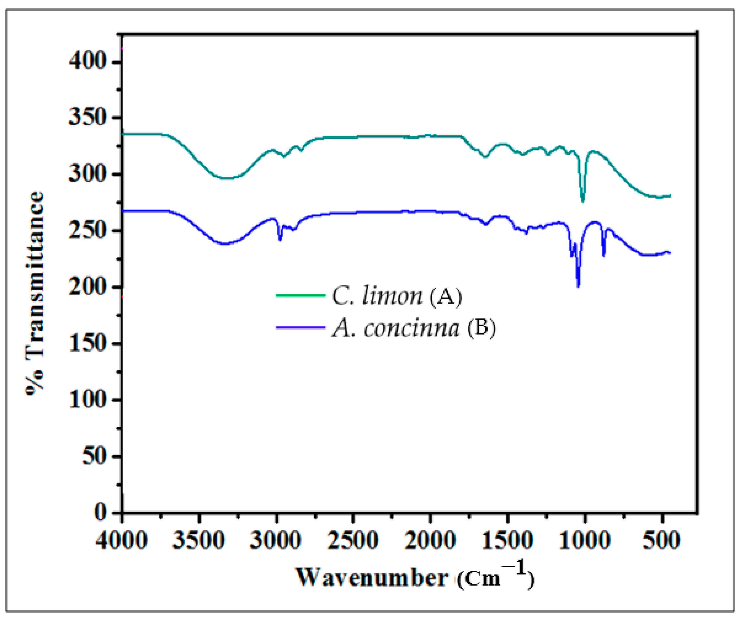
Figure 1. FTIR analysis of phytochemicals in the plant extracts. (A) FTIR trace of Citrus limon; (B) FTIR trace of Acacia concinna.
Molecules 27 02715 g001
Figure 2. HPLC chromatograph of quercetin from A. concinna extract. The pink coloured lines represent the retention/peak values.
Molecules 27 02715 g002
Figure 3. HPLC chromatograph of vanillin, quercetin, gallic acid, and caffeine from C. limon extract. The pink coloured lines represent the retention/peak values.
Molecules 27 02715 g003
Figure 4. Incubation of silver nitrate solution with A. concinna leaf extract.
Molecules 27 02715 g004
Figure 5. UV–vis spectrum showing formation of silver nanoparticles.
Molecules 27 02715 g005
Figure 6. Antimicrobial activity of synthesized nanoparticles by A. concinna extract against Z. mobilis.
Molecules 27 02715 g006
Figure 7. Inhibition kinetics of amylase activity.
Molecules 27 02715 g007
Figure 8. Inhibition kinetics of α-glucosidase activity.
Molecules 27 02715 g008
Figure 9. Inhibition kinetics of urease activity.
Molecules 27 02715 g009
Table 1. Phytochemicals observed in A. concinna and C. limon after methanol extraction. +: present, −: absent.
PhytoconstituentsA. concinnaC. limon
Alkaloids++
Flavonoids++
Saponins++
Steroids and triterpenoids++
Tannins++
Phenols++
Glycosides++
Quinones++
Coumarins++
Diterpenes+
Phlobatannins++
Anthocyanosides++
Catechins
Table 2. Concentration of some phytochemicals observed from plant extracts.
Medicinal PlantAlkaloids (mg/L)Flavonoids (mg/L)Phenols (mg/L)
A. concinna0.1860 ± 0.00010.2768 ± 0.00030.2431 ± 0.0002
C. limon0.0002 ± 0.00000.1752 ± 0.00010.3401 ± 0.0007
Table 3. Zone of bacterial clearance of various extracts in mm.
Medicinal PlantE. coliB. subtilisZ. mobilisM. luteusS. aureusB. cereusP. aeruginosa
A. concinna22 ± 0.7637 ± 0.12324 ± 0.8017 ± 0.28520 ± 0.3655 ± 0.1695 ± 0.401
C. limon22 ± 0.67117 ± 0.24922 ± 0.59332 ± 0.58227 ± 0.80122 ± 0.46721 ± 0.397
Table 4. MIC of A. concinna and C. limon at 600 nm.
A. concinnaE. coliB. subtilisZ. mobilisM. luteusS. aureusP. aeruginosa
50 µg/mL
100 µg/mL
150 µg/mL
C. limon
50 µg/mL++
100 µg/mL
150 µg/mL
Table 5. Anticancer activity of A. concinna extract.
BlankUntreatedCisplatin 15 µg/mL100200300400500 µg/mL
Reading 10.0090.630.0680.3750.2290.190.0110.007
Reading 20.0050.690.0680.3870.2020.1720.0170.009
Mean OD0.0070.6850.0680.3810.21550.1810.0140.008
Mean OD-Mean blank 0.6780.06810.3740.20850.1740.0070.001
Standard deviation 0.016970600.0084850.0190920.0127280.0042430.001414
Standard Error 0.01200.0060.01350.0090.0030.001
% Standard error 1.769911500.8849561.991151.3274340.4424780.1474
% Viability 1008.99705055.1622430.7522125.663721.0324480.147493
Table 6. Anticancer activity of C. limon extracts.
BlankUntreatedCisplatin 15 µg/mL100 µg/mL200 µg/mL300 µg/mL400 µg/mL500 µg/mL
Reading 10.0090.630.0680.3650.210.1940.020.008
Reading 20.0050.690.0680.3670.2040.1820.0220.007
Mean OD0.0070.660.0680.3660.2070.1880.0210.007
Mean OD-Mean blank 0.6530.0610.3590.20.1810.0140.0005
Standard deviation 0.0400.0010.0040.0080.0010
Standard Error 0.0300.0010.0030.0060.0010.0005
% Standard error 4.5900.150.450.910.150.07
% Viability 1009.3454.9730.6227.712.140.076
Publisher’s Note: MDPI stays neutral with regard to jurisdictional claims in published maps and institutional affiliations.