Article

Phosphorylation of cPLA2α at Ser505 Is Necessary for Its Translocation to PtdInsP2-Enriched Membranes

1
Instituto de Biología y Genética Molecular, Consejo Superior de Investigaciones Científicas (CSIC), 47003 Valladolid, Spain
2
Centro de Investigación Biomédica en Red de Diabetes y Enfermedades Metabólicas Asociadas (CIBERDEM), 28029 Madrid, Spain
*
Authors to whom correspondence should be addressed.
Molecules 2022, 27(7), 2347; https://doi.org/10.3390/molecules27072347
Submission received: 21 March 2022 / Revised: 30 March 2022 / Accepted: 1 April 2022 / Published: 6 April 2022

Abstract

:
Group IVA cytosolic phospholipase A2α (cPLA2α) is a key enzyme in physiology and pathophysiology because it constitutes a rate-limiting step in the pathway for the generation of pro- and anti-inflammatory eicosanoid lipid mediators. cPLA2α activity is tightly regulated by multiple factors, including the intracellular Ca2+ concentration, phosphorylation reactions, and cellular phosphatidylinositol (4,5) bisphosphate levels (PtdInsP2). In the present work, we demonstrate that phosphorylation of the enzyme at Ser505 is an important step for the translocation of the enzyme to PtdInsP2–enriched membranes in human cells. Constructs of eGFP-cPLA2 mutated in Ser505 to Ala (S505A) exhibit a delayed translocation in response to elevated intracellular Ca2+, and also in response to increases in intracellular PtdInsP2 levels. Conversely, translocation of a phosphorylation mimic mutant (S505E) is fully observed in response to cellular increases in PtdInsP2 levels. Collectively, these results suggest that phosphorylation of cPLA2α at Ser505 is necessary for the enzyme to translocate to internal membranes and mobilize arachidonic acid for eicosanoid synthesis.

1. Introduction

The eicosanoids constitute an ample family of bioactive lipids with potent pro- and anti-inflammatory activities. They are not stored in the cells but produced in response to specific cellular stimulation [1,2]. The common precursor of the eicosanoids is arachidonic acid (AA), a fatty acid that is mostly found esterified in the sn-2 position of cellular glycerophospholipids, primarily those containing choline, ethanolamine, and inositol as polar headgroups [3,4,5]. Although the cells possess multiple phospholipase A2 (PLA2) enzymes potentially capable of liberating AA from membrane phospholipids [6,7,8,9], the major PLA2 involved in receptor-mediated AA mobilization is the group IVA PLA2, also known as cytosolic PLA2α (cPLA2α) [10,11].
cPLA2α is tightly regulated in cells, not only to finely regulate the amount of AA mobilized for eicosanoid synthesis but also because both of its by-products, free fatty acids and lysophospholipids, could be deleterious to the cells if accumulating at high levels. Increases in the intracellular Ca2+ concentration constitute one of the key regulators of cPLA2α activity in cells and mediate translocation of the enzyme to a variety of cytoplasmic membranes [10,11]. This is driven by the presence in the enzyme of a calcium-binding domain, or C2 domain. Unlike other PLA2 family members, cPLA2α does not require Ca2+ for enzyme activity, but to dock and penetrate into the membrane interface [12,13,14].
In addition to Ca2+, cPLA2α is also regulated by intracellular lipids. The C2 domain also has a site for ceramide 1-phosphate, produced by activated ceramide kinase in situ [15,16]. This lipid allosterically activates the enzyme and increases the residence time of the enzyme in membranes [15,16]. cPLA2α also binds phosphatidylinositol 4,5-bisphosphate (PtdInsP2) via a 4-Lys cluster present in the catalytic domain [17,18]. In vitro, PtdInsP2 increases the catalytic activity of the enzyme in a calcium-independent manner, likely by enhancing its capacity to penetrate membranes, especially those enriched in choline phospholipids [17,18]. Mutations in the residues where PtdInsP2 binds give rise to an enzyme that, when transfected into cells, manifests a reduced ability to translocate to intracellular membranes and mobilize AA [19,20].
cPLA2α can also be phosphorylated in cells at residues Ser505, Ser515, and Ser727, and all of these phosphorylation reactions have been suggested to be involved in the regulation of agonist-induced AA mobilization [10,11,21]. While the functional relevance of cPLA2α phosphorylation at Ser515 and Ser727 may depend on cell type and stimulation conditions, there is general agreement that phosphorylation at Ser505 represents a key regulatory event under nearly all cellular conditions examined; thus, it has been the most extensively studied [10,11,21]. In general terms, it appears that the extracellular-regulated kinases p42/p44 are responsible for cPLA2α phosphorylation at Ser505 in cells of murine origin [22,23,24], and the related kinases p38 and JNK are involved in cells of human origin [25,26,27].
Several lines of evidence have suggested that cPLA2α phosphorylation at Ser505 is necessary for the enzyme to be fully active in cells; however, the molecular reasons for this still remain elusive [21]. In vitro studies have indicated that phosphorylation of cPLA2α at Ser505 is not required for activity but for proper binding of the enzyme to membranes in a Ca2+-dependent fashion [28].
Previous work from our laboratory has highlighted the importance of intracellular PtdInsP2 levels in regulating the physical state of cPLA2α at Ca2+ levels equaling those present in resting cells [19]. The phosphorylation state of cPLA2α does not seem to influence the translocation of the enzyme to model membranes in the presence of high Ca2+ [28]. In the present study, we have studied the influence of cPLA2α phosphorylation at Ser505 on PtdInsP2 binding and in the translocation capacity of the enzyme in a cellular scenario. We show that cPLA2α phosphorylation at Ser505 is necessary for the translocation of the enzyme to membranes and to promote AA release in response to PtdInsP2 elevations. These studies provide new insights into the complex regulation of cPLA2α, thereby expanding and deepening our knowledge of the cellular mechanisms controlling the production of pro- and anti-inflammatory lipid mediators.

2. Results

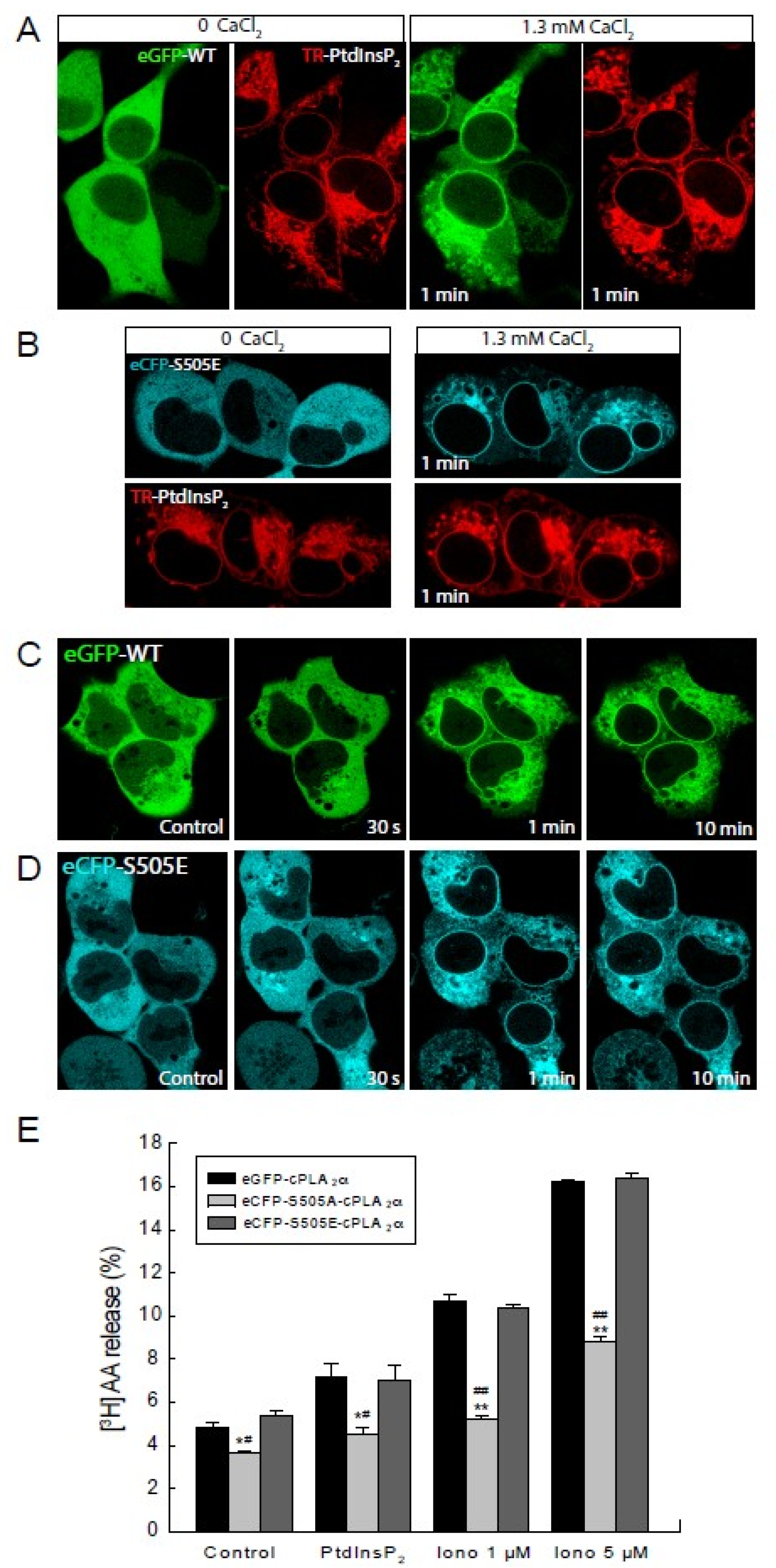
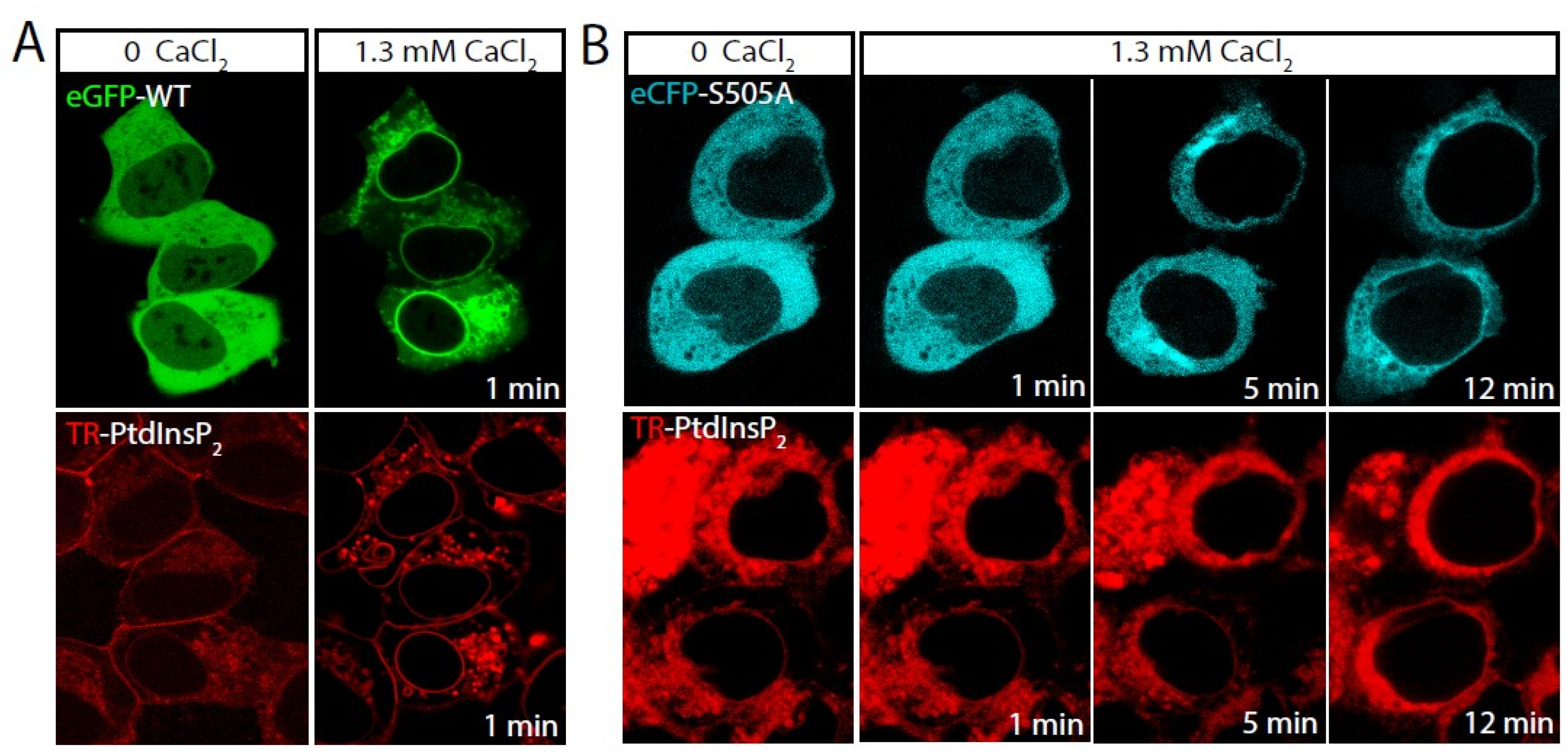
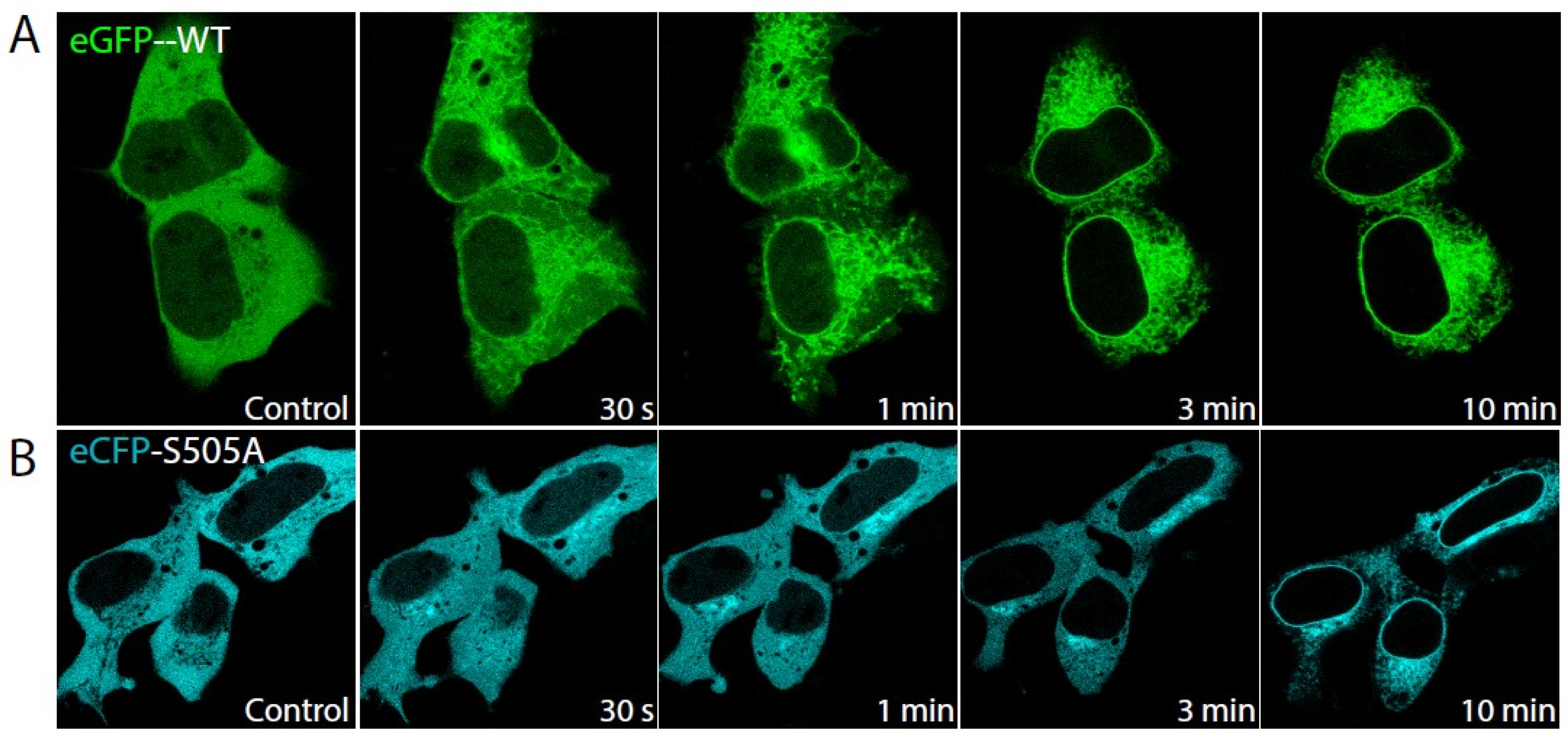
2.1. Role of cPLA2α Phosphorylation in Membrane Translocation in Response to Increases in PtdInsP2 and Calcium

In previous work, we showed that increasing PtdInsP2 levels in intact cells in the absence of a rise in intracellular Ca2+ is sufficient to trigger cPLA2α activation and attendant AA mobilization [19]. We began the current study by assessing whether such an effect of PtdInsP2 requires the enzyme to be phosphorylated at Ser505. To this end, cells stably expressing eGFP-cPLA2α or the mutant eCFP-S505A-cPLA2α were incubated with exogenous PtdsInsP2 complexed with a histone shuttle to facilitate entry into the cells. We and others have previously used this technique successfully [19,29,30], which results in cells displaying increased PtdInsP2 levels, and no influence on the physical state of cPLA2α, as the incubations are carried out in the absence of extracellular Ca2+ [19]. The PtdsInsP2 contained a fluorescent tag that allowed its monitoring within the cells by confocal microscopy. Once the cells incorporated all the PtdsInsP2 (as assessed by the cellular fluorescence associated with it, which remained stable and did not increase further), restoring extracellular Ca2+ levels triggered the immediate translocation of cPLA2α, i.e., 1 min, to internal membranes (Figure 1A). Importantly, the non-phosphorylatable eCFP-S505A-cPLA2α mutant required considerably more time, i.e., 5–12 min, to translocate to perinuclear membranes (Figure 1B).
Because PtdInsP2 is known to reduce the Ca2+ threshold for cPLA2α to translocate to membranes in vitro [17,31], in the next series of experiments, we examined the behavior of the enzyme and the S505A mutant under the opposite circumstances, i.e., in the presence of a sustained rise of the intracellular Ca2+ level. As shown in Figure 2, cell treatment with 5 µM ionomycin, which raises the intracellular Ca2+ concentration up to 4 µM [19,32], the eCFP-S505A-cPLA2α translocated to inner membranes after experiencing a marked delay compared with wild type EGFP-cPLA2α. The former required approx. 10 min to target the perinuclear membranes (Figure 2B), while the latter was translocated completely to perinuclear membranes within the first 3 min of treatment (Figure 2A). Collectively, these data indicate that phosphorylation of cPLA2α at Ser505 is a key step for the enzyme to readily translocate to its target membranes even at high intracellular Ca2+ levels.

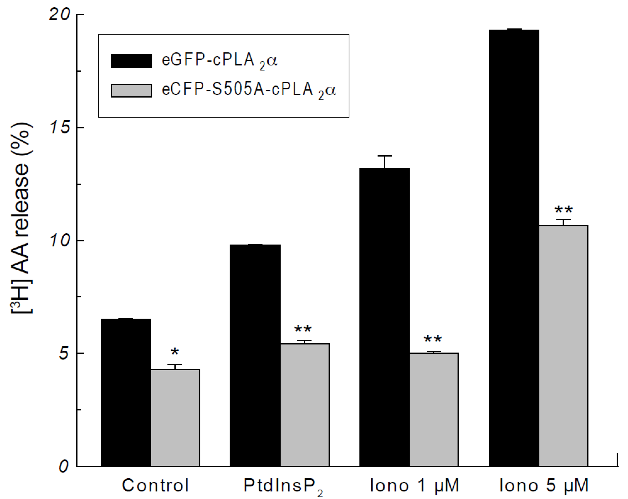
2.2. Phosphorylation of cPLA2α Is Necessary for AA Release in Response to PtdInsP2

To assess the possible physiological/pathophysiological relevance of the delay in membrane translocation of the eCFP-S505A-cPLA2α, we also conducted AA mobilization experiments under identical experimental conditions. The cells were prelabeled with [3H]AA, and the release of radiolabeled fatty acid was measured after exposing the cells to PtdInsP2 and ionomycin. As shown in Figure 3, [3H]AA release in cells transfected with the eCFP-S505A-cPLA2α mutant was significantly lower than that of cells transfected with the wild-type enzyme, and this occurred at all conditions tested. Thus, delayed cPLA2α translocation results in diminished fatty acid mobilization.
To further characterize the importance of Ser505 phosphorylation in cPLA2α translocation in response to PtdInsP2, mutants were constructed where Ser505 was replaced with Glu (S505E), which mimics phosphorylation at that residue [25,28]. The ability of this S505E mutant to translocate in response to PtdInsP2 elevations and to sustained increases in intracellular Ca2+ was then evaluated, and the results are shown in Figure 4. As expected, the S505E mutant behaved the same as the wild-type mutant (Figure 4A–D). Note that, in common with many other cells in culture [24,33], most of the wild-type cPLA2α in the HEK cells is already phosphorylated at Ser505. This explains why the S505E mutant behaves the same as the wild-type enzyme. Moreover, cells expressing the S505E mutant manifested an AA release response to either PtdInsP2 elevations or ionomycin which was essentially identical to that of cells expressing the wild-type enzyme (Figure 4E). Thus, these data show that cPLA2α phosphorylation at Ser505 capacitates the enzyme for a full functional response.

3. Discussion

The mechanisms responsible for the translocation of cPLA2α to cellular membranes in the absence of sustained increases in intracellular calcium have remained a subject of debate [10,11,34]. Early studies utilizing purified cPLA2α showed that the binding of the enzyme to vesicles and micelles increased in the presence of PtdInsP2, resulting in enhanced activity even at nanomolar Ca2+ levels [17,18,31]. Studies in intact cells have also provided evidence that increased PtdInsP2 levels in cells can sustain cPLA2α activation and attendant AA mobilization at Ca2+ levels equaling those of resting cells [34,35]. Finally, a four-Lys cluster was described in the enzyme, which binds PtdInsP2 tightly and may help regulate the cellular location of the enzyme under stimulatory conditions [19,20]. In this work, we extend our knowledge of PtdInsP2 regulation of cPLA2α by showing for the first time that phosphorylation of the enzyme at Ser505 is necessary for the full regulatory effect of PtdInsP2 to take place. Thus, these results establish a hitherto unrecognized link between two major mechanisms of cPLA2α regulation, namely PtdInsP2 and Ser505 phosphorylation.
While phosphorylation of cPLA2α at Ser505 has been recognized for a long time, its full physiological significance remains unclear. Reasons for this include the finding that in resting cells, most of the cPLA2α is already phosphorylated at Ser505 and that the specific activity of the non-phosphorylated enzyme differs little from that of the phosphorylated one [24,33,36]. Our data support the view that PtdInsP2 may help the cPLA2α to achieve the appropriate conformation for optimal interaction of the enzyme with its target membrane, in agreement with previous observations [28]. cPLA2α is a rather ‘promiscuous’ enzyme, being able to translocate to different membranes depending on cell type and stimulatory conditions [11,37]. Whether PtdInsP2 regulates the translocation of cPLA2α to all kinds of intracellular membranes or its regulatory function is limited to targeting the enzyme to specific membranes is unknown at present. It is also interesting to note that the kinases involved in phosphorylating cPLA2α at Ser505 under activation conditions appear to greatly depend on cell type and stimulation conditions [22,23,24,25,26,27]. Whether these differences are due to species-specific features or reflect distinctive regulatory attributes of the enzyme is also unknown. Future work in the laboratory will be aimed to investigate whether the PtdInsP2 effects on enzyme translocation are related to the involvement of a specific kinase or intracellular membrane.
Stimulatory cell conditions of physiological/pathophysiological relevance do not lead to high calcium concentrations inside the cell. Rather, receptor-mediated activation promotes low and transient increases in Ca2+ concentration, which on many occasions lead to cPLA2α activation [10,11,12]. Thus, it seems necessary to define the factors that regulate the translocation of cPLA2α to membranes under physiological Ca2+ conditions. Several lines of evidence have suggested that the enzyme behaves differently depending on Ca2+ availability. Under high Ca2+ concentrations (>1 µM), the C2 domain of the enzyme is fully active and can drive translocation of the cPLA2α to membranes without any other requirement [12,34]. On the contrary, under physiologically relevant Ca2+ conditions (up to 400 nM), multiple regulatory components may have to be set into motion to achieve full translocation of the enzyme. We have shown here that cPLA2α phosphorylation at Ser505 is one of these components.
The finding that the non-phosphorylatable mutant S505A shows a reduced ability to translocate to membranes in response to PtdInsP2 elevations suggests that, in the absence of phosphorylation, cPLA2α is not capable of binding productively to PtdInsP2, probably because the affinity for the phospholipid is decreased. Alternatively, it is also possible that the S505A mutant requires higher Ca2+ levels to translocate to the membrane, even in the presence of PtdInsP2. The experiments conducted with the phosphorylation mimetic mutant S505E further support the idea that cPLA2α phosphorylation at Ser505 is required for the enzyme to recognize and respond to PtdInsP2 elevations optimally.
Overall, the findings described here demonstrate that cPLA2α has multiple mechanisms to circumvent its necessity for high Ca2+ concentrations to translocate to membranes and that those mechanisms interact with each other. Moreover, the diminished translocation ability of the nonphosphorylated enzyme in response to PtdInsP2 elevations (at least 11 min delay compared with the wild-type enzyme) underscores the importance of these interactions for cPLA2α to display full biological activity.

4. Materials and Methods

4.1. Plasmids

The plasmid eGFP-cPLA2α has been described elsewhere [19,32]. For the construction of the eCFP-S505A-cPLA2α, the eGFP was substituted in the plasmid eGFP-cPLA2α by the eCFP by using the restriction enzymes Agel and BsrGI. Subsequently, Ser505 was replaced with Ala (S505A) by using the QuikChange XL Site-Directed Mutagenesis kit (Stratagene, San Diego, CA, USA), and the oligonucleotides 5′-CAATACATCTTATCCATGG- CGCCTTTGAGTGACTT-3′ (forward) and 5′-GCAAAGTCACTCAAAGGCGCCAGTGGATAAGATGTA-3′ (reverse). For the mutagenesis of Ser505 to Glu (S505E) the oligonucleotides used were: 5′-GAATCTCAATACATCTTATCCACTGGAGCCTTTGAGTGACTTTGC-3′ (forward) and 5′-GCAAAGTCACTCAAAGGCTCCAGTGGATAAGATGTATTGAGATTC-3x (reverse). Mutagenesis was confirmed by sequencing.

4.2. Cells

HEK cells were cultured in Dulbecco’s Modified Essential Medium (Gibco, Carlsbad, CA, USA) supplemented with 2 mM glutamine, 10% fetal calf serum, 100 U/mL penicillin, and 100 µg/mL streptomycin at 37 °C in a 5% CO2 humidified incubator. Cells were passaged twice a week by trypsinization. Cells (40–70% confluence) were transfected with 1 µg plasmid/mL using Lipofectamine PlusTM (Invitrogen, Carlsbad, CA, USA), following the manufacturer’s instructions. For stably transfected cells, 1 mg/mL G418 was used for selection and subsequent passages.

4.3. Lipid Preparation

PtdInsP2 was added to the cells as previously described [19,30]. Briefly, 2 µg of phospholipid was mixed with 2 µL of the carrier (histone, 0.5 mM), resuspended in Hank’s balanced salt solution containing 10 mM HEPES, sonicated in a water bath for 2 min, and allowed to rest at 37 °C for 10 min before use. The final concentration of PtdInsP2 in the incubation media was 5.7 µM.

4.4. Confocal Microscopy

The cells were seeded on glass-bottom culture dishes (MatTek Corp., Ashland, MA, USA) and allowed to adhere for 24 h. The medium was then replaced by Hanks’ buffered saline containing 10 mM HEPES and 1.3 mM CaCl2. For some experiments, cells were incubated without CaCl2, which was added back when needed. Fluorescence was monitored by confocal microscopy using a Bio-Rad Radiance 2100 laser-scanning system coupled to a Nikon TE-2000U with a termostatized chamber (Warner Instruments, Holliston, MA, USA). The objective was CFI Plan Apo 60X, 1.4 numerical aperture, and oil immersion. The fluorescence of eCFP was monitored at 457 nm argon excitation using the combination of a long pass barrier filter HQ470LP and a short pass filter HQ520SP. The fluorescence of eGFP was monitored at 488 nm Argon excitation using the combination of a long pass filter HQ500LP and a short pass filter HQ560SP. The Alexa-Fluo 594 fluorescence was monitored at 543 nm HeNe excitation using a long band pass filter HQ570LP. Red fluorescence from BODYPI-TRx was monitored at 543 nm HeNe laser excitation using an HQ590/570 long band pass blocking filter.

4.5. AA Release

The cells were labeled with 0.5 µCi/mL [3H]AA (sp. act. 200 Ci/mmol; HartBiomédica, Madrid, Spain) for 18 h. Afterward, they were washed extensively and overlaid with 0.5 mL of serum-free medium supplemented with 0.5 mg/mL albumin and treated with 5 µM thimerosal for 15 min to block fatty acid reacylation [38,39]. The cells were then stimulated for 60 min. Supernatants were removed, and cell monolayers were overlaid with ice-cold phosphate buffer containing 0.05% Triton X-100 and scraped. Total lipids from supernatants and cells were extracted according to Bligh and Dyer [40]. After extraction, lipids were separated by thin-layer chromatography using the system n-hexane/diethyl ether/acetic acid (70:30:1 by volume) [41]. Spots corresponding to free AA and phospholipid were scraped, and radioactivity was quantified by liquid scintillation counting.

5. Conclusions

cPLA2α-mediated production of bioactive lipid mediators represents a key event in the execution of physiological and pathophysiological responses to external stimuli. This study has focused on the complex interactions that govern cPLA2α translocation to membranes and the multiple factors that may be involved in its regulation. Specifically, we have described the interconnected role of two of these factors, phosphorylation of the enzyme at Ser505 and cellular PtdInsP2 levels. Both of them seem to work together to promote membrane translocation and activation of cPLA2α under low intracellular Ca2+ levels. Together, the studies described here represent a relevant working model to further understand the intricacies of the cellular regulation of cPLA2α and the molecular mechanisms underlying it.

Author Contributions

Conceptualization, J.C., J.B. and M.A.B.; Data curation, J.C.; Formal analysis, J.C. and M.A.B.; Funding acquisition, J.B. and M.A.B.; Investigation, J.C.; Methodology, J.C., J.B. and M.A.B.; Project administration, J.B. and M.A.B.; Resources, J.B. and M.A.B.; Supervision, J.B. and M.A.B.; Validation, J.C., J.B. and M.A.B.; Writing—original draft, J.C. and J.B.; Writing—review & editing, J.C., J.B. and M.A.B. All authors have read and agreed to the published version of the manuscript.

Funding

This research was funded by the Spanish Ministry of Science and Innovation (MICIN/AEI/10.13039/501100011033), grant number PID2019-105989RB-I00, and the the Regional Government of Castile and Leon, grant number CSI141P20 co-financed by the European Union through the European Regional Development Fund (ERDF A Way of Making Europe). The APC was funded by the Spanish National Research Council (CSIC). CIBERDEM is an initiative of Instituto de Salud Carlos III.

Institutional Review Board Statement

Not applicable.

Informed Consent Statement

Not applicable.

Data Availability Statement

Data are contained within the article.

Acknowledgments

We thank Montse Duque and Eva Merino for excellent technical assistance.

Conflicts of Interest

The authors declare no conflict of interest. The funders had no role in the design of the study; in the collection, analyses, or interpretation of data; in the writing of the manuscript, or in the decision to publish the results.

References

  1. Dennis, E.A.; Norris, P.C. Eicosanoid storm in infection and inflammation. Nat. Rev. Immunol. 2015, 15, 511–523. [Google Scholar] [CrossRef] [PubMed] [Green Version]
  2. Astudillo, A.M.; Balboa, M.A.; Balsinde, J. Selectivity of phospholipid hydrolysis by phospholipase A2 enzymes in activated cells leading to polyunsaturated fatty acid mobilization. Biochim. Biophys. Acta 2019, 1864, 772–783. [Google Scholar] [CrossRef] [PubMed]
  3. Gil-de-Gómez, L.; Astudillo, A.M.; Guijas, C.; Magrioti, V.; Kokotos, G.; Balboa, M.A.; Balsinde, J. Cytosolic group IVA and calcium-independent group VIA phospholipase A2s act on distinct phospholipid pools in zymosan-stimulated mouse peritoneal macrophages. J. Immunol. 2014, 192, 752–762. [Google Scholar] [CrossRef] [PubMed] [Green Version]
  4. Rubio, J.M.; Astudillo, A.M.; Casas, J.; Balboa, M.A.; Balsinde, J. Regulation of phagocytosis in macrophages by membrane ethanolamine plasmalogens. Front. Immunol. 2018, 9, 1723. [Google Scholar] [CrossRef] [PubMed]
  5. Lebrero, P.; Astudillo, A.M.; Rubio, J.M.; Fernández-Caballero, J.; Kokotos, G.; Balboa, M.A.; Balsinde, J. Cellular plasmalogen content does not influence arachidonic acid levels or distribution in macrophages: A role for cytosolic phospholipase A2γ in phospholipid remodeling. Cells 2019, 8, 799. [Google Scholar] [CrossRef] [PubMed] [Green Version]
  6. Murakami, M. Novel functions of phospholipase A2s: Overview. Biochim. Biophys. Acta 2019, 1864, 763–765. [Google Scholar] [CrossRef]
  7. Mouchlis, V.D.; Dennis, E.A. Phospholipase A2 catalysis and lipid mediator lipidomics. Biochim. Biophys. Acta 2019, 1864, 766–771. [Google Scholar] [CrossRef] [PubMed]
  8. Jarc, E.; Petan, T. A twist of FATe: Lipid droplets and inflammatory lipid mediators. Biochimie 2020, 169, 69–87. [Google Scholar] [CrossRef] [PubMed]
  9. Peng, Z.; Chang, Y.; Fan, J.; Ji, W.; Su, C. Phospholipase A2 superfamily in cancer. Cancer Lett. 2021, 497, 165–177. [Google Scholar] [CrossRef] [PubMed]
  10. Kita, Y.; Shindou, H.; Shimizu, T. Cytosolic phospholipase A2 and lysophospholipid acyltransferases. Biochim. Biophys. Acta 2019, 1864, 838–845. [Google Scholar] [CrossRef] [PubMed]
  11. Leslie, C.C. Cytosolic phospholipase A2: Physiological function and role in disease. J. Lipid Res. 2015, 56, 1386–1402. [Google Scholar] [CrossRef] [PubMed] [Green Version]
  12. Dessen, A. Structure and mechanism of human cytosolic phospholipase A2. Biochim. Biophys. Acta 2000, 488, 40–47. [Google Scholar] [CrossRef]
  13. Hirano, Y.; Gao, Y.G.; Stephenson, D.J.; Vu, N.T.; Malinina, L.; Simanshu, D.K.; Chalfant, C.E.; Patel, D.J.; Brown, R.E. Structural basis of phosphatidylcholine recognition by the C2–domain of cytosolic phospholipase A2α. eLife 2019, 8, e44760. [Google Scholar] [CrossRef]
  14. Ward, K.E.; Sengupta, R.; Ropa, J.P.; Amiar, S.; Stahelin, R.V. The cytosolic phospholipase A2α N-terminal C2 domain binds and oligomerizes on membranes with positive curvature. Biomolecules 2020, 10, 647. [Google Scholar] [CrossRef]
  15. Ward, K.E.; Bhardwaj, N.; Vora, M.; Chalfant, C.E.; Lu, H.; Stahelin, R.V. The molecular basis of ceramide-1-phosphate recognition by C2 domains. J. Lipid Res. 2013, 54, 636–648. [Google Scholar] [CrossRef] [Green Version]
  16. Stahelin, R.V.; Subramanian, P.; Vora, M.; Cho, W.; Chalfant, C.E. Ceramide-1-phosphate binds group IVA cytosolic phospholipase A2 via a novel site in the C2 domain. J. Biol. Chem. 2007, 282, 20467–20474. [Google Scholar] [CrossRef] [PubMed] [Green Version]
  17. Mosior, M.; Six, D.A.; Dennis, E.A. Group IV cytosolic phospholipase A2 binds with high affinity and specificity to phosphatidylinositol 4,5-bisphosphate resulting in dramatic increases in activity. J. Biol. Chem. 1998, 273, 2184–2191. [Google Scholar] [CrossRef] [PubMed] [Green Version]
  18. Das, S.; Cho, W. Roles of catalytic domain residues in interfacial binding and activation of group IV cytosolic phospholipase A2. J. Biol. Chem. 2002, 277, 23838–23846. [Google Scholar] [CrossRef] [PubMed] [Green Version]
  19. Casas, J.; Gijón, M.A.; Vigo, A.G.; Crespo, M.S.; Balsinde, J.; Balboa, M.A. Phosphatidylinositol 4,5-bisphosphate anchors cytosolic group IVA phospholipase A2 to perinuclear membranes and decreases its calcium requirement for translocation in live cells. Mol. Biol. Cell 2006, 17, 155–162. [Google Scholar] [CrossRef] [PubMed] [Green Version]
  20. Casas, J.; Valdearcos, M.; Pindado, J.; Balsinde, J.; Balboa, M.A. The cationic cluster of group IVA phospholipase A2 (Lys488/Lys541/Lys543/Lys544) is involved in translocation of the enzyme to phagosomes in human macrophages. J. Lipid Res. 2010, 51, 388–399. [Google Scholar] [CrossRef] [PubMed] [Green Version]
  21. Pérez-Chacón, G.; Astudillo, A.M.; Balgoma, D.; Balboa, M.A.; Balsinde, J. Control of free arachidonic acid levels by phospholipases A2 and lysophospholipid acyltransferases. Biochim. Biophys. Acta 2009, 1791, 1103–1113. [Google Scholar] [CrossRef] [PubMed] [Green Version]
  22. Yun, B.; Lee, H.; Jayaraja, S.; Suram, S.; Murphy, R.C.; Leslie, C.C. Prostaglandins from cytosolic phospholipase A2α/cyclooxygenase-1 pathway and mitogen-activated protein kinases regulate gene expression in Candida albicans-infected macrophages. J. Biol. Chem. 2016, 291, 7070–7086. [Google Scholar] [CrossRef] [PubMed] [Green Version]
  23. Ruipérez, V.; Astudillo, M.A.; Balboa, M.A.; Balsinde, J. Coordinate regulation of TLR-mediated arachidonic acid mobilization in macrophages by group IVA and group V phospholipase A2s. J. Immunol. 2009, 182, 3877–3883. [Google Scholar] [CrossRef] [PubMed] [Green Version]
  24. Balboa, M.A.; Balsinde, J.; Dillon, D.A.; Carman, G.M.; Dennis, E.A. Proinflammatory macrophage-activating properties of the novel phospholipid diacylglycerol pyrophosphate. J. Biol. Chem. 1999, 274, 522–526. [Google Scholar] [CrossRef] [Green Version]
  25. Guijas, C.; Pérez-Chacón, G.; Astudillo, A.M.; Rubio, J.M.; Gil-de-Gómez, L.; Balboa, M.A.; Balsinde, J. Simultaneous activation of p38 and JNK by arachidonic acid stimulates the cytosolic phospholipase A2-dependent synthesis of lipid droplets in human monocytes. J. Lipid Res. 2012, 53, 2343–2354. [Google Scholar] [CrossRef] [Green Version]
  26. Casas, J.; Meana, C.; Esquinas, E.; Valdearcos, M.; Pindado, J.; Balsinde, J.; Balboa, M.A. Requirement of JNK-mediated phosphorylation for translocation of group IVA phospholipase A2 to phagosomes in human macrophages. J. Immunol. 2009, 183, 2767–2774. [Google Scholar] [CrossRef] [Green Version]
  27. Tang, F.; Chen, Z.; Ciszewski, C.; Setty, M.; Solus, J.; Tretiakova, M.; Ebert, E.; Han, J.; Lin, A.; Guandalini, S.; et al. Cytosolic PLA2 is required for CTL-mediated immunopathology of celiac disease via NKG2D and IL-15. J. Exp. Med. 2009, 206, 707–719. [Google Scholar] [CrossRef] [Green Version]
  28. Das, S.; Rafter, J.D.; Kim, K.P.; Gygi, S.P.; Cho, W. Mechanism of group IVA cytosolic phospholipase A2 activation by phosphorylation. J. Biol. Chem. 2003, 278, 41431–41442. [Google Scholar] [CrossRef] [Green Version]
  29. Ozaki, S.; DeWald, D.B.; Shope, J.C.; Chen, J.; Prestwich, G.D. Intracellular delivery of phosphoinositides and inositol phosphates using polyamine carriers. Proc. Natl. Acad. Sci. USA 2000, 97, 11286–11291. [Google Scholar] [CrossRef] [Green Version]
  30. Gil-de-Gómez, L.; Astudillo, A.M.; Meana, C.; Rubio, J.M.; Guijas, C.; Balboa, M.A.; Balsinde, J. A phosphatidylinositol species acutely generated by activated macrophages regulates innate immune responses. J. Immunol. 2013, 190, 5169–5177. [Google Scholar] [CrossRef] [Green Version]
  31. Six, D.A.; Dennis, E.A. Essential Ca2+-independent role of the group IVA cytosolic phospholipase A2 C2 domain for interfacial activity. J. Biol. Chem. 2003, 278, 23842–23850. [Google Scholar] [CrossRef] [PubMed] [Green Version]
  32. Casas, J.; Gijón, M.A.; Vigo, A.G.; Crespo, M.S.; Balsinde, J.; Balboa, M.A. Overexpression of cytosolic group IVA phospholipase A2 protects cells from Ca2+-dependent death. J. Biol. Chem. 2006, 281, 6106–6116. [Google Scholar] [CrossRef] [PubMed] [Green Version]
  33. Balboa, M.A.; Balsinde, J.; Dennis, E.A. Phosphorylation of cytosolic group IV phospholipase A2 is necessary but not sufficient for arachidonic acid release in P388D1 macrophages. Biochem. Biophys. Res. Commun. 2000, 267, 145–148. [Google Scholar] [CrossRef] [PubMed]
  34. Dennis, E.A.; Cao, J.; Hsu, Y.H.; Magrioti, V.; Kokotos, G. Phospholipase A2 enzymes: Physical structure, biological function, disease implication, chemical inhibition, and therapeutic intervention. Chem. Rev. 2011, 111, 6130–6185. [Google Scholar] [CrossRef] [PubMed] [Green Version]
  35. Balsinde, J.; Balboa, M.A.; Li, W.H.; Llopis, J.; Dennis, E.A. Cellular regulation of cytosolic group IV phospholipase A2 by phosphatidylinositol 4,5-bisphosphate. J. Immunol. 2000, 164, 5398–5402. [Google Scholar] [CrossRef] [Green Version]
  36. Börsch-Haubold, A.G.; Bartoli, F.; Asselin, J.; Dudler, T.; Kramer, R.M.; Apitz-Castro, R.; Watson, S.P.; Gelb, M.H. Identification of the phosphorylation sites of cytosolic phospholipase A2 in agonist-stimulated human platelets and HeLa cells. J. Biol. Chem. 1998, 273, 4449–4458. [Google Scholar] [CrossRef] [Green Version]
  37. Guijas, C.; Rodríguez, J.P.; Rubio, J.M.; Balboa, M.A.; Balsinde, J. Phospholipase A2 regulation of lipid droplet formation. Biochim. Biophys. Acta 2014, 1841, 1661–1671. [Google Scholar] [CrossRef] [PubMed] [Green Version]
  38. Balboa, M.A.; Pérez, R.; Balsinde, J. Amplification mechanisms of inflammation: Paracrine stimulation of arachidonic acid mobilization by secreted phospholipase A2 is regulated by cytosolic phospholipase A2-derived hydroperoxyeicosatetraenoic acid. J. Immunol. 2003, 171, 989–994. [Google Scholar] [CrossRef] [Green Version]
  39. Pérez, R.; Matabosch, X.; Llebaria, A.; Balboa, M.A.; Balsinde, J. Blockade of arachidonic acid incorporation into phospholipids induces apoptosis in U937 promonocytic cells. J. Lipid Res. 2006, 47, 484–491. [Google Scholar] [CrossRef] [PubMed] [Green Version]
  40. Bligh, E.G.; Dyer, W.J. A rapid method of total lipid extraction and purification. Can. J. Biochem. Physiol. 1959, 37, 911–917. [Google Scholar] [CrossRef]
  41. Diez, E.; Balsinde, J.; Aracil, M.; Schüller, A. Ethanol induces release of arachidonic acid but not synthesis of eicosanoids in mouse peritoneal macrophages. Biochim. Biophys. Acta 1987, 921, 82–89. [Google Scholar] [CrossRef]
Figure 1. Translocation of eGFP-cPLA2α and eCFP-S505A-cPLA2α in response to PtdInsP2. HEK cells stably transfected with eGFP-cPLA2α (A) or the mutant eCFP-S505A-cPLA2α (B) were incubated with TR-PtdInsP2/histone in the absence of extracellular Ca2+ (labeled as 0 CaCl2) for 10 min. Afterward, 1.3 mM CaCl2 was added to the medium to restore extracellular calcium levels (labeled as 1.3 mM CaCl2). Pictures were taken under the confocal microscope at the indicated time points. Upper panels show the fluorescence from the cPLA2α constructs, while lower panels show fluorescence from the TR-PtdInsP2.
Molecules 27 02347 g001
Figure 2. Translocation of eGFP-cPLA2α and eCFP-S505A-cPLA2α in response to high Ca2+ concentrations. Fluorescence from HEK cells stably transfected with eGFP-cPLA2α (A) or the mutant eCFP-S505A-cPLA2α (B) was analyzed by confocal microscopy before (Control) or after stimulation with 5 µM ionomycin for the indicated periods of time.
Molecules 27 02347 g002
Figure 3. cPLA2α phosphorylation is necessary for AA release. HEK cells stably transfected with the construct eGFP-cPLA2α (black bars) or eCFP-S505A-cPLA2α (gray bars) were prelabeled with [3H]AA and treated with vehicle (Control), PtdInsP2/histone, 1 µM or 5 µM ionomycin (iono), as indicated. AA release was assessed at different times. Data are shown as means ± standard error of the mean (n = 4). * p < 0.05, ** p < 0.01, significantly different, eCFP-S505A-cPLA2α versus eGFP-cPLA2α at each condition.
Molecules 27 02347 g003
Figure 4. Translocation capabilities and cellular activity of the mutant eCFP-S505E-cPLA2α. HEK cells stably transfected with eGFP-cPLA2α (A,C) or the mutant eCFP-S505E-cPLA2α (B,D) were incubated with TR-PtdInsP2/histone in the absence of extracellular Ca2+ (labeled as 0 CaCl2) for 10 min (A,B). Afterward, 1.3 mM CaCl2 was added to the medium to restore extracellular Ca2+ levels (labeled as 1.3 mM CaCl2) (A,B). In (C,D) HEK cells were stimulation with 5 µM ionomycin for the indicated periods of time. Pictures were taken to live cells under the confocal microscope at the indicated time points. (E) Cells stably transfected with the construct eGFP-cPLA2α (black bars), eCFP-S505A-cPLA2α (gray bars), or eCFP-S505E-cPLA2α (dark gray bars) were prelabeled with [3H]AA and treated with vehicle (Control), PtdInsP2/histone, 1 µM or 5 µM ionomycin, as indicated. AA release was assessed at different times. Data are shown as means ± standard error of the mean (n = 4). * p< 0.05, ** p < 0.01, significantly different, eCFP-S505A-cPLA2α versus eGFP-cPLA2α at each condition. # p < 0.05, ## p < 00.1, significantly different, eCFP-S505A-cPLA2α versus eCFP-S505E-cPLA2α at each condition.
Molecules 27 02347 g004
Publisher’s Note: MDPI stays neutral with regard to jurisdictional claims in published maps and institutional affiliations.

