Review

Quinazoline Based HDAC Dual Inhibitors as Potential Anti-Cancer Agents

1
Mitchell Cancer Institute, University of South Alabama, 1660 SpringHill Ave., Mobile, AL 36604, USA
2
College of Pharmacy and Health Sciences, Western New England University, 1215 Wilbraham Road, Springfield, MA 01119, USA
*
Author to whom correspondence should be addressed.
Molecules 2022, 27(7), 2294; https://doi.org/10.3390/molecules27072294
Submission received: 9 March 2022 / Revised: 24 March 2022 / Accepted: 25 March 2022 / Published: 31 March 2022
(This article belongs to the Special Issue Novel Anti-cancer Lead Compounds)

Abstract

:
Cancer is the most devastating disease and second leading cause of death around the world. Despite scientific advancements in the diagnosis and treatment of cancer which can include targeted therapy, chemotherapy, endocrine therapy, immunotherapy, radiotherapy and surgery in some cases, cancer cells appear to outsmart and evade almost any method of treatment by developing drug resistance. Quinazolines are the most versatile, ubiquitous and privileged nitrogen bearing heterocyclic compounds with a wide array of biological and pharmacological applications. Most of the anti-cancer agents featuring quinazoline pharmacophore have shown promising therapeutic activity. Therefore, extensive research is underway to explore the potential of these privileged scaffolds. In this context, a molecular hybridization approach to develop hybrid drugs has become a popular tool in the field of drug discovery, especially after witnessing the successful results during the past decade. Histone deacetylases (HDACs) have emerged as an important anti-cancer target in the recent years given its role in cellular growth, gene regulation, and metabolism. Dual inhibitors, especially based on HDAC in particular, have become the center stage of current cancer drug development. Given the growing significance of dual HDAC inhibitors, in this review, we intend to compile the development of quinazoline based HDAC dual inhibitors as anti-cancer agents.

1. Introduction

Cancer is the most fatal disease affecting millions of people worldwide and has resulted in 9,958,133 mortalities in 2020 alone [1]. Among the various types of cancers that include breast, lungs, liver, prostrate, esophagus, cervix, etc., the most common and frequently encountered are the breast, prostate, and lung cancers [1,2]. There are several types of treatments for cancer such as chemotherapy, hormone therapy, radiation therapy, photodynamic therapy, immunotherapy, hyperthermia, stem cell therapy, targeted therapy, and, in some cases, surgery is the best option [3]. Despite the numerous drug classes available in the market for cancer treatment, challenges like multidrug resistance by cancer cells, as well as the intolerable side effects of drugs, hindered the efficiency of chemotherapy, and add an overwhelming and frustrating experience for the patients undergoing chemotherapy.

2. History and Significance of Quinazolines

Heterocyclic compounds and, in particular, nitrogen bearing compounds have tremendous values in the field of medicinal chemistry. With a plethora of potential pharmacological activities, nitrogen containing derivatives continue to enthrall scientists worldwide with their therapeutic properties, and this is the reason why heterocyclic compounds are the building blocks of many drugs and pharmaceutical products [4,5,6,7]. Among the nitrogen containing heterocyclic aromatic scaffolds, quinazolines are considered as privileged compounds. Quinazoline scaffolds represent the ubiquitous class of heterocyclic compounds that are found in over 200 naturally occurring alkaloids [4,8,9]. They have gained wide recognition and also constitute the most studied moieties in heterocyclic chemistry. Ever since their discovery, these fused ring compounds have been the center of interest for medicinal chemists due to their attractive biological and pharmacological applications. Although quinazolines were first recognized for their anti-malarial activity, their derivatives later found diverse applications such as antimicrobial, anti-fungal, anti-convulsant, anti-tumor, anti-hypertensive, anti-diabetic, anti-inflammatory, anti-HIV, and kinase inhibition [10,11,12,13]. Besides their pharmacological applications, structural modification of these compounds rendered them attractive fluorescent probes, which was vastly exploited by researchers expanding their applications in to optical imaging and sensing [14,15].

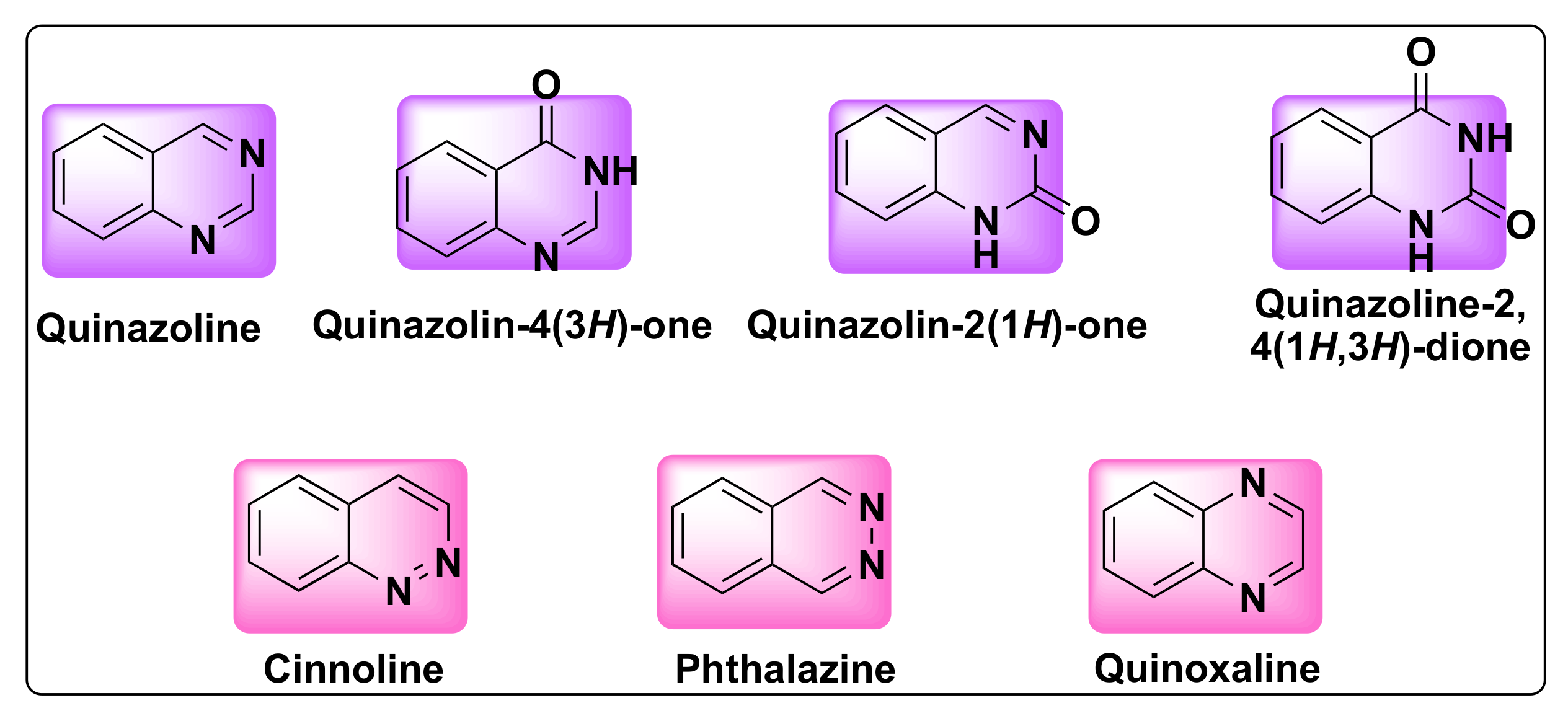
3. Structure, Isomerism, and Synthesis of Quinazolines

Quinazoline is a bicyclic heterocyclic compound formed from the fusion of two six-membered aromatic rings of benzene and pyrimidine. These compounds belong to 1,3-benzodiazine (diazonaphthalene) category which contains two nitrogen atoms. Based on the position of nitrogen atoms, the quinazoline ring can exist in four isomeric forms such as quinazoline, quinoxaline, cinnolines, and pthalazines (Figure 1). The oxidized form of quinazolines are called quinazolinones, which can be further classified into 2(1H)-quinazolinones, 4(3H)-quinazolinones, and 2,4(1H,3H)-quinazolinedione based on the position of the keto group (Figure 1) [16].
The first synthesis of quinazoline was accomplished by Bischler and Lang in the year 1895 [17]. Although these fused ring compounds were known as ‘benzo-1,3-diazine’ in the beginning of their discovery, Weddige later named them as ‘quinazolines’ in the year 1887. The discovery of febrifugine, an anti-malarial drug marked the beginning of an extensive research on the pharmacological activities of quinazolines [18]. The history of quinazoline dates back to 1869, when Griess synthesized 2-cyano-3,4-dihydro-4-oxoquinazoline, the first derivative of quinazoline. Vasicine (peganine), known for its bronchodilatory and anti-tuberculosis activity, was the first quinazoline alkaloid isolated from Justicia adhatoda [19].
Owing to its ubiquitous nature, synthetic versatility and widespread biological and pharmacological applications, numerous synthetic methods have come to light for the facile synthesis of quinazoline and its derivatives both for therapeutic and imaging purposes [15]. The most notable among them is the copper-catalyzed intramolecular N-arylation, Rhodium-catalyzed ortho-amidation, cyclocondensation, tetrabutyl ammonium fluoride promoted cyclization of N-imidoyl-o-alkynylanilines, Niementowski reaction, and benzotrizol-1-yloxytris(dimethylamino) phosphonium hexafluorophosphate (BOP)-mediated ring closures [8,17,20].

4. Quinazolines as Anti-Cancer Agents

Quinazolines are widely acclaimed for their efficiency as anti-cancer agents. Several quinazoline based kinase inhibitors such as afatinib, lapatinib, gefitinib, dacomitinib, tucatinib, and erlotinib are FDA approved for cancer treatment which prove their therapeutic efficiency as anti-tumor agents (Figure 2). Various approaches were proposed to explain the mechanism of their anti-tumor activity, which can be attributed to one or more of the following pathways: (1) Tubulin inhibition, (2) Epidermal growth factor receptor (EGFR) inhibition, (3) Inhibition of DNA damage/repair mechanism, and (4) Inhibition of thymidylate synthetase [21,22].

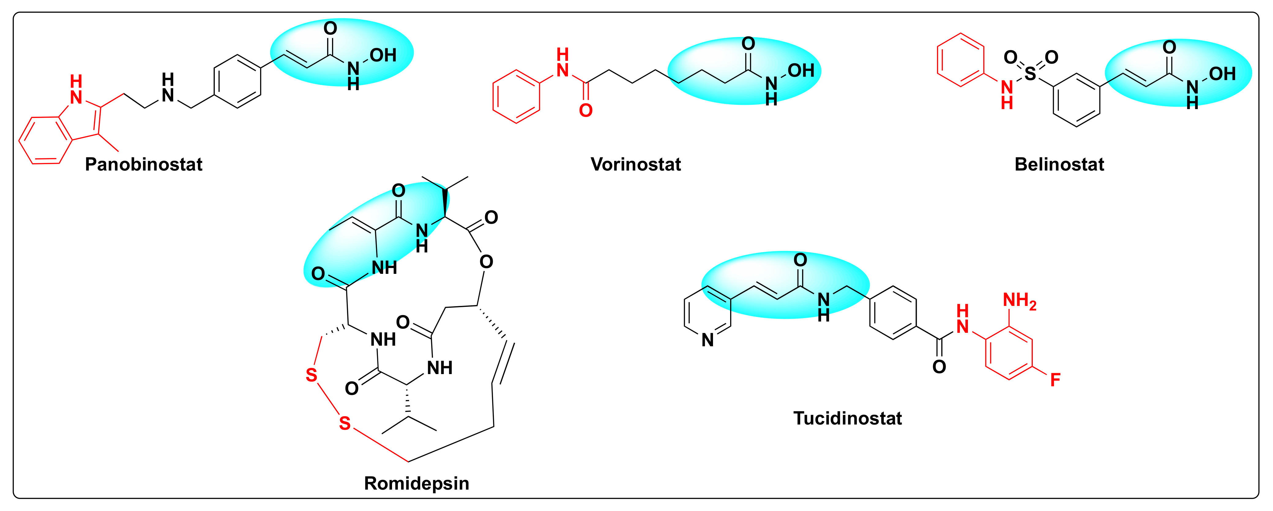
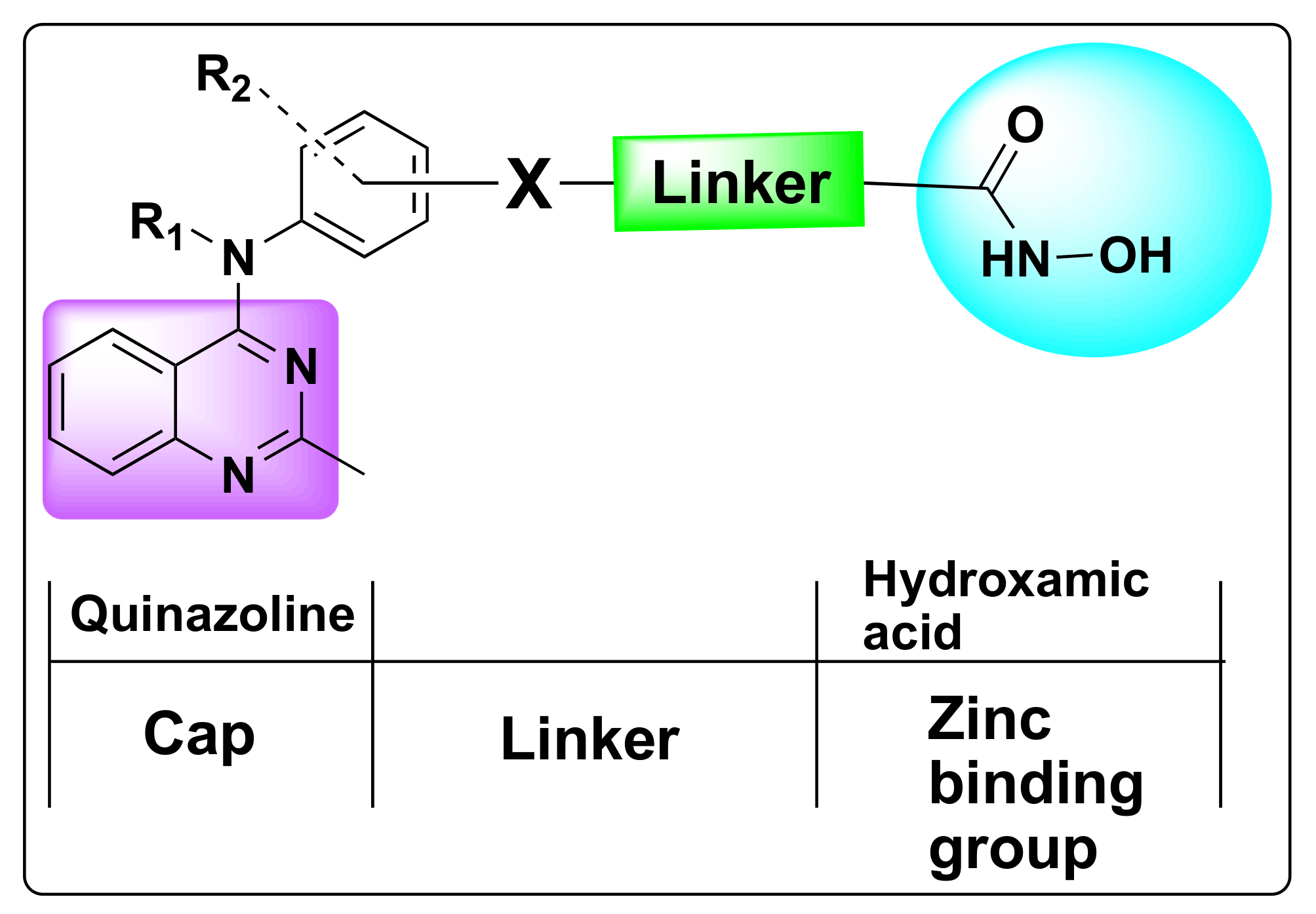
5. Histone Deacetylase Inhibition as Anti-Cancer Agents

Histone deacetylases (HDACs) were discovered by Vincent Allfrey who identified that this set of enzymes can remove acetyl groups from histones [23]. Based on the homology sequence and subcellular localization, HDACs are divided in to four classes—Class I (HDAC-1, 2, 3 and 8), Class II (HDAC-4, 5, 6, 7, 9 and 10), Class III (SERT protein 1–7), and Class IV (HDAC-11). All the HDACs are Zn+2 dependent enzymes except Class III that are NAD+ dependent and not Zn+2 dependent [24]. The fact that these enzymes are overexpressed in several human cancers and are involved in several critical biological events such as DNA damage repair, regulation of transcription, cell cycle, autophagy, and other stress responses besides deacetylation of histone and non-histone residues rendered these substrates as attractive cancer-therapeutic targets [25,26,27]. Therefore, the development of small molecule HDAC inhibitors has gained a lot of attention in the past decade [28,29,30,31]. Currently, there are five FDA approved HDAC inhibitors available for cancer treatment: Panobinostat, vorinostat (SAHA), belinostat (PXD-101), romidepsin (FK-228), and tucidinostat (HBI-8000) (Figure 3).

6. Multi-Targeted Therapy and Multi-Drug Resistance

As commonly known, drug resistance is the one of the major challenges encountered in the cancer treatment by chemotherapy [3,32]. Currently, anti-cancer drugs available in the market suffer from two major challenges: toxicity and drug resistance [33]. Based on the time of development, resistance to cancer drugs can be either intrinsic or acquired and can be the outcome of several factors. Some of them include drug metabolism, sequestration of anti-cancer drugs in lysosomes, overexpression of ATP binding cassette (ABC) transporters leading to the extrusion of anti-cancer drugs, alterations in the tumor micro-environment, genetic mutations, drug inactivation, and inhibition of apoptosis just to name a few [34,35,36]. Given the complexity and heterogeneity in the tumor invasion and progression and considering the quick adaptation of cancer drugs to any single chemotherapeutic agent, cocktail or multi-drug therapy which includes treatment with a combination of two or more drugs has become a most common approach in the recent years to treat tumor malignancies, in order to circumvent the drug resistance and improve the safety and efficacy of cancer treatment with less toxicity, multi-targeted therapy has evolved as an effective tool in the field of drug discovery [3,37,38].
However, multi-drug resistance (MDR) is another major hurdle encountered in cancer chemotherapy, which is a phenomenon in which cancer cells develop resistance to structurally or pharmacologically unrelated drugs. Several strategies were developed to overcome the MDR that include inhibition of membrane transport proteins (ABC), gene silencing by turning off the drivers of MDR, transcriptional regulation, targeted delivery of anti-cancer drugs, and hybrid/chimeric drug approaches are some of them [3,32].

7. Synergistic Effect of the Dual Inhibition/Hybrid Approach

Hybrid drugs are obtained by the fusion of one or more pharmacophore units or bioactive molecules using a molecular hybridization technique [37]. The resulting hybrid or chimeric molecules have a chemical structure different from the individual parent molecules and are connected by a labile spacer or linker. Several reports appeared in the literature which indicate that the hybrid derivatives have improved affinity, efficacy, and less toxicity compared to the parent entities. Hybrid drugs can be an alternative path to avoid MDR.
The pharmacophoric scaffold of HDAC inhibitor usually consists of a capping group, a zinc-binding group—which is most commonly hydroxamate/hydroxamic acid—and a proper linker (Figure 4). The hydroxamate group is critical for efficient binding of the small molecule inhibitor to the enzyme. However, given the multi-drug resistance and the dearth of potent HDAC inhibitor with high efficacy, development of dual HDAC inhibitors has evolved as a lucrative approach to cancer treatment [39,40,41]. Several reports came to light in the past decade highlighting the pharmacological significance of the fusion of the quinazoline pharmacophore with various FDA approved HDAC inhibitors to harness the synergistic effects of these two pharmacophoric units.
The main focus of this review is to shed light on the quinazoline based dual HDAC inhibitors with other cancer epigenetic targets. Given the pharmacological significance of quinazoline pharmacophore and the growing importance of HDAC inhibitors in cancer treatment, we herein compiled the drug design and activity of quinazoline based dual inhibitors targeting HDAC enzyme with itself and other therapeutic targets such as PI3K, EGFR, VEGFR, and G9a/GLPand will be fully discussed in the following sections.

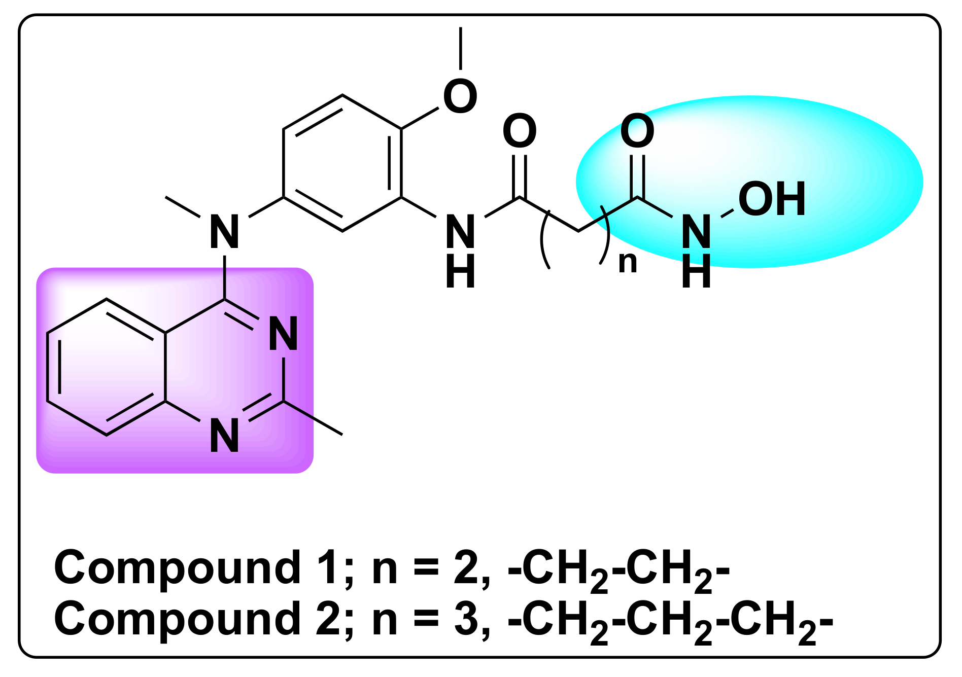
7.1. Quinazoline Based Dual Inhibition of HDAC1 and HDAC6 Enzymes

In order to develop potent dual inhibitors of HDAC1 and 6, Chen et al. designed and synthesized nearly fifty-eight quinazoline based derivatives [42]. Of all the derivatives, two compounds (1 and 2) in particular demonstrated synergistic inhibition of HDAC1 and 6 (Figure 5) [42]. Both the compounds exhibited 10-fold selectivity to other HDACs in addition to tubulin acetylation and induction of histone H3 acetylation in Hela cells. Compound 1 exhibited IC50 values of 31 nM and 16 nM against HDAC1 and 6, respectively, whereas compound 2 showed IC50 of 37 nM and 25 nM against HDAC1 and 6, respectively (Table 1).
Remarkably, both the compounds demonstrated strong anti-proliferative activity against various tumor cell lines including solid tumors (HepG2, M-M-231, MCF-7, H1975, H460, and Hela cells) with IC50 less than 40 nM values and importantly in hematological tumors’ cells (U266 and RPMI8226) with IC50 > 1 Nm (Table 2) [42].
Compound 1 in particular showed significant inhibition of tumor growth in a resistant MCF-7/ADR xenograft animal model with no significant changes in the body weight and behavior [42].

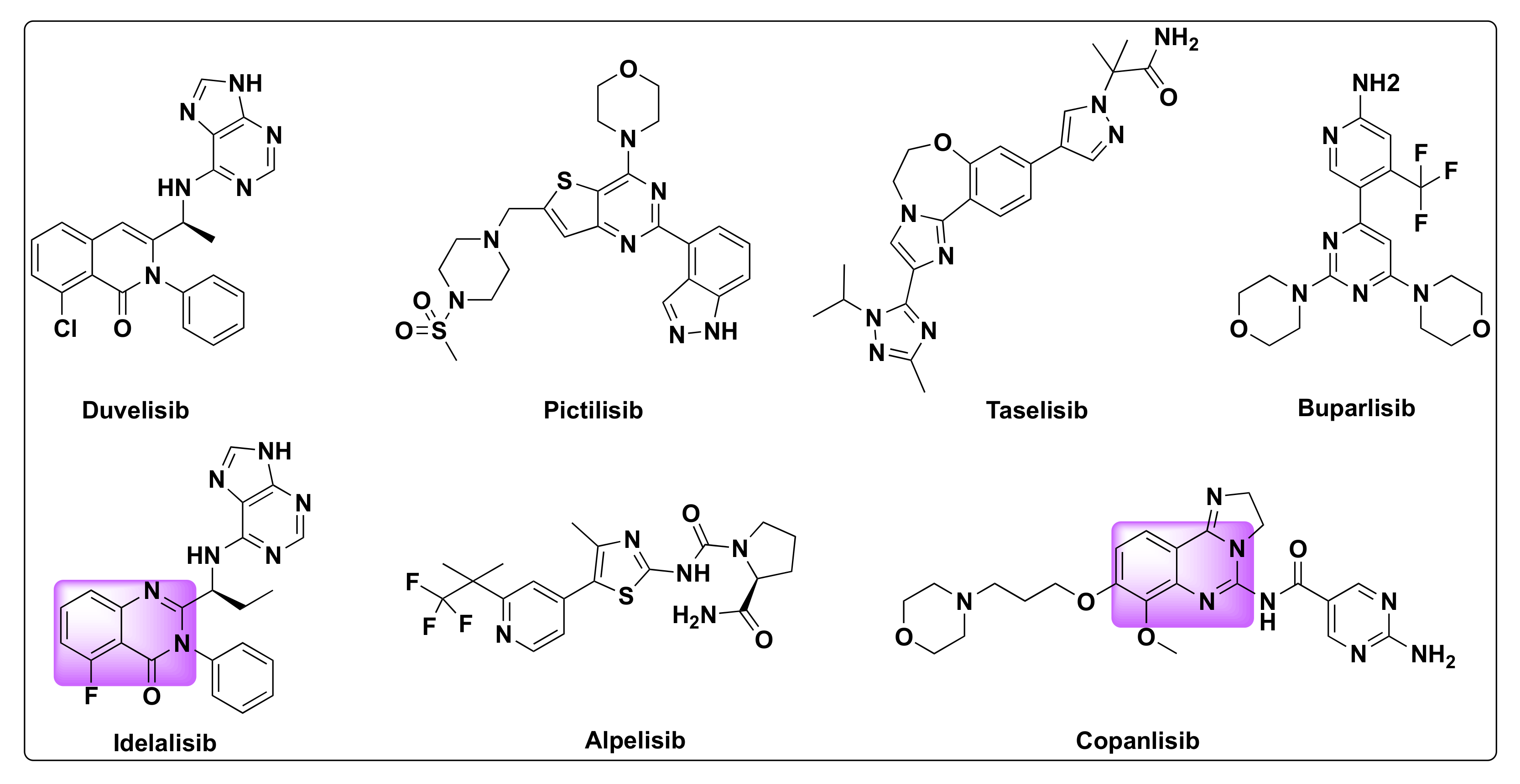
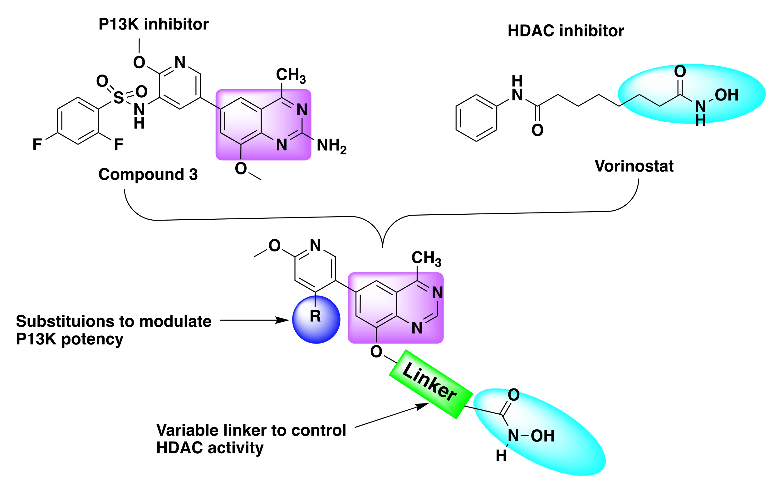
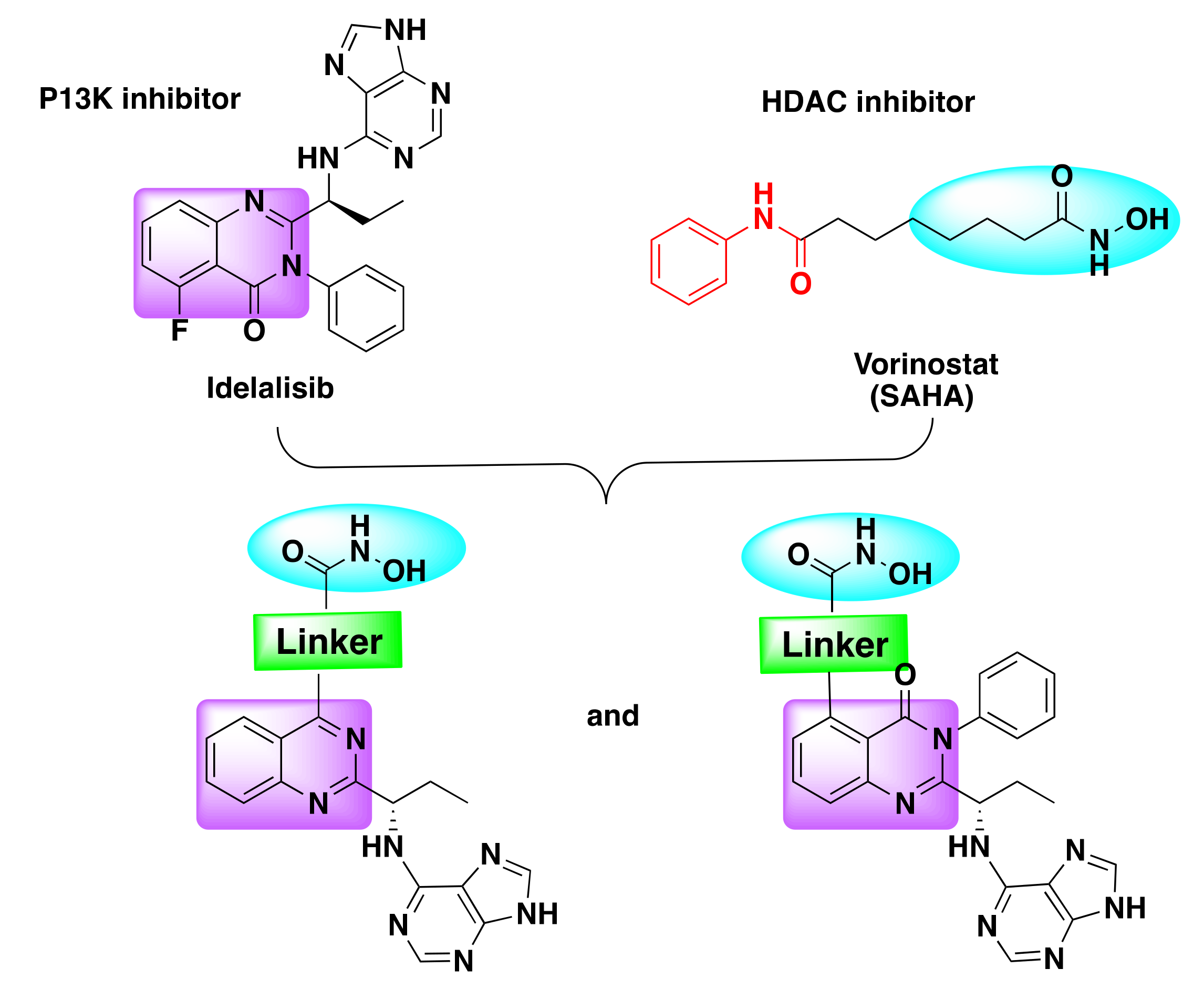
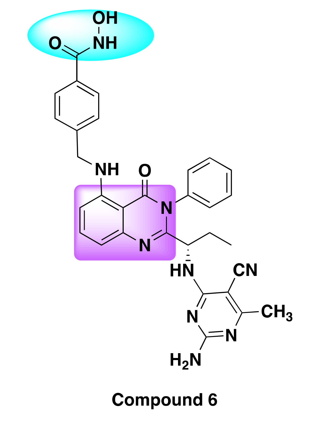
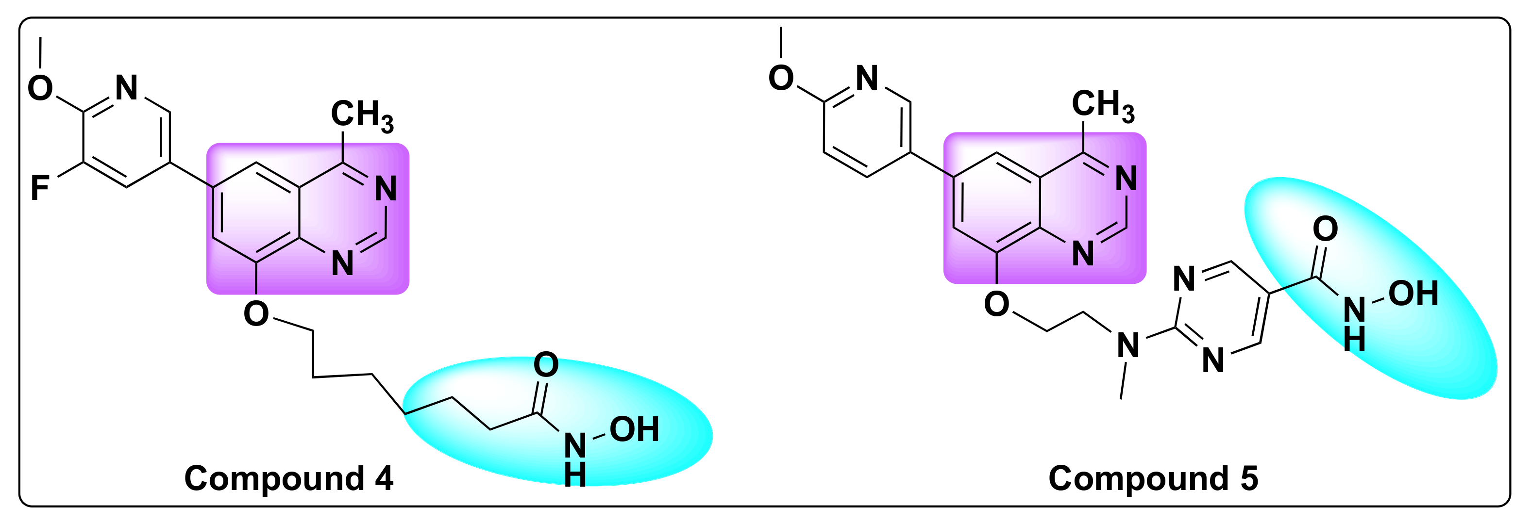
7.2. Quinazoline Based Dual Inhibition of HDAC and PI3K

Phosphoinositide 3-kinases (PI3Ks) are a class of lipid kinases that are involved in the regulation of a range of cellular events such as cell division, apoptosis, cell growth, DNA repair, angiogenesis, cell survival, and motility. These are key signaling intermediates in the PI3K/AKT/mTOR signaling pathways [43]. Dysregulation of PI3K pathway is observed in several different types of cancer making them an attractive target for the development of cancer therapeutics [44,45,46]. PI3K inhibitors find applications in the treatment of variety of human cancers such as non-small cell lung cancer (NSCLC), breast cancer, head and neck squamous cell carcinomas (HNSCC), and other solid tumors [43,47]. Duvelisib, pictilisib, taselisib, alpelisib, idelalisib, buparlisib, and copanlisib are some of the PI3K selective inhibitors in various stages of clinical development for the treatment of different types of cancers (Figure 6) [48,49].
Recently, Zhang et al. designed a series of dual PI3K and HDAC inhibitors incorporating a quinazoline based PI3K pharmacophore (compound 3) with the hydroxamic acid targeting the HDAC (Figure 7) [44,50].
The resulting hybrid scaffold can be effectively tuned to modulate the activities of PI3K and HDAC by varying the substitutions at the 3-position of methoxy pyridine of the PI3K inhibitor and the linker length of hydroxamate, respectively, as shown in Figure 7 [50].
When the resulting hybrid derivatives were tested for potency in various cancer cell lines, they found that two compounds (4 and 5)-compound 4 with an alkyl linker and compound 5 with a pyrimidine linker showed excellent HDAC and PI3K inhibition compared to the reference HDAC inhibitor SAHA and PI3K inhibitor: BKM120, respectively (Figure 8). In addition to the HDAC and PI3K inhibitor, activity was also compared to a dual HDAC/PI3K inhibitor: CUDC-907 as shown in Table 3.
Compound 5 in particular demonstrated excellent anti-proliferative activity against several cancer cell lines with IC50 values ranging from 0.05–2.7 µM range (Table 4) [50]. Both the compounds showed target modulation such as increased histone-H3 acylation and decreased p-AKT in addition to fast clearance and poor bioavailability similar to other HDAC inhibitors. On further investigation of the mechanism of action in hematologic tumors, it was revealed that compound 4 caused induction of apoptosis and G1-phase arrest in vitro and in vivo by the regulation of PI3K and HDAC signaling pathways [51].
Using a similar approach, Thakur et al. developed a series of dual PI3K and HDAC inhibitors by integration of HDAC targeting hydroxamic acid to a known PI3K inhibitor drug (idelalisib) through an optimized linker (Figure 9) [52]. The resulting dual inhibitors were identified to be potent against multiple cancer lines with excellent selectivity against PI3K γ, δ isoforms, and HDAC6 enzymes (Table 5).
SAR studies conducted against a panel of 60 cancer cell lines clearly demonstrated the selectivity and potency of most of the derivatives against PI3Kγ, δ, and HDAC6 enzymes.
Though most of the inhibitors exhibited nanomolar potency against the target enzymes (IC50 < 10 nM), one compound, notably compound 6, was found to be the best of all the derivatives, as it showed remarkable anti-proliferative activity and high potency against a variety of cancer cell lines including NSCLC, leukemia, breast cancer, renal cancer, CNS cancer, and melanoma (Table 6, Figure 10).
The potency of compound 6 was further demonstrated from the induction of cell death via necrosis in FLT3-ITD mutant and FLT3-inhibitor resistant cell lines and primary blasts from acute myeloid leukemia (AML) patients. In addition to excellent pharmacokinetic profile and the dual inhibition, compound 6 was further evaluated using cellular thermal shift assay (CETSA) in MV411 cell line.52

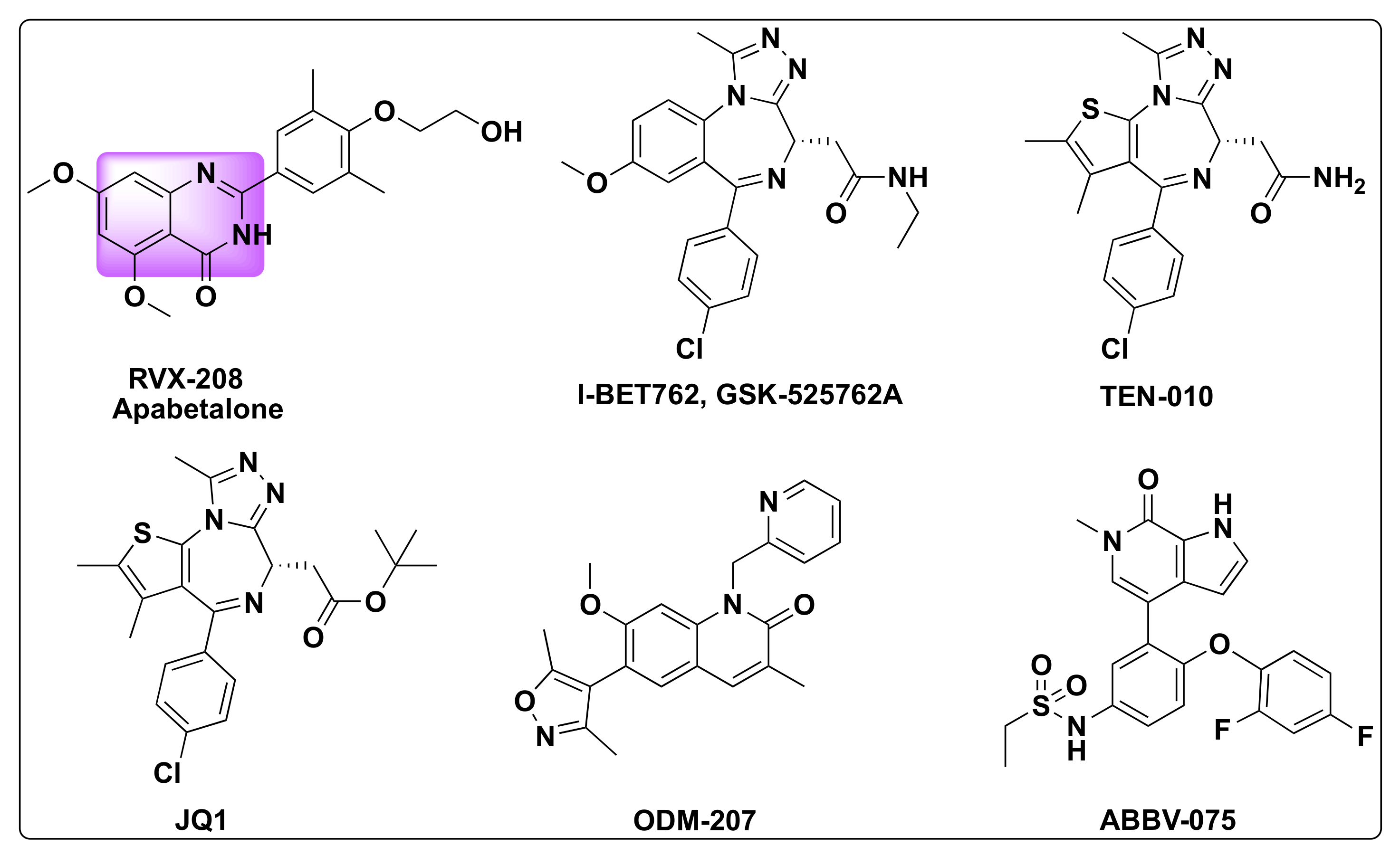
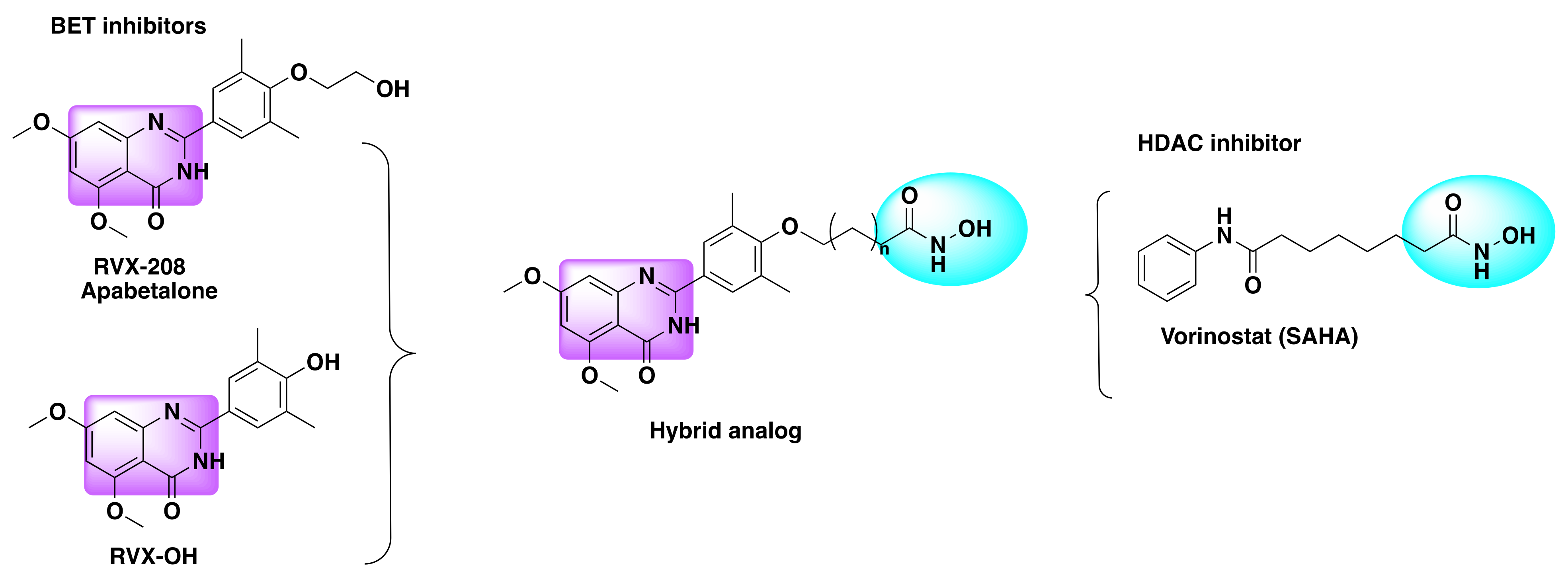
7.3. Quinazoline Based Dual Inhibition of HDAC and BET

The bromodomain and extra-terminal domain (BET) family consists of four types of proteins: bromodomain containing protein 2 (BRD2), BRD3, and BRD4 that are found to be ubiquitous while bromodomain testis specific protein (BRDT) is localized to testes and oocytes [53]. BET family proteins are an important set of proteins involved in the acetylation of histone proteins and key players in cellular events such as cell-division, cell growth, apoptosis, and in the regulation of gene expression [54,55]. BET proteins are critical in the transcription of oncogenes and their dysregulation was implicated in several diseases including cancer as a consequence of triggering of oncogene production [56,57]. Consequently, the development of inhibitors targeting BET family members has evolved as a promising approach to treat specific cancers [58,59,60,61] Several small molecule BET inhibitors were developed in the recent years with some of them even entering clinical trials for cancer therapies such as I-BET762/GSK-525762A, TEN-010, JQ1, ODM-207, ABBV-075, and a quinazolone based BET inhibitor—Apabetalone (RVX-208) developed by Resverlogix (Figure 11) [62,63].
Reports have indicated that BET and HDAC inhibitors induce similar genes and synergize to kill Myc-linked murine lymphoma [55]. In this direction, several reports came to light to harness the collaborative benefits of HDAC/BET proteins [64]. Fiskus and team found that co-treatment of BRD4 antagonist (JQ1) and HDAC inhibitor panobinostat showed promising results against human acute myeloid leukemia (AML) and the co-treatment was more effective in the induction of apoptosis than individual treatment with either of the inhibitors [65].
Based on all of the above findings, Shao and his team developed a series of dual HDAC/BRD4 inhibitors by amalgamating the structural features of RVX-208, RVX-OH, and SAHA (Figure 12) [66]. Their selection was based on the fact that RVX-208 exhibits preferential binding (over 25-fold) towards BD2 (second bromo domain of BET protein) than BD1. On the other hand, RVX-OH, although lacking the selectivity towards the BET proteins comes with an advantage to form a hydrogen bond with N140 due to the free hydroxy group of its phenyl ring that acts as an acetyl-lysine mimetic group. In order to harness the structural advantages offered by both the pharmacophores of HDAC and BRD4 inhibitors, researchers used various types of linkers to connect the zinc-binding hydroxamate group with the free hydroxy group of the phenyl ring [66].
During their studies, research team found that RVX-208 had no biological activity on HDAC1 and similarly vorinostat (SAHA) by itself showed no activity against BRET proteins (BRD4/BD2), whereas the amalgamate derivatives (7 and 8, Figure 13) showed potent activity against HDAC1 and BRD4 with IC50 values of 32 and 204 nM, respectively, against HDAC and IC50 of 225 and 401 nM against BRD4, respectively (Table 7).
Most remarkably, compound 8 exhibited more pronounced growth inhibition of AML cells compared to single inhibitors of RVX-208 or SAHA alone, with an IC50 of 0.38 µM demonstrating the potency of dual inhibitors (Table 8) [66].

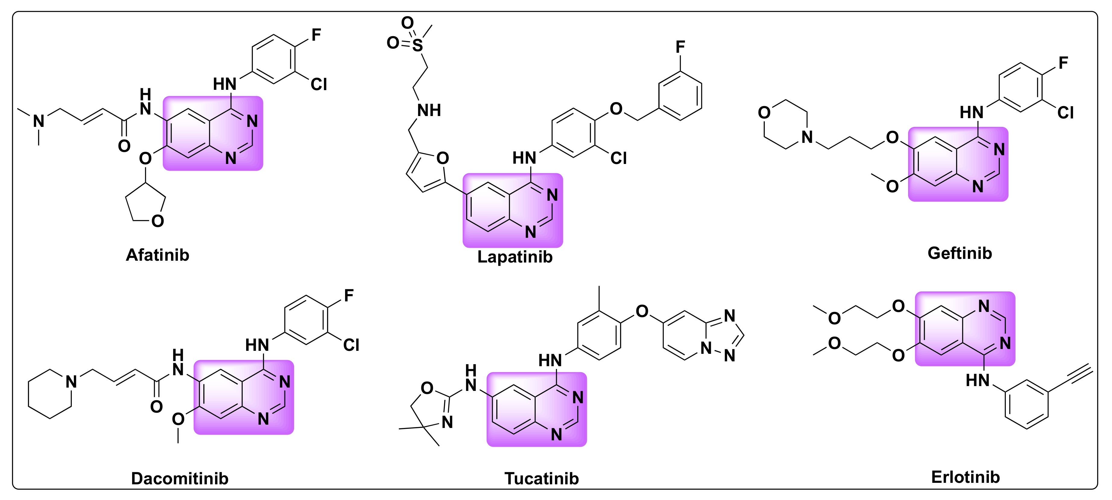
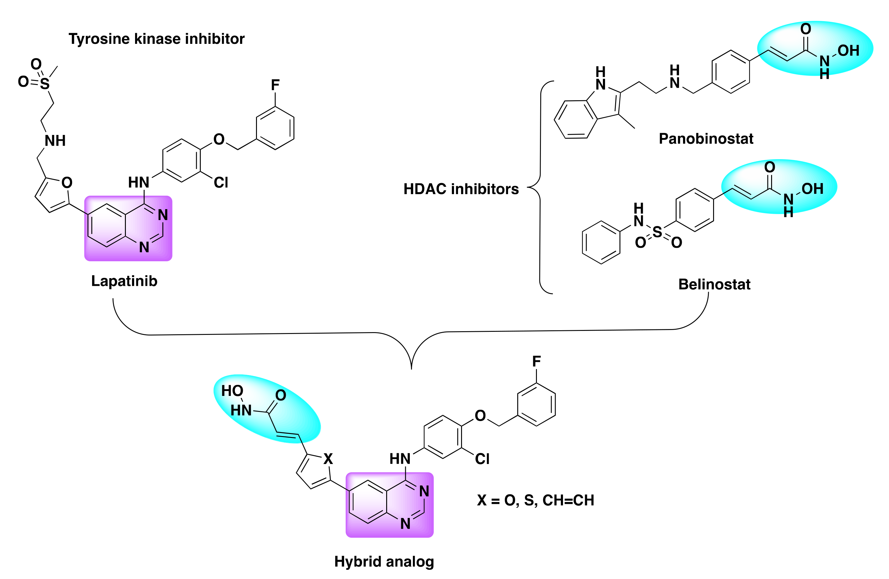
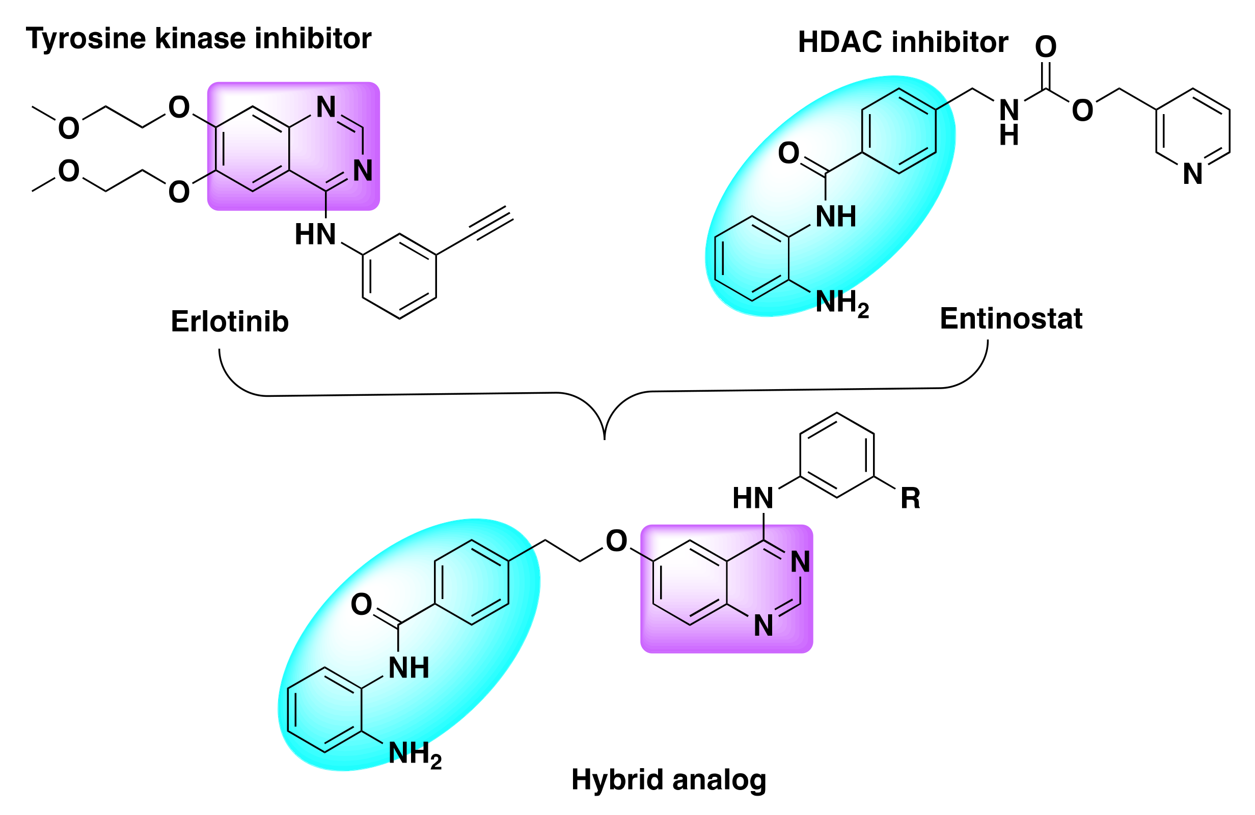
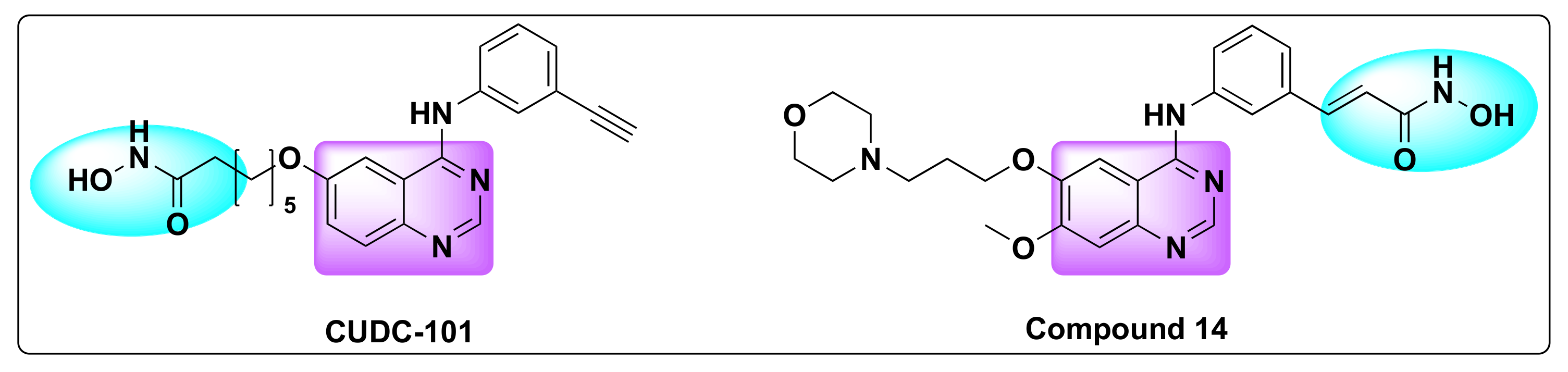
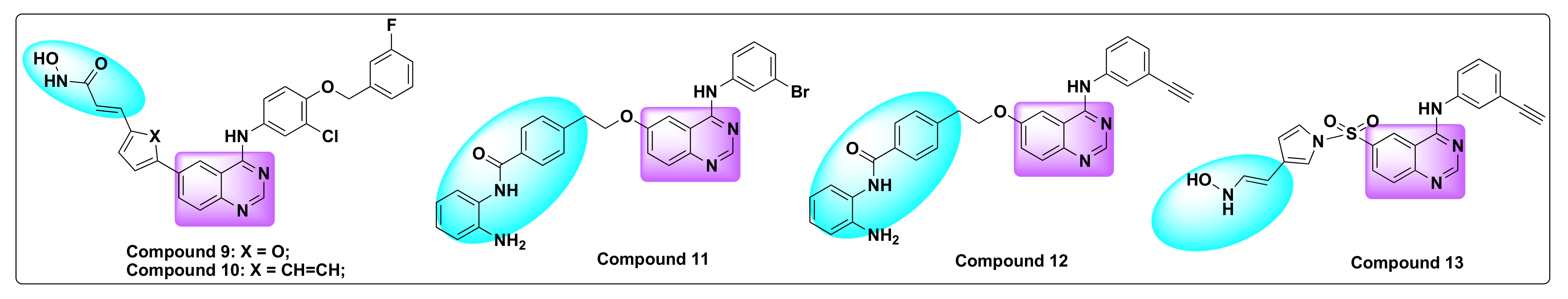
7.4. Quinazoline Based Dual Inhibition of HDAC and EGFR

Epidermal growth factor receptor (HER1/ErbB1) belongs to the ErbB receptor tyrosine kinase family [14,67]. Extracellular ligand binding results in the activation of the receptor leading to homodimerization or heterodimerization of the ErbB receptor, which ultimately activates multiple signal transduction pathways such as the mitogen activated protein kinase (MAPK) and PI3K pathways.
Dysregulation of EGFR is encountered in several types of cancers including head and neck, breast, lung cancers, prostrate, ovarian, glioblastomas, and other solid tumor malignancies [14]. Overexpression or dysregulation can be the outcome of relocation, mutations (deletions) in the EGFR gene, or aberrant signaling [68,69,70]. Consequently, small molecule inhibitors targeting EGFR have evolved as an attractive strategy to control the tumor growth and treatment of some types of cancers [22,67,71]. Extensive research is focused on the development of small molecule inhibitors and some have already been approved by FDA for the cancer treatment such as bosutinib, dasatinib, erlotinib, gefitinib, lapatinib, sunitinib, pazopanib, vemurafenib, and crizotinib (Figure 2 and Figure 14) [72]. Among these drugs, erlotinib, gefitinib, and lapatinib are the quinazoline based kinase inhibitors (Figure 2) [15,73].
Given the importance of HDAC inhibitors in arresting the cell growth, differentiation and apoptosis, the concept of dual inhibition of HDAC and EGFR is receiving attention from drug discovery scientists worldwide. Studies have demonstrated the successful design of dual inhibitors incorporating the pharmacophores of HDAC and EGFR inhibitors and obtained hybrid derivatives with strong anti-proliferative activity.
In this direction, Mahboobi et al. developed dual inhibitors of HDAC and EGFR by integrating the pharmacological activity of HDAC (Class I/II) enzyme inhibition with EGFR/HER2 kinase inhibition (Figure 15) [74].
For the drug design, their team selected hydroxamic acid and benzamide motifs as the zinc binding hydrophobic region of the HDAC inhibitors and integrated it with the 4-amino-substituted quinazoline pharmacophore to design the desired chimeric scaffolds.
As lapatinib was an established EGFR inhibitor, it was selected as a scaffold to design the dual inhibitors [75]. The resulting derivatives were tested for the activity of HDAC and protein kinase inhibition in various biochemical assays and EGFR or HER2 overexpressing cancer cell lines were further used to determine the cytotoxicity of the chimeric derivatives (Table 9 and Table 10).
Two hybrid derivatives, namely compounds 9 and 10, were identified to be best in terms of selectivity and potency of HDAC and EGFR/HER2 inhibition (Figure 16). When the activity was tested in the head and neck cancer cell lines CAL 27, compound 10 resulted in concurrent inhibition of EGFR and induction of histone H3 acetylation (Table 11).
Detailed cytotoxicity studies revealed that the chimeric inhibitors with benzamide motif were not as potent as those integrated with hydroxamic acid, as the former ones failed to induce the HDAC inhibition although they demonstrated kinase inhibition. Compound 10 with hydroxamic acid moiety demonstrated a dual biological activity with an IC50 of 0.59 μM (Table 11) [74].
The same group developed another set of dual inhibitors targeting HDAC class I and II enzymes and EGFR/HER2 kinases (Figure 17) [76]. Although the pharmacological targets remained the same as previously reported, the authors selected erlotinib in lieu of lapatinib and fused it with the hydroxamic acid and benzamide motifs from the established HDAC inhibitors to develop the chimeric inhibitors [74,77].
Further investigation of the biological activity of the resulting hybrids revealed that the derivatives 11 and 12 in which erlotinib and the benzamide moiety were separated by the methylene linker resulted in the selective inhibition of HDAC class I isoforms including the successful inhibition of nuclear and cell HDAC (Table 12, Figure 16).
When tested against nuclear and cellular HDACs, compound 11 showed IC50 of 0.25 μM and 2.46 μM, respectively, and compound 12 behaved similarly with IC50 values of 0.2 and 1.85 μM, respectively (Table 12). It is to be noted that only these two benzamide derivatives exhibited dual inhibition of HDAC and EGFR along with other tumor cell lines with IC50 in the micromolar range (Table 13).
On the other hand, if the benzamide moiety was replaced with the hydroxamic acid in the inhibitors, the resulting derivative—compound 13 successfully inhibited both HDAC I and II isoforms in the nanomolar range along with nuclear and cellular HDACs with an IC50 of 6.4 and 3.4 nM, respectively (Table 12). Remarkably, these values were less than the reference compound SAHA, indicating the potency of the hybrid analog.
In addition to the dual inhibition of HDAC and EGFR, compound 13 was found to show cytotoxicity against all the tumor cell lines (HeLa, A549, A431, Cal27, SKBR3, and SKOV3) (Table 14). Although compounds 11 and 12 were also cytotoxic against other tumor cell lines, compound 13 was identified to be more potent from its IC50 values (Table 14).
In another attempt to circumvent the limitations in cancer treatment, Cai et al. synthesized a series of multi-acting HDAC/EGFR/HER2 inhibitors, and their research culminated in the development of CUDC-101—a potent, hybrid derivative of quinazoline based anti-cancer drug, combining HDAC inhibitory pharmacophore into EGFR and human epidermal growth factor receptor 2 (HER2) (Table 15, Figure 18) [78].
CUDC-101 has undergone Phase-I clinical trials for the treatment of solid tumors and head and neck squamous cell carcinoma, alongside tumor inhibition in various types of cancers such as breast, NSCLC, liver, head and neck, and pancreatic cancers [79,80,81].
The team studied the anti-proliferative effects of CUDC-101 on human acute promyelocytic leukemia (APL) and arsenic trioxide (ATO) resistant APL cell lines using a CCK-8 assay. The IC50 of CUDC-101 was found to be 0.6–5 µM, which indicated that NB4 and HL-60 cell lines were more sensitive to CUDC-101 than ATO. In addition, the drug evaluation on ATO-resistant APL cell lines revealed that, at a concentration of 0–5 µM, the drug was able to significantly inhibit the cell proliferation of NB4/As cells in a time-dependent manner [80]. All the above findings demonstrate that CUDC-101 is more potent over ATO in APL and ATO-resistant APL cell lines.
Following the above strategy, Zhang et al. designed and synthesized a series of dual inhibitors incorporating the pharmacophores of HDAC and EGFR inhibitors (Figure 2 and Figure 18) [82]. The so-called hybrids were designed based on the EGFR inhibitors: erlotinib and gefitinib and two established HDAC inhibitors: dacinostat and belinostat (Figure 2 and Figure 3). Furthermore, the activity of the resulting hybrids was tested against HDAC, EGFR, and HER2 receptors to evaluate the potency of the derivatives (Table 16 and Table 17). Studies revealed that the hydroxamate moiety is critical for potent HDAC activity and the linker aids in the zinc binding of the hydroxamate portion.
Although one of the hybrid compounds (compound 14) showed anti-HDAC activity against HDAC 1, 2, and 6 with IC50 of 0.16, 0.18, and 0.56 µM, respectively, the same derivative was ineffective against EGFR and HER2 (Table 16) [82]. Overall, the SAR studies demonstrated that polar groups like hydroxamate (as in compound 14) at 4th position of quinazoline ring will produce a potent HDAC/HER2 dual inhibitor rather than a HDAC/EGFR chimeric derivative.
Ding et al. used the similar concept to generate hybrid derivatives by combining the pharmacophores of the kinase inhibitors such as vandetanib, neratinib, BMS-690514, and TAK-285 with the HDAC inhibitor vorinostat (Figure 19) [83].
A triazole linker was used to append vorinostat to the kinase inhibitor and inhibition studies revealed that the resulting fusion hybrids exhibited different selectivity profiles relative to their parent compounds or individual inhibitors (Table 18 and Table 19).
Notably, one compound (15) demonstrated excellent inhibition of EGFR with IC50 of 10.3 nM and HDAC1 and HDAC6 with IC50 values of 1.1 and 4.3 nM, respectively (Table 19, Figure 20).
In addition to the inhibition of EGFR phosphorylation, compound 15 successfully demonstrated induction of hyperacetylation of histone H3 on the cellular level, and it showed excellent anti-proliferative activities against five cancer cell lines (A549, BT-474, A431, SK-BR-3, and NCI-H1975) with IC50 in the range of 0.2–7.8 µM (Table 20) [83].
Following a similar strategy, Goehringer et al. recently developed novel chimeric inhibitors targeting epidermal growth factor receptors (EGFRs) and HDACs [84]. These molecules were designed by coupling the quinazoline pharmacophore of EGFR inhibitors (e.g., gefitinib) and the zinc binding group of HDAC inhibitors like SAHA. Furthermore, the anti-cancer efficiency of the resulting hybrid derivatives was tested in prostrate and hepatocellular solid cancer cell lines in addition to leukemia/lymphoma cell models (Table 21) [84].
Of all the chimeric derivatives, compounds 16 and 17 demonstrated potent inhibition of tumor cell growth with IC50 values comparable to the reference compounds (SAHA and gefitinib) and, in some cases, even better than the individual or parent pharmacophores (Figure 20). These derivatives stand apart with their remarkable anti-proliferative, anti-angiogenic effects, and potent apoptotic induction in solid tumor cell models with less cytotoxic side-effects [84].

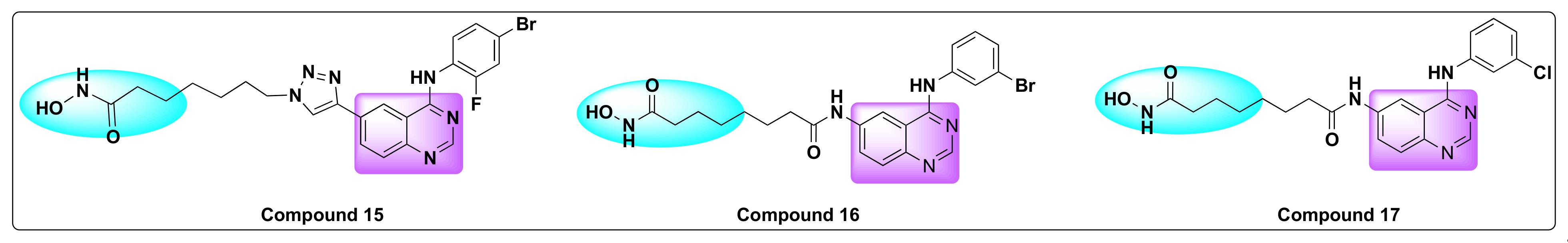
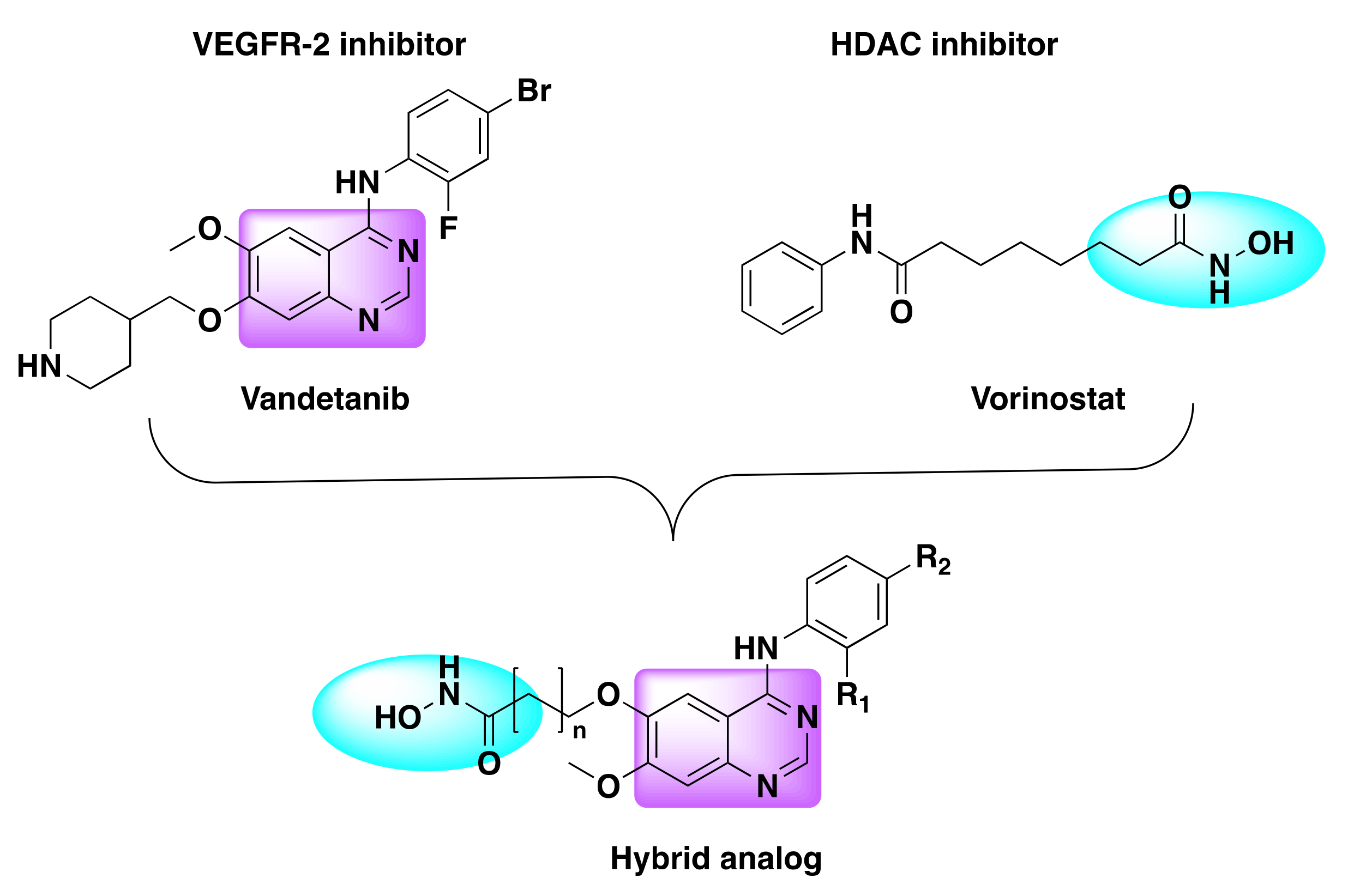
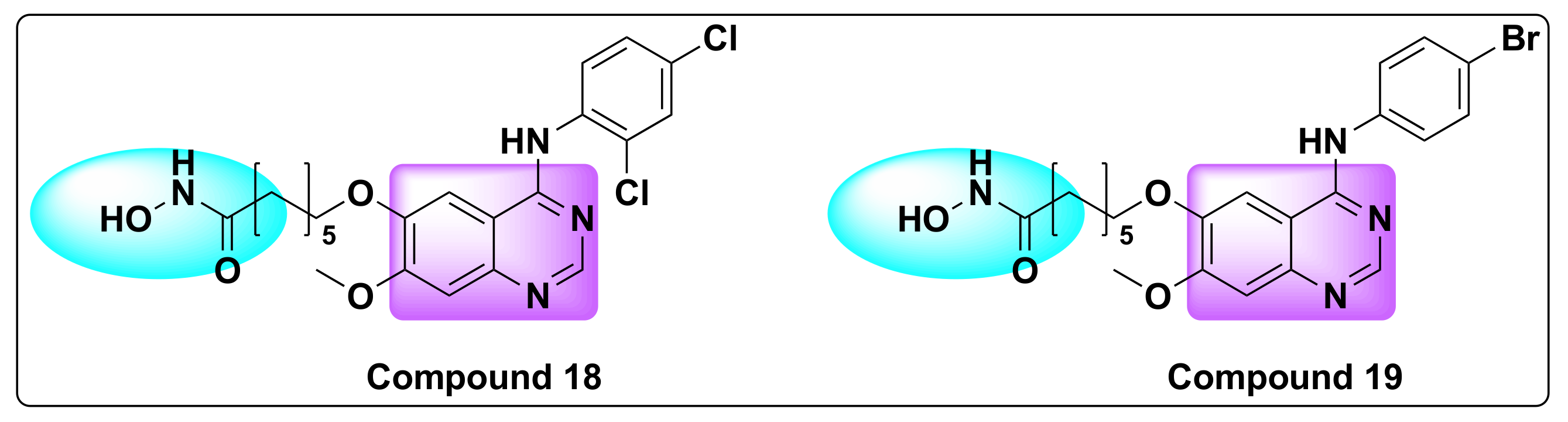
7.5. Quinazoline Based Dual Inhibition of HDAC and VEGFR

Vascular endothelial growth factor receptor (VEGFR) belongs to the family of tyrosine kinase receptors. VEGFR is involved in the regulation of both vasculogenesis and angiogenesis and is therefore not only essential for the physiological functions of normal cells but also plays a vital role in the pathophysiological conditions such as rheumatoid arthritis, inflammation, psoriasis, and in several types of cancers [85,86]. In the VEGFR family, VEGFR-2 and 3 are upregulated in most common human cancers and therefore form an attractive therapeutic target for cancer treatment. VEGFR-2 is activated by binding to VEGF-A which is essential for neovascular growth needed to support the tumor progression, while activation of VEGFR-3 induces lymphangiogenesis [87,88]. This binding triggers the signaling cascade that regulates cell survival, migration, differentiation into mature blood vessels and vasodialation.
Thus, drugs targeting VEGF signaling to inhibit angiogenesis have resulted in positive outcomes in certain cancers which include VEGF-A neutralizing antibody bevacizumab, ramucirumab, and small molecule inhibitors such as sorafenib, sunitinib, pazopanib, vandetanib, axitinib, regorafenib, cabozantinib, nintedanib, lenvatinib, and apatinib (Figure 21) [30,86,89,90]. FDA approval of these nine small molecule inhibitors demonstrates the anti-cancer efficacy of VEGFR-2 inhibitors.
Peng et al. designed and developed a series of dual hybrids by combining 4-anilinoquinazoline and hydroxamic acid targeting vascular endothelial growth factor (VEGFR) and HDAC (Figure 22) [91]. Although all the resulting hybrids were found to be potent inhibitors of HDAC and VEGFR, one compound in particular (compound 18) was identified as most potent against VEGFR-2 and HDAC enzymes with nanomolar IC50 values (Table 22, Figure 23).
In addition, compound 18 was also found to be most potent against HDAC1, HDAC2, HDAC6, and HDAC8, and showed strong anti-proliferative activity against human breast cancer cell lines—MCF-7 lines. The experimental results were substantiated with the molecular docking studies, which demonstrated that compound 18 can be a potential HDAC and VEGFR inhibitor.
The same group developed dual inhibitors targeting VEGFR and HDAC, by integrating 4-amino-N-phenyl-quinazoline and hydroxamic acid moieties (Figure 22) [92]. Among the series, compound 19 turned out to be a best dual inhibitor with IC50 of 2.2 nM against HDAC and IC50 of 74 nM against VEGFR-2 (Table 23 and Table 24, Figure 23).
In addition, compound 19 showed excellent inhibition against breast cancer cell lines (MCF-7) with IC50 of 0.85 µM (Table 23). Molecular docking studies of the compound aligned well with the experimental results, and the results showed the compound interacted well with the active binding sites of HDLP and VEGFR-2, indicating the potential of compound 18 as an anti-cancer agent [92].

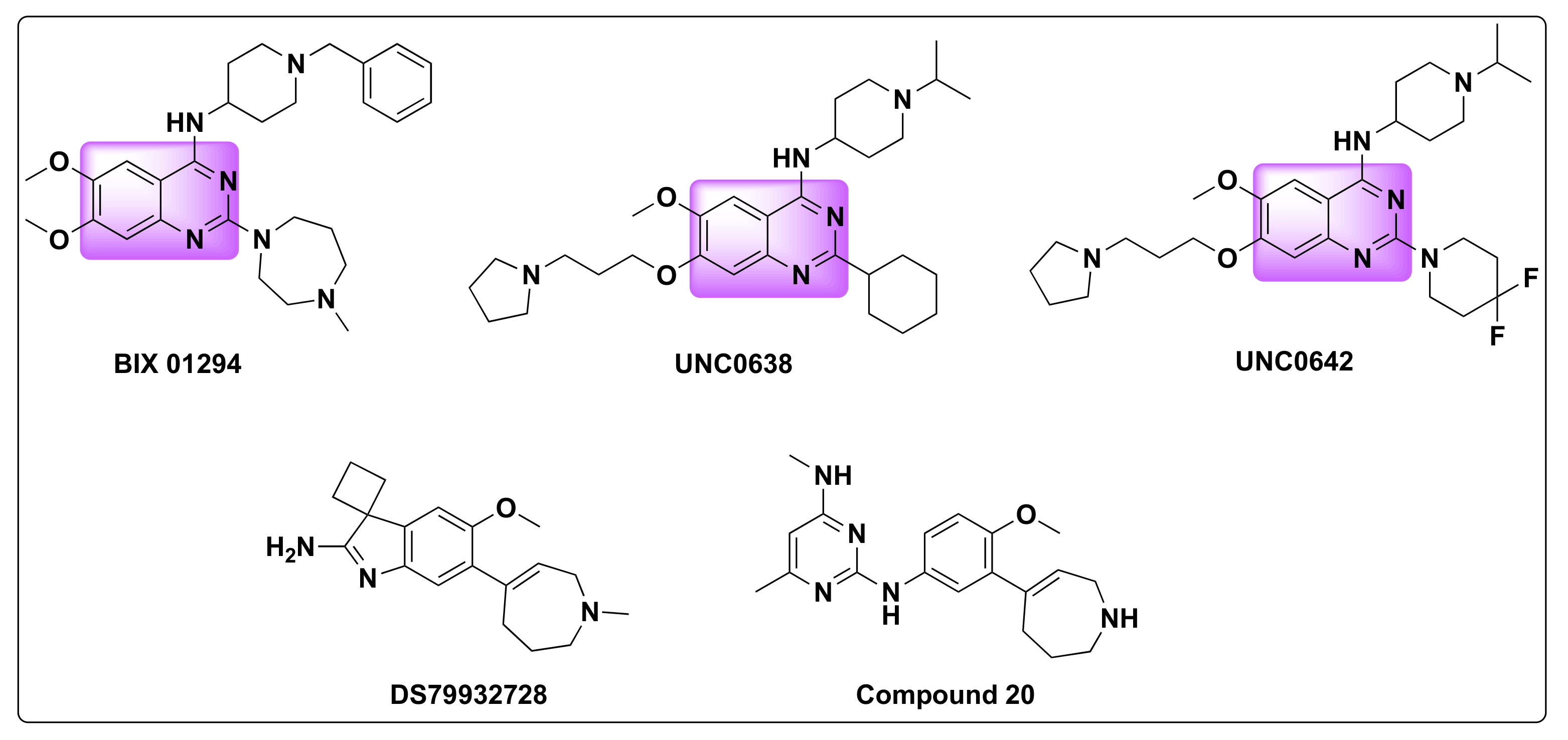
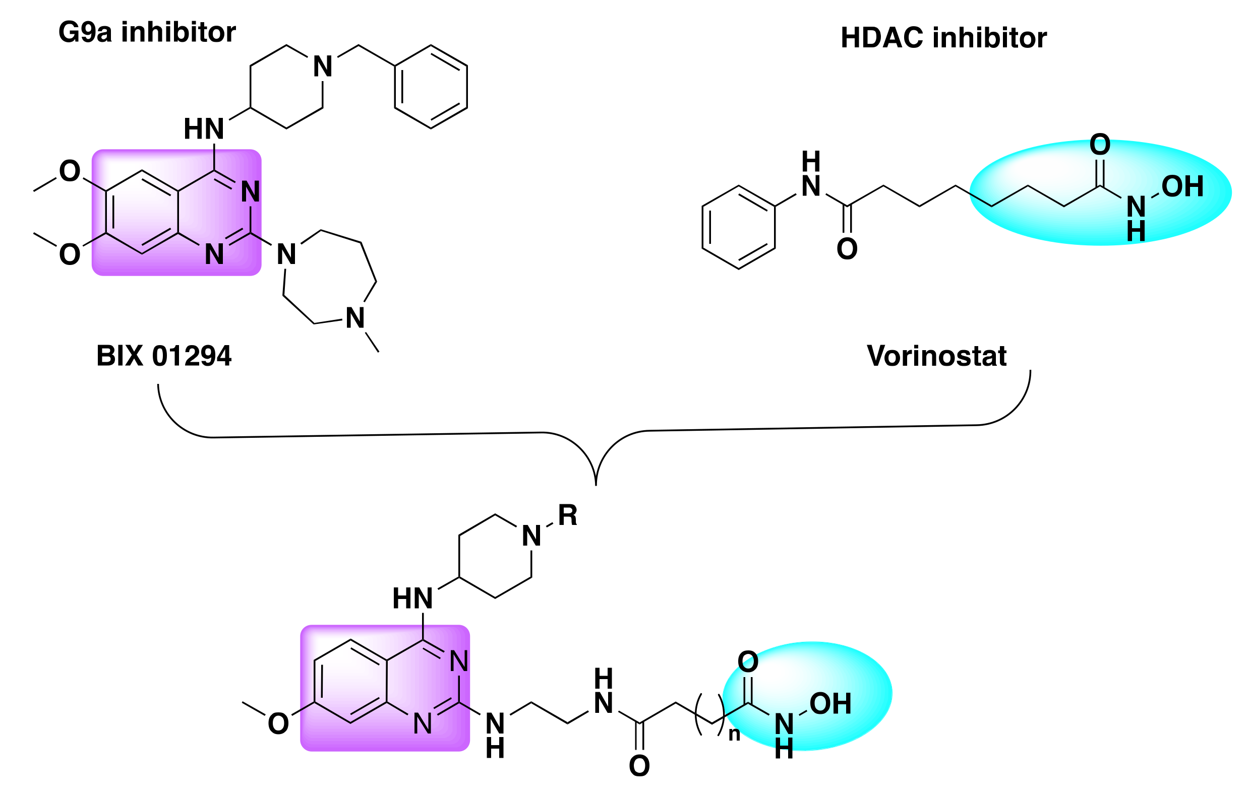
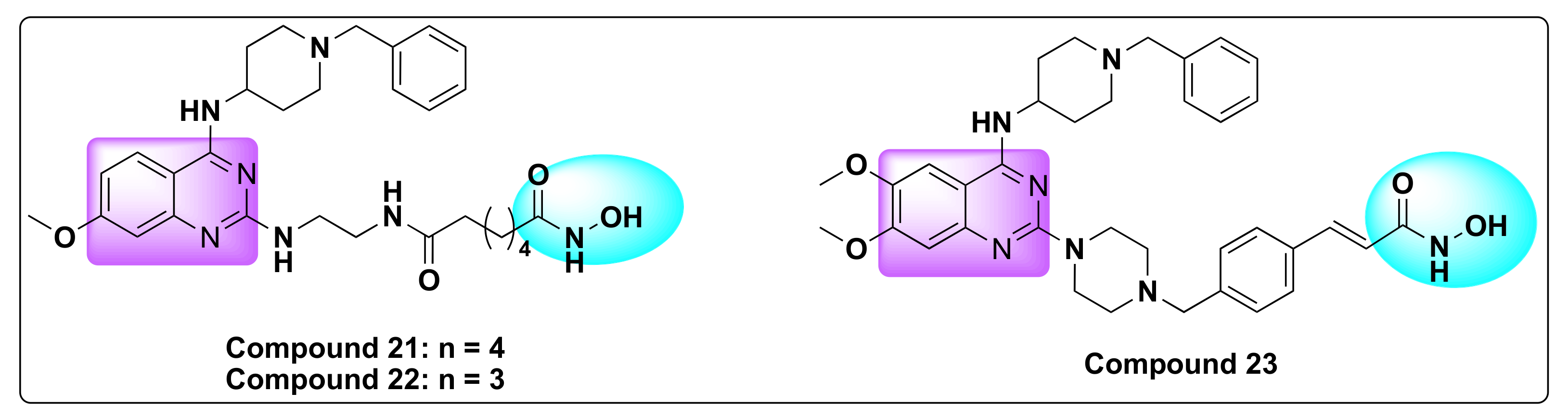
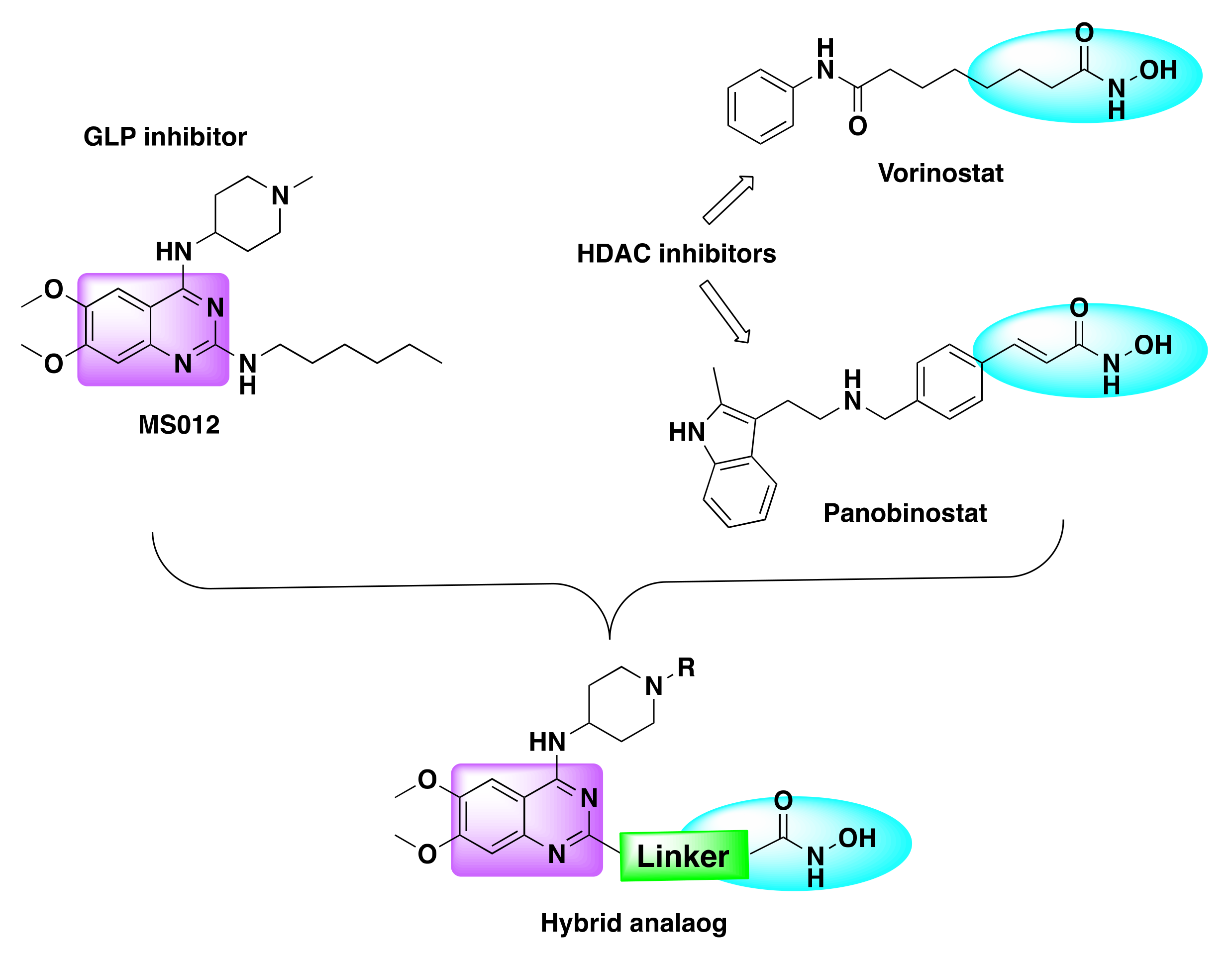
7.6. Quinazoline Based Dual Inhibitors of HDACs and Histone Methyltransferases (G9a)

Histone methylation is a complex post translational modification which involves the addition of one or more methyl groups from S-adenosylmethionine (SAM) to lysine and arginine residues on histone proteins [93]. This histone modification is a dynamic, irreversible process that is associated with chromatin structure, induction, or repression of gene expression and therefore is critical for cell development and differentiation, stress response, DNA damage repair and other important cellular events. Lysine methylation is a critical epigenetic modification regulating a number of cellular signal transduction pathways and is catalyzed by lysine methyltransferases and lysine demethylases [94]. G9a also known as lysine methyl transferase 1C or euchromatic histone lysine N-methyltransferase 2 (EHMT2) and G-9a like protein (GLP) also called as lysine methyl transferase 1D or euchromatic histone lysine N-methyltransferase (EHMT1) are critical epigenetic enzymes that catalyze the methyl transfer from SAM to the amino acid residues in histone and non-histone substrates [93,94,95,96]. Besides histone methylation, studies have shown that G9a and GLP are involved in the regulation of diverse cellular processes, DNA repair, chromatin remodeling and are overexpressed in a variety of cancers such as ovarian, lung, liver, breast, prostrate, leukemia, and colorectal cancers. Considering their unique physiological and pathophysiological functions, EHMT1/2 have emerged as cancer therapeutic targets and, as such, inhibitors targeting G9a and GLP evolved as a novel approach to cancer treatment [97,98,99]. Some of the examples of G9a/GLP inhibitors include BIX-01294, UNC0638, UNC0642, DS79932728, and compound 20 (Figure 24) [99,100,101].
Since both G9a/GLP and HDAC enzymes are overexpressed in several cancers and given their role in the tumor growth and progression, scientists developed dual inhibitors incorporating the pharmacophores of G9a/GLP and HDAC inhibitors to simultaneously block both the oncogenic targets to harvest the synergistic action of the resulting hybrids.
Zang et al. developed a series of dual inhibitors targeting HDAC and histone methyltransferases (G9a) [102]. After investigating the structural details of the HDAC and G9a inhibitors, the authors coupled the lipophilic quinazoline core with the zinc binding portion of the HDAC inhibitor using a variable linker (Figure 25).
The resulting hybrid molecules consisted of linker and hydroxamic acid at C2 and C4 position of the quinazoline ring.
In total, twenty different hybrid molecules were synthesized and cell-based assays were conducted to investigate their inhibition potential against several cell lines (Table 25).
Of all the synthesized compounds, two of them in particular, that include compounds 21 and 22, showed potent inhibition against HDAC and G9a with activity comparable to BIX-01294 (Table 26, Figure 26).
Recently, Zheng et al. designed and synthesized a series of quinazoline based dual inhibitors targeting HDAC and GLP enzymes by integrating quinazoline pharmacophore with the zinc binding element of HDAC inhibitor (Figure 27) [103].
The resulting hybrid derivatives turned out to be very potent against GLP and HDAC1/6 enzymes with nanomolar IC50 values (IC50 < 190 nM) and cytotoxic against several cancer cell lines with micromolar IC50 values (IC50 < 50 µM) (Table 27 and Table 28).
Remarkably, among all the synthesized derivatives, compound 23 proved to be highly potent with the best dual inhibition ability demonstrated with an IC50 of 89 and 13 nM against HDAC1 and 6, respectively, and IC50 of 1.3 nM against GLP (Table 28, Figure 26).
In addition to the potent anti-proliferative effect against cancer cell lines, compound 23 was also capable of inhibition of HDAC and GLP on the protein level, G0/G1 arrest in HepG2cells, and induction of apoptosis in cancer cells.

8. Conclusions

Owing to the limitations and hurdles in the single drug therapy of cancer, the multi drug approach has evolved as an attractive tool for the drug discovery scientists to fight the battle against cancer. Given the tendency of cancer cell to bypass or outsmart any treatment, the technique of combination therapy is the most common method of treatment used in cancer chemotherapy. However, toxicity and undesired side effects from the use of multi-drugs is a major shortcoming with chemotherapy. In this context, the concept of molecular hybridization which involves the combination of two or more pharmacophores to obtain a hybrid drug has gained lot of attention in the past decade.
Despite the challenges involved in the design of the hybrid drugs like retaining the selectivity, pharmacological activity, and avoiding unfavorable drug–drug interactions, in most cases, hybrid drugs often proved to be more potent and showed high efficacy compared to the parent or individual pharmacophores. FDA approved quinazoline based drugs are reported to be successful in controlling the tumor growth and progression in several clinical studies. As the number of HDAC inhibitors gaining FDA approval for the treatment of various cancers has increased in the recent years, the combination of quinazoline and HDAC pharmacophores appears to be a promising approach to produce effective anti-cancer drugs. As suggested from different examples of dual HDAC inhibitors and their successful experimental results presented in this review, quinazoline based HDAC dual inhibitors show great potential to enter the clinical trials and soon emerge as effective anti-cancer tools.

Funding

This research received no external funding.

Institutional Review Board Statement

Not applicable.

Informed Consent Statement

Not applicable.

Data Availability Statement

Not applicable.

Conflicts of Interest

The authors declare no conflict of interest.

References

  1. Sung, H.; Ferlay, J.; Siegel, R.L.; Laversanne, M.; Soerjomataram, I.; Jemal, A.; Bray, F. Global Cancer Statistics 2020: GLOBOCAN Estimates of Incidence and Mortality Worldwide for 36 Cancers in 185 Countries. CA Cancer J. Clin. 2021, 71, 209–249. [Google Scholar] [CrossRef] [PubMed]
  2. International Agency for Research on Cancer. Available online: https://gco.iarc.fr/today/data/factsheets/cancers/20-Breast-fact-sheet.pdf (accessed on 9 March 2022).
  3. Vasan, N.; Baselga, J.; Hyman, D.M. A View on Drug Resistance in Cancer. Nature 2019, 575, 299–309. [Google Scholar] [CrossRef] [PubMed] [Green Version]
  4. Hameed, A.; Al-Rashida, M.; Uroos, M.; Ali, S.A.; Arshia; Ishtiaq, M.; Khan, K.M. Quinazoline and Quinazolinone as Important Medicinal Scaffolds: A Comparative Patent Review (2011–2016). Expert Opin. Ther. Pat. 2018, 28, 281–297. [Google Scholar] [CrossRef] [PubMed]
  5. Dhuguru, J.; Zviagin, E.; Skouta, R. FDA-Approved Oximes and Their Significance in Medicinal Chemistry. Pharmaceuticals 2022, 15, 66. [Google Scholar] [CrossRef] [PubMed]
  6. Ghoneim, O.M.; Bill, A.; Dhuguru, J.; Szollosi, D.E.; Edafiogho, I.O. Design, Synthesis and Biological Evaluation of Piperazino-Enaminones as Novel Suppressants of pro-Inflammatory Cytokines. Bioorgan. Med. Chem. 2018, 26, 3890–3898. [Google Scholar] [CrossRef]
  7. Szollosi, D.E.; Ghoneim, O.A.M.; Manzoor, M.K.; Dhuguru, J.; Edafiogho, I.O. Novel Piperazino-Enaminones Suppress Pro-Inflammatory Cytokines and Inhibit Chemokine Receptor CCR2. Inflammation 2016, 39, 2053–2061. [Google Scholar] [CrossRef]
  8. Khan, I.; Zaib, S.; Batool, S.; Abbas, N.; Ashraf, Z.; Iqbal, J.; Saeed, A. Quinazolines and Quinazolinones as Ubiquitous Structural Fragments in Medicinal Chemistry: An Update on the Development of Synthetic Methods and Pharmacological Diversification. Bioorgan. Med. Chem. 2016, 24, 2361–2381. [Google Scholar] [CrossRef]
  9. Karan, R.; Agarwal, P.; Sinha, M.; Mahato, N. Recent Advances on Quinazoline Derivatives: A Potential Bioactive Scaffold in Medicinal Chemistry. ChemEngineering 2021, 5, 73. [Google Scholar] [CrossRef]
  10. El-Azab, A.S.; ElTahir, K.E.H. Design and Synthesis of Novel 7-Aminoquinazoline Derivatives: Antitumor and Anticonvulsant Activities. Bioorgan. Med. Chem. Lett. 2012, 22, 1879–1885. [Google Scholar] [CrossRef]
  11. Liu, T.; Peng, F.; Cao, X.; Liu, F.; Wang, Q.; Liu, L.; Xue, W. Design, Synthesis, Antibacterial Activity, Antiviral Activity, and Mechanism of Myricetin Derivatives Containing a Quinazolinone Moiety. ACS Omega 2021, 6, 30826–30833. [Google Scholar] [CrossRef]
  12. Wang, H.-X.; Liu, H.-Y.; Li, W.; Zhang, S.; Wu, Z.; Li, X.; Li, C.-W.; Liu, Y.-M.; Chen, B.-Q. Design, Synthesis, Antiproliferative and Antibacterial Evaluation of Quinazolinone Derivatives. Med. Chem. Res. 2019, 28, 203–214. [Google Scholar] [CrossRef]
  13. Shagufta, S.; Ahmad, I. An Insight into the Therapeutic Potential of Quinazoline Derivatives as Anticancer Agents. Med. Chem. Commun. 2017, 8, 871–885. [Google Scholar] [CrossRef] [PubMed]
  14. Sicard, R.; Dhuguru, J.; Liu, W.; Patel, N.; Landgraf, R.; Wilson, J.N. A Fluorescent Reporter of ATP Binding-Competent Receptor Kinases. Bioorgan. Med. Chem. Lett. 2012, 22, 5532–5535. [Google Scholar] [CrossRef] [PubMed]
  15. Dhuguru, J.; Liu, W.; Gonzalez, W.G.; Babinchak, W.M.; Miksovska, J.; Landgraf, R.; Wilson, J.N. Emission Tuning of Fluorescent Kinase Inhibitors: Conjugation Length and Substituent Effects. J. Org. Chem. 2014, 79, 4940–4947. [Google Scholar] [CrossRef] [PubMed]
  16. Wdowiak, P.; Matysiak, J.; Kuszta, P.; Czarnek, K.; Niezabitowska, E.; Baj, T. Quinazoline Derivatives as Potential Therapeutic Agents in Urinary Bladder Cancer Therapy. Front. Chem. 2021, 9, 765552. [Google Scholar] [CrossRef]
  17. Asif, M. Chemical Characteristics, Synthetic Methods, and Biological Potential of Quinazoline and Quinazolinone Derivatives. Int. J. Med. Chem. 2014, 2014, 395637. [Google Scholar] [CrossRef]
  18. Fishman, M.; Cruickshank, P.A. Febrifugine Antimalarial Agents. I. Pyridine Analogs of Febrifugine. J. Med. Chem. 1970, 13, 155–156. [Google Scholar] [CrossRef]
  19. Sharma, S.; Kumar, M.; Kumar, V.; Kumar, N. Metal-Free Transfer Hydrogenation of Nitroarenes in Water with Vasicine: Revelation of Organocatalytic Facet of an Abundant Alkaloid. J. Org. Chem. 2014, 79, 9433–9439. [Google Scholar] [CrossRef]
  20. Faisal, M.; Saeed, A. Chemical Insights into the Synthetic Chemistry of Quinazolines: Recent Advances. Front. Chem. 2021, 8, 594717. [Google Scholar] [CrossRef]
  21. Jafari, E.; Khajouei, M.R.; Hassanzadeh, F.; Hakimelahi, G.H.; Khodarahmi, G.A. Quinazolinone and Quinazoline Derivatives: Recent Structures with Potent Antimicrobial and Cytotoxic Activities. Res. Pharm. Sci. 2016, 11, 1–14. [Google Scholar]
  22. Mirgany, T.O.; Abdalla, A.N.; Arifuzzaman, M.; Motiur Rahman, A.F.M.; Al-Salem, H.S. Quinazolin-4(3H)-One Based Potential Multiple Tyrosine Kinase Inhibitors with Excellent Cytotoxicity. J. Enzym. Inhib. Med. Chem. 2021, 36, 2055–2067. [Google Scholar] [CrossRef] [PubMed]
  23. Mukhopadhyay, R. Vincent Allfrey’s Work on Histone Acetylation. J. Biol. Chem. 2012, 287, 2270–2271. [Google Scholar] [CrossRef] [Green Version]
  24. Hesham, H.M.; Lasheen, D.S.; Abouzid, K.A.M. Chimeric HDAC Inhibitors: Comprehensive Review on the HDAC-Based Strategies Developed to Combat Cancer. Med. Res. Rev. 2018, 38, 2058–2109. [Google Scholar] [CrossRef] [PubMed]
  25. Li, G.; Tian, Y.; Zhu, W.-G. The Roles of Histone Deacetylases and Their Inhibitors in Cancer Therapy. Front. Cell Dev. Biol. 2020, 8, 1004. [Google Scholar] [CrossRef] [PubMed]
  26. Wilson, A.J.; Byun, D.-S.; Popova, N.; Murray, L.B.; L’Italien, K.; Sowa, Y.; Arango, D.; Velcich, A.; Augenlicht, L.H.; Mariadason, J.M. Histone Deacetylase 3 (HDAC3) and Other Class I HDACs Regulate Colon Cell Maturation and P21 Expression and Are Deregulated in Human Colon Cancer. J. Biol. Chem. 2006, 281, 13548–13558. [Google Scholar] [CrossRef] [Green Version]
  27. Choi, J.-H.; Kwon, H.J.; Yoon, B.-I.; Kim, J.-H.; Han, S.U.; Joo, H.J.; Kim, D.-Y. Expression Profile of Histone Deacetylase 1 in Gastric Cancer Tissues. Jpn. J. Cancer Res. 2001, 92, 1300–1304. [Google Scholar] [CrossRef] [PubMed]
  28. Al-Janadi, A.; Chandana, S.R.; Conley, B.A. Histone Deacetylation: An Attractive Target for Cancer Therapy? Drugs R D 2008, 9, 369–383. [Google Scholar] [CrossRef]
  29. Eckschlager, T.; Plch, J.; Stiborova, M.; Hrabeta, J. Histone Deacetylase Inhibitors as Anticancer Drugs. IJMS 2017, 18, 1414. [Google Scholar] [CrossRef] [PubMed]
  30. Dhuguru, J.; Skouta, R. Role of Indole Scaffolds as Pharmacophores in the Development of Anti-Lung Cancer Agents. Molecules 2020, 25, 1615. [Google Scholar] [CrossRef] [Green Version]
  31. Smalley, J.P.; Cowley, S.M.; Hodgkinson, J.T. Bifunctional HDAC Therapeutics: One Drug to Rule Them All? Molecules 2020, 25, 4394. [Google Scholar] [CrossRef]
  32. Persidis, A. Cancer Multidrug Resistance. Nat. Biotechnol. 2000, 18, IT18–IT20. [Google Scholar] [CrossRef]
  33. Wang, X.; Zhang, H.; Chen, X. Drug Resistance and Combating Drug Resistance in Cancer. Cancer Drug Resist. 2019, 2, 141–160. [Google Scholar] [CrossRef] [PubMed] [Green Version]
  34. Dallavalle, S.; Dobričić, V.; Lazzarato, L.; Gazzano, E.; Machuqueiro, M.; Pajeva, I.; Tsakovska, I.; Zidar, N.; Fruttero, R. Improvement of Conventional Anti-Cancer Drugs as New Tools against Multidrug Resistant Tumors. Drug Resist. Updates 2020, 50, 100682. [Google Scholar] [CrossRef] [PubMed]
  35. Gottesman, M.M. Mechanisms of Cancer Drug Resistance. Annu. Rev. Med. 2002, 53, 615–627. [Google Scholar] [CrossRef] [Green Version]
  36. Ward, R.A.; Fawell, S.; Floc’h, N.; Flemington, V.; McKerrecher, D.; Smith, P.D. Challenges and Opportunities in Cancer Drug Resistance. Chem. Rev. 2021, 121, 3297–3351. [Google Scholar] [CrossRef]
  37. Szumilak, M.; Wiktorowska-Owczarek, A.; Stanczak, A. Hybrid Drugs—A Strategy for Overcoming Anticancer Drug Resistance? Molecules 2021, 26, 2601. [Google Scholar] [CrossRef]
  38. Teiten, M.-H.; Dicato, M.; Diederich, M. Hybrid Curcumin Compounds: A New Strategy for Cancer Treatment. Molecules 2014, 19, 20839–20863. [Google Scholar] [CrossRef]
  39. Cheng, J.; Qin, J.; Guo, S.; Qiu, H.; Zhong, Y. Design, Synthesis and Evaluation of Novel HDAC Inhibitors as Potential Antitumor Agents. Bioorgan. Med. Chem. Lett. 2014, 24, 4768–4772. [Google Scholar] [CrossRef]
  40. Zhang, Y.; Feng, J.; Jia, Y.; Wang, X.; Zhang, L.; Liu, C.; Fang, H.; Xu, W. Development of Tetrahydroisoquinoline-Based Hydroxamic Acid Derivatives: Potent Histone Deacetylase Inhibitors with Marked in Vitro and in Vivo Antitumor Activities. J. Med. Chem. 2011, 54, 2823–2838. [Google Scholar] [CrossRef]
  41. Wu, Y.-W.; Chao, M.-W.; Tu, H.-J.; Chen, L.-C.; Hsu, K.-C.; Liou, J.-P.; Yang, C.-R.; Yen, S.-C.; HuangFu, W.-C.; Pan, S.-L. A Novel Dual HDAC and HSP90 Inhibitor, MPT0G449, Downregulates Oncogenic Pathways in Human Acute Leukemia in Vitro and in Vivo. Oncogenesis 2021, 10, 39. [Google Scholar] [CrossRef]
  42. Chen, J.; Sang, Z.; Jiang, Y.; Yang, C.; He, L. Design, Synthesis, and Biological Evaluation of Quinazoline Derivatives as Dual HDAC1 and HDAC6 Inhibitors for the Treatment of Cancer. Chem. Biol Drug Des. 2019, 93, 232–241. [Google Scholar] [CrossRef]
  43. Akinleye, A.; Avvaru, P.; Furqan, M.; Song, Y.; Liu, D. Phosphatidylinositol 3-Kinase (PI3K) Inhibitors as Cancer Therapeutics. J. Hematol. Oncol. 2013, 6, 88. [Google Scholar] [CrossRef] [PubMed] [Green Version]
  44. Lin, S.; Wang, C.; Ji, M.; Wu, D.; Lv, Y.; Zhang, K.; Dong, Y.; Jin, J.; Chen, J.; Zhang, J.; et al. Discovery and Optimization of 2-Amino-4-Methylquinazoline Derivatives as Highly Potent Phosphatidylinositol 3-Kinase Inhibitors for Cancer Treatment. J. Med. Chem. 2018, 61, 6087–6109. [Google Scholar] [CrossRef] [PubMed]
  45. Vivanco, I.; Sawyers, C.L. The Phosphatidylinositol 3-Kinase–AKT Pathway in Human Cancer. Nat. Rev. Cancer 2002, 2, 489–501. [Google Scholar] [CrossRef] [PubMed]
  46. Engelman, J.A. Targeting PI3K Signalling in Cancer: Opportunities, Challenges and Limitations. Nat. Rev. Cancer 2009, 9, 550–562. [Google Scholar] [CrossRef] [PubMed]
  47. Vanhaesebroeck, B.; Perry, M.W.D.; Brown, J.R.; André, F.; Okkenhaug, K. PI3K Inhibitors Are Finally Coming of Age. Nat. Rev. Drug Discov. 2021, 20, 741–769. [Google Scholar] [CrossRef] [PubMed]
  48. Yang, J.; Nie, J.; Ma, X.; Wei, Y.; Peng, Y.; Wei, X. Targeting PI3K in Cancer: Mechanisms and Advances in Clinical Trials. Mol. Cancer 2019, 18, 26. [Google Scholar] [CrossRef] [Green Version]
  49. Markham, A. Copanlisib: First Global Approval. Drugs 2017, 77, 2057–2062. [Google Scholar] [CrossRef]
  50. Zhang, K.; Lai, F.; Lin, S.; Ji, M.; Zhang, J.; Zhang, Y.; Jin, J.; Fu, R.; Wu, D.; Tian, H.; et al. Design, Synthesis, and Biological Evaluation of 4-Methyl Quinazoline Derivatives as Anticancer Agents Simultaneously Targeting Phosphoinositide 3-Kinases and Histone Deacetylases. J. Med. Chem. 2019, 62, 6992–7014. [Google Scholar] [CrossRef]
  51. Yan, Z.; Zhang, K.; Ji, M.; Xu, H.; Chen, X. A Dual PI3K/HDAC Inhibitor Downregulates Oncogenic Pathways in Hematologic Tumors In Vitro and In Vivo. Front. Pharmacol. 2021, 12, 741697. [Google Scholar] [CrossRef]
  52. Thakur, A.; Tawa, G.J.; Henderson, M.J.; Danchik, C.; Liu, S.; Shah, P.; Wang, A.Q.; Dunn, G.; Kabir, M.; Padilha, E.C.; et al. Design, Synthesis, and Biological Evaluation of Quinazolin-4-One-Based Hydroxamic Acids as Dual PI3K/HDAC Inhibitors. J. Med. Chem. 2020, 63, 4256–4292. [Google Scholar] [CrossRef] [PubMed]
  53. Wang, N.; Wu, R.; Tang, D.; Kang, R. The BET Family in Immunity and Disease. Signal Transduct. Target. Ther. 2021, 6, 23. [Google Scholar] [CrossRef] [PubMed]
  54. Zhang, G.; Sanchez, R.; Zhou, M.-M. Scaling the Druggability Landscape of Human Bromodomains, a New Class of Drug Targets. J. Med. Chem. 2012, 55, 7342–7345. [Google Scholar] [CrossRef] [PubMed] [Green Version]
  55. Bhadury, J.; Nilsson, L.M.; Veppil Muralidharan, S.; Green, L.C.; Li, Z.; Gesner, E.M.; Hansen, H.C.; Keller, U.B.; McLure, K.G.; Nilsson, J.A. BET and HDAC Inhibitors Induce Similar Genes and Biological Effects and Synergize to Kill in Myc-Induced Murine Lymphoma. Proc. Natl. Acad. Sci. USA 2014, 111, E2721–E2730. [Google Scholar] [CrossRef] [PubMed] [Green Version]
  56. Alqahtani, A.; Choucair, K.; Ashraf, M.; Hammouda, D.M.; Alloghbi, A.; Khan, T.; Senzer, N.; Nemunaitis, J. Bromodomain and Extra-Terminal Motif Inhibitors: A Review of Preclinical and Clinical Advances in Cancer Therapy. Future Sci. 2019, 5, FSO372. [Google Scholar] [CrossRef] [Green Version]
  57. Mita, M.M.; Mita, A.C. Bromodomain Inhibitors a Decade Later: A Promise Unfulfilled? Br. J. Cancer 2020, 123, 1713–1714. [Google Scholar] [CrossRef]
  58. Qi, J.; Shi, Y. Selective Targeting of Different Bromodomains by Small Molecules. Cancer Cell 2020, 37, 764–766. [Google Scholar] [CrossRef]
  59. Shorstova, T.; Foulkes, W.D.; Witcher, M. Achieving Clinical Success with BET Inhibitors as Anti-Cancer Agents. Br. J. Cancer 2021, 124, 1478–1490. [Google Scholar] [CrossRef]
  60. Gilan, O.; Rioja, I.; Knezevic, K.; Bell, M.J.; Yeung, M.M.; Harker, N.R.; Lam, E.Y.N.; Chung, C.; Bamborough, P.; Petretich, M.; et al. Selective Targeting of BD1 and BD2 of the BET Proteins in Cancer and Immunoinflammation. Science 2020, 368, 387–394. [Google Scholar] [CrossRef]
  61. Faivre, E.J.; McDaniel, K.F.; Albert, D.H.; Mantena, S.R.; Plotnik, J.P.; Wilcox, D.; Zhang, L.; Bui, M.H.; Sheppard, G.S.; Wang, L.; et al. Selective Inhibition of the BD2 Bromodomain of BET Proteins in Prostate Cancer. Nature 2020, 578, 306–310. [Google Scholar] [CrossRef]
  62. Tsujikawa, L.M.; Fu, L.; Das, S.; Halliday, C.; Rakai, B.D.; Stotz, S.C.; Sarsons, C.D.; Gilham, D.; Daze, E.; Wasiak, S.; et al. Apabetalone (RVX-208) Reduces Vascular Inflammation in Vitro and in CVD Patients by a BET-Dependent Epigenetic Mechanism. Clin. Epigenetics 2019, 11, 102. [Google Scholar] [CrossRef]
  63. Liu, Z.; Wang, P.; Chen, H.; Wold, E.A.; Tian, B.; Brasier, A.R.; Zhou, J. Drug Discovery Targeting Bromodomain-Containing Protein 4. J. Med. Chem. 2017, 60, 4533–4558. [Google Scholar] [CrossRef] [PubMed]
  64. Ren, Q.; Gao, W. Current Status in the Discovery of Dual BET/HDAC Inhibitors. Bioorgan. Med. Chem. Lett. 2021, 31, 127671. [Google Scholar] [CrossRef] [PubMed]
  65. Fiskus, W.; Sharma, S.; Qi, J.; Valenta, J.A.; Schaub, L.J.; Shah, B.; Peth, K.; Portier, B.P.; Rodriguez, M.; Devaraj, S.G.T.; et al. Highly Active Combination of BRD4 Antagonist and Histone Deacetylase Inhibitor against Human Acute Myelogenous Leukemia Cells. Mol. Cancer Ther. 2014, 13, 1142–1154. [Google Scholar] [CrossRef] [PubMed] [Green Version]
  66. Shao, M.; He, L.; Zheng, L.; Huang, L.; Zhou, Y.; Wang, T.; Chen, Y.; Shen, M.; Wang, F.; Yang, Z.; et al. Structure-Based Design, Synthesis and in Vitro Antiproliferative Effects Studies of Novel Dual BRD4/HDAC Inhibitors. Bioorgan. Med. Chem. Lett. 2017, 27, 4051–4055. [Google Scholar] [CrossRef]
  67. Ismail, R.S.M.; Ismail, N.S.M.; Abuserii, S.; Abou El Ella, D.A. Recent Advances in 4-Aminoquinazoline Based Scaffold Derivatives Targeting EGFR Kinases as Anticancer Agents. Future J. Pharm. Sci. 2016, 2, 9–19. [Google Scholar] [CrossRef]
  68. Schram, A.M.; Chang, M.T.; Jonsson, P.; Drilon, A. Fusions in Solid Tumours: Diagnostic Strategies, Targeted Therapy, and Acquired Resistance. Nat. Rev. Clin. Oncol. 2017, 14, 735–748. [Google Scholar] [CrossRef]
  69. Blume-Jensen, P.; Hunter, T. Oncogenic Kinase Signalling. Nature 2001, 411, 355–365. [Google Scholar] [CrossRef]
  70. Kannaiyan, R.; Mahadevan, D. A Comprehensive Review of Protein Kinase Inhibitors for Cancer Therapy. Expert Rev. Anticancer Ther. 2018, 18, 1249–1270. [Google Scholar] [CrossRef]
  71. Eroglu, Z.; Tagawa, T.; Somlo, G. Human Epidermal Growth Factor Receptor Family-Targeted Therapies in the Treatment of HER2-Overexpressing Breast Cancer. Oncologist 2014, 19, 135–150. [Google Scholar] [CrossRef] [Green Version]
  72. Bhullar, K.S.; Lagarón, N.O.; McGowan, E.M.; Parmar, I.; Jha, A.; Hubbard, B.P.; Rupasinghe, H.P.V. Kinase-Targeted Cancer Therapies: Progress, Challenges and Future Directions. Mol. Cancer 2018, 17, 48. [Google Scholar] [CrossRef] [PubMed]
  73. Opdam, F.L.; Guchelaar, H.; Beijnen, J.H.; Schellens, J.H.M. Lapatinib for Advanced or Metastatic Breast Cancer. Oncologist 2012, 17, 536–542. [Google Scholar] [CrossRef] [PubMed] [Green Version]
  74. Mahboobi, S.; Sellmer, A.; Winkler, M.; Eichhorn, E.; Pongratz, H.; Ciossek, T.; Baer, T.; Maier, T.; Beckers, T. Novel Chimeric Histone Deacetylase Inhibitors: A Series of Lapatinib Hybrides as Potent Inhibitors of Epidermal Growth Factor Receptor (EGFR), Human Epidermal Growth Factor Receptor 2 (HER2), and Histone Deacetylase Activity. J. Med. Chem. 2010, 53, 8546–8555. [Google Scholar] [CrossRef] [PubMed]
  75. Voigtlaender, M.; Schneider-Merck, T.; Trepel, M. Lapatinib. In Small Molecules in Oncology; Martens, U.M., Ed.; Recent Results in Cancer Research; Springer International Publishing: Cham, Switzerland, 2018; Volume 211, pp. 19–44. [Google Scholar] [CrossRef]
  76. Beckers, T.; Mahboobi, S.; Sellmer, A.; Winkler, M.; Eichhorn, E.; Pongratz, H.; Maier, T.; Ciossek, T.; Baer, T.; Kelter, G.; et al. Chimerically Designed HDAC- and Tyrosine Kinase Inhibitors. A Series of Erlotinib Hybrids as Dual-Selective Inhibitors of EGFR, HER2 and Histone Deacetylases. Med. Chem. Commun. 2012, 3, 829. [Google Scholar] [CrossRef]
  77. Mahboobi, S.; Dove, S.; Sellmer, A.; Winkler, M.; Eichhorn, E.; Pongratz, H.; Ciossek, T.; Baer, T.; Maier, T.; Beckers, T. Design of Chimeric Histone Deacetylase- and Tyrosine Kinase-Inhibitors: A Series of Imatinib Hybrides as Potent Inhibitors of Wild-Type and Mutant BCR-ABL, PDGF-Rβ, and Histone Deacetylases. J. Med. Chem. 2009, 52, 2265–2279. [Google Scholar] [CrossRef]
  78. Cai, X.; Zhai, H.-X.; Wang, J.; Forrester, J.; Qu, H.; Yin, L.; Lai, C.-J.; Bao, R.; Qian, C. Discovery of 7-(4-(3-Ethynylphenylamino)-7-Methoxyquinazolin-6-Yloxy)-N-Hydroxyheptanamide (CUDC-101) as a Potent Multi-Acting HDAC, EGFR, and HER2 Inhibitor for the Treatment of Cancer. J. Med. Chem. 2010, 53, 2000–2009. [Google Scholar] [CrossRef]
  79. Wang, J.; Pursell, N.W.; Samson, M.E.S.; Atoyan, R.; Ma, A.W.; Selmi, A.; Xu, W.; Cai, X.; Voi, M.; Savagner, P.; et al. Potential Advantages of CUDC-101, a Multitargeted HDAC, EGFR, and HER2 Inhibitor, in Treating Drug Resistance and Preventing Cancer Cell Migration and Invasion. Mol. Cancer Ther. 2013, 12, 925–936. [Google Scholar] [CrossRef] [Green Version]
  80. Zhang, T.; Ma, D.; Wei, D.; Lu, T.; Yu, K.; Zhang, Z.; Wang, W.; Fang, Q.; Wang, J. CUDC-101 Overcomes Arsenic Trioxide Resistance via Caspase-Dependent Promyelocytic Leukemia-Retinoic Acid Receptor Alpha Degradation in Acute Promyelocytic Leukemia. Anti-Cancer Drugs 2020, 31, 158–168. [Google Scholar] [CrossRef]
  81. Galloway, T.J.; Wirth, L.J.; Colevas, A.D.; Gilbert, J.; Bauman, J.E.; Saba, N.F.; Raben, D.; Mehra, R.; Ma, A.W.; Atoyan, R.; et al. A Phase I Study of CUDC-101, a Multitarget Inhibitor of HDACs, EGFR, and HER2, in Combination with Chemoradiation in Patients with Head and Neck Squamous Cell Carcinoma. Clin. Cancer Res. 2015, 21, 1566–1573. [Google Scholar] [CrossRef] [Green Version]
  82. Zhang, X.; Su, M.; Chen, Y.; Li, J.; Lu, W. The Design and Synthesis of a New Class of RTK/HDAC Dual-Targeted Inhibitors. Molecules 2013, 18, 6491–6503. [Google Scholar] [CrossRef]
  83. Ding, C.; Li, D.; Wang, Y.-W.; Han, S.-S.; Gao, C.-M.; Tan, C.-Y.; Jiang, Y.-Y. Discovery of ErbB/HDAC Inhibitors by Combining the Core Pharmacophores of HDAC Inhibitor Vorinostat and Kinase Inhibitors Vandetanib, BMS-690514, Neratinib, and TAK-285. Chin. Chem. Lett. 2017, 28, 1220–1227. [Google Scholar] [CrossRef]
  84. Goehringer, N.; Biersack, B.; Peng, Y.; Schobert, R.; Herling, M.; Ma, A.; Nitzsche, B.; Höpfner, M. Anticancer Activity and Mechanisms of Action of New Chimeric EGFR/HDAC-Inhibitors. IJMS 2021, 22, 8432. [Google Scholar] [CrossRef] [PubMed]
  85. Shibuya, M. Vascular Endothelial Growth Factor (VEGF) and Its Receptor (VEGFR) Signaling in Angiogenesis: A Crucial Target for Anti- and Pro-Angiogenic Therapies. Genes Cancer 2011, 2, 1097–1105. [Google Scholar] [CrossRef]
  86. Zhang, Y.; Chen, Y.; Zhang, D.; Wang, L.; Lu, T.; Jiao, Y. Discovery of Novel Potent VEGFR-2 Inhibitors Exerting Significant Antiproliferative Activity against Cancer Cell Lines. J. Med. Chem. 2018, 61, 140–157. [Google Scholar] [CrossRef] [PubMed]
  87. Smith, N.R.; Baker, D.; James, N.H.; Ratcliffe, K.; Jenkins, M.; Ashton, S.E.; Sproat, G.; Swann, R.; Gray, N.; Ryan, A.; et al. Vascular Endothelial Growth Factor Receptors VEGFR-2 and VEGFR-3 Are Localized Primarily to the Vasculature in Human Primary Solid Cancers. Clin. Cancer Res. 2010, 16, 3548–3561. [Google Scholar] [CrossRef] [Green Version]
  88. Tammela, T.; Zarkada, G.; Wallgard, E.; Murtomäki, A.; Suchting, S.; Wirzenius, M.; Waltari, M.; Hellström, M.; Schomber, T.; Peltonen, R.; et al. Blocking VEGFR-3 Suppresses Angiogenic Sprouting and Vascular Network Formation. Nature 2008, 454, 656–660. [Google Scholar] [CrossRef] [PubMed]
  89. Neves, K.B.; Rios, F.J.; van der Mey, L.; Alves-Lopes, R.; Cameron, A.C.; Volpe, M.; Montezano, A.C.; Savoia, C.; Touyz, R.M. VEGFR (Vascular Endothelial Growth Factor Receptor) Inhibition Induces Cardiovascular Damage via Redox-Sensitive Processes. Hypertension 2018, 71, 638–647. [Google Scholar] [CrossRef] [Green Version]
  90. Harrison, C. A Deeper Understanding of VEGFR Inhibitors. Nat. Rev. Drug Discov. 2012, 11, 831. [Google Scholar] [CrossRef]
  91. Peng, F.-W.; Wu, T.-T.; Ren, Z.-W.; Xue, J.-Y.; Shi, L. Hybrids from 4-Anilinoquinazoline and Hydroxamic Acid as Dual Inhibitors of Vascular Endothelial Growth Factor Receptor-2 and Histone Deacetylase. Bioorgan. Med. Chem. Lett. 2015, 25, 5137–5141. [Google Scholar] [CrossRef]
  92. Peng, F.-W.; Xuan, J.; Wu, T.-T.; Xue, J.-Y.; Ren, Z.-W.; Liu, D.-K.; Wang, X.-Q.; Chen, X.-H.; Zhang, J.-W.; Xu, Y.-G.; et al. Design, Synthesis and Biological Evaluation of N-Phenylquinazolin-4-Amine Hybrids as Dual Inhibitors of VEGFR-2 and HDAC. Eur. J. Med. Chem. 2016, 109, 1–12. [Google Scholar] [CrossRef]
  93. Greer, E.L.; Shi, Y. Histone Methylation: A Dynamic Mark in Health, Disease and Inheritance. Nat. Rev. Genet. 2012, 13, 343–357. [Google Scholar] [CrossRef] [PubMed] [Green Version]
  94. Han, D.; Huang, M.; Wang, T.; Li, Z.; Chen, Y.; Liu, C.; Lei, Z.; Chu, X. Lysine Methylation of Transcription Factors in Cancer. Cell Death Dis. 2019, 10, 290. [Google Scholar] [CrossRef] [Green Version]
  95. Riahi, H.; Brekelmans, C.; Foriel, S.; Merkling, S.H.; Lyons, T.A.; Itskov, P.M.; Kleefstra, T.; Ribeiro, C.; van Rij, R.P.; Kramer, J.M.; et al. The Histone Methyltransferase G9a Regulates Tolerance to Oxidative Stress–Induced Energy Consumption. PLoS Biol. 2019, 17, e2006146. [Google Scholar] [CrossRef] [PubMed]
  96. Watson, Z.L.; Yamamoto, T.M.; McMellen, A.; Kim, H.; Hughes, C.J.; Wheeler, L.J.; Post, M.D.; Behbakht, K.; Bitler, B.G. Histone Methyltransferases EHMT1 and EHMT2 (GLP/G9A) Maintain PARP Inhibitor Resistance in High-Grade Serous Ovarian Carcinoma. Clin. Epigenetics 2019, 11, 165. [Google Scholar] [CrossRef] [PubMed]
  97. Ciechomska, I.A.; Przanowski, P.; Jackl, J.; Wojtas, B.; Kaminska, B. BIX01294, an Inhibitor of Histone Methyltransferase, Induces Autophagy-Dependent Differentiation of Glioma Stem-like Cells. Sci. Rep. 2016, 6, 38723. [Google Scholar] [CrossRef] [PubMed] [Green Version]
  98. Xiong, Y.; Li, F.; Babault, N.; Dong, A.; Zeng, H.; Wu, H.; Chen, X.; Arrowsmith, C.H.; Brown, P.J.; Liu, J.; et al. Discovery of Potent and Selective Inhibitors for G9a-Like Protein (GLP) Lysine Methyltransferase. J. Med. Chem. 2017, 60, 1876–1891. [Google Scholar] [CrossRef] [Green Version]
  99. Haebe, J.R.; Bergin, C.J.; Sandouka, T.; Benoit, Y.D. Emerging Role of G9a in Cancer Stemness and Promises as a Therapeutic Target. Oncogenesis 2021, 10, 76. [Google Scholar] [CrossRef]
  100. Katayama, K.; Ishii, K.; Terashima, H.; Tsuda, E.; Suzuki, M.; Yotsumoto, K.; Hiramoto, K.; Yasumatsu, I.; Torihata, M.; Ishiyama, T.; et al. Discovery of DS79932728: A Potent, Orally Available G9a/GLP Inhibitor for Treating β-Thalassemia and Sickle Cell Disease. ACS Med. Chem. Lett. 2021, 12, 121–128. [Google Scholar] [CrossRef]
  101. Katayama, K.; Ishii, K.; Tsuda, E.; Yotsumoto, K.; Hiramoto, K.; Suzuki, M.; Yasumatsu, I.; Igarashi, W.; Torihata, M.; Ishiyama, T.; et al. Discovery of Novel Histone Lysine Methyltransferase G9a/GLP (EHMT2/1) Inhibitors: Design, Synthesis, and Structure-Activity Relationships of 2,4-Diamino-6-Methylpyrimidines. Bioorgan. Med. Chem. Lett. 2020, 30, 127475. [Google Scholar] [CrossRef]
  102. Zang, L.; Kondengaden, S.M.; Zhang, Q.; Li, X.; Sigalapalli, D.K.; Kondengadan, S.M.; Huang, K.; Li, K.K.; Li, S.; Xiao, Z.; et al. Structure Based Design, Synthesis and Activity Studies of Small Hybrid Molecules as HDAC and G9a Dual Inhibitors. Oncotarget 2017, 8, 63187–63207. [Google Scholar] [CrossRef] [Green Version]
  103. Zheng, H.; Dai, Q.; Yuan, Z.; Fan, T.; Zhang, C.; Liu, Z.; Chu, B.; Sun, Q.; Chen, Y.; Jiang, Y. Quinazoline-Based Hydroxamic Acid Derivatives as Dual Histone Methylation and Deacetylation Inhibitors for Potential Anticancer Agents. Bioorgan. Med. Chem. 2022, 53, 116524. [Google Scholar] [CrossRef] [PubMed]
Figure 1. Structures of isomeric forms of quinazolines and its oxidized forms.
Molecules 27 02294 g001
Figure 2. FDA approved quinazoline based drugs for the treatment of lung and breast cancer.
Molecules 27 02294 g002
Figure 3. Some selected examples of HDAC inhibitors.
Molecules 27 02294 g003
Figure 4. Design strategy of quinazoline based HDAC dual inhibitors.
Molecules 27 02294 g004
Figure 5. Chemical structure of compounds 1 and 2.
Molecules 27 02294 g005
Figure 6. Some selected examples of PI3K inhibitors.
Molecules 27 02294 g006
Figure 7. Design strategy of dual inhibitors.
Molecules 27 02294 g007
Figure 8. Chemical structures of compounds 4 and 5.
Molecules 27 02294 g008
Figure 9. Design strategy of dual inhibitors.
Molecules 27 02294 g009
Figure 10. Chemical structure of compound 6.
Molecules 27 02294 g010
Figure 11. Some selected examples of BET inhibitors.
Molecules 27 02294 g011
Figure 12. Design strategy of dual inhibitors.
Molecules 27 02294 g012
Figure 13. Chemical structure of compounds 7 and 8.
Molecules 27 02294 g013
Figure 14. Chemical structures of EGFR inhibitors.
Molecules 27 02294 g014
Figure 15. Design strategy of dual inhibitors.
Molecules 27 02294 g015
Figure 16. Chemical structures of compounds 913.
Molecules 27 02294 g016
Figure 17. Design strategy of dual inhibitors.
Molecules 27 02294 g017
Figure 18. Chemical structures of CUDC-101 and compound 14.
Molecules 27 02294 g018
Figure 19. Design strategy of dual inhibitors.
Molecules 27 02294 g019
Figure 20. Chemical structures of compounds 1517.
Molecules 27 02294 g020
Figure 21. Some selected examples of VEGFR inhibitors.
Molecules 27 02294 g021
Figure 22. Design strategy of dual inhibitors.
Molecules 27 02294 g022
Figure 23. Chemical structures of compounds 18 and 19.
Molecules 27 02294 g023
Figure 24. Some selected examples of G9a/GLP inhibitors.
Molecules 27 02294 g024
Figure 25. Design strategy of dual inhibitors.
Molecules 27 02294 g025
Figure 26. Chemical structures of compounds 2123.
Molecules 27 02294 g026
Figure 27. Design strategy of dual inhibitors.
Molecules 27 02294 g027
Table 1. Inhibition of HDAC1 and HDAC6 by compounds 1, 2 and SAHA.
CompdIC50 (nM)
HDAC1HDAC6
131 ± 0.3716.15 ± 0.62
237 ± 0.2435 ± 0.71
SAHA11.4 ± 0.3916.01 ± 1.37
Table 2. Anti-proliferative activity of compounds 1, 2 and SAHA against various cell lines.
CompdIC50 (nM)
HepG2M-M-231MCF-7H1975H460HelaU266RPMI8226
13.5 ± 0.482.65 ± 0.712.55 ± 0.402.88 ± 0.691.05 ± 0.022.41 ± 0.59<0.01<0.01
24.51 ± 0.337.41 ± 0.818.62 ± 1.0139.51 ± 1.0037.59 ± 2.623.16 ± 0.150.15 ± 0.010.37 ± 0.21
SAHA>1000>1000>1000>1000>1000720 ± 13.02569 ± 21.00420 ± 9.37
Table 3. Inhibition of PI3Kα, HDAC1, mTOR and HCT116 by compounds 4, 5, CUDC-907, BKM120, and SAHA (Data undetermined is shown as “—”).
CompdIC50 (nM)IC50 (µM)
HCT116
PI3KαHDAC1mTOR
4421.428610.15
52261.162620.007
CUDC-907690.364310.005
BKM120201.3
SAHA321.9
Table 4. Anti-proliferative activity of compounds 4, 5, CUDC-907, BKM120, and SAHA against various cell lines (Data undetermined is shown as “—”).
Cancer TypeCell LineIC50 (µM)
Compd 4Compd 5SAHABKM120CUDC-907
AMLTHP-15.40.919.512
CMLK5626.81.2107.90.28
Breast cancerMCF-72.01.14.15.70.041
MDA-MB-4530.300.0040.370.370.009
ColonHCT-80.320.0590.351.70.005
HCT1160.150.0071.91.30.005
Brain cancerU873.20.34104.80.007
NSCLCNCI-H4603.02.7>1003.20.15
Lung cancerNCI-H12991.00.185.13.60.025
Pancreas cancerCapan20.430.273.0420.007
SW19900.90.233.81.30.18
ProstrateDU1450.540.0661.8450.56
StomachHGC-270.110.0140.350.990.041
LiverHepG21.10.0762.03.20.015
Huh72.50.244.32.00.56
BEL-74025.21.67.1130.013
Table 5. Inhibition of PI3Kα, PI3Kβ, PI3Kγ, PI3Kδ, HDAC6, and HDAC isoform selectivity by compounds 6, PI-103, and TSA (Data undetermined is shown as “—”).
CompdIC50 (nM)
PI3KαPI3KβPI3KγPI3KδHDAC6HDAC Isoform Selectivity
64797.012>39
PI-10347555
TSA6
Table 6. Anti-proliferative activity of compounds 6, Idelalisib, and SAHA against various cancer cell lines.
Cancer TypeCell LineGI50 (µM)
Compd 6IdelalisibSAHA
LeukemiaCCRF-CEM0.722.30.7
SR0.70.4
NSCLCHOP-622.4>1001.6
HOP-920.914.12.9
Colon cancerHCT-29982.138.51.9
KM-121.71.20.8
CNS cancerSNB-750.71.20.8
U2511.753.21.6
MelanomaLOX IMV11.633.51.2
M141.837.81.3
Ovarian cancerIGROV11.54.81.1
OVCAR31.217.71.4
Renal cancerA4981.01.11.4
CAKI-11.320.51.2
RXF 3931.21.41.3
Breast cancerMDA-MB-232/ATCC1.842.32.5
HS 578T2.06.03.6
T-47D1.25.20.5
Table 7. Inhibition of BRD4/BD1 and BRD4/BD2 by compounds 7, 8, RVX-208, and SAHA TSA (Data undetermined is shown as “—”).
CompdIC50 (nM)
BRD4/BD1BRD4/BD2HDAC1
7>5000225 ± 3232 ± 10
8>5000401 ± 21204 ± 21
RVX-2081985 ± 23367 ± 9
SAHA10 ± 3
Table 8. Activity of compounds 7 and 8 compared to standards RVX-208 and SAHA against AML cell lines.
CompdIC50 (µM)
MV4-11OCI-AML2OCI-AML3
71.67 ± 0.211.52 ± 0.211.09 ± 0.18
80.56 ± 0.090.38 ± 0.080.43 ± 0.18
RVX-2084.48 ± 0.218.31 ± 0.327.17 ± 0.34
SAHA0.98 ± 0.270.78 ± 0.320.85 ± 0.29
Table 9. Inhibition of HDAC HeLa nuclear extract, HDAC activity of recombinant HDAC isoenzymes, and induction of H3K hyperacylation by compounds 9 and 10.
CompdIC50 (µM)
HDACrHDAC1rHDAC3rHDAC6rHDAC8H3K (EC50)
90.630.616.300.23113.65
100.0470.0350.0660.0860.634.70
SNDX/MS 275110.0820.62>30≥252.35
Table 10. Activity of compounds 9 and 10 in selected recombinant protein tyrosine and serine/threonine kinases.
CompdIC50 (µM)
EGFRHER2PDGF-RβInsRAb11CDK2PKAPIK1
90.0250.00413.37.347.70.4412012
100.0180.0112.53.65.30.92>1006.80
Lapatinib0.00290.00458.5172311>100>100
Table 11. Anti-proliferative activity of compounds 9 and 10 against various cancer cell lines.
CompdIC50 (µM)
A549HeLaA431Cal27SKOV3SKBR3
93.93.21.90.822.10.51
101.20.970.720.591.00.35
Lapatinib8.55.90.970.0070.0030.002
SAHA1.81.54.23.22.22.6
Table 12. Inhibition of nuclear extract HDAC, cell HDAC and rHDAC1, rHDAC3, rHDAC6, and rHDAC8 enzymes by compounds 11, 12, 13, SAHA and erlotinib.
CompdIC50 (µM)
nHDACcHDACrHDAC1rHDAC3rHDAC6rHDAC8
110.252.460.0740.51>32>32
120.201.850.0410.55>32>32
130.00640.0340.00650.0150.00730.59
SAHA0.0790.0630.0130.0370.0251.2
Erlotinib>32>15.8>32>32>32>32
Table 13. Inhibition of selected recombinant protein tyrosine and serine/threonine kinases in biochemical assays by compounds 11, 12, 13 and erlotinib.
CompdIC50 (mM)
EGFRHER2PDGF-RβInsRAb11CDK2PKAPIK1
110.0330.03928.015011.022.0>100>100
120.0780.0667.2367.013.0>100>100
131.23.470.065.027.0>100>100>100
Erlotinib0.0050.122.3>1002.1>100>100>100
Table 14. Anti-proliferative activity of compounds 11, 12, 13, SAHA, and erlotinib against various cancer cell lines.
CompdIC50 (µM)
A549HeLaA431Cal27SKOV3SKBR3
111.31.451.70.625.16.3
120.91.351.60.613.45.1
130.240.50.260.160.190.56
SAHA1.81.54.23.22.62.2
Erlotinib>508.32.50.0741531
Table 15. Activity of CUDC101, SAHA, erlotinib, and lapatinib against HDAC, EGFR, and HER2 receptors (Data undetermined is shown as “—”).
CompdIC50 (nM)
HDACEGFRHER2
CUDC1014.42.415.7
SAHA40.0
Erlotinib48.0134.8
Lapatinib11.210.2
Table 16. Anti-proliferative activity of compound 14, SAHA, and lapatinib (Data undetermined is shown as “—”).
CompdIC50 (µM)
HDACHDAC3HDAC6
140.16 ± 0.020.18 ± 0.050.56 ± 0.06
SAHA0.25 ± 0.040.17 ± 0.020.23 ± 0.06
Lapatinib
Table 17. Comparison of RTK inhibition of compound 14 with lapatinib.
CompdInvitro RTK Inhibition (%)
EGFR (%)HER2 (%)
149.747.2
SAHA00
Lapatinib92.792.0
Table 18. Percent inhibition of VEGFR2, HER2 and EGFR by compound 15, lapatinib, and staurosporine at 1 µmol/L (Data undetermined is shown as “—”).
CompdVEGFR2 (%)HER2 (%)EGFR (%)
1514.57 ± 26.24−76.87 ± 4.4999.26 ± 0.48
Lapatinib91.38 ± 1.2899.46 ± 1.14
Staurosporine97.90 ± 1.78
Table 19. IC50 value of compound 15, erlotinib, lapatinib, and SAHA against EGFR, HER2, HDAC1, and HDAC6 (Data undetermined is shown as “—”).
CompdIC50 (nM)
EGFRHER2HDAC1HDAC6
1510.3 ± 0.8>10001.1 ± 0.14.3 ± 0.2
Erlotinib13.3 ± 0.9
Lapatinib23.9 ± 1.4
SAHA12.2 ± 0.611.4 ± 1.1
Table 20. Anti-proliferative activity of compound 15, erlotinib, and SAHA against A549, BT-474, A431, SK-BR-3, and NCI-H1975 cell lines.
CompdIC50 (µM)
A549BT-474A431SK-BR-3NCI-H1975
150.71 ± 0.045.13 ± 0.620.26 ± 0.020.91 ± 0.057.85 ± 0.62
Erlotinib16.83 ± 1.462.26 ± 0.171.85 ± 0.213.43 ± 0.1923.76 ± 1.58
SAHA2.57 ± 0.372.67 ± 0.382.29 ± 0.042.58 ± 0.131.90 ± 0.09
Table 21. IC50 values of compounds 16, 17, SAHA, and gefitinib in human prostate cancer (DU145), hepatoma (Hep-G2), and human T-cell lymphoma cell lines (Data undetermined is shown as “—”).
CompdIC50 (µM)
DU145Hep-G2JurkatHut78SupT11SMZ1
163.53 ± 0.234.94 ± 0.38
173.23 ± 0.183.92 ± 0.251.40 ± 0.121.18 ± 0.226.22 ± 0.252.24 ± 0.17
SAHA0.68 ± 0.043.22 ± 0.441.7 ± 0.175.07 ± 0.424.67 ± 0.312.87 ± 0.33
Gefitinib11.88 ± 2.1318.53 ± 1.7810.95 ± 0.28>20>20>20
Table 22. Inhibitory activity of compound 18, vandetanib, and SAHA against VEGFR2, HDAC enzymes, and MCF-7 (breast cancer) cell line.
CompdIC50
VEGFR2 (nM)HDAC (nM)MCF-7 (µM)
18842.81.2
Vandetanib62>10,00018.5
SAHA>10,000124.5
Table 23. Inhibitory activity of compound 19, vandetanib, and SAHA against VEGFR2, HDAC enzymes and MCF-7 (breast cancer) cell line.
CompdIC50
VEGFR-2 (nM)HDAC (nM)MCF-7 (µM)
19742.20.85
Vandetanib54>10,00018.5
SAHA>10,000154.2
Table 24. Inhibition activity of compound 19 against HDAC isoenzymes (1, 2, 6, and 8).
CompdIC50 (nM)
HDAC1HDAC2HDAC6HDAC8
191.83.316.44.6
Table 25. Anti-proliferative activity of compounds 21 and 22 compared to standards BIX-01294 and SAHA against various cancer cell lines.
CompdIC50 (µM)
MDA-MB-231MCF-7A549HEK293
2189.33 ± 1.2379.43 ± 2.72>10056.96 ± 1.12
2210.02 ± 1.6637.36 ± 2.2036.24 ± 1.7619.95 ± 0.19
BIX-012942.155 ± 0.888.103 ± 1.9921.74 ± 2.732.048 ± 0.98
SAHA2.874 ± 0.848.124 ± 4.9819.31 ± 1.262.482 ± 1.13
Table 26. IC50 data of compounds 21, 22, BIX-01294, and SAHA against G9a receptor, Hela, and K562 cells. (* G9a activity was assessed by H3K9Me2 cell immunofluorescence In-Cell Western (ICW) assay in (MDA-MB-231 cell line); ** Evaluation of HDAC activity in Hela and K562 using a homogeneous cellular assay) (Data undetermined is shown as “—”).
CompdIC50 (µM)
G9a *Hela **K562 **
2137.79 ± 2.8015.33 ± 0.7927.75 ± 0.59
227.136 ± 1.6213.8 ± 1.225.735 ± 1.23
BIX-012944.563 ± 1.2
SAHA5.044 ± 0.532.056 ± 0.59
Table 27. Anti-proliferative activity of compound 23, SAHA, and UNC0642 against various cancer cell lines.
CompdIC50 (µM)
A431HT-29HeLaK562MDA-MB-231HCT-116HEPG2HL-7702
232.38 ± 1.106.99 ± 2.773.34 ± 0.172.6 ± 0.959.3 ± 0.1411.05 ± 0.042.74 ± 0.0539.81 ± 8.86
SAHA0.63 ± 2.030.56 ± 0.140.48 ± 0.570.68 ± 0.72.36 ± 0.361.08 ± 0.1>501.35 ± 1.03
UNC06423.38 ± 1.221.35 ± 2.41.74 ± 0.070.92 ± 0.563.23 ± 0.285.01 ± 0.882.69 ± 0.063.23 ± 0.19
Table 28. Inhibition of HDAC enzymes and GLP by compound 23, SAHA, and UNC0642 (Data undetermined is shown as “—”).
CompdIC50 (nM)
HDAC1HDAC6GLP
2389131.3
SAHA1613
UNC06422.8
Publisher’s Note: MDPI stays neutral with regard to jurisdictional claims in published maps and institutional affiliations.

