Abstract
Pentacyclic triterpenoic acids (betulinic, oleanolic, ursolic, and platanic acid) were selected and subjected to acetylation followed by the formation of amides derived from either piperazine or homopiperazine. These amides were coupled with either rhodamine B or rhodamine 101. All of these compounds were screened for their cytotoxic activity in SRB assays. As a result, the cytotoxicity of the parent acids was low but increased slightly upon their acetylation while a significant increase in cytotoxicity was observed for piperazinyl and homopiperazinyl amides. A tremendous improvement in cytotoxicity was observed; however, for the rhodamine B and rhodamine 101 conjugates, and compound 27, an ursolic acid derived homopiperazinyl amide holding a rhodamine 101 residue showed an EC50 = 0.05 µM for A2780 ovarian cancer cells while being less cytotoxic for non-malignant fibroblasts. To date, the rhodamine 101 derivatives presented here are the first examples of triterpene derivatives holding a rhodamine residue different from rhodamine B.
1. Introduction
Despite significant progress, cancer therapy still falls short of the expectations placed in it many years ago [1,2]. The prognosis for a complete cure is very good for some types of cancer, but still poor for many others, especially when regular screening is taken into account. The high cost of therapy [3,4,5] is often offset especially for cancers that are difficult to treat by only a slight increase in life expectancy and, at the same time, a significantly reduced quality of life. Thus, the survival rate [6] for testicular cancer is approximately 98% while for pancreatic cancer it is about 1%. The main reason for the reduced quality of life and, thus, a reduced compliance by the affected patients is more or less often insufficient selectivity of the chemotherapeutic agents used. As a consequence, there has been no lack of attempts to improve the efficacy but also to reduce the side effects caused by antitumor drugs (such as weight loss, hair loss, etc.). Furthermore, many different strategies have been tested for a successful drug targeting of tumors—whereby the real problem is usually not the solid primary tumor but the metastases that have already formed and spread throughout the body. These attempts [7] included the use of micelles [8], antibodies [9], liposomes [10], polymers but also of drug-loaded nanoparticles [7].
Although first described several years ago, so-called mitocans (i.e., mitochondria targeting anticancer drugs) [11,12,13,14,15,16] are currently experiencing a scientific renaissance. Mitocans, which specifically induce a programmed cell death in tumor cells, can be considered as one of the most innovative therapeutic approaches of drug targeting of the “next generation”. In the past, we could already show with some examples that compounds derived from pentacyclic triterpenes (such as ursolic, oleanolic, betulinic, platanic, glycyrrhetinic, β-boswellic, tormentic, euspaphic, or asiatic acid) exhibit high cytotoxicity, and can act as mitocans [17,18,19,20,21,22]. These mitocans might cause either membrane permeabilization but also the opening of the mitochondrial permeability pore [16,17]. However, the deactivation of mitochondrial enzymes cannot completely be ruled out [17].
This cytotoxicity (but also to some extent their pronounced tumor/non-tumor cell selectivity) seems to depend on many parameters. On the one hand, this concerns a dependence on the type of terpene (Figure 1) used: compounds derived from dehydroabietylamine [23] were—by and large—less cytotoxic than those with a pentacyclic triterpenoid backbone [17]. Amides at position C-28 were mostly more active than the analogous esters [22], whereas a direct attachment of a rhodamine B moiety to the triterpenoid backbone resulted in compounds of significantly lowered selectivity [17]. Therefore, the use of a suitable spacer is of crucial importance. Furthermore, triterpene/rhodamine B hybrids holding an ethylenediamine spacer [20] were significantly less active than those with a piperazine spacer; in some cases, the use of a homopiperazine spacer [17] proved successful. However, the presence of a distal cationic center alone is not sufficient for achieving good cytotoxic activity [17,24,25,26]. Only special delocalized lipophilic cations are useful for a successful mitochondria-targeted chemotherapy. Thereby, quaternary ammonium salts [24] but also malachite green-derived compounds [27] proved to be significantly less cytotoxic than their rhodamine B analogs [17]. Furthermore, the presence of a rhodamine residue is of crucial importance, which is why we decided to extend our studies to rhodamine B and other rhodamines, and to investigate especially the synthesis and cytotoxic activity of (homo)-piperazinyl-spaced triterpenes holding a rhodamine 101 residue in more detail, and to compare their cytotoxic activity with those carrying a rhodamine B unit.
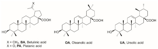
Figure 1.
Structure of betulinic acid (BA), platanic acid (PA), oleanolic acid (OA), and UA (ursolic acid).
2. Results
Acetylation (Scheme 1) of betulinic (BA, Figure 1), oleanolic (OA), ursolic (UA), and platanic (PA) acid gave well known acetates 1–4; their carboxyl group was activated with oxalyl chloride followed by the addition of either piperazine or homopiperazine to furnish amides 5–8 and 9–12, respectively. Activation of rhodamine B or rhodamine 101 with oxalyl chloride and reaction with amides 5–12 furnished piperazine/rhodamine B derived conjugates 13–16 and 17–20 as well as rhodamine 101 derived hybrids 21–24 and 25–28, respectively. All of these conjugates were violet in color, hence, indicating the presence of an intact cationic rhodamine moiety. This is regarded as a prerequisite for obtaining high cytotoxicity due to interaction with the mitochondrial membrane(s).

Scheme 1.
Reactions and conditions: (a) Ac2O, NEt3, DMAP (cat.), DCM, 20 °C, 1 d; (b) (COCl)2, DMF (cat.), DCM, then DCM (homo)piperazine, 20 °C, 1 h; (c) rhodamine B or rhodamine 101, (COCl)2, NEt3, DMAP (cat.), DCM, 20 °C, 1 d; T—triterpenoic acid; OA—oleanolic acid; UA—ursolic acid; BA—betulinic acid; PA—platanic acid; Rh B—rhodamine B; Rh 101—rhodamine 101.
The cytotoxicity of the compounds was determined in sulforhodamine B (SRB) assays employing several human tumor cell lines (A375, HT29, MCF-7, A2780, FaDu) as well as two non-malignant cell lines (NIH 3T3, HEK293). The results from these assays are summarized in Table 1, Table 2, Table 3 and Table 4.

Table 1.
Cytotoxicity of parent compounds BA, OA, UA, and PA as well as of their corresponding acetates 1–4 (EC50-values in µM from SRB-assays) after 72 h of treatment; the values are averaged from three independent experiments performed each in triplicate, confidence interval CI = 95%; mean ± standard mean error); n.d. not determined; doxorubicin (DX) was used as a positive control. Cell lines: malignant: A375 (melanoma), HT29 (colon adenocarcinoma), MCF-7 (breast adenocarcinoma), A2780 (ovarian carcinoma), FaDu (hypopharyngeal carcinoma); non-malignant: NIH 3T3 (fibroblasts).
Table 2.
Cytotoxicity of 3-O-acetylated triterpenoic amides 5–8 (piperazinyl amimides) and 9–12 (homopiperazinyl amides) (EC50-values in µM from SRB-assays) after 72 h of treatment; the values are averaged from three independent experiments performed each in triplicate, confidence interval CI = 95%; mean ± standard mean error); n.d. not determined; n.s. not soluble; doxorubicin (DX) was used as a positive control. Cell lines: malignant: A375 (melanoma), HT29 (colon adenocarcinoma), MCF-7 (breast adenocarcinoma), A2780 (ovarian carcinoma); non-malignant: NIH 3T3 (fibroblasts).
Table 3.
Cytotoxicity of 3-O-acetylated triterpenoic amides holding a distal rhodamine B unit 13–20 (EC50-values in µM from SRB-assays) after 72 h of treatment; the values are averaged from three independent experiments performed each in triplicate, confidence interval CI = 95%; mean ± standard mean error); n.d. not determined; n.s. not soluble; doxorubicin (DX) was used as a positive control. Cell lines: malignant: A375 (melanoma), HT29 (colon adenocarcinoma), MCF-7 (breast adenocarcinoma), A2780 (ovarian carcinoma); non-malignant: NIH 3T3 (fibroblasts).
Table 4.
Cytotoxicity of 3-O-acetylated triterpenoic amides holding a distal rhodamine 101 unit 13–20 (EC50-values in µM from SRB-assays) after 72 h of treatment; the values are averaged from three independent experiments performed each in triplicate, confidence interval CI = 95%; mean ± standard mean error); n.d. not determined; n.s. not soluble; doxorubicin (DX) was used as a positive control. Cell lines: malignant: A375 (melanoma), HT29 (colon adenocarcinoma), MCF-7 (breast adenocarcinoma), A2780 (ovarian carcinoma), HeLa (cervical cancer); non-malignant: NIH 3T3 (fibroblasts), HEK293 (human embryonic kidney).
Table 1 shows the results from the SRB assays for the parent compounds and their acetates. Except for BA and UA, all other triterpenoids held EC50 values > 30 µM (cut-off of the assay) for the cancer cell lines but also for the non-malignant fibroblasts NIH 3T3. Acetates 1–4 showed slightly improved cytotoxicity (except PA derived 4); by-and-large, EC50 values between 7.2 µM (1 for FaDu cells) and 21.3 (1 for A375 cells) were observed. Highest cytotoxicity was found for 1 and FaDu cells, for 2 with respect to A2780 and for 3 also with A2780 cells, respectively. Interestingly, PA derived acetate 4 was not cytotoxic at all within the limits of the assay.
Significant improvement was observed for the piperazinyl amides (Table 2), and EC50 values between 1.00 (5 for HT29) and 3.86 (for PA derived 8 and HT29 cells) were determined. Except for the latter, all EC50 values were smaller than 3 µM. While 5 was cytotoxic with an EC50 = 1.5 µM for A375 cells, its homopiperazinyl analog 9 was significantly less active (EC50 = 18.7 µM). However, by-and-large, the cytotoxicity of the homopiperazinyl derivatives 9–12 was of the same order as that of the piperazinyl analogs 5–8.
A dramatic improvement of cytotoxicity, however, was observed for the piperazinyl and homopiperazinyl spacered triterpenoic acid–rhodamine B conjugates 13–20 (Table 3).
Thereby, all of the compounds showed high cytotoxicity for all human tumor cell lines; EC50 values ranged from EC50 = 0.02 µM (compound 18 and A2780 cells) to EC50 = 0.76 µM (compound 17 and A375 cells). Thus, the former compound was as cytotoxic as standard doxorubicin.
Previously, we have shown the high cytotoxicity of several rhodamine B conjugates. Hence, it became of interest to investigate whether this high cytotoxicity is limited to rhodamine B conjugates or can also be found in conjugates holding a rhodamine 101 scaffold. As a result (Table 4), conjugates holding either a piperazinyl or homopiperazinyl spacer were only slightly less cytotoxic than those holding a rhodamine B moiety.
Interestingly enough, in this series of compounds, UA derived 27 (carrying a homopiperazine spacer and a rhodamine 101 residue) held the highest cytotoxicity, and the EC50 values for this compound were as low as EC50 = 0.05 µM (A2780 cells). Cytotoxicity for non-malignant fibroblasts NIH 3T3 were approximately five times lower for both rhodamine scaffolds. Extra staining experiments of A375 cells (acridine orange (AO), Hoechst 33342, rhodamine 123 (Figure 2)) showed 26 to act as a mitocan.

Figure 2.
Staining experiments: A375 cells, 24 h; (A) control (AO); (B) in the presence of 26; (C) merged (AO, 26); (D) control (AO); (E) Hoechst 33,342 staining; (F) merged (Hoechst 33,342).
A summary of the hitherto known structural prerequisites to obtain pentacyclic triterpenoids of high cytotoxicity is depicted in Figure 3.
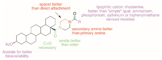
Figure 3.
Hitherto known SAR parameters for obtaining pentacyclic triterpenoids of high cytotoxicity.
3. Conclusions
Four representative pentacyclic triterpenoic acids (BA, OA, UA, and PA) were selected for a systematic evaluation of cytotoxic derivatives. As a result—and as exemplified for A2780 cancer cells—the cytotoxicity of the parent acids is low but increased slightly upon their acetylation. A significant increase in cytotoxicity was observed when acetates 1–4 were transformed into their piperazinyl amides 5–8. For the latter, compounds EC50 values between EC50 = 2.6 to 1.7 µM have been determined. The same trend was observed for the homopiperazinyl derivatives 9–12. Interestingly, betulinic acid derived 9 (EC50 = 12.0 µM) was significantly less cytotoxic than its piperazinyl derivative 5 (EC50 = 1.9 µM). A tremendous improvement in cytotoxicity was observed, however, for the rhodamine conjugates, and EC50 values between EC50 = 0.05–0.032 µM were observed for the piperazinyl rhodamine B conjugates. The corresponding piperazinyl rhodamine 101 conjugates were of comparable bioactivity (EC50 = 0.09–0.17 µM). A similar trend was observed for the homopiperazinyl rhodamine conjugates, and EC50 = 0.02–0.45 µM (for rhodamine B derived 17–20) and EC50 = 0.05–0.19 µM (for rhodamine 101 derived 25–28) were determined. Thus, it can be concluded that an optimal combination of pentacyclic triterpene, a suitable spacer and a lipophilic cationic residue must be found to achieve good cytotoxic activity. It was shown that both piperazinyl and homopiperazinyl spacers are equally suitable to serve as anchors for the binding of either rhodamine B or rhodamine 101. Furthermore, the conjugates derived from either rhodamine B or rhodamine 101 are of comparable cytotoxicity.
4. Experimental
NMR spectra were recorded using the Varian spectrometers (Darmstadt, Germany) DD2 and VNMRS (400 and 500 MHz, respectively). MS spectra were taken on a Advion expressionL CMS mass spectrometer (Ithaca, NY, USA); positive ion polarity mode, solvent: methanol, solvent flow: 0.2 mL/min, spray voltage: 5.17 kV, source voltage: 77 V, APCI corona discharge: 4.2 μA, capillary temperature: 250 °C, capillary voltage: 180 V, sheath gas: N2). Thin-layer chromatography was performed on pre-coated silica gel plates supplied by Macherey-Nagel (Düren, Germany). IR spectra were recorded on a Spectrum 1000 FT-IR-spectrometer from Perkin Elmer (Rodgau, Germany). The UV/Vis-spectra were recorded on a Lambda 14 spectrometer from Perkin Elmer (Rodgau, Germany); optical rotations were measured at 20 °C using a JASCO-P2000 instrument (JASCO Germany GmbH, Pfungstadt, Germany) The melting points were determined using the Leica hot stage microscope Galen III (Leica Biosystems, Nussloch, Germany) and are uncorrected. The solvents were dried according to usual procedures. Microanalyses were performed with an Elementar Vario EL (CHNS) instrument (Elementar Analysensysteme GmbH, Elementar-Straße 1, D-63505, Langenselbold, Germany).
4.1. General Procedure for the Synthesis of Acetates 1–4 (GPA)
To a solution of the triterpenoic acid (OA, UA, BA, PA, 1 equiv.) in dry DCM, acetic anhydride (3 equiv.), triethylamine (3 equiv.), and DMAP (cat.) were added, and the mixture was stirred at 20 °C for 1 day. Usual aqueous work-up followed by re-crystallization from ethanol furnished products 1–4.
4.2. 3β-Acetyloxy-lup-20(29)-en-28-oic Acid (1)
Following GPA, compound 1 (4.90 g, 90%) was obtained from BA as a colorless solid; Rf = 0.59 (n-hexane/ethyl acetate, 4:1); m.p. 277–279 °C (lit.: [28] 277–278 °C); [α]D = +20.7° (c 0.42, CHCl3) [(lit.: [29] [α]D = +20.7° (c 0.42, CHCl3)]; MS (ESI, MeOH): m/z 487.1 (32%, [M − H]−, 995.0 (100%, [2M − H]−, 1018.6 (29% [2M − 2H + Na]−).
4.3. 3β-Acetyloxy-olean-12-en-oic Acid (2)
Following GPA, compound 2 (2.45 g, 89%) was obtained from OA as a colorless solid; Rf = 0.72 (toluene/ethyl acetate/heptane/formic acid, 80:26:10:5); m.p. 286–289 °C (lit.: [30] 264–265 °C); [α]D = +69.4° (c 0.30, CHCl3) [(lit.: [31] [α]D = +74.0° (c 1.0, CHCl3)]; MS (ESI, MeOH): m/z 499.4 ([100%, M + H]+), 516.3 (36%, [M + NH4]+, 521.5 [35%, [M + Na]+.
4.4. 3β-Acetyloxy-urs-12-en-oic Acid (3)
Following GPA, compound 3 (2.41 g, 89%) was obtained from UA as a colorless solid; Rf = 0.57 (n-hexane/ethyl acetate, 3:1); m. p. 281–283 °C (lit.: [32] 280 °C); [α]D = +66.5° (c 0.42, CHCl3) [lit.: [33] [α]D = +63.5° (c 0.5, CHCl3)]; MS (ESI, MeOH): m/z 499.3 (17%, [M + H]+), 521.2 (31%, [M + H]+), 1019.4 (100%, [2M + Na]+).
4.5. 3β-Acetyloxy-20-oxo-30-norlupan-28-oic Acid (4)
Following GPA, compound 4 (6.9 g, 84%) was obtained from PA as a colorless solid; Rf = 0.50 (toluene/ethyl acetate/heptane/formic acid, 80:26:10.5); m.p. 265–268 °C (lit.: [34] 252–255 °C); [α]D = −9.4° (c 0.32, CHCl3) [(lit.: [34] [α]D = −9.5° (c 0.5, CHCl3)]; MS (ESI, MeOH): m/z 999.5 (100%, [2M − H]−).
4.6. General Procedure for the Synthesis of Acetylated (homo)piperazinyl Amides 5–12 (GPB)
To a solution of the acetylated triterpenoic acid 1–4 (1 equiv.) in dry DCM, DMF (cat.) and oxalyl chloride (4 equiv.) were added followed by the addition of (homo)piperazine (4 equiv.). After stirring for 1h at 20 °C followed by usual aqueous work-up and column chromatography, products 5–12 were obtained.
4.7. 3β-Acetyloxy-28-(1-piperazinyl)-lup-20(29)en-28-one (5)
Following GPB from 1 (2.5 g, 5 mmol) and piperazine (1.6 g, 20.0 mmol), compound 5 (2.07 g, 73%) was obtained as a colorless solid; Rf = 0.40 (SiO2, CHCl3/MeOH, 9:1); m.p. = 166–173 °C (lit.: [35] 162–167 °C); [α]D = −1.4° (c 0.21, MeOH), [lit.: [35] [α]D = −1.8° (c 0.32, MeOH); MS (ESI, MeOH): m/z (%) = 567.3 ([100%, M + H]+).
4.8. 3β-Acetyloxy-28-(1-piperazinyl)-olean-12-en-28-one (6)
Following GPB from 2 (2.5 g, 5.0 mmol) and piperazine (1.6 g, 29 mmol), 6 (2.16 g, 76%) was obtained as a colorless solid; Rf = 0.40 (SiO2, CHCl3/MeOH, 9:1); m.p. = 172–175 °C (lit.: [35] 173–175 °C); [α]D = +23.4° (c 0.18, CHCl3), [lit.: [35] [α]D = +26.6° (c 0.35, MeOH); MS (ESI, MeOH): m/z (%) = 567.4 (100%, [M + H]+).
4.9. 3β-Acetyloxy-28-(1-piperazinyl)-urs-12-en-28-one (7)
Following GPB from 3 (2.5 g, 5.0 mmol) and piperazine (1.6 g, 20.0 mmol), 7 (1.84 g, 65%) was obtained as a colorless solid; Rf = 0.40 (SiO2, CHCl3/MeOH, 9:1); m.p. = 187–190 °C (lit.: [35] 187–188 °C); [α]D = +25.1° (c 0.24, MeOH), [lit.: [35] [α]D = +24.5° (c 0.29, MeOH); MS (ESI, MeOH): m/z (%) = 567.4 (100%, [M + H]+).
4.10. 3β-Acetyloxy-28-(1-piperazinyl)-30-norlupane-20,28-dione (8)
Following GPB from 4 (2.5 g, 5.0 mmol) and piperazine (1.6 g, 20.0 mmol), 17 (1.93 g, 68%) was obtained as a colorless solid; Rf = 0.40 (SiO2, CHCl3/MeOH, 9:1); m.p. = 127–130 °C (lit.: [20] 115–125 °C); [α]D = −20.3° (c 0.13, CHCl3); MS (ESI, MeOH): m/z (%) = 569.3 (100%, [M + H]+).
4.11. 3β-Acetyloxy-28-(1-homopiperazinyl)-lup-20(29)en-28-one (9)
Following GPB from 1 (1.0 g, 2.02 mmol) and homopiperazine (9.8 g, 8.0 mmol), 9 (1.02 g, 88%) was obtained as a colorless solid; Rf = 0.4 (SiO2, CHCl3/MeOH, 9:1); m.p. 190–193°C (lit.: [21] 196–199 °C); [α]D = +107.6° (c 0.20, CHCl3), [lit.: [21] [α]D = +109.8° (c 0.38, CHCl3); MS (ESI, MeOH): m/z = 581.4 (100%, [M + H]+).
4.12. 3β-Acetyloxy-28-(1-homopiperazinyl)-olean-12-en-28-one (10)
Following GPB from 2 (1.0 g, 2.02 mmol) and homopiperazine (0.8 g, 8.0 mmol), 10 (1.4 g, 90%) was obtained as a colorless solid; Rf = 0.4 (SiO2, CHCl3/MeOH, 9:1); m.p. 182–185 °C (lit.: [21] 187–190 °C); [α]D = 12.4° (c 0.14, CHCl3), [lit.: [21] [α]D = +9.9° (c 0.35, CHCl3); MS (ESI, MeOH): m/z = 581.4 (100%, [M + H]+).
4.13. 3β-Acetyloxy28-(1-homopierazinyl)-urs-12-en-28-one (11)
Following GPB from 3 (1.0 g, 2.02 mmol) and homopiperazine (0.8 g, 8.0 mmol), 11 (1.1 g, 86%) was obtained as a colorless solid; Rf = 0.4 (SiO2, CHCl3/MeOH, 9:1); m.p. 171–175 °C (lit.: [21] 178–180 °C); [α]D = +27.0° (c 0.21, CHCl3), [lit.: [21] [α]D = +29.7° (c 0.34, CHCl3); MS (ESI, MeOH): m/z = 581.4 (100%, [M + H]+, 100%).
4.14. 3β-Acetyloxy-28-(1-homopiperazinyl)-30-norlupane-20,28-dione (12)
Following GPB from 4 (1.0 g, 2.02 mmol) and homopiperazine (0.8 g, 8.0 mmol), 12 (1.1 g, 94%) was obtained as a colorless solid; Rf = 0.4 (SiO2, CHCl3/MeOH, 9:1); m.p. 162–165 °C; [α]D = −29.2° (c 0.16, CHCl3); IR (ATR): ν = 2941m, 2866w, 1732m, 1708m, 1622m, 1464m, 1453m, 1408m, 1367m, 1244vs, 1191m, 1149m, 1133m, 1109w, 1026m, 979m, 947w, 934w, 900w, 750s, 665m, 610w, 557m, 506m, 455w, 410w cm−1; 1H NMR (400 MHz, CDCl3): δ = 4.44 (dd, J = 10.5, 5.5 Hz, 1H, 3-H), 3.89–3.60 (m, 4H, 33-H), 3.40–3.09 (m, 5H, 19-H, 33-H), 2.62 (td, J = 12.1, 4.1 Hz, 1H, 13-H), 2.25 (s, 2H, 35-H), 2.17 (s, 3H, 29-H), 2.14–2.06 (m, 2H, 16-Ha + 18-H), 2.02 (s, 3H, 32-H), 1.97–1.84 (m, 2H, 21-Ha + 22-Ha), 1.68–1.14 (m, 15H + 1-Ha + 2-H + 6-Ha + 6-Hb + 7-H + 9-H + 11-Ha + 11-Hb + 15-H + 16-Hb + 21-Hb + 22-Hb), 1.06–0.98 (m, 2H, 12-H), 0.96 (s, 4H, 1-Hb + 27-H), 0.89 (s, 3H, 26-H), 0.82 (s, 3H, 24-H), 0.82 (s, 3H, 25-H), 0.81 (s, 3H, 23-H), 0.79–0.74 (m, 1H, 5-H) ppm; 13C NMR (101 MHz, CDCl3): δ = 213.0 (C-20), 175.0 (C-28), 171.2 (C-31), 81.0 (C-3), 55.4 (C-5), 54.9 (C-17), 52.6 (C-18), 50.6 (C-9), 49.9 (C-19), 46.6 (C-33 + C-34), 41.8 (C-14), 40.6 (C-8), 38.3 (C-1), 37.8 (C-4), 37.1 (C-10), 36.0 (C-13), 35.7 (C-22), 34.1 (C-7), 31.7 (C-16), 30.3 (C-29), 29.9 (C-15), 28.6 (C-21), 27.9 (C-24), 27.3 (C-12), 25.7 (C-35), 23.6 (C-2), 21.3 (C-32), 21.1 (C-11), 18.1 (C-6), 16.4 (C-23), 16.2 (C-25), 15.9 (C-26), 14.7 (C-27) ppm; MS (ESI, MeOH): m/z = 583.3 (100%, [M + H]+; analysis calcd for C36H58N2O4 (582.87): C 74.18, H 10.03, N 4.81; found: C 73.95, H 10.34, N 4.63.
4.15. General Procedure for the Synthesis of the Rhodamine Conjugates 13–28 (GPC)
To a solution of the respective rhodamine (1.15 eq.) in dry DCM at 0 °C, DMF (cat.) and oxalyl chloride (4 eq.) were added (vide supra). This acid chloride was slowly added to the solution of the respective triterpene (1 eq. in DCM) in the presence of triethylamine (1 eq.). After stirring for 1 day at 20 °C, aqueous work-up was carried out as usual and the residue was purified by column chromatography.
4.16. 9-[2-[[4-(3β-Acetyloxy-28-oxo-lup-20(29)en-28-yl)-1-piperazinyl]carbonyl]phenyl]-3,6-bis(diethylamino)-xanthylium chloride (13)
Following GPC from 5 (180 mg, 0.32 mmol) and rhodamine B, 13 (220 mg, 67%) was obtained as a pink solid; Rf = 0.39 (SiO2, CHCl3/MeOH, 9:1); m.p. 247–252 °C (lit.: [20] m.p. 246–250 °C); MS (ESI, MeOH): m/z = 991.6 (100%, [M − Cl]+).
4.17. 9-[2-[[4-(3β-Acetyloxy-28-oxo-olean-12-en-28-yl)-1-piperazinyl]carbonyl]phenyl]-3,6-bis(diethylamino)-xanthylium chloride (14)
Following GPC from 6 (180 mg, 0.32 mmol) and rhodamine B, 14 (250 mg, 76%) was obtained as a pink solid; Rf = 0.40 (SiO2, CHCl3/MeOH, 9:1); m.p. 246–252 °C (lit.: [20] 245–248 °C); MS (ESI, MeOH): m/z = 991.9 (100%, [M − Cl]+).
4.18. 9-[2-[[4-(3β-Acetyloxy-28-oxo-ursan-12-en-28-yl)-1-piperazinyl]carbonyl]phenyl]-3,6-bis(diethylamino)-xanthylium chloride (15)
Following GPC from 7 (180 mg, 0.32 mmol) and rhodamine B, 15 (190 mg, 58%) was obtained as a pink solid; Rf = 0.40 (SiO2, CHCl3/MeOH, 9:1); m.p. 245–251 °C (lit.: [20] 243–245 °C); MS (ESI, MeOH): m/z = 991.7 (100%, [M − Cl]+).
4.19. 9-[2-[[4-(3β-Acetyloxy-20,28-dioxo-30-norlupan-28-yl)-1-piperazinyl]carbonyl]phenyl]-3,6-bis(diethylamino)-xanthylium chloride (16)
Following GPC from 8 (180 mg, 0.32 mmol) and rhodamine B, 16 (230 mg, 70%) was obtained as a pink solid; Rf = 0.37 (SiO2, CHCl3/MeOH, 9:1); m.p. 247–254 °C (lit.: [20] 235–243 °C); MS (ESI, MeOH): m/z = 993.7 (100%, [M − Cl]+).
4.20. 9-[2-[[4-(3β-Acetyloxy-28-oxo-30-norlupan-28-yl)-1-homopiperazinyl]carbonyl]phenyl]- 3,6-bis(diethylamino)-xanthylium chloride (17)
Following GPC from 9 (260 mg, 0.32 mmol) and rhodamine B, 17 (229 mg, 69%) was obtained as a pink solid; Rf = 0.50 (SiO2, MeCN/CH2Cl2/H2O, 10:1:1); m.p. 261–266 °C (lit.: [21] 256–260 °C); MS (ESI, MeOH): m/z = 1005.7 (100%, [M − Cl]+).
4.21. 9-[2-[[4-(3β-Acetyloxy-28-oxo-olean-12-en-28-yl]-1-homopiperazinyl]carbonyl]phenyl]- 3,6-bis(diethylamino)-xanthylium chloride (18)
Following GPC from 10 (270 mg, 0.32 mmol) and rhodamine B, 18 (267 mg, 80%) was obtained as a pink solid; Rf = 0.52 (SiO2, MeCN/CH2Cl2/H2O, 10:1:1); m.p. 235–241 °C (lit.: [21] 238–245 °C); MS (ESI, MeOH): m/z = 1005.8 (100%, [M − Cl]+).
4.22. 9-[2-[[4-(3β-Acetyloxy-28-oxo-urs-12-en-28-yl]-1-homopiperazinyl]carbonyl]phenyl]- 3,6-bis(diethylamino)-xanthylium chloride (19)
Following GPC from 11 (270 mg, 0.32 mmol) and rhodamine B, 19 (253 mg, 76%) was obtained as a pink solid; Rf = 0.51 (SiO2, MeCN/CH2Cl2/H2O, 10:1:1); m.p. 238–246 °C (lit.: [21] 238–245 °C); MS (ESI, MeOH): m/z = 1005.7 (100%, [M − Cl]+).
4.23. 9-[2-[[4-(3β-Acetyloxy-20,28-dioxo-30-norlupan-28-yl)-1-homopiperazinyl]carbonyl]phenyl]-3,6-bis(diethylamino)-xanthylium chloride (20)
Following GPC from 10 (125 mg, 0.21 mmol) and rhodamine B, 18 (190 mg, 87%); was obtained as a pink solid; m.p. 248–250 °C (decomp.); Rf = 0.30 (SiO2, CHCl3/MeOH, 9:1); UV-Vis (CHCl3): λmax (log ε) = 562 nm (5.05); IR (ATR): ν = 2937w, 1730w, 1585s, 1466m, 1411m, 1334s, 1273m, 1244m, 1178s, 1131m, 1072m, 977w, 920w, 822w, 746m, 683m cm−1; 1H NMR (500 MHz, CDCl3): δ = 7.66–7.57 (m, 2H, 42-H, 43-H), 7.42–7.36 (m, 1H, 40-H), 7.31–7.27 (m, 1H, 41-H), 7.25–7.15 (m, 2H, 50-H), 6.99–6.83 (m, 2H, 49-H), 6.79–6.68 (m, 2H, 47-H), 4.40 (dd, J = 10.4, 5.6 Hz, 1H, 3-H), 3.85–3.31 (m, 16H, 32-H, 33-H, 34-H, 36-H, 51-H), 3.27–3.12 (m, 1H, 13-H), 2.72–2.57 (m, 1H, 19-H), 2.11 (s, 3H, 29-H), 2.18–2.06 (m, 1H, 16-Ha), 1.98 (s, 3H, 31-H), 1.96 (s, 1H, 18-H), 1.95–0.97 (m, 21H, 22-Ha, 21-Ha, 35-H, 1-Ha, 2-H, 16-Hb, 21-Hb, 6-H, 22-Hb, 11-Ha, 7-H, 11-Hb, 9-H, 15-H, 12-H), 1.32–1.26 (m, 12H, 52-H), 0.92 (s, 3H, 27-H), 0.89 (s, 3H, 24-H), 0.94–0.86 (m, 1H, 1-Hb), 0.79 (s, 9H, 23-H, 25-H, 26-H), 0.73 (s, 1H, 5-Hb) ppm; 13C NMR (126 MHz, CDCl3): δ = 212.8 (C-20), 174.7 (C-28), 170.7 (30 C-), 168.3 (C-37), 157.5 (C-48), 157.5 (C-44), 155.4 C- (46), 136.5 (C-38) 135.9 (C-39), 132.1 (C-50), 130.1 (C-41), 129.6 (C-42), 129.4 (C-43), 126.3 (C-40), 113.9 (C-49), 113.4 (C-45), 96.2 (C-47), 80.7 (C-3), 55.3 (C-5), 54.7 (C-17), 52.7 (C-18), 50.4 (C-9), 50.0 (C-19), 46.0 (C-51), 41.6 (C-14), 40.4 (C-8), 38.2 (C-1), 37.6 (C-4), 36.9 (C-10), 35.8 (C-13), 35.3 (C-22), 34.1 (C-7), 31.7 (C-35), 31.2 (C-16), 30.0 (C-29), 29.7 (C-15), 28.7 (C-21), 27.7 (C-23), 27.2 (C-12), 23.5 (C-2), 21.1 (C-31), 21.0 (C-11), 18.0 (C-6), 16.3 (C-25), 16.1 (C-26), 16.0 (C-24), 14.4 (C-27), 12.5 (C-52). ppm; MS (ESI, MeOH): m/z 1007 (100%, [M]+); analysis calculated for C64H85ClN4O6 (1041.84): C 73.78, H 8.22, N 5.38; found: C 73.55, H 8.41, N 5.19.
4.24. 3β-Acetyloxy-28-[4-[3-(2,3,6,7,12,13,16,17-octahydro-1H,5H,11H,15H-pyrido[3,2,1-ij] pyrido[1”,2”,3”:1′,8′]quinolino[6′,5′:5,6]pyrano[2,3-f]quinolin-4-ium-9-yl)benzoyl]piperazine-1-yl]-28-oxo-lup-20(29)-en chloride (21)
Following GPC from 5 (0.25 g, 0.44 mmol) and rhodamine 101 (0.25 g, 0.51 mmol), 21 (0.24 g, 52%) was obtained as a pink colored solid; Rf = 0.4 (SiO2, CHCl3/MeOH, 9:1); m.p. >300 °C; IR (ATR): ν = 3400w, 2939w, 2862w, 1728w, 1630m, 1595m, 1542w, 1493m, 1434m, 1360m, 1294vs, 1267s, 1248s, 1195s, 1182s, 1099s, 1035m, 1003m, 978m, 896w, 827w, 772m, 747m, 637m, 560m, 506m, 421s cm−1; 1H NMR (400 MHz, CDCl3): δ = 7.67 (dd, J = 5.7, 3.3 Hz, 2H, 37-H, 39-H), 7.53–7.48 (m, 1H, 38-H), 7.31 (dd, J = 5.7, 3.2 Hz, 1H, 40-H), 6.68 (d, J = 6.0 Hz, 2H, 48-H), 4.68 (s, 1H, 29-Ha), 4.56 (s, 1H, 29-Hb), 4.44 (dd, J = 10.2, 5.9 Hz, 1H, 3-H), 3.60–3.48 (m, 4H, 49-H), 3.48–3.40 (m, 4H, 52-H), 3.40–3.31 (m, 8H, 33-H + 34-H), 3.04–2.97 (m, 4H, 54-H), 2.91 (dt, J = 11.9, 6.1 Hz, 1H, 19-H), 2.81–2.74 (m, 1H, 13-H), 2.74–2.60 (m, 4H, 51-H), 2.14–2.04 (m, 4H, 53-H), 2.02 (s, 3H, 32-H), 1.98–1.91 (m, 4H, 50-H), 1.77 (q, J = 33.5, 10.4 Hz, 4H, 12-H + 16-Ha + 22-Ha), 1.65 (s, 3H, 30-H), 1.63–1.44 (m, 8H, 1-H + 2-H + 6-Ha + 11-H + 16-Hb + 18-H), 1.44–1.08 (m, 10H, 6-Hb + 7-H + 9-H + 15-H + 21-H + 22-Hb), 0.92 (s, 3H, 27-H), 0.88 (s, 3H, 26-H), 0.82 (s, 6H, 23-H + 24-H), 0.81 (s, 3H, 25-H), 0.77 (s, 1H, 5-H) ppm; 13C NMR (101 MHz, CDCl3): δ = 174.1 (C-28), 171.0 (C-31), 167.9 (C-35), 162.7 (C-20), 152.0 (C-43), 151.2 (C-44), 150.9 (C-46), 134.8 (C-41), 131.8 (C-36), 130.7 (C-40), 130.2 (C-39), 129.7 (C-37), 127.5 (C-38), 126.5 (C-48), 123.6 (C-45), 123.6 (C-45), 113.2 (C-42), 109.4 (C-29), 105.4 (C-47), 80.9 (C-3), 55.5 (C-5), 54.6 (C-17), 52.5 (C-18), 51.0 (C-49), 50.7 (C-9), 50.5 (C-52,), 45.7 (C-19), 41.9 (C-14), 40.6 (C-8), 38.4 (C-1), 37.8 (C-4), 37.1 (C-10), 36.9 (C-13), 35.8 (C-22), 34.3 (C-7), 32.5 (C-16), 31.2 (C-21), 29.8 (C-15), 27.9 (C-24), 27.7 (C-51), 25.5 (C-12), 23.7 (C-2), 21.3 (C-32), 20.6 (C-11, C-50), 19.9 (C-54), 19.6 (C-53), 19.5 (C-30), 18.1 (C-6), 16.5 (C-23), 16.2 (C-25), 16.1 (C-26), 14.6 (C-27) ppm; MS (ESI, MeOH): m/z = 1039.3 (100%, [M − Cl]+); analysis calculated for C68H87N4O5Cl (1075.92): C 75.91, H 8.15, N 5.21; found: C 75.77, H 8.36, N 5.05.
4.25. 3β-Acetyloxy-28-[4-[3-(2,3,6,7,12,13,16,17-octahydro-1H,5H,11H,15H-pyrido[3,2,1-ij]pyrido[1”,2”,3”:1′,8′]quinolino[6′,5′:5,6]pyrano[2,3-f]quinolin-4-ium-9-yl)benzoyl]piperazine-1-yl]-28-oxoolean-12-en chloride (22)
Following GPC from 6 (0.25 g, 0.44 mmol) and rhodamine 101 (0.25 g, 0.51 mmol), 22 (0.33 g, 71%) was obtained as a pink solid; Rf = 0.4 (SiO2, CHCl3/MeOH, 9:1); m.p. >300 °C; IR (ATR): ν = 3398w, 2941w, 2859w, 1728w, 1631m, 1594s, 1543w, 1494s, 1458m, 1435m, 1361m, 1295Vs, 1267s, 1248s, 1196s, 1182s, 1100s, 1077m, 1035m, 1002m, 897w, 862w, 773w, 733m, 640m, 560m, 498m, 421s cm−1; 1H NMR (500 MHz, CDCl3) δ = 7.70–7.66 (m, 2H, 37-H, 39-H), 7.53–7.49 (m, 1H, 38-H), 7.34–7.30 (m, 1H, 40-H), 6.67 (d, J = 7.5 Hz, 2H, 48-H), 5.23 (t, J = 3.5 Hz, 1H, 12-H), 4.48 (dd, J = 9.9, 6.0 Hz, 1H, 3-H), 3.63–3.51 (m, 4H, 49-H), 3.52–3.40 (m, 4H, 52-H), 3.39–3.29 (m, 8H, 33-H + 34-H), 3.09–3 (m, 4H, 54-H), 3.00–2.96 (m, 1H, 18-H), 2.76–2.62 (m, 4H, 51-H), 2.15–2.07 (m, 5H, 16-Ha + 53-H), 2.04 (s, 3H, 32-H), 2.01–1.91 (m, 4H, 50-H), 1.89–1.81 (m, 1H, 11-H), 1.71–1.48 (m, 10H, 1-Ha + 2-H + 6-Ha + 7-H + 9-H + 15-Ha + 16-Hb + 19-Ha), 1.48–1.13 (m, 7H, 6-Hb + 19-Hb + 21-H + 22-Ha + 22-Hb), 1.11 (s, 3H, 27-H), 1.08–0.94 (m, 2H, 1-Hb + 15-Hb), 0.91 (s, 3H, 25-H), 0.89 (s, 3H, 29-H), 0.88 (s, 3H, 30-H), 0.86 (s, 3H, 24-H), 0.85 (s, 3H, 23-H), 0.82 (s, 1H, 5-H), 0.66 (s, 3H, 26-H) ppm; 13C NMR (126 MHz, CDCl3): δ = 176.4 (C-28), 171.0 (C-31), 167.8 (C-35), 152.8 (C-43), 152.0 (C-44), 152.0 (C-44), 151.3 (C-46), 151.2 (C-46), 144.4 (C-13), 134.8 (C-41), 131.8 (C-36), 130.8 (C-40), 130.2 (C-39), 129.7 (C-37), 127.4 (C-36), 126.5 (C-48), 126.5 (C-48), 123.6 (C-45), 123.5 (C-45), 121.7 (C-12), 113.3 (C-42), 105.5 (C-47), 105.5 (C-47), 80.8 (C-3), 55.3 (C-5), 51.0 (C-49, 49), 50.6 (C-52), 47.6 (C-9, 33, 34), 47.5 (C-17), 46.2 (C-19), 43.5 (C-18), 41.8 (C-14), 39.1 (C-8), 38.0 (C-1), 37.7 (C-4), 37.0 (C-10), 33.9 (C-21), 32.9 (C-30), 32.8 (C-22), 30.3 (C-20), 30.0 (C-7), 28.0 (C-24), 27.8 (C-15), 27.7 (C-51), 25.9 (C-27), 24.0 (C-29), 23.5 (C-2), 23.3 (C-11), 22.5 (C-16), 21.3 (C-32), 20.6 (C-50), 19.9 (C-54), 19.7 (C-53), 18.2 (C-6), 16.9 (C-26), 16.7 (C-23), 15.4 (C-25) ppm; MS (ESI, MeOH): m/z = 1039.3 (100%, [M − Cl]+); analysis calculated for C68H87N4O5Cl (1075.64): C 75.91, H 8.15, N 5.21; found: C 75.72, H 8.29, N 5.01.
4.26. 3β-Acetyloxy-28-[4-[3-(2,3,6,7,12,13,16,17-octahydro-1H,5H,11H,15H-pyrido[3,2,1-ij] pyrido[1”,2”,3”:1′,8′]quinolino[6′,5′:5,6]pyrano[2,3-f]quinolin-4-ium-9-yl)benzoyl]piperazine-1-yl]-28-oxo-urs-12-en chloride (23)
Following GPC from 7 (0.25 g, 0.44 mmol) and rhodamine 101 (0.25 g, 0.51 mmol), 23 (0.27 g, 58%) was obtained as a pink solid; Rf = 0.4 (SiO2, CHCl3/MeOH, 9:1); m.p. >300 °C; IR (ATR): ν = 3392w, 2926w, 2867w, 1728w, 1626m, 1594s, 1542w, 1493s, 1457m, 1435m, 1361m, 1294vs, 1266s, 1246s, 1196s, 1181s, 1099s, 1035m, 1004m, 897w, 863w, 773m, 732m, 653m, 561m, 420s cm−1; 1H NMR (500 MHz, CDCl3): δ = 7.71–7.65 (m, 2H, 37-H, 39-H), 7.54–7.48 (m, 1H, 38-H), 7.34–7.30 (m, 1H, 40-H), 6.67 (d, J = 4.7 Hz, 2H, 48-H), 5.17 (s, 1H, 12-H), 4.48 (dd, J = 10.4, 5.5 Hz, 1H, 3-H), 3.56 (dt, J = 17.2, 6.1 Hz, 4H, 49-H), 3.51–3.41 (m, 4H, 52-H), 3.33 (s, 8H, 33-H + 34-H), 3.02 (q, J = 6.0 Hz, 4H, 54-H), 2.68 (ddt, J = 22.7, 15.6, 7.3 Hz, 4H, 51-H), 2.39–2.32 (m, 1H, 18-H), 2.15–2.06 (m, 4H, 53-H), 2.04 (s, 3H, 32-H), 1.99–1.94 (m, 4H, 50-H), 1.91–1.87 (m, 1H + 11-Ha), 1.79–1.57 (m, 7H + 1-Ha + 2-H + 11-Hb + 16-H + 22-Ha), 1.56–1.40 (m, 5H, 6-Ha + 7-Ha + 9-H + 21-Ha + 22-Hb), 1.40–1.20 (m, 4H, 6-Hb + 7-Hb + 19-H + 21-Hb), 1.05 (s, 6H, 1-Hb + 15-H + 27-H), 0.99–0.95 (m, 1H, 20-H), 0.93 (s, 3H, 30-H), 0.91 (s, 3H, 25-H), 0.86 (s, 3H, 24-H), 0.86 (s, 3H, 26-H), 0.84 (s, 3H, 29-H), 0.81–0.80 (m, 1H, 5-H), 0.67 (s, 3H, 23-H) ppm; 13C NMR (126 MHz, CDCl3): δ = 174.8 (C-28), 171.0 (C-31), 167.8 (C-35), 152.8 (C-46), 152.0 (C-43), 151.3 (C-44), 151.2 (C-44), 134.8 (C-41), 130.8 (C-40), 130.2 (C-37), 129.8 (C-39), 127.5 (C-38), 126.5 (C-48), 125.2 (C-12), 123.6, 123.5 (C-45), 113.2 (C-42), 105.5 (C-47), 105.5 (C-47), 80.8 (C-3), 55.3 (C-5, 18), 51.0 (C-49), 50.6 (C-52), 48.6 (C-17), 47.5 (C-9, C-33, C-34), 42.1 (C-14), 39.8 (C-19), 39.4 (C-8), 38.7 (C-20), 34.4 (C-22), 32.9 (C-7), 30.4 (C-21), 28.1 (C-15), 28.1 (C-24), 27.7 (C-51), 27.7 (C-51), 23.5 (C-2, 16, 27), 23.3 (C-11), 21.3 (C-30), 21.2 (C-32), 20.6 (C-50), 19.9 (C-54), 19.7 (C-53), 18.2 (C-6), 17.4 (C-29), 16.9 (C-26), 16.7 (C-23), 15.5 (C-25) ppm; MS (ESI, MeOH): m/z = 1039.4 ([M − Cl]+, 100%); analysis calculated for C68H87N4O5Cl (1075.64): C 75.91, H 8.15, N 5.21; found: C 75.64, H 8.29, N 4.96.
4.27. 3β-Acetyloxy-28-[4-[3-(2,3,6,7,12,13,16,17-octahydro-1H,5H,11H,15H-pyrido[3,2,1-ij]pyrido[1”,2”,3”:1′,8′]quinolino[6′,5′:5,6]pyrano[2,3-f]quinolin-4-ium-9-yl)benzoyl]piperazine-1-yl]-20,28-dioxo-30-norlupan-12-en chloride (24)
Following GPC from 8 (0.25 g, 0.44 mmol) and rhodamine 101 (0.25 g, 0.51 mmol), 24 (0.30 g, 65%) was obtained as a pink solid; Rf = 0.4 (SiO2, CHCl3/MeOH, 9:1); m.p. >300 °C; IR (ATR): ν = 3396w, 2940w, 2863w, 1727w, 1627m, 1594s, 1543w, 1493s, 1459m, 1446m, 1439m, 1419m, 1361m, 1295Vs, 1267s, 1195s, 1182s, 1099s, 1035m, 1004m, 979m, 897w, 863w, 773m, 732m, 561m, 505m, 420s cm−1; 1H NMR (500 MHz, CDCl3): δ = 7.70–7.67 (m, 2H, 37-H, 39-H), 7.53–7.49 (m, 1H, 38-H), 7.33–7.30 (m, 1H, 40-H), 6.66 (d, J = 6.8 Hz, 2H, 48-H), 4.44 (dd, J = 10.9, 5.2 Hz, 1H, 3-H), 3.62–3.51 (m, 4H, 49-H), 3.51–3.43 (m, 4H, 52-H), 3.43–3.29 (m, 8H, 33-H + 34-H), 3.15 (td, J = 11.1, 3.1 Hz, 1H, 19-H), 3.06–2.98 (m, 4H, 54-H), 2.78–2.63 (m, 4H, 51-H), 2.58 (td, J = 12.1, 3.8 Hz, 1H, 13-H), 2.14 (s, 3H, 29-H), 2.13–2.08 (m, 4H, 53-H), 2.08–2.04 (m, 1H, 18-H), 2.02 (s, 3H, 32-H), 1.97 (q, J = 6.9 Hz, 5H, 16-Ha + 50-H), 1.88–1.80 (m, 2H, 21-Ha + 22-Ha), 1.67–1.54 (m, 4H, 1-Ha + 2-H + 16-Hb), 1.54–0.98 (m, 14H, 6-Ha + 6-Hb + 7-H + 9-H + 11-Ha + 11-Hb + 12-H + 15-H + 21-Hb + 22-Hb), 0.95 (s, 4H, 1-Hb + 27-H), 0.87 (s, 3H, 26-H), 0.82 (s, 3H, 24-H), 0.82 (s, 3H, 25-H), 0.81 (s, 3H, 23-H), 0.79–0.75 (m, 1H, 5-H) ppm; 13C NMR (126 MHz, CDCl3) δ = 212.6 (C-20), 173.9 (C-28), 170.9 (C-31), 167.8 (C-35), 152.8 (C-43), 152.0 (C-44), 152.0 (C-44), 151.3 (C-46), 151.2 (C-46), 134.7 (C-41), 131.9 (C-36), 130.8 (C-40), 130.3 (C-39), 129.7 (C-37), 127.4 (C-38), 126.5 (C-48), 123.6 (C-45), 123.5 (C-45), 113.2 (C-42), 105.5 (C-47), 55.4 (C-5), 54.5 (C-17), 52.5 (C-18), 51.0 (C-49), 51.0 (C-49), 50.6 (C-52), 50.6 (C-52), 50.5 (C-9), 50.0 (C-19), 41.7 (C-14), 40.5 (C-8), 38.3 (C-1), 37.8 (C-4), 37.1 (C-10), 35.9 (C-13), 35.6 (C-22), 34.2 (C-7), 32.0 (C-16), 30.2 (C-29), 29.8 (C-15), 28.7 (C-21), 27.9 (C-24), 27.7 (C-51), 27.4 (C-12), 23.6 (C-2), 21.3 (C-32), 21.1 (C-11), 20.6 (C-50), 19.9 (C-54), 19.7 (C-53), 18.1 (C-6), 16.5 (C-23), 16.2 (C-25), 16.0 (C-26), 14.6 (C-27) ppm; MS (ESI, MeOH): m/z = 1041.3 ([M − Cl]+, 100%); analysis calculated for C67H85N4O6Cl (1077.89): C 74.66, H 7.95, N 5.20; found: C 74.50, H 8.14, N 5.03.
4.28. 3β-Acetyloxy-28-[4-[3-(2,3,6,7,12,13,16,17-octahydro-1H,5H,11H,15H-pyrido[3,2,1-ij] pyrido[1”,2”,3”:1′,8′]quinolino[6′,5′:5,6]pyrano[2,3-f]quinolin-4-ium-9-yl)benzoyl]homopiperazine-1-yl]-28-oxo-lup-20(29)-en chloride (25)
Following GPC from 9 (0.35 g, 0.60 mmol) and rhodamine 101 (0.25 g, 0.51 mmol), 25 (0.37 g, 68%) was obtained as a pink solid; Rf = 0.4 (SiO2, CHCl3/MeOH, 9:1); m.p. >300 °C; IR (ATR): ν = 2940w, 2865w, 1730w, 1624m, 1595s, 1542w, 1493s, 1459m, 1376m, 1361m, 1294vs, 1268s, 1247m, 1196s, 1180s, 1098s, 1035m, 1018m, 978m, 895w, 772w, 747m, 622m, 421s cm−1; 1H NMR (400 MHz, CDCl3): δ = 7.63–7.51 (m, 2H, 37-H, 39-H), 7.41 (t, J = 9.2 Hz, 1H, 38-H), 7.21 (d, J = 7.4 Hz, 1H, 40-H), 6.68 (dd, J = 26.0, 20.3 Hz, 2H, 48-H), 4.70 (d, J = 18.2 Hz, 1H, 29-Ha), 4.55 (d, J = 17.7 Hz, 1H, 29-Hb), 4.45–4.38 (m, 1H, 3-H), 3.85–3.62 (m, 4H, 33-H), 3.55–3.45 (m, 8H, 49-H + 52-H), 3.44–3.24 (m, 4H, 34-H), 3.08–2.91 (m, 5H, 19-H + 54-H), 2.89–2.80 (m, 1H, 13-H), 2.78–2.59 (m, 4H), 2.07 (s, 7H, 16-Ha + 53-H + 55-H), 1.99 (s, 3H, 32-H), 1.93 (s, 4H, 50-H), 1.83 (d, J = 4.6 Hz, 2H, 21-Ha + 22-Ha), 1.66 (s, 2H, 12-H), 1.62 (s, 3H, 30-H), 1.55 (dd, J = 22.6, 11.1 Hz, 3H, 1-H + 2-H), 1.50–1.40 (m, 3H, 6-Ha + 16-Hb + 18-H), 1.40–1.30 (m, 5H, 7-H + 11-Ha + 21-Hb + 22-Hb), 1.30–1.04 (m, 5H, 6-Hb + 9-H + 11-Hb + 15-Ha + 15-Hb), 0.93–0.89 (m, 5H, 1-H + 12-H + 27-H), 0.88 (s, 3H, 26-H), 0.82–0.78 (m, 9H, 23-H + 24-H + 25-H), 0.77–0.72 (m, 1H, 5-H) ppm; 13C NMR (126 MHz, CDCl3): δ = 175.5 (C-28), 170.9 (C-31), 168.9 (C-35), 152.8, 152.0 (C-43), 151.9 (C-43), 151.3 (C-44), 151.3 (C-44), 151.2 (C-20), 151.1 (C-46), 136.0 (C-41), 131.1 (C-36), 130.5 (C-40), 129.8 (C-39), 129.5 (C-37), 127.6 (C-38), 126.6 (C-48), 126.4 (C-48), 123.4 (C-45), 123.4 (C-45), 112.9 (C-42), 109.4 (C-29), 105.4 (C-47), 105.3 (C-47), 80.9 (C-3), 55.5 (C-5), 54.8 (C-17), 52.7 (C-18), 51.0 (C-49), 50.5 (C-52), 46.1 (C-33 + 34), 45.9 (C-19), 41.9 (C-14), 40.7 (C-8), 38.4 (C-1), 37.8 (C-4), 37.1 (C-10), 36.8 (C-13), 36.0 (C-22), 34.3 (C-7), 32.3 (C-16), 31.5 (C-21), 29.9 (C-15), 28.9 (C-55), 27.9 (C-24), 27.5 (C-51), 25.5 (C-12), 23.6 (C-2), 21.3 (C-32), 21.1 (C-11), 20.6 (C-50), 19.9 (C-54), 19.7 (C-53), 19.5 (C-30), 18.2 (C-6), 16.4 (C-23), 16.2 (C-25), 16.1 (C-26), 14.6 (C-27) ppm; MS (ESI, MeOH): m/z = 1052.9 ([M − Cl]+, 100%); analysis calculated for C69H89N4O5Cl (1089.94): C 76.04, H 8.23, N 5.14; found: C 75.76, H 8.31, N 5.02.
4.29. 3β-Acetyloxy-28-[4-[3-(2,3,6,7,12,13,16,17-octahydro-1H,5H,11H,15H-pyrido[3,2,1-ij] pyrido[1′′,2′′,3′′:1′,8′]quinolino[6′,5′:5,6]pyrano[2,3-f]quinolin-4-ium-9-yl)benzoyl]homopiperazine-1-yl]-28-oxo-olean-12-en chloride (26)
Following GPC from 10 (0.25 g, 0.60 mmol) and rhodamine 101 (0.25 g, 0.51 mmol), 26 (0.38 g, 70%) was obtained as a pink solid; Rf = 0.4 (SiO2, CHCl3/MeOH, 9:1); m.p. > 300 °C; IR (ATR): ν = 3369vw, 2942w, 2863w, 1729w, 1623m, 1595s, 1543w, 1493s, 1459m, 1361m, 1294vs, 1268s, 1246s, 1195s, 1180s, 1150m, 1098s, 1035m, 985m, 896w, 862w, 771m, 746m, 622m, 575w, 560w, 498m, 421s cm−1; 1H NMR (400 MHz, CDCl3): δ = 7.63–7.58 (m, 2H, 37-H + 39-H), 7.55–7.50 (m, 1H, 38-H), 7.23–7.17 (m, 1H, 40-H), 6.72–6.60 (m, 2H, 48-H), 5.25–5.21 (m, 1H, 12-H), 4.45 (t, J = 7.8 Hz, 1H, 3-H), 3.95–3.60 (m, 4H, 33-H + 34-H), 3.58–3.43 (m, 8H, 49-H + 52-H), 3.41–3.11 (m, 4H, 34-H), 3.06 (s, 1H, 18-H), 3.03–2.95 (m, 4H, 54-H), 2.74–2.61 (m, 4H, 51-H), 2.14–2.02 (m, 7H, 16-Ha + 53-H + 55-H), 2.00 (s, 3H, 32-H), 1.94 (dq, J = 11.1, 5.3 Hz, 4H, 50-H), 1.88–1.79 (m, 2H + 11-H), 1.71–1.41 (m, 10H, 1-Ha + 2-H + 6-Ha + 7-H + 9-H + 15-Ha + 16-Hb + 19-Ha), 1.41–1.26 (m, 3H, 6-Hb, 21 + 22-Ha), 1.26–1.12 (m, 3H, 19-Hb + 22-Hb), 1.09 (s, 3H, 27-H), 1.06–0.96 (m, 2H, 1-Hb + 15-Hb), 0.94 (s, 3H, 29-H), 0.88 (d, J = 2.0 Hz, 6H, 25-H + 30-H), 0.82 (s, 3H, 24-H), 0.81 (s, 3H, 23-H), 0.79–0.76 (m, 1H, 5-H), 0.66 (s, 3H, 26-H) ppm; 13C NMR (101 MHz, CDCl3): δ = 176.1 (C-28), 171.0 (C-31), 169.0 (C-35), 152.8 (C-43), 152.0 (C-44), 151.9 (C-44), 151.3 (C-46), 151.2 (C-46), 144.8 (C-13), 136.0 (C-41), 130.4 (C-40), 129.9 (C-39), 129.5 (C-37), 127.5 (C-36), 126.5 (C-48), 126.2 (C-48), 123.6 (C-45), 123.5 (C-45), 121.4 (C-12), 113.3 (C-42), 105.5 (C-47), 105.4 (C-47), 80.9 (C-3), 55.3, 51.0 (C-49), 50.5 (C-52), 47.7 (C-17), 47.6 (C-9), 47.6 (C-33 + 34), 46.6 (C-19), 43.5 (C-18), 42.0 (C-14), 39.0 (C-8), 38.0 (C-1), 37.7 (C-4), 37.0 (C-10), 34.0 (C-21), 32.9 (C-30), 32.7 (C-22), 30.5 (C-7), 30.3 (C-20, C-55), 28.0 (C-24), 27.8 (C-15), 27.6 (C-51), 25.9 (C-27), 24.2 (C-29), 23.5 (C-2), 23.3 (C-11), 22.5 (C-16), 21.3 (C-32), 20.6 (C-50), 19.9 (C-54), 19.7 (C-53), 19.6 (C-53), 18.2 (C-6), 17.0 (C-26), 16.6 (C-23), 15.4 (C-25) ppm; MS (ESI, MeOH): m/z = 1053.0 (100%, [M − Cl]+); analysis calculated for C69H89N4O5Cl (1089.94): C 76.04, H 8.23, N 5.14; found: C 75.81, H 8.40, N 4.86.
4.30. 3β-Acetyloxy-28-[4-[3-(2,3,6,7,12,13,16,17-octahydro-1H,5H,11H,15H-pyrido[3,2,1-ij]pyrido[1′′,2′′,3′′:1′,8′]quinolino[6′,5′:5,6]pyrano[2,3-f]quinolin-4-ium-9-yl)benzoyl]homopiperazine-1-yl]-28-oxo-urs-12-en chloride (27)
Following GPC from 11 (0.35 g, 0.60 mmol) and rhodamine 101 (0.25 g, 0.51 mmol), 27 (0.39 g, 72%) was obtained as a pink solid; Rf = 0.4 (SiO2, CHCl3/MeOH, 9:1); m.p. >300 °C; IR (ATR): ν = 2934w, 2866w, 1730w, 1622w, 1594w, 1543w, 1493w, 1459w, 1435w, 1376w, 1361w, 1293w, 1268w, 1246w, 1195w, 1180w, 1143w, 1098w, 1035w, 985w, 966w, 941w, 896w, 862w, 772w, 744w, 715w, 661w, 622w, 574w, 560w, 496w cm−1; 1H NMR (400 MHz, CDCl3): δ = 7.59 (dd, J = 5.7, 3.3 Hz, 2H, 37-H + 39-H), 7.43–7.34 (m, 1H, 38-H), 7.22 (dd, J = 5.7, 3.3 Hz, 1H, 40-H), 6.61 (d, J = 24.5 Hz, 2H, 48-H), 5.18–5.11 (m, 1H-H), 4.44 (dd, J = 11.0, 5.2 Hz, 1H, 3-H), 4.08–3.66 (m, 4H, 34-H), 3.59–3.41 (m, 8H, 49-H + 52-H), 3.38–3.07 (m, 4H, 33-H), 3.02–2.93 (m, 4H, 54-H), 2.67 (h, J = 9.5, 8.8 Hz, 4H, 51-H), 2.45–2.34 (m, 1H, 18-H), 2.11–2.03 (m, 4H,55-H + 53-H), 2.00 (s, 3H, 32-H), 1.96–1.81 (m, 6H, 11-H + 50-H), 1.79–1.52 (m, 7H, 1-Ha + 2-H + 16-H + 22-H), 1.44 (dt, J = 14.3, 7.8 Hz, 4H, 6-Ha + 7-Ha + 9-H + 21-Ha), 1.37–1.11 (m, 4H, 6-Hb + 7-Hb + 19-H + 21-Hb), 1.01 (d, J = 11.5 Hz, 6H, 1-Hb + 15-H + 27-H), 0.92 (s, 1H, 20-H), 0.88 (s, 6H, 25-H + 30-H), 0.81 (s, 6H, 24-H + 29-H), 0.80 (s, 3H, 23-H), 0.75 (s, 1H, 5-H), 0.66 (s, 3H, 26-H) ppm; 13C NMR (126 MHz, CDCl3): δ = 176.4 (C-28), 170.9 (C-31), 168.1 (C-35), 152.9 (C-46), 151.9 (C-43), 151.3 (C-44), 138.7 (C-13), 135.7 (C-41), 131.4 (C-36), 130.4 (C-40), 129.8, 129.6 (C-37), 129.5 (C-39), 127.5 (C-38), 126.8 (C-48), 125.0 (C-12), 123.7 (C-45), 113.3 (C-42), 105.3 (C-47), 105.1 (C-47), 80.9 (C-3), 55.3 (C-5, 18), 51.0 (C-49), 50.5 (C-52), 48.8 (C-17), 47.5 (C-9, 33, 34), 42.8 (C-14), 39.3 (C-8, 19), 38.7 (C-20), 38.2 (C-1), 37.6 (C-4), 36.9 (C-10), 34.4 (C-22), 32.9 (C-7), 30.4 (C-21), 30.3 (C-55), 28.0 (C-15, 24), 27.6 (C-51), 27.5 (C-51), 23.5 (C-2, 16, 27), 23.2 (C-11), 21.3 (C-30), 21.2 (C-32), 20.6 (C-50), 20.6 (C-50), 19.9 (C-54), 19.7 (C-53), 19.6 (C-53), 18.1, 17.4 (C-29), 17.0 (C-26), 16.7 (C-23), 15.5 (C-25) ppm; MS (ESI, MeOH): m/z = 1053.1 (100%, [M − Cl]+); analysis calculated for C69H89N4O5Cl (1089.94): C 76.04, H 8.23, N 5.14; found: C 75.76, H 8.51, N 4.97.
4.31. 3β-Acetyloxy-28-[4-[3-(2,3,6,7,12,13,16,17-octahydro-1H,5H,11H,15H-pyrido[3,2,1-ij]pyrido[1′′,2′′,3′′:1′,8′]quinolino[6′,5′:5,6]pyrano [2,3-f]quinolin-4-ium-9-yl)benzoyl]homopiperazine-1-yl]-20,28-dioxo-30-norlupan-12-en chloride (28)
Following GPC from 12 (0.35 g, 0.60 mmol) and rhodamine 101 (0.25 g, 0.51 mmol), 28 (0.35 g, 66%) was obtained as a pink solid; Rf = 0.4 (SiO2, CHCl3/MeOH, 9:1); m.p. > 300 °C; IR (ATR): ν = 3383vw, 2940w, 2863w, 1731w, 1623m, 1594s, 1542w, 1493s, 1459m, 1436m, 1376m, 1361m, 1294vs, 1268s, 1247m, 1195s, 1180s, 1143m, 1099s, 1075m, 1035m, 1018m, 978w, 897w, 862w, 746m, 623w, 561m, 506m, 421s cm−1; 1H NMR (500 MHz, CDCl3): δ = 7.63–7.57 (m, 2H, 37-H + 39-H), 7.41 (d, J = 23.5 Hz, 1H, 38-H), 7.29–7.21 (m, 1H, 40-H), 6.67 (dd, J = 27.4, 18.9 Hz, 2H, 48-H), 4.42 (dt, J = 10.6, 5.4 Hz, 1H, 3-H), 3.93–3.58 (m, 4H, 33-H), 3.58–3.40 (m, 8H, 49-H, 52-H), 3.40–3.08 (m, 5H, 19-H, 34-H), 3.06–2.91 (m, 4H, 54-H), 2.70 (d, J = 27.3 Hz, 5H, 13-H + 51-H), 2.12 (s, 3H, 29-H), 2.10–2.02 (m, 7H, 16-Ha + 53-H + 55-H), 2.00 (s, 4H, 18-H + 32-H), 1.98–1.88 (m, 5H, 22-Ha + 50-H), 1.87–1.80 (m, 1H, 21-Ha), 1.66–1.52 (m, 4H, 1-H + 16-Hb), 1.45 (q, J = 16.9, 12.1 Hz, 3H, 6-Ha + 21-Hb + 22-Hb), 1.39–1.30 (m, 5H, 6-Hb + 7-H + 11-Ha + 12-Ha), 1.29–1.19 (m, 4H, 9-H + 11-Hb + 15-H), 0.95–0.93 (m, 2H, 1-H + 12-Hb), 0.91 (s, 6H, 26-H + 27-H), 0.83–0.78 (m, 9H, 23-H + 24-H + 25-H), 0.76–0.72 (m, 1H, 5-H) ppm; 13C NMR (126 MHz, CDCl3): δ = 213.0 (C-20), 174.5 (C-28), 170.9 (C-31), 168.9 (C-35), 152.6 (C-43), 151.9 (C-44), 151.3 (C-46), 136.1 (C-41), 131.1 (C-36), 130.5 (C-40), 129.8 (C-39), 129.5 (C-37), 127.5 (C-38), 126.7 (C-48), 126.2 (C-48), 123.7 (C-45), 123.5 (C-45), 112.9 (C-42), 105.3 (C-47), 80.9 (C-3), 55.5 (C-5), 54.9 (C-17), 52.8 (C-18), 51.0 (C-49), 50.6 (C-9), 50.5 (C-52), 50.2 (C-19), 48.0 (C-33 + 34), 41.8 (C-14), 40.6 (C-8), 38.4 (C-1), 37.8 (C-4), 37.1 (C-10), 36.0 (C-13), 35.6 (C-22), 34.2 (C-7), 31.9 (C-16), 30.2 (C-29), 29.9 (C-15), 29.6 (C-55), 28.9 (C-21), 27.9 (C-24), 27.6 (C-51), 27.6 (C-51), 27.3 (C-12), 23.6 (C-2), 21.3 (C-32), 21.2 (C-11), 20.6 (C-50), 19.9 (C-54), 19.7 (C-53), 19.6 (C-53), 18.2 (C-6), 16.5 (C-23), 16.2 (C-25), 16.1 (C-26), 14.7 (C-27) ppm; MS (ESI, MeOH): m/z = 1055.0 (100%, [M − Cl]+); analysis calcd for C68H87N4O6Cl (1091.92): C 74.80, H 8.03, N 5.13; found: C 74.61, H 8.27, N 4.95.
4.32. Cytotoxicity Assay (SRB)
The cell lines were obtained from Department of Oncology (Martin-Luther-University Halle Wittenberg; they were bought from ATCC; A 375 (CRL-1619), HT29 (HTB-38), MCF7 (HTM-22), A2780 (HTP-77), FaDu (HTP-43), NIH 3T3 (CRL-1658), HEK-293 (CRL-1573)). Cultures were maintained as monolayers in RPMI 1640 medium with l-glutamine (Capricorn Scientific GmbH, Ebsdorfergrund, Germany) supplemented with 10% heat inactivated fetal bovine serum (Sigma-Aldrich Chemie GmbH, Steinheim, Germany) and penicillin/streptomycin (1%, Capricorn Scientific GmbH, Ebsdorfergrund, Germany) at 37 °C in a humidified atmosphere with 5% CO2. The cytotoxicity of the compounds was evaluated using the sulforhodamine-B (Kiton-Red S, ABCR) micro culture colorimetric assay using confluent cells in 96-well plates with the seeding of the cells on day 0 applying appropriate cell densities to prevent confluence of the cells during the period of the experiment. On day 1, the cells were treated with six different concentrations (1, 3, 7, 12, 20, and 30 μM); thereby, the final concentration of DMSO was always <0.5%, generally regarded as non-toxic to the cells. On day 4, the supernatant medium was discarded; the cells were fixed with 10% trichloroacetic acid. After another day at 4 °C, the cells were washed in a strip washer and dyed with the SRB solution (100 μL, 0.4% in 1% acetic acid) for about 20 min to be followed by washing of the plates (four times, 1% acetic acid) and air-drying overnight. Furthermore, tris base solution (200 μL, 10 mM) was added to each well and absorbance was measured at λ = 570 nm employing a reader (96 wells, Tecan Spectra, Crailsheim, Germany). The EC50 values were averaged from three independent experiments performed each in triplicate calculated from semi logarithmic dose response curves applying a non-linear four-parameter Hills-slope equation (GraphPad Prism5; variables top and bottom were set to 100 and 0, respectively).
4.33. Acridine Orange (AO) Staining
On the first day, A375 cells were counted and seeded 1 × 10⁵ in a Petri dish (diameter 4 cm) with coverslips (22 mm × 22 mm) in 2 mL medium. After 24 h, the medium was removed, and treatment was performed with 2 mL of new medium (control) and 2 mL each of 2 times the EC50 concentration of compounds 27. After 24 h, the medium was removed from the samples, the coverslip was rinsed with 1 mL of PBS (with Ca2+ and Mg2+), placed on a slide containing 20 µL of AO solution (2.5 µg/mL in PBS), and measured directly on the fluorescence microscope.
4.34. Hoechst 33,3342 and Rhodamine 123 Staining
On day 1, A375 cells were counted and seeded 1 × 10⁵ in a Petri dish (diameter 4 cm) with coverslips (22 mm × 22 mm) in 2 mL medium. After 24 h, the medium was removed, and treatment was performed with 1 mL of new medium containing 1 µL of compound 27 (0.08 mM solution). After another 24 h, additional treatment was performed with 1 µL rhodamine (1 mg/mL in EtOH) and 2 µL Hoechst 33342 (100 µg/mL in DMSO) for (at least) 30 min. The medium was then removed, rinsed once with PBS (with Ca2+ and Mg2+), placed on a slide containing 20 µL PBS (with Ca2+ and Mg2+), and measured directly on the fluorescence microscope.
Author Contributions
Conceptualization, R.C.; validation, R.C., N.V.H. and M.K.; investigation, N.V.H., S.H. and M.K.; writing—original draft preparation, R.C.; writing—review and editing, N.V.H., D.M., S.H., M.K., I.S. and R.C. All authors have read and agreed to the published version of the manuscript.
Funding
This research received no external funding.
Institutional Review Board Statement
Not applicable.
Informed Consent Statement
Not applicable.
Data Availability Statement
The data presented in this study are available on request from the corresponding author.
Acknowledgments
We like to thank D. Ströhl, Y. Schiller and S. Ludwig for the NMR spectra and the late R. Kluge as well as T. Schmidt for recording numerous MS spectra; IR spectra, micro-analyses and optical rotations were measured by M. Schneider. The cell lines were provided by Th. Müller (Dept. Oncology, Martin-Luther-University Halle-Wittenberg).
Conflicts of Interest
The authors declare no conflict of interest.
Sample Availability
Samples of the compounds are available from the authors.
References
- Gambardella, V.; Fleitas, T.; Tarazona, N.; Papaccio, F.; Huerta, M.; Rosello, S.; Gimeno-Valiente, F.; Roda, D.; Cervantes, A. Precision Medicine to Treat Advanced Gastroesophageal Adenocarcinoma: A Work in Progress. J. Clin. Med. 2020, 9, 3049. [Google Scholar] [CrossRef]
- Gambardella, V.; Tarazona, N.; Cejalvo, J.M.; Lombardi, P.; Huerta, M.; Rosello, S.; Fleitas, T.; Roda, D.; Cervantes, A. Personalized Medicine: Recent Progress in Cancer Therapy. Cancers 2020, 12, 1009. [Google Scholar] [CrossRef] [PubMed] [Green Version]
- Hofmarcher, T.; Lindgren, P.; Wilking, N.; Jonsson, B. The cost of cancer in Europe 2018. Eur. J. Cancer 2020, 129, 41–49. [Google Scholar] [CrossRef] [Green Version]
- Wilking, N.; Brådvik, G.; Lindgren, P.; Svedman, C.; Jönsson, B.; Hofmarcher, T. A comparative study on costs of cancer and access to medicines in Europe. Ann. Oncol. 2020, 31, S1197. [Google Scholar] [CrossRef]
- Wilking, N.E.; Brådvik, G.; Lindgren, P.; Svedman, C.; Jönsson, B.; Hofmarcher, T. A comparative study on costs of cancer and access to medicines in Europe. J. Clin. Oncol. 2020, 38, e19051. [Google Scholar] [CrossRef]
- Nuffieldtrust. Available online: https://www.nuffieldtrust.org.uk/resource/cancer-survival-rates (accessed on 28 March 2020).
- Lammers, T.; Kiessling, F.; Hennink, W.E.; Storm, G. Drug targeting to tumors: Principles, pitfalls and (pre-) clinical progress. J. Control. Release 2012, 161, 175–187. [Google Scholar] [CrossRef]
- Klochkov, S.G.; Neganova, M.E.; Nikolenko, V.N.; Chen, K.; Somasundaram, S.G.; Kirkland, C.E.; Aliev, G. Implications of nanotechnology for the treatment of cancer: Recent advances. Semin. Cancer Biol. 2021, 69, 190–199. [Google Scholar] [PubMed]
- Schrama, D.; Reisfeld, R.A.; Becker, J.C. Antibody targeted drugs as cancer therapeutics. Nat. Rev. Drug Discov. 2006, 5, 147–159. [Google Scholar] [CrossRef]
- Moosavian, S.A.; Bianconi, V.; Pirro, M.; Sahebkar, A. Challenges and pitfalls in the development of liposomal delivery systems for cancer therapy. Semin. Cancer Biol. 2021, 69, 337–348. [Google Scholar] [CrossRef]
- Chiu, H.Y.; Tay, E.X.Y.; Ong, D.S.T.; Taneja, R. Mitochondrial Dysfunction at the Center of Cancer Therapy. Antioxid. Redox Signal. 2020, 32, 309–330. [Google Scholar] [CrossRef] [PubMed]
- Dong, L.F.; Gopalan, V.; Holland, O.; Neuzil, J. Mitocans Revisited: Mitochondrial Targeting as Efficient Anti-Cancer Therapy. Int. J. Mol. Sci. 2020, 21, 7941. [Google Scholar] [CrossRef]
- Fialova, J.L.; Raudenska, M.; Jakubek, M.; Kejik, Z.; Martasek, P.; Babula, P.; Matkowski, A.; Filipensky, P.; Masarik, M. Novel Mitochondria-targeted Drugs for Cancer Therapy. Mini-Rev. Med. Chem. 2021, 21, 816–832. [Google Scholar] [CrossRef]
- Macasoi, I.; Mioc, A.; Mioc, M.; Racoviceanu, R.; Soica, I.; Cheveresan, A.; Dehelean, C.; Dumitrascu, V. Targeting Mitochondria through the Use of Mitocans as Emerging Anticancer Agents. Curr. Med. Chem. 2020, 27, 5730–5757. [Google Scholar] [CrossRef] [PubMed]
- Mani, S.; Swargiary, G.; Singh, K.K. Natural Agents Targeting Mitochondria in Cancer. Int. J. Mol. Sci. 2020, 21, 6992. [Google Scholar] [CrossRef]
- Nguyen, C.; Pandey, S. Exploiting Mitochondrial Vulnerabilities to Trigger Apoptosis Selectively in Cancer Cells. Cancers 2019, 11, 916. [Google Scholar] [CrossRef] [Green Version]
- Hoenke, S.; Serbian, I.; Deigner, H.P.; Csuk, R. Mitocanic Di- and Triterpenoid Rhodamine B Conjugates. Molecules 2020, 25, 5443. [Google Scholar] [CrossRef] [PubMed]
- Kahnt, M.; Wiemann, J.; Fischer, L.; Sommerwerk, S.; Csuk, R. Transformation of asiatic acid into a mitocanic, bimodal-acting rhodamine B conjugate of nanomolar cytotoxicity. Eur. J. Med. Chem. 2018, 159, 143–148. [Google Scholar] [CrossRef]
- Serbian, I.; Hoenke, S.; Csuk, R. Synthesis of some steroidal mitocans of nanomolar cytotoxicity acting by apoptosis. Eur. J. Med. Chem. 2020, 199, 112425. [Google Scholar] [CrossRef] [PubMed]
- Sommerwerk, S.; Heller, L.; Kerzig, C.; Kramell, A.E.; Csuk, R. Rhodamine B conjugates of triterpenoic acids are cytotoxic mitocans even at nanomolar concentrations. Eur. J. Med. Chem. 2017, 127, 1–9. [Google Scholar] [CrossRef]
- Wolfram, R.K.; Fischer, L.; Kluge, R.; Strohl, D.; Al-Harrasi, A.; Csuk, R. Homopiperazine-rhodamine B adducts of triterpenoic acids are strong mitocans. Eur. J. Med. Chem. 2018, 155, 869–879. [Google Scholar] [CrossRef] [PubMed]
- Wolfram, R.K.; Heller, L.; Csuk, R. Targeting mitochondria: Esters of rhodamine B with triterpenoids are mitocanic triggers of apoptosis. Eur. J. Med. Chem. 2018, 152, 21–30. [Google Scholar] [CrossRef]
- Wiemann, J.; Al-Harrasi, A.; Csuk, R. Cytotoxic Dehydroabietylamine Derived Compounds. Anti-Cancer Agents Med. Chem. 2020, 20, 1756–1767. [Google Scholar] [CrossRef]
- Brandes, B.; Koch, L.; Hoenke, S.; Deigner, H.P.; Csuk, R. The presence of a cationic center is not alone decisive for the cytotoxicity of triterpene carboxylic acid amides. Steroids 2020, 163, 108713. [Google Scholar] [CrossRef] [PubMed]
- Heise, N.; Hoenke, S.; Simon, V.; Deigner, H.P.; Al-Harrasi, A.; Csuk, R. Type and position of linkage govern the cytotoxicity of oleanolic acid rhodamine B hybrids. Steroids 2021, 172, 108876. [Google Scholar] [CrossRef]
- Hoenke, S.; Christoph, M.A.; Friedrich, S.; Heise, N.; Brandes, B.; Deigner, H.P.; Al-Harrasi, A.; Csuk, R. The Presence of a Cyclohexyldiamine Moiety Confers Cytotoxicity to Pentacyclic Triterpenoids. Molecules 2021, 26, 2102. [Google Scholar] [CrossRef]
- Friedrich, S.; Serbian, I.; Hoenke, S.; Wolfram, R.K.; Csuk, R. Synthesis and cytotoxic evaluation of malachite green derived oleanolic and ursolic acid piperazineamides. Med. Chem. Res. 2020, 29, 926–933. [Google Scholar] [CrossRef] [Green Version]
- Urban, M.; Sarek, J.; Klinot, J.; Korinkova, G.; Hajduch, M. Synthesis of A-Seco Derivatives of Betulinic Acid with Cytotoxic Activity. J. Nat. Prod. 2004, 67, 1100–1105. [Google Scholar] [CrossRef]
- Thibeault, D.; Gauthier, C.; Legault, J.; Bouchard, J.; Dufour, P.; Pichette, A. Synthesis and structure-activity relationship study of cytotoxic germanicane- and lupane-type 3β-O-monodesmosidic saponins starting from betulin. Bioorg. Med. Chem. 2007, 15, 6144–6157. [Google Scholar] [CrossRef] [PubMed]
- Ruzicka, L.; Hofmann, K. Polyterpenes and polyterpenoids. C. Transpositions in the rings A and E of oleanolic acid. Carbon skeleton of pentacyclic triterpenes. Helv. Chim. Acta 1936, 19, 114–128. [Google Scholar] [CrossRef]
- Topcu, G.; Altiner, E.N.; Gozcu, S.; Halfon, B.; Aydogmus, Z.; Pezzuto, J.M.; Zhou, B.-N.; Kingston, D.G.I. Studies on Di- and triterpenoids from Salvia staminea with cytotoxic activity. Planta Med. 2003, 69, 464–467. [Google Scholar]
- Corbett, R.E.; McDowell, M.A. Extractives from the New Zealand Myrtaceae. III. Triterpene acids from the bark of Leptospermum scoparium. J. Chem. Soc. 1958, 3715–3716. [Google Scholar] [CrossRef]
- Taketa, A.T.C.; Breitmaier, E.; Schenkel, E.P. Triterpenes and triterpenoidal glycosides from the fruits of Ilex paraguariensis (Mate). J. Braz. Chem. Soc. 2004, 15, 205–211. [Google Scholar] [CrossRef] [Green Version]
- Vystrcil, A.; Budesinsky, M. Triterpenes. XVI. Unusual epimerization of the C-19 acetyl group in 20-oxo-30-norlupane derivatives. Collect. Czech. Chem. Commun. 1970, 35, 295–311. [Google Scholar] [CrossRef]
- Brandes, B.; Hoenke, S.; Fischer, L.; Csuk, R. Design, synthesis and cytotoxicity of BODIPY FL labelled triterpenoids. Eur. J. Med. Chem. 2020, 185, 111858. [Google Scholar] [CrossRef] [PubMed]
Publisher’s Note: MDPI stays neutral with regard to jurisdictional claims in published maps and institutional affiliations. |
© 2022 by the authors. Licensee MDPI, Basel, Switzerland. This article is an open access article distributed under the terms and conditions of the Creative Commons Attribution (CC BY) license (https://creativecommons.org/licenses/by/4.0/).