Article

Secondary Metabolites from Hericium erinaceus and Their Anti-Inflammatory Activities

1
School of Life Science and Technology, University of Electronic Science and Technology of China, Chengdu 610054, China
2
Sichuan Institute of Edible Fungi, Sichuan Academy of Agricultural Sciences, Chengdu 610066, China
*
Authors to whom correspondence should be addressed.
Molecules 2022, 27(7), 2157; https://doi.org/10.3390/molecules27072157
Submission received: 28 February 2022 / Revised: 18 March 2022 / Accepted: 21 March 2022 / Published: 27 March 2022
(This article belongs to the Special Issue Chemical Compositions and Bioactivities of Foods)

Abstract

:
Hericium erinaceus, a culinary and medicinal mushroom, is widely consumed in Asian countries. Chemical investigation on the fruiting bodies of Hericium erinaceus led to the isolation of one new ergostane-type sterol fatty acid ester, erinarol K (1); and eleven known compounds: 5α,8α -epidioxyergosta-6,22-dien-3β-yl linoleate (2); ethyl linoleate (3); linoleic acid (4); hericene A (5); hericene D (6); hericene E (7); ergosta-4,6,8(14),22-tetraen-3-one (8); hericenone F (9); ergosterol (10); ergosterol peroxide (11); 3β,5α,6α,22E-ergosta-7,22-diene-3,5,6-triol 6-oleate (12). The chemical structures of the compounds were determined by 1D and 2D NMR (nuclear magnetic resonance) spectroscopy, mass spectra, etc. Anti-inflammatory effects of the isolated aromatic compounds (57, 9) were evaluated in terms of inhibition of pro-inflammatory mediator (TNF-α, IL-6 and NO) production in lipopolysaccharide (LPS)-stimulated murine RAW 264.7 macrophage cells. The results showed that compounds 5 and 9 exhibited moderate activity against TNF-α (IC50: 78.50 μM and 62.46 μM), IL-6 (IC50: 56.33 μM and 48.50 μM) and NO (IC50: 87.31 μM and 76.16 μM) secretion. These results supply new information about the secondary metabolites of Hericium erinaceus and their anti-inflammatory effects.

1. Introduction

Mushrooms are familiar food ingredients and frequently appear on the daily dining table. Their wide consumption is not only due to their unique flavor and texture as an attractive food, but also to their beneficial effects on human health. Hericium erinaceus (Bull.) Pers. (family Hericiaceae), also known as Houtougu (monkey head) in Chinese, Lion’s Mane in English and Yamabushitake in Japanese after its shape, is a popular edible and medicinal mushroom widely consumed in Asian countries (China, Japan and Korea, etc.) [1]. H. erinaceus grows on old or dead trunks of hard woods and its fruiting bodies have been used in traditional Chinese medicine for treatment of gastritis for more than 1000 years [2]. Recently, the beneficial effects of the fruiting bodies of H. erinaceus on depression, anxiety and cognitive impairment were also reported [3,4]. Previous chemical investigations on H. erinaceus have established the presence of an exceptionally large amount of structurally different bioactive and potential bioactive components, such as diterpenoids (erinacines) [5], aromatic compounds (hericerins, erinacerins and erinaceolactones) [6,7,8,9,10], sterols [11,12], polysaccharides and glycoproteins [13,14,15]. These isolated components of H. erinaceus were reported to possess various bioactivities, such as cytotoxicity [9,10], immunomodulation [16,17], nerve growth factor (NGF) promotion [18,19], and antidiabetic [7,8] properties.
In our continuing investigation on edible and medicinal mushrooms [20,21,22,23], one new (1) and eleven known compounds (212, Figure 1) were isolated from the fruiting bodies of H. erinaceus. Here, we report the structural elucidation of the isolated components and the anti-inflammatory effects of the isolated aromatic compounds.

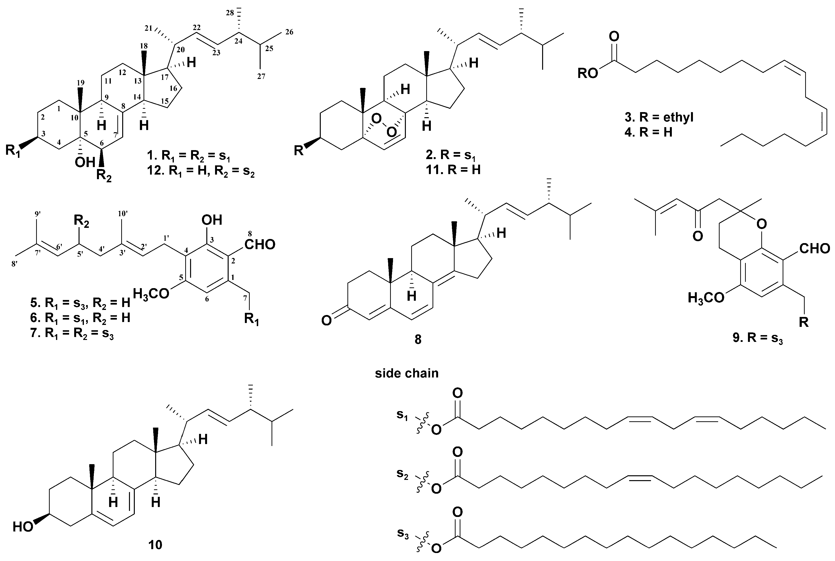
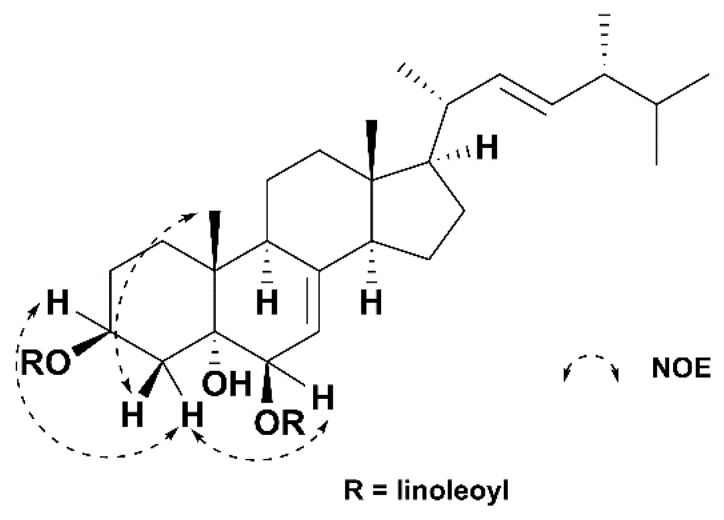
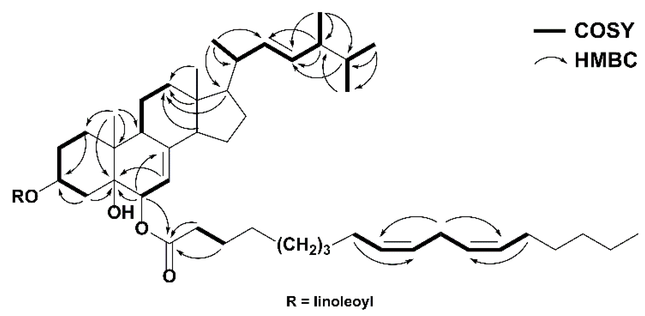
2. Results and Discussion

Compound 1 was isolated as a colorless, oily solid. Its molecular formula was established as C64H106O5 using HRESIMS (high resolution electrospray ionization mass spectrometry) (m/z 955.8112 [M + H]+; calcd 955.8119), indicating 12 degrees of unsaturation. The IR spectrum indicated the presence of hydroxyl group (3435 cm−1) and carbonyl group (1736 cm−1). The 1H NMR data of 1 (Table 1) showed six methyls [δH 0.57 (s, Me-18), 0.82 (d, J = 6.4 Hz, Me-27), 0.83 (d, J = 6.4 Hz, Me-26), 0.91 (d, J = 6.8 Hz, Me-28), 1.02 (d, J = 6.5 Hz, Me-21), 1.06 (s, Me-19)], two oxymethines [δH 4.80 (d, J = 4.7 Hz, H-6α), 5.14 (m, H-3α)], and three olefinic protons [δH 5.19 (m, H-22), 5.21 (m, H-23), 5.28 (m, H-7)]. Additionally, two typical linoleic acid residues were observed, including eight olefinic protons [δH 5.30–5.40 (H-9′, 9″, 10′, 10″, 12′, 12″, 13′, 13″)], twenty-four methylenes [δH 1.23–2.03 (m, H-3′-8′, 14′-17′, 3″-8″, 14″-17″), 2.25 (t, J = 7.5 Hz, Ha-2′/2″), 2.30 (t, J = 7.2 Hz, Hb-2′/2″), 2.77 (t, J = 6.3 Hz, H-11′ and 11″)], and two terminal methyls [δH 0.88 (H-18′ and 18″)]. Its 13C NMR spectrum (Table 1) showed 28 resonances of the sterol moiety, including six methyls [δC 12.3 (C-18), 17.6 (C-28), 18.2 (C-19), 19.6 (C-27), 19.9 (C-26), 21.1 (C-21)], seven sp3 methylenes [δC 21.9 (C-11), 22.8 (C-15), 26.9 (C-2), 27.8 (C-16), 32.2 (C-1), 35.7 (C-4), 39.1 (C-12)], eight sp3 [two oxygenated at δC 70.4 (C-3), 73.4 (C-6)] and three sp2C 114.0 (C-7), 132.1 (C-23), 135.3 (C-22)] methines, and three sp3 [one oxygenated at δC 74.9 (C-5)] and one sp2C 145.6 (C-8)] quaternary carbons. Signals of two linoleic acid residues were also observed in the 13C NMR spectrum, showing eight olefinic carbons [δC 127.8, 127.9, 18.0, 128.0 (C-9′, 9″, 10′, 10″), 130.0, 130.0, 130.2, 130.2 (C-12′, 12″, 13′, 13″)], twenty-four methylene groups [δC 22.5–34.6 (C-2′-8′, C-2″-8″, C-11′, C-11″, C-14′-17′, C-14″-17″)], two methyl groups [δC 14.0, 14.1 (C-18′, 18″)], and two carbonyl groups [δC 173.0 (C-1″), 173.3 (C-1′)]. The data above indicates the sterol moiety of 1 was a Δ7,8 ergostane derivative, closely resembling those of (22E,24R)-ergosta-7,22-diene-3β,5α,6β-triol, a known sterol previously isolated from the fruiting bodies of H. erinaceum [24]. The only difference between these two compounds is that 1 had two additional linoleic acid residues at C-3 and C-6, which were supported by the key HMBC (heteronuclear multiple bond correlation) correlations between H-3 (δH 5.14) and C-1′ (δC 173.3), H-6 (δH 4.80) and C-1″ (δC 173.0) (Figure 2). The relative configuration of 1 was determined using a NOESY (nuclear overhauser effect spectroscopy) NMR experiment (Figure 3). Therefore, compound 1 was determined to be (22E,24R)-ergosta-7,22-diene-3β,5α,6β-triol 3,6-dilinoleate, and named erinarol K.
The structures of the eleven known compounds were identified by comparing HRESIMS, 1H NMR and 13C NMR data with the literature, as 5α,8α-epidioxyergosta-6,22-dien-3β-yl linoleate (2) [25], ethyl linoleate (3) [26], linoleic acid (4) [27], hericene A (5) [7], hericene D (6) [10], hericene E (7) [8], ergosta-4,6,8(14),22-tetraen-3-one (8) [28], hericenone F (9) [29], ergosterol (10) [30], ergosterol peroxide (11) [31], 3β,5α,6α,22E-ergosta-7,22-diene-3,5,6-triol 6-oleate (12) [32].
TNF-α, IL-6 and NO, the major pro-inflammatory mediators, are able to induce inflammation due to overproduction in abnormal situations [33,34,35], and the inhibition effects on their secretion are often used in evaluating the potential anti-inflammatory activities of the isolated natural products [12,36]. Bacterial lipopolysaccharide (LPS) is the best characterized stimulus for the induction of inflammatory mediators in macrophage RAW 264.7 [37]. On the basis of the traditional use in treating gastritis by H. erinaceus [2], we evaluated the potential anti-inflammatory activity of hericene A, D and E (57), hericenone F (9), one type of characteristic aromatic compound only isolated from H. erinaceus, using LPS-stimulated RAW 264.7 mouse cells as the cell model.
First, cell viability was evaluated using the CCK-8 assay. The results showed that compounds 57 and 9 did not affect cell viability at the tested concentrations. As shown in Figure 4, the secretion of TNF-α was significantly inhibited by compounds 57 and 9 in a dose-dependent manner, and compounds 5 and 9 showed the most potent inhibitory activities on the production of inflammatory factor TNF-α, with IC50 values of 78.50 and 62.46 μM, respectively, compared with the positive control (Aspirin, IC50 27.08 μM) (Table 2). We, therefore, further evaluated compounds 5 and 9 for their inhibition on the secretion of IL-6 and NO, another two pro-inflammatory mediators, in LPS-stimulated RAW 264.7 mouse cells. As shown in Figure 5 and Table 2, compounds 5 and 9 also inhibited the secretion of IL-6 and NO in a dose-dependent manner, with IC50 values of 56.33 and 48.5 μM (IL-6), 87.31 and 76.16 μM (NO), respectively, compared with aspirin (IC50, 28.43 μM for IL-6; 51.82 μM for NO).

3. Materials and Methods

3.1. General Experimental Procedures

Optical rotation was measured using a Rudolph Research Analytical APVI/6W automatic polarimeter (Hackettstown, NJ, USA). The FT-IR spectrum was recorded on a ThermoFisher Nicolet 6700 FT-IR spectrometer (Waltham, MA, USA). The NMR spectra were recorded using Bruker AV II 600 and 400 (Billerica, MA, USA), with tetramethylsilane as an internal standard. The high-resolution electrospray ionization mass spectra (HRESIMS) were obtained using a Water Q-TOF Premier (Milford, MA, USA). Column chromatography was performed using silica gel (200–300 mesh, Qingdao Marine Chemical Company, Qingdao, China) and Sephedex LH-20 (GE Healthcare Bio-Sciences AB, Uppsala, Sweden); thin-layer chromatography (TLC) was performed using precoated silica gel GF254 (0.2–0.25 mm, Qingdao Haiyang Chemical Co., Qingdao, China).

3.2. Fungal Material

Mature fruiting bodies of H. erinaceus were collected from a planting base in Jintang District, Chengdu, China, in September 2018 and identified by one of the authors (L.X.). A voucher specimen (HE-201809) was deposited at the Sichuan Institute of Edible Fungi, Sichuan Academy of Agricultural Sciences.

3.3. Extraction and Isolation

Oven-dried fruiting bodies (10 kg) of H. erinaceus were extracted with 95% EtOH (45 L × 3) under room temperature (7d each time). The EtOH extract was concentrated in vacuo to yield a residue (2.1 L), which was further suspended in water and partitioned with EtOAc (6 L × 3), yielding EtOAc fractions (125 g).
The EtOAc fraction (125 g) was subjected to column chromatography over silica gel (200–300 mesh, 1.8 kg) and eluted with petroleum ether-EtOAc (120:1–1:1, gradient system) to yield eighteen fractions (Fr. 1–18). The fraction Fr. 3 (3 g) was separated using silica gel column chromatography with a gradient of cyclohexane-EtOAc (100:1 to 10:1) to yield 5 subfractions (Fr. 3-1–3-5). The fraction Fr. 3-2 was isolated by silica gel column chromatography (cyclohexane-EtOAc, 45:1) and then purified by Sephadex LH-20 column chromatography (CHCl3-MeOH, 2:1) to yield compound 2 (20 mg). The fraction Fr. 3-5 was subjected to silica gel column chromatography (cyclohexane-EtOAc, 80:1) and then purified by Sephadex LH-20 column chromatography (CHCl3-MeOH, 2:1) to yield compound 3 (8 mg). The fraction Fr. 5 (0.8 g) was separated using silica gel column chromatography (petroleum ether-EtOAc, 65:1) to yield 5 subfractions (Fr. 5-1–5-5). The fraction Fr. 5-3 was purified by Sephadex LH-20 column chromatography (CHCl3-MeOH, 2:1) to yield compound 4 (10 mg). The fraction Fr. 6 (0.82 g) was separated using silica gel column chromatography with a gradient of petroleum ether-EtOAc (60:1 to 1:1) to yield 6 subfractions (Fr. 6-1–6-6). The fraction Fr. 6-2 was purified by Sephadex LH-20 column chromatography (CHCl3-MeOH, 2:1) to yield compound 5 (50 mg). The fraction Fr. 8 (0.75 g) was separated using silica gel column chromatography (cyclohexane-EtOAc, 40:1) to yield 5 subfractions (Fr. 8-1–8-5). The fraction Fr. 8-3 was purified by Sephadex LH-20 column chromatography (CHCl3-MeOH, 2:1) to yield compound 6 (10 mg). The fraction Fr. 9 (0.62 g) was separated using silica gel column chromatography with a gradient of petroleum ether-EtOAc (80:1 to 1:1) to yield 7 subfractions (Fr. 9-1–9-7). The fraction Fr. 9-5 was purified by Sephadex LH-20 column chromatography (CHCl3-MeOH, 2:1) to yield compound 7 (10 mg). The fraction Fr. 9-6 was subjected to silica gel column chromatography (petroleum ether-EtOAc, 40:1) and then purified by Sephadex LH-20 column chromatography (CHCl3-MeOH, 2:1) to yield compound 1 (10 mg). The fraction Fr. 10 (0.6 g) was separated using silica gel column chromatography (petroleum ether-EtOAc, 60:1) to yield 6 subfractions (Fr. 10-1–10-6). The fraction Fr. 10-3 was subjected to silica gel column chromatography (petroleum ether-EtOAc, 40:1) and then purified by Sephadex LH-20 column chromatography (CHCl3-MeOH, 2:1) to yield compound 8 (8 mg). The fraction Fr. 11 (3.2 g) was separated using silica gel column chromatography (petroleum ether-EtOAc, 60:1) to yield 7 subfractions (Fr. 11-1–11-7). The fraction Fr. 11-4 was subjected to silica gel column chromatography (petroleum ether-EtOAc, 30:1) and then purified by Sephadex LH-20 column chromatography (CHCl3-MeOH, 2:1) to yield compound 9 (30 mg). The insoluble part of Fr. 12 was recrystallized using EtOAc to yield compound 10 (300 mg). The fraction Fr. 13 (2.1 g) was separated using silica gel column chromatography (petroleum ether-EtOAc, 50:1) to yield 5 subfractions (Fr. 13-1–13-5). The fraction Fr. 13-5 was subjected to silica gel column chromatography (cyclohexane-EtOAc, 20:1) and then purified by Sephadex LH-20 column chromatography (CHCl3-MeOH, 2:1) to yield compound 11 (11 mg). The fraction Fr. 15 (1.3 g) was separated using silica gel column chromatography (petroleum ether-EtOAc, 40:1) to yield 5 subfractions (Fr. 15-1–15-5). The fraction Fr. 15-3 was subjected to silica gel column chromatography (petroleum ether-EtOAc, 20:1) and then purified by Sephadex LH-20 column chromatography (CHCl3-MeOH, 2:1) to yield compound 12 (18 mg).
Erinarol K (1). Colorless oily solid; [α] 21 D −35.9 (c 2.08 × 10⁻³, CHCl3); IR (KBr) νmax 3435, 2925, 2854, 1736, 1461, 1377, 1259, 1168, 758 cm−1; 1H and 13C NMR spectroscopic data, see Table 1; HRESIMS m/z 955.8112 [M + H]+ (calcd for C64H107O5+, 955.8119).

3.4. Anti-Inflammatory Activity Assay

3.4.1. Cell Culture

Raw 264.7 mouse cells (ATCC, Rockville, MD, USA) were cultured in RPMI 1640 (GIBCO, Invitrogen Corporation, Carlsbad, CA, USA) supplemented with 10% fetal bovine serum (FBS) (GIBCO, Invitrogen Corporation, Carlsbad, CA, USA), 100 units/mL penicillin and 100 μg/mL streptomycin (all from Sigma, St. Louis, MO, USA), and then cultured in an incubator at 37 °C under a 5% CO2 atmosphere. Compounds were dissolved in chloroform to make stock solutions of 10 mM (compounds 5, 9 and aspirin) and 50 mM (compounds 6 and 7), which were then diluted in culture medium to obtain the desired concentrations. Aspirin was used as the positive control.

3.4.2. Cell Viability

Cell viability was evaluated by the CCK-8 (Cell Counting Kit-8) method. Compounds in different concentration were added to the cells and incubated for 2 h, and then CCK-8 solution (10 μL, Beyotime, Shanghai, China) was added. The cells were further incubated for 4 h and then the absorbance was measured at 450 nm.

3.4.3. Pro-Inflammatory Cytokines (TNF-α and IL-6) Assay

The production of TNF-a and IL-6 was measured according to the literature with minor modification [38]. RAW 264.7 cells were cultured at a density of 1 × 10⁵ cells/well in RPMI 1640. Cells were pretreated with different concentrations of compounds for 2 h before LPS stimulation. Twenty-four hours after LPS (200 ng/mL) stimulation, TNF-α (TNF-α Elisa kit, Boster Biological Technology Co. Ltd., Wuhan, China) and IL-6 (Mouse IL-6 Elisa kit, Beyotime, Shanghai, China) levels in the supernatant were measured by the ELISA test according to the manufacturer’s instructions.

3.4.4. Nitric Oxide (NO) Assay

The production of NO was measured using the Griess method as previously reported with minor modification [39]. Briefly, the RAW 264.7 cells were pretreated with different concentrations of compounds for 2 h before LPS stimulation. Twenty-four hours after LPS (200 ng/mL) stimulation, 50 μL Griess reagent I and 50 μL Griess reagent II (Beyotime, Shanghai, China) were added into the 50 μL supernatant, respectively. This mixture was incubated for 10 min at room temperature, and the absorbance was measured at 540 nm using a microplate reader (LB 941, Berthold Technologies, Bad Wildbad, Germany). The amount of nitrite in the samples was obtained by a calibration curve using NaNO2 as the standard.

3.4.5. Statistical Analysis

GraphPad Prism 6 (GraphPad Software Inc., San Diego, CA, USA) was used for data processing and analysis. The data obtained are presented as the means ± SD of five independent experiments. A one-way analysis of variance (ANOVA) followed by Tukey’s test was used to determine significant differences between each treated group and the LPS group. Values of p < 0.05 (*), p < 0.01 (**) and p < 0.001 (***) were considered to indicate statistical significance.

4. Conclusions

In this study, the chemical constitution of the fruiting bodies of H. erincacus was studied, and twelve compounds, including one new compound and eleven known compounds were isolated. The four typical aromatic compounds were evaluated for their inhibition effects on the secretion of TNF-α, IL-6 and NO, three major pro-inflammatory mediators, in the macrophage RAW 264.7 model. Two of them showed moderate inhibitory effects indicating their potential anti-inflammatory activity, which may provide the basis for the traditional medical use of H. erincacus.

Author Contributions

Conceptualization, G.X. and L.X.; methodology, G.X., L.T. and Y.X.; resources, G.X. and L.X.; data curation, L.T. and Y.X.; writing―original draft preparation, L.T. and G.X.; writing―review and editing, G.X. and L.X.; project administration, G.X.; funding acquisition, G.X. and L.X. All authors have read and agreed to the published version of the manuscript.

Funding

This research was funded by the Sichuan Science and Technology Program (grant number 2021YFN0094) and the Chengdu Science and Technology Program (grant number 2020-YF09-00080-SN).

Institutional Review Board Statement

Not applicable.

Informed Consent Statement

Not applicable.

Data Availability Statement

Not applicable.

Acknowledgments

The authors thank the Analytical and Measuring Centre, Sichuan University for the spectra measurements.

Conflicts of Interest

The authors declare no conflict of interest. The funders had no role in the design of the study; in the collection, analyses, or interpretation of data; in the writing of the manuscript, or in the decision to publish the results.

Sample Availability

Samples of the compounds are available from the authors.

References

  1. Friedman, M. Chemistry, nutrition, and health-promoting properties of Hericium erinaceus (lion’s mane) mushroom fruiting bodies and mycelia and their bioactive compounds. J. Agric. Food Chem. 2015, 63, 7108–7123. [Google Scholar] [CrossRef]
  2. Mizuno, T. Bioactive substances in Hericium erinaceus (Bull.: Fr.) Pers. and its medicinal utilization. Int. J. Med. Mushrooms 1999, 1, 105–119. [Google Scholar] [CrossRef]
  3. Nagano, M.; Shimizu, K.; Kondo, R.; Hayashi, C.; Sato, D.; Kitagawa, K.; Ohnuki, K. Reduction of depression and anxiety by 4 weeks Hercium erinaceus intake. Biomed. Res. 2010, 31, 231–237. [Google Scholar] [CrossRef] [Green Version]
  4. Mori, K.; Inatomi, S.; Ouchi, K.; Azumi, Y.; Tuchida, T. Improving effects of the mushroom Yamabushitake (Hercium ericaceus) on mild cognitive impairment: A double-blind placebo-controlled clinical trial. Phytother. Res. 2009, 23, 367–372. [Google Scholar] [CrossRef]
  5. Tang, H.Y.; Yin, X.; Zhang, C.C.; Jia, Q.; Gao, J.M. Structure diversity, synthesis, and biological activity of cyathane diterpenoids in higher fungi. Curr. Med. Chem. 2015, 22, 2375–2391. [Google Scholar] [CrossRef] [PubMed]
  6. Li, W.; Sun, Y.N.; Zhou, W.; Shim, S.H.; Kim, Y.H. Erinacene D, a new aromatic compound from Hericium erinaceum. J. Antibiot. 2014, 67, 727–729. [Google Scholar] [CrossRef] [PubMed]
  7. Miyazawa, M.; Takahashi, T.; Horibe, I.; Ishikawa, R. Two new aromatic compounds and a new D-arabinitol ester from the mushroom Hericium erinaceum. Tetrahedron 2012, 68, 2007–2010. [Google Scholar] [CrossRef]
  8. Chen, B.; Han, J.; Bao, L.; Wang, W.; Ma, K.; Liu, H. Identification and α-glucosidase inhibitory activity of meroterpenoids from Hericium erinaceus. Planta Med. 2020, 86, 571–578. [Google Scholar] [CrossRef]
  9. Li, W.; Zhou, W.; Kim, E.J.; Shim, S.H.; Kang, H.K.; Kim, Y.H. Isolation and identification of aromatic compounds in Lion’s Mane mushroom and their anticancer activities. Food Chem. 2015, 170, 336–342. [Google Scholar] [CrossRef]
  10. Ma, B.J.; Yu, H.Y.; Shen, J.W.; Ruan, Y.; Zhao, X.; Zhou, H.; Wu, T.T. Cytotoxic aromatic compounds from Hericium erinaceus. J. Antibiot. 2010, 63, 713–715. [Google Scholar] [CrossRef]
  11. Li, W.; Zhou, W.; Song, S.B.; Shim, S.H.; Kim, Y.H. Sterol fatty acid esters from the mushroom Hericium erinaceum and their PPAR transactivational effects. J. Nat. Prod. 2014, 77, 2611–2618. [Google Scholar] [CrossRef]
  12. Li, W.; Zhou, W.; Cha, J.Y.; Kwon, S.U.; Baek, K.H.; Shim, S.H.; Lee, Y.M.; Kim, Y.H. Sterols from Hericium erinaceum and their inhibition of TNF-α and NO production in lipopolysaccharide-indeced RAW 264.7 cells. Phytochemistry 2015, 115, 231–238. [Google Scholar] [CrossRef]
  13. Hou, Y.; Ding, X.; Hou, W. Composition and antioxidant activity of water-soluble oligosaccharide from Hericium erinaceus. Mol. Med. Rep. 2015, 11, 3794–3799. [Google Scholar] [CrossRef] [Green Version]
  14. Li, Q.Z.; Wu, D.; Chen, X.; Zhou, S.; Liu, Y.; Yang, Y.; Cui, F. Chemical compositions and macrophage activation of polysaccharides from Lion’s Mane culinary-medicinal mushroom Hericium erinaceus (higher Basidiomycetes) in different maturation stages. Int. J. Med. Mushrooms 2015, 17, 443–452. [Google Scholar] [CrossRef]
  15. Cui, F.J.; Li, Y.H.; Zan, X.Y.; Yang, Y.; Sun, W.J.; Qian, J.Y.; Zhou, Q.; Yu, S.L. Purification and partial characterization of a novel hemagglutinating glycoprotein from the cultured mycelia of Hericium erinaceus. Process Biochem. 2014, 49, 1362–1369. [Google Scholar] [CrossRef]
  16. Yu, R.; Sun, M.; Meng, Z.; Zhao, J.; Qin, T.; Ren, Z. Immunomodulatory effects of polysaccharides enzymatic hydrolysis from Hericium erinaceus on the MODE-K/DCs co-culture model. Int. J. Biol. Macromol. 2021, 187, 272–280. [Google Scholar] [CrossRef]
  17. Sheu, S.C.; Lyu, Y.; Lee, M.S.; Cheng, J.H. Immunomodulatory effects of polysaccharides isolated from Hericium erinaceus on dendritic cells. Process Biochem. 2013, 48, 1402–1408. [Google Scholar] [CrossRef]
  18. Kawagishi, H.; Ando, M.; Sakamoto, H.; Yoshida, S.; Ojima, F.; Ishiguro, Y.; Ukai, N.; Furukawa, S. Hericenones C, D and E, stimulators of nerve growth factor (NGF) synthesis, from the mushroom Hericium erinaceum. Tetrahedron Lett. 1991, 32, 4561–4564. [Google Scholar] [CrossRef]
  19. Ma, B.J.; Shen, J.W.; Yu, H.Y.; Ruan, Y.; Wu, T.T.; Zhao, X. Hericenones and erinacines: Stimulators of nerve growth factor (NGF) biosynthesis in Hericium erinaceus. Mycology 2010, 1, 92–98. [Google Scholar] [CrossRef] [Green Version]
  20. Tu, X.; Tang, L.; Xie, G.; Deng, K.; Xie, L. Chemical composition of aromas and lipophilic extracts from black morel (Morchella importuna) grown in China. Mycobiology 2021, 49, 78–85. [Google Scholar] [CrossRef]
  21. Tu, X.M.; Xie, G.B.; Tang, L.; Deng, K.J.; Xie, L.Y. Chemical composition of Morchella sextelata (Pezizales, Ascomycota). Mycosystema 2021, 40, 2134–2147. [Google Scholar]
  22. Deng, K.; Lan, X.; Fang, Q.; Li, M.; Xie, G.; Xie, L. Untargeted metabolomics reveals alterations in the primary metabolites and potential pathways in the vegetative growth of Morchella sextelata. Front. Mol. Biosci. 2021, 8, 632341. [Google Scholar] [CrossRef] [PubMed]
  23. Deng, K.; Lan, X.; Chen, Y.; Wang, T.; Li, M.; Xu, Y.; Cao, X.; Xie, G.; Xie, L. Integration of transcriptomics and metabolomics for understanding the different vegetative growth in Morchella sextelata. Front. Genet. 2022, 12, 829379. [Google Scholar] [CrossRef]
  24. Takaishi, Y.; Uda, M.; Ohashi, T.; Nakano, K.; Murakami, K.; Tomimatsu, T. Glycosides of ergosterol derivatives from Hericum erinacens. Phytochemistry 1991, 30, 4117–4120. [Google Scholar] [CrossRef]
  25. Lin, C.N.; Tome, W.P. Novel cytotoxic principles of Formosan Ganoderma lucidum. J. Nat. Prod. 1991, 54, 998–1002. [Google Scholar] [CrossRef]
  26. Huh, S.; Kim, Y.S.; Jung, E.; Lim, J.; Jung, K.S.; Kim, M.O.; Lee, J.; Park, D. Melanogenesis inhibitory effect of fatty acid alkyl esters isolated from Oxalis triangularis. Biol. Pharm. Bull. 2010, 33, 1242–1245. [Google Scholar] [CrossRef] [Green Version]
  27. Kim, H.J.; Kim, H.M.; Ryu, B.; Lee, W.S.; Shin, J.S.; Lee, K.T.; Jang, D.S. Constituents of PG201 (Layla®), a multi-component phytopharmaceutical, with inhibitory activity on LPS-induced nitric oxide and prostaglandin E2 productions in macrophages. Arch. Pharm. Res. 2016, 39, 231–239. [Google Scholar] [CrossRef]
  28. Gan, K.H.; Kuo, S.H.; Lin, C.N. Steroidal constituents of Ganoderma applanatum and Ganoderma neo-japonicum. J. Nat. Prod. 1998, 61, 1421–1422. [Google Scholar] [CrossRef]
  29. Kawagishi, H.; Ando, M.; Shinba, K.; Sakamoto, H.; Yoshida, S.; Ojima, F.; Ishiguro, Y.; Ukai, N.; Furukawa, S. Chromans, hericenones F, G and H from the mushroom Hericium erinaceum. Phytochemistry 1993, 32, 175–178. [Google Scholar] [CrossRef]
  30. Shirane, N.; Takenaka, H.; Ueda, K.; Hashimoto, Y.; Katoh, K.; Ishii, H. Sterol analysis of DMI-resistant and -sensitive strains of Venturia inaequalis. Phytochemistry 1996, 41, 1301–1308. [Google Scholar] [CrossRef]
  31. Hybelbauerová, S.; Sejbal, J.; Dračínský, M.; Hahnová, A.; Koutek, B. Chemical constituents of Stereum subtomentosum and two other birch-associated Basidiomycetes: An interspecies comparative study. Chem. Biodivers. 2008, 5, 743–750. [Google Scholar] [CrossRef] [PubMed]
  32. Gao, H.; Hong, K.; Zhang, X.; Liu, H.W.; Wang, N.L.; Zhuang, L.; Yao, X.S. New steryl esters of fatty acids from the mangrove fungus Aspergillus awamori. Helv. Chim. Acta 2007, 90, 1165–1178. [Google Scholar] [CrossRef]
  33. Bazzoni, F.; Beutler, B. Seminars in medicine of the Beth Israel hospital, Boston: The tumor necrosis factor ligand and receptor families. N. Engl. J. Med. 1996, 334, 1717–1725. [Google Scholar] [CrossRef]
  34. Tanaka, T.; Narazaki, M.; Kishimoto, T. IL-6 in inflammation, immunity, and disease. Cold Spring Harb. Perspect. Biol. 2014, 6, a016295. [Google Scholar] [CrossRef] [PubMed]
  35. Sharma, J.N.; AI-Omran, A.; Parvathy, S.S. Role of nitric oxide in inflammatory diseases. Inflammopharmacology 2007, 15, 252–259. [Google Scholar] [CrossRef] [PubMed]
  36. Perrone, A.; Plaza, A.; Ercolino, S.F.; Hamed, A.I.; Parente, L.; Pizza, C.; Piacente, S. 14,15-secopregnane derivatives from the leaves of Solenostemma argel. J. Nat. Prod. 2006, 69, 50–54. [Google Scholar] [CrossRef] [PubMed]
  37. MacMicking, J.; Xie, Q.W.; Nathan, C. Nitric oxide and macrophage function. Annu. Rev. Immunol. 1997, 15, 323–350. [Google Scholar] [CrossRef]
  38. Jeong, Y.H.; Oh, Y.C.; Cho, W.K.; Lee, B.; Ma, J.Y. Anti-inflammatory effects of melandrii herba ethanol extract via inhibition of NF-κB and MAPK signaling pathways and induction of HO-1 in RAW 264.7 cells and mouse primary macrophages. Molecules 2016, 21, 818. [Google Scholar] [CrossRef] [Green Version]
  39. Wang, L.; Yang, X.; Zhang, Y.; Chen, R.; Cui, Y.; Wang, Q. Anti-inflammatory chalcone-isoflavone dimers and chalcone dimers from Caragana jubata. J. Nat. Prod. 2019, 82, 2761–2767. [Google Scholar] [CrossRef]
Figure 1. Chemical structures of compounds 112.
Molecules 27 02157 g001
Figure 2. 1H-1H COSY and key HMBC correlations of 1. (COSY: correlation spectroscopy).
Molecules 27 02157 g002
Figure 3. NOESY correlations of 1.
Molecules 27 02157 g003
Figure 4. Effects of compounds 57 and 9 on TNF-α production in LPS-stimulated raw 264.7 cells. * p < 0.05, ** p < 0.01, and *** p < 0.001 compared with LPS-stimulated group. Aspirin (positive control).
Molecules 27 02157 g004
Figure 5. Effects of compounds 5 and 9 on IL-6 (A) and NO (B) production in LPS-stimulated raw 264.7 cells. ** p < 0.01, and *** p < 0.001 compared with LPS-stimulated group. Aspirin (positive control).
Molecules 27 02157 g005
Table 1. 1H NMR (400 MHz) and 13C NMR (100 MHz) Spectroscopic Data for 1 in CDCl3.
PositionδCδH (J in Hz)PositionδCδH (J in Hz)
132.21.55 (m); 1.70 (m)2442.81.84 (m)
226.91.49 (m); 1.89 (m)2533.01.46 (m)
370.45.14 (m)2619.90.83 (d, J = 6.48)
435.71.71 (m); 1.96 (m)2719.60.82 (d, J = 6.44)
574.9-2817.60.91 (d, J = 6.80)
673.44.80 (d, J = 4.7)1′/1″173.3, 173.0-
7114.05.28 (m)2′/2″34.6, 34.62.25 (t, J = 7.5); 2.30 (t, J = 7.2)
8145.6-3′/3″24.9, 25.01.60 (m)
943.22.02 (m)4′/4″29.1–29.71.23–1.35 (m)
1037.3-5′/5″29.1–29.71.23–1.35 (m)
1121.91.57 (m)6′/6″29.1–29.71.23–1.35 (m)
1239.11.32 (m); 2.05 (m)7′/7″29.1–29.71.23–1.35 (m)
1343.7-8′/8″27.2, 27.22.03 (m)
1454.81.91 (m)9′/9″127.8, 127.95.30–5.40 (m)
1522.81.39 (m); 1.42 (m)10′/10″128.0, 128.05.30–5.40 (m)
1627.81.72 (m)11′/11″25.6, 25.62.77 (t, J = 6.3)
1755.91.27 (m)12′/12″130.0, 130.05.30–5.40 (m)
1812.30.57 (s)13′/13″130.2, 130.2,5.30–5.40 (m)
1918.21.06 (s)14′/14″27.2, 27.22.03 (m)
2040.42.01 (m)15′/15″29.1–29.71.23–1.35 (m)
2121.11.02 (d, J = 6.56)16′/16″31.5, 31.91.26 (m)
22135.35.19 (m)17′/17″22.5. 22.71.29 (m)
23132.15.21 (m)18′/18″14.0, 14.10.88 (m)
Table 2. IC50 values a (μM) of compounds 57 and 9 as inhibitors of TNF-α, IL-6 and NO.
CompoundsTNF-αIL-6NO
578.50 ± 3.7256.33 ± 6.8187.31 ± 8.77
6298.50 ± 18.77--
7168.30 ± 9.69--
962.46 ± 3.1848.50 ± 6.5476.16 ± 9.11
Aspirin b27.08 ± 1.8628.43 ± 4.4651.82 ± 8.62
a Data are expressed as mean ± SD; n = 5 independent experiments. b Positive control.
Publisher’s Note: MDPI stays neutral with regard to jurisdictional claims in published maps and institutional affiliations.

