Communication

Identification of Antibacterial Sterols from Korean Wild Mushroom Daedaleopsis confragosa via Bioactivity- and LC-MS/MS Profile-Guided Fractionation

1
School of Pharmacy, Sungkyunkwan University, Suwon 16419, Korea
2
Research Institute of Pharmaceutical Sciences, College of Pharmacy, Sookmyung Women’s University, Seoul 04310, Korea
3
College of Pharmacy and Research Institute of Pharmaceutical Sciences, Gyeongsang National University, Jinju 52828, Korea
4
Special Forest Products Division, Forest Bioresources Department, National Institute of Forest Science, Suwon 16631, Korea
5
College of Pharmacy, Hanyang University, Ansan 15588, Korea
*
Authors to whom correspondence should be addressed.
Molecules 2022, 27(6), 1865; https://doi.org/10.3390/molecules27061865
Submission received: 19 January 2022 / Revised: 8 March 2022 / Accepted: 11 March 2022 / Published: 14 March 2022
(This article belongs to the Section Natural Products Chemistry)

Abstract

:
As part of an ongoing natural product chemical research for the discovery of bioactive secondary metabolites with novel structures, wild fruiting bodies of Daedaleopsis confragosa were collected and subjected to chemical and biological analyses. We subjected the fractions derived from the methanol extract of the fruiting bodies of D. confragosa to bioactivity-guided fractionation because the methanol extract of D. confragosa showed antibacterial activity against Helicobacter pylori strain 51, according to our bioactivity screening. The n-hexane and dichloromethane fractions showed moderate to weak antibacterial activity against H. pylori strain 51, and the active fractions were analyzed for the isolation of antibacterial compounds. Liquid chromatography-tandem mass spectrometry (LC–MS/MS) analysis revealed that the n-hexane fraction contains several compounds which are absent in the other fractions, so the fraction was prioritized for further fractionation. Through chemical analysis of the active n-hexane and dichloromethane fractions, we isolated five ergosterol derivatives (15), and their chemical structures were determined to be demethylincisterol A3 (1), (20S,22E,24R)-ergosta-7,22-dien-3β,5α,6β-triol (2), (24S)-ergosta-7-ene-3β,5α,6β-triol (3), 5α,6α-epoxy-(22E,24R)-ergosta-7,22-dien-3β-ol (4), and 5α,6α-epoxy-(24R)-ergosta-7-en-3β-ol (5) by NMR spectroscopic analysis. This is the first report on the presence of ergosterol derivatives (15) in D. confragosa. Compound 1 showed the most potent anti-H. pylori activity with 33.9% inhibition, rendering it more potent than quercetin, a positive control. Compound 3 showed inhibitory activity comparable to that of quercetin. Distribution analysis of compound 1 revealed a wide presence of compound 1 in the kingdom Fungi. These findings indicate that demethylincisterol A3 (1) is a natural antibiotic that may be used in the development of novel antibiotics against H. pylori.

1. Introduction

Mushrooms have been used for treating various diseases, including diabetes, cancer, and arteriosclerosis, in oriental medicine [1]. In addition, in recent decades, numerous pharmacological and phytochemical studies on mushrooms have proven that they are rich sources of novel compounds with various desirable pharmacological activities such as anti-mutagenic, antioxidant, immunomodulatory, and angiostatic activities, as well as cytotoxicity against cancer cells [1,2,3,4,5,6,7]. Based on this experimental evidence, mushrooms have emerged as potential natural sources for the discovery of bioactive lead molecules; however, most studies have focused on well-known edible ones and little is known regarding biologically active secondary metabolites derived from wild or inedible mushrooms.
As part of our continuing natural product discovery program for the identification of novel bioactive compounds from miscellaneous natural resources [8,9,10,11,12,13,14], we continued chemical and biological analyses of methanolic extracts of the fruiting bodies of D. confragosa, because the methanol extract of D. confragosa showed antibacterial activity against Helicobacter pylori strain 51 in our bioactivity screening. D. confragosa has rarely been investigated in terms of its chemical constituents. In our recent untargeted metabolomics study on 40 species of Polyporaceae, Daedaleopsis tricolor was highlighted as one of the promising producers of unique specialized metabolites [15]. As the appearance of D. confragosa is similar to that of D. tricolor and a recent ITS sequences-based phylogenetic analysis suggested that D. confragosa and D. tricolor may be two morpho-ecological varieties of one species [16], consistent with the suggestion of a mitochondrial small subunit rDNA sequences-based study in 1999 [17], we hypothesized that D. confragosa would show unique specialized metabolites as D. tricolor.
We applied the fractions derived from the methanol extract of D. confragosa into bioactivity-guided fractionation and observed that the n-hexane and CH2Cl2 fractions showed moderate to weak antibacterial activity against H. pylori strain 51. Based on the screening results, we aimed to isolate antibacterial compounds from the active n-hexane and CH2Cl2 fractions, aided by a liquid chromatography-tandem mass spectrometry (LC–MS/MS)-based chemical profiling. Herein, we describe the chemical profile- and bioactivity-guided fractionation of the fractions, isolation and structural determination of compounds 15, and evaluation of their anti-H. pylori activity.

2. Results and Discussion

2.1. Extraction of Metabolites from D. confragosa and Bioactivity-Guided Fractionation

Dried D. confragosa fruiting bodies were extracted with 80% methanol under reflux, yielding the crude methanol extract by rotary evaporation. The resultant methanol extract was sequentially subjected to solvent partitioning with four organic solvents, namely n-hexane, dichloromethane (CH2Cl2), ethyl acetate (EtOAc), and n-butanol (BuOH). Thus, four main solvent fractions with increasing polarity were obtained: n-hexane, CH2Cl2, EtOAc, and BuOH-soluble fractions. The anti-H. pylori activity of the fractions was evaluated using a clinical strain of H. pylori 51. Although the four fractions showed weak or mild inhibitory activity against H. pylori strain 51, n-hexane- and CH2Cl2-soluble fractions showed more potent and similar inhibitory activity, respectively, than the positive control, quercetin (Table 1).

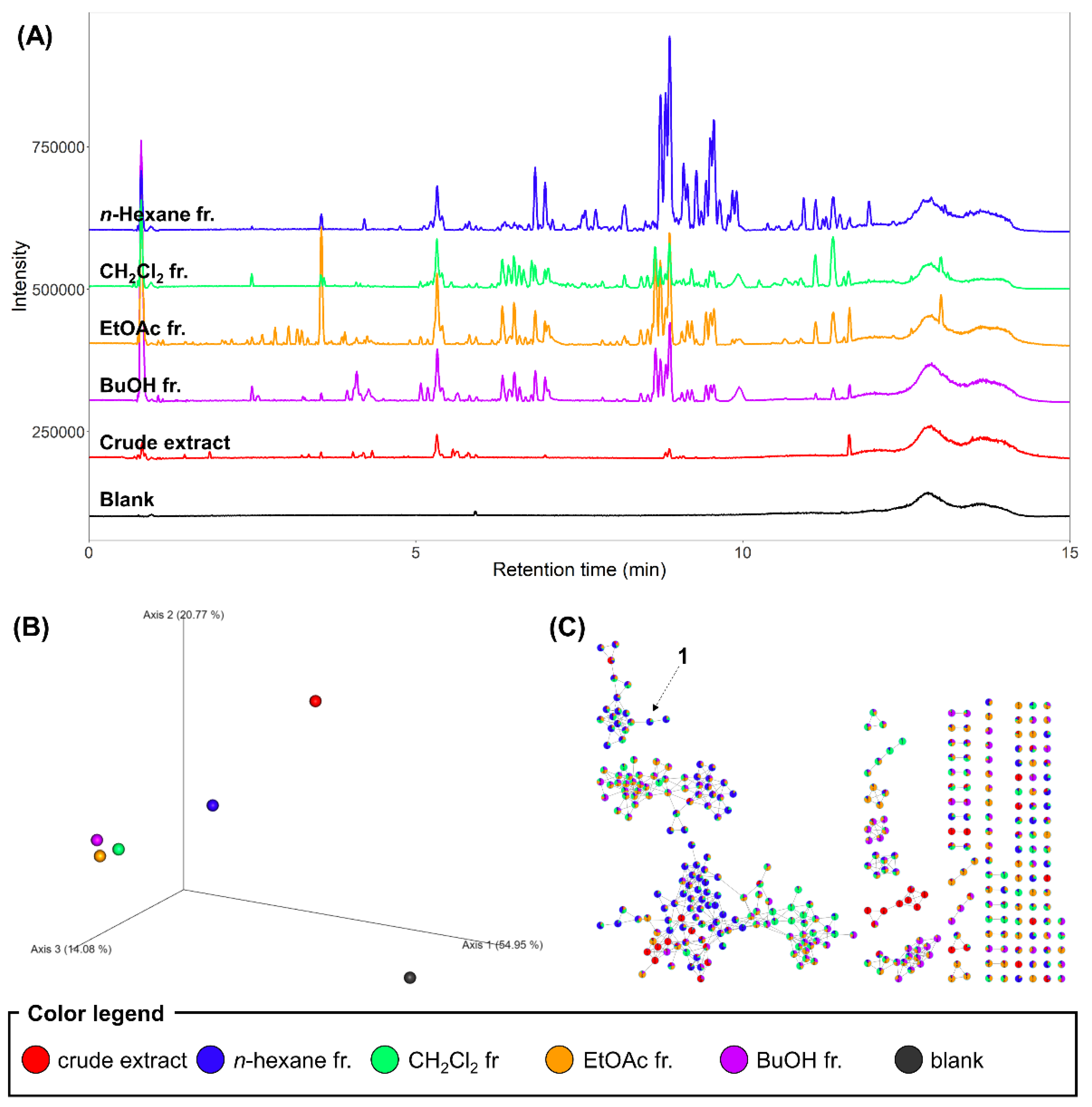
2.2. LC–MS/MS Analysis of the Extract and Fractions

As the crude methanol extract and fractions showed different bioactivities, we performed chemical profiling of the samples to obtain insights into the bioactive components. Contrary to our hypothesis, MS ions detected in the crude extract did not overlap with those from the extract of the isolate culture of D. tricolor [15]. However, we continued the investigation on the fractions, as they showed the bioactivity. Most specialized metabolites showed relatively high intensities in the fractions compared to the crude methanol extract, which suggested that fractions were more concentrated with semi-polar metabolites, mainly due to the removal of the polar primary metabolites (Figure 1A). The LC–MS chromatograms showed that the n-hexane fraction exhibited the most distinct chemical compositions, which was observed in the principal coordinate analysis (PCoA) based on the Bray-Curtis similarity between the samples (Figure 1B). For annotation of metabolites, the MS features were organized into a spectral similarity network through the application of the feature-based molecular networking workflow in GNPS [18]. No molecular families were annotated because their spectra did not match the reference spectra; however, the molecular networking analysis revealed that the n-hexane fraction contained numerous metabolites that were not abundant in the other fractions (Figure 1C). Thus, we hypothesized that some of these metabolites are the bioactive constituents contributing to the antibacterial activity of the n-hexane fraction and performed targeted isolation of the metabolites. In addition, the CH2Cl2 fraction was applied to the isolation, based on the weak antibacterial activity observed in the bioactivity-guided fractionation.

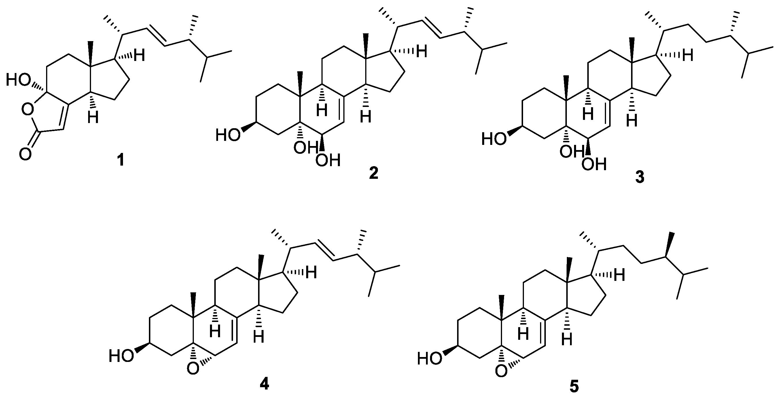
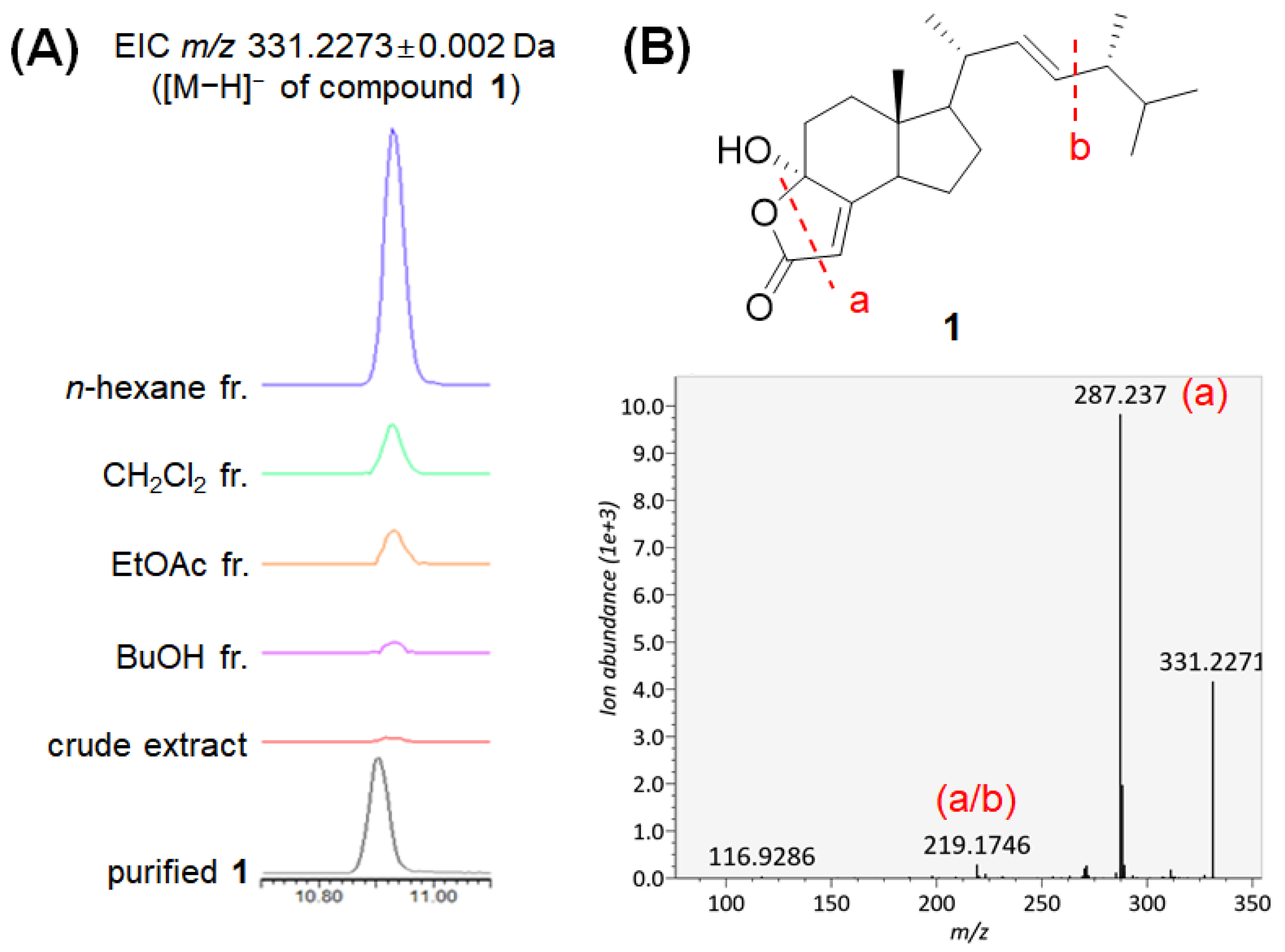
2.3. Isolation and Chemical Characterization of the Compounds

Based on the LC-MS/MS analysis and bioactivity-guided fractionation, the n-hexane- and CH2Cl2-soluble fractions were subjected to analysis using sequential column chromatography, as well as preparative and semi-preparative HPLC, which resulted in the isolation of five ergosterol derivatives (15) (Figure 2).
Compounds 15 were identified as demethylincisterol A3 (1) [19], (20S,22E,24R)-ergosta-7,22-dien-3β,5α,6β-triol (2) [20], (24S)-ergosta-7-ene-3β,5α,6β-triol (3) [21], 5α,6α-epoxy-(22E,24R)-ergosta-7,22-dien-3β-ol (4) [22], and 5α,6α-epoxy-(24R)-ergosta-7-en-3β-ol (5) [23] (Figure 3) by comparing their NMR spectroscopic data with reported values from previously published studies and LC/MS data (Figures S1–S10). This is the first report regarding the presence of the identified ergosterol derivatives (15) in D. confragosa.

2.4. Evaluation of Antibacterial Activity of the Isolated Compounds against H. pylori

The isolated compounds 15 were tested for antibacterial activity against H. pylori strain 51. Among the isolates from the active fractions, compound 1 showed the most potent anti-H. pylori activity with 33.9% inhibition (Table 2); the activity was more potent than that of quercetin, a positive control. Furthermore, compound 3 showed an inhibitory activity comparable to that of quercetin. The other compounds failed to show inhibitory activity. H. pylori is a major public health problem worldwide, affecting approximately 50% of the global population [24]. Extermination of H. pylori leads to the resolution of both gastritis and gastric ulcers, even gastric cancer [25]; however, clinical failures due to antibiotic resistance are of increasing concern. Thus, there is an urgent need to develop novel antibiotics against H. pylori infection. Recent studies have shown that cholesterol, one of the most representative sterols, can play a vital role in virulence and contribute to the intrinsic antibiotic resistance of H. pylori [24]. Another sterol cholestenone exhibited antibiotic activity against H. pylori, including a clarithromycin-resistant H. pylori strain, by suppressing biosynthesis of the cell-wall component cholesteryl-α-ᴅ-glucopyranoside [26]. Based on these findings, we suggest that the antibacterial sterols identified in the present study can potentially serve as clinical antibiotics against H. pylori for use; however, further studies on their mechanism and toxicity are needed.

2.5. Distribution Analysis of Compound 1 in D. confragosa and Other Fungi

The relative abundance of bioactive compound 1 in each fraction was analyzed based on the LC–MS data of the fractions. The extracted ion chromatogram of m/z 331.2273 representing the deprotonated ion of 1 revealed that compound 1 was significantly abundant in the n-hexane fraction (Figure 4A), which might contribute to the high bioactivity of the fraction. We further searched the MS/MS spectrum of compound 1 (Figure 4B) against all publicly available MS/MS datasets deposited in the GNPS MassIVE database through MASST [27] to determine the distribution of this antibacterial sterol among the fungal species. In the MASST search, 20 datasets containing MS/MS spectra matched the spectrum of 1. After removing the matches showing Δm/z > 0.02 Da, four datasets remained, all of which were from fungi. Among these four datasets, three were available for metadata and two were obtained from our previous studies. Compound 1 was present in the fungal cell extracts of Fusarium graminearum (dataset MSV000084977) [28] and fungal culture extracts from eight species in our previous Polyporaceae dataset (MSV000085974) [15], Cerrena unicolor, Coriolopsis strumosa, Daedaleopsis tricolor, Polyporus brumalis, Microporus vernicipes, Trametes orientalis, Trametes suaveolens, and Trametopsis cervina. Interestingly, the extract of D. conragosa in the Polyporaceae dataset did not contain 1, which suggests that the biosynthesis of 1 was not activated under the laboratory culture conditions used in this study. Another dataset, MSV000085071, which contains data from mushroom samples collected during the Mount Pisgah mushroom festival in September 2019, contained 66 files matched to the spectrum of 1. The deposited metadata provided 48 species names for the matched files (Table 3). This result revealed the wide contribution of compound 1 in the kingdom Fungi.

3. Materials and Methods

3.1. General Experimental Procedure

Optical rotations were measured using a Jasco P-1020 polarimeter (Jasco, Easton, MD, USA). Nuclear magnetic resonance (NMR) spectra were recorded using a Bruker Avance III HD 850 NMR spectrometer operating at 850 MHz (1H), with chemical shifts given in ppm (δ). Preparative high-performance liquid chromatography (HPLC) was performed using a Waters 1525 Binary HPLC pump with a Waters 996 Photodiode Array Detector (Waters Corporation, Milford, CT, USA) and an Agilent Eclipse C18 column (250 mm × 21.2 mm, 5 μm; flow rate: 5 mL/min; Agilent Technologies). Semi-preparative HPLC was performed using a Shimadzu Prominence HPLC System with SPD-20A/20AV Series Prominence HPLC UV-Vis Detectors (Shimadzu, Tokyo, Japan). LC/MS analysis was performed on an Agilent 1200 Series HPLC system (Agilent Technologies, Santa Clara, CA, USA) equipped with a diode array detector and a 6130 Series ESI mass spectrometer using an analytical Kinetex C18 Å column (100 mm × 2.1 mm, 5 μm; flow rate: 0.3 mL/min; Phenomenex). Spots were detected on a TLC plate under UV light or following heating after spraying with anisaldehyde–sulfuric acid. Silica gel 60 (230–400 mesh; Merck, Darmstadt, Germany) was used for column chromatography. Sephadex LH-20 (Pharmacia, Uppsala, Sweden) was used as the packing material for molecular sieve column chromatography. Merck precoated silica gel F254 plates and RP-18 F254s plates were used for thin-layer chromatography (TLC).

3.2. Mushroom Material

Fresh fruiting bodies of D. confragosa were collected at the Herb Garden of the College of Pharmacy, Hanyang University, Ansan, Korea in August 2019. DNA sequences were analyzed for identifying the species of this material using a modified method [29]. PCR amplification was performed using the fungal-specific PCR primers ITS1 and ITS4 for the internal transcribed spacer (ITS) region using the modified method [30]. The homology of this region sequence was matched with Deadaleopsis confragosa with the highest matching scores in the NCBI BLAST database. A voucher specimen (SKKU-DJBS-2019-08) of the mushroom was deposited at the herbarium of the School of Pharmacy, Sungkyunkwan University, Korea.

3.3. Extraction of D. Confragosa and Solvent Partition

Completely dried D. confragosa fruiting bodies (2 kg) were extracted with 80% methanol under reflux three times (6.0 L × 3) and subsequently filtered. The filtrate was subsequently evaporated in vacuo to obtain a crude methanol extract (59.3 g). The resultant extract was dissolved in distilled water (700 mL) and then solvent partitioned with hexane, CH2Cl2, EtOAc, and n-BuOH (700 mL × 3). Four layers with increasing polarity were obtained: hexane (3.4 g), CH2Cl2 (6.5 g), EtOAc (0.7 g), and n-BuOH-soluble (2.6 g) fractions.

3.4. LC–MS/MS Analysis

The crude extract and fractions were dissolved in 50% methanol at a concentration of 1.0 mg/mL and filtered through a 0.22 μm PTFE syringe filter (Altoss, Sejong, Korea). LC–MS/MS analysis was performed using a Waters Acquity I-Class UPLC system (Waters Co., Milford, MA, USA) hyphenated to a Waters VION IMS QTOF mass spectrometer (Waters MS Technologies, Manchester, UK). Chromatographic separation was performed on a Waters Acquity UPLC BEH C18 (100 mm × 2.1 mm, 1.7 μm) column maintained at 40 °C. Elution was performed with a mixture of 0.1% formic acid in H2O (A) and MeCN (B) in a linear gradient of 10–100% B (0–12 min) followed by 3 min of washing with 100% B and 3 min of column re-conditioning with 10% B at a flow rate of 0.3 mL/min. The samples (2.0 μL of injection volume) were analyzed in the negative ion mode. MS/MS spectra were acquired in the MSE data-independent acquisition (DIA) mode with a low collision energy of 6 eV for the detection of precursor ions and high collision energy of 20–40 eV for fragmentation. The [M−H] ion of leucine enkephalin at m/z 554.2615 was used as the lock mass to ensure mass accuracy and reproducibility. Chromatographic deconvolution and feature finding were performed using MS-DIAL 4.60 [31], and the resulting feature table and MS/MS spectral list were subjected to the feature-based molecular networking workflow [18] of the Global Natural Products Social Molecular Networking (GNPS; https://gnps.ucsd.edu, accessed on 5 December 2021) environment [32].
Raw LC-MS/MS data and the result files from MS-DIAL processing were deposited in the MassIVE (https://massive.ucsd.edu, accessed on 5 December 2021) with the accession no. MSV000089017.
The MS/MS spectral network can be accessed via the following link: https://gnps.ucsd.edu/ProteoSAFe/status.jsp?task=66d2218f575849a8aecbdb379ae99386, accessed on 5 December 2021.
The experimental spectrum of 1 was searched against public datasets deposited in GNPS MassIVE via MASST [27]. The MASST search results can be accessed via https://gnps.ucsd.edu/ProteoSAFe/status.jsp?task=f0e9eb1240894f838b5079f4f34bc5c6, accessed on 5 December 2021.

3.5. Isolation of Compounds from the Hexane and CH2Cl2 Fractions

The hexane-soluble fraction (3.4 g) was loaded onto a silica gel chromatography column and fractionated using a gradient solvent system of hexane-EtOAc (30:1–1:1, v/v) to yield five fractions (H1–H5). Seven subfractions (H51–H57) were obtained from the fraction H5 (486 mg) by preparative reversed-phase HPLC using acetonitrile–H2O (2:1–1:0, v/v, gradient solvent system, flow rate: 5 mL/min). The H54 subfraction (109 mg) was applied to a Sephadex LH-20 column using CH2Cl2–MeOH (1:1, v/v) to obtain six subfractions (H541-H546). Compound 1 (1.0 mg) was isolated from subfraction H543 (12 mg) via semi-preparative reversed-phase HPLC with 88% MeOH/H2O (isocratic solvent system, flow rate: 2 mL/min). The CH2Cl2 fraction (6.5 g) was loaded onto a silica gel chromatography column and fractionated using a gradient solvent system of CH2Cl2–MeOH (29:1–1:1, v/v) to yield seven fractions (M1–M7). Four subfractions (M51–M54) were obtained from fraction M5 (143 mg) by preparative reversed-phase HPLC using acetonitrile-H2O (70–100% MeOH/H2O, v/v, gradient solvent system, flow rate: 5 mL/min). Compounds 2 (0.8 mg, tR = 40.0 min) and 3 (0.5 mg, tR = 44.0 min) were isolated from subfraction M54 (34 mg) via semi-preparative reversed-phase HPLC using 85% MeOH/H2O (isocratic solvent system, flow rate: 2 mL/min). Five subfractions (M61–M65) were obtained from fraction M6 (140 mg) using a Sephadex LH-20 column using CH2Cl2–MeOH (1:2, v/v). Finally, subfraction M63 (14 mg) was purified by semi-preparative reversed-phase HPLC with 55% CH3CN/H2O (isocratic solvent system, flow rate: 2 mL/min) to afford compounds 4 (1.0 mg, tR = 33.0 min) and 5 (0.7 mg, tR = 38.0 min).

3.6. Helicobacter Pylori Culture

A clinical strain of H. pylori 51 was provided by the H. pylori Korean Type Culture Collection, School of Medicine, Gyeongsang National University, Korea. The strain was grown and maintained on Brucella agar medium (BD Co., Sparks, MD, USA) supplemented with 10% horse serum (Gibco, New York, USA). The culture conditions were 37 °C, 100% humidity, and 10% CO2 for 2–3 days.

3.7. Anti-Helicobacter Pylori Activity

Twenty microliters of the bacterial colony suspension equivalent to 2 × 10⁸–3 × 10⁸ cfu/mL and 20 µL of test samples or controls were added to Brucella broth medium supplemented with 10% horse serum to each well in a 6-well plate. The final volume was 2 mL, and the final concentrations were 100 μg/mL and 100 μM for the fractions and compounds, respectively. After 24 h of incubation at 37 °C, growth was assessed by measuring the optical density at 600 nm using a spectrophotometer. Quercetin and metronidazole were purchased from Sigma (St. Louis, MO, USA) and used as positive controls.

4. Conclusions

In this study, we isolated and characterized the potential anti-H. pylori compounds, five ergosterol derivatives (15) from the methanolic extracts of D. confragosa fruiting bodies via bioactivity-guided fractionation and LC–MS/MS profiling. Their structures were established using NMR spectroscopy and LC-MS analysis. In the anti-H. pylori activity test, we demonstrated that compound 1 showed the most potent anti-H. pylori activity with 33.9% inhibition and compound 3 showed inhibitory activity comparable to that of quercetin, a positive control. Distribution analysis of compound 1 by LC–MS/MS-based analysis indicated the wide distribution of compound 1 in the kingdom Fungi. Based on these findings, we conclude that active demethylincisterol A3 (1) may be used to develop novel antibiotics against H. pylori.

Supplementary Materials

The following supporting information can be downloaded at: https://www.mdpi.com/article/10.3390/molecules27061865/s1, Figure S1: 1H-NMR spectrum of compound 1 (in CD3OD); Figure S2: LC/MS data of compound 1; Figure S3: 1H-NMR spectrum of compound 2 (in CD3OD); Figure S4: LC/MS data of compound 2; Figure S5: 1H-NMR spectrum of compound 3 (in CD3OD); Figure S6: LC/MS data of compound 3; Figure S7: 1H-NMR spectrum of compound 4 (in CDCl3); Figure S8: LC/MS data of compound 4; Figure S9: 1H-NMR spectrum of compound 5 (in CDCl3); Figure S10: LC/MS data of compound 5.

Author Contributions

Conceptualization, K.B.K. and K.H.K.; formal analysis, M.W.N., E.L., D.-M.K., S.Y.J., R.R., C.-Y.K. and M.-J.A.; investigation, M.W.N., E.L. and D.-M.K.; writing—original draft preparation, K.B.K. and K.H.K.; writing—review and editing, K.H.K.; visualization, M.W.N. and K.B.K.; supervision, M.-J.A., K.B.K. and K.H.K.; project administration, K.H.K.; funding acquisition, K.H.K. All authors have read and agreed to the published version of the manuscript.

Funding

This work was supported by a National Research Foundation of Korea (NRF) grant funded by the Korean government (MSIT; grant numbers 2019R1A5A2027340, NRF-2020R1C1C1004046, and 2021R1A2C2007937).

Institutional Review Board Statement

Not applicable.

Informed Consent Statement

Not applicable.

Conflicts of Interest

The authors declare that they have no conflicts of interest.

References

  1. Patel, S.; Goyal, A. Recent developments in mushrooms as anti-cancer therapeutics: A review. 3 Biotech 2011, 2, 1–15. [Google Scholar] [CrossRef] [PubMed] [Green Version]
  2. Wasser, S.P. Current findings, future trends, and unsolved problems in studies of medicinal mushrooms. Appl. Microbiol. Biotechnol. 2011, 89, 1323–1332. [Google Scholar] [CrossRef] [PubMed]
  3. Paterson, R.; Lima, N. Biomedical effects of mushrooms with emphasis on pure compounds. Biomed. J. 2014, 37, 357–368. [Google Scholar] [CrossRef]
  4. Ha, J.W.; Kim, J.; Kim, H.; Jang, W.; Kim, K.H. Mushrooms: An important source of natural bioactive compounds. Nat. Prod. Sci. 2020, 26, 118–131. [Google Scholar]
  5. Lee, S.; Ryoo, R.; Choi, J.H.; Kim, J.H.; Kim, S.H.; Kim, K.H. Trichothecene and tremulane sesquiterpenes from a hallucinogenic mushroom Gymnopilus junonius and their cytotoxicity. Arch. Pharmacal Res. 2020, 43, 214–223. [Google Scholar] [CrossRef] [PubMed]
  6. Lee, S.; Lee, D.; Ryoo, R.; Kim, J.-C.; Park, H.B.; Kang, K.S.; Kim, K.H. Calvatianone, a Sterol Possessing a 6/5/6/5-Fused Ring System with a Contracted Tetrahydrofuran B-ring, from the fruiting bodies of Calvatia nipponica. J. Nat. Prod. 2020, 83, 2737–2742. [Google Scholar] [CrossRef] [PubMed]
  7. Lee, S.; Kim, C.S.; Yu, J.S.; Kang, H.; Yoo, M.J.; Youn, U.J.; Ryoo, R.; Bae, H.Y.; Kim, K.H. Ergopyrone, a Styrylpyrone-Fused Steroid with a Hexacyclic 6/5/6/6/6/5 Skeleton from a Mushroom Gymnopilus orientispectabilis. Org. Lett. 2021, 23, 3315–3319. [Google Scholar] [CrossRef] [PubMed]
  8. Rho, T.; Jeong, H.W.; Hong, Y.D.; Yoon, K.; Cho, J.Y.; Yoon, K.D. Identification of a novel triterpene saponin from Panax ginseng seeds, pseudoginsenoside RT8, and its antiinflammatory activity. J. Ginseng Res. 2020, 44, 145–153. [Google Scholar] [CrossRef]
  9. Lee, S.R.; Kang, H.; Yoo, M.J.; Yu, J.S.; Lee, S.; Yi, S.A.; Kim, K.H. Anti-adipogenic pregnane steroid from a Hydractinia-associated fungus, Cladosporium sphaerospermum SW67. Nat. Prod. Sci. 2020, 26, 230–235. [Google Scholar]
  10. Yu, J.S.; Park, M.; Pang, C.; Rashan, L.; Jung, W.H.; Kim, K.H. Antifungal Phenols from Woodfordia uniflora Collected in Oman. J. Nat. Prod. 2020, 83, 2261–2268. [Google Scholar] [CrossRef]
  11. Lee, D.; Choi, S.; Yamabe, N.; Kim, K.H.; Kang, K.S. Recent Findings on the Mechanism of Cisplatin-Induced Renal Cytotoxicity. Nat. Prod. Sci. 2020, 26, 28–49. [Google Scholar]
  12. Baek, S.C.; Lee, B.S.; Yi, S.A.; Yu, J.S.; Lee, J.; Ko, Y.-J.; Pang, C.; Kim, K.H. Discovery of Dihydrophaseic Acid Glucosides from the Florets of Carthamus tinctorius. Plants 2020, 9, 858. [Google Scholar] [CrossRef] [PubMed]
  13. Jo, M.S.; Lee, S.; Yu, J.S.; Baek, S.C.; Cho, Y.-C.; Kim, K.H. Megastigmane Derivatives from the Cladodes of Opuntia humifusa and Their Nitric Oxide Inhibitory Activities in Macrophages. J. Nat. Prod. 2020, 83, 684–692. [Google Scholar] [CrossRef] [PubMed]
  14. Lee, K.H.; Kim, J.K.; Yu, J.S.; Jeong, S.Y.; Choi, J.H.; Kim, J.-C.; Ko, Y.-J.; Kim, S.-H.; Kim, K.H. Ginkwanghols A and B, osteogenic coumaric acid-aliphatic alcohol hybrids from the leaves of Ginkgo biloba. Arch. Pharmacal Res. 2021, 44, 514–524. [Google Scholar] [CrossRef]
  15. Pham, H.T.; Lee, K.H.; Jeong, E.; Woo, S.; Yu, J.; Kim, W.-Y.; Lim, Y.W.; Kim, K.H.; Bin Kang, K. Species Prioritization Based on Spectral Dissimilarity: A Case Study of Polyporoid Fungal Species. J. Nat. Prod. 2021, 84, 298–309. [Google Scholar] [CrossRef]
  16. Mukhin, V.A.; Zhuykova, E.V.; Vladykina, V.D.; Badalyan, S.M. Notes on medicinal polypore species from the genus Daedaleopsis (Agaricomycetes), distributed in the Asian part of Russia. Int. J. Med. 2020, 22, 775–780. [Google Scholar] [CrossRef]
  17. Ko, K.S.; Jung, H. Molecular phylogeny of Trametes and related genera. Antonie Van Leeuwenhoek 1999, 75, 191–199. [Google Scholar] [CrossRef]
  18. Nothias, L.F.; Petras, D.; Schmid, R.; Dührkop, K.; Rainer, J.; Sarvepalli, A.; Dorrestein, P.C. Feature-based molecular networking in the GNPS analysis environment. Nat. Methods. 2020, 17, 905–908. [Google Scholar] [CrossRef]
  19. Zhao, J.-Y.; Feng, T.; Li, Z.-H.; Dong, Z.-J.; Zhang, H.-B.; Liu, J.-K. Sesquiterpenoids and an ergosterol from cultures of the fungus Daedaleopsis tricolor. Nat. Prod. Bioprospecting 2013, 3, 271–276. [Google Scholar] [CrossRef] [Green Version]
  20. Lee, S.H.; Shim, S.H.; Kim, J.S.; Kang, S.S. Constituents from the fruiting bodies of Ganoderma applanatum and their aldose reductase inhibitory activity. Arch. Pharmacal Res. 2006, 29, 479–483. [Google Scholar] [CrossRef]
  21. Iorizzi, M.; Minale, L.; Riccio, R.; Lee, J.-S.; Yasumoto, T. Polar Steroids from the Marine Scallop Patinopecten yessoensis. J. Nat. Prod. 1988, 51, 1098–1103. [Google Scholar] [CrossRef]
  22. Bok, J.W.; Lermer, L.; Chilton, J.; Klingeman, H.G.; Towers, G.N. Antitumor sterols from the mycelia of Cordyceps sinensis. Phytochemistry 1999, 51, 891–898. [Google Scholar] [CrossRef] [Green Version]
  23. Xu, S.; Liao, X.; Du, B.; Zhou, X.; Huang, Q.; Wu, C. A series of new 5, 6-epoxysterols from a Chinese sponge Ircinia aruensis. Steroids 2008, 73, 568–573. [Google Scholar] [CrossRef] [PubMed]
  24. McGee, D.J.; George, A.E.; Trainor, E.A.; Horton, K.E.; Hildebrandt, E.; Testerman, T.L. Cholesterol Enhances Helicobacter pylori Resistance to Antibiotics and LL-37. Antimicrob. Agents Chemother. 2011, 55, 2897–2904. [Google Scholar] [CrossRef] [Green Version]
  25. Chey, W.D.; Wong, B.C. American College of Gastroenterology guideline on the management of Helicobacter pylori infection. Am. J. Gastroenterol. 2007, 102, 1808–1825. [Google Scholar] [CrossRef]
  26. Kobayashi, J.; Kawakubo, M.; Fujii, C.; Arisaka, N.; Miyashita, M.; Sato, Y.; Nakayama, J. Cholestenone functions as an antibiotic against Helicobacter pylori by inhibiting biosynthesis of the cell wall component CGL. Proc. Natl. Acad. Sci. USA 2021, 118, e2016469118. [Google Scholar] [CrossRef]
  27. Wang, M.; Jarmusch, A.K.; Vargas, F.; Aksenov, A.A.; Gauglitz, J.M.; Weldon, K.; Petras, D.; Da Silva, R.; Quinn, R.; Melnik, A.V.; et al. Mass spectrometry searches using MASST. Nat. Biotechnol. 2020, 38, 23–26. [Google Scholar] [CrossRef]
  28. Kim, D.; Shin, Y.; Lee, S.; Wimonmuang, K.; Bin Kang, K.; Lee, Y.; Yun, S. FgPKS7 is an essential player in mating-type-mediated regulatory pathway required for completing sexual cycle in Fusarium graminearum. Environ. Microbiol. 2021, 23, 1972–1990. [Google Scholar] [CrossRef]
  29. Lee, S.B.; Taylor, J.W. Isolation of DNA from fungal mycelia and single spores. In PCR Protocols: A Guide to Methods and Applications; Innis, M.A., Sninsky, D.H.G.J.J., White, T.J., Eds.; Academic Press: San Diego, CA, USA, 1990; pp. 282–287. [Google Scholar]
  30. Gardes, M.; Bruns, T.D. ITS primers with enhanced specificity for basidiomycetes-application to the identification of mycorrhizae and rusts. Mol. Ecol. 1993, 2, 113–118. [Google Scholar] [CrossRef]
  31. Tsugawa, H.; Cajka, T.; Kind, T.; Ma, Y.; Higgins, B.; Ikeda, K.; Kanazawa, M.; VanderGheynst, J.; Fiehn, O.; Arita, M. MS-DIAL: Data-independent MS/MS deconvolution for comprehensive metabolome analysis. Nat. Methods 2015, 12, 523–526. [Google Scholar] [CrossRef]
  32. Wang, M.; Carver, J.J.; Phelan, V.V.; Sanchez, L.M.; Garg, N.; Peng, Y.; Nguyen, D.D.; Watrous, J.; Kapono, C.A.; Luzzatto-Knaan, T.; et al. Sharing and community curation of mass spectrometry data with Global Natural Products Social Molecular Networking. Nat. Biotechnol. 2016, 34, 828–837. [Google Scholar] [CrossRef] [PubMed] [Green Version]
Figure 1. (A) LC–MS base peak ion (BPI) chromatograms of the crude extract and fractions of D. confragosa. Gaps between chromatograms were added for visualization of their difference; therefore, the y-axis values are not equal to the absolute intensities. (B) PCoA plot based on the Bray–Curtis similarity between the MS data of the crude extract and fractions of D. confragosa. (C) MS/MS spectral network of the crude extract and fractions of D. confragosa. The spectral node representing compound 1 is highlighted.
Molecules 27 01865 g001
Figure 2. The separation scheme of compounds 15.
Molecules 27 01865 g002
Figure 3. Chemical structures of compounds 15.
Molecules 27 01865 g003
Figure 4. (A) Detection of compound 1 in the crude extract and fractions of D. confragosa. EICs of the extract and fractions are in the same scale; the peak height represents the relative abundance of 1. (B) The MS/MS spectrum of compound 1. The putative substructural annotations on the fragment ions are shown.
Molecules 27 01865 g004
Table 1. Anti-H. pyroli activity of the MeOH extract and fractions derived from the solvent partitioning.
SampleConcentrationsInhibition (%)
Methanolic extract100 μg/mL22.4
n-hexane fraction38.2
CH2Cl2 fraction23.3
EtOAc fraction16.0
BuOH fraction15.6
Quercetin a100 μM22.2
Metronidazole a73.5
a Positive controls.
Table 2. Anti-H. pyroli activity of compounds 15.
CompoundConcentrationsInhibition (%)
1100 μM33.9
28.6
318.5
46.8
53.4
Quercetin a100 μM22.2
Metronidazole a73.5
a Positive controls.
Table 3. Fungal species found to contain compound 1, according to the MASST analysis.
DatasetSpeciesFamily
MSV000084977Fusarium graminearumNectraiaceae
MSV000085071Leucoagaricus atrodiscaAgaricaceae
Lepiota spheniscuspora
Amanita pantherinoidesAmanitaceae
Amanita protecta
Phylloporus arenicolaBoletaceae
Suillelus amygdalinus
Bondarzewia occidentalisBondarzewiaceae
Cantharellus californicusCantharellaceae
Cantharellus subalbidus
Craterellus tubaeformis
Clavariadelphus mucronatusClavariadelphaceae
Clavariadelphus occidentalis
Clavulina cinereaClavulinaceae
Cortinarius alboglobosusCortinariaceae
Cortinarius albogfragrans
Cortinarius alboviolaceus
Cortinarius anomalus
Cortinarius cinnamomeus
Cortinarius collinitus
Cortinarius caperatus
Cortinarius causticus
Cortinarius croceus
Clitopilus hobsoniiEntolomataceae
Gomphus vlavatusGomphaceae
Ramaria araispora var. araiospora
Ramaria formosa
Ramaria gelatonosa var. oregonensis
Ramaria rubripermanens
Ramaria sandaracina var. sandaracina
Hydnum umbilicatumHydnaceae
Armillaria solidipesPhysalacriaceae
Polyporus leptocephalusPolyporaceae
Trametes hirsuta
Trametes versicolor
Coprinopsis atramentariaPsathyrellaceae
Lactarius aestivusRussulaceae
Lactarius rubrilacteus
Lactarius scrobiculatus
Russula americana
Russula xerampelina
Pholiota flammansStrophariaceae
Clitocybe nudaTricholomataceae
Collybia cirrhata
Leucopaxillus albissimus
Tricholoma inamoenum
Tricholoma mutabile
Tricholoma pardinum
Tricholoma portentosum
MSV000085974Cerrena unicolorPolyporaceae
Coriolopsis strumose
Daedaleopsis tricolor
Microporus vernicipes
Polyporus brumalis
Trametes orientalis
Trametes suaveolens
Trametopsis cervina
Publisher’s Note: MDPI stays neutral with regard to jurisdictional claims in published maps and institutional affiliations.

