Article

Recombinant Spider Silk Fiber with High Dimensional Stability in Water and Its NMR Characterization

1
Department of Biotechnology, Tokyo University of Agriculture and Technology, 2-24-16 Nakacho, Koganei, Tokyo 184-8588, Japan
2
Department of Applied Chemistry and Biotechnology, Graduate School of Engineering, University of Fukui, 3-9-1 Bunkyo, Fukui 910-8507, Japan
3
Spiber Inc., 234-1 Mizukami, Kakuganji, Tsuruoka 997-0052, Japan
*
Author to whom correspondence should be addressed.
Molecules 2022, 27(23), 8479; https://doi.org/10.3390/molecules27238479
Submission received: 11 October 2022 / Revised: 1 December 2022 / Accepted: 1 December 2022 / Published: 2 December 2022
(This article belongs to the Special Issue The Chemical Properties of Silk Raw Materials)

Abstract

:
Spider dragline silk has unique characteristics of strength and extensibility, including supercontraction. When we use it as a biomaterial or material for textiles, it is important to suppress the effect of water on the fiber by as much as possible in order to maintain dimensional stability. In order to produce spider silk with a highly hydrophobic character, based on the sequence of ADF-3 silk, we produced recombinant silk (RSSP(VLI)) where all QQ sequences were replaced by VL, while single Q was replaced by I. The artificial RSSP(VLI) fiber was prepared using formic acid as the spinning solvent and methanol as the coagulant solvent. The dimensional stability and water absorption experiments of the fiber were performed for eight kinds of silk fiber. RSSP(VLI) fiber showed high dimensional stability, which is suitable for textiles. A remarkable decrease in the motion of the fiber in water was made evident by 13C solid-state NMR. This study using 13C solid-state NMR is the first trial to put spider silk to practical use and provide information regarding the molecular design of new recombinant spider silk materials with high dimensional stability in water, allowing recombinant spider silk proteins to be used in next-generation biomaterials and materials for textiles.

Graphical Abstract

1. Introduction

Spider dragline silks have attracted much attention as a potential source of next-generation biomaterials and textiles because of their outstanding mechanical properties and biocompatibility [1,2,3,4,5,6]. The spider silks have a unique property, supercontraction, which occurs in the hydration process, i.e., interaction with water causes the spider dragline silk fiber to contract by up to 50% in length and to transition from glassy to rubbery phases [7,8,9,10,11]. However, this latter characteristic is not suitable for use in biomaterials, because biomaterials are generally used in water, and therefore, the maintenance of the dimensional stability of biomaterials in water is required [3,4,5]. In addition, for textiles, it is generally required to overcome some inferior characteristics of the fiber, i.e., low water repellency, vulnerability to friction, low dimensional stability, being prone to wrinkles, and so on, and therefore, dimensional stability in water is also necessary [12,13,14].
All spider silks are mainly composed of spidroins. The best characterized silk is the major ampullate silk. It is known that the assembly of major ampullate spidroin 1 (MaSp1) and spidroin 2 (MaSp2) into a fiber demonstrates outstanding mechanical properties [1,2,3,4,5,15,16,17]. The repetitive domains of MaSpl protein are mainly composed of polyalanine (poly-Ala), which forms antiparallel β-sheets (AP-β) in a crystalline region, and Gly-rich regions, which form an amorphous region. The former region has been considered as the origin of high fiber strength, and the latter as the source of the high elasticity of spider dragline silk fiber [1,2,3,4,5].
Because of their cannibalistic behavior, we cannot farm spiders. Furthermore, collecting the silk fiber from spiders is time consuming. Therefore, silk genes have been transferred from spiders to other host organisms to construct recombinant spider silk proteins [18]. In our previous work [19,20,21,22], recombinant spider silk protein (RSP) based on the sequence of ADF(A. diadematus fibroin)-3 silk from the European garden spider Araneus diadematus was produced with E. coli. The two major ampullate silk components of A. diadematus are historically referred to as ADF-3 and -4 [18]. The primary structure and amino acid composition of RSP are shown in Figure S1 [19]. In addition, another spider silk protein named RSSP(QQQ), with a slightly modified primary structure of RSP was also produced using E. coli [23]. We used the abbreviation RSSP(QQQ) in reference to the focus on three types of Q. The primary structure and amino acid composition of RSSP(QQQ) are shown in Figure S2 [23]. Then, the regenerated fibers of these recombinant spider silk proteins were prepared by the wet spinning process, and the secondary structures of the fibers were investigated using 13C solid-state NMR methods.
In this paper, a new recombinant spider silk protein, RSSP(VLI) (Figure 1), based on RSSP(QQQ) [23], where all QQ sequences are replaced by VL while single Q is replaced by I, is produced with E. coli to overcome a critical defect: the low dimensional stability of RSSP(QQQ) fiber in water. In addition, to clarify the structural and dynamical change in the fiber formation process of the recombinant silk protein at the molecular level, solid-state NMR was used.
Because the recombinant spider silk proteins are generally obtained in a powder state, a spinning process to prepare the fibers is required in order to use them widely, and the wet spinning method in particular is frequently used [24]. The dissolution of the powders is a critical step in producing the fibers. The spinning solvents used previously for native spider silks [25] and recombinant spider silks [26,27,28,29,30,31,32] were mainly hexafluoroisopropanol (HFIP) and water. More recently, by learning the spinning system of native spiders, water and aqueous buffers were used for preparing recombinant spider silk fibers with both N-terminal and C-terminal domains [33,34,35,36,37,38,39,40,41]. The relatively small molecular weight makes the recombinant spider silk soluble in water, but the strength of the fiber is quite low at present. The RSSP(VLI) powder was insoluble in water, as well as RSSP(QQQ) powder. In addition, there were many voids in the fiber prepared from the HFIP solution of RSSP(QQQ) [23], which was not suitable for textiles.
On the other hand, formic acid (FA) [42,43,44,45,46,47] and CaCl2-FA [48,49,50,51] have been used as the spinning solvents for regenerated B. mori silk fibroin (RSF), and excellent physical properties have been obtained. Thus, we select FA as the spinning solvent [21,22,23,42,52,53]. FA induces chemical modifications, namely, the formylation of proteins [21,22,23,54,55,56,57]. In addition, the selection of the coagulation solvents is also important. Thus far, alcohols such as methanol (MeOH), ethanol, and isopropanol have been used [24]. We select MeOH as the coagulation solvent [21,22,23,24,25,26,27]. Previous reports have shown the esterification of Ser residue in recombinant spider silk proteins incubated in FA [21,22,23]. Thus, first, the formylation of Ser residue in RSSP(VLI) was studied using the 13C solution NMR method. The mechanical properties of the RSSP(VLI) fibers were examined before and after hydration treatments. Then, dimensional stability was determined for the RSSP(VLI) and RSSP(QQQ) fibers to examine changes in the stability when all QQ sequences were replaced by VL, while single Q was replaced by I. The dimensional stability experiments were expanded by adding the water absorption experiments to more samples, i.e., acetylated RSSP(VLI) and acetylated RSSP(QQQ) fibers, RSP and acetylated RSP fibers, and regenerated B. mori silk fibroin and regenerated acetylated B. mori silk fibroin fibers. The dimensional stability of the latter four samples was reported previously [22,58]. Finally, in order to clarify what happened in the wet spinning process of RSSP(VLI) from FA solution to fiber formation, we observed three kinds of 13C solid-state NMR, i.e., 13C refocused insensitive nuclei enhanced by polarization transfer (r-INEPT), 13C dipolar decoupled-magic angle spinning (DD/MAS), and 13C cross polarization-magic angle spinning (CP/MAS) NMR experiments are performed in the dry and hydrated states [19,22,23,58,59,60,61,62,63,64]. This study, including 13C solid-state NMR is the first trial to put spider silk to practical use, providing information about the molecular design of new recombinant spider silk materials with high dimensional stability in water, allowing the use of recombinant spider silk proteins for next-generation biomaterials and materials for textiles.

2. Results and Discussion

2.1. 13C Solution NMR Spectra of RSSP(VLI) Samples Prepared by Changing the Storage Times in FA

FA is known as a formylating reagent. We previously reported the conformations of RSP and RSSP(QQQ) samples in FA and the formylation of its amino acid side chains [21,23]. These samples consist of Ser and Tyr residues, which are candidates to form formyl groups, but only the Ser side chain was found to be formylated. Thus, we evaluated the formylation of RSSP(VLI) dissolved in FA by 13C solution NMR and confirmed the formylation of the Ser side chain.
First, we assigned the 13C solution NMR peaks of most of the amino acid residues of RSSP(VLI). Figure 2 shows the 13C solution NMR spectrum of the RSSP(VLI) sample in FA after dissolving it for 15 h, together with the peak assignments. All CHn (n = 1–3) groups for most amino acid residues of RSSP(VLI), i.e., Gly, Ala, Pro, Ser, Tyr, Val, Leu, and Ile, were assigned by using 13C HSQC and 1D 13C NMR spectra. The 13C NMR chemical shifts are listed in Table S1. The 13C HSQC spectra of RSSP(VLI) were measured several times continuously to characterize the formylation of the Ser side chain in real time after the dissolution of RSSP(VLI) in FA.
The 13C chemical shifts of Ser CαH and Ser CβH2 peaks changed after the dissolution of RSSP(VLI) in FA. As shown in Figure 3, unformylated Ser CαH and Ser CβH2 peaks were observed in the first 13C HSQC spectrum at 0.7 h after dissolution in FA.
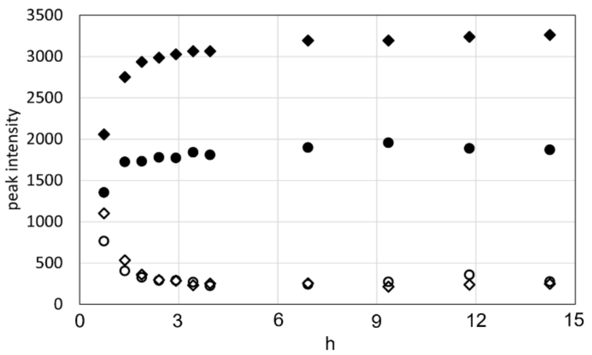
The 1H and 13C chemical shifts were 4.73 ppm and 57.5 ppm for the CαH and 4.08 ppm and 63.3 ppm and 4.17 ppm and 63.3 ppm for the CβH2, respectively. The intensity of these peaks gradually decreased with time and became negligible at 7 h after dissolution. On the other hand, formylated Ser peaks were observed at 4.95 ppm and 54.6 ppm for the CαH and 4.62 ppm and 64.6 ppm and 4.66 ppm and 64.6 ppm for the CβH2, and the intensity of these peaks increased with time. The time-dependent changes in intensity for the CαH and CβH2 peaks of unformylated and formylated Ser residues are plotted in Figure 4.
These measurements show that most of the Ser residues in RSSP (VLI) were formylated within 2 h after dissolution in FA. The formylation rate of RSSP (VLI) is faster than that of RSP and RSSP (QQQ) reported previously [21,23]. This may be due to the fact that the reaction temperature of RSSP(VLI) is higher than that of RSP and RSSP (QQQ), being 313 K for RSSP (VLI) but 293 K for RSP and 298 K for RSP (QQQ). It was necessary to dissolve RSSP (VLI) in FA at a higher temperature because the solubility of RSSP (VLI) was lower than that of RSP and RSSP(QQQ).

2.2. Stress–Strain Curves of RSSP(VLI) Fiber after Repeated Immersion in Water and Drying of the Fiber

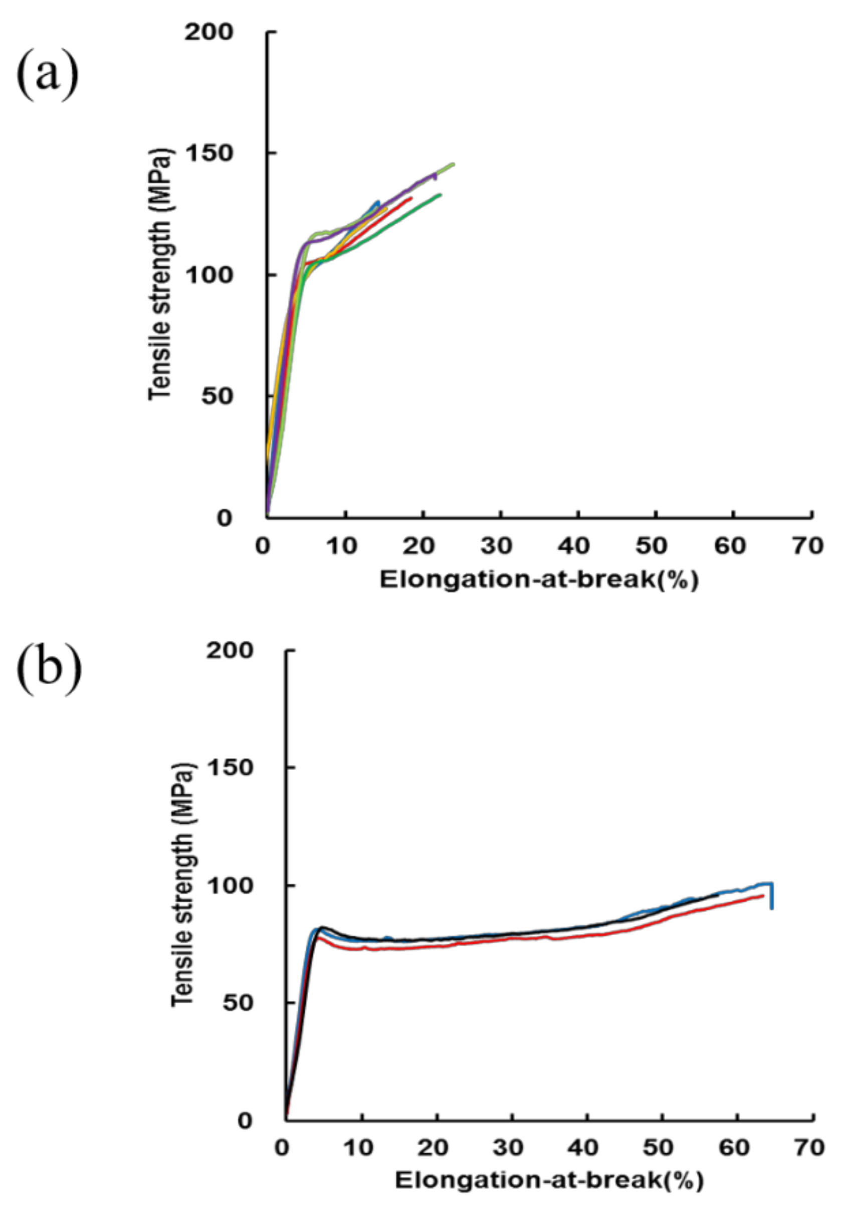
The calculated tensile strength (MPa) and elongation-at-break (%) calculated from the stress–strain curves of RSSP(VLI) fiber prepared by the wet-spinning method from the FA solutions of RSSP(VLI) powder (storage time in FA was 20 h at 40 °C) are shown in Figure 5a.
Stress–strain curves of RSSP(VLI) fiber after hydration treatment, i.e., repeated immersion of the fiber in water and drying at room temperature, are also shown in Figure 5b. The mechanical properties of RSSP(VLI) fiber were compared before and after hydration treatments. The tensile strength (MPa) and elongation-at-break (%) were 134 ± 10 and 19 ± 4 for RSSP(VLI) fiber, respectively, before hydration treatment. The value of the tensile strength (MPa) was lower than the previous value of RSSP(QQQ) fiber, i.e., 191 ± 4 (storage time in FA, 40 h) and 238 ± 5 (storage time in FA, 4 h) [23]. This is potentially due to the difference in the Mw of the samples, i.e., the Mw of RSSP(QQQ) fiber is 210 kDa, which is remarkably higher than the value of Mw 51.6 kDa for RSSP(VLI) fiber. By hydration treatment, the tensile strength (MPa) of RSSP(VLI) fiber decreased from 134 ± 10 to 97 ± 3, but the elongation-at-break (%) increased remarkably from 19 ± 4 to 62 ± 3. These changes are a general matter due to the hydration effect of the silk fibers. Namely, in general, because of the presence of water molecules, the inter-molecular hydrogen bonding of protein fiber such as silk fiber weakens; the molecules are separated from each other, and the space for the movement of the molecules becomes greater, namely, the fiber becomes more plasticized by water. As a result, the tensile strength of silk protein fiber decreases and the elongation-at-break increases by hydration [22,23,24].

2.3. Dimensional Stability Experiments of Eight Kinds of Silk Fiber Samples

At first, we compared the dimensional stabilities of RSSP(QQQ) and RSSP(VLI) fibers, determined by repeated immersion in water and drying, as shown in Figure 6. Because changes in the P ratio largely depend on the condition of the fiber formation, we consider only the S ratio for dimensional stability. Here, the definitions of the P and S ratios are given in the Materials and Methods section. The change is remarkable in the S ratio, i.e., the S ratio of RSSP(VLI) fiber becomes almost half that of the RSSP(QQQ) fiber. This is due to the hydrophobic effect as a result of replacing the QQQ amino acid residues with VLI in the recombinant spider silk.
Next, the dimensional stability experiments were expanded for eight kinds of silk fiber, as shown in the histogram in Figure 7.
We added the S ratio of RSP fibers reported previously [22] for recombinant spider silk fibers other than RSSP(QQQ) and RSSP(VLI) fibers. During the acetylation of the silk fiber, it is known that the fiber becomes more hydrophobic due to the acetylation of Ser and Tyr residues in the silk molecules, and therefore, the acetylated RSSP(QQQ), RSSP(VLI), and RSP fibers were selected for dimensional stability experiments. For this purpose, we prepared acetylated RSSP(QQQ) and RSSP(VLI) fibers, as described in the Materials and Methods section. The regenerated B. mori silk fiber and acetylated regenerated B. mori silk fiber reported previously [58] were also compared with the corresponding data of the recombinant spider silk fibers. The S ratios of both non-acetylated and acetylated recombinant spider silk fibers decreased in the order of RSP, RSSP(QQQ), and RSSP(VLI). The amino acid composition (%) of RSP (Mw = 47.5 kDa) is G:37, Q:19, A:16, P:15, S:6, and Y:4 and that of RSSP(QQQ) (Mw = 210 kDa) is G:31, Q:17, A:20, P:14, S:9, and Y:7. Because the amino acid composition does not change significantly between two samples, the cause of the decrease in the S ratio of the RSSP(QQQ) sample is likely due to the remarkably larger molecular weight compared with that of the RSP sample. The smallest S ratio of the RSSP(VLI) (Mw = 51.6 kDa and G:31, A:20, P:14, S:10, Y:7, V:6, L:6, and I:6) sample is clearly due to the hydrophobic effect as a result of substituting QQQ with VLI, although the Mw is smaller than that of the RSSP(QQQ) sample. In addition, all of the S ratios of the acetylated fibers were smaller than those of the non-acetylated fibers. This is due to the acetylation of the Ser and Tyr residues mentioned above. For B. mori silk fibroin fiber, the decrease in the S ratio was relatively larger than the S ratios of recombinant spider silk fibers. Because both Ser and Tyr residues (amino acid composition (%) of S:12 and Y:5.3) are acetylated in B. mori silk fibroin fiber [58], the hydrophobic effect by acetylation seems greater than that of recombinant spider silks.

2.4. Water Absorption Experiments of Eight Kinds Using Silk Fiber Samples

Eight kinds of silk fiber samples were used for the water absorption experiment, as shown in Figure 8a. The water absorption is generally a reflection of the hydrophilic character of the fiber samples.
To obtain a more detailed insight into the interaction of silk fiber and water molecules, the 2H solution NMR relaxation and exchange measurements of water molecules interacting with silk fibers are very useful, as reported previously by us [59,61]. Namely, (a) bulk water outside the fiber, (b) water molecules trapped weakly on the surface of the fiber, (c) bound water molecules located in the inner surface of the fiber, and (d) bound water molecules located in the inner part of the fiber were distinguishable in the experiments. We would like to focus on these experiments in future work. The water adsorption is far greater for RSP fiber, including acetylated RSP fiber, compared with that of other fibers. Figure 8b shows the plot of the S ratio against the value of water absorption. It can be noticed that there is a correlation between the water absorption and S ratio, with only regenerated B. mori silk fibroin fiber deviating from the trend of the whole plot. Thus, both the RSSP(VLI) fiber and acetylated RSSP(VLI) fiber show the low water absorption and high dimensional stability of recombinant spider silk fiber.

2.5. 13C Solid-State NMR Spectra of RSSP(VLI) Powders and Fibers in the Dry and Hydrated States

A variety of techniques, such as X-ray diffraction, Fourier transform infrared/Raman spectroscopy, transmission electron microscopy, and so on, were used to elucidate the structure of spider dragline silk, from its secondary structure, to its molecular arrangement, to its hierarchical structure [1,2,3,4,5,6]. However, the most detailed picture of the structure and dynamics of spider silk at the molecular level was obtained from NMR spectroscopy. The conformation-dependent 13C chemical shifts coupled with selective 13C labeling can be used effectively to determine the secondary structure of spider silks in an amino-acid-specific manner. Moreover, many kinds of advanced solid-state NMR techniques have been used to obtain the local structure and dynamics of spider silk at atomic resolution [19,20,21,22,23,52,53,65,66,67,68,69,70,71,72]. In this paper, three kinds of 13C solid-state NMR, i.e., 13C r-INEPT, 13C DD/MAS, and 13C CP/MAS NMR spectroscopy [19,22,23,58,59,60,61,62,63,64], were used to clarify the secondary structure and dynamics of RSSP(VLI) powder and fiber in the dry and hydrated states.
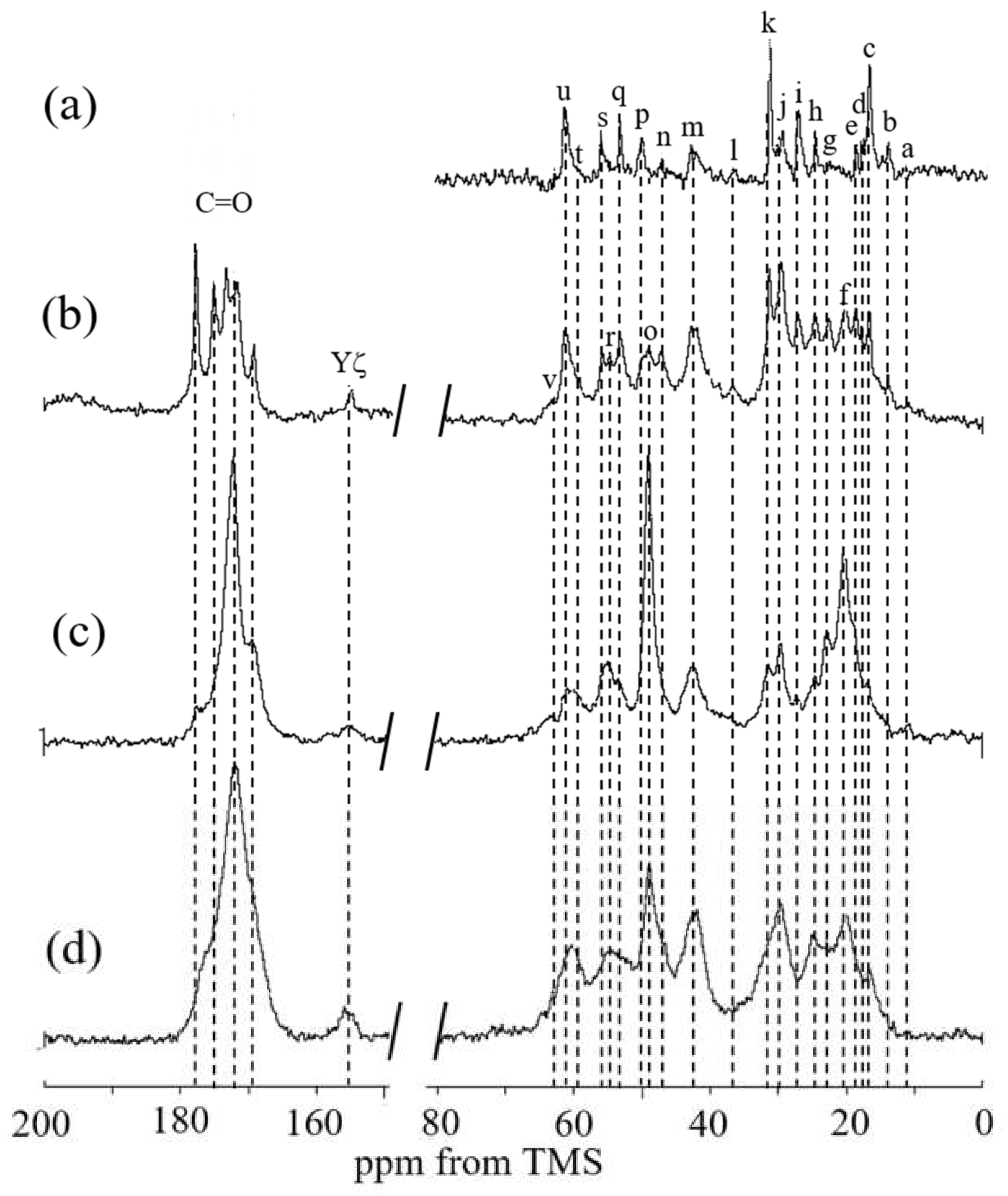
The 13C solid-state NMR spectra of RSSP(VLI) powder before MeOH treatment were first observed in the dry and hydrated states. Figure 9 shows (a) 13C r-INEPT, (b) 13C DD/MAS, and (c) 13C CP/MAS NMR spectra in the hydrated state, together with (d) the 13C CP/MAS NMR spectrum in the dry state. Four kinds of spectra are considerably changed and the spectra look sharper gradually from Figure 9(d) to 9(a). The intensity of the 13C CP/MAS NMR signal is sensitive to the components of very slow motion (< 104 Hz). In the 13C CP/MAS NMR spectrum of RSSP(VLI) in the dry state, the main 13C Ala Cβ and Ala Cα peaks were observed at 20.2 (f) and 48.9 ppm (o), respectively (see Table 1). This means that the conformation of the poly-Ala region has an AP-β structure [73,74]. The 13C chemical shifts of the Ser Cα and Ser Cβ peaks were 55.0 (r) and 63.2 ppm (v), respectively, indicating that the Ser residue also has an AP-β structure [73,74]. Other peaks from the Gly-rich region were also observed. This means that there are amino acid residues forming inter-molecular hydrogen bonds and/or a spatially densely packed region. These amino acid residues are in a restricted state, even if the conformation of the amino acid residue takes the form of random coil.
When the powder is hydrated in water, water molecules cause a significant increase in the motion of the amino acid residues in the powder. In the 13C CP/MAS NMR spectrum Figure 9c, a loss in CP signal occurs because a mobile domain cannot be observed [63,64,70]. Thus, spectrum (c) became sharper than spectrum (d), and the β-sheet peaks of Ala Cα, Cβ, and CO carbons became the main peaks of RSSP(VLI) powder. On the other hand, the 13C r-INEPT was sensitive to the component of fast motion (> 105 Hz) in hydrated powder. Therefore, only the peaks that were very mobile in solution NMR could be observed in the 13C r-INEPT spectrum (a). Only random coil and sharp peaks induced by water absorption were observed for the Cα and Cβ carbons of Ala and Ser residues. All carbons of other residues in the Gly-rich region were also observed as sharp peaks. Thus, mobile components of RSSP(VLI) powder were observed in water. All of the 1H attached carbons in the amino acid residues were observed, although the carbonyl and aromatic peaks with no protons were not observed in the 13C r-INEPT spectrum. In the 13C DD/MAS spectrum (b), both mobile and immobile components in the hydrated state can be observed, namely, the β-sheet peaks of the Cα and Cβ carbons of Ala and Ser residues were observed together with the mobile peaks observed in the 13C r-INEPT spectrum. The carbonyl peaks were also a mixture of both sharp and broad peaks. These results indicate that both rigid β-sheet and flexible random coil structures can be observed in the hydrated state.
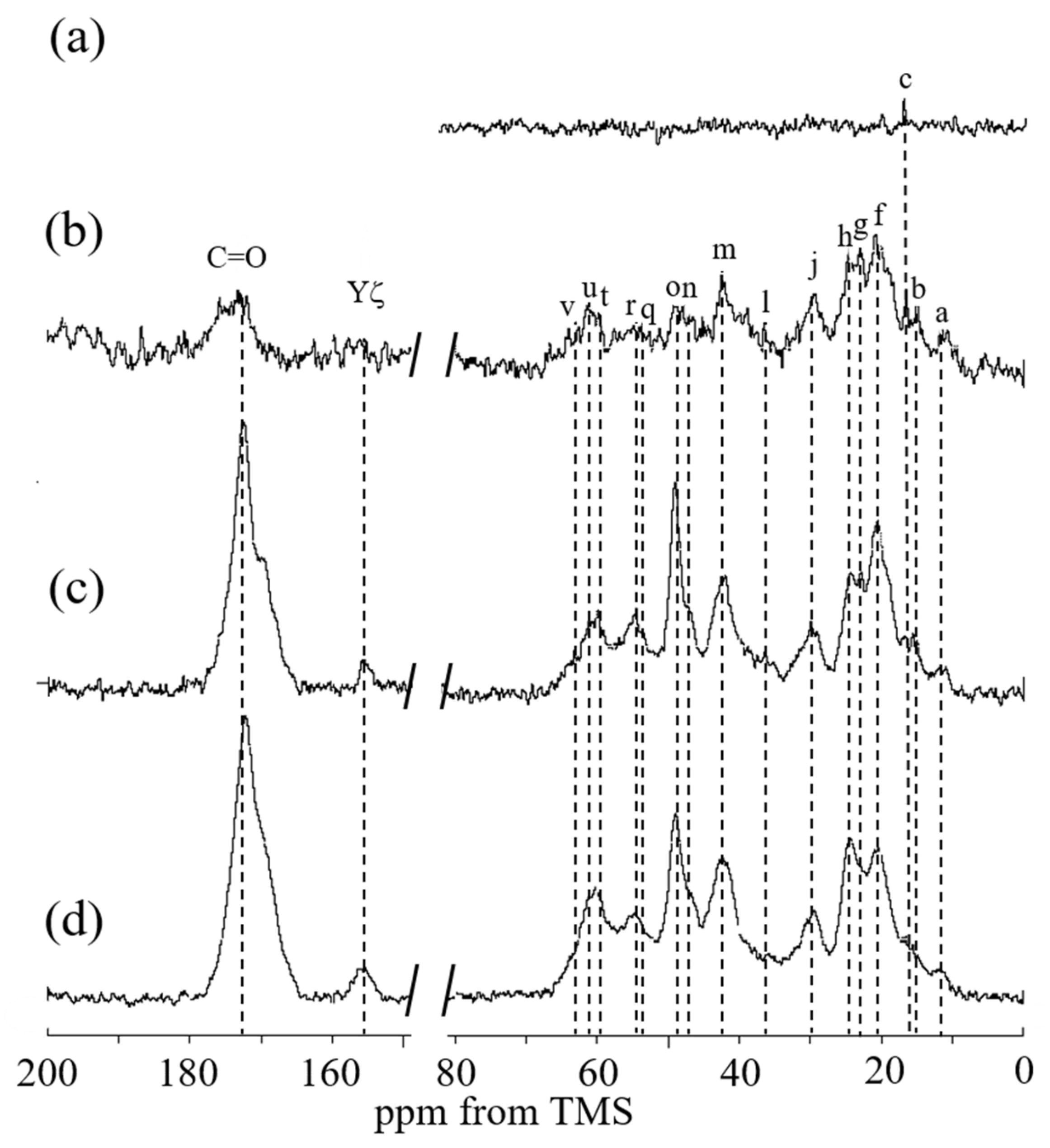
Figure 10 shows the (a) 13C r-INEPT, (b) 13C DD/MAS, and (c) 13C CP/MAS NMR spectra of RSSP(VLI) powder after MeOH treatment in the hydrated state, together with (d) the 13C CP/MAS NMR spectrum in the dry state. The β-sheet peaks of Ala Cα, Ala Cβ, Ser Cα, and Ser Cβ carbons were observed together with other peaks from the Gly-rich region in spectrum (d). With hydration, the β-sheet peaks of Ala residues were also relatively larger in spectrum (c) compared with spectrum (d). The 13C DD/MAS NMR spectrum of RSSP(VLI) powder in water became slightly sharper compared with spectrum (c), but the change was as that shown in Figure 9. The 13C r-INEPT spectrum (a) in Figure 10 shows only a small Ala Cβ peak, and other peaks disappeared completely, which is remarkably different from spectrum (a) in Figure 9. Thus, by MeOH treatment, the mobility of RSSP(VLI) powder does not increase significantly, even in the hydrated state. Figure 11 shows the (a) 13C r-INEPT, (b) 13C DD/MAS, and (c) 13C CP/MAS NMR spectra of RSSP(VLI) fiber in the hydrated state, together with (d) the 13C CP/MAS NMR spectrum in the dry state. The spectra are very similar between Figure 10 and Figure 11 because both samples were obtained after MeOH treatment.
Table 1. 13C solid-state NMR chemical shifts (ppm) of RSSP(VLI) powder and fiber.
Gly42.0, 42.8
m m
Ala50.0 (r.c.)16.6 (r.c)
pc
48.9 (α)20.2 (β)
of
Pro61.431.327.047.1
ukin
Ser53.3 (r.c.), 52.4 (a)61.4 (r.c)
q q*u
55.0 (α)63.2 (β)
rv
Tyr56.136.4
sl
Val61.429.317.8, 18.5
ujd e
Leu53.342.827.022, 24.6
qmig h
Ile59.236.413.9, 24.611.4
tlb ha
(a). The 52.4 (a) ppm peak (q*) was assigned to Cα peak of formylated Ser residue. r.c.: random coil and β: β-sheet.
However, there are two different points in Figure 11 compared with Figure 10. Namely, as mentioned above, the Ser residue of RSSP(VLI) is formylated after FA treatment. Therefore, the Ser Cα peak (q*) of formylated RSSP(VLI) was observed at approximately 1 ppm higher than the Ser Cα peak (q) of unformylated RSSP(VLI) (see Table 1). In addition, the formyl peak was newly observed at 161 ppm in Figure 11.

3. Materials and Methods

3.1. Preparation of RSSP(VLI) Powder Sample

The RSSP(VLI) sample (MW = 51.6 kDa) based on RSSP(QQQ), where all QQ sequences are replaced by VL, while single Q is replaced by I, is produced using E. coli. and purified with a Ni column [17,18]. The His-tag is attached to the N-terminal side of the amino acid sequence and used for sample purification. The primary structure is shown in Figure 1. We tried to produce RSSP(VLI) with Mw = 210 kDa, because RSSP(QQQ) with Mw = 210 kDa was used for a comparison in further experiments, but we could not obtain sufficient amounts of the samples. The RSSP(VLI) powder was used for the observation of solution NMR, and the powder samples before and after MeOH treatments were used for 13C solid-state NMR observations. All of the reagents used here were purchased from FUJIFILM Wako Pure Chemical Corporation, Japan.

3.2. Preparation of Acetylated RSSP(VLI) and Acetylated RSSP(QQQ) Powder Samples

For acetylation, RSSP(VLI) powder (1.0 g) was dissolved in dimethylformamide (DMF, 40 mL) with LiCl (1.6 g) at 80 °C with a stirrer [22]. Then, acetic anhydrate (5 mL) was added and stirred at 80 °C for 6 h. The DMF solution was poured in MeOH (400 mL). The precipitate was collected by centrifugal separation and washed a few times with MeOH. The precipitate suspension in distilled water was dried by lyophilization and then dried at 80 °C overnight. The acetylation of RSSP(QQQ) powders was performed with a similar method. These two acetylated samples, acetylated RSSP(VLI) and RSSP(QQQ), were used for dimensional stability and water absorption experiments after becoming fibrous together with the non-acetylated RSSP(VLI) and RSSP(QQQ) fibers.

3.3. 13C Solution NMR Observation of RSSP(VLI) Sample

The RSSP(VLI) powder was dissolved in formic acid-d1 (Cambridge Isotope Laboratories, Inc., Andover, MA.) to a concentration of 5 w/v% and stored in a 5 mm Shigemi microtube. The solution NMR experiments were performed on a JEOL Resonance ECZ500 spectrometer equipped with a HCN triple resonance inverse probe at 313 K. The assignments of the 13C peaks were accomplished using 1H-13C HSQC and 1D 13C NMR spectra. In order to characterize the formylation of RSSP(VLI) in FA, the 1H-13C HSQC spectra were observed as a function of time after the RSSP(VLI) powder was dissolved in FA. Spectra were processed using JEOL Delta and NMR Pipe. The TMS proton signal at 0 ppm was used as a chemical shift reference for 1H signals. The 13C chemical shifts were indirectly referenced by using the 1H chemical shift value of TMS.

3.4. Preparation of RSSP(VLI) Fiber, Acetylated RSSP(VLI), and Acetylated RSSP(QQQ) Fibers

The preparation of RSSP(VLI) fiber was performed as follows. The RSSP(VLI) powder was dissolved in FA (conc. 20 w/v%) with a stirrer for 16 h at 40 °C and dissolution using rotation/revolution mixer (Awatori Rentaro® ARE-310: THINKY Co., Ltd., Tokyo, Japan) within 4 h. After the FA solution of RSSP(VLI) was filtered with a 5 m steel filter, the FA solution was extruded through a stainless steel spinneret with 0.2 mm inner diameter into the MeOH coagulation bath at room temperature without an air gap [22,23,58,75,76,77]. The coagulation time of the fiber in MeOH was set for 1 min, and then, the fiber was stretched by 4 times with a manual uniaxial stretching machine (Imoto machinery Co. Ltd., Japan) and dried at room temperature. The fibers were used for further experiments, i.e., the determination of the mechanical properties, dimensional stability evaluation, water absorption experiments, and the 13C solid-state NMR measurements. The fibers of acetylated RSSP(VLI) and acetylated RSSP(QQQ) powders were prepared according to a similar method, mentioned above.

3.5. Mechanical Property Measurements of RSSP(VLI) Fibers

The stress–strain curves of the fibers were measured with an EZ-Graph tensile testing machine (EZ-Graph, SHIMADZU Co. Ltd. Japan) at room temperature with a 5 Newton (N) load cell. In the measurement of the stress–strain curve, both ends of the fibers were mounted on Scotch tape with a base length of 20 mm and fixed with ethyl cyanoacrylate [22,23,58,65,66,67]. The rate of crosshead was 3 mm/min on samples of 20 mm length. The breaking strength (MPa) measured as the highest stress value attained during the test was calculated by dividing the cross-sectional area of the fiber. The elongation-at-break (%) was measured as the change in length divided by the initial length. Each value was obtained by averaging over 3–6 measurements. Before starting the tensile test, the diameters of these fibers were measured with an optical microscope (KEYENCE BIOREVO BZ-9000, Japan). The stress–strain curves of the fibers were also observed after the dimensional stability measurements described below. The ends of the fibers were fixed to maintain the length constant at room temperature before the stress–strain experiments.

3.6. Dimensional Stability Experiments with Eight Kinds of Silk Fiber Samples by Repeated Immersion in Water and Drying

Eight kinds of silk fibers, i.e., the RSSP(VLI) (Mw:51.6 kDa) and acetylated RSSP(VLI) fibers, RSSP(QQQ) (Mw:210 kDa) and acetylated RSSP(QQQ) fibers, and RSP (Mw:47.5 kDa) and acetylated RSP fibers reported previously [22], and regenerated B. mori silk fibroin and regenerated acetylated B. mori silk fibroin fibers, also reported previously [58], were used for the dimensional stability measurements. The dimensional stabilities were determined by repeated immersion in water and drying at room temperature as follows [22,58]. A bundle of 24 fibers was loaded onto a 7.84 mN weight installed at the bottom edge of the bundle. The length of the bundle in the dry state, Ldry0, was determined. After immersing the bundles in water for 30 min, the length in the hydrated state, Lhydrated1, was determined. Here, the primary contraction, or P ratio, of the bundles is defined as the change in length when the bundles are first immersed in water, expressed by the following formula: (Lhydrated1 − Ldry0) /Ldry0 [22,58]. Then, the bundles were dried at room temperature for 30 min and Ldry2 is determined. This hydration and drying treatment was repeated three times. The averaged value over (Lhydrated3 − Ldry2) /Ldry2, (Lhydrated3 − Ldry4)/Ldry4, (Lhydrated5 − Ldry4) /Ldry4 and (Lhydrated5 − Ldry6)/Ldry6 was defined to determine the secondary contraction, S ratio [22,58]. We compared only the S ratio as dimensional stability among samples because the P ratio changed greatly depending on the condition of the fiber formation.

3.7. Water Absorption Experiments of Eight Kinds Using Silk Fiber Samples

Eight kinds of silk fibers were used for water absorption experiments. Approximately 35 mg of each dry sample of the eight kinds of silk fiber was weighed after drying the samples at 80 °C for 16 h. Then, these samples were pressed at 20 MPa for 5 min using a tablet modeling machine for infrared measurement, and a tablet of diameter ca. 10 mm and thickness ca. 1mm was obtained. The weight, A mg, of the dry tablet was measured. Then, the tablet was immersed in water for 30 min and the water quickly wiped from the surface of the tablet. The weight, B mg, of the wet tablet was measured. The water absorption (%) of the tablet was calculated using the equation [(B – A)/A] × 100 (%).

3.8. 13C r-INEPT, 13C CP/MAS and 13C DD/MAS NMR Measurements of RSSP(VLI) Powders and Fibers in the Dry and Hydrated States

The 13C solid-state NMR spectra of RSSP(VLI) powders before and after MeOH treatments, as well as the RSSP(VLI) fibers, were observed in the dry and hydrated states using a Bruker Avance 400 NMR spectrometer with a 4 mm double resonance MAS probe and a MAS rate of 8.5 kHz at room temperature. For the NMR observations in the hydrated state, the samples were carefully inserted into a zirconia rotor, sealed with polytetrafluoroethylene (PTFE) insert to prevent the dehydration of the hydrated samples [19,22,23,33,34,35,36,37,38,39,40,41,42,43]. Typical parameters for the 13C CP/MAS NMR experiments were a 3.5 μs 1H 90° pulse, a 1 ms ramped CP pulse with 71.4 kHz rf field strength, two pulse phase modulation (TPPM) 1H decoupling during acquisition, 2176 data points, 8 k scans, 310 ppm sweep width, and a 4 s recycle delay. Lorentzian line broadening of 20 Hz was applied prior to Fourier transformation. To observe both the mobile and immobile parts of PSSP(VIL), the 13C DD/MAS NMR method was used. In the 13C DD/MAS NMR experiments, 13C nuclei were excited using a single 90° pulse, and 13C free induction signals were subsequently acquired under a high power 1H decoupling pulse. A recycle delay of 5 s was used to prevent the saturation of 13C NMR signals. A repetition time of 5 s was selected because the reduction in 13C NMR signals by saturation caused by long 13C spin lattice relaxation time in the rigid part was negligible, as indicated in our previous repetition time variation experiments [59]. Details of the NMR conditions for the 13C DD/MAS NMR experiments were described previously [59], including 8 k scans, a recycle delay of 5 s, and a 13C 90° pulse of 3.5 μs. Typical experimental parameters for the r-INEPT NMR experiments include 3.5 μs 1H and 3.6 μs 13C pulses, an inter-pulse delay of 1/41JCH (1JCH =145 Hz), a refocusing delay of 1/61JCH or 1/31JCH, TPPM 1H decoupling during acquisition, 1438 data points, 4 k scans, 200 ppm sweep width, and a 3.5 s recycle delay. The 13C chemical shifts were calibrated externally through the methylene peak of adamantane observed at 28.8 ppm relative to TMS at 0 ppm.

4. Conclusions

A new recombinant spider silk protein, RSSP(VLI) based on the RSSP(QQQ), where all QQ sequences are replaced by VL, while single Q is replaced by I, is produced with E. coli to overcome a critical defect: the low dimensional stability of RSSP(QQQ) fiber in water, preventing its practical use. The fiber was prepared from powder using formic acid as the spinning solvent and methanol as the coagulant solvent during the wet spinning process. A higher dimensional stability of RSSP(VLI) fiber than other silk fibers was obtained. A remarkable decrease in the motion of the fiber in water was evident by 13C solid-state NMR studies. This study, including 13C solid-state NMR is the first trial to put spider silk to practical use, providing information for the molecular design of new recombinant spider silk materials with high dimensional stability in water, allowing the use of recombinant spider silk proteins in next-generation biomaterials and materials for textiles.

Supplementary Materials

The following supporting information can be downloaded at: https://www.mdpi.com/article/10.3390/molecules27238479/s1, Figure S1: Primary structure and amino acid compositions (%) of recombinant spider silk protein, RSP based on the primary sequence of Araneus diadematus silk. Each amino acid was colored in G(black), Q(violet), A(red), P(green), S(blue), Y(orange), and others (grey); Figure S2: Primary structure and amino acid compositions (%) of recombinant spider silk protein (RSSP(QQQ): Mw = 210 kDa) based on the primary sequence of Araneus diadematus silk. Polyalanine-rich sequences were colored red and others black; Table S1: 13C solution NMR chemical shifts (ppm) of RSSP(VLI) in formic acid.

Author Contributions

T.A.: conceptualization, investigation, project administration, writing—original draft, review and editing; H.M.: investigation, methodology; A.N.: investigation, writing—review and editing; H.O.: investigation; Y.S.: investigation, writing—review and editing, and Y.A.: investigation, writing—review and editing. All authors have read and agreed to the published version of the manuscript.

Funding

This work was funded by a JSPS KAKENHI, Grant-in-Aid for Scientific Research (C), Grant Number JP19K05609.

Institutional Review Board Statement

Not applicable.

Informed Consent Statement

Not applicable.

Data Availability Statement

The data presented in this study are available in this article.

Acknowledgments

T.A. acknowledges support by a JSPS KAKENHI, Grant-in-Aid for Scientific Research (C), Grant Number JP19K05609.

Conflicts of Interest

The authors declare no conflict of interest.

Sample Availability

Samples of the compounds are not available from the authors.

References

  1. Gosline, J.M.; DeMont, M.E.; Denny, M.W. The structure and properties of spider silk. Endeavour 1986, 10, 37–43. [Google Scholar] [CrossRef]
  2. Vollrath, F.; Knight, D.P. Liquid crystalline spinning of spider silk. Nature 2001, 410, 541–548. [Google Scholar] [CrossRef] [PubMed]
  3. Heidebrecht, A.; Scheibel, T. Chapter four. In Recombinant Production of Spider Silk Proteins; Sariaslani, S., Gadd, G.M., Eds.; Academic Press: Cambridge, MA, USA, 2013; pp. 115–153. [Google Scholar] [CrossRef]
  4. Asakura, T.; Miller, T. (Eds.) Biotechnology of Silk; Biologically-Inspired Systems; Springer: Dordrecht, The Netherlands, 2014; Volume 5, ISBN 978-94-007-7118-5. [Google Scholar]
  5. Tokareva, O.; Jacobsen, M.; Buehler, M.; Wong, J.; Kaplan, D.L. Structure–function–property–design interplay in biopolymers: Spider silk. Acta Biomater. 2014, 10, 1612–1626. [Google Scholar] [CrossRef] [PubMed] [Green Version]
  6. Wang, Q.; McArdle, P.; Wang, S.L.; Wilmington, R.L.; Xing, Z.; Greenwood, A.; Cotten, M.L.; Qazilbash, M.M.; Schniepp, H.C. Protein secondary structure in spider silk nanofibrils. Nat. Commun. 2022, 13, 4329. [Google Scholar] [CrossRef] [PubMed]
  7. Savage, K.N.; Gosline, J.M. The effect of proline on the network structure of major ampullate silks as inferred from their mechanical and optical properties. J. Exp. Biol. 2008, 211, 1937–1947. [Google Scholar] [CrossRef] [PubMed] [Green Version]
  8. Liu, Y.; Sponner, A.; Porter, D.; Vollrath, F. Proline and Processing of Spider Silks. Biomacromolecules 2008, 9, 116–121. [Google Scholar] [CrossRef]
  9. Cohen, N.; Levin, M.; Eisenbach, C.D. On the Origin of Supercontraction in Spider Silk. Biomacromolecules 2021, 22, 993–1000. [Google Scholar] [CrossRef]
  10. Work, R.W.; Morosoff, N. A Physico-Chemical Study of the Supercontraction of Spider Major Ampullate Silk Fibers. Text. Res. J. 1982, 52, 349–356. [Google Scholar] [CrossRef]
  11. Elices, M.; Plaza, G.R.; Arnedo, M.A.; Pérez-Rigueiro, J.; Torres, F.G.; Guinea, G.V. Mechanical Behavior of Silk During the Evolution of Orb-Web Spinning Spiders. Biomacromolecules 2009, 10, 1904–1910. [Google Scholar] [CrossRef]
  12. Arai, T.; Freddi, G.; Innocent, R.; Kaplan, D.L.; Tsukada, M. Acylation of silk and wool with acid anhydrides and preparation of water-repellent fibers. J. Appl. Polym. Sci. 2001, 82, 2832–2841. [Google Scholar] [CrossRef]
  13. John, M.J.; Anandjiwala, R.D. Recent developments in chemical modification and characterization of natural fiber-reinforced composites. Polym. Compos. 2008, 29, 187–207. [Google Scholar] [CrossRef]
  14. Li, G.; Liu, H.; Li, T.; Wang, J. Surface modification and functionalization of silk fibroin fibers/fabric toward high performance applications. Mater. Sci. Eng. C. 2012, 32, 627–636. [Google Scholar] [CrossRef]
  15. Xu, M.V.; Lewis, R. Structure of a protein superfiber: Spider dragline silk. Proc. Natl. Acad. Sci. USA 1990, 87, 7120–7124. [Google Scholar] [CrossRef] [Green Version]
  16. Guerette, P.A.; Ginzinger, D.G.; Weber, B.H.; Gosline, J.M. Silk properties determined by gland-specific expression of a spider fibroin gene family. Science 1996, 272, 112–115. [Google Scholar] [CrossRef]
  17. Rising, A.; Widhe, M.; Johansson, J.; Hedhammar, M. Spider silk proteins: Recent advances in recombinant production, structure–function relationships and biomedical applications. Cell. Mol. Life Sci. 2011, 68, 169–184. [Google Scholar] [CrossRef]
  18. Humenik, M.; Smith, A.M.; Scheibel, T. Recombinant Spider Silks—Biopolymers with Potential for Future Applications. Polymers 2011, 3, 640–661. [Google Scholar] [CrossRef] [Green Version]
  19. Tasei, Y.; Nishimura, A.; Suzuki, Y.; Sato, T.K.; Sugahara, J.; Asakura, T. NMR Investigation about Heterogeneous Structure and Dynamics of Recombinant Spider Silk in the Dry and Hydrated States. Macromolecules 2017, 50, 8117–8128. [Google Scholar] [CrossRef]
  20. Asakura, T. Structure and Dynamics of Spider Silk Studied with Solid-State Nuclear Magnetic Resonance and Molecular Dynamics Simulation. Molecules 2020, 25, 2634. [Google Scholar] [CrossRef]
  21. Suzuki, Y.; Higashi, T.; Yamamoto, T.; Okamura, H.; Sato, T.K.; Asakura, T. Presence of β-Turn Structure in Recombinant Spider Silk Dissolved in Formic Acid Revealed with NMR. Molecules 2022, 27, 511. [Google Scholar] [CrossRef] [PubMed]
  22. Asakura, T.; Matsuda, H.; Aoki, A.; Naito, A. Acetylation and hydration treatment of recombinant spider silk fiber, and their characterization using 13C NMR spectroscopy. Polymer 2022, 243, 124605. [Google Scholar] [CrossRef]
  23. Asakura, T.; Matsuda, H.; Naito, A.; Abe, Y. Formylation of Recombinant Spider Silk in Formic Acid and Wet Spinning Studied Using Nuclear Magnetic Resonance and Infrared Spectroscopies. ACS Biomater. Sci. Eng. 2022, 8, 2390–2402. [Google Scholar] [CrossRef] [PubMed]
  24. Koeppel, A.; Holland, C. Progress and Trends in Artificial Silk Spinning: A Systematic Review. ACS Biomater. Sci. Eng. 2017, 3, 226–237. [Google Scholar] [CrossRef] [PubMed] [Green Version]
  25. Seidel, A.; Liivak, O.; Calve, S.; Adaska, J.; Ji, G.; Yang, Z.; Grubb, D.; Zax, D.B.; Jelinski, L.W. Regenerated Spider Silk: Processing, Properties, and Structure. Macromolecules 2000, 33, 775–780. [Google Scholar] [CrossRef]
  26. Lazaris, A.; Arcidiacono, S.; Huang, Y.; Zhou, J.F.; Duguay, F.; Chretien, N.; Welsh, E.A.; Soares, J.W.; Karatzas, C.N. Spider Silk Fibers Spun from Soluble Recombinant Silk Produced in Mammalian Cells. Science 2002, 295, 472–476. [Google Scholar] [CrossRef] [PubMed] [Green Version]
  27. Xia, X.-X.; Qian, Z.-G.; Ki, C.S.; Park, Y.H.; Kaplan, D.L.; Lee, S.Y. Native-sized recombinant spider silk protein produced in metabolically engineered Escherichia coli results in a strong fiber. Proc. Natl. Acad. Sci. USA 2010, 107, 14059–14063. [Google Scholar] [CrossRef] [Green Version]
  28. Teulé, F.; Addison, B.; Cooper, A.R.; Ayon, J.; Henning, R.W.; Benmore, C.J.; Holland, G.P.; Yarger, J.L.; Lewis, R.V. Combining flagelliform and dragline spider silk motifs to produce tunable synthetic biopolymer fibers. Biopolymers 2012, 97, 418–431. [Google Scholar] [CrossRef] [Green Version]
  29. Lin, Z.; Deng, Q.; Liu, X.-Y.; Yang, D. Engineered Large Spider Eggcase Silk Protein for Strong Artificial Fibers. Adv. Mater. 2013, 25, 1216–1220. [Google Scholar] [CrossRef]
  30. Albertson, A.E.; Teulé, F.; Weber, W.; Yarger, J.L.; Lewis, R.V. Effects of different post-spin stretching conditions on the mechanical properties of synthetic spider silk fibers. J. Mech. Behav. Biomed. Mater. 2014, 29, 225–234. [Google Scholar] [CrossRef] [Green Version]
  31. Copeland, C.G.; Bell, B.E.; Christensen, C.D.; Lewis, R.V. Development of a Process for the Spinning of Synthetic Spider Silk. ACS Biomater. Sci. Eng. 2015, 1, 577–584. [Google Scholar] [CrossRef]
  32. Li, X.; Mi, J.; Wen, R.; Zhang, J.; Cai, Y.; Meng, Q.; Lin, Y. Wet-Spinning Synthetic Fibers from Aggregate Glue: Aggregate Spidroin 1 (AgSp1). ACS Appl. Bio Mater. 2020, 3, 5957–5965. [Google Scholar] [CrossRef]
  33. Wohlrab, S.; Thamm, C.; Scheibel, T. The Power of Recombinant Spider Silk Proteins. In Biotechnology of Silk; Asakura, T., Miller, T., Eds.; Springer: Dordrecht, The Netherlands, 2014; pp. 179–201. ISBN 978-94-007-7119-2. [Google Scholar]
  34. Jones, J.A.; Harris, T.I.; Tucker, C.L.; Berg, K.R.; Christy, S.Y.; Day, B.A.; Gaztambide, D.A.; Needham, N.J.C.; Ruben, A.L.; Oliveira, P.F.; et al. More Than Just Fibers: An Aqueous Method for the Production of Innovative Recombinant Spider Silk Protein Materials. Biomacromolecules 2015, 16, 1418–1425. [Google Scholar] [CrossRef]
  35. Heidebrecht, A.; Eisoldt, L.; Diehl, L.J.; Schmidt, A.; Geffers, M.; Lang, G.; Scheibel, T. Biomimetic Fibers Made of Recombinant Spidroins with the Same Toughness as Natural Spider Silk. Adv. Mater. 2015, 27, 2189–2194. [Google Scholar] [CrossRef]
  36. Andersson, M.; Jia, Q.; Abella, A.; Lee, X.-Y.; Landreh, M.; Purhonen, P.; Hebert, H.; Tenje, M.; Robinson, C.V.; Meng, Q.; et al. Biomimetic spinning of artificial spider silk from a chimeric minispidroin. Nat. Chem. Biol. 2017, 13, 262–264. [Google Scholar] [CrossRef]
  37. Salehi, S.; Scheibel, T. Biomimetic spider silk fibres: From vision to reality. Biochemist 2018, 40, 4–7. [Google Scholar] [CrossRef]
  38. Malay, A.D.; Suzuki, T.; Katashima, T.; Kono, N.; Arakawa, K.; Numata, K. Spider silk self-assembly via modular liquid-liquid phase separation and nanofibrillation. Sci. Adv. 2020, 6, eabb6030. [Google Scholar] [CrossRef]
  39. Zhu, H.; Rising, A.; Johansson, J.; Zhang, X.; Lin, Y.; Zhang, L.; Yi, T.; Mi, J.; Meng, Q. Tensile properties of synthetic pyriform spider silk fibers depend on the number of repetitive units as well as the presence of N- and C-terminal domains. Int. J. Biol. Macromol. 2020, 154, 765–772. [Google Scholar] [CrossRef]
  40. Hu, C.F.; Qian, Z.-G.; Peng, Q.; Zhang, Y.; Xia, X.-X. Unconventional Spidroin Assemblies in Aqueous Dope for Spinning into Tough Synthetic Fibers. ACS Biomater. Sci. Eng. 2021, 7, 3608–3617. [Google Scholar] [CrossRef]
  41. Greco, G.; Mirbaha, H.; Schmuck, B.; Rising, A.; Pugno, A.N.M. Artificial and natural silk materials have high mechanical property variability regardless of sample size. Sci. Rep. 2022, 12, 3507. [Google Scholar] [CrossRef]
  42. Um, I.C.; Kweon, H.; Park, Y.H.; Hudson, S. Structural characteristics and properties of the regenerated silk fibroin prepared from formic acid. Int. J. Biol. Macromol. 2001, 29, 91–97. [Google Scholar] [CrossRef]
  43. Um, I.C.; Kweon, H.Y.; Lee, K.G.; Park, Y.H. The role of formic acid in solution stability and crystallization of silk protein polymer. Int. J. Biol. Macromol. 2003, 33, 203–213. [Google Scholar] [CrossRef]
  44. Um, I.C.; Ki, C.S.; Kweon, H.; Lee, K.G.; Ihm, D.W.; Park, Y.H. Wet spinning of silk polymer: II. Effect of drawing on the structural characteristics and properties of filament. Int. J. Biol. Macromol. 2004, 34, 107–119. [Google Scholar] [CrossRef] [PubMed]
  45. Ha, S.-W.; Tonelli, A.E.; Hudson, S.M. Structural Studies of Bombyx mori Silk Fibroin during Regeneration from Solutions and Wet Fiber Spinning. Biomacromolecules 2005, 6, 1722–1731. [Google Scholar] [CrossRef] [PubMed]
  46. Liu, Q.; Wang, F.; Gu, Z.; Ma, Q.; Hu, X. Exploring the Structural Transformation Mechanism of Chinese and Thailand Silk Fibroin Fibers and Formic-Acid Fabricated Silk Films. Int. J. Mol. Sci. 2018, 19, 3309. [Google Scholar] [CrossRef] [PubMed] [Green Version]
  47. Zhang, T.; Xiong, Q.; Shan, Y.; Zhang, F.; Lu, S. Porous Silk Scaffold Derived from Formic Acid: Characterization and Biocompatibility. Adv. Mater. Sci. Eng. 2021, 2021, 3245587. [Google Scholar] [CrossRef]
  48. Wang, Q.; Chen, Q.; Yang, Y.; Shao, Z. Effect of various dissolution systems on the molecular weight of regenerated silk fibroin. Biomacromolecules 2013, 14, 285–289. [Google Scholar] [CrossRef] [PubMed]
  49. Yue, X.; Zhang, F.; Wu, H.; Ming, J.; Fan, Z.; Zuo, B. A novel route to prepare dry-spun silk fibers from CaCl2–formic acid solution. Mater. Lett. 2014, 128, 175–178. [Google Scholar] [CrossRef]
  50. Zhang, F.; Lu, Q.; Yue, X.; Zuo, B.; Qin, M.; Li, F.; Kaplan, D.L.; Zhang, X. Regeneration of high-quality silk fibroin fiber by wet spinning from CaCl2–formic acid solvent. Acta Biomater. 2015, 12, 139–145. [Google Scholar] [CrossRef]
  51. Zhang, F.; You, X.; Dou, H.; Liu, Z.; Zuo, B.; Zhang, X. Facile Fabrication of Robust Silk Nanofibril Films via Direct Dissolution of Silk in CaCl2–Formic Acid Solution. ACS Appl. Mater. Interfaces 2015, 7, 3352–3361. [Google Scholar] [CrossRef]
  52. Hijirida, D.H.; Do, K.G.; Michal, C.; Wong, S.; Zax, D.; Jelinski, L.W. 13C NMR of Nephila clavipes major ampullate silk gland. Biophys. J. 1996, 71, 3442–3447. [Google Scholar] [CrossRef]
  53. Yao, J.; Ohgo, K.; Sugino, R.; Kishore, R.; Asakura, T. Structural Analysis of Bombyx mori Silk Fibroin Peptides with Formic Acid Treatment Using High-Resolution Solid-State 13C NMR Spectroscopy. Biomacromolecules 2004, 5, 1763–1769. [Google Scholar] [CrossRef]
  54. Klunk, W.E.; Pettegrew, J.W. Alzheimer’s β-Amyloid Protein Is Covalently Modified when Dissolved in Formic Acid. J. Neurochem. 1990, 54, 2050–2056. [Google Scholar] [CrossRef]
  55. Klunk, W.E.; Xu, C.-J.; Pettegrew, J.W. NMR Identification of the Formic Acid-Modified Residue in Alzheimer’s Amyloid Protein. J. Neurochem. 1994, 62, 349–354. [Google Scholar] [CrossRef]
  56. Zheng, S.; Doucette, A.A. Preventing N- and O-formylation of proteins when incubated in concentrated formic acid. Proteomics 2016, 16, 1059–1068. [Google Scholar] [CrossRef]
  57. Lenčo, J.; Khalikova, M.A.; Švec, F. Dissolving Peptides in 0.1% Formic Acid Brings Risk of Artificial Formylation. J. Proteome Res. 2020, 19, 993–999. [Google Scholar] [CrossRef]
  58. Asakura, T.; Matsuda, H.; Naito, A. Acetylation of Bombyx mori silk fibroin and their characterization in the dry and hydrated states using 13C solid-state NMR. Int. J. Biol. Macromol. 2020, 155, 1410–1419. [Google Scholar] [CrossRef]
  59. Asakura, T.; Isobe, K.; Aoki, A.; Kametani, S. Conformation of Crystalline and Noncrystalline Domains of [3-13C]Ala-, [3-13C]Ser-, and [3-13C]Tyr-Bombyx mori Silk Fibroin in a Hydrated State Studied with 13C DD/MAS NMR. Macromolecules 2015, 48, 8062–8069. [Google Scholar] [CrossRef]
  60. Asakura, T.; Endo, M.; Tasei, Y.; Ohkubo, T.; Hiraoki, T. Hydration of Bombyx mori silk cocoon, silk sericin and silk fibroin and their interactions with water as studied by 13C NMR and 2H NMR relaxation. J. Mater. Chem. B 1017, 5, 1624–1632. [Google Scholar] [CrossRef]
  61. Asakura, T.; Endo, M.; Fukuhara, R.; Tasei, Y. 13C NMR characterization of hydrated 13C labeled Bombyx mori silk fibroin sponges prepared using glycerin, poly(ethylene glycol diglycidyl ether) and poly(ethylene glycol) as porogens. J. Mater. Chem. B 2017, 5, 2152–2160. [Google Scholar] [CrossRef]
  62. Asakura, T.; Isobe, K.; Kametani, S.; Ukpebor, O.T.; Silverstein, M.C.; Boutis, G.S. Characterization of water in hydrated Bombyx mori silk fibroin fiber and films by 2H NMR relaxation and 13C solid state NMR. Acta Biomater. 2017, 50, 322–333. [Google Scholar] [CrossRef]
  63. Nishimura, A.; Matsuda, H.; Tasei, Y.; Asakura, T. Effect of Water on the Structure and Dynamics of Regenerated [3-13C] Ser, [3-13C] Tyr, and [3-13C] Ala-Bombyx mori Silk Fibroin Studied with 13C Solid-State Nuclear Magnetic Resonance. Biomacromolecules 2018, 19, 563–575. [Google Scholar] [CrossRef]
  64. Asakura, T.; Matsuda, H.; Aoki, A.; Kataoka, N.; Imai, A. Conformational change of 13C-labeled 47-mer model peptides of Nephila clavipes dragline silk in poly(vinyl alcohol) film by stretching studied by 13C solid-state NMR and molecular dynamics simulation. Int. J. Biol. Macromol. 1019, 131, 654–665. [Google Scholar] [CrossRef] [PubMed]
  65. Simmons, A.H.; Michal, C.A.; Jelinski, L.W. Molecular orientation and two-component nature of the crystalline fraction of spider dragline silk. Science 1996, 271, 84–87. [Google Scholar] [CrossRef]
  66. Van Beek, J.D.; Hess, S.; Vollrath, F.; Meier, B.H. The molecular structure of spider dragline silk: Folding and orientation of the protein backbone. Proc. Natl. Acad. Sci. USA 2002, 99, 10266–10271. [Google Scholar] [CrossRef] [Green Version]
  67. Holland, G.P.; Jenkins, J.E.; Creager, M.S.; Lewis, R.V.; Yarger, J.L. Solid-State NMR Investigation of Major and Minor Ampullate Spider Silk in the Native and Hydrated States. Biomacromolecules 2008, 9, 651–657. [Google Scholar] [CrossRef]
  68. Yamaguchi, E.; Yamauchi, K.; Terry, G.; Asakura, T. Structural analysis of the Gly-rich region in spider dragline silk using stable-isotope labeled sequential model peptides and solid-state NMR. Chem. Commun. 2009, 28, 4176–4178. [Google Scholar] [CrossRef]
  69. Asakura, T.; Okonogi, M.; Horiguchi, K.; Aoki, A.; Saitô, H.; Knight, D.P.; Williamson, M.P. Two different packing arrangements of antiparallel polyalanine. Angew. Chem. Int. Ed. Eng. 2012, 51, 1212–1215. [Google Scholar] [CrossRef]
  70. Gray, G.; van der Vaart, A.; Guo, C.; Jones, J.; Onofrei, D.; Cherry, B.; Lewis, R.; Yarger, J.; Holland, G. Secondary structure adopted by the Gly-Gly-X repetitive regions of dragline spider silk. Int. J. Mol. Sci. 2016, 17, 2023–2035. [Google Scholar] [CrossRef] [Green Version]
  71. Asakura, T.; Nishimura, A.; Tasei, Y. Determination of local structure of 13C selectively labeled 47-mer peptides as a model for Gly-rich region of Nephila clavipes dragline silk using a combination of 13C solid-state NMR and MD simulation. Macromolecules 2018, 51, 3608–3619. [Google Scholar] [CrossRef]
  72. Asakura, T.; Tasei, Y.; Matsuda, H.; Naito, A. Dynamics of alanine methyl groups in alanine oligopeptides and spider dragline silks with different packing structures as studied by 13C solid-state NMR relaxation. Macromolecules 2018, 51, 6746–6756. [Google Scholar] [CrossRef]
  73. Asakura, T.; Kuzuhara, A.; Tabeta, R.; Saito, H. Conformational Characterization of Bombyx mori Silk Fibroin in the Solid State by High-Frequency 13C Cross Polarization-Magic Angle Spinning NMR, X-Ray Diffraction, and Infrared Spectroscopy. Macromolecules 1985, 18, 1841–1845. [Google Scholar] [CrossRef]
  74. Asakura, T.; Iwadate, M.; Demura, M.; Williamson, M.P. Structural analysis of silk with 13C NMR chemical shift contour plots. Int. J. Biol. Macromol. 1999, 24, 167–171. [Google Scholar] [CrossRef]
  75. Zhao, C.; Yao, J.; Masuda, H.; Raghuvansh, K.; Asakura, T. Structural characterization and artificial fiber formation of Bombyx mori silk fibroin in hexafluoro-iso-propanol solvent system. Biopolymers 2003, 69, 253–259. [Google Scholar] [CrossRef]
  76. Zhu, Z.; Ohgo, K.; Watanabe, R.; Takezawa, T.; Asakura, T. Preparation and characterization of regenerated Bombyx mori silk fibroin fiber containing recombinant cell-adhesive proteins; Nonwoven fiber and monofilament. J. Appl. Polym. Sci. 2008, 109, 2956–2963. [Google Scholar] [CrossRef]
  77. Zhu, Z.; Kikuchi, Y.; Kojima, K.; Tamura, T.; Kuwabara, N.; Nakamura, T.; Asakura, T. Mechanical Properties of Regenerated Bombyx mori Silk Fibers and Recombinant Silk Fibers Produced by Transgenic Silkworms. J. Biomater. Sci. Polym. Ed. 2010, 21, 395–411. [Google Scholar] [CrossRef]
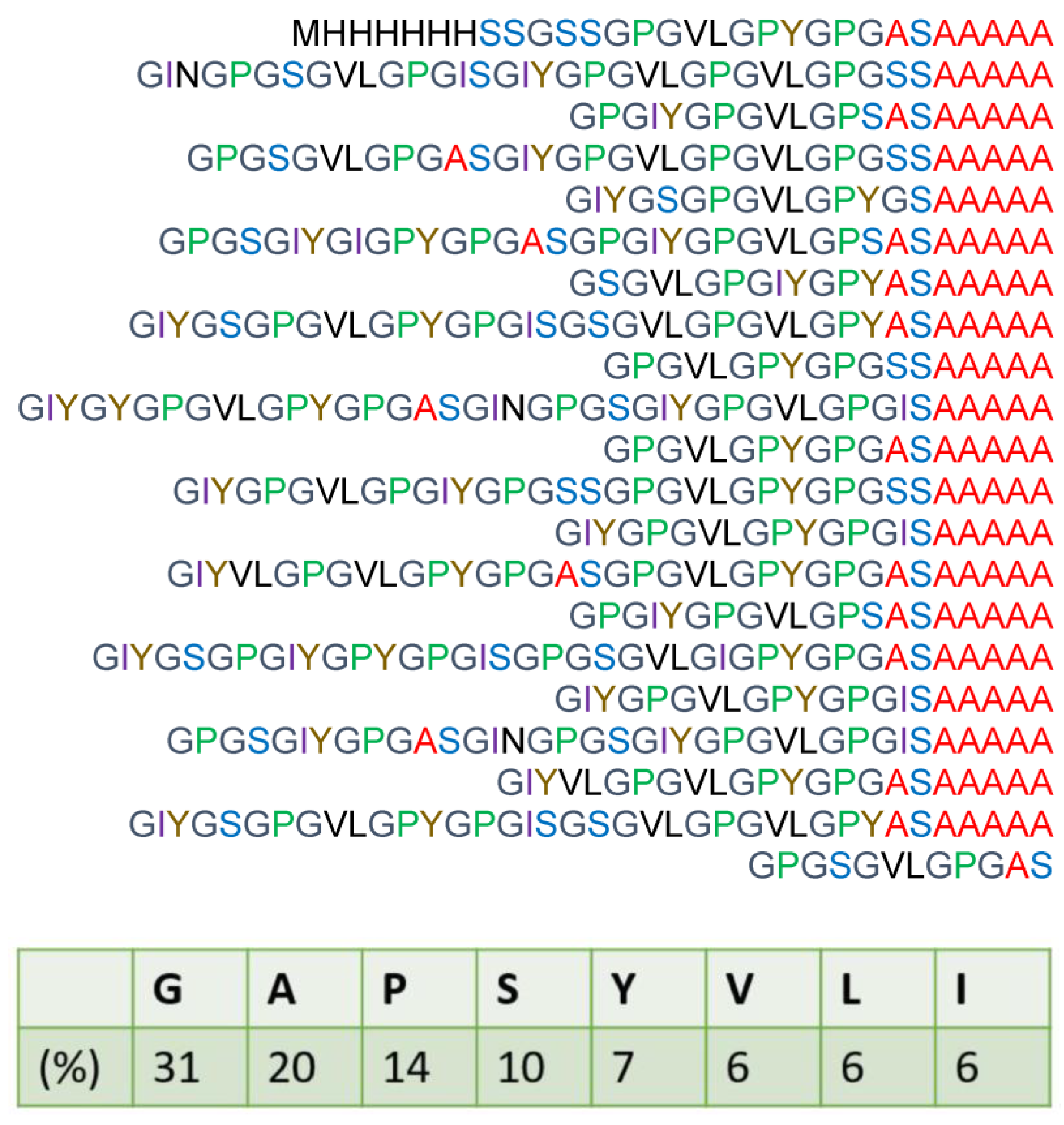
Figure 1. Amino acid composition and primary structure of recombinant spider silk protein RSSP(VLI) based on the primary sequence of ADF-3 Araneus diadematus silk protein with the Gln site replaced by Val, Leu, and Ile.
Molecules 27 08479 g001
Figure 2. 13C solution NMR spectrum of RSSP(VLI) sample in formic acid after dissolving it for 15 h together with the peak assignments.
Molecules 27 08479 g002
Figure 3. 13C HSQC spectra of RSSP(VLI) measured several times continuously to observe the formylation in real time after the dissolution of RSSP(VLI) in FA. The elapsed time from start of the measurements is denoted in the top left of each box. Peaks derived from unformylated and formylated Ser residues are denoted in the spectra.
Molecules 27 08479 g003
Figure 4. Peak intensity for the CαH and CβH2 protons of unformylated and formylated Ser residues with time after the dissolution of RSSP(VLI) in formic acid. (● formylated Hα, ◆ formylated Hβ, 〇 unformylated Hα, ◇ unformylated Hβ).
Molecules 27 08479 g004
Figure 5. Stress–strain curves of (a) RSSP(VLI) fiber prepared by the wet spinning method from FA solutions of RSSP(VLI) powder (storage time in FA was 40 h at 40 °C) before hydration treatment and (b) RSSP(VLI) fiber after dimensional stability measurement, i.e., repeated immersion of the fiber in water and drying at room temperature. The calculated tensile strength (MPa), elongation-at-break (%), and diameter (μm) were (a) 134 ± 10, 19 ± 4, and 61.9 and (b) 97 ± 3, 62 ± 3, and 59.6, respectively.
Molecules 27 08479 g005
Figure 6. Dimensional stabilities of regenerated fibers of (a) RSSP(QQQ) and (b) RSSP(VLI) after repeated immersion in water. L0:Ldry0, L1:Lhydrated1, L2:Ldry2, L3:Lhydrated3, L4:Ldry4, L5:Lhydrated5, and L6:Ldry6.
Molecules 27 08479 g006
Figure 7. The secondary contraction, S ratio, of eight kinds of silk fiber, i.e., non-acetylated and acetylated fibers of RSSP(QQQ), RSSP(VLI), RSP, and regenerated B. mori silk. The S ratio is defined in the text.
Molecules 27 08479 g007
Figure 8. (a) The water absorption (%) and (b) the correlation between the water absorption (%) and the S ratio (%) observed for eight kinds of silk fibers, i.e., non-acetylated and acetylated fibers of RSSP(QQQ), RSSP(VLI), RSP, and regenerated B. mori silk. Water absorption and S ratio are defined in the text. The data of both non-acetylated and acetylated fibers of RSSP(QQQ), RSSP(VLI), RSP, and regenerated B. mori silk are the circles colored in orange, red, blue, and green.
Molecules 27 08479 g008
Figure 9. (a) 13C r-INEPT, (b) 13C DD/MAS, and (c) 13C CP/MAS NMR spectra of RSSP(VLI) powders before methanol treatment in the hydrated states and the (d) 13C CP/MAS NMR spectrum in the dry state. Peak assignments are as follows: a: Iδ, b: Iγ, c: Aβ(r.c.), d: Vγ, e: Vγ, f: Aβ(β), g: Lδ, h: Iγ, Lδ, i: Pγ, Lγ, j: Vβ, k: Pβ, l: Iβ, Yβ, m: Gα, Lβ, n: Pδ, o: Aα(β), p: Aα(r.c.), q: Lα, Sα(r.c.), r: Sβ(β), s: Yα, t: Iα, u: Pα, Sβ(r.c.), Vα, and v: Sα(β).
Molecules 27 08479 g009
Figure 10. (a) 13C r-INEPT, (b) 13C DD/MAS, (c) 13C CP/MAS NMR spectra of RSSP(VLI) powder after methanol treatment in the hydrated states, and (d) 13C CP/MAS NMR spectrum in the dry state. The peak assignment is shown in Figure 9.
Molecules 27 08479 g010
Figure 11. (a) 13C r-INEPT, (b) 13C DD/MAS, and (c) 13C CP/MAS NMR spectra of RSSP(VLI) fiber in the hydrated states and (d) 13C CP/MAS NMR spectrum in the dry state. The peak assignment is shown in Figure 9. A peak at around 30 ppm in the spectrum (a) was assigned to impurity.
Molecules 27 08479 g011
Publisher’s Note: MDPI stays neutral with regard to jurisdictional claims in published maps and institutional affiliations.