Article

Facile Synthesis of Some Coumarin Derivatives and Their Cytotoxicity through VEGFR2 and Topoisomerase II Inhibition

by
Mohamed S. Gomaa
1,
Ibrahim A. I. Ali
2,
Gaber El Enany
3,4,
El Sayed H. El Ashry
5,
Samir M. El Rayes
2,*,
Walid Fathalla
4,
Abdulghany H. A. Ahmed
6,
Samar A. Abubshait
7,8,
Haya A. Abubshait
9 and
Mohamed S. Nafie
2
1
Department of Pharmaceutical Chemistry, College of Clinical Pharmacy, Imam Abdulrahman Bin Faisal University, P.O. Box 1982, Dammam 31441, Saudi Arabia
2
Department of Chemistry, Faculty of Science, Suez Canal University, Ismailia 41522, Egypt
3
Department of Physics, College of Science and Arts in Uglat Asugour, Qassim University, Buraidah 52571, Saudi Arabia
4
Scientific Department, Faculty of Engineering, Port Said University, Port Said 42526, Egypt
5
Chemistry Department, Faculty of Science, University of Alexandria, Alexandria 21526, Egypt
6
Chemistry Department, Faculty of Medicinal Science, University of Science and Technology, Aden 15201, Yemen
7
Chemistry Department, College of Science, Imam Abdulrahman Bin Faisal University, P.O. Box 1982, Dammam 31441, Saudi Arabia
8
Basic and Applied Scientific Research Center, Imam Abdulrahman Bin Faisal University, P.O. Box 1982, Dammam 31441, Saudi Arabia
9
Basic Science Department, Deanship of Preparatory Year and Supporting Studies, Imam Abdulrahman Bin Faisal University, P.O. Box 1982, Dammam 31441, Saudi Arabia
*
Author to whom correspondence should be addressed.
Molecules 2022, 27(23), 8279; https://doi.org/10.3390/molecules27238279
Submission received: 25 October 2022 / Revised: 15 November 2022 / Accepted: 18 November 2022 / Published: 28 November 2022
(This article belongs to the Special Issue Design, Synthesis and Evaluation of Novel Anticancer Agents)

Abstract

:
Novel semisynthetic coumarin derivatives were synthesized to be developed as chemotherapeutic anticancer agents through topoisomerase II, VEGFR2 inhibition that leads to apoptotic cancer cell death. The coumarin amino acids and dipeptides derivatives were prepared by the reaction of coumarin-3-carboxylic acid with amino acid methyl esters following the N,N-dicyclohexylcarbodiimide (DCC) method and 1-hydroxy-benzotriazole (HOBt), as coupling reagents. The synthesized compounds were screened towards VEGFR2, and topoisomerase IIα proteins to highlight their binding affinities and virtual mechanism of binding. Interestingly, compounds 4k (Tyr) and 6c (β-Ala-L-Met) shared the activity towards the three proteins by forming the same interactions with the key amino acids, such as the co-crystallized ligands. Both compounds 4k and 6c exhibited potent cytotoxic activities against MCF-7 cells with IC50 values of 4.98 and 5.85 µM, respectively causing cell death by 97.82 and 97.35%, respectively. Validating the molecular docking studies, both compounds demonstrated promising VEGFR-2 inhibition with IC50 values of 23.6 and 34.2 µM, compared to Sorafenib (30 µM) and topoisomerase-II inhibition with IC50 values of 4.1 and 8.6 µM compared to Doxorubicin (9.65 µM). Hence, these two promising compounds could be further tested as effective and selective target-oriented active agents against cancer.

Graphical Abstract

1. Introduction

Cancer is the leading or second leading cause of death from noncommunicable diseases in the world [1]. Cancer is a multifaceted disease characterized by the growth of abnormal cells that are out of control. Tumor formation and development are linked to apoptosis, which is thought to be a highly programmed mechanism of cell death. One of the main reasons for their prominence was their wide range of pharmacological actions. The design of coumarin skeleton substitutions has a significant impact on the therapeutic applications of these compounds [2].
Anticancer, cytotoxic, and antiproliferative activity were among the many biological effects shown by coumarin derivatives. It’s not just that they work well against cancer, but that they rarely cause side effects [3]. Herbal medicines containing coumarins were traditionally derived from various plant, fungal, and bacterial sources [4,5]. Coumarin is frequently associated with biological activity, such as anti-cancer [6], antifungal [7], anti-HIV [8,9], inhibit lipid peroxidation [10] and anti-clotting. For many years, the therapy of angina pectoris has included the use of carbochromen, a powerful selective coronary vasodilator [11]. Seseline has been shown to have anti-HIV effects [12,13], while novobiocin [14] has antibiotic properties and wedelolactone [15] is used as venomous snakebite antidote Figure 1A.
Coumarins are a class of natural compounds commonly found in a variety of plant families [16], as naturally occurring antitumor compounds. Hyaluronic acid, a key component in tumor growth and progression, is inhibited by 4-methylumbelliferone [17]. Cancers of the pancreas, kidney, prostate, ovaries, and breasts are all effectively targeted by this chemopreventive and chemotherapeutic drug [18]. Many different semi-synthetic coumarin compounds, with varying substitution patterns, have been identified as powerful anticancer drugs, as shown in Figure 1B, as examples of some anticancer coumarin-based derivatives (IVI) [19,20,21,22].
Recently, studies have demonstrated that coumarin-based compounds can inhibit immune regulation, cell growth and differentiation, and cell differentiation through VEGFR2 [23,24,25], and topoisomerase II [26,27,28] inhibition, which have been considered attractive targets for the development of anticancer agents. Almost all cellular responses to VEGF are mediated by VEGFR-2, a type III transmembrane receptor tyrosine kinase (RTK) present in a wide variety of cancer cells. It is considered as the most important transducer of VEGF-dependent apoptosis and angiogenesis [29]. Hence, the VEGFR-2 inhibitory signaling pathway has become a crucial strategy for the identification and development of novel therapeutics for a variety of human malignancies [30].
N.S. Reddy et al. [31] have outlined the growth inhibitory properties of the novel coumarin sulfonamide. These coumarin derivatives activated c-Jun N-terminal kinase 1 (JNK 1) [32]. Reddy et al. have designed and synthesized a series of novel coumarin-3-carboxamides and examined the antioxidant, anti-inflammatory and against anti-human immunodeficiency activities [33,34]. For a long time, organic and medicinal chemists have been interested in the synthesis of coumarins and their derivatives. Structure modification on coumarins focused on the attachment of pharmacophoric groups at C-3, C-4 and C-7 positions for biological evaluation, including anti-microbial, anti-HIV, anti-cancer, lipid-lowering, anti-oxidant, and anti-coagulation activities [35]. Considering the highly valuable biological and pharmaceutical properties of coumarins, it was aimed to synthesize a series of coumarins containing a variety of pharmacophores, amino acids and peptides at position 3, and to investigate the hit compounds through the molecular docking screening as cytotoxic against HepG2, and MCF-7 cancer cells.

2. Results and Discussion

2.1. Chemistry

In continuation of our efforts in synthesizing various bioactive molecules [36], it was found desirable to synthesize a series of coumarin-3-amino acid methyl esters and dipeptides. By interacting with the receptor recognition sites, these compounds were designed to enhance the binding affinity of the coumarin moiety.
Condensation of diethyl malonate with salicylaldehyde afforded the coumarin-3-carboxylic acid ethyl ester, which upon hydrolysis gave coumarin-3-carboxylic acid (3, Scheme 1) [32].
The acid 3 is an excellent precursor for the structure modification via peptide coupling. Peptide bonds were first introduced in the literature using a variety of coupling reagents, which react carboxylic acid with amino acid methyl ester [37]. A convenient coupling method [38] was employed for the formation of peptides by reaction of the carboxylic acid group with amino acid esters, using N,N-dicyclohexylcarbodiimide (DCC) and 1-hydroxy-benzotriazole (HOBt), as coupling reagents. With its ability to reduce racemization during carbodiimide peptide coupling, HOBt is a common method [39].
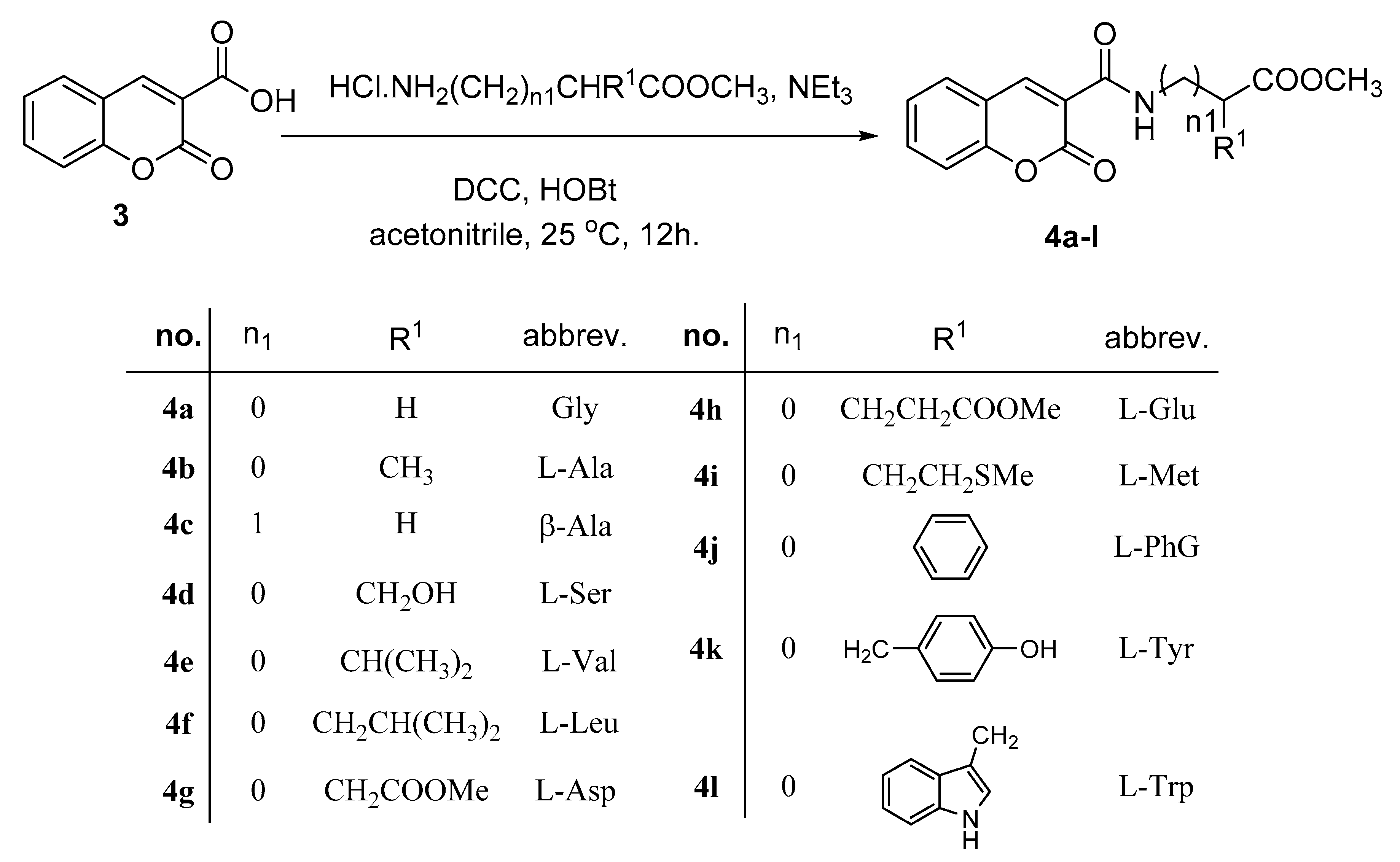
Thus, the treatment of coumarin-3-carboxylic acid (3) with amino acid methyl esters (glycine, L-alanine, L-leucine, L-serine, L-phenylglycine, β-alanine, L-valine, L-methionine, L-tryptophan, L-tyrosine) in the presence of coupling reagents, DCC and HOBT, afforded the amino ester derivatives 4al, respectively, in the 63–87% yield, Scheme 2.
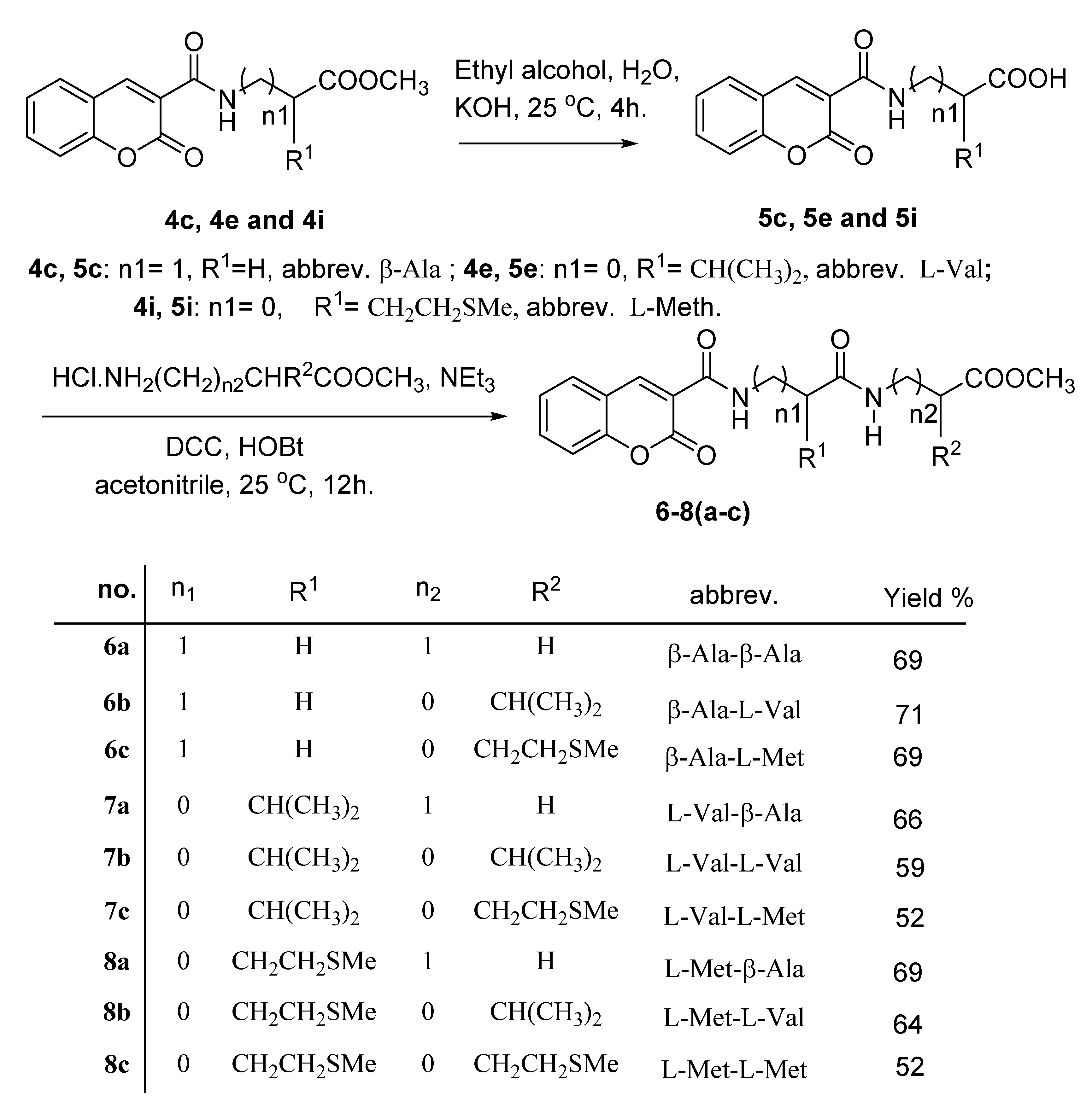
Saponification of esters 4c, 4e and 4i (β-Ala, L-Val, and L Met) in alcoholic KOH afforded the free acid derivatives 5c, 5e and 5i (β-Ala, L-Val, and L Met) in 58–82% yield, Scheme 3. Next, our target was the formation of dipeptide derivatives following the above procedure by the treatment of acid derivatives 5c, 5e and 5i (β-Ala, L-Val, and L Met) with amino acid esters, via the DCC coupling method to give the dipeptides 68(ac) in 52–69% yield, Scheme 3.
The structure assignment of the amino acid ester 4al and dipeptide 68(ac) based on spectral and physicochemical analysis; Figure 2. All the isolated products exhibited a rather interesting, fixed conformation, as represented in Figure 2, and indicated from all 1H NMR spectra. Thus, the 1H NMR spectrum of 3-methyl-2-[(2-oxo-2H-chromene-3-carbonyl)-amino]-butyric acid methyl ester 4e (L-Val) demonstrated an interesting exchangeable singlet down fielded signal at δ 9.24 ppm corresponding to the NH group participating in an intramolecular hydrogen bond interaction of the type N–H···O=C [40]. The 1H NMR spectrum of 4e (L-Val) also demonstrated signals at δ 1.01, 3.75, 4.65 and 8.87 corresponding to 2CH3, OCH3, CH(Val) and CH(coumarin), respectively.
The 1H NMR spectrum of methyl 3-{3-methyl-2-[(2-oxo-2H-chromene-3-carbonyl)-amino]-butyryl-amino} propanoate (7a) (L-Val-β-Ala) similarly demonstrated signals at δ 9.20, 8.87, 6.60, 4.35, 3.59–3.47, 2.53 and 3.65 ppm corresponding NH, CH(coumarin), NH, CH(Val), 2CH2 and OCH3 groups, respectively; Figure 2. The 13C NMR spectrum of (7a) (L-Val-β-Ala) reveal signals at δ 171.4, 170.2, 167.3, 166.4, 61.8, 56.9, 34.3, 38.7, 17.5, and 17.3 ppm corresponding to four C=O, CH, OCH3, CH2, CH2, CH3 and CH3 groups, respectively.

2.2. In Silico Studies

2.2.1. Molecular Docking

The synthesized derivatives were screened for their targets using a molecular docking study. They were screened towards VEGFR-2, and topoisomerase IIα, based on the previous studies that highlighted their binding affinities. As seen in Table 1, the compounds/protein interactions were summarized as a result of docking analysis.
Regarding the molecular docking study towards VEGFR2, the co-crystallized ligand of VEGFR2 protein formed three HB interactions with Asp 1064, Cys 919 and Lys 868 as the key amino acids. Interestingly, most compounds formed one important interaction like the native ligand. Additionally, for the molecular docking study towards topoisomerase IIα, the co-crystallized ligand of Topoisomerase IIα protein formed one HB with Ser 149 as the key amino acid. Thus, both compound 4k and 6c structures represents the important pharmacophoric regions that made them share the activity towards the three proteins by forming the same interactions with the key amino acids, such as the co-crystallized ligands. As observed in Figure 3 with the surface and interactive representations, compound 4k maintained the binding disposition of the co-crystallized ligand of VEGFR2 (A), and Topoisomerase II (B).

2.2.2. ADME Pharmacokinetics

In order to determine the physicochemical and drug-like properties of compounds 4k and 6c towards the examined proteins, bioinformatics study was performed. All of the tested chemicals were readily permeable to and absorbed. As shown in Table 2, typically, compounds contained 2 H-bonding donors and 6 to 7 H-bonding acceptors. In addition, they exhibited log p values (1.81–2.69), so they were well tolerated by cell membranes. The ratable bond number (nrotb) should be less than 10 for effective regulation of conformational changes and oral bioavailability. In addition, the BOILED-Egg model found that the compounds had good absorption in the gastrointestinal tract, as shown in Figure 4.

2.3. Biological Assessment

2.3.1. Cytotoxicity against MCF-7 and HepG2 Cancer Cell Lines

Based on the preliminary of the molecular docking studies of all tested compounds, two hit compounds 4k and 6c with the highest binding affinities towards the tested targets to be screened as cytotoxic against MCF-7 and HepG2 cell lines. As seen in Figure 5, compounds 4k and 6c exhibited promising cytotoxicity against MCF-7 cells with IC50 values of 4.98 and 5.85 µM, respectively, causing cell death by 97.82 and 97.35%, respectively. Additionally, compound 4k exhibited promising cytotoxicity against HepG2 cells with an IC50 value of 9.4 µM, while compound 6c showed moderate cytotoxicity with an IC50 value of 33.88 µM. Hence, these compounds exhibited promise cytotoxic activity.

2.3.2. Enzyme Target Activity

Based on the primarily molecular docking studies of the highest binding affinities of 4k and 6c, enzymatic target activities of both compounds towards VEGFR2, topoisomerase II proteins were tested. As summarized in Table 3, compounds 4k and 6c demonstrated promising VEGFR-2 inhibition with IC50 values of 23.6 and 34.2 µM, compared to Sorafenib (30 µM) and topoisomerase-II inhibition with IC50 values of 4.1 and 8.6 µM compared to Doxorubicin (9.65 µM). Our results of VEGFR2 inhibition agreed with previous studies [23,24] that exhibited promising VEGFR2 inhibition of newly synthesized coumarin derivatives aligned with cytotoxicity in breast cancer cells with promising IC50 values. Other studies [26,41,42] designed some novel coumarin derivatives as cytotoxic activities through topoisomerase inhibition in some cancer cell lines including MCF-7 cells. There were previous studies [43,44] linking apoptosis as the mechanistic cell death upon treatment to VEGFR2 and topoisomerase inhibition, that are emerged as a prime approach for discovering new therapies for many apoptosis-dependent anticancer drugs.

3. Experimental Methods

3.1. Synthesis

Solvent was purified and dried in the usual way. The boiling range of the petroleum ether used was 40–60 °C. Thin layer chromatography (TLC): silica gel 60 F254 plastic plates (E. Merck, layer thickness 0.2 mm) detected by UV absorption. Melting points were determined on a Buchi 510 melting-point apparatus and the values are uncorrected. NMR spectra were measured with Bruker (300 MHz) (Waltham, MA, USA). Tetramethylsilane (TMS, 0.00 ppm) was used as the internal standard. The mass spectra were recorded on a Finnigan (MAT312) and Jeol (JMS.600H); HRMS were recorded with Thermo Finnegan (MAT 95XP) (San Jose, CA, USA).
General procedure for the preparation of coumarin-3-amino acid methyl esters (4a–l): To a cold solution (−5 °C) of the amino acid methyl ester hydrochloride (2.0 mmol) in acetonitrile (20 mL) containing triethyl amine (0.28 mL, 2.0 mmol), coumarin-3-carboxylic acid (3) (0.38 g, 2.0 mmol), dicyclohexylcarbodiimide (DCC) (0.414 g, 2.0 mmol) and 1-hydroxybenzotriazole (HOBT) (0.27 g, 2.0 mmol) were added successively. The reaction mixture was stirred at 0 °C for one hour, at 5 °C for one hour, and then at room temperature for 12 h. The precipitated dicyclohexylurea was filtered off and the filtrate was evaporated under reduced pressure. The residue was dissolved in ethyl acetate, filtered and the filtrate was washed with 5% NaHCO3, 1N HCl and saturated solution of sodium chloride then dried over anhydrous sodium sulphate. After evaporation of the solvent, the remaining oily residue was triturated with petroleum ether (b.p. 40–60 °C) at 0 °C with scratching. The solid residue was filtered off and crystallized from petroleum ether/ethyl acetate. Charcterization analyses including NMR and MS for the synthesized compounds were supported as supplementry.
Methyl [(2-oxo-2H-chromene-3-carbonyl)-amino] ethanoate (4a) From glycine methyl ester hydrochloride, yield, 0.45 g (87%); mp 173–175 °C. 1H NMR (300 MHz, CDCl3): δppm: 3.77 (s, 3H, OCH3), 4.24 (d, J = 5.6 Hz, 2H, CH2), 7.34–7.41 (m, 2H, Ar-H), 7.63–7.69 (m, 2H, Ar-H), 8.89 (s, 1H, CH), 9.22 (br s, 1H, NH); HR EIMS: Calcd for C13H11NO5 (261.2301): Found (261.2110).
Methyl 2-[(2-oxo-2H-chromene-3-carbonyl)-amino] propanoate (4b). From L-alanine methyl ester hydrochloride, yield, 0.46 g (84%); mp 104–106 °C. 1H NMR (300 MHz, CDCl3): δppm: 1.52 (d, J = 7.1 Hz, 3H, CH3), 3.76 (s, 3H, OCH3), 4.73 (m, 1H, CH), 7.33–7.40 (m, 2H, Ar-H), 7.62–7.68 (m, 2H, Ar-H), 8.86 (s, 1H, CH), 9.22 (d, J = 6.5 Hz, 1H, NH); HR EIMS: Calcd for C14H13NO5 (275.2567): Found (275.2390).
Methyl 3-[(2-oxo-2H-chromene-3-carbonyl)-amino] propanoate (4c). From β-alanine methyl ester hydrochloride, yield, 0.44 g (81%); mp 135–137 °C. 1H NMR (300 MHz, CDCl3): δppm: 2.65 (t, J = 6.4 Hz, 2H, CH2), 3.70–3.76 (m, 5H, CH2, OCH3), 7.35–7.40 (m, 2H, Ar-H),7.64–7.68 (m, 2H, Ar-H), 8.87 (s, 1H, CH), 9.11 (bs, 1H, NH); HR EIMS: Calcd for C14H13NO5 (275.2567): Found (275.1971).
Methyl 3-hydroxy-2-[(2-oxo-2H-chromene-3-carbonyl)-amino]-propanoate (4d). From L-serine methyl ester hydrochloride, yield, 0.44 g (76%); mp 177–179 °C. 1H NMR (300 MHz, CDCl3): δppm: 1.66 (bs, D2O Exchangeable, 1H, OH), 3.81 (s, 3H, OCH3), 4.02–4.11 (m, 2H, CH2), 4.82–4.87 (m, 1H, CH), 7.34–7.41 (m, 2H, Ar-H), 7.64–7.69 (m, 2H, Ar-H), 8.88 (s, 1H, CH), 9.56 (d, J = 6.9 Hz, 1H, NH); HR EIMS: Calcd for C14H13NO6 (291.2561): Found (291.2110).
Methyl 3-methyl-2-[(2-oxo-2H-chromene-3-carbonyl)-amino] butanoate (4e). From L-valine methyl ester hydrochloride, yield, 0.42 g (69%); mp 75–77 °C. 1H NMR (300 MHz, CDCl3): δppm: 1.01 (d, J = 6.0 Hz, 6H, 2CH3), 2.27–2.33 (m, 1H, CH), 3.75 (s, 3H, OCH3), 4.65 (dd, J = 5.3, 7.9 Hz, 1H, CH), 7.24–7.41 (m, 2H, Ar-H), 7.62–7.68 (m, 2H, Ar-H), 8.87 (s, 1H, CH), 9.24 (d, J = 7.9 Hz, 1H, NH); HR EIMS: Calcd for C16H17NO5 (303.1067): Found (303.1097).
Methyl 4-methyl-2-[(2-oxo-2H-chromene-3-carbonyl)-amino] pentan-oate (4f). From L-leucine methyl ester hydrochloride, yield, 0.40 g (63%); mp 95–97 °C. 1H NMR (300 MHz, CDCl3): δppm: 0.96 (2d, J = 3.1 Hz, 6H, 2CH3),1.72–1.75 (m, 3H, CH, CH2), 3.74 (s, 3H, OCH3), 4.73–4.76 (m, 1H, CH), 7.33–7.41 (m, 2H, Ar-H), 7.62–7.68 (m, 2H, Ar-H), 8.87 (s, 1H, CH), 9.10 (d, J = 7.4 Hz, 1H, NH); HR EIMS: Calcd for C17H19NO5 (317.1263): Found (317.1178).
Dimethyl 2-[(2-oxo-2H-chromene-3-carbonyl)-amino] succinate (4g). From L-aspartic methyl ester hydrochloride, yield, 0.47 g (70%); mp 69–71 °C. 1H NMR (300 MHz, CDCl3): δppm: 2.87–2.91 (m, 2H, CH2), 3.66 (s, 3H, OCH3), 3.72 (s, 3H, OCH3), 4.87–5.09 (m, 1H, CH), 7.31–7.35 (m, 2H, Ar-H), 7.55–7.58 (m, 2H, Ar-H), 8.83 (s, 1H, CH), 9.11 (d, J = 7.9 Hz, 1H, NH). 13C NMR spectrum, (75.0 MHz, CDCl3), δ, ppm: 35.2, 51.6, 55.7, 56.9, 123.4, 125.5, 126.3, 127.4, 128.8, 130.2, 148.6, 149.8, 165.2 (C=O), 166.4 (C=O), 172.6 (C=O), 172.7 (C=O). HR EIMS: Calcd for C16H15NO7 (333.1077): Found (333.1087).
Dimethyl 2-[(2-oxo-2H-chromene-3-carbonyl)-amino] pentanedioate (4h). From L- Glutamic methyl ester hydrochloride, yield, 0.48 g (69%); mp 64–66 °C. 1H NMR (300 MHz, CDCl3): δppm: 2.27–2.31 (m, 2H, CH2), 2.35–2.38 (m, 2H, CH2), 3.71 (s, 3H, OCH3), 3.75 (s, 3H, OCH3), 4.37–4.44 (m, 1H, CH), 7.33–7.36 (m, 2H, Ar-H), 7.49–7.52 (m, 2H, Ar-H), 8.80 (s, 1H, CH), 9.17 (d, J = 7.9 Hz, 1H, NH). 13C NMR spectrum, (75.0 MHz, CDCl3), δ, ppm: 24.4, 25.2, 53.6, 56.1, 57.3, 123.4, 125.1, 126.4, 127.7, 128.5, 130.6, 148.8, 149.7, 165.4 (C=O), 166.3 (C=O), 172.4 (C=O), 172.6 (C=O). HR EIMS: Calcd for C17H17NO7 (347.1065): Found (347.1077).
Methyl 4-methylsulfanyl-2-[(2-oxo-2H-chromene-3-carbonyl)-amino] butanoate (4i). From L-methionine methyl ester hydrochloride, yield, 0.46 g (68%); mp 81–83 °C. 1H NMR (300 MHz, CDCl3): δppm: 2.09 (s, 3H, SCH3). 2.12–2.29 (m, 2H, CH2), 2.55–2.60 (m, 2H, CH2), 3.77 (s, 3H, OCH3), 4.85–4.92 (m,1H, CH), 7.34–7.41 (m, 2H, Ar-H), 7.63–7.68 (m, 2H, Ar-H), 8.87 (s, 1H, CH), 9.25 (d, J = 7.5 Hz, 1H, NH). 13C NMR spectrum, (75.0 MHz, CDCl3), δ, ppm: 17.1, 29.5, 33.9, 54.9, 56.5 (OCH3), 123.2, 126.4, 127.1, 128.2, 128.6, 130.1, 148.5, 149.9, 164.8 (C=O), 169.2 (C=O), 171.4 (C=O). HR EIMS: Calcd for C16H17NO5S (335.0827): Found (335.0861).
Methyl [(2-oxo-2H-chromene-3-carbonyl)-amino]-phenylacetate (4j). From DL-phenylglycine methyl ester hydrochloride, yield, 0.49 g (73%); mp 189–192 °C. 1H NMR (300 MHz, CDCl3): δppm: 3.75 (s, 3H, OCH3), 5.70 (d, J = 6.4 Hz, 2H, CH), 7.32–7.47 (m, 7H, Ar-H), 7.64–7.67 (m, 2H, Ar-H), 8.85 (s, 1H, CH), 9.73 (d, J = 6.4 Hz, 1H, NH). 13C NMR spectrum, (75.0 MHz, CDCl3), δ, ppm: 55.3, 56.6, 121.7, 125.3, 126.2, 127.7, 127.9, 128.8, 128.6, 129.3, 130.1, 135.4, 136.1, 148.5, 149.7, 166.6 (C=O), 167.2 (C=O), 172.4 (C=O). HR EIMS: Calcd for C19H15NO5 (337.0950): Found (337.0977).
Methyl 3-(4-hydroxyphenyl)-2-[(2-oxo-2H-chromene-3-carbonyl)-amino]-propanoate (4k). From L-tyrosine methyl ester hydrochloride, yield, 0.55 g (76%); mp 112–114 °C. 1H NMR (300 MHz, CDCl3): δppm: 2.98–3.23 (m, 2H, CH2), 3.73 (s, 3H, OCH3), 4.88–4.98 (m,1H, CH), 6.62–6.76 (m, 3H, Ar-H), 6.90 (bs, D2O Exchangeable, 1H, OH), 7.06–7.95 (m, 2H, Ar-H), 7.57–7.66 (m, 3H, Ar-H), 8.77 (s, 1H, CH), 9.29 (d, J = 7.4 Hz, 1H, NH). HR EIMS: Calcd for C20H17NO6 (367.0950): Found (367.0923).
Methyl 3-(1H-Indol-3-yl)-2-[(2-oxo-2H-chromene-3-carbonyl)-amino]-propanoate (4l). From L-tryptophan methyl ester hydrochloride, yield, 0.49 g (64%); mp 138–139 °C. 1H NMR (300 MHz, CDCl3): δppm: 3.30–3.38 (m, 2H, CH2), 3.65 (s, 3H, OCH3), 4.85–4.89 (m,1H, CH), 6.93–7.07 (m, 2H, Ar-H), 7.10–7.23 (m, 2H, Ar-H), 7.33–7.49 (m, 3H, Ar-H), 10.97 (bs, 1H, NH), 7.69–7.98 (m, 2H, Ar-H), 8.89 (s, 1H, CH), 9.14 (d, J = 7.2 Hz, 1H, NH). 13C NMR spectrum, (75.0 MHz, CDCl3), δ, ppm: 34.1, 55.7, 56.9, 104.7, 117.7, 118.4, 120.3, 121.2, 125.4, 126.7, 127.1, 127.6, 128.2, 128.8, 130.3, 135.4, 136.2, 148.7, 149.9, 166.2, (C=O), 167.4 (C=O), 171.4 (C=O). HR EIMS: Calcd for C22H18N2O5 (390.1237): Found (390.1201).
General procedure for preparation of coumarin-3-amino acids (5c, 5e and 5i): A solution of 4c, 4e and 4i (10.0 mmol) in ethanol (40 mL) and 5% KOH (15 mL) was stirred for 2–4h. The solution was evaporated to dryness and the residue was dissolved in water and acidified with dil HCl. The white precipitate was filtered, dried and crystallized from ethanol to give 5c, 5e and 5i.
3-[(2-Oxo-2H-chromene-3-carbonyl)-amino]-propanoic acid (5c): yield, 0.42 g (82%); mp 195–197 °C. 1H NMR (300 MHz, CDCl3): δppm: 1.67 (bs, D2O Exchangeable,1H, OH), 2.72 (t, J = 6.2 Hz, 2H, CH2), 3.72–3.75 (m, 2H, CH2), 7.24–7.40 (m, 2H, Ar-H), 7.63–7.69 (m, 2H, Ar-H), 8.89 (s, 1H, CH), 9.16 (bs, 1H, NH). 13C NMR spectrum, (75.0 MHz, CDCl3), δ, ppm: 36.1, 36.9, 125.4, 126.7, 127.6, 128.2, 128.8, 130.3, 148.7, 149.9, 166.5 (C=O), 167.3 (C=O), 170.4 (C=O). HR EIMS: Calcd for C13H11NO5 (261.0637): Found (261.0704).
3-Methyl-2-[(2-oxo-2H-chromene-3-carbonyl)-amino] butanoic acid (5e) yield, 0.45 g (77%); mp 210–212 °C. 1H NMR (300 MHz, CDCl3): δppm: 1.06 (d, J = 6.8 Hz, 6H, 2CH3), 2.36–2.42 (m, 1H, CH), 3.43 (bs, D2O Exchangeable, 1H, OH), 4.65 (dd, J = 4.8, 7.8 Hz,1H, CH), 7.34–7.42 (m, 2H, Ar-H), 7.64–7.69 (m, 2H, Ar-H), 8.90 (s, 1H, CH), 9.30 (d, J = 7.8 Hz, 1H, NH). 13C NMR spectrum, (75.0 MHz, CDCl3), δ, ppm: 17.1, 17.3, 28.0, 63.7, 125.7, 126.3, 127.4, 128.6, 128.9, 130.7, 149.4, 149.8, 166.8 (C=O), 167.7(C=O), 177.4 (C=O). HR EIMS: Calcd for C15H15NO5 (289.0950): Found (289.1135).
4-Methylsulfanyl-2-[(2-oxo-2H-chromene-3-carbonyl)-amino]-butanoic acid (5i): yield, 0.37 g (58%); mp 108–109 °C. 1H NMR (300 MHz, CDCl3): δppm: 2.11 (s, 3H, SCH3), 2.15–2.36 (m, 2H, CH2), 2.61–2.65 (m, 2H, CH2), 3.92 (bs, D2O Exchangeable, 1H, OH), 4.85–4.92 (m,1H, CH), 7.35–7.42 (m, 2H, Ar-H), 7.65–7.70 (m, 2H, Ar-H), 8.91 (s, 1H, CH), 9.31 (d, J = 6.8 Hz, 1H, NH). 13C NMR spectrum, (75.0 MHz, CDCl3), δ, ppm: 16.9, 29.4, 33.6, 55.3, 125.7, 126.6, 127.4, 128.6, 128.8, 130.4, 148.8, 149.7, 166.8 (C=O), 171.2 (C=O), 177.1 (C=O). HR EIMS: Calcd for C15H15NO5S (321.0671): Found (321.1127).
General procedure for preparation of coumarin-3-dipeptides methyl esters 68 (ac). Dipeptides 68 (ac) were prepared according to above method.
Methyl 3-{3-[(2-oxo-2H-chromene-3-carbonyl)-amino]-propionyl amino} propanoate (6a): (abbreviated: β-Ala-β-Ala) yield, 0.48 g (69%); mp 127–8 °C. 1H NMR (300 MHz, CDCl3): δppm: 2.49–2.55 (m, 4H, 2CH2),3.33–3.72 (m, 4H, 2CH2), 3.74 (s, 3H, OCH3), 6.55 (bs,1H, NH), 7.34–7.43 (m, 2H, Ar-H), 7.61–7.68 (m, 2H, Ar-H), 8.86 (s, 1H, CH), 9.11 (bs, 1H, NH). 13C NMR spectrum, (75.0 MHz, CDCl3), δ, ppm: 33.8, 34.9, 37.8, 38.4, 56.8 (OCH3), 125.3, 126.5, 127.4, 128.8, 128.4, 130.6, 148.5, 149.1, 166.2 (C=O), 167.6 (C=O), 170.3 (C=O), 171.4 (C=O). HR EIMS: Calcd for C17H18N2O6 (346.3346): Found (346.2521).
Methyl 3-Methyl-2-{3-[(2-oxo-2H-chromene-3-carbonyl)-amino]-propionyl-amino}-butanoate (6b): (abbreviated: β-Ala-L-Val) yield, 0.53 g (71%); mp 101–103 °C. 1H NMR (300 MHz, CDCl3): δppm: 1.08 (d, J = 6.8 Hz, 6H, 2CH3), 2.42–2.49 (m, 2H, CH2), 2.99–3.06 (m, 1H, CH), 3.29–3.41 (m, 2H, CH2), 3.66 (s, 3H, OCH3), 4.40–4.46 (m, 1H, CH), 6.71 (bs,1H, NH), 7.26–7.33 (m, 2H, Ar-H), 7.52–7.57 (m, 2H, Ar-H), 8.63 (s, 1H, CH), 9.04 (bs, 1H, NH). 13C NMR spectrum, (75.0 MHz, CDCl3), δ, ppm: 17.1, 17.3, 28.0, 34.8, 38.6, 56.7 (OCH3), 61.3, 125.3, 126.2, 127.6, 128.4, 128.3, 130.2, 149.3, 149.9, 166.6 (C=O), 167.5 (C=O), 170.1 (C=O), 171.2 (C=O). HR EIMS: Calcd for C19H22N2O6 (374.3235): Found (374.3248).
Methyl 4-methylsulfanyl-2-{3-[(2-oxo-2H-chromene-3-carbonyl)-amino]-propionylamino}-butanoate (6c): (abbreviated: β-Ala-L-Met) yield, 0.51 g (69%); mp 122–123 °C. 1H NMR (300 MHz, CDCl3): δppm: 2.12 (s, 3H, CH3), 2.33–2.37 (m, 2H, CH2), 2.42–2.46 (m, 2H, CH2), 2.49–2.53 (m, 2H, CH2), 3.08–3.14 (m, 2H, CH2), 3.65 (s, 3H, OCH3), 4.41–4.45 (m, 1H, CH), 6.66 (bs,1H, NH), 7.20–7.27 (m, 2H, Ar-H), 7.51–7.54 (m, 2H, Ar-H), 8.72 (s, 1H, CH), 9.10 (bs, 1H, NH). 13C NMR spectrum, (75.0 MHz, CDCl3), δ, ppm: 17.2, 28.7, 33.3, 34.9, 38.3, 54.2, 55.7 (OCH3), 125.7, 126.3, 127.0, 128.4, 128.6, 130.5, 149.2, 149.7, 166.1 (C=O), 167.7 (C=O), 170.2 (C=O), 171.6 (C=O). HR EIMS: Calcd for C19H22N2O6S (406.3125): Found (406.3133).
Methyl 3-{3-methyl-2-[(2-oxo-2H-chromene-3-carbonyl)-amino]-butyryl-amino}-propanoate (7a): (abbreviated: L-Val-β-Ala) yield, 0.49 g (66%); mp 149–151 °C. 1H NMR (300 MHz, CDCl3): δppm: 0.98 (d, J = 6.8 Hz, 6H, 2CH3), 2.30–2.36 (m, 1H, CH), 2.53 (t, J = 5.3 Hz, 2H, CH2), 3.47–3.59 (m, 2H, CH2), 3.65 (s, 3H, OCH3), 4.35 (dd, J = 6.1, 7.9 Hz, 1H, CH), 6.60 (bs, 1H, NH), 7.34–7.41 (m, 2H, Ar-H), 7.63–7.69 (m, 2H, Ar-H), 8.87 (s, 1H, CH), 9.20 (d, J = 7.9 Hz, 1H, NH). 13C NMR spectrum, (75.0 MHz, CDCl3), δ, ppm: 17.3, 17.5, 28.2, 34.3, 38.7, 56.9 (OCH3), 61.8, 125.2, 126.1, 127.4, 128.2, 128.7, 130.5, 149.2, 149.8, 166.4 (C=O), 167.3 (C=O), 170.2 (C=O), 171.4 (C=O). HR EIMS: Calcd for C19H22N2O6 (374.1477): Found (374.1451).
Methyl 3-Methyl-2-{3-methyl-2-[(2-oxo-2H-chromene-3-carbonyl)-amino]-butyrylamino}-butanoate (7b): (abbreviated: L-Val-L-Val) yield, 0.47 g (59%); mp 97–99 °C. 1H NMR (300 MHz, CDCl3): δppm: 0.91 (d, J = 7.1 Hz, 6H, 2CH3), 1.01 (d, J = 6.5 Hz, 6H, 2CH3), 2.13–2.19 (m, 1H, CH), 2.31–2.38 (m, 1H, CH), 3.70 (s, 3H, OCH3), 4.46 (dd, J = 6.4, 8.0 Hz, 1H, CH), 4.53 (dd, J = 4.8, 8.6 Hz, 1H, CH), 6.60 (d, J = 8.6 Hz, 1H, NH), 7.34–7.41 (m, 2H, Ar-H), 7.56–7.70 (m, 2H, Ar-H), 8.89 (s, 1H, CH), 9.25 (d, J = 8.0 Hz, 1H, NH). 13C NMR spectrum, (75.0 MHz, CDCl3), δ, ppm: 16.9, 17.0, 17.1, 17.3, 28.9, 28.9, 55.8 (OCH3), 61.9, 62.1, 125.5, 126.3, 127.1, 128.4, 128.9, 130.7, 149.4, 149.7, 166.8 (C=O), 167.4 (C=O), 170.5 (C=O), 171.7 (C=O). HR EIMS: Calcd for C21H26N2O6 (402.1791): Found (402.1829).
Methyl 2-{3-methyl-2-[(2-oxo-2H-chromene-3-carbonyl)-amino]-butyrylamino} -4-methylsulfanyl butanoate (7c): (abbreviated: L-Val-L-Met) yield, 0.45 g (52%); mp 100–101 °C. 1H NMR (300 MHz, CDCl3): δppm: 1.02 (d, J = 5.7 Hz, 6H, 2CH3), 2.04 (s, 3H, SCH3), 2.12–2.22 (m, 2H, CH2), 2.33–2.39 (m, 1H, CH), 2.47–2.56 (m, 2H, CH2), 3.73 (s, 3H, OCH3), 4.42 (dd, J = 6.3, 7.9 Hz, 1H, CH), 4.67–4.74 (m, 1H, CH), 6.76 (d, J = 7.7 Hz,1H, NH), 7.34–7.42 (m, 2H, Ar-H), 7.64–7.69 (m, 2H, Ar-H), 8.88 (s, 1H, CH), 9.23 (d, J = 7.9 Hz, 1H, NH). 13C NMR spectrum, (75.0 MHz, CDCl3), δ, ppm: 17.0, 17.1, 17.3, 28.9, 31.3, 33.9, 54.1, 56.2 (OCH3), 61.7, 125.2, 126.1, 127.3, 128.5, 128.8, 130.4, 149.1, 149.9, 166.5, (C=O), 167.6 (C=O), 170.1 (C=O), 171.4 (C=O). HR EIMS: Calcd for C21H26N2O6S (434.1512): Found (434.1518).
Methyl 3-{4-methylsulfanyl-2-[(2-oxo-2H-chromene-3-carbonyl)-amino]-butyrylamino}-propanoate (8a): (abbreviated: L-Met-β-Ala) yield, 0.51 g (69%); mp 104–105 °C. 1H NMR (300 MHz, CDCl3): δppm: 2.17 (s, 3H, CH3), 2.21–2.25 (m, 2H, CH2), 2.40–2.44 (m, 2H, CH2), 2.43–2.48 (m, 2H, CH2), 3.11–3.15 (m, 2H, CH2), 3.71 (s, 3H, OCH3), 4.48–4.52 (m, 1H, CH), 6.71 (bs,1H, NH), 7.27–7.31 (m, 2H, Ar-H), 7.54–7.58 (m, 2H, Ar-H), 8.68 (s, 1H, CH), 9.07 (bs, 1H, NH). 13C NMR spectrum, (75.0 MHz, CDCl3), δ, ppm: 16.9, 27.4, 30.1, 33.6, 38.4, 54.3, 57.1 (OCH3), 125.2, 126.4, 127.3, 128.7, 128.6, 130.3, 148.7, 149.5, 166.6 (C=O), 167.2 (C=O), 170.1 (C=O), 171.0 (C=O). HR EIMS: Calcd for C19H22N2O6S (406.3244): Found (406.3252).
Methyl 3-methyl-2-{4-methylsulfanyl-2-[(2-oxo-2H-chromene-3-carbonyl)-amino]-butyrylamino}-butanoate (8b): (abbreviated: L-Met-L-Val) yield, 0.56 g (64%); mp 98–99 °C. 1H NMR (300 MHz, CDCl3): δppm: 0.92 (d, J = 7.1 Hz, 6H, 2CH3), 2.12 (s, 3H, SCH3), 2.15–2.25 (m, 3H, CH, CH2), 2.63–2.68 (m, 2H, CH2), 3.72 (s, 3H, OCH3), 4.52 (dd, J = 4.7, 7.5 Hz, 1H, CH), 4.82 (m, 1H, CH), 6.86 (d, J = 7.5 Hz,1H, NH), 7.35–7.42 (m, 2H, Ar-H), 7.64–7.69 (m, 2H, Ar-H), 8.89 (s, 1H, CH), 9.26 (d, J = 7.4 Hz, 1H, NH). 13C NMR spectrum, (75.0 MHz, CDCl3), δ, ppm: 17.1, 17.3, 17.6, 28.8, 33.5, 33.9, 54.4, 56.2 (OCH3), 61.7, 125.0, 126.5, 127.4, 128.3, 128.7, 130.1, 148.9, 149.8, 166.3 (C=O), 167.7 (C=O), 170.4 (C=O), 171.2 (C=O). HR EIMS: Calcd for C21H26N2O6S (434.1512): Found (434.1518).
Methyl 4-methylsulfanyl-2-{4-methylsulfanyl-2-[(2-oxo-2H-chromene-3-carbonyl)-amino]-butyrylamino}-butanoate (8c): (abbreviated: L-Met-L-Met) yield, 0.45 g (52%); mp 109–111 °C. 1H NMR (300 MHz, CDCl3): δppm: 2.03 (s, 3H, SCH3), 2.08 (s, 3H, SCH3), 2.12–2.22 (m, 2H, CH2), 2.33–2.39 (m, 2H, CH2), 2.41–2.44 (m, 2H, CH2), 2.47–2.56 (m, 2H, CH2), 3.70 (s, 3H, OCH3), 4.43–4.49 (m, 1H, CH), 4.61–4.66 (m, 1H, CH), 6.74 (d, J = 7.7 Hz,1H, NH), 7.32–7.37 (m, 2H, Ar-H), 7.61–7.67 (m, 2H, Ar-H), 8.83 (s, 1H, CH), 9.15 (d, J = 7.9 Hz, 1H, NH). 13C NMR spectrum, (75.0 MHz, CDCl3), δ, ppm: 17.1, 17.4, 28.7, 28.6, 33.5, 33.9, 54.4, 54.7, 66.2 (OCH3), 125.0, 126.5, 127.4, 128.3, 128.7, 130.1, 148.9, 149.8, 166.3 (C=O), 167.7 (C=O), 170.4 (C=O), 171.2 (C=O). HR EIMS: Calcd for C21H26N2O6S2 (466.2611):Found (466.2656).

3.2. In Silico Studies

3.2.1. Molecular Docking

The synthesized compounds were screened for binding to demonstrate possible interactions towards some molecular targets of VEGFR2 (PDB = 3U6J), and Topoisomerase IIα (PDB = 1ZXM) using AutoDock Vina as the docking software. In each protein, water molecules and ions were removed from each receptor (protein) using Maestro. In addition, hydrogens were incorporated into the protein during the optimization step. Amino acid residues with missing atoms and those with alternate positions for some of their atoms were optimized. All synthesized derivatives were chemically and energetically optimized according to Nafie et al., 2019 [45,46]. The grid-box size (center_x_y_z: –24.11, −13.01, and 3.64; spacing dimensions of the grid were of 1 Å) was prepared, AutoDock Vina was used for molecular docking for binding poses generation and docking score calculations [47], and Finally, the docked chemical and protein interaction with the key amino acids was analysed using Chimera, a visualisation software.

3.2.2. ADME Pharmacokinetics

In silico ADME pharmacokinetics parameters of the lead compounds were calculated using online websources including “MolSoft”, “Molinspiration”, and “SwissADME”, with their websites, as previously described [48].

3.3. Biological Assessment

3.3.1. Cytotoxicity Using MTT Assay

Breast cancer (MCF-7) and liver cancer (HepG2) cell lines were collected from National Cancer Institute in Cairo and grown in “RPMI-1640 medium L-Glutamine. The cells were grown in 10% fetal bovine serum (FBS) and 1% penicillin-streptomycin using standard tissue culture protocol. On the second day, cells were grown in triplicate on a 96-well plate at a density of 5 × 10⁴ cells and incubated with the investigated compounds at “0.01, 0.1, 1, 10, and 100 µM”. Cell viabilioty was measured with MTT solution. The absorbance was measured using an ELISA microplate reader. After comparing the treated groups to the control, the IC50 values were recorded to indicate the level of viability following previous work [49,50,51].

3.3.2. Enzyme Target Assays

VEGFR2 (KDR) kinase assay kit “BPS Bioscience Corporation catalog#40325” and Topo isomearse II screening kit “TopoGEN, Inc., Columbus, OH, USA” were utilized to invetstiage the enzyme activity for the most active compounds following the manufacturer instructions. Inhibition of autophosphorylation by tested compounds was determined. The IC50 values were determined using GraphPad Prism 7 software from curves depicting the percentage of inhibition using eight concentrations of each compound [43,44].

4. Conclusions

Herein, a novel series of coumarin-based amino acid and dipeptide derivatives synthesized as cytotoxic agents through VEGFR2 and Topoisomerase II inhibition. The coumarin amino acids and dipeptides derivatives were prepared by the reaction of coumarin-3-carboxylic acid with amino acid methyl esters following N,N-dicyclohexylcarbodiimide (DCC) and 1-hydroxy-benzotriazole (HOBt), as coupling reagents. Both compounds 4k (Tyr) and 6c (β-Ala-L-Met) exhibited potent cytotoxic activities against MCF-7 cells with IC50 values of 4.98 and 5.85 µM, respectively, causing cell death by 97.82 and 97.35%, respectively. Validating the molecular docking results, they exhibited promising VEGFR-2 inhibition with IC50 values of 23.6 and 34.2 µM, compared to Sorafenib (30 µM) and topoisomerase-II inhibition with IC50 values of 4.1 and 8.6 µM compared to Doxorubicin (9.65 µM). Accordingly, compounds 4k and 6c were singled out as promising therapeutic candidates for the research and development of new anticancer medicines due to their dual inhibitory effect.

Supplementary Materials

The following supporting information can be downloaded at: https://www.mdpi.com/article/10.3390/molecules27238279/s1. The characterization NMR analyses of compounds.

Author Contributions

Conceptualization, S.M.E.R., I.A.I.A., W.F., M.S.G., S.A.A., H.A.A. and M.S.N.; methodology, M.S.G., I.A.I.A., G.E.E., E.S.H.E.A., S.M.E.R., W.F., A.H.A.A. and M.S.N.; software, M.S.N.; validation, S.M.E.R., I.A.I.A., W.F., M.S.G., S.A.A., H.A.A. and M.S.N.; formal analysis, S.M.E.R., I.A.I.A., W.F., M.S.G., S.A.A., H.A.A. and M.S.N.; investigation, S.M.E.R., I.A.I.A., W.F., M.S.G. and M.S.N.; resources, M.S.G., S.M.E.R., I.A.I.A., and M.S.N.; data curation, S.M.E.R., I.A.I.A., W.F. and M.S.N.; writing—original draft preparation, S.M.E.R., I.A.I.A., W.F., M.S.G. and M.S.N.; writing—review and editing, S.M.E.R., I.A.I.A., W.F., M.S.G. and M.S.N.; visualization, S.M.E.R. and M.S.N.; supervision, S.M.E.R., M.S.N. and I.A.I.A.; project administration, S.M.E.R., and M.S.N.; funding acquisition, S.A.A., H.A.A. and M.S.G. All authors have read and agreed to the published version of the manuscript.

Funding

This research received no external funding.

Institutional Review Board Statement

This study did not require ethical approval.

Informed Consent Statement

Not applicable.

Data Availability Statement

Data are contained within the article and supplementary material.

Conflicts of Interest

The authors declare no conflict of interest. This research did not receive any specific grant from funding agencies in the public, commercial, or not-for-profit sectors.

Sample Availability

Samples of the compounds are not available from the authors.

References

  1. Wild, C.P.; Weiderpass, E.; Stewart, B.W. World Cancer Report: Cancer Research for Cancer Prevention; International Agency for Research on Cancer WHO: Geneva, Switzerland, 2020. [Google Scholar]
  2. Zhu, J.-J.; Jiang, J.-G. Pharmacological and Nutritional Effects of Natural Coumarins and Their Structure–Activity Relationships. Mol. Nutr. Food Res. 2018, 62, 1701073. [Google Scholar] [CrossRef] [PubMed]
  3. Rawat, A.; Reddy, A.V.B. Recent advances on anticancer activity of coumarin derivatives. Eur. J. Med. Chem. Rep. 2022, 5, 100038. [Google Scholar] [CrossRef]
  4. Murray, R.D.H.; Mndez, J.; Brown, S.A. The Natural Coumarins: Occurrence, Chemistry, and Biochemistry; Wiley: Chichester, UK; New York, NY, USA, 1982. [Google Scholar]
  5. Banerji, A.; Mallick, B.; Chatterjee, A.; Budzikiewics, H.; Breuer, M. Assafoetidin and ferocolicin, two sesquiterpenoid coumarins from ferula assafoetida regel. Tetrahedron Lett. 1988, 29, 1557–1560. [Google Scholar] [CrossRef]
  6. Lacy, A.; O’Kennedy, R. Studies on coumarins and coumarin-related compounds to determine their therapeutic role in the treatment of cancer. Curr. Pharm. Des. 2004, 10, 3797–3811. [Google Scholar] [CrossRef] [Green Version]
  7. Sardari, S.; Nishibe, S.; Daneshtalab, M. Coumarins, the bioactive structures with antifungal property. Stud. Nat. Prod. Chem. 2000, 23, 335–393. [Google Scholar] [CrossRef]
  8. Kojima, E.; Iimuro, A.; Nakajima, M.; Kinuta, H.; Asada, N.; Sako, Y.; Nakata, Z.; Uemura, K.; Arita, S.; Miki, S.; et al. Pocket-to-Lead: Structure-Based De Novo Design of Novel Non-peptidic HIV-1 Protease Inhibitors Using the Ligand Binding Pocket as a Template. J. Med. Chem. 2022, 65, 6157–6170. [Google Scholar] [CrossRef]
  9. Kaye, P.T.; Musa, M.A.; Nchinda, A.T.; Nocanda, X.W. Novel Heterocyclic Analogues of the HIV-1 Protease Inhibitor, Ritonavir. Synth. Commun. 2004, 34, 2575–2589. [Google Scholar] [CrossRef]
  10. O’Kennedy, R.; Thornes, R.D. Coumarins: Biology, Applications, and Mode of Action; John Wiley & Sons: Chichester, UK; New York, NY, USA, 1997. [Google Scholar]
  11. Fiedler, V.B.; Scholtholt, J. Effects of carbocromene on myocardial oxygen consumption in isolated dog hearts. J. Pharmacol. Exp. Ther. 1981, 217, 306–313. [Google Scholar] [PubMed]
  12. Yu, D.; Suzuki, M.; Xie, L.; Morris-Natschke, S.L.; Lee, K.-H. Recent progress in the development of coumarin derivatives as potent anti-HIV agents. Med. Res. Rev. 2003, 23, 322–345. [Google Scholar] [CrossRef] [PubMed]
  13. Huang, L.; Kashiwada, Y.; Cosentino, L.M.; Fan, S.; Chen, C.-H.; McPhail, A.T.; Fujioka, T.; Mihashi, K.; Lee, K.-H. Anti-AIDS Agents. 15. Synthesis and Anti-HIV Activity of Dihydroseselins and Related Analogs. J. Med. Chem. 1994, 37, 3947–3955. [Google Scholar] [CrossRef]
  14. Pereira, N.A.; Pereira, B.M.; Nascimento, M.C.; Parente, J.P.; Mors, W.B. Pharmacological Screening of Plants Recommended by Folk Medicine as Snake Venom Antidotes; IV. Protection against Jararaca Venom by Isolated Constituents1. Planta Med. 1994, 60, 99–100. [Google Scholar] [CrossRef]
  15. Sarveswaran, S.; Gautam, S.C.; Ghosh, J. Wedelolactone, a medicinal plant-derived coumestan, induces caspase-dependent apoptosis in prostate cancer cells via downregulation of PKCε without inhibiting Akt. Int. J. Oncol. 2012, 41, 2191–2199. [Google Scholar] [CrossRef] [PubMed] [Green Version]
  16. García-Vilas, J.A.; Quesada, A.R.; Medina, M.Á. 4-Methylumbelliferone Inhibits Angiogenesis in Vitro and in Vivo. J. Agric. Food Chem. 2013, 61, 4063–4071. [Google Scholar] [CrossRef] [PubMed]
  17. Nagy, N.; Kuipers, H.F.; Frymoyer, A.R.; Ishak, H.D.; Bollyky, J.B.; Wight, T.N.; Bollyky, P.L. 4-Methylumbelliferone Treatment and Hyaluronan Inhibition as a Therapeutic Strategy in Inflammation, Autoimmunity, and Cancer. Front. Immunol. 2015, 6, 123. [Google Scholar] [CrossRef] [PubMed] [Green Version]
  18. Bhattacharyya, S.S.; Paul, S.; Mandal, S.K.; Banerjee, A.; Boujedaini, N.; Khuda-Bukhsh, A.R. A synthetic coumarin (4-Methyl-7 hydroxy coumarin) has anti-cancer potentials against DMBA-induced skin cancer in mice. Eur. J. Pharmacol. 2009, 614, 128–136. [Google Scholar] [CrossRef]
  19. Duangdee, N.; Mahavorasirikul, W.; Prateeptongkum, S. Design synthesis and anti-proliferative activity of some new coumarin substituted hydrazide–hydrazone derivatives. J. Chem. Sci. 2020, 132, 66. [Google Scholar] [CrossRef]
  20. Chimichi, S.; Boccalini, M.; Salvador, A.; Dall’Acqua, F.; Basso, G.; Viola, G. Synthesis and Biological Evaluation of New Geiparvarin Derivatives. Chem. Med. Chem. 2009, 4, 769–779. [Google Scholar] [CrossRef] [PubMed]
  21. Konc, J.T.; Hejchman, E.; Kruszewska, H.; Wolska, I.; Maciejewska, D. Synthesis and pharmacological activity of O-aminoalkyl derivatives of 7-hydroxycoumarin. Eur. J. Med. Chem. 2011, 46, 2252–2263. [Google Scholar] [CrossRef]
  22. Ghany, L.M.A.A.; El-Dydamony, N.M.; Helwa, A.A.; Abdelraouf, S.M.; Abdelnaby, R.M. Coumarin-acetohydrazide derivatives as novel antiproliferative agents via VEGFR-2/AKT axis inhibition and apoptosis triggering. New J. Chem. 2022, 46, 17394–17409. [Google Scholar] [CrossRef]
  23. Batran, R.Z.; Dawood, D.H.; El-Seginy, S.A.; Ali, M.M.; Maher, T.J.; Gugnani, K.S.; Rondon-Ortiz, A.N. New Coumarin Derivatives as Anti-Breast and Anti-Cervical Cancer Agents Targeting VEGFR-2 and p38α MAPK. Archiv. Der. Pharm. 2017, 350, 1700064. [Google Scholar] [CrossRef]
  24. Ahmed, E.Y.; Latif, N.A.A.; El-Mansy, M.F.; Elserwy, W.S.; Abdelhafez, O.M. VEGFR-2 inhibiting effect and molecular modeling of newly synthesized coumarin derivatives as anti-breast cancer agents. Bioorganic Med. Chem. 2020, 28, 115328. [Google Scholar] [CrossRef]
  25. Mohamed, T.K.; Batran, R.Z.; Elseginy, S.A.; Ali, M.M.; Mahmoud, A.E. Synthesis, anticancer effect and molecular modeling of new thiazolylpyrazolyl coumarin derivatives targeting VEGFR-2 kinase and inducing cell cycle arrest and apoptosis. Bioorganic Chem. 2019, 85, 253–273. [Google Scholar] [CrossRef]
  26. Liu, H.; Ren, Z.-L.; Wang, W.; Gong, J.-X.; Chu, M.-J.; Ma, Q.-W.; Wang, J.-C.; Lv, X.-H. Novel coumarin-pyrazole carboxamide derivatives as potential topoisomerase II inhibitors: Design, synthesis and antibacterial activity. Eur. J. Med. Chem. 2018, 157, 81–87. [Google Scholar] [CrossRef]
  27. Konkoľová, E.; Hudáčová, M.; Hamuľaková, S.; Jendželovský, R.; Vargová, J.; Ševc, J.; Fedoročko, P.; Kožurková, M. Tacrine-Coumarin Derivatives as Topoisomerase Inhibitors with Antitumor Effects on A549 Human Lung Carcinoma Cancer Cell Lines. Molecules 2021, 26, 1133. [Google Scholar] [CrossRef]
  28. Hueso-Falcón, I.; Amesty, Á.; Anaissi-Afonso, L.; Lorenzo-Castrillejo, I.; Machín, F.; Estévez-Braun, A. Synthesis and biological evaluation of naphthoquinone-coumarin conjugates as topoisomerase II inhibitors. Bioorg. Med. Chem. Lett. 2017, 27, 484–489. [Google Scholar] [CrossRef] [PubMed]
  29. Holmes, K.; Roberts, O.L.; Thomas, A.M.; Cross, M.J. Vascular endothelial growth factor receptor-2: Structure, function, intracellular signalling and therapeutic inhibition. Cell Signal 2007, 19, 2003–2012. [Google Scholar] [CrossRef]
  30. Fontanella, C.; Ongaro, E.; Bolzonello, S.; Guardascione, M.; Fasola, G.; Aprile, G. Clinical advances in the development of novel VEGFR2 inhibitors. Ann. Transl. Med. 2014, 2, 123. [Google Scholar] [CrossRef] [PubMed]
  31. Reddy, N.S.; Mallireddigari, M.R.; Cosenza, S.; Gumireddy, K.; Bell, S.C.; Reddy, E.P.; Reddy, M.V.R. Synthesis of new coumarin 3-(N-aryl) sulfonamides and their anticancer activity. Bioorganic Med. Chem. Lett. 2004, 14, 4093–4097. [Google Scholar] [CrossRef] [PubMed]
  32. Reddy, N.S.; Gumireddy, K.; Mallireddigari, M.R.; Cosenza, S.C.; Venkatapuram, P.; Bell, S.C.; Reddy, E.P.; Reddy, M.V.R. Novel coumarin-3-(N-aryl)carboxamides arrest breast cancer cell growth by inhibiting ErbB-2 and ERK1. Bioorg. Med. Chem. 2005, 13, 3141–3147. [Google Scholar] [CrossRef]
  33. Pereira, T.M.; Franco, D.P.; Vitorio, F.; Kummerle, A.E. Coumarin Compounds in Medicinal Chemistry: Some Important Examples from the Last Years. Curr. Top. Med. Chem. 2018, 18, 124–148. [Google Scholar] [CrossRef]
  34. Naik, M.D.; Bodke, Y.D.; M, V.K.; BC, R. An efficient one-pot synthesis of coumarin-amino acid derivatives as potential anti-inflammatory and antioxidant agents. Synth. Commun. 2020, 50, 1210–1216. [Google Scholar] [CrossRef]
  35. Kulkarni, M.V.; Kulkarni, G.M.; Lin, C.-H.; Sun, C.-M. Recent Advances in Coumarins and 1-Azacoumarins as Versatile Biodynamic Agents. Curr. Med. Chem. 2006, 13, 2795–2818. [Google Scholar] [CrossRef]
  36. Al-Masoudi, N.A.; Al-Masoudi, I.A.; Ali, I.A.I.; Al-Soud, Y.A.; Saeed, B.; la Colla, P. Amino acid derivatives. Part I. Synthesis, antiviral and antitumor evaluation of new alpha-amino acid esters bearing coumarin side chain. Acta Pharm. 2006, 56, 175–188. [Google Scholar]
  37. Asagarasu, A.; Uchiyama, T.; Achiwa, K. Syntheses of HIV-protease inhibitors having a peptide moiety which binds to GP120. Chem. Pharm. Bull. 1998, 46, 697–703. [Google Scholar] [CrossRef] [PubMed] [Green Version]
  38. Davies, J.S.; Mohammed, A.K. Assessment of racemisation in N-alkylated amino-acid derivatives during peptide coupling in a model dipeptide system. J. Chem. Soc. Perkin Trans. 1981, 1, 2982–2990. [Google Scholar] [CrossRef]
  39. König, W.; Geiger, R. A new method for synthesis of peptides: Activation of the carboxyl group with dicyclohexylcarbodiimide using 1-hydroxybenzotriazoles as additives. Chem. Ber. 1970, 103, 788–798. [Google Scholar] [CrossRef]
  40. Strakova, I.; Petrova, M.; Belyakov, S.; Strakovs, A. Reactions of 4-chloro-3-formyl-coumarin with primary amines. Chem. Heterocycl. Compd. 2006, 42, 574–582. [Google Scholar] [CrossRef]
  41. Abdel-Hafez, G.A.; Mohamed, A.-M.I.; Youssef, A.F.; Simons, C.; Aboraia, A.S. Synthesis, computational study and biological evaluation of 9-acridinyl and 1-coumarinyl-1,2,3-triazole-4-yl derivatives as topoisomerase II inhibitors. J. Enzym. Inhib. Med. Chem. 2022, 37, 502–513. [Google Scholar] [CrossRef]
  42. Guruge, A.G.; Udawatte, C.; Weerasinghe, S.; Guruge, A.G.; Udawatte, C.; Weerasinghe, S. An In Silico Approach of Coumarin-Derived Inhibitors for Human DNA Topoisomerase I. Aust. J. Chem. 2016, 69, 1005–1015. [Google Scholar] [CrossRef]
  43. Khalifa, M.M.; Al-Karmalawy, A.A.; Elkaeed, E.B.; Nafie, M.S.; Tantawy, M.A.; Eissa, I.H.; Mahdy, H.A. Topo II inhibition and DNA intercalation by new phthalazine-based derivatives as potent anticancer agents: Design, synthesis, anti-proliferative, docking, and in vivo studies. J. Enzym. Inhib. Med. Chem. 2022, 37, 299–314. [Google Scholar] [CrossRef]
  44. Nafie, M.S.; Boraei, A.T.A. Exploration of novel VEGFR2 tyrosine kinase inhibitors via design and synthesis of new alkylated indolyl-triazole Schiff bases for targeting breast cancer. Bioorganic Chem. 2022, 122, 105708. [Google Scholar] [CrossRef] [PubMed]
  45. Nafie, M.S.; Tantawy, M.A.; Elmgeed, G.A. Screening of different drug design tools to predict the mode of action of steroidal derivatives as anti-cancer agents. Steroids 2019, 152, 108485. [Google Scholar] [CrossRef]
  46. Matin, P.; Hanee, U.; Alam, M.S.; Jeong, J.E.; Matin, M.M.; Rahman, M.R.; Mahmud, S.; Alshahrani, M.M.; Kim, B. Novel Galactopyranoside Esters: Synthesis, Mechanism, In Vitro Antimicrobial Evaluation and Molecular Docking Studies. Molecules 2022, 27, 4125. [Google Scholar] [CrossRef] [PubMed]
  47. Trott, O.; Olson, A.J. AutoDock Vina: Improving the speed and accuracy of docking with a new scoring function, efficient optimization, and multithreading. J. Comput. Chem. 2010, 31, 455–461. [Google Scholar] [CrossRef] [PubMed] [Green Version]
  48. Boraei, A.T.A.; Eltamany, E.H.; Ali, I.A.I.; Gebriel, S.M.; Nafie, M.S. Synthesis of new substituted pyridine derivatives as potent anti-liver cancer agents through apoptosis induction: In vitro, in vivo, and in silico integrated approaches. Bioorganic Chem. 2021, 111, 104877. [Google Scholar] [CrossRef]
  49. Nafie, M.S.; Amer, A.M.; Mohamed, A.K.; Tantawy, E.S. Discovery of novel pyrazolo [3,4-b]pyridine scaffold-based derivatives as potential PIM-1 kinase inhibitors in breast cancer MCF-7 cells. Bioorganic Med. Chem. 2020, 28, 115828. [Google Scholar] [CrossRef] [PubMed]
  50. Mosmann, T. Rapid colorimetric assay for cellular growth and survival: Application to proliferation and cytotoxicity assays. J. Immunol. Methods 1983, 65, 55–63. [Google Scholar] [CrossRef] [PubMed]
  51. Nafie, M.S.; Mahgoub, S.; Amer, A.M. Antimicrobial and antiproliferative activities of novel synthesized 6-(quinolin-2-ylthio)pyridine derivatives with molecular docking study as multi-targeted JAK2/STAT3 inhibitors. Chem. Biol. Drug Des. 2021, 97, 553–564. [Google Scholar] [CrossRef]
Figure 1. (A): Representative biological active coumarins; (B) structures of some of anticancer coumarin derivatives through VEGFR2 inhibition.
Molecules 27 08279 g001
Scheme 1. Synthesis of coumarin-3-carboxylic acid (3).
Molecules 27 08279 sch001
Scheme 2. Preparation of coumarin-3-amino acid methyl esters 4al.
Molecules 27 08279 sch002
Scheme 3. Preparation of coumarin-3-dipeptide methyl esters 68(ac).
Molecules 27 08279 sch003
Figure 2. Selected 1H NMR δ values of compounds 4e and 7a.
Molecules 27 08279 g002
Figure 3. Binding disposition of the co-crystallized ligand (cyan-colored) and the docked compound 4k (orange-colored) towards (A): VEGFR2 (PDB = 3U6J); (B): Topoisomerase II (PDB = 1ZXM). Left panel is the surface representation, while right panel is the interactive binding mode with the key amino acids. The 3D representation was generated by Chimera-UCSF.
Molecules 27 08279 g003
Figure 4. BOILED-Egg model for compounds 4k (A) and 6c (B) using the Swiss ADME. “Points located in the BOILED-Egg’s yolk are molecules predicted to passively permeate through the blood–brain barrier, while points located in the BOILED-Egg’s white are molecules predicted to be passively absorbed by gastrointestinal tract”.
Molecules 27 08279 g004
Figure 5. Dose-response curve for the cytotoxicity of two hit compounds 6c and 4k against (A): MCF-7 and (B): HepG2 cell lines for 48 h using MTT assay. Half minimal inhibitory concentration (IC50).
Molecules 27 08279 g005
Table 1. Analysis of ligand-receptor interactions of the docked compounds towards two proteins VEGFR2 (PDB = 3U6J), and Topoisomerase IIα (PDB = 1ZXM) using AutoDock Vina *.
Tested TargetsVEGFR2
(PDB = 3U6J) a
Topoisomerase IIα
(PDB = 1ZXM) b
Compounds
Co-crystallized ligand (key interactions)3 HB with Asp 1064, Cys 919 and Lys 8681 HB with Ser 149
4a-1 HB with Ser 149
4b1HB with Lys 868-
4c1HB with Asp 1046-
4d3 HB with Tyr 916, Lys 868, and Asp 10461 HB with Ser 149
4e1HB with Cys 919-
4fArene-cation with Lys 8681 HB with Asn 150
4g3HB with Tyr 916, Lys 868 and Asp 10462 HB with Ser 149 and Asn 91
4h2 HB with Lys 868 and Cys 916-
4i2HB with Lys 868 and Cys 9191 HB with Ser 149
4j2HB with Thr 916 and Lys 8681 HB with Ser 149 and Arene-cation with Arg 98
4k1 HB with Asp 1046 and Arene-cation with Lys 8683HB with Ser 149, Thr 215 and Asn 120
4l1HB with Asp 10462 HB with Ser 149 and Asn 150
5c2HB with Cys 919 and Lys 868-
5e-2 HB with Asn 150 and Lys 168
5i2HB with Thr 916 and Cys 9191 HB with Ala 167
6a2HB with Thr 916 and Asp 10461 HB with Ser 149
6b1 HB with Cys 919-
6c1 HB with Cys 9192 HB with Arg 98 and Ser 149
7a--
7b1HB with Asp 10462 HB with Ser 149 and Asn 120
7c-1 HB with Lys 157 and arene-cation Arg 98
8aArene-cation Lys 868arene-cation Arg 98 and Lys 157
8bArene-cation Lys 8681 HB with Ser 149
8c1 HB with Lys 920-
* Docking calculation using AutoDock Vina software was validated by self-docking step that fulfil RMSD lower than 2. a Binding energy of the docked compounds towards VEGFR2 protein from −9.36 to −24.64 Kcal/mol, and b Binding energy of the docked compounds towards topoisomerase IIα protein from −7.64 to −29.46 Kcal/mol.
Table 2. In silico ADME pharmacokinetics properties of the lead compounds.
#Molinspiration 2018.10MolSoftSwissADME
MWt
(D)
MV (A3)PSA (A2)Log pNrotbNviolationsHBAHBDSolubility
(mg/L)
Drug Likeness (Lipinski Pfizer Filter)
4k381.3334.3105.82.697062519.9“Yes, drug like”
MW ≤ 500, Log p ≤ 4.15, HBA ≤ 10 and HDD ≤ 5
6c434.5388.2114.71.8110072485.3
Mwt: Molecular Weight, MV: Molecular Volume, PAS: Polar Surface Area, Log p: Octanol-water partition coefficient, nrotb: number of rotatable bond, nviolations: number of violations, HBA: Hydrogen Bond Acceptor, HBD: Hydrogen Bond Donor.
Table 3. Inhibitory activities of the hit compounds towards topoisomerase II, VEGFR2 target enzymes.
CompoundIC50 ± SD * (µM)
VEGFR2Topoisomerase II
4k23.6 ± 1.194.1 ± 0.64
6c34.2 ± 1.068.6 ± 0.15
Doxorubicin---9.65 ± 0.72
Sorafenib30.0 ± 0.42----
* IC50 values were calculated using a sigmoidal nonlinear regression curve fit of percentage inhibition against five concentrations of each compound.
Publisher’s Note: MDPI stays neutral with regard to jurisdictional claims in published maps and institutional affiliations.