Article

Sustainable and Efficacy Approach of Green Synthesized Cobalt Oxide (Co3O4) Nanoparticles and Evaluation of Their Cytotoxicity Activity on Cancerous Cells

Chemistry Department, Faculty of Science, Taif University, Al-Hawiah, Taif City P.O. Box 11099, Saudi Arabia
Molecules 2022, 27(23), 8163; https://doi.org/10.3390/molecules27238163
Submission received: 2 October 2022 / Revised: 8 November 2022 / Accepted: 21 November 2022 / Published: 23 November 2022

Abstract

:
In this study, rosemary leaf extract was effectively used to synthesize cobalt oxide nanoparticles (Co3O4 NPs) using a rapid, low-cost, and environmentally friendly approach. The prepared Co3O4 NPs were examined using various analytical techniques. However, UV spectrum analysis displayed two sharp absorption peaks at ~350 and 745 nm. The dynamic light scattering and zeta potential measurements were used to evaluate the particle size and the effective stabilization of the synthetic nanoparticles in the suspensions. A semi-triangular pyramidal shape of the Co3O4 NPs with a wide particle-size distribution could be observed in the scanning electron microscopy images. The energy-dispersive X-ray spectrum confirmed their successful synthesis, as the experimental atomic percentages agreed with the theoretical values. Moreover, X-ray diffraction analysis revealed that the synthesized Co3O4 NPs had a cubic crystalline structure corroborating that of theoretical Co3O4. Additionally, the Co3O4 NPs were not toxic at ≤62.5 µg/mL for Hep G2 and at ≤31.25 µg/mL for Mcf7. Therefore, these unique environmentally friendly Co3O4 NPs at this safe concentration could be studied in the future for their therapeutic activity.

1. Introduction

Extensive research on nanostructured materials has provided access to new classes of functional materials with novel characteristics and uses. Owing to their large surface areas, unusual adsorptive characteristics, and rapid diffusivities, nanosized crystalline metal oxides have attracted increasing attention in recent years. P-type semiconductor cobalt oxide nanoparticles (Co3O4 NPs) are transition metal oxides that can exist in various oxidation states, including Co2+, Co3+, and Co4+ [1,2,3,4]. Among them, the spinel structure is the most stable form of Co3O4 NPs, which can be used in a wide range of applications, including lithium-ion batteries, gas sensors, supercapacitors, solar selective absorbers, drug delivery, anticancer and antimicrobial agents, water remediation, and photocatalysis [2,5,6,7,8,9,10]. Additionally, the outstanding magnetic, optical, chemical, physical, thermal, and biomedical characteristics of Co3O4 NPs are of particular interest. Further, Co3O4 NPs are more economical metal oxide nanoparticles than other noble metals [11,12]. Co3O4 NPs had previously been investigated as therapeutic agents for treating disorders such as microbial infections, rendering them attractive for use in biomedical applications [13]. In comparison to antibiotics, Co3O4 NPs have fewer side effects, and higher antibacterial and antifungal properties at low concentrations, and are nontoxic to the body at low levels [14].
Coprecipitation, sonochemistry, chemical spray pyrolysis, thermal decomposition, solution combustion, microwave-assisted, microemulsion, and hydrothermal reactions are examples of physical and chemical processes that have been used to synthesize Co3O4 NPs [4,15,16,17]. The abovementioned synthesis techniques, although effective, have a number of disadvantages, such as being expensive, time-consuming, energy-intensive, and harmful to the environment [5]. The use of plants, plant components, and microorganisms (algae, bacteria, and fungi) that can produce NPs is a more environmentally friendly approach that meets the demands of minimal hazardous waste [2]. Various studies have reported the synthesis of Co3O4 NPs using plants such as Calotropis gigantea, Moringa oleifera, Aspalathus linearis, Terminalia chebula, Sageretia thea, Calotropis procera, Manihot esculenta Crantz, and Euphorbia heterophylla L. [18,19,20,21,22,23].
Rosemary (Rosmarinus officinalis Linn.), a common household plant, an evergreen perennial aromatic shrub native to the northern and southern coasts of the Mediterranean Sea [24]. Recent research has suggested that traditional medicinal herbs may help in preventing or treating certain metabolic problems, including diabetes, heart disease, and some types of cancer [25]. Dried rosemary leaves and flowers are particularly interesting sources of biologically active phytochemicals because they contain a number of phenolic compounds with significant in vitro antioxidant activity, such as carnosol, carnosic acid, rosmanol, 7-methyl-epirosmanol, isorosmanol, rosmadial, and caffeic acid [26,27], as shown in Figure 1.
Using rosemary leaf extract as a capping and reducing agent, Co3O4 NPs were synthesized using a completely green approach wherein no organic or inorganic solvents, surfactants, or other chemicals were used. The characterization and cytotoxic activity of the Co3O4 NPs on liver and breast cancer cells are also described in this study.

2. Results and Discussion

2.1. UV Spectroscopy

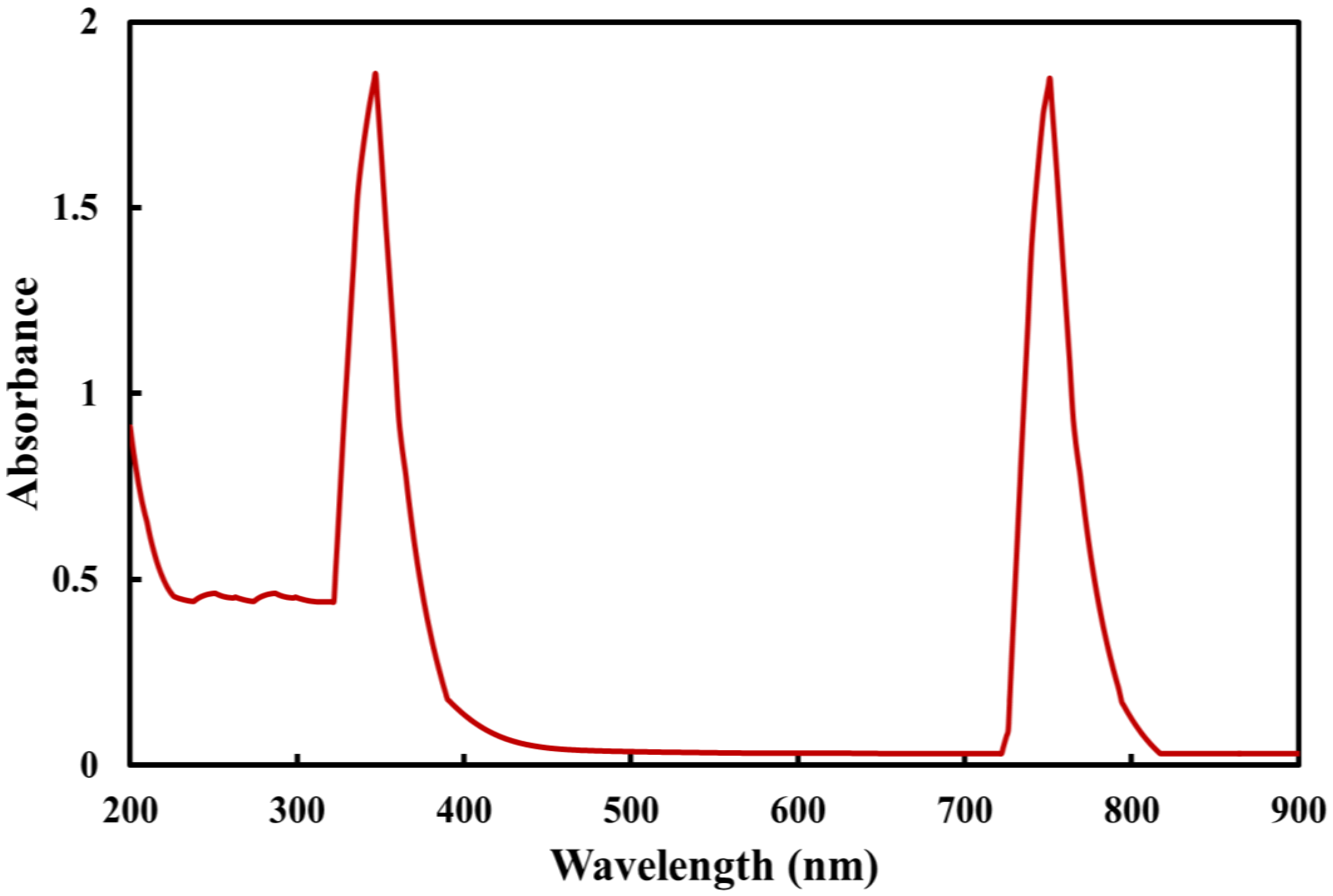
A color change into dark brown was initially used to identify the green synthesis of Co3O4 NPs. The chemical process between the components of rosemary leaf extract and metal ions was examined using the UV–visible spectrum. The UV spectrum of Co3O4 NPs synthesized using rosemary leaves indicated nanoparticle formation by demonstrating two sharp absorption peaks at approximately 350 and 745 nm (Figure 2). However, these two different wavelengths might have arisen from the charge transfer process from the ligand to the metal as O2 → Co2+ for 350 nm absorption peak, and O2 → Co3+ for the 745 nm absorption peak. In addition, these peaks were attributed to surface plasmon resonance behavior [28].

2.2. Dynamic Light Scattering (DLS) and Zeta Potential

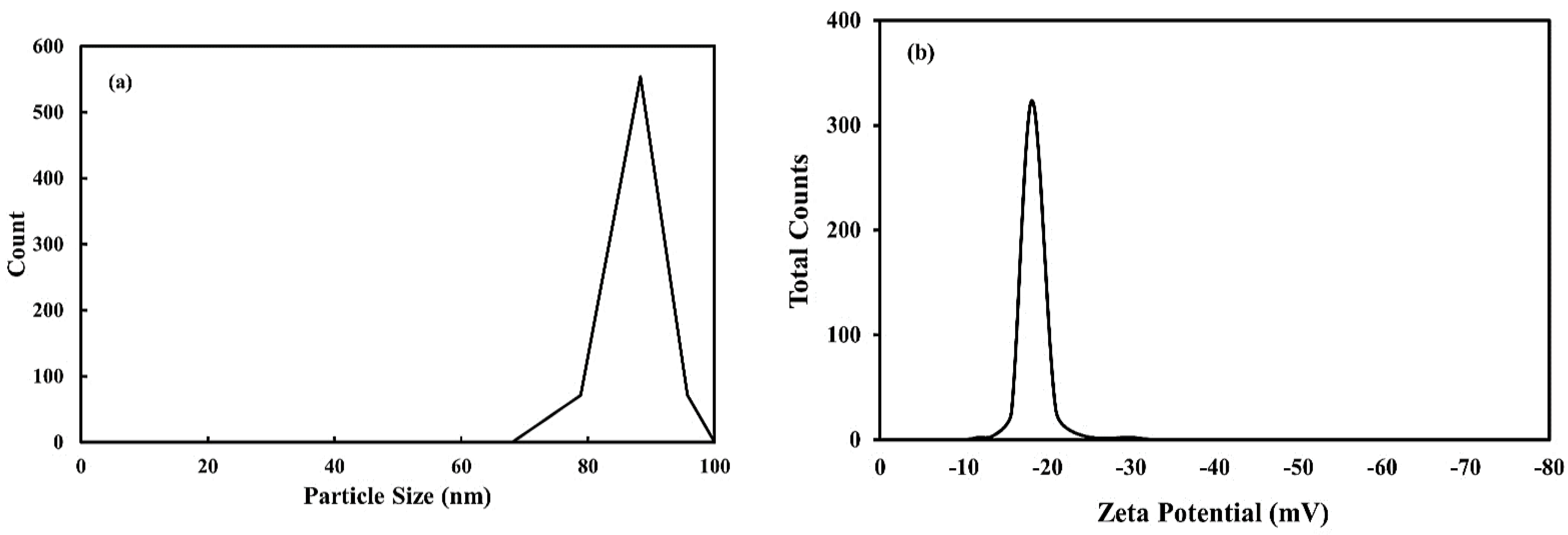
The particle size distribution for the Co3O4 NPs synthesized using rosemary leaf extract, as determined by the DLS method, is displayed in Figure 3a. The average particle size distribution of the synthesized Co3O4 NPs was 89 nm depending on size distribution data. Analysis revealed a unimodal size distribution with polydispersity indices; the suspension was monodispersed and produced high colloidal stability. Moreover, the zeta potential of the synthesized Co3O4 NPs showed a peak at −18 mV (Figure 3b). This result reveals that the surface of the synthesized nanoparticles acquired a negative charge, and the nanoparticles dispersed significantly throughout the medium. Therefore, the determined negative value was responsible for the good stabilization of prepared NPs in the suspensions. Furthermore, because the average size is a measure of hydrodynamic size, its value indicates both the availability of nanoparticles and any solvent molecules linked to the tumbling particle.

2.3. TEM

The microstructure of the synthesized Co3O4 NPs using rosemary leaf extract was investigated using the TEM and HRTEM images shown in Figure 4a,b. As depicted in Figure 4a, the prepared nanoparticles had an almost irregular cluster shape, with the presence of some rod-like and sub-spherical regions with a diameter of approximately 100 nm. The HRTEM image (Figure 4b) reveals agglomerated spherical nanoparticles. The average particle size was in the approximate range of 50–100 nm.

2.4. SEM

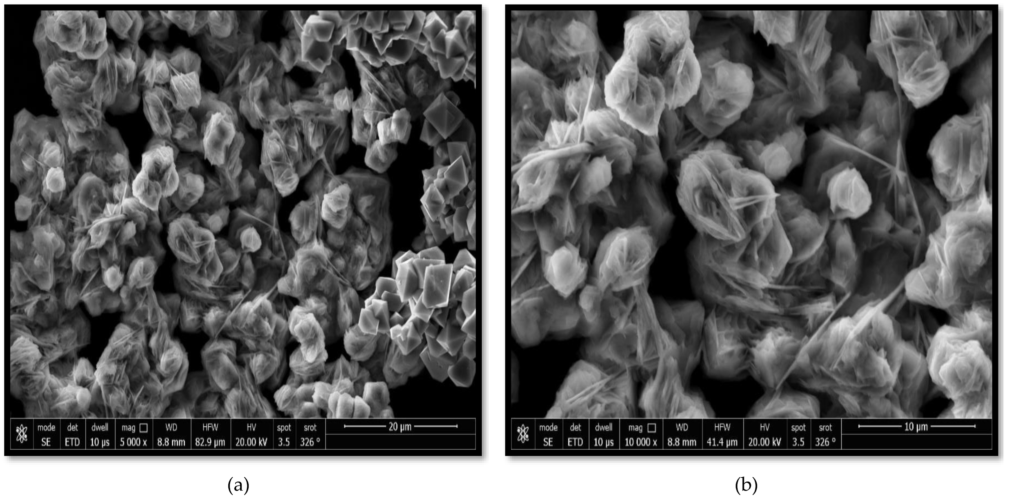
The morphologies of the Co3O4 NPs synthesized using rosemary leaf extract at various magnifications are shown in Figure 5a,b, where a semi-triangular pyramidal shape with various-sized particles is shown. The magnetic induction characteristics of Co3O4 NPs may have been the reason for the highly agglomerated synthesized nanoparticles [1]. This result is consistent with that of TEM studies.

2.5. EDX Spectrum

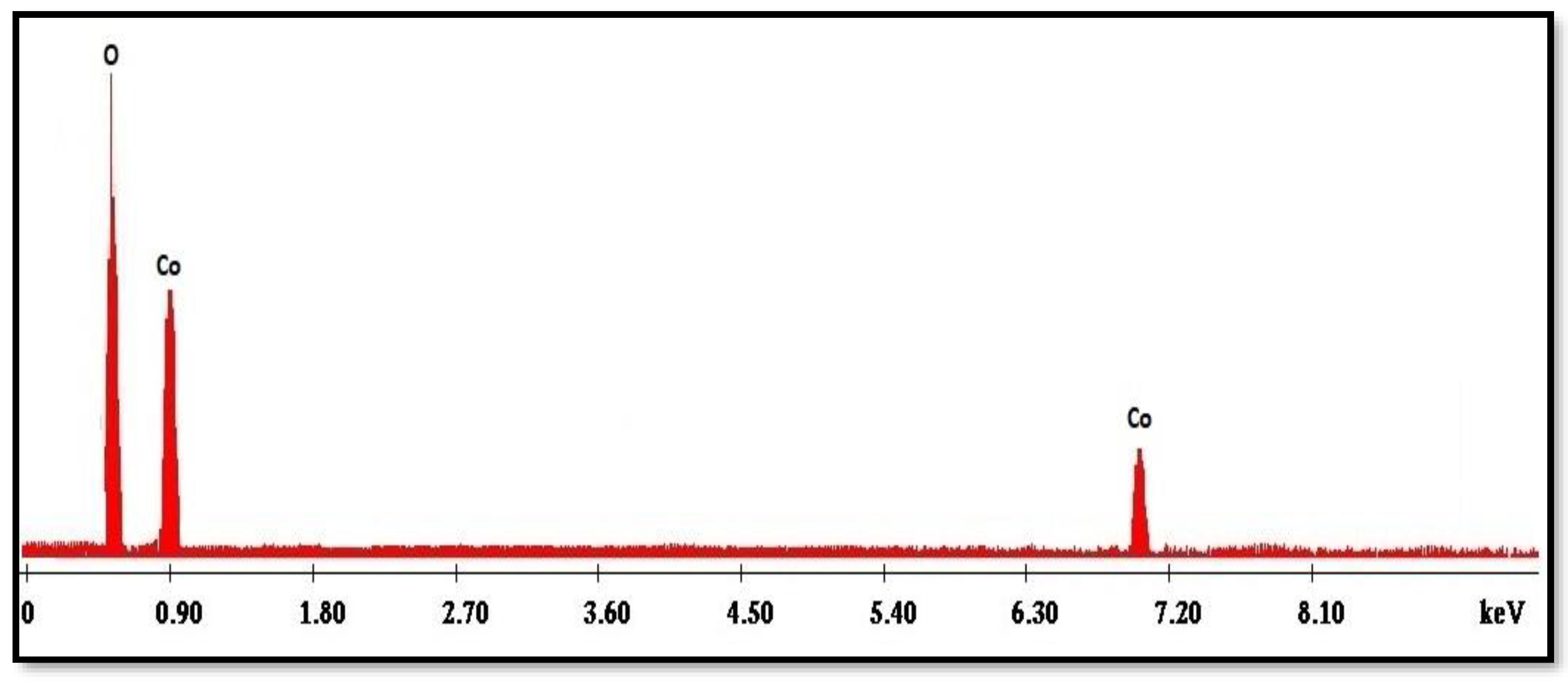
Figure 6 displays the EDX spectrum of the Co3O4 NPs synthesized using the rosemary leaf extract. The major components of the synthesized nanoparticles were only O and Co, with atomic percentages of 24 and 75.9%, respectively; this is almost identical to the theoretical 3:4 ratio in Co3O4 NPs [14].

2.6. Crystalline Structure

The XRD pattern of the Co3O4 NPs synthesized using rosemary leaf extract is depicted in Figure 7, which shows that the NPs had a cubic phase structure. The peak positions (2θ = 31.27°, 36.85°, 38.55°, 44.82°, 59.37°, 60.37°, and 65.25°) and relative intensities of the Co3O4 NPs matched those of the JCPDS card no. COD 9005887 file, confirming Co3O4 with a cubic structure Fd-3m (227) space group. However, no typically observed impurity peaks [29] were observed. Using the Scherrer relation, the average crystalline size was approximately 90 nm.

2.7. Cytotoxicity Activity

The in vitro cytotoxicity evaluation of synthesized Co3O4 NPs is crucial for biomedical applications. To determine the optimal concentration where there is no cytotoxic effect on living tissue, and to demonstrate the safety of synthesized NPs for further research, different concentrations of the synthesized Co3O4 NPs using rosemary leaf extract were tested against Hep G2 and Mcf7 cancer cell lines.
As the liver is the primary site for chemical and food metabolism, liver carcinoma cells were used. Regardless of the method of administration (intravenous injection or orally via the digestive tract to reach the blood circulation system), the drug must pass through the liver. In addition, many chemicals undergo a metabolic process in the liver before elimination, leading to the possibility of liver poisoning. Therefore, the liver is a major organ that must be protected when receiving chemicals. Hepatic cell cancer is the fourth most common cause of cancer-related death. Additionally, because of its distinct structure and function, the liver plays a significant role in the immune system and inflammation [30,31].
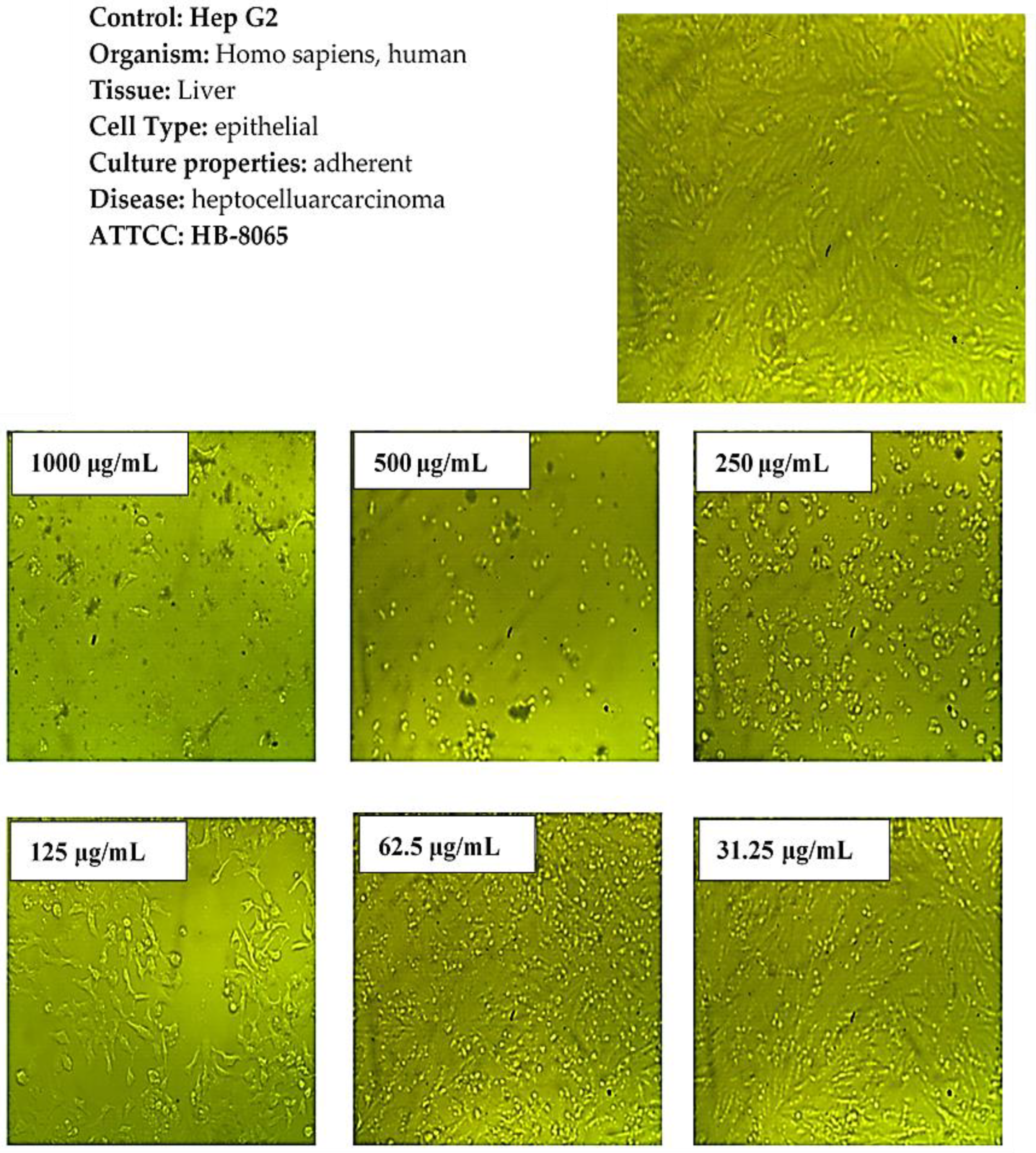
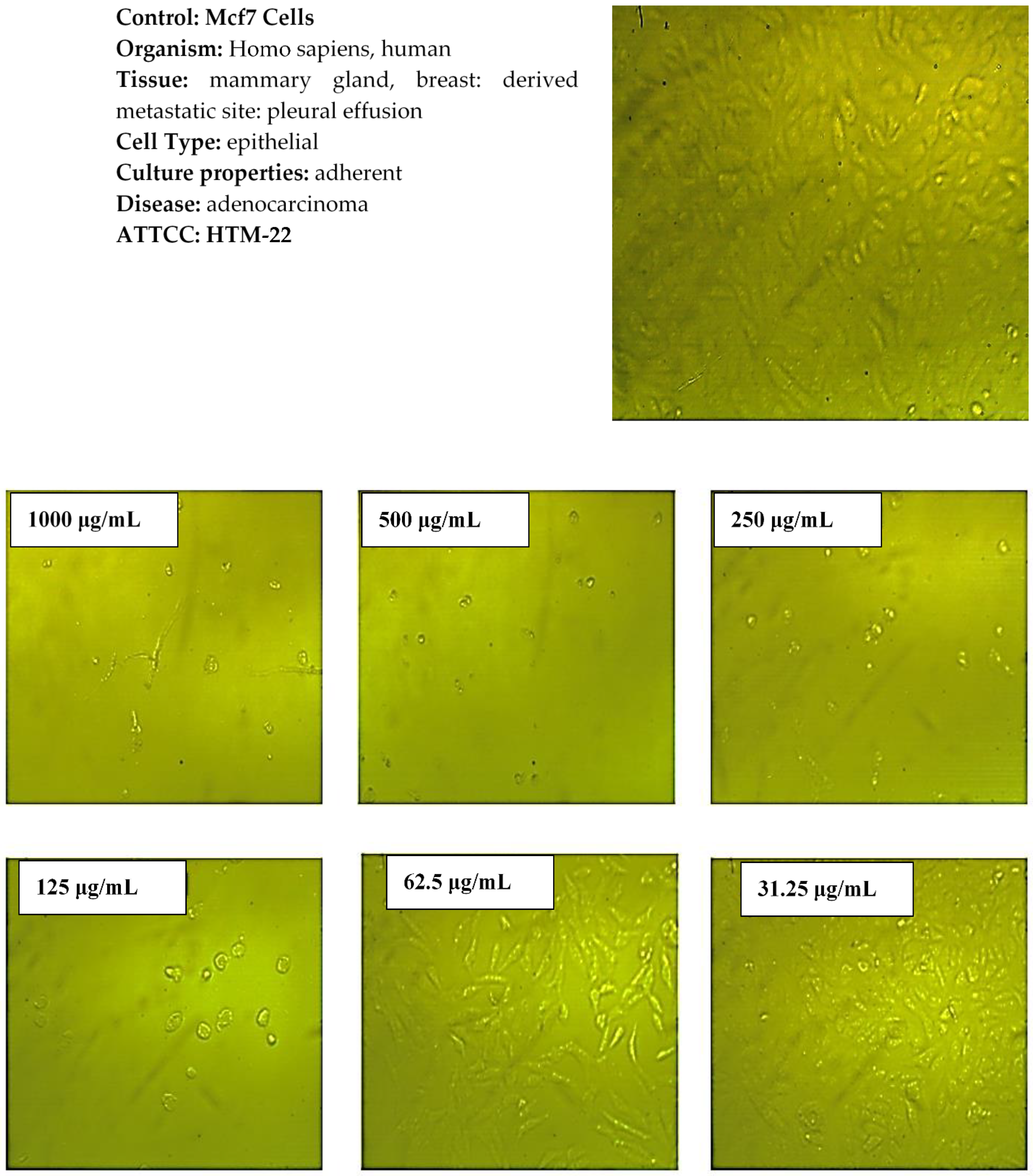
Figure 8 demonstrates the effect of the synthesized Co3O4 NPs using rosemary leaf extract on the metabolic activity of the Hep G2 and Mcf7 cancer cell lines following a 24 h incubation time at various concentrations (31.25, 62.5, 125, 250, 500, and 1000 μg/mL) by using MTT assay. The results show that the metabolism of the Hep G2 and Mcf7 cell lines, when compared to the positive control, was not affected by the concentration of Co3O4 NPs ≤62.5 µg/mL for Hep G2 and ≤31.25 µg/mL for Mcf7. This indicates that Co3O4 NPs are not toxic up to ≤62.5 µg/mL for Hep G2, and ≤31.25 µg/mL for Mcf7, which was also observed from microscopic images of Hep G2 and Mcf7 after applying those lower concentrations. The examined nanoparticles had a noticeable effect on the cell viability of both cancerous cell lines at higher concentrations. As illustrated in Figure 8, when the concentration of synthesized Co3O4 NPs was increased to 125 μg/mL, the metabolic activity of the Hep G2 and Mcf7 cancer cell lines decreased. This indicates that Co3O4 NPs had a toxic effect on Hep G2 and Mcf7, which was observed more when higher concentrations were applied. The microscopic images of the Hep G2 and Mcf7 cancer cell lines that were exposed to various doses of the synthesized Co3O4 NPs using rosemary leaf extract support these outcomes (Figure 9 and Figure 10). The half-maximal inhibitory concentrations values (IC50) were also determined to be 112 μg/mL for Hep G2 and 86 μg/mL for Mcf7. These in vitro findings indicate that the synthesized Co3O4 NPs are safe to be used in concentrations ≤ 31.25 µg/mL.

3. Experiments

3.1. Materials

Cobalt chloride hexahydrate (CoCl2·6H2O) was purchased from Sigma-Aldrich (St. Louis, MI, USA) and used as received. Double-distilled water was used in this study.

3.2. Preparation of the Rosemary Leaf Extract

The rosemary was purchased from a local market and rinsed with tap water. Subsequently, it was cleaned with double-distilled water to eliminate any residues and then air-dried for six days. Approximately 5 g of dried leaf was combined with 100 mL of double-distilled water to create the extract, which was then heated for 20 min at 50 °C. The extract was then filtered and stored for further research [32].

3.3. Biosynthesis of the Co3O4 NPs

In a flask, 100 mL of double-distilled water was used to dissolve 10 g of CoCl2·6H2O. The solution was heated to 50 °C and kept at this temperature for 5 min on a hot plate. The salt solution was then supplemented with 20 mL of rosemary extract while being vigorously stirred. During the reaction, the solution turned dark brown, indicating the synthesis of Co3O4 NPs. Following this, the mixture was centrifuged for 20 min at 15,000 rpm. The synthesized Co3O4 NPs were subsequently rinsed with double-distilled water and dried for 24 h at 80 °C. Subsequently, the dried Co3O4 NPs were annealed at 600 °C for 2 h in a muffle furnace [14] for stability. The synthesized Co3O4 NPs were then placed in a glass vial and stored for characterization.

3.4. Characterization of the Co3O4 NPs

UV, VIS, and NIR spectrometers were used to investigate the synthesized Co3O4 NPs spectrum (Lambda 750, Parkin Elmer). Furthermore, the surface charge and particle size of the prepared Co3O4 NPs were determined using a zeta seizer instrument (NanoSight NS500, Malvern Panalytical, Malvern, UK). The morphology and elemental composition of the produced Co3O4 NPs were investigated using energy-dispersive X-ray spectroscopy (EDX) analysis and scanning electron microscopy (SEM; JSM-6701FPlus, JEOL Ltd., Tokyo, Japan) at an acceleration voltage of 18 kV and magnifications of 5000× and 30,000×. Transmission electron microscopy (TEM) was performed using a high-resolution JEM-2100 instrument (JEOL Ltd.). The X-ray diffraction (XRD; EQUINOX 1000, Thermo Fisher Scientific, Waltham, MA, USA) patterns of powdered Co3O4 NPs were obtained using Cu–Kα radiation with a current of 35 mA and an applied voltage of 39 kV. The 2θ angles ranged from 20° to 90° with a scan speed of 0.3°/min.

3.5. In Vitro Cytotoxicity Evaluation

An MTT assay was used to evaluate the in vitro cytotoxicity of the synthesized Co3O4 NPs against liver cancer cells (Hep G2, ATCC number HB-8065) and cancerous breast cells (Mcf7, ATCC number HTB-22). To fabricate a complete monolayer sheet, a 96-well tissue culture plate was incubated with 1 × 10⁵ cells/mL (100 µL/well) at 37 °C for 24 h. After the formation of a confluent sheet, the growth medium was isolated from the 96-well microtiter plates, and the cell monolayer was washed twice with the wash medium. The analyzed samples were diluted twice in an RPMI-1640 medium containing 2% serum (maintenance medium). Three wells served as the control and received only the maintenance medium, while 0.1 mL of each dilution was tested in different wells. After testing, the plates were incubated at 37 °C. Physical characteristics of toxicity were examined in the cells, such as the complete or partial loss of the monolayer, rounding, shrinkage, and cell granulation. An MTT solution (5 mg/mL in PBS) was prepared (Bio Basic Inc., Markham, ON, Canada), and 20 µL of the solution was added to each well. To completely blend MTT into the medium, the samples were shaken at 150 rpm for 5 min. The samples were then incubated for 4 h at 37 °C with 5% CO2 to allow for MTT to be metabolized. The medium was then removed, and the plates were dried on paper towels to remove any residues. To thoroughly mix the formazan and solvent, 200 µL of DMSO was used to resuspend the formazan (MTT metabolic products) before shaking at 150 rpm for 5 min. The background was subtracted at 620 nm before calculating the optical density at 560 nm. The cell number and optical density were directly correlated.

4. Conclusions

Co3O4 NPs were prepared with a practical ecofriendly approach using an aqueous extract of rosemary leaves. Different analytical methods were used to analyze the synthesized Co3O4 NPs. The UV spectrum revealed two distinct sharp absorption peaks that confirmed the successful synthesis of Co3O4 NPs using the rosemary leaf extract. The excellent stability of synthesized nanoparticles in the suspensions and the particle size were evaluated using studies of dynamic light scattering and zeta potential. SEM images showed that the synthesized Co3O4 NPs had a semi-triangular pyramidal form with wide particle-size distribution, while EDX analysis confirmed their successful synthesis. XRD analysis confirmed the crystal structure of the prepared NPs. Furthermore, the synthesized Co3O4 NPs were not toxic to Hep G2 (at ≤62.5 µg/mL) and Mcf7 (at ≤31.25 µg/mL), so this novel ecofriendly Co3O4 NPs at this safe concentration can be evaluated in the future for its therapeutic activity.

Funding

This research received no external funding.

Institutional Review Board Statement

Not applicable.

Informed Consent Statement

Not applicable.

Data Availability Statement

Data are contained within the article.

Acknowledgments

The author acknowledges the support of the Chemistry Department at Taif University and the Ministry of Education in the Kingdom of Saudi Arabia. The author also appreciates Fahad A. Almughem from National Center for Biotechnology, Life Science and Environment Research Institute, King Abdulaziz City for Science and Technology (KACST for assisting in explination the cytotoxicity activity of the synthesized nanoparticles.

Conflicts of Interest

The author declares no conflict of interest.

References

  1. Manigandan, R.; Giribabu, K.; Suresh, R.; Vijayalakshm, L.; Stephen, A.; Narayanan, V. Cobalt oxide nanoparticles: Characterization and its electrocatalytic activity towards nitrobenzene. Chem. Sci. Trans. 2013, 2, S47–S50. [Google Scholar]
  2. Waris, A.; Misbahud, D.; Asmat, A.; Shakeeb, A.; Abdul, B.; Atta Ullah, K.; Muhammad, A. Green fabrication of Co and Co3O4 nanoparticles and their biomedical applications: A review. Open Life Sci. 2021, 16, 14–30. [Google Scholar] [CrossRef]
  3. Anele, A.; Obare, S.; Wei, J. Recent Trends and Advances of Co3O4 Nanoparticles in Environmental Remediation of Bacteria in Wastewater. Nanomaterials 2022, 12, 1129. [Google Scholar] [CrossRef]
  4. Sharma, J.; Pratibha, S.; Gurdip, M.; Shaheer, S.; Ameen, S. Green synthesis of Co3O4 nanoparticles and their applications in thermal decomposition of ammonium perchlorate and dye-sensitized solar cells. Mater. Sci. Eng. B 2015, 193, 181–188. [Google Scholar] [CrossRef]
  5. Iravani, S.; Varma, R.S. Sustainable synthesis of cobalt and cobalt oxide nanoparticles and their catalytic and biomedical applications. Green Chem. 2020, 22, 2643–2661. [Google Scholar] [CrossRef]
  6. Hafeez, M.; Shaheen, R.; Akram, B.; Haq, S. Green synthesis of cobalt oxide nanoparticles for potential biological applications. Mater. Res. Express 2020, 7, 025019. [Google Scholar] [CrossRef]
  7. Qiu, H.; Wang, Y.; Liu, Y.; Li, D.; Zhu, X.; Ji, Q.; Quan, F. Synthesis of Co/Co3O4 nanoparticles embedded in porous carbon nanofibers for high performance lithium-ion battery anodes. J. Porous Mater. 2017, 24, 551–557. [Google Scholar] [CrossRef]
  8. Hsueh, T.; Wu, S.-S. Highly sensitive Co3O4 nanoparticles/MEMS NO2 gas sensor with the adsorption of the Au nanoparticles. Sens. Actuators B Chem. 2021, 329, 129201. [Google Scholar] [CrossRef]
  9. Khalid, N.; Gull, A.; Ali, F.; Tahir, M.; Iqbal, T. Bi-functional green-synthesized of Co3O4 NPs for photocatalytic and electrochemical applications. Ceram. Int. 2022, 48, 32009–32021. [Google Scholar] [CrossRef]
  10. Jarestan, M.; Khalatbari, K.; Sadat Shandiz, S.; Beigi, S. Preparation, characterization, and anticancer efficacy of novel cobalt oxide nanoparticles conjugated with thiosemicarbazide. Biotech 2020, 10, 230. [Google Scholar] [CrossRef]
  11. Bhargava, R.; Khan, S.; Ahmad, N. Investigation of structural, optical and electrical properties of Co3O4 nanoparticles. In AIP Conference Proceedings; AIP Publishing LLC: Melville, NY, USA, 2018. [Google Scholar]
  12. Farhadi, S.; Sepahdar, A.; Jahanara, K. Spinel-type cobalt oxide (Co3O4) nanoparticles from the mer-Co (NH3)3(NO2)3 complex: Preparation, characterization, and study of optical and magnetic properties. J. Nanostruct. 2013, 3, 199–207. [Google Scholar]
  13. Khan, M.; Ali, F.; Faisal, S.; Rizwan, M.; Hussain, Z. Exploring the therapeutic potential of Hibiscus rosa sinensis synthesized cobalt oxide (Co3O4-NPs) and magnesium oxide nanoparticles (MgO-NPs). Saudi J. Biol. Sci. 2021, 28, 5157–5167. [Google Scholar]
  14. Anuradha, C.; Raji, P. Citrus limon fruit juice-assisted biomimetic synthesis, characterization and antimicrobial activity of cobalt oxide (Co3O4) nanoparticles. Appl. Phys. A 2021, 127, 55. [Google Scholar] [CrossRef]
  15. Ngnintedem Yonti, C.; Patrice, K.; Roussin, L.; Francois, D.; John Lambi, N. Green Synthesis of Iron-Doped Cobalt Oxide Nanoparticles from Palm Kernel Oil via Co-Precipitation and Structural Characterization. Nanomaterials 2021, 11, 2833. [Google Scholar] [CrossRef]
  16. Niu, M.; Wang, Y.; Cheng, Y.; Chen, G.; Cui, L. Fabrication of Co3O4 cubic nanoframes: Facet-preferential chemical etching of Fe3+ ions to Co3O4 nanocubes. Mater. Lett. 2009, 63, 837–839. [Google Scholar] [CrossRef]
  17. Shaalan, N.; Rashad, M.; Moharram, A. Promising methane gas sensor synthesized by microwave-assisted Co3O4 nanoparticles. Mater. Sci. Semicond. Process. 2016, 46, 1–5. [Google Scholar] [CrossRef]
  18. Matinise, N.; Mayedwa, N.; Fuku, X. Green synthesis of cobalt (II, III) oxide nanoparticles using Moringa Oleifera natural extract as high electrochemical electrode for supercapacitors. In AIP Conference Proceedings; AIP Publishing LLC: Melville, NY, USA, 2018. [Google Scholar]
  19. Diallo, A.; Beye, A.; Doyle, T.; Park, E. Green synthesis of Co3O4 nanoparticles via Aspalathus linearis: Physical properties. Green Chem. Lett. Rev. 2015, 8, 30–36. [Google Scholar] [CrossRef] [Green Version]
  20. Khalil, A.; Muhammad, O.; Ikram, U.; Muhammad, A.; Zabta, K.; Malik, M. Physical properties, biological applications and biocompatibility studies on biosynthesized single phase cobalt oxide (Co3O4) nanoparticles via Sageretia thea (Osbeck.). Arab. J. Chem. 2020, 13, 606–619. [Google Scholar] [CrossRef]
  21. Ikhuoria, E.U.; Omorogbe, S.O.; Sone, B.T.; Maaza, M. Bioinspired shape controlled antiferromagnetic Co3O4 with prism like-anchored octahedron morphology: A facile green synthesis using Manihot esculenta Crantz extract. Sci. Technol. Mater. 2018, 30, 92–98. [Google Scholar] [CrossRef]
  22. Edison, T.; Raji, A.; Gopalakrishnan, S.; Lee, Y. Supercapacitor performance of carbon supported Co3O4 nanoparticles synthesized using Terminalia chebula fruit. J. Taiwan Inst. Chem. Eng. 2016, 68, 489–495. [Google Scholar] [CrossRef]
  23. Pagar, T.; Suresh, G.; Khanderao, P.; Shreyas, P.; Rajeshwari, O. A review on bio-synthesized Co3O4 nanoparticles using plant extracts and their diverse applications. J. Chem. Rev. 2019, 1, 260–270. [Google Scholar]
  24. Labban, L.; Mustafa, U.E.-S.; Ibrahim, Y.M. The effects of rosemary (Rosmarinus officinalis) leaves powder on glucose level, lipid profile and lipid perodoxation. Int. J. Clin. Med. 2014, 2014, 44285. [Google Scholar]
  25. Hammer, M.; Junghanns, W. Rosmarinus officinalis L.: Rosemary. In Medicinal, Aromatic and Stimulant Plants; Springer: Berlin/Heidelberg, Germany, 2020; pp. 501–521. [Google Scholar]
  26. Ribeiro-Santos, R.; Carvalho-Costaad, D.; Helena, C.; Gonçalves, A.T.; Conceição, C.M.; Fernando, R.; Nathália, R.; Ana, S. A novel insight on an ancient aromatic plant: The rosemary (Rosmarinus officinalis L.). Trends Food Sci. Technol. 2015, 45, 355–368. [Google Scholar] [CrossRef]
  27. Tyagi, A. The effects of rosemary (Rosemarinus officinalis) leaves powder on glucose level. World J. Pharm. Res. 2017, 6, 548–554. [Google Scholar]
  28. Badreah, A.A.; Ahmed, A.; Mohamed, M.; Kamel, S. Phytosynthesis of Co3O4 Nanoparticles as the High Energy Storage Material of an Activated Carbon/Co3O4 Symmetric Supercapacitor Device with Excellent Cyclic Stability Based on a Na2SO4 Aqueous Electrolyte. ACS Omega 2022, 7, 23673–23684. [Google Scholar]
  29. Dewi, N.O.M.; Yulizar, Y.; Apriandanu, D.O.B. Green synthesis of Co3O4 nanoparticles using Euphorbia heterophylla L. leaves extract: Characterization and photocatalytic activity. In IOP Conference Series: Materials Science and Engineering; IOP Publishing: Bristol, UK, 2019. [Google Scholar]
  30. Leone, P.; Antonio, G.; Rossella, F.; Antonella, A.; Eleonora, M.; Alessio, B.; Luigi, G.; Valli, D.; Nicola, S.; Vito, R. The evolving role of immune checkpoint inhibitors in hepatocellular carcinoma treatment. Vaccines 2021, 9, 532. [Google Scholar] [CrossRef] [PubMed]
  31. Al-Qasmi, N.; Almughem, F.A.; Jarallah, S.J.; Almaabadi, A. Efficient Green Synthesis of (Fe3O4) and (NiFe2O4) Nanoparticles Using Star Anise (Illicium verum) Extract and Their Biomedical Activity against Some Cancer Cells. Materials 2022, 15, 4832. [Google Scholar] [CrossRef]
  32. Al-Qasmi, N. Facial eco-friendly synthesis of copper oxide nanoparticles using chia seeds extract and evaluation of its electrochemical activity. Processes 2021, 9, 2027. [Google Scholar] [CrossRef]
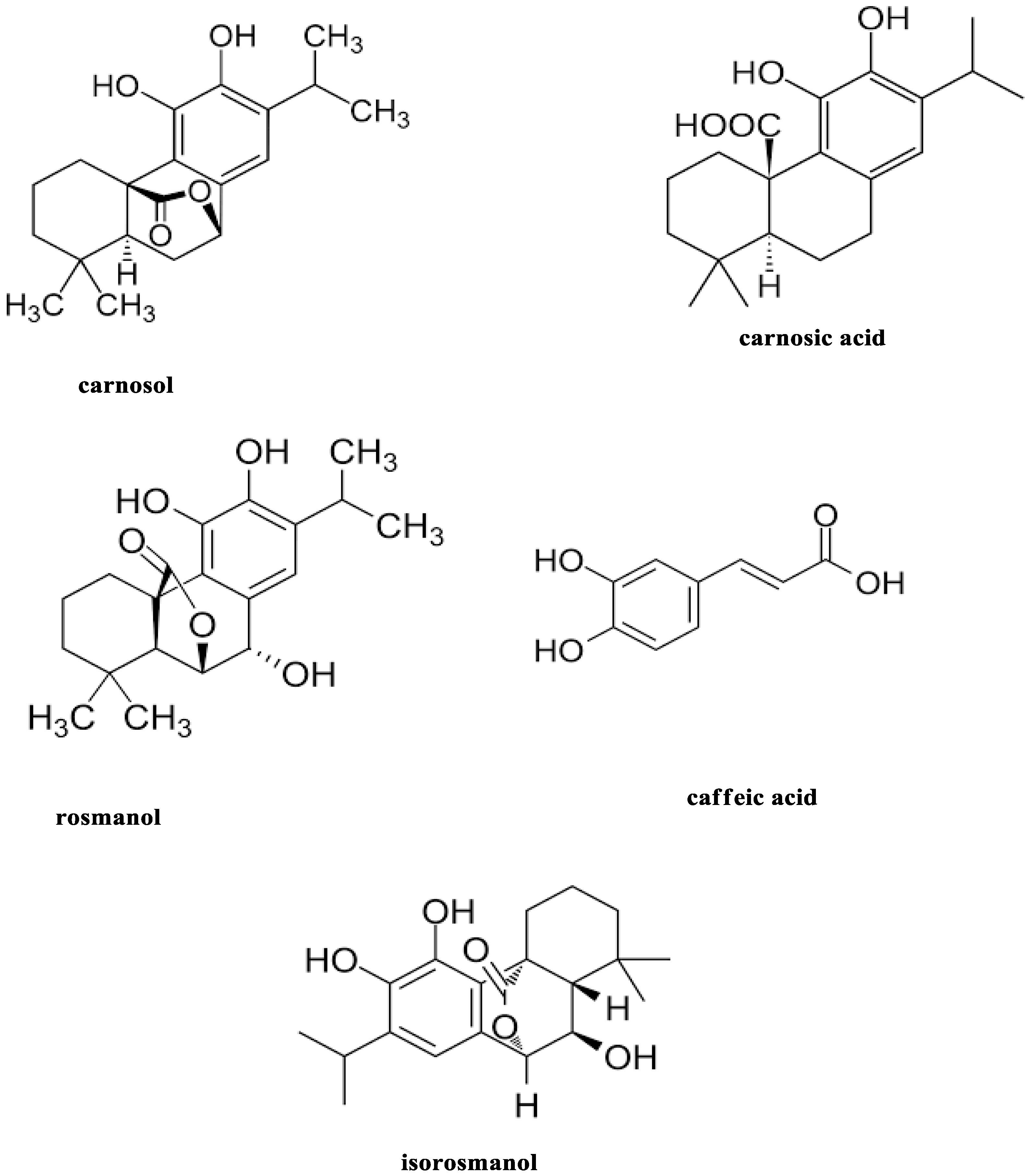
Figure 1. Chemical structure of biologically active phytochemicals in rosemary leaves.
Molecules 27 08163 g001
Figure 2. UV spectrum of Co3O4 NPs synthesized using rosemary leaf extract.
Molecules 27 08163 g002
Figure 3. (a) DLS measurements of particle size distribution and (b) charge zeta potential of Co3O4 NPs synthesized using rosemary leaf extract.
Molecules 27 08163 g003
Figure 4. (a) Transmission electron microscopy image and (b) high-resolution transmission electron microscopy image of the Co3O4 NPs synthesized using rosemary leaf extract.
Molecules 27 08163 g004
Figure 5. (a,b) Scanning electron microscopy images of Co3O4 NPs synthesized using rosemary leaf extract at different magnifications.
Molecules 27 08163 g005
Figure 6. Energy-dispersive X-ray spectrum of the Co3O4 NPs synthesized using rosemary leaf extract.
Molecules 27 08163 g006
Figure 7. X-ray diffraction pattern of Co3O4 NPs synthesized using rosemary leaf extract.
Molecules 27 08163 g007
Figure 8. Relative percentages of living cells of Hep G2 and Mcf7 cancer cells after exposure to different concentrations of Co3O4 NPs synthesized using rosemary leaf extract. The results are from an MTT assay and are displayed as the mean ± SD (n = 3).
Molecules 27 08163 g008
Figure 9. Microscopic images of Hep G2 cancer cells after treatment with various concentrations of Co3O4 NPs synthesized using rosemary leaf extract.
Molecules 27 08163 g009
Figure 10. Microscopic images of Mcf7 cancer cells after treatment with various concentrations of Co3O4 NPs synthesized using rosemary leaf extract.
Molecules 27 08163 g010
Publisher’s Note: MDPI stays neutral with regard to jurisdictional claims in published maps and institutional affiliations.