Article

A Background-Free SERS Strategy for Sensitive Detection of Hydrogen Peroxide

Key Laboratory of Biomaterials of Guangdong Higher Education Institutes, Department of Biomedical Engineering, Jinan University, Guangzhou 510632, China
*
Author to whom correspondence should be addressed.
These authors contributed equally to this work.
Molecules 2022, 27(22), 7918; https://doi.org/10.3390/molecules27227918
Submission received: 2 November 2022 / Revised: 11 November 2022 / Accepted: 14 November 2022 / Published: 16 November 2022

Abstract

:
The accurate and sensitive detection of biomolecules by surface-enhanced Raman spectroscopy (SERS) is possible, but remains challenging due to the interference from biomolecules in complex samples. Herein, a new SERS sensor is developed for background-free detection of hydrogen peroxide (H2O2) with an ultralow detection limit (1 × 10⁻¹⁰ mol/L), using a Raman-silent strategy. The Au microparticles (Au-RSMPs) resembling rose-stones are devised as SERS substrates with a high enhancement effect, and 4-mercaptophenylboronic acid (4-MPBA) is selected as an H2O2-responsive Raman reporter. Upon the reaction with H2O2, the phenylboronic group of 4-MPBA was converted to a phenol group, which subsequently reacted with 4-diazonium-phenylalkyne (4-DP), an alkyne-carrying molecule via the azo reaction. The formed product exhibits an intense and sharp SERS signal in the Raman-silent region, avoiding interference of impurities and biomolecules. As a proof-of-concept demonstration, we show that this SERS sensor possesses significant merits towards the determination of H2O2 in terms of broad linear range, low limit of detection, and high selectivity, showing promise for the quantitative analysis of H2O2 in complicated biological samples.

1. Introduction

Surface-enhanced Raman scattering (SERS) spectroscopy is a highly sensitive technique that enhances the Raman scattering of molecules supported by some nanostructured materials [1]. SERS allows for high specificity because it reveals the vibrational fingerprints of the analytes [2]. Compared with other detection techniques for biomolecules, SERS simultaneously possesses several key merits for bioimaging and biosensing, including its ability to characterize vibrational peaks, insusceptibility to photobleaching/photodegradation, negligible interference of water, multiple detection capabilities, and unmatched detection sensitivity [3,4,5].
Quantitative and precise measurements of metabolites or biomarkers in biological samples are critical in fundamental research and clinical diagnosis [6,7,8,9,10,11,12,13]. Among them, some SERS-based methods have been developed; however, it remains a challenge to differentiate traditional Raman tags or reporters from biomolecules since the proteins and lipids can generate a strong background signal in the “fingerprint region” (<1800 cm−1). To accurately assess metabolites in complex samples, a new concept of “silent-region” Raman reporters was reported [14,15,16], which shows the characteristic Raman peaks in the range of 1800–2800 cm−1; most of the biomolecules do not generate any Raman vibrational signal in this region [3].
To date, “silent-region” Raman tags and reporters were mostly applied to SERS imaging focusing on tumor cell targeting [17] and therapy [18], tissue, and living-cell imaging [19]. There are several reports based on SERS “silent-region” measurement for physiological detection [20,21,22,23]. However, these SERS sensors suffer from two major drawbacks: (1) the nanoscale SERS substrates lack high enhancement for trace detection with poor reproducibility and signal instability [24,25,26,27]; (2) the extensive application of “silent-region” reporters is impeded due to the complicated chemical synthesis or bioconjugation [3,28].
We herein demonstrate a new probe for H2O2 detection through the development of a highly-enhanced SERS substrate and a “silent-region” Raman reporter. The SERS probe contains “rose-stone-like” Au microparticles (RSMPs) and an H2O2-responsive Raman reporter. The as-synthesized RSMPs can generate intense SERS enhancement for the ultrasensitive determination, while the Raman reporter can produce the silent-range Raman signals upon the reaction with H2O2, enabling the detection of H2O2 with high sensitivity and selectivity.

2. Materials and Methods

2.1. Reagents and Materials

HAuCl4·3H2O, HCl, Polyvinylpyrrolidone (PVP, Mw = 40,000 Da), 4-mercaptobenzoic acid (4-MBA), 4-mercaptophenylboronic acid (4-MPBA), hydrogen peroxide (H2O2, 30%), NaNO2, 4-ethynylaniline (4-ENA), NaBF4, N-methyl pyrrolidone (NMP), and ethanol were purchased from Sigma. N-(3-Amidino)-aniline (NAAN) was synthesized according to the reported method [29]. All reagents were analytical grade and used as received. Deionized water (DI) was acquired by a Milli-Q water system (18.25 MΩ·cm) and used for all the aqueous samples.

2.2. Characterization Conditions

Field emission scanning electron microscopy (FESEM, Ultra-55, ZEISS, Oberkochen, Germany) was used to study the morphology of RSMPs. The size of RSMPs and their standard deviation (SD) were calculated by measuring over 100 individual particles. Laser Confocal Micro Raman Spectrometer (LabRAM HR Evolution, HORIBA Jobin Yvon S.A.S, Bensheim, Germany) was employed to measure the signal of SERS spectra by using a 633 nm laser excitation source. To measure the SERS signals, 1.5 μL SERS particles were dropped on a glass slide, and the measurement was conducted on a single particle after being dried in the air.

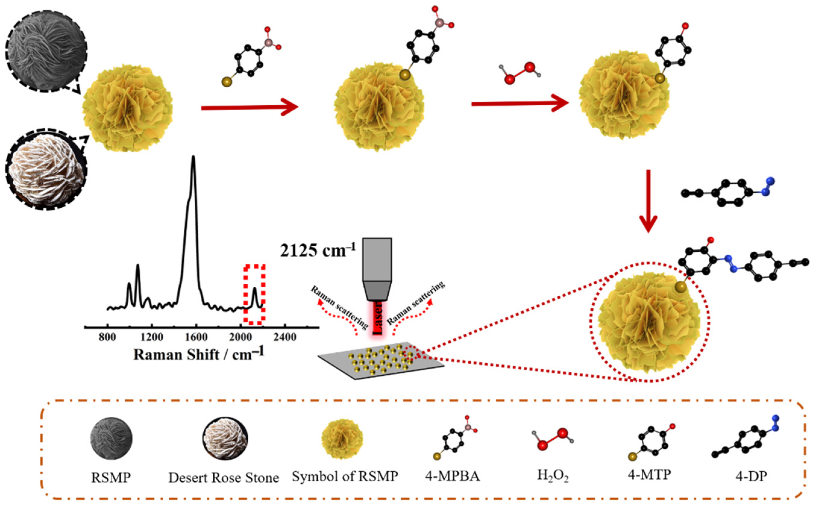
2.3. Synthesis of RSMPs

The RSMPs were prepared via the reduction of HAuCl4 using NAAN as a reducing agent [24]. Simply put, 25 μL 16 mg/mL PVP (0.40 mg/mL) was added into 1 mL 1 mM HCl solution, followed by the addition of 8 μL 10% HAuCl4 solution (2 mM, dissolved in 1 mM HCl). After cooling the mixture to 4 °C, 50 μL of 100 mg/mL NAAN (16 mM) in 1 mM HCl was injected, the mixture was stirred for 30 s and allowed to react at 4 °C for 24 h without further stirring. The final solution was centrifuged at 6000 rpm for 5 min, and the collected pellets were subsequently washed three times with NMP and DI to remove the impurities. The eventual pellets were collected as RSMPs and stored in the fridge for further usage. To acquire the appropriate particles as a good SERS substrate, the concentrations of NAAN (3, 4, and 5 mg/mL), PVP (0.40, 3.20, and 12.80 mg/mL), and the reaction time (24, 72, and 120 h) were optimized.

2.4. Synthesis of 4-Diazonium-Phenylaklyne (4-DP)

4-DP was prepared according to the typical diazonium reaction. 4-Ethynylaniline (4-ENA) (100 mg, 0.59 mmol) was dissolved in 1 mL 1 M HCl solution and cooled by ice water. In another flask, NaNO2 (45 mg, 0.65 mmol) was dissolved in cooled DI water (0.25 mL), which was subsequently added to the 4-ENA solution dropwise. The mixture was left to stir at 4 °C for 1 h and room temperature for 30 min, followed by the addition of 2 mL NaBF4 saturated solution. The resulting product was filtered and the precipitate was collected and thoroughly washed with cooled water. After drying under a high vacuum, the 4-DP was stored in the fridge.

2.5. Preparation of SERS Probe

200 μL of RSMPs aqueous solution was mixed with 1 mL 1 mM 4-MPBA solution (dissolved in ethanol), and the mixture was stirred for 2 h by a vortex at room temperature. After washing with ethanol and DI, the 4-MPBA modified RSMPs were distributed in 50 μL DI water as the SERS probe. To measure the SERS performance of the resulting SERS probe, 10 μL of SERS probe solution was dropped on a cleaned glass slide and dried in the air, and the SERS signals on each particle were measured.

2.6. H2O2 Detection

100 μL H2O2 solutions with varying concentrations were incubated with the SERS probes for 1 h. After washing with DI water, the resulting SERS probes were dissolved in 20 μL DI, followed by the addition of 10 μL 100 mM 4-DP and stirring for 1 h. After washing with NMP and DI water, 10 μL of the eventual SERS probes was dropped on a glass slide for SERS measurements after being dried in air.

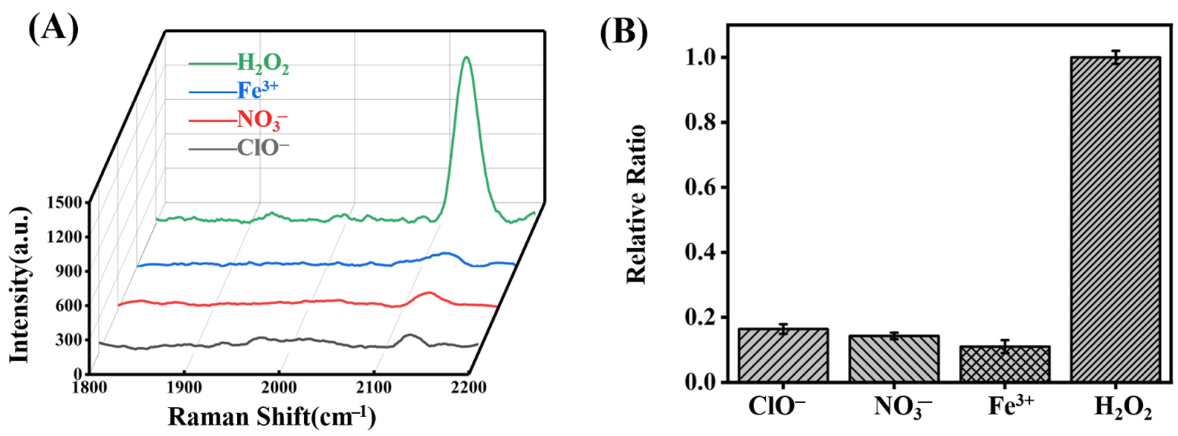
2.7. Interference Experiment

10 mM NaClO was prepared by mixing 10 mM NaOH and HClO solutions. 300 μL 10 mM Cu(NO3)2, FeCl3, and NaClO were mixed with 20 μL SERS probe solution. After 1 h incubation and washed with DI water, the SERS probes were mixed with 10 μL 100 mM 4-DP for 1 h. The SERS probes were washed successively with NMP and DI water. 5 μL of each SERS probe solution was dropped on a glass slide for SERS measurements after being dried in air. Each spectrum was processed by subtracting the baseline and normalized using the intensity at 1071 cm−1 due to the C-H stretching vibration of the benzene.

2.8. Real Sample Test

The standard additional method was applied to test the applicability of the resulting SERS probes. Human serum solution was centrifuged and the collected supernatant was diluted 1000-fold with PBS, followed by spiking with different concentrations of H2O2. After incubation of SERS probes with H2O2 solutions, and subsequently reacting with 4-DP, the SERS probes were collected and their SERS signals were measured.

3. Results and Discussion

The SERS sensing mechanism is shown in Figure 1. Firstly, we synthesized Au-based ”rose stone-like” microparticles named RSMPs due to their appearance which is similar to rose stones found in deserts. Their layered structure carrying multiple nanoplates provides abundant nanogaps and edges within RSMPs, which can generate hotspots for the SERS enhancement, resulting in the RSMPs being an excellent SERS substrate. 4-MPBA can be modified on RSMPs via the Au-S bond, and the reaction of 4-MPBA with H2O2 can transform the phenylboronic acid group into a phenol group [30]. Subsequently, 4-DP (4-Diazonium-phenylalkyne) can react with phenol to form an azobenzene compound carrying an alkyne group, which has a “silent region” Raman fingerprint (2125 cm−1). The amount of alkyne group on the SERS particle surface is dependent on the formation of the phenol group, which is determined by the H2O2 concentration, therefore, H2O2 can be profiled with significant sensitivity owing to the high signal/noise SERS measurement. Figure 2 demonstrates the synthesis of 4-DP and the H2O2-induced azo reaction. 4-ENA reacts with NaNO2 in acidic conditions to generate 4-DP. Eventually, the diazonium group of 4-DP reacts with the phenol generated by the reaction of 4-MPBA and H2O2 to form an azobenzene derivative EEDM ((E)-2-((4-ethynylphenyl)diazenyl)-4-mercaptophenol).
To study the mechanism of RSMP growth, samples at different time intervals were collected. In the early stage, HAuCl4 was reduced to form Au nanoplates by NAAN, while NAAN was oxidized to form PNAAN, which has a highly selective binding ability on the Au (111) facet, and the growth on this facet was inhibited, resulting in the growth of Au nanoplates. A mixture of cubic Au nanoplates with a diameter of 200–800 nm was observed after 10 min reaction (Figure 3A). As the growth continued, the Au nanoplates further grew into microplates (>1 μm) and new Au microplates were generated on the surface of prior plates to form ”rose stone-like” microparticles (RSMPs) (Figure 3B). After a 4 h reaction, RSMPs with a diameter of 2–3 μm were observed (Figure 3C), which further grew into the microparticles with an average size of up to 10 μm and exhibit densely-packed layer structures (Figure 3D).
To better control the anisotropic growth of Au microparticles and obtain uniform RSMPs as SERS substrates, three parameters including the concentrations of NAAN, PVP, and the reaction time were investigated. As shown in Figure 4A–C, the change of NAAN concentration from 3 to 5 mg/mL does not lead to the obvious change in RSMPs, and the corresponding histogram (Figure 4D) illustrates the diameters of 9.54 ± 1.01, 9.90 ± 1.31, and 9.05 ± 1.54 μm, respectively. As a shape control agent, PVP has a high binding ability to Au (111) facets, and its presence is important for the growth of Au microparticles. In the presence of 0.40 and 3.20 mg/mL PVP, the resulting RSMPs exhibit soft lamellae structures (Figure 4E,F). Further increasing the PVP concentration to 12.8 mg/mL results in the RSMPs containing few Au nanoplates with a decreased diameter to 4 μm (Figure 4G). The histogram of corresponding samples represents the RSMP diameter of 11.50 ± 1.11, 8.65 ± 1.90, and 4.40 ± 0.84 μm, respectively (Figure 4H). This demonstrates that a low concentration of PVP (0.4 mg/mL) is favorable for the formation of RSMPs. When the reaction was conducted for 24, 72, and 120 h (Figure 4I–K), the collected RSMPs showed similar morphology and average size (11.46 ± 1.08, 11.98 ± 0.99, and 12.69 ± 1.74 μm, Figure 4L). However, the Au microplates become thick and fold inwards or merge in a continuous stacking process as the reaction time increases, which can eliminate the nanogaps and is unfavorable for SERS enhancement. Therefore, 24 h was set as the optimal reaction time.
In addition, we investigated the yield of the resulting RSMPs prepared at different reaction conditions. As shown in Figure 5A–C, the proportion of satisfactory particles slightly increased with the increase of NAAN concentration, and the corresponding histogram in Figure 5D shows the percentages of 27.66%, 30.75%, and 34.92%. As the PVP concentration increases from 0.4–3.2 mg/mL, the percentage of RSMPs decreases from 48.78% to 36.36% (Figure 5E). Further increasing PVP concentration up to 12.8 mg/mL results in a mixture of small gold-based microparticles (Figure 5G), which is caused by the fast reduction of HAuCl4 to Au atoms in the presence of excessive PVP. Therefore, the presence of PVP for the formation of RSMPs is necessary, while an overly high concentration is unfavorable. As shown in Figure 5I–K, the corresponding yields are 27.59%, 25.89%, and 22.15% for the reaction times of 24, 72, and 120 h, respectively (Figure 5L), revealing that the reaction time has less effect on the yield of RSMPs. Based on the results mentioned above, we can conclude that the effects of NAAN concentration and synthesis time on RSMP morphology are not significant, while the concentration of PVP has a dramatic impact on the particle size and morphology. In order to obtain SERS substrates with uniform size and morphology, we finally chose 5 mg/mL NAAN, 0.4 mg/mL PVP, and a synthesis time of 24 h as the optimal condition for the preparation of RSMPs.
The microplates and the densely-packed layered structures provide a large number of sharp edges as well as nanogaps among different layers, which can generate hotspots for electromagnetic enhancement (EM) [31]. In order to evaluate the SERS enhancement of RSMPs, commercial SERS chips with SERS enhancement factors (EF) of 1.0 × 10⁴ were applied for the comparison (Xiamen Spectroscopy Scientific Instruments Co., Ltd., Xiamen, China). The average size of RSMPs is 11.5 ± 1.1 μm (Figure 6A), which is much larger than the diameter of the Raman laser spot size (about 2 μm, Figure 6B), allowing the measurement of SERS signals on an individual RSMP. In contrast, the Raman chips were prepared by the self-assembly of spherical Au nanoparticles (NPs) (diameter of 44–56 nm) into a well-organized array (Figure 6C). After the modification of 4-MBA via the Au-S bond, the SERS intensity using RSMPs as SERS substrate is almost four times higher compared to that using the commercial chip as SERS substrate (Figure 6D), revealing that the as-prepared RSMPs demonstrate a better enhancement effect. The corresponding SERS enhancement factors (EF) were calculated to be 2.8×10⁶ for the RSMPs, according to the reported method [24].
Thanks to the superb SERS enhancement, RSMPs can be utilized for ultrasensitive H2O2 detection. Hydrogen peroxide (H2O2) is a key redox signaling agent produced under the control of growth factors and cytokines. At low physiological levels in the nanomolar range, H2O2 is the primary agent signaling through specific protein targets, and it participates in metabolic regulation and stress response to support cell adaptation to changing environments and stress [32]. A large number of studies have focused on the analysis or imaging of intracellular H2O2 levels [33,34,35,36,37]. In addition, it has been suggested that extracellular H2O2 is also a fundamental signaling molecule, and it could activate related pathways to cause the corresponding response or the progress of tissues, leading to tumor progression, vascular damage, and even Alzheimer’s disease [38,39,40]. To better understand the production and role of H2O2 in physiology and disease, it is important to achieve sensitive detection of physiological extracellular/exogenous H2O2.
Research has been carried out for H2O2 detection. In particular, phenylboronic acid was mostly applied as a probe since it could selectively react with H2O2 to produce phenol. Several sensing methods were developed based on this reaction including electrochemical luminescence (ECL) [41,42], fluorescence (FL) [43,44,45], chemiluminescence (CL) [46,47,48,49] and SERS [50,51,52]. Among these methods, SERS has attracted great attention due to its ability to characterize vibrational peaks, insusceptibility to photobleaching, and unmatched detection sensitivity [3]. The typical SERS sensing of H2O2 is based on the mercaptophenylboronic acid (MPBA) self-assembled monolayer on the SERS substrate. The presence of H2O2 leads to the transformation of the phenylboronic acid group into the phenol group, resulting in the major shift of typical C=C stretching vibration from 1569 cm−1 to 1600 cm−1 [53]. However, this SERS sensor has shown some major drawbacks in two ways: (1) the readout signal was recorded based on the shift of the phenyl group, but a slight shift of the benzene group will lead to the overlap of these two peaks, and it is difficult to distinguish this change, resulting in severe error. (2) The SERS signals were collected in the range of 1000–1700 cm−1, where most of biomolecules may generate strong interference signals from the lipid and proteins, significantly hindering the SERS measurement. Therefore, the interference could be efficiently eliminated or avoided, which could lead to false-positive results as characteristic peaks located in the “fingerprint” region were used as analysis standard. To overcome these limitations, we further function the phenol group with a new compound 4-DP via the azo coupling reaction, the introduction of alkyne group results in a new Raman shift in 2125 cm−1 which is located in the Raman-silent region and the surface ligand of SERS substrate or biomolecules have no Raman signal in this range [54,55,56]. This results in a high signal/noise ratio, and greatly improves the sensitivity of the H2O2 sensor.
4-DP was synthesized according to the diazonium reaction, and its IR spectrum displays a characteristic absorption peak of 1415 cm−1, which is ascribed to the N=N stretch of the diazonium group (Figure 7A) [57]. The absorption peaks at 1506 cm−1, 1084 cm−1, and 2110 cm−1 are due to the stretching of benzene, C-N, and C≡N groups, respectively. After the modification of 4-MPBA on RSMPs, two Raman peaks at 1071 and 1569 cm−1 were observed, which are attributed to the plane deformation of C-H and the stretching vibration of C=C on the benzene ring (Figure 7B line a). 4-DP could not be attached to the RSMPs or 4-MPBA directly since there was no new Raman peak after the incubation with SERS probes (Figure 7B line b). Upon the reaction with H2O2, the peaks at 1569 cm−1 become slightly broadened with a side peak at 1600 cm−1, which is consistent with the reference [53]. Apparently, most of the newly-formed peak overlaps with the original peak, resulting in a difficult separation. After further incubation with 4-DP, new peaks due to the formation of azobenzene located at 1141, 1391 and 1438 cm−1 can be observed, revealing the formation of azobenzene (Figure 7B line c). However, the peaks in this range suffered from the substantial interference from the background signal of RSMP, and the accurate measurement of its intensity for the H2O2 detection is difficult. Fortunately, an additional peak located at 2125 cm−1 was observed, which is due to the presence of alkyne group, enabling the high signal/noise determination of H2O2.
After incubation of the 4-MPBA with different concentrations of H2O2 and subsequent reaction with excess 4-DP, their SERS spectra were measured and are shown in Figure 7A. As the H2O2 concentration gradually increased, the intensity of the SERS peak at 2125 cm−1 increased. Therefore, we selected two characteristic peaks at 2125 cm−1 and 1071 cm−1 and used the ratio of their corresponding intensity for the H2O2 detection. As shown in Figure 8B, I2125/I1071 is linearly correlated to H2O2 concentration in the concentration range of 0.1 nM–1.5 mM (Figure 8A), and the calibration equation is Y = 0.072X + 0.759 with R2 of 0.976 (Figure 8B). The corresponding limit of detection (LOD) is 0.1 nM, which is much lower than those reported in the previous work (Table 1). The significant improvement is owing to the high EF of the SERS substrate; more importantly, the introduction of Raman peak in the silent range plays a crucial role.
Next, we investigated the selectivity of this sensor by testing the response of ClO, NO3−, and Fe3+. After incubation of the SERS probe with 10 mM of each sample and subsequent reaction with 4-DP, their corresponding SERS spectra are shown in Figure 9A. It can be seen that these three interferences can only induce slight SERS signals. A comparison with the spectra of the blank SERS probe molecule and the probe molecule after reaction with 0.06 mM H2O2 in the silent region is shown in Figure 9B. Normalizing the intensity of the peak at 2125 cm−1 after the reaction with 0.06 mM H2O2, the relative intensities of ClO, NO3−, and Fe3+ are 0.16, 0.14, and 0.10, respectively (Figure 9B). The concentration of interferents in the experiment is about 166 times that of H2O2, but the peak intensity at 2125 cm−1 was still much less than that of H2O2, demonstrating that our SERS probe was able to specifically identify H2O2.
In order to verify the feasibility of this method in a biological environment, the determination of H2O2 in human serum was conducted via the typical additional method. Simply put, the human serum was diluted 1000-fold by PBS, followed by the addition of H2O2 with known concentrations. After the treatment of the SERS probe with prepared H2O2 solution and 4-DP, their corresponding SERS spectra were measured. As shown in Table 2, the recovery rates were in the range of 96.52% and 116.62% with relative standard deviations (RSDs) less than 9.22%. This result reveals that the as-developed SERS sensor could be applied for the clinical determination of H2O2.

4. Conclusions

In this study, we have successfully developed a new SERS method for the specific detection of H2O2 based on the Raman-silent region signal molecule. The resulting SERS sensor demonstrated excellent performance in terms of high sensitivity, broad detection range, and good selectivity. Owing to the good capability against interference, especially the near-zero signals of biomolecules in the Raman-silent range, we envision that this strategy can be applied for the sensitive and specific monitoring of the various physiological processes that produce H2O2.

Author Contributions

Conceptualization, N.L.; methodology, H.C. and K.C.; validation, P.Z., S.L., J.W. and N.L.; investigation, K.C. and H.C.; writing—original draft preparation, H.C.; writing—review and editing, K.C. and N.L.; supervision, N.L.; project administration, N.L.; funding acquisition, N.L. All authors have read and agreed to the published version of the manuscript.

Funding

This research was funded by the Guangdong Basic and Applied Basic Research Foundation (2020A1515010957).

Institutional Review Board Statement

Not applicable.

Informed Consent Statement

Not applicable.

Data Availability Statement

Not applicable.

Conflicts of Interest

The authors declare no conflict of interest.

Sample Availability

Not applicable.

References

  1. Langer, J.; Jimenez de Aberasturi, D.; Aizpurua, J.; Alvarez-Puebla, R.A.; Auguié, B.; Baumberg, J.J.; Bazan, G.C.; Bell, S.E.J.; Boisen, A.; Brolo, A.G.; et al. Present and Future of Surface-Enhanced Raman Scattering. ACS Nano 2020, 14, 28–117. [Google Scholar] [CrossRef] [PubMed] [Green Version]
  2. Sharma, B.; Frontiera, R.R.; Henry, A.-I.; Ringe, E.; Van Duyne, R.P. SERS: Materials, applications, and the future. Mater. Today 2012, 15, 16–25. [Google Scholar] [CrossRef]
  3. Yin, Y.; Li, Q.; Ma, S.; Liu, H.; Dong, B.; Yang, J.; Liu, D. Prussian Blue as a Highly Sensitive and Background-Free Resonant Raman Reporter. Anal. Chem. 2017, 89, 1551–1557. [Google Scholar] [CrossRef] [PubMed]
  4. Liu, H.; Gao, X.; Xu, C.; Liu, D. SERS Tags for Biomedical Detection and Bioimaging. Theranostics 2022, 12, 1870–1903. [Google Scholar] [CrossRef] [PubMed]
  5. Tahir, M.A.; Dina, N.E.; Cheng, H.; Valev, V.K.; Zhang, L. Surface-enhanced Raman spectroscopy for bioanalysis and diagnosis. Nanoscale 2021, 13, 11593–11634. [Google Scholar] [CrossRef]
  6. Jain, M.; Nilsson, R.; Sharma, S.; Madhusudhan, N.; Kitami, T.; Souza Amanda, L.; Kafri, R.; Kirschner Marc, W.; Clish Clary, B.; Mootha Vamsi, K. Metabolite Profiling Identifies a Key Role for Glycine in Rapid Cancer Cell Proliferation. Science 2012, 336, 1040–1044. [Google Scholar] [CrossRef] [Green Version]
  7. Fernie, A.R.; Trethewey, R.N.; Krotzky, A.J.; Willmitzer, L. Metabolite profiling: From diagnostics to systems biology. Nat. Rev. Mol. Cell Biol. 2004, 5, 763–769. [Google Scholar] [CrossRef]
  8. Wang, T.J.; Larson, M.G.; Vasan, R.S.; Cheng, S.; Rhee, E.P.; McCabe, E.; Lewis, G.D.; Fox, C.S.; Jacques, P.F.; Fernandez, C.; et al. Metabolite profiles and the risk of developing diabetes. Nat. Med. 2011, 17, 448–453. [Google Scholar] [CrossRef] [Green Version]
  9. Asiago, V.M.; Alvarado, L.Z.; Shanaiah, N.; Gowda, G.A.N.; Owusu-Sarfo, K.; Ballas, R.A.; Raftery, D. Early Detection of Recurrent Breast Cancer Using Metabolite Profiling. Cancer Res. 2010, 70, 8309–8318. [Google Scholar] [CrossRef] [Green Version]
  10. Gong, J.; Tang, H.; Wang, M.; Lin, X.; Wang, K.; Liu, J. Novel three-dimensional graphene nanomesh prepared by facile electro-etching for improved electroanalytical performance for small biomolecules. Mater. Des. 2022, 215, 110506. [Google Scholar] [CrossRef]
  11. Gong, J.; Zhang, T.; Luo, T.; Luo, X.; Yan, F.; Tang, W.; Liu, J. Bipolar silica nanochannel array confined electrochemiluminescence for ultrasensitive detection of SARS-CoV-2 antibody. Biosens. Bioelectron. 2022, 215, 114563. [Google Scholar] [CrossRef] [PubMed]
  12. Zhou, H.; Ma, X.; Sailjoi, A.; Zou, Y.; Lin, X.; Yan, F.; Su, B.; Liu, J. Vertical silica nanochannels supported by nanocarbon composite for simultaneous detection of serotonin and melatonin in biological fluids. Sens. Actuators B Chem. 2022, 353, 131101. [Google Scholar] [CrossRef]
  13. Gong, J.; Zhang, T.; Chen, P.; Yan, F.; Liu, J. Bipolar silica nanochannel array for dual-mode electrochemiluminescence and electrochemical immunosensing platform. Sens. Actuators B Chem. 2022, 368, 132086. [Google Scholar] [CrossRef]
  14. Hong, S.; Chen, T.; Zhu, Y.; Li, A.; Huang, Y.; Chen, X. Live-Cell Stimulated Raman Scattering Imaging of Alkyne-Tagged Biomolecules. Angew. Chem. Int. Ed. 2014, 53, 5827–5831. [Google Scholar] [CrossRef] [PubMed]
  15. Jin, Q.; Fan, X.; Chen, C.; Huang, L.; Wang, J.; Tang, X. Multicolor Raman Beads for Multiplexed Tumor Cell and Tissue Imaging and in Vivo Tumor Spectral Detection. Anal. Chem. 2019, 91, 3784–3789. [Google Scholar] [CrossRef] [Green Version]
  16. Tian, S.; Li, H.; Li, Z.; Tang, H.; Yin, M.; Chen, Y.; Wang, S.; Gao, Y.; Yang, X.; Meng, F.; et al. Polydiacetylene-based ultrastrong bioorthogonal Raman probes for targeted live-cell Raman imaging. Nat. Commun. 2020, 11, 81. [Google Scholar] [CrossRef] [Green Version]
  17. Kennedy, D.C.; Hoop, K.A.; Tay, L.-L.; Pezacki, J.P. Development of nanoparticle probes for multiplex SERS imaging of cell surface proteins. Nanoscale 2010, 2, 1413–1416. [Google Scholar] [CrossRef] [Green Version]
  18. Zhu, W.; Gao, M.-Y.; Zhu, Q.; Chi, B.; Zeng, L.-W.; Hu, J.-M.; Shen, A.-G. Monodispersed plasmonic Prussian blue nanoparticles for zero-background SERS/MRI-guided phototherapy. Nanoscale 2020, 12, 3292–3301. [Google Scholar] [CrossRef]
  19. Zhang, L.; Zhang, R.; Gao, M.; Zhang, X. Facile synthesis of thiol and alkynyl contained SERS reporter molecular and its usage in assembly of polydopamine protected bioorthogonal SERS tag for live cell imaging. Talanta 2016, 158, 315–321. [Google Scholar] [CrossRef]
  20. Li, N.; Chen, H.L.; Zhang, M.Y.; Zha, Y.C.; Mu, Z.X.; Ma, Y.; Chen, P. A universal ultrasensitive platform for enzyme-linked immunoassay based on responsive surface-enhanced Raman scattering. Sens. Actuators B Chem. 2020, 315, 128135. [Google Scholar] [CrossRef]
  21. Lin, L.; Tian, X.; Hong, S.; Dai, P.; You, Q.; Wang, R.; Feng, L.; Xie, C.; Tian, Z.-Q.; Chen, X. A Bioorthogonal Raman Reporter Strategy for SERS Detection of Glycans on Live Cells. Angew. Chem. Int. Ed. 2013, 52, 7266–7271. [Google Scholar] [CrossRef] [PubMed]
  22. Hanif, S.; Liu, H.-L.; Ahmed, S.A.; Yang, J.-M.; Zhou, Y.; Pang, J.; Ji, L.-N.; Xia, X.-H.; Wang, K. Nanopipette-Based SERS Aptasensor for Subcellular Localization of Cancer Biomarker in Single Cells. Anal. Chem. 2017, 89, 9911–9917. [Google Scholar] [CrossRef] [PubMed]
  23. Lin, X.; Wang, Y.; Wang, L.; Lu, Y.; Li, J.; Lu, D.; Zhou, T.; Huang, Z.; Huang, J.; Huang, H.; et al. Interference-free and high precision biosensor based on surface enhanced Raman spectroscopy integrated with surface molecularly imprinted polymer technology for tumor biomarker detection in human blood. Biosens. Bioelectron. 2019, 143, 111599. [Google Scholar] [CrossRef] [PubMed]
  24. Ma, Y.; Yung, L.Y.L. Gold Nanoplate-Based 3D Hierarchical Microparticles: A Single Particle with High Surface-Enhanced Raman Scattering Enhancement. Langmuir 2016, 32, 7854–7859. [Google Scholar] [CrossRef]
  25. Alvarez-Puebla, R.A.; Liz-Marzán, L.M. SERS-Based Diagnosis and Biodetection. Small 2010, 6, 604–610. [Google Scholar] [CrossRef]
  26. Xu, L.; Yan, W.; Ma, W.; Kuang, H.; Wu, X.; Liu, L.; Zhao, Y.; Wang, L.; Xu, C. SERS Encoded Silver Pyramids for Attomolar Detection of Multiplexed Disease Biomarkers. Adv. Mater. 2015, 27, 1706–1711. [Google Scholar] [CrossRef]
  27. Li, N.; Zhang, M.; Zha, Y.; Cao, Y.; Ma, Y. π-π stacking-directed self-assembly of nanoplatelets into diversified three-dimensional superparticles for high surface-enhanced Raman scattering. J. Colloid Interface Sci. 2020, 575, 54–60. [Google Scholar] [CrossRef]
  28. Yamakoshi, H.; Dodo, K.; Palonpon, A.; Ando, J.; Fujita, K.; Kawata, S.; Sodeoka, M. Alkyne-Tag Raman Imaging for Visualization of Mobile Small Molecules in Live Cells. J. Am. Chem. Soc. 2012, 134, 20681–20689. [Google Scholar] [CrossRef]
  29. Ma, Y.; Yung, L.Y.L. Synthesis of Self-Stabilized Poly(N-(3-Amidino)-Aniline) Particles and their CO2-Responsive Properties. Part Part Syst. Char. 2015, 32, 743–748. [Google Scholar] [CrossRef]
  30. Cheng, W.; Tang, P.; He, X.; Xing, X.; Xing, M.; Lu, L.; Ju, P.; Wang, Y.; Liu, S.; Lu, X.; et al. Dynamic monitoring and quantitative characterization of intracellular H2O2 content by using SERS based boric acid nanoprobe. Talanta 2020, 214, 120863. [Google Scholar] [CrossRef]
  31. Wang, Y.Q.; Yan, B.; Chen, L.X. SERS Tags: Novel Optical Nanoprobes for Bioanalysis. Chem Rev 2013, 113, 1391–1428. [Google Scholar] [CrossRef] [PubMed]
  32. Sies, H.; Jones, D.P. Reactive oxygen species (ROS) as pleiotropic physiological signalling agents. Nat. Rev. Mol. Cell Biol. 2020, 21, 363–383. [Google Scholar] [CrossRef] [PubMed]
  33. Nordberg, J.; Arnér, E.S.J. Reactive oxygen species, antioxidants, and the mammalian thioredoxin system. Free Radic. Biol. Med. 2001, 31, 1287–1312. [Google Scholar] [CrossRef]
  34. Belousov, V.V.; Fradkov, A.F.; Lukyanov, K.A.; Staroverov, D.B.; Shakhbazov, K.S.; Terskikh, A.V.; Lukyanov, S. Genetically encoded fluorescent indicator for intracellular hydrogen peroxide. Nat. Methods 2006, 3, 281–286. [Google Scholar] [CrossRef] [PubMed]
  35. Bienert, G.P.; Møller, A.L.B.; Kristiansen, K.A.; Schulz, A.; Møller, I.M.; Schjoerring, J.K.; Jahn, T.P. Specific Aquaporins Facilitate the Diffusion of Hydrogen Peroxide across Membranes*. J. Biol. Chem. 2007, 282, 1183–1192. [Google Scholar] [CrossRef] [Green Version]
  36. Liu, J.; Ren, J.; Bao, X.; Gao, W.; Wu, C.; Zhao, Y. pH-Switchable Fluorescent Probe for Spatially-Confined Visualization of Intracellular Hydrogen Peroxide. Anal. Chem. 2016, 88, 5865–5870. [Google Scholar] [CrossRef]
  37. Huang, X.; Zhou, H.; Huang, Y.; Jiang, H.; Yang, N.; Shahzad, S.A.; Meng, L.; Yu, C. Silver nanoparticles decorated and tetraphenylethene probe doped silica nanoparticles: A colorimetric and fluorometric sensor for sensitive and selective detection and intracellular imaging of hydrogen peroxide. Biosens. Bioelectron. 2018, 121, 236–242. [Google Scholar] [CrossRef]
  38. Wu, W.-S. The signaling mechanism of ROS in tumor progression. Cancer Metastasis Rev. 2006, 25, 695–705. [Google Scholar] [CrossRef]
  39. Cai, H. Hydrogen peroxide regulation of endothelial function: Origins, mechanisms, and consequences. Cardiovasc. Res. 2005, 68, 26–36. [Google Scholar] [CrossRef] [Green Version]
  40. Tabner, B.J.; El-Agnaf, O.M.A.; Turnbull, S.; German, M.J.; Paleologou, K.E.; Hayashi, Y.; Cooper, L.J.; Fullwood, N.J.; Allsop, D. Hydrogen Peroxide Is Generated during the Very Early Stages of Aggregation of the Amyloid Peptides Implicated in Alzheimer Disease and Familial British Dementia *. J. Biol. Chem. 2005, 280, 35789–35792. [Google Scholar] [CrossRef]
  41. Zhao, S.; Zang, G.; Zhang, Y.; Liu, H.; Wang, N.; Cai, S.; Durkan, C.; Xie, G.; Wang, G. Recent advances of electrochemical sensors for detecting and monitoring ROS/RNS. Biosens. Bioelectron. 2021, 179, 113052. [Google Scholar] [CrossRef] [PubMed]
  42. Guo, X.; Li, Y.; Li, Y.; Ye, Z.; Zhang, J.; Zhu, T.; Li, F. An L012@PAni-PAAm hydrogel composite based-electrochemiluminescence biosensor for in situ detection of H2O2 released from cardiomyocytes. Electrochim. Acta 2020, 354, 136763. [Google Scholar] [CrossRef]
  43. Ma, J.; Wang, X.; Li, N.; Cheng, Y. A bifunctional probe that allows dual-channel fluorescence turn-on detection of protein aggregates and hydrogen peroxide in neurodegenerative diseases. Sens. Actuators B Chem. 2021, 346, 130536. [Google Scholar] [CrossRef]
  44. Ren, M.; Dong, D.; Xu, Q.; Yin, J.; Wang, S.; Kong, F. A biotin-guided two-photon fluorescent probe for detection of hydrogen peroxide in cancer cells ferroptosis process. Talanta 2021, 234, 122684. [Google Scholar] [CrossRef]
  45. Xu, M.; Han, J.M.; Zhang, Y.; Yang, X.; Zang, L. A selective fluorescence turn-on sensor for trace vapor detection of hydrogen peroxide. Chem. Commun. 2013, 49, 11779–11781. [Google Scholar] [CrossRef]
  46. Tsaplev, Y.B. Chemiluminescence determination of hydrogen peroxide. J. Anal. Chem. 2012, 67, 506–514. [Google Scholar] [CrossRef]
  47. He, S.; Shi, W.; Zhang, X.; Li, J.; Huang, Y. β-Cyclodextrins-based inclusion complexes of CoFe2O4 magnetic nanoparticles as catalyst for the luminol chemiluminescence system and their applications in hydrogen peroxide detection. Talanta 2010, 82, 377–383. [Google Scholar] [CrossRef]
  48. Ramos, M.C.; Torijas, M.C.; Navas Díaz, A. Enhanced chemiluminescence biosensor for the determination of phenolic compounds and hydrogen peroxide. Sens. Actuators B Chem. 2001, 73, 71–75. [Google Scholar] [CrossRef]
  49. Hu, X.; Han, H.; Hua, L.; Sheng, Z. Electrogenerated chemiluminescence of blue emitting ZnSe quantum dots and its biosensing for hydrogen peroxide. Biosens. Bioelectron. 2010, 25, 1843–1846. [Google Scholar] [CrossRef]
  50. Li, Q.; Ge, X.; Ye, J.; Li, Z.; Su, L.; Wu, Y.; Yang, H.; Song, J. Dual Ratiometric SERS and Photoacoustic Core–Satellite Nanoprobe for Quantitatively Visualizing Hydrogen Peroxide in Inflammation and Cancer. Angew. Chem. Int. Ed. 2021, 60, 7323–7332. [Google Scholar] [CrossRef]
  51. Bai, L.; Wang, X.; Zhang, K.; Tan, X.; Zhang, Y.; Xie, W. Etchable SERS nanosensor for accurate pH and hydrogen peroxide sensing in living cells. Chem. Commun. 2019, 55, 12996–12999. [Google Scholar] [CrossRef] [PubMed]
  52. Peng, R.; Si, Y.; Deng, T.; Zheng, J.; Li, J.; Yang, R.; Tan, W. A novel SERS nanoprobe for the ratiometric imaging of hydrogen peroxide in living cells. Chem. Commun. 2016, 52, 8553–8556. [Google Scholar] [CrossRef] [PubMed]
  53. Qu, L.-L.; Liu, Y.-Y.; He, S.-H.; Chen, J.-Q.; Liang, Y.; Li, H.-T. Highly selective and sensitive surface enhanced Raman scattering nanosensors for detection of hydrogen peroxide in living cells. Biosens. Bioelectron. 2016, 77, 292–298. [Google Scholar] [CrossRef] [PubMed]
  54. Liu, X.; Liu, X.; Rong, P.; Liu, D. Recent advances in background-free Raman scattering for bioanalysis. TrAC Trends Anal. Chem. 2020, 123, 115765. [Google Scholar] [CrossRef]
  55. Plakas, K.; Rosch, L.E.; Clark, M.D.; Adbul-Rashed, S.; Shaffer, T.M.; Harmsen, S.; Gambhir, S.S.; Detty, M.R. Design and evaluation of Raman reporters for the Raman-silent region. Nanotheranostics 2022, 6, 1–9. [Google Scholar] [CrossRef]
  56. Qiu, C.; Cheng, Z.; Lv, C.; Wang, R.; Yu, F. Development of bioorthogonal SERS imaging probe in biological and biomedical applications. Chin. Chem. Lett. 2021, 32, 2369–2379. [Google Scholar] [CrossRef]
  57. Zimmermann, F.L.T.; Beyer, C.; Stebani, J.; Nuyken, O.; Wokaun, A. N=N Vibrational Frequencies and Fragmentation Patterns of Substituted 1-Aryl-3,3-Dialkyl-Triazenes: Comparison with Other High-Nitrogen Compounds. Appl. Spectrosc. 1993, 47, 986–993. [Google Scholar] [CrossRef]
  58. Wang, Y.; Jiang, D.; Chen, H.-Y. Electrochemiluminescence Analysis of Hydrogen Peroxide Using L012 Modified Electrodes. J. Anal. Test. 2020, 4, 122–127. [Google Scholar] [CrossRef]
  59. Gao, J.; Liu, H.; Tong, C.; Pang, L.; Feng, Y.; Zuo, M.; Wei, Z.; Li, J. Hemoglobin-Mn3(PO4)2 hybrid nanoflower with opulent electroactive centers for high-performance hydrogen peroxide electrochemical biosensor. Sens. Actuators B Chem. 2020, 307, 127628. [Google Scholar] [CrossRef]
  60. Manavalan, S.; Ganesamurthi, J.; Chen, S.-M.; Veerakumar, P.; Murugan, K. A robust Mn@FeNi-S/graphene oxide nanocomposite as a high-efficiency catalyst for the non-enzymatic electrochemical detection of hydrogen peroxide. Nanoscale 2020, 12, 5961–5972. [Google Scholar] [CrossRef]
  61. Ye, S.; Hu, J.J.; Zhao, Q.A.; Yang, D. Fluorescent probes for in vitro and in vivo quantification of hydrogen peroxide. Chem. Sci. 2020, 11, 11989–11997. [Google Scholar] [CrossRef]
  62. Dervisevic, E.; Voelcker, N.H.; Risbridger, G.; Tuck, K.L.; Cadarso, V.J. Colorimetric Detection of Extracellular Hydrogen Peroxide Using an Integrated Microfluidic Device. Anal. Chem. 2022, 94, 1726–1732. [Google Scholar] [CrossRef] [PubMed]
  63. Chen, Y.; Zhong, Q.; Wang, Y.; Yuan, C.; Qin, X.; Xu, Y. Colorimetric detection of hydrogen peroxide and glucose by exploiting the peroxidase-like activity of papain. RSC Adv. 2019, 9, 16566–16570. [Google Scholar] [CrossRef] [PubMed] [Green Version]
  64. Gu, X.; Wang, H.; Schultz, Z.D.; Camden, J.P. Sensing Glucose in Urine and Serum and Hydrogen Peroxide in Living Cells by Use of a Novel Boronate Nanoprobe Based on Surface-Enhanced Raman Spectroscopy. Anal. Chem. 2016, 88, 7191–7197. [Google Scholar] [CrossRef] [PubMed] [Green Version]
  65. Li, Y.; Wang, Y.; Fu, C.; Wu, Y.; Cao, H.; Shi, W.; Jung, Y.M. A simple enzyme-free SERS sensor for the rapid and sensitive detection of hydrogen peroxide in food. Analyst 2020, 145, 607–612. [Google Scholar] [CrossRef]
  66. Li, X.; Duan, X.; Yang, P.; Li, L.; Tang, B. Accurate In Situ Monitoring of Mitochondrial H2O2 by Robust SERS Nanoprobes with a Au–Se Interface. Anal. Chem. 2021, 93, 4059–4065. [Google Scholar] [CrossRef]
  67. Lyu, Q.; Zhai, Q.; Dyson, J.; Gong, S.; Zhao, Y.; Ling, Y.; Chandrasekaran, R.; Dong, D.; Cheng, W. Real-Time and In-Situ Monitoring of H2O2 Release from Living Cells by a Stretchable Electrochemical Biosensor Based on Vertically Aligned Gold Nanowires. Anal. Chem. 2019, 91, 13521–13527. [Google Scholar] [CrossRef]
Figure 1. Schematic illustration of the procedure for H2O2 detection by SERS responsive probe with “Raman-silent region” reporter.
Molecules 27 07918 g001
Figure 2. The synthesis of 4-DP and the mechanism of H2O2 detection.
Molecules 27 07918 g002
Figure 3. The time-dependent growth of RSMPs. (A) 10 min, (B) 1.5 h, (C) 4 h, (D) 24 h.
Molecules 27 07918 g003
Figure 4. SEM images of RSMP prepared under different conditions. (AC) RSMP prepared with 3/4/5 mg/mL NAAN, 1.6 mg/mL PVP, 24 h; (EG) RSMP prepared with 0.4/3.2/12.8 mg/mL PVP, 5 mg/mL NAAN, 24 h; (IK) The time of RSMP’s preparation was 24/72/120 h, respectively, with 0.4 mg/mL PVP, 5 mg/mL NAAN; (D,H,L) Histogram of the average diameter of the particles under different synthesis conditions.
Molecules 27 07918 g004
Figure 5. SEM images of RSMP proportion under different conditions. (AC) RSMP prepared with 3/4/5 mg/mL NAAN, 1.6 mg/mL PVP, 24 h. (EG) RSMP prepared with 0.4/3.2/12.8 mg/mL PVP, 5 mg/mL NAAN, 24 h. (IK) The time of RSMPs preparation was 24/72/120 h, respectively, with 0.4 mg/mL PVP, 5 mg/mL NAAN; (D,H,L) Histogram of the proportion of the generated particles under different synthesis conditions.
Molecules 27 07918 g005
Figure 6. The characterization of RSMP, (A) SEM, (B) optical microscope. (C) SEM image of commercial Raman chip. (D) The Raman spectrums of 4-MBA powder (line a), commercial chip@4-MBA (line b), and RSMP@4-MBA (line c).
Molecules 27 07918 g006
Figure 7. (A) FT-IR spectrum of 4-DP; (B) the Raman spectrum of RSMP@4-MPBA (line a), probe+4-DP (line b), probe@H2O2@4-DP (line c).
Molecules 27 07918 g007
Figure 8. (A) The Raman spectrum of RSMP@4-MPBA that reacted with different H2O2 concentrations of (a–i): 0, 0.1 nM, 1 nM, 10 nM, 100 nM, 1 μM, 10 μM, 60 μM and 1.5 mM, respectively, then connected with 4-DP; (B) The calibration curve of I2125/I1071 ratio of concentration of H2O2 from 0.1 nM to 1.5 mM.
Molecules 27 07918 g008
Figure 9. (A) Raman spectra of probes reacted with ClO, NO3, Fe3+ and H2O2 in the range of 1800–2200 cm−1 in the silent region; (B) Normalizing the intensity of the peak at 2125 cm−1 after reaction with 0.06 mM H2O2 and the relative ratios of 10 mM of ClO, NO3 and Fe3+.
Molecules 27 07918 g009
Table 1. H2O2 detection of other SERS methods VS. our method.
MethodMaterialLinear Range (M)Limit of Detection (M)Reference
ElectrochemiluminescenceHydrogel composite1.0 × 10−8–5.0 × 10−52.9 × 10−9[42]
ElectrochemiluminescenceModified Electrodes1.0 × 10−5–5.0 × 10−34.3 × 10−6[58]
ElectrochemistryHybrid nanoflower2.0 × 10−8–3.6 × 10−67.0 × 10−9[59]
ElectrochemistryGraphene oxide nanocomposite5.5 × 10−7–5.2 × 10−48.8 × 10−9[60]
FluorescenceArylboronate-pyridinium0–1.5 × 10−55.6 × 10−6[43]
FluorescenceHKPerox-Red0–1.0 × 10−44.8 × 10−9[61]
ColorimetryOxidized HRP and ABTS5.0 × 10−7–6.5 × 10−51.7 × 10−9[62]
ColorimetryPapain and TMB55.0 × 10−6–9.0 × 10−52.1 × 10−6[63]
SERSGold Nanorod1.0 × 10−6–10−43.0 × 10⁻⁷[52]
SERSAuNPs1.0 × 10−7–2.5 × 10−67.0 × 10−8[64]
SERSAuNPs1.0 × 10−7–2.0 × 10−58.0 × 10−8[53]
SERSAgNPs1.0 × 10−6–10−21.0 × 10⁻⁶[65]
SERSZIF-81.0 × 10⁻⁹–10−33.6 × 10⁻¹⁰[66,67]
SERSAuNPs1.0 × 10−6–10−42.0 × 10⁻⁷[66]
SERSRSMPs1.0 × 10−10–1.5 × 10−31.0 × 10−10this work
Table 2. SERS detection of H2O2 in serum (n = 3).
SampleSpiking Conc. (mM)Detected Conc. (mM)Recovery (%)RSD (%)
11.2001.180 ± 0.24098.6406.630
20.5000.580 ± 0.120116.6209.220
30.0100.009 ± 0.00396.5201.760
Publisher’s Note: MDPI stays neutral with regard to jurisdictional claims in published maps and institutional affiliations.

