Article

Comparative Study of Antimicrobial Activity of Silver, Gold, and Silver/Gold Bimetallic Nanoparticles Synthesized by Green Approach

1
Department of Biotechnology, University of Science and Technology, Bannu 28100, Pakistan
2
Department of Biology, College of Science, Princess Nourah bint Abdulrahman University, P.O. Box 84428, Riyadh 11671, Saudi Arabia
3
Department of Botany, University of Science and Technology, Bannu 28100, Pakistan
4
Department of Biochemistry, Rehman Medical Institute, Peshawar 25000, Pakistan
5
Department of Biochemistry, KMU Institute of Medical Sciences, Kohat 26000, Pakistan
6
Botany and Microbiology Department, Faculty of Science, Beni-Suef University, Beni-Suef 62521, Egypt
7
Department of Clinical Laboratory Sciences, College of Applied Medical Sciences, King Saud University, P.O. Box 10219, Riyadh 11433, Saudi Arabia
*
Authors to whom correspondence should be addressed.
Molecules 2022, 27(22), 7895; https://doi.org/10.3390/molecules27227895
Submission received: 21 October 2022 / Revised: 4 November 2022 / Accepted: 8 November 2022 / Published: 15 November 2022

Abstract

:
Nanotechnology is one of the most recent technologies. It is uncertain whether the production of small-size nanoparticles (NPs) can be achieved through a simple, straightforward, and medicinally active phytochemical route. The present study aimed to develop an easy and justifiable method for the synthesis of Ag, Au, and their Ag/Au bimetallic NPs (BNPs) by using Hippeastrum hybridum (HH) extract, and then to investigate the effects of Ag, Au, and their Ag/Au BNPs as antimicrobial and phytotoxic agents. Ag, Au, and their Ag/Au BNPs were characterized by UV-visible spectroscopy, FT-IR spectroscopy, XRD, EDX, and SEM analysis. XRD analysis conferring to the face of face-centered cubic crystal structure with an average size of 13.3, 10.72, and 8.34 nm of Ag, Au, and Ag/Au BNPs, respectively. SEM showed that Ag, Au, and Ag/Au BNPs had spherical morphologies, with calculated nano measurements of 40, 30, and 20 nm, respectively. The EDX analysis confirmed the composition of elemental Ag signal of the HH-AgNPs with 22.75%, Au signal of the HH-AuNPs with 48.08%, Ag signal with 12%, and Au signal with 38.26% of the Ag/Au BNPs. The Ag/Au BNPs showed an excellent antimicrobial efficacy against Gram-positive Staphylococcus aureus, Actinomycetes meriye, Bacillus cereus, Streptococcus pyogenes, Methicillin-resistant Staphylococcus aureus, Micrococcus luteus, Streptococcus pneumonia, and Gram-negative Klebsiella pneumonia, Escherichia coli, and Serratia marcescens bacterial strains, as well as against three fungal strains (Aspergillus niger, Aspergillus fumigatus, and Aspergillus flavus) compared to HH extract, HH-AgNPs, and HH-AuNPs. However, further investigations are recommended to be able to minimize potential risks of application.

1. Introduction

The green synthesis (phytosynthesis) of nanoparticles (NPs) is one of the emergent fields in nanotechnology and nanoscience. Different properties such as electrical, physical, optical, and chemical significantly affect the size and shapes of NPs. Nowadays, NPs, mostly silver (Ag) and gold (Au), are extensively used in biomedicine [1], drug delivery [2], agriculture [3,4], antioxidants [5], as anticancers [6], antibacterials [7], antifungals [8], and ant biofilms agents [9]. For centuries, silver nanoparticles (AgNPs) have been the most widely used antibacterial agents in the healing of wounds, bandages, and medication. [10]. Currently, due to the characteristic antimicrobial potential, AgNPs are also used in other fields, such as medical instrumental devices, cosmetic products, packaging, textiles, food processing, and biosensors [11,12,13]. Apart from AgNPs, gold nanoparticles (AuNPs) are also among the most valuable metals, due to their unique optical, catalytic, and physical properties [14]. The catalytic properties of MNPs can be enhanced to a greater extent by mixing them with some other metals, such as Ag, Ni, Co, and Au, etc. However, bimetallic nanoparticles (BNPs) of Ag and Au metals are of great interest due to their dual influence which can modify their applications. The Ag/Au BNPs showed improved potential as catalytic, optical, electronic, and biomedical applications compared to single metals. BNPs are more important due to the presence of extra degree of freedom. BNPs have shown greater surface area, which increases their adsorption power and means they act as efficient catalysts compared to those of MNPs. BNPs have characteristic mixing patterns and geometrical architecture, which enhances their functionality [15,16].
NPs possess a strong toxicity, which helps to reduce pathogens in a wound, and also possess anti-inflammatory properties which promote nano crystalline bandages to heal chronic ulcers. Recently, different pathogenic microbes of humans have developed resistance to the commercially antimicrobial drugs that are commonly employed in the management of infectious diseases. This undesirable side effect of antibiotics has forced researchers to look for new favorable antimicrobial agents [17]. Currently, metal and their oxides NPs have been used to inhibit the growth of microbes [18,19,20]. The green synthesis AgNPs suggests high antimicrobial activity against both fungal and bacterial pathogens [21]. Similarly, the green-synthesized gold nanoparticles (AuNPs) also demonstrate excellent antimicrobial capabilities [22].
Green synthesis is the method by which the NPs are synthesized using plant extract as a reducing and stabilizing mediator. These plant mediators include Citric acid, saponins, flavonoids, tannins, phenols, Ascorbic acids, dehydrogenases, and extracellular electron shuttles that directly reduce the metal ions into stable NPs. Nowadays, plant-based synthesis of Ag, Au NPs, and Ag/Au BNPs is emergent because of its simplicity, cost-effectiveness, and environmentally friendly synthesis protocols [23,24,25,26].
Hippeastrum hybridum (HH) belongs to the Amaryllidaceae family. It is an ornamental bulbous flowering plant, commonly known as Royal Dutch Amaryllis [27]. In ecological and physiological research this plant is commonly used, but the scope of its genomic properties remains limited [28]. HH and other members of the family contain alkaloids, such as montanine, and tazetine [29]. The present research work was conducted on HH extract-induced Ag, Au, and Ag/Au BNPs synthesis, their characterization (UV-visible spectroscopy, FT-IR, XRD, SEM, and EDX), and then their antimicrobial potential.

2. Results

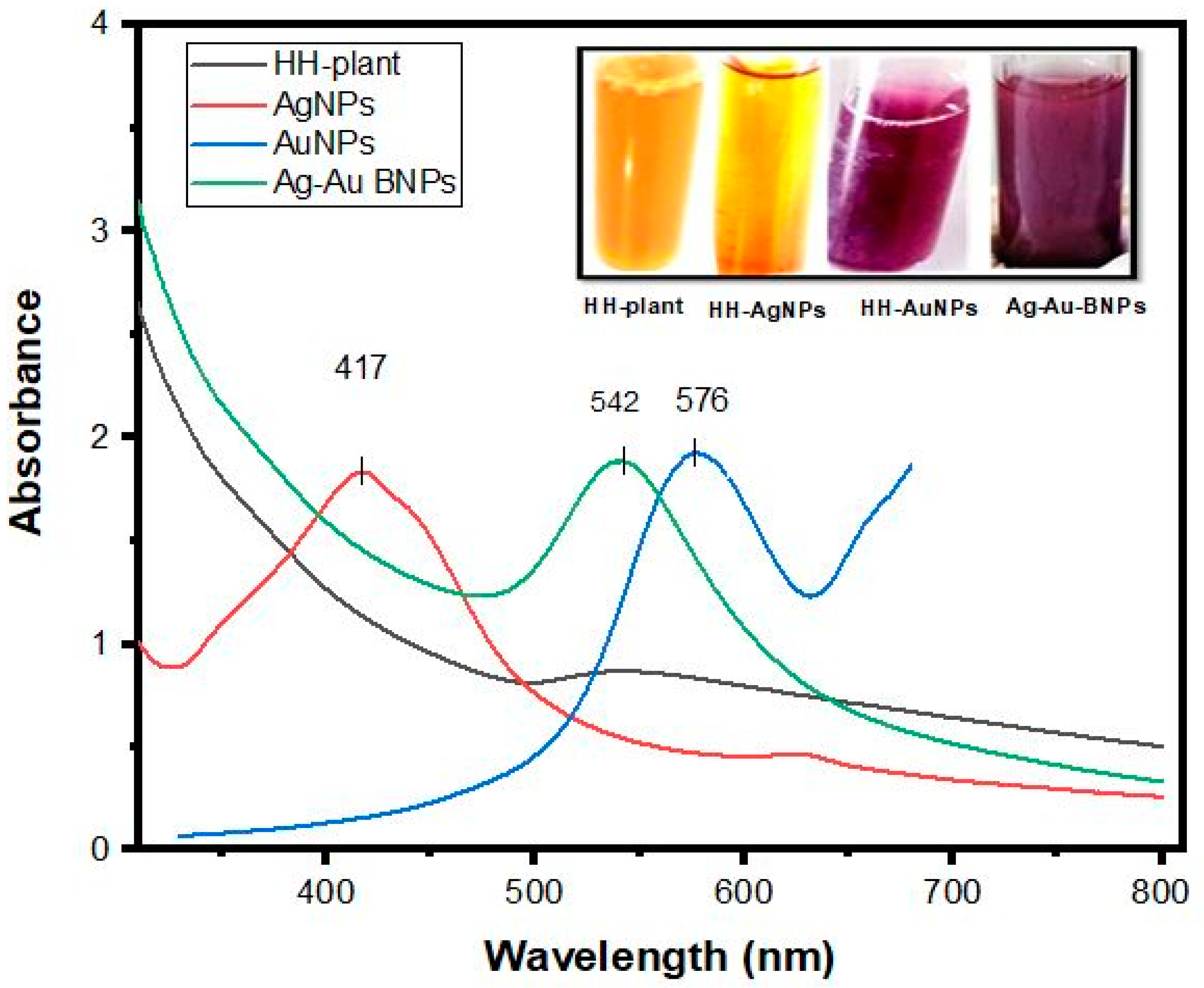
2.1. UV-Visible Spectrophotometric Analysis of NPs

In the present research study, we observed the visual reduction of Ag, Au, and Ag/Au BNPs from the addition of AgNO3, HAuCl4·3H2O, and AgNO3/HAuCl4·3H2O (1:1) to HH aqueous solution. The initially uncolored solution changed to brown, ruby red, and reddish-brown colors, indicating Ag, Au, and Ag/Au BNPs formation, respectively. The spectra recorded for the HH plant extract did not exhibit any absorption peak between 200 to 800 nm region, while absorption peaks at 417, 576, and 542 nm, which correspond to Ag, Au, and Ag/Au BNPs, were recorded during the rapid reduction process after 24 h. The difference in the peaks of plant and NPs is due to the reduction of the phytochemicals with the salt Ag and Au ions (Figure 1).

2.2. Factors Effecting Synthesis of NPs

The plant extract, induced HH-AgNPs, HH-AuNPs, and Ag/Au BNPs solutions were exposed to UV-vis spectroscopy during the reduction of ions by HH extract to study the mechanism of Ag, Au, and Ag/Au BNPs formation. The recorded spectra at different pH, such as 4, 5, 6, 7, 8, 9, 10, 11, and 12, are shown in Figure S1A–C. The spectra recorded during the reduction of ions at various pH showed the maximum absorbance with a sharp peak of 417 nm at pH 12, 576 nm at pH 4, and 542 nm at pH 6, which corresponds to the Ag, Au, and Ag/Au BNPs, respectively. The spectra recorded at different concentrations of HH plant, such as 0.5, 1, 1.5, and 2 mL, are shown in Figure S2A–C. The recorded spectra at different HH plant concentrations showed the maximum absorbance with a sharp peak of 417 nm, 576 nm, and 542 nm at 1 mL concentration of plant, which corresponds to the Ag, Au, and Ag/Au BNPs. The UV-vis spectra of Ag, Au, and Ag/Au bimetallic NPs in an aqueous medium at different AgNO3/HAuCl4·3H2O concentrations (0.25, 0.5, 1, and 1.5 mM) were noted at the range of 200 to 800 nm wavelength, which indicated the sharp peaks of 417 nm, 576 nm, and 542 nm at 1 mM salt concentration, which corresponds to the Ag, Au, and Ag/Au BNPs (Figure S3A–C). In the present research work, NPs were studied at different temperature ranges (20 to 100 °C). Broadband appeared at a lower temperature of 20 °C for Ag, Au, and Ag/Au BNPs, while sharp bands of 417 nm, 576 nm, and 542 nm, which correspond to the Ag, Au, and Ag/Au BNPs, appeared at 40 °C; however, with further increase in temperature beyond 40 °C, the band becomes broad and final broadband with lower absorbance appeared at 100 °C (Figure S4A–C). Figure S5A–C indicated the reaction time effect on HH-reduced Ag, Au, and Ag/Au BNPs. A broad peak with lower absorption appeared after 1, 2, and 3 h of the stirring. Due to the continuous reduction of Ag and Au ions by HH extract, the absorption peak increased with time. A final, clear, sharp peak of 417 nm, 576 nm, and 542 nm corresponds to the Ag, Au, and Ag/Au BNPs observed after 24 h of the HH and Ag/Au ions’ reaction. The stability of the HH-reduced Ag, Au, and Ag/Au BNPs was studied at different periods (1 day, 3 months, and 6 months). Sharp peaks with maximum absorbance are reported after 1 day and 3 months of NPs formation; however, after 6 months these peaks became broad and also showed low absorbance for all three types of NPs (Figure S6A–C).

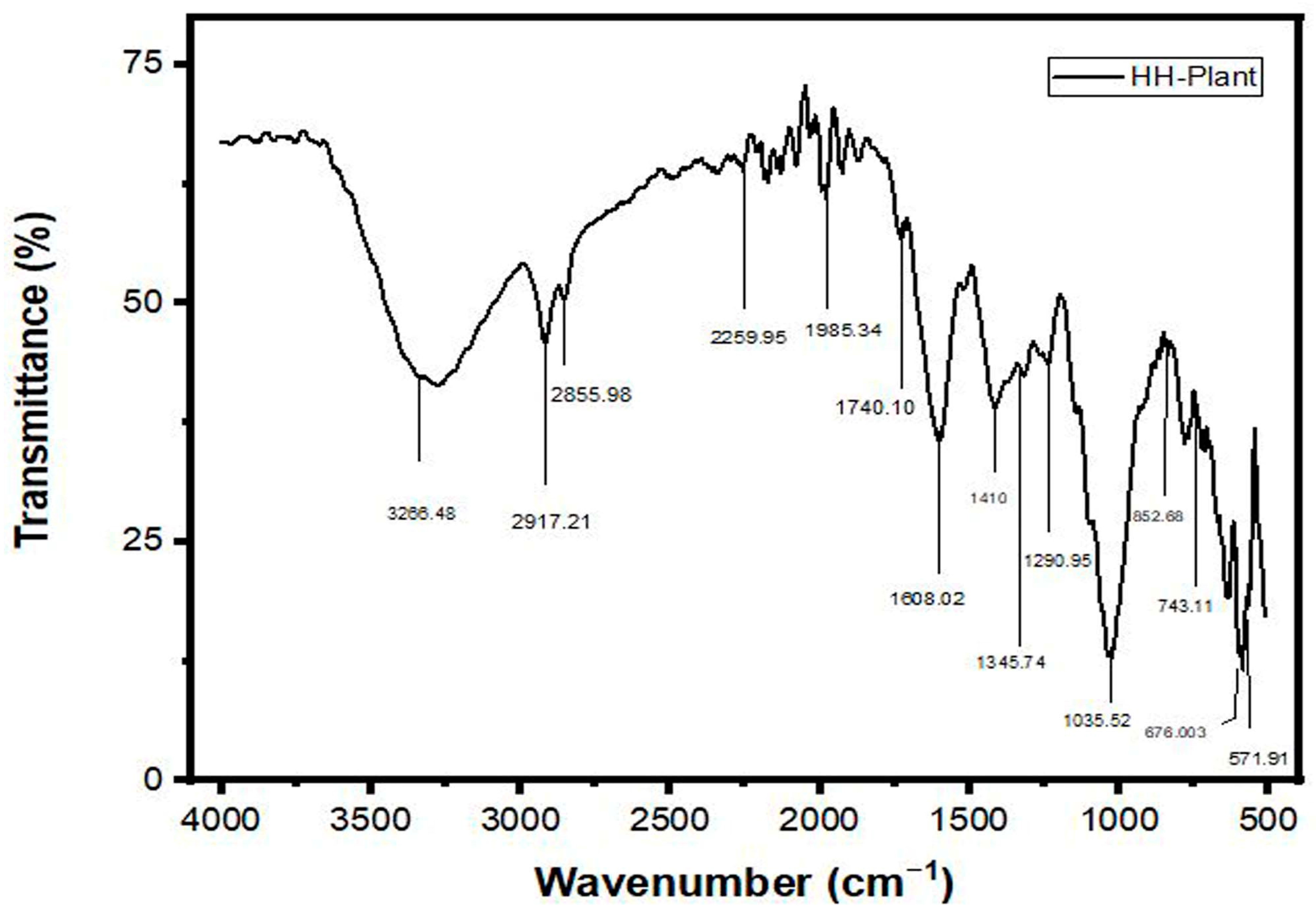
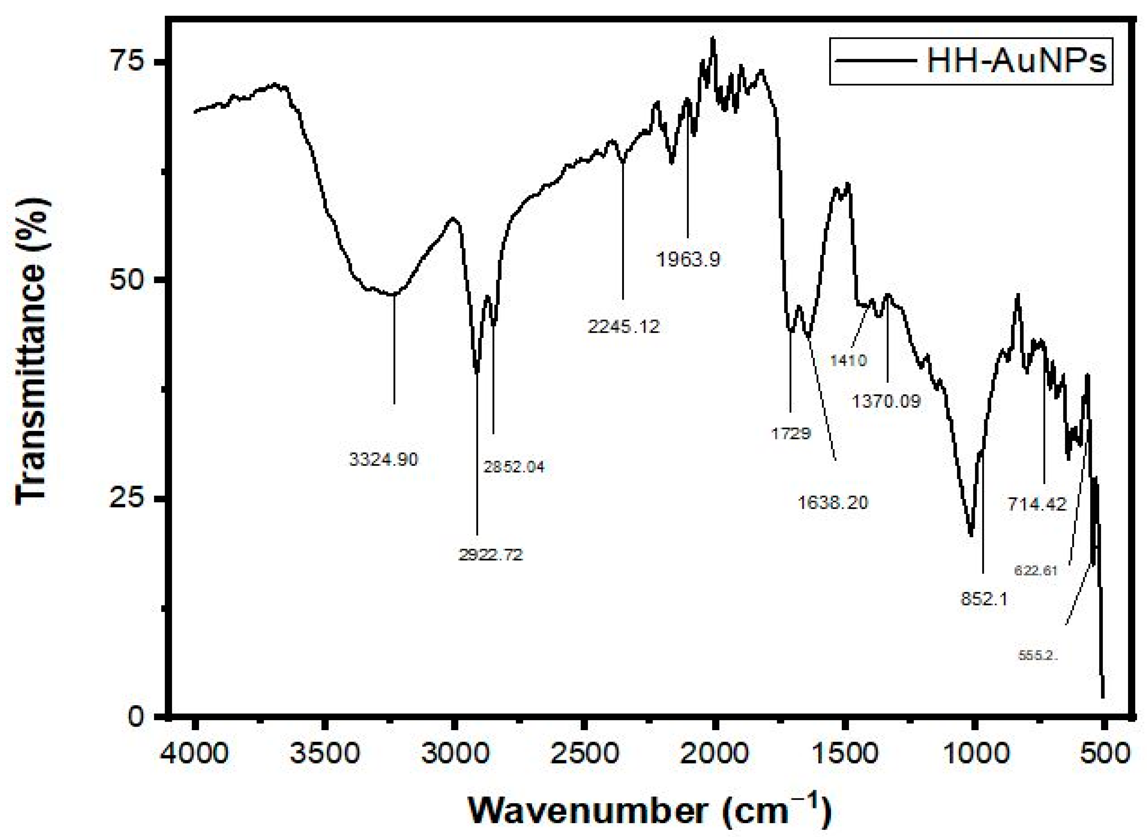
2.3. FT-IR Analysis of NPs

The FTIR spectrum analysis of HH extracts Ag, Au, and Ag/Au BNPs (prepared in water) is presented by Figure 2, Figure S7, Figure 3, and Figure S8, respectively. The peak values data and their corresponding functional groups existing in the HH, Ag, Au, and Ag/Au BNPs are presented in Table 1. The characteristic absorption bands were exhibited in the range 3400–3200 cm−1 (for O-H stretch), 2935–2915 cm−1 (for –CH (CH2) vibration), 2865–2845 cm−1 (for –CH (CH2), 2260–2100 cm−1 (for C≡C stretch), 2100–1800 cm−1 (for C=O frequency), 1740–1725 cm−1 (for C=O stretch), 1650–1600 cm−1 (for C=O stretch), 1410–1310 cm−1 (for O-H bend), 1340–1250 cm−1 (for CN stretch), 995–850 cm−1 (for P-O-C stretch), 800–700 cm−1 (for C-Cl stretch), 700–600 cm−1 (for C-Br stretch), 690–550 cm−1 (for C-Br stretch) by HH extract, Ag, Au, and Ag/Au BNPs.

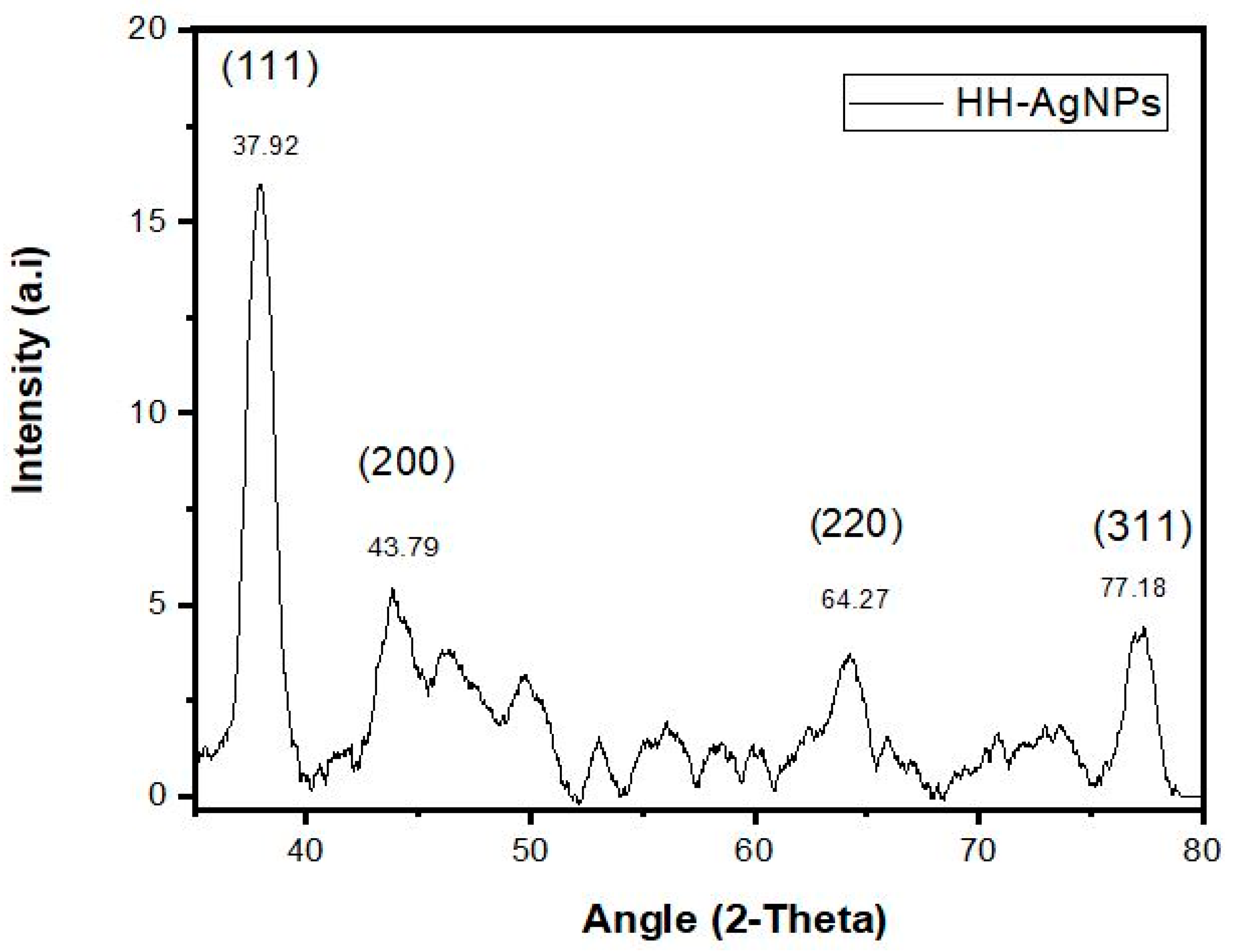
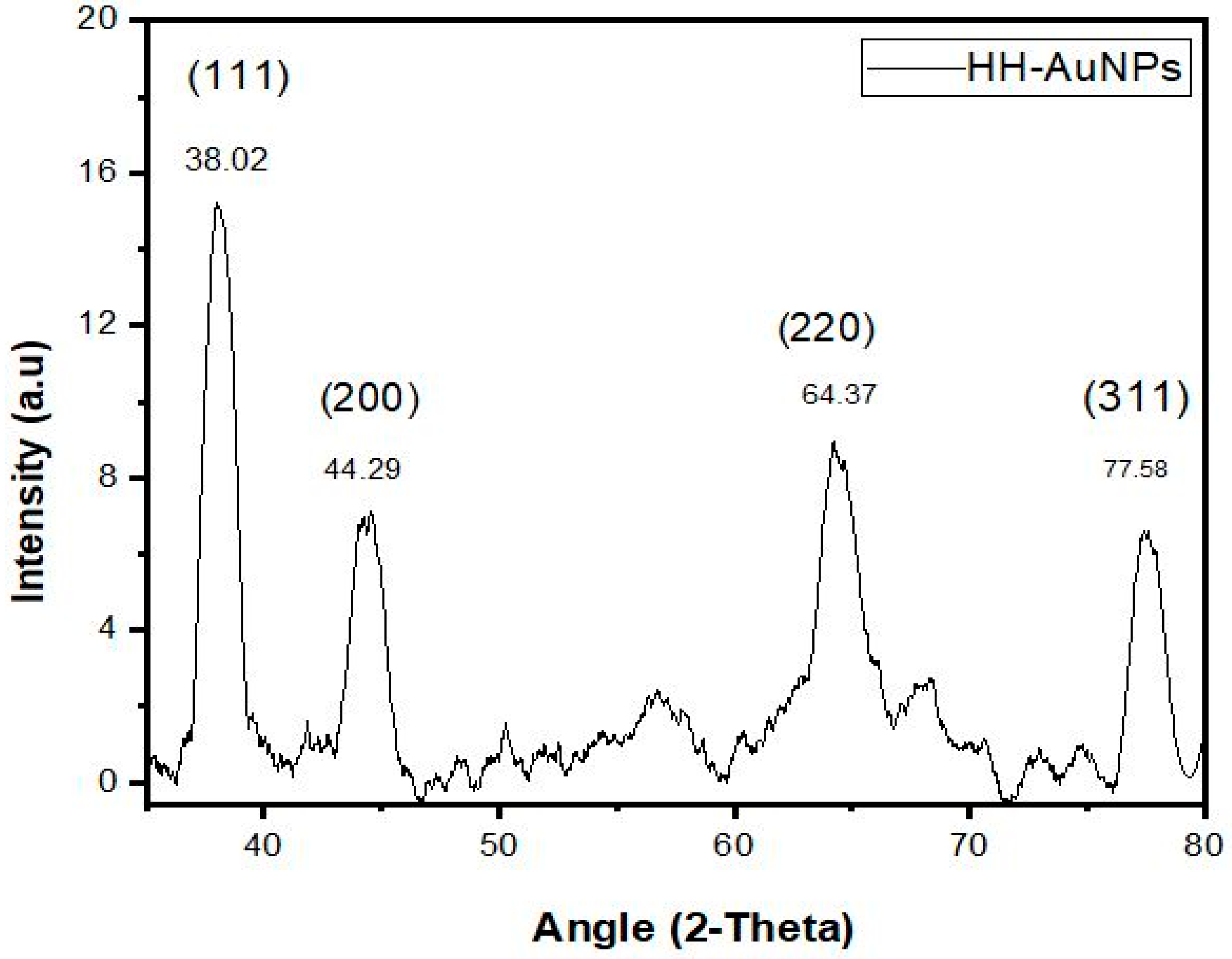
2.4. XRD Analysis of NPs

XRD is a technique that is used for determining the size and crystalline nature of the sample. In the present study, the Ag, Au, and Ag/Au BNPs were analyzed by XRD. Figure 4 indicated four Bragg reflections at angles of 37.92°, 43.79°, 64.27°, and 77.18° for AgNPs, Figure 5 showed 37.02°, 44.29°, 64.37°, and 77.58° for AuNPs, and Figure S9 showed 38.12°, 44.09°, 64.76°, and 77.48° for Ag/Au BNPs, which are related to the planes (1 1 1), (2 0 0), (2 2 0), and (3 1 1), respectively. These resulting reflections can be indexed conferring to the face of face-centered cubic crystal structures of Ag and Au ions. The “d” (interplanar spacing) and “a” (Miller constants) values were calculated by using the Debye-Sherrer’s Equations (1) and (2), respectively; results are tabulated in Table 2.
d hkl = π 2 sin θ hkl
å = d hkl h 2 + k 2 + l 2
The average crystalline size of HH-induced NPs is calculated by using Debye-Sherrer’s Formula (3);
D = k λ / β cos θ
“D” is the average crystalline size, k is a geometric factor (0.9), λ is the wavelength of the X-ray radiation source, and β is the angular full width at half maximum (FWHM) of the XRD peak at the diffraction angle θ. For the four major peaks, the average crystalline size for each of the samples Ag, Au, and Ag/Au BNPs was found to be 13.3, 10.72, and 8.34 nm, respectively.

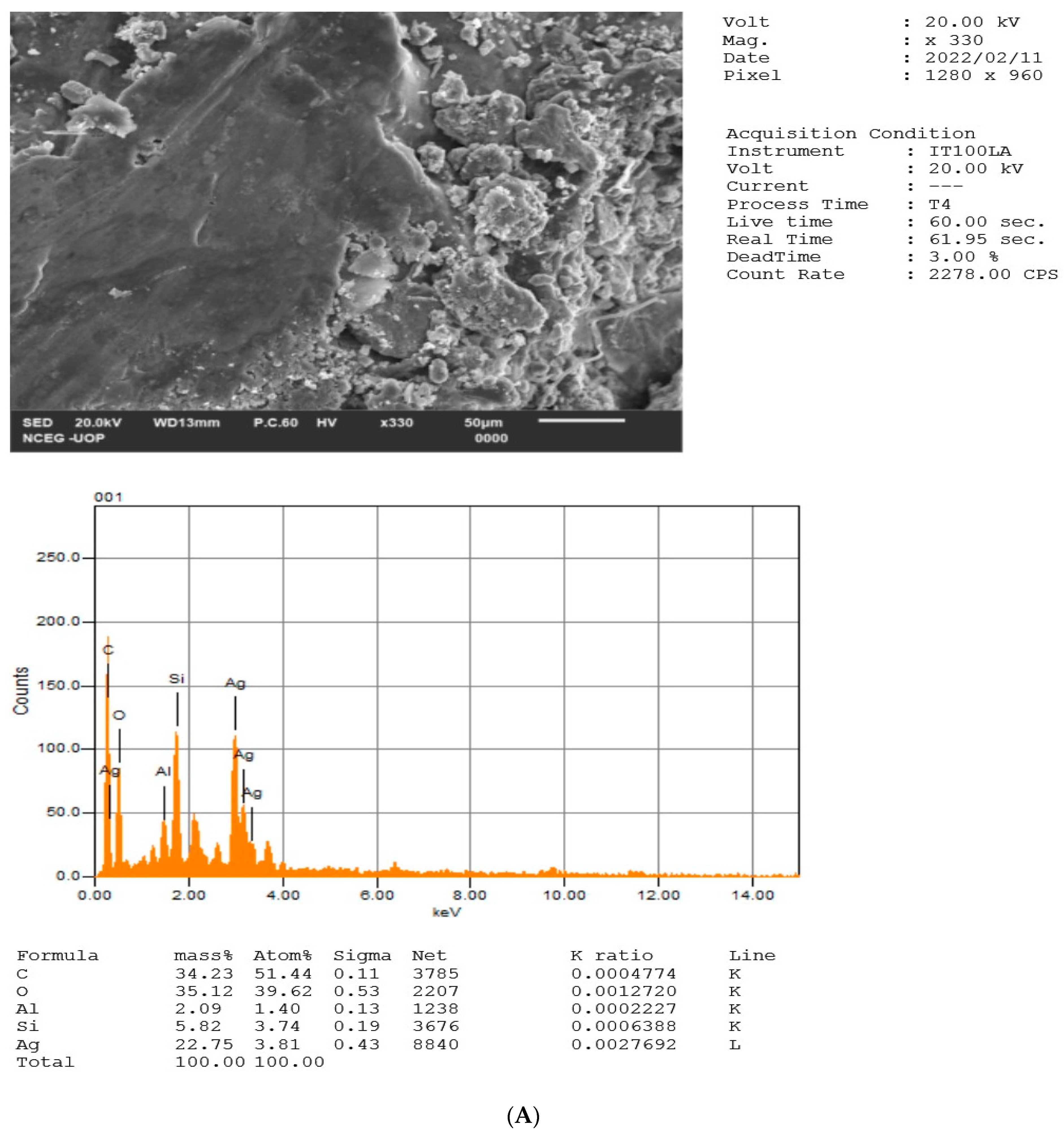
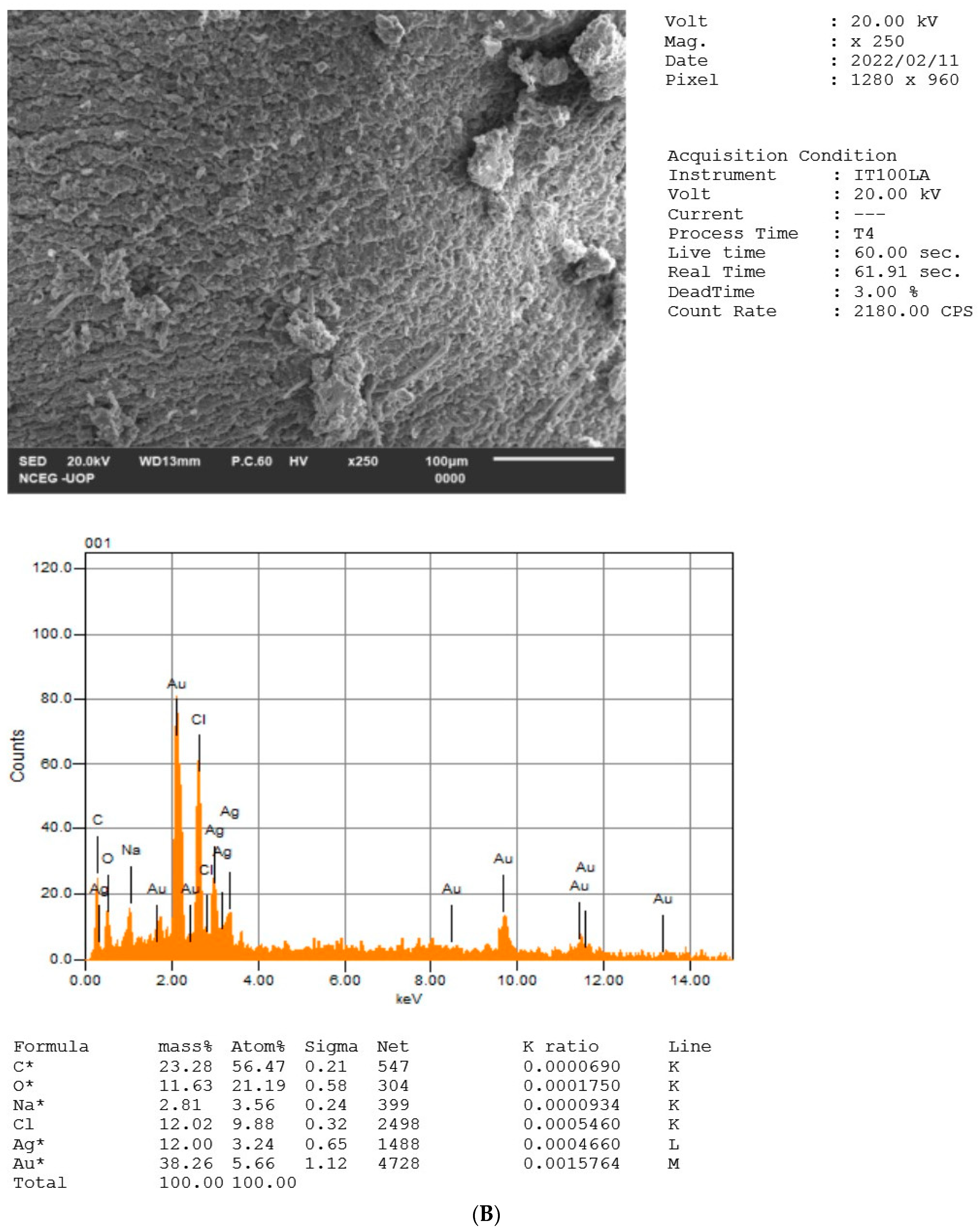
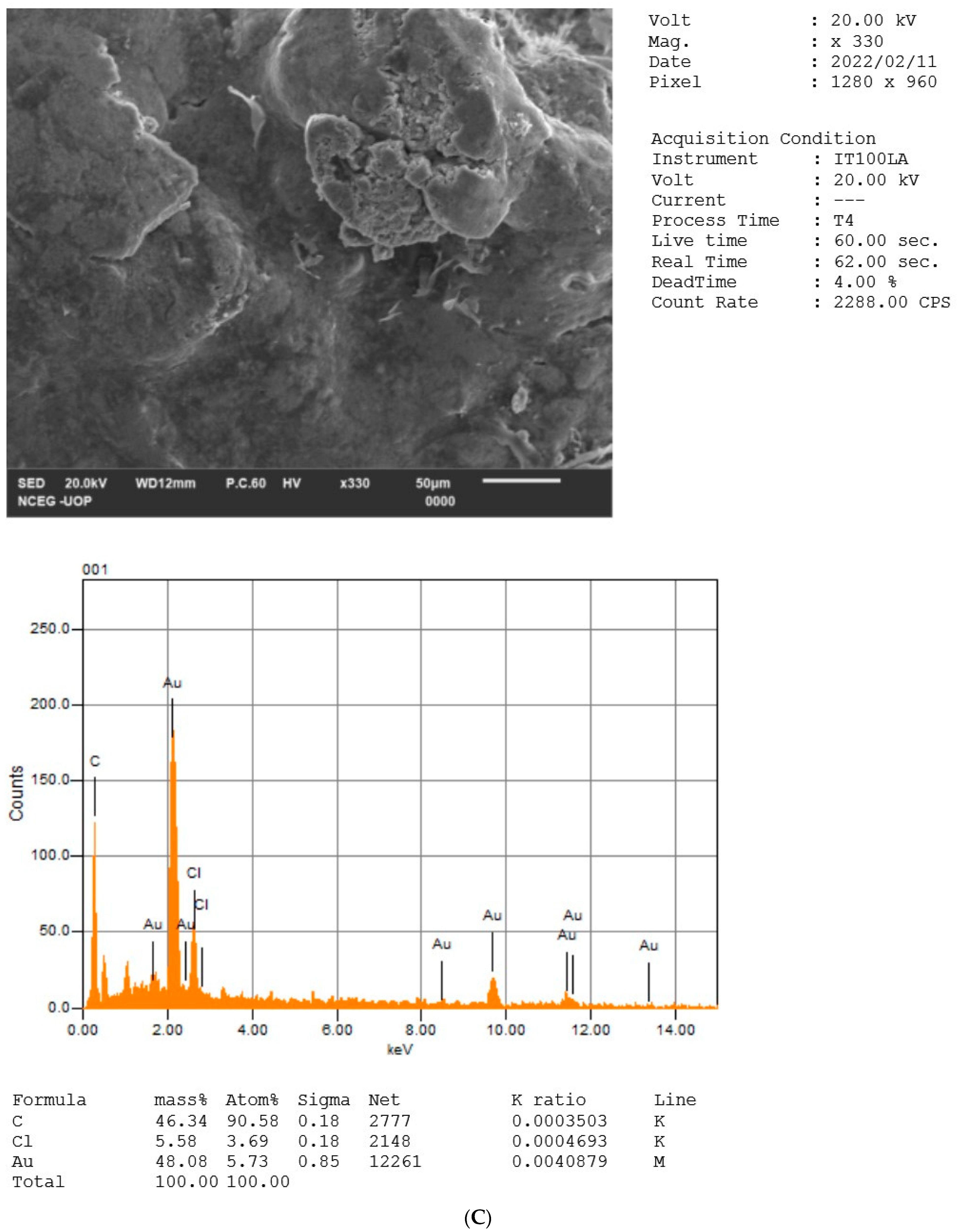
2.5. EDX Analysis of NPs

The elemental constituents and relative abundance of the biosynthesized Ag, Au, and Ag/Au BNPs were obtained from EDX analysis, as shown in Figure 6A–C. EDX analysis established the existence of elemental Ag signal of the AgNPs with 22.75%, Au signal of the AuNPs with 48.08%, and Ag signal with 12%/Au signal with 38.26% of the Ag/Au BNPs. The vertical axis displays the number of X-ray counts, while the horizontal axis displays energy in KeV. Identification lines for the major emission energies for Ag and Au are displayed, and these correspond with peaks in the spectrum, thus giving confidence that Ag and Au have been correctly identified in Ag, Au, and Ag/Au BNPs. Thus, the EDX analysis unveils the complete chemical alignment and purity of HH-induced Ag, Au, and Ag/Au BNPs, while the other elements served as coating organic agents bound on the Ag, Au, and Ag/Au BNPs surface. See Table 3 for elemental composition of NPs.

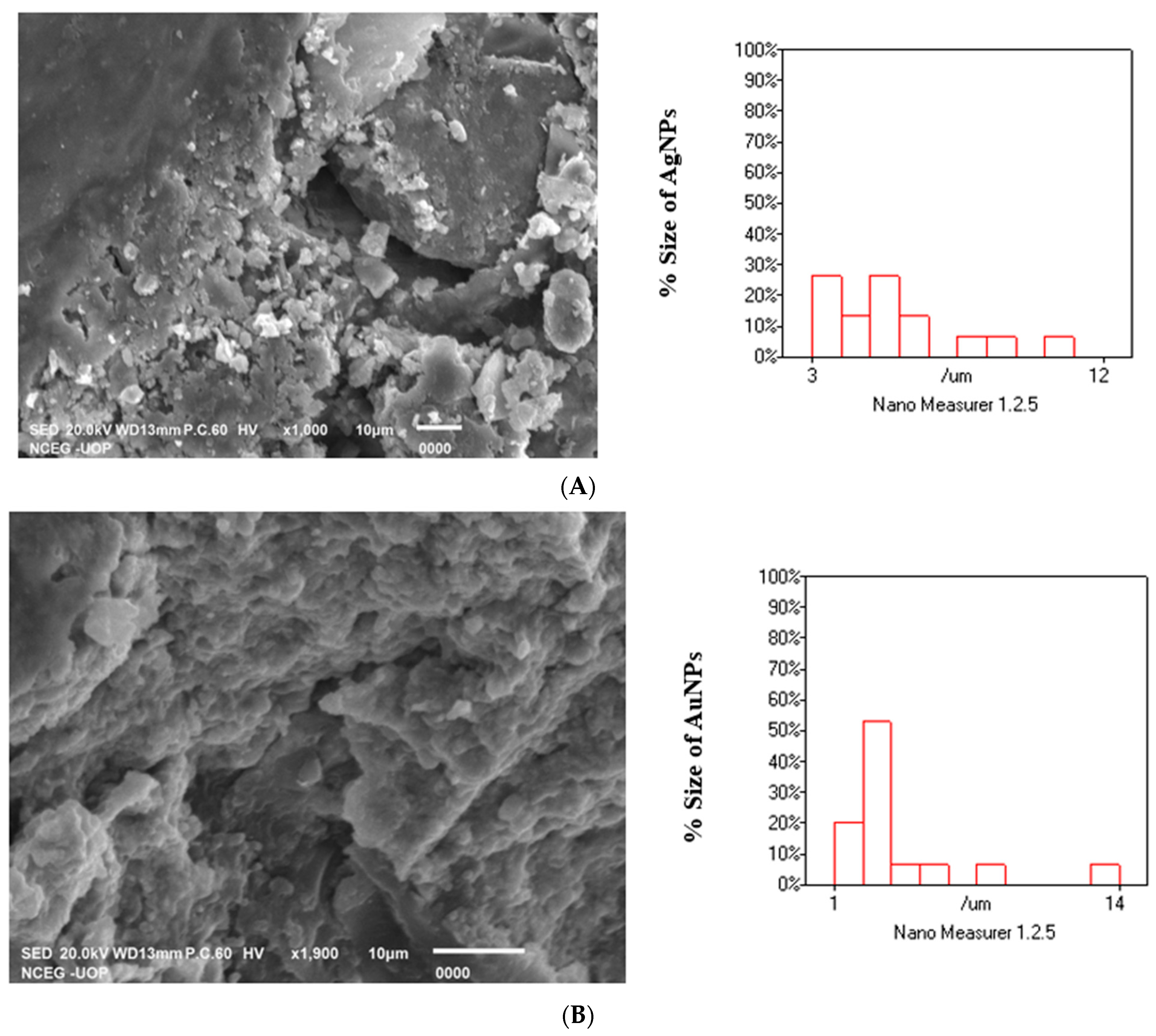
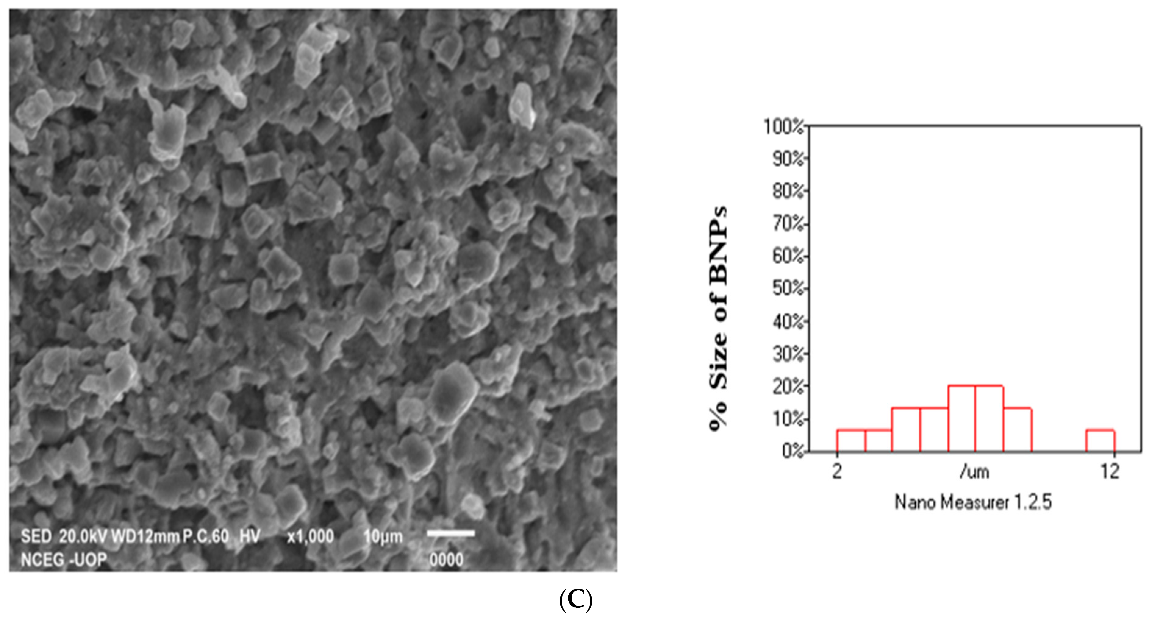
2.6. SEM Analysis of Ag, Au, MNPs and Ag-Au BNPs

Figure 7A–C indicated the SEM analysis of Ag, Au, and Ag/Au BNPs synthesized from HH extract: The SEM analysis showed relatively spherical shaped NPs. The morphology of NPs synthesized by the HH extract was confirmed by the SEM analysis. The size was calculated by Nano Measurer 1.2 software by marking 15 particles; size for each of the samples Ag, Au, and Ag/Au BNPs was found to be 40, 30, and 20 nm, respectively.

2.7. Antibacterial Activity

The AgNO3 salt, HAuCl4·3H2O salt, HH extracts, HH-AgNPs, HH-AuNPs, and BNPs showed antibacterial activities at various doses (30, 60, and 90 µg/mL). At the highest 90 µg/mL concentration the HH extracts, HH-AgNPs, HH-AuNPs, and BNPs showed inhibition of 8.5 ± 0.04, 10.5 ± 0.03, 14 ± 0.22, and 15 ± 0.12 mm against S. aureus. Against K. pneumonia HH extracts, HH-AgNPs, HH-AuNPs, and BNPs showed 2.5 ± 0.11, 5.5 ± 0.1, 9 ± 0.21, and 9.6 ± 0.12 mm inhibition. Against A. meriye HH extracts, HH-AgNPs, HH-AuNPs, and BNPs exhibited 3.5 ± 0.012, 9.5 ± 0.1, 6.5 ± 0.05, and 11.5 ± 0.11 mm inhibition. Against S. pyogen HH extracts, HH-AgNPs, HH-AuNPs, and BNPs showed 0, 6 ± 23, 9 ± 0.23, and 10 ± 0.08 mm zone inhibition. Against E. coli HH extracts, HH-AgNPs, HH-AuNPs, and BNPs showed 4.5 ± 0.011, 6.5 ± 0.17, 7.5 ± 0.12, and 7.8 ± 0.01 mm inhibition. Against S. marcescens HH extracts, HH-AgNPs, HH-AuNPs, and BNPs exhibited 2 ± 0.011, 3.5 ± 0.012, 7 ± 0.15, 12.6 ± 0.11 mm inhibition, respectively. Against B. cereus HH extracts, HH-AgNPs, HH-AuNPs, and BNPs showed 5.5 ± 0.01, 7 ± 0.03, 2.5 ± 0.11, and 9.5 ± 0.13 mm zone inhibition. Against MRSA HH extracts, HH-AgNPs, HH-AuNPs, and BNPs exhibited 6.5 ± 0.011, 6 ± 0.21, 7.5 ± 0.13, and 8.5 ± 0.11 mm inhibition. Against M. luteus 7 ± 0.014, 8.5 ± 0.23, 8 ± 0.11, and 10 ± 0.34 mm inhibition were reported for HH extracts, HH-AgNPs, HH-AuNPs, and BNPs, respectively. Against S. pneumonia HH extracts, HH-AgNPs, HH-AuNPs, and BNPs showed 4 ± 0.011, 8.5 ± 0.11, 5.5 ± 0.11, and 9 ± 0.11 mm zone inhibition, respectively. AgNO3 salt exhibited no zone inhibition against S. aureus, K. pneumonia, B. cereus, and S. pneumonia, while 3 ± 0.11, 2.4 ± 0.11, 3.1 ± 0.1, 2 ± 0.13, 0.2 ± 0.11, and 4 ± 0.11 mm inhibition reported against A. meriye, S. pyogen, E. coli, S. marcescens, MRSA, and M. luteus, respectively; similarly, HAuCl4·3H2O showed no zone inhibition against S. aureus, K. pneumonia, A. meriye, S. marcescens, B. cereus, MRSA, M. luteus, and S. pneumonia, while 2 ± 0.11 mm inhibition was reported against S. pyogen at the highest 90 µg/mL concentration (Figure S10A–J). DMSO served as negative control and levofloxacin (1 g/mL) as a positive control. See Table 4A–F.

2.8. Antifungal Activity

From the current study, it has been found that AgNO3 salt, HAuCl4·3H2O salt, HH extracts, HH-AgNPs, HH-AuNPs, and BNPs significantly inhibited the growth of tested fungal strains and proved a valuable antifungal source. At 20 µg/mL, no growth inhibition of A. niger and A. flavus was reported by AgNO3 and HAuCl4·3H2O salt, while 7 ± 0.12 and 10 ± 0.21 mm inhibition was reported against A. fumigatus by AgNO3 and HAuCl4·3H2O salt, respectively. At the highest concentration, 100 µg/mL, AgNO3 showed 15 ± 0.11, 31 ± 0.11, and 33 ± 0.14 mm inhibition against A. niger, A. fumigatus, and A. flavus, respectively; similarly, HAuCl4·3H2O salt showed 11 ± 0.21, 37 ± 04, and 27 ± 0.13 mm inhibition against A. niger, A. fumigatus, and A. flavus, respectively. Consequently, higher growth inhibition of 67 ± 0.13, 75 ± 0.13, 82.5 ± 0.18, and 93 ± 0.18 mm against A. niger, 70 ± 0.11, 111 ± 0.15, 94.5 ± 0.21, and 114 ± 0.21 mm against A. fumigatus, and 80 ± 0.08, 92.5 ± 0.13, 92 ± 0.04, 112 ± 0.04 mm against A. flavus was reported by HH extracts, HH-AgNPs, HH-AuNPs, and BNPs, respectively, at the highest 100 µg/mL concentration (Figure S11A–C). DMSO served as a negative control. No fungal growth was observed in positive control test tubes (terbinafine). See Table 5A–F.

3. Materials and Methods

3.1. Materials

The Hippeastrum hybridum plant was collected in April from Bannu Khyber Pakhtunkhwa (city), and was identified by a taxonomist, Dr. Tahir Iqbal, at the department of Botany, University of Science and Technology, Bannu. Ten clinically isolated bacterial strains, seven Gram-positive Actinomycetes meriye ATCC 35568, Streptococcus pyogenes ATCC 700294, Bacillus cereus ATCC 14579, Staphylococcus aureus ATCC 29213, Methicillin-resistant Staphylococcus aureus ATCC 43300, Micrococcus luteus ATCC 4698, Streptococcus pneumonia ATCC 6303, and three Gram-negative Klebsiella pneumonia ATCC 700603, Escherichia coli ATCC BAA-2471, and Serratia marcescens ATCC BAA-3111 were used. Three fungus strains, Aspergillus fumigatus, Aspergillus niger, and Aspergillus flavus, were obtained from the microbiology lab of Biotechnology. Brine shrimps, sea salts, nutrient agar, and Sabourad Dextrose Agar (SDA) were obtained from Sigma. All other reagents used were of analytical grade.

3.2. Plant’s Extraction

After identification, the Hippeastrum hybridum plant was washed using water, shade dried, and ground to a fine powder. About 250 g of the fine plant powder was mixed with 70% methanol in 1:3, and then retained for extraction at room temperature for 7 days. After that, it was filtered by using Whatman No 1 filter paper, after which the methanol was vaporized at 37 °C to obtain a pure crude methanolic extract of sample, which was then kept in the refrigerator at 4 °C for more studies [30].

3.3. Synthesis of Ag, Au, and Ag/Au BNPs

For the synthesis of Ag, Au, and Ag-Au BNPs from the HH extract, the standard protocol of [31] was followed with minor modification. Solutions of about 10 mM (0.01 M) of AgNO3 and HAuCl4·3H2O were prepared in 50 mL deionized water. The 10 mM AgNO3 and HAuCl4·3H2O solutions were further diluted 10 times to obtain 1 mM AgNO3 and HAuCl4·3H2O solutions. 0.1 M NaOH, ≥98%, and 0.1 M HCl were used for pH regulation. The HH plant extract was prepared in deionized water (1 gm/100 mL) and agitated quietly for about 1 h on a magnetic stirrer for complete dissolution. The solution was then centrifuged at 6000 rpm for 30 min to obtain the desired product. The supernatant was reserved for further use in NPs synthesis. About 50 mL of the plant supernatant was mixed with 500 mL of: 1 mM AgNO3 (pH 12), 1 mM HAuCl4·3H2O (pH 4), and AgNO3/HAuCl4·3H2O (1:1) solution (pH 6). After 4 h, the reduction of Ag, Au, and Ag/Au BNPs was detected visually using color change. For the complete settlement and stabilization of NPs, the resultant solution was stored for 24 h at room temperature. After that, the NPs were monitored using a UV–Visible spectrophotometer. The colloidal suspension was centrifuged at −4 °C at 10,000 rpm by cold centrifuge for about 10 min; the supernatants were discarded and the pellet (containing NPs) was lyophilized to obtain the powder form. The powder was further characterized and tested for different biological activities.

3.4. Factors Affecting Synthesis Rate, Size, and Shape of NPs

Ag, Au, and Ag/Au BNPs synthesis was determined by using different factors such as pH, AgNO3/HAuCl4·3H2O concentration, HH plant extract concentration, temperature, time, and stability time. To study the effect of basic and acidic conditions, pH of the reaction mixture ranged from 4 to 12 by using NaOH and HCl (0.1 M) solution. To study the effect of AgNO3 and HAuCl4·3H2O salt concentration, its concentration varied to 0.25, 0.5, 1, and 1.5 mM. To study the effect of HH extract concentration on NPs synthesis, its concentrations varied from 0.5, 1, 1.5, and 2 mL. To study the temperature effect, the NPs synthesis was carried out at different reaction temperature ranges (20, 40, 60, 80, and 100 °C). To study the rate of completion of the reaction, NPs were synthesized at different time intervals (1, 2, 3, and 24 h). The synthesized NPs’ stability was studied after 1 day, 3 months, and 6 months.

3.5. Characterization

In the aqueous solution, Ag, Au, and Ag/Au BNPs’ concentrations were confirmed by using a SHIMADZU UV SPECTROPHOTOMETER (UV-1800). The purified Ag, Au, Ag/Au BNPs, and HH plants extract were examined for the presence of different phytochemicals by using a Fourier Transform-Infrared (FT-IR) Shimadzu (IR Prestige-21) spectrometer (Japan). The crystalline nature of the Ag, Au, and Ag/Au BNPs was determined by using the JDX-3532 (JEOL JAPAN) X-ray diffractometer (XRD) with λ-1.54 A° wavelength. The size and shape of Ag, Au, and Ag/Au BNPs were determined by using JEOL Scanning Electron Microscope (SEM) Model JSM-5910 (Japan). The presence of elements in synthesized Ag, Au, and Ag/Au BNPs was determined by using electron diffraction X-ray spectroscopy (EDX).

3.6. Antibacterial Activity (Agar Diffusion Method)

Antimicrobial activities of AgNO3 salt, HAuCl4·3H2O salt, HH extracts, HH-AgNPs, HH-AuNPs, and BNPs were determined according to the procedure [32]. Bacterial suspensions were prepared by caring 1 CFU of bacterial strain from the maintained slants in 0.9% NaCl solution, followed by incubation at 37 °C for 24 h. Nutrient agar (2.8 g/100 mL dH2O), autoclaved at 121 °C for 15 min. About 25 mL media (cooled to room temperature) were added to each Petri plate and then kept back for solidification. By swab, a good amount of each bacterial suspension was taken and streaked on the Petri plates. Per plate, 5 wells (3–6 mm) were dug with a sterilized cork borer, with 1 central hole surrounded by 4 holes. All of the wells were properly labeled. DMSO (20 µL, as well as AgNO3 salt, HAuCl4·3H2O salt, HH extracts, HH-AgNPs, HH-AuNPs, and BNPs (concentrations of 30, 60, and 90 µg/mL), were added to the surrounding holes. Levofloxacin (final concentration of 1 mg/mL in DMSO) was added to the central holes. DMSO served as a negative control. Zones of inhibition were measured after a day with a graduated ruler. A clear zone of bacterial inhibition was observed around the holes. The diameter of the clear zone was measured in mm. Inhibition by active ingredients of the extract was determined by measuring linear growth (mm) in Petri plates concerning levofloxacin (mm), a standard antibiotic (positive control).

3.7. Antifungal Bio-Assay

SDA media (6.5 g/100 mL dH2O) were autoclaved at 121 °C for 15 min and kept at room temperature for cooling (40–50 °C). To each test tube, 7 mL media, along with 67 µL AgNO3 salt, HAuCl4·3H2O salt, HH extracts, HH-AgNPs, HH-AuNPs, and BNPs from the concentrations 20, 40, 60, 80, and 100 µg/mL were added, and kept in slant position for solidifying inside the laminar flow hood. DMSO served as negative control and terbinafine as a positive control. Then, the particular fungal strains (A. niger, A. flavus, and A. fumigatus) were homogeneously spotted in each test tube. The test tubes were then sealed with cotton plugs. The entire setup was placed in an incubator with open water at 30 °C. Growth inhibition was determined by measuring linear growth [32].

3.8. Statistical Analysis

All the data were expressed as mean ± SE. Normal data distribution was confirmed.

4. Discussion

In nanoscience, the design and development of nanomaterials with unusual optoelectronic and physicochemical features act as a cornerstone. The present study discloses the bioreduction properties of Ag and Au ions into Au, Ag, and Ag/Au BNPs using ethnopharmacologically important HH plant extract. In the present investigation, Ag and Au NPs were synthesized from AgNO3 and HAuCl4·3H2O, while Ag/Au BNPs were manufactured by treating the HH extract with a mixture of both AgNO3/HAuCl4·3H2O (1:1). The synthesis of NPs was monitored visually with a change in the color of reaction mixtures under optimum conditions. After 24 h, the NPs became stable and no more synthesis was detected, which was monitored with UV–vis spectroscopy. These results are supported by the [33,34] report of bimetallic alloys.
To accomplish maximum production of NPs, different intrinsic factors, such as pH, salt concentrations, plant concentration, time, temperature, and stability period were studied. The present results justify the earlier report [35] and confirm the effect of different intrinsic factors on the maximum synthesis of NPs. An ideal pH is required for the synthesis of controlled shape/size NPs [36,37]. As a result of extensive screening with different pH, syntheses of Ag, Au, and Ag/Au BNPs were successful with pH 12, 4, and 6, respectively. These results showed that AgNPs’ synthesis is supported by basic conditions and suppressed by acidic conditions; AuNPs’ is supported by highly acidic conditions, and Ag/Au BNPs’ synthesis is supported by nearly neutral conditions [23,38]. In the current study, NPs’ reduction and capping were achieved with the different AgNO3, HAuCl4·3H2O salt, and HH plant concentrations. Screening with various concentrations of AgNO3, HAuCl4·3H2O salt, and plant synthesis of Ag, Au, and Ag/Au BNPs was successful, with 1 mM salt and 1 mL plant concentration. Medicinal plants are a rich source of phytochemicals that act as reducing, capping, and mediators for NPs. However, the composition of these active secondary metabolites varies from plant to plant, dependent on the nature, part, type of plant, and method followed for the extraction of these metabolites [39]. Thus, a slight increase in the HH extract and slat concentration beyond 1 mL and 1 mM, respectively, increases the wavelength. Thus, this slight variation in the absorbance values is because of the change in the particle size. The results of the present research are also supported by [23], who reported an absorbance at 414 nm for the 1 mL Calligonum polygonoides and 1 mM salt with high absorbance of 2.375. The results of the present research work are also supported by [31], who reported the synthesis of NPs by using Gloriosa superba leaf extract. The Ag/Au BNPs showed an absorption peak at 542 nm, which is in good agreement with the previous results of the BNPs (Ag:Au) 1:1 ratio [30]. The temperature of the reaction medium determines the nature of NPs [40]. In this study, NPs were synthesized at varying temperature ranges (20 to 100 °C). At a lower temperature of 20 °C, a broad peak with low absorbance was obtained, but with an increase in temperature up to 40 °C, a sharp peak was recorded. With a further increase in temperature, the peak become broader and broader and final broad peaks were revealed for Ag, Au, and Ag/Au BNPs at the highest temperature of 100 °C. Thus, from these changes in peaks with temperature, it is revealed that NPs synthesis required an optimal temperature for its stability [23,41,42]. The quality, size, shape, and type of NPs are greatly influenced by incubated reaction time [43]. After mixing the HH extract with 1 mM salt, no color change was observed after 1 h, 2 h, and 3 h, but after 24 h of incubation an absorption spectrum of 417 nm, 576 nm, and 542 nm were reported, which corresponds to the Ag, Au, and Ag/Au BNPs. From the time study, it is revealed that an optimum time is required for stable NPs synthesis [23,44]. The reactivity and potency of the NPs greatly depends on their stability. The NPs have an optimum stability time, after which their potency is decreased or completely lost. In the present research, the stability of the NPs was checked at different periods of 1 day, 3 months, and 6 months. Sharp peaks at 17 nm, 576 nm, and 542 nm, which correspond to the Ag, Au, and Ag/Au BNPs, appeared after incubation in the dark for 1 day to 3 months. However, after 6 months the absorption band became broad and showed low absorbance. The present research study indicated that NPs remained stable for about 3 months, but after 6 months they lost their stability. The results are best supported by [45], who used Kiwifruit juice at room temperature for NPs’ synthesis and revealed NPs in the range of 5 to 25 nm which remain stable for more than one month. Mean particle size and stability of NPs increased at 72 h of stirring time [44]. Thus, the variations in the reaction time may occur due to reasons such as: particle aggregation due to long time storage; particles shrinking or growing during long storage; the shelf life of particles, and so forth. All of these affect the reactivity and potential of the particles [46].
The phyto-components present in aqueous plant extract that are responsible for stabilizing and mediating the NPs were illustrated using FTIR analysis. The scientific studies reported that FTIR is ideal for forecasting functional moieties. In the present study, various vibrational stretches occurred at different peaks, which correspond to polyhydroxy, phenol, carboxyl, proteins, lipids, amide, alkynes, alkene, etc. The FTIR analysis delineated the different functional groups, such as hydroxyl, ketones, carboxyl, amino, alkaloids, amide, etc., that are responsible for the induction of metal ions to NPs [47,48]. The present result of FTIR analysis is justified by the prior findings of [49]. Stimulatingly, in plant-induced NPs, the phytochemicals of the plant played a significant role in the stabilization of NPs, which is critical for transcription and its applicative properties. These results are also supported by the earlier reports of [50,51].
The XRD peaks of Ag, Au, and Ag/Au BNPs exhibited four brag reflections which agree with the face-centric cube crystalline nature of Ag, Au, and Ag/Au BNPs. The well-resolved and strong pattern of the XRD showed that both Ag and Au NPs that are formed due to the reduction of Ag+ and Au+ ions are crystal-like. The Ag/Au BNPs that are formed by the combined reduction of Ag and Au ions are not different from the Ag and Au monometallic NPs, which can be similarly described as a crystal lattice structure. The mean calculated sizes were found to be 13.3, 10.72, and 8.34 nm for Ag, Au, and Ag/Au BNPs, respectively. The obtained results are justified by the earlier reports of [31,52].
The EDX analysis was used for the confirmation of elemental composition, purity, and relative abundance of NPs [53,54]. The percentages of Ag, Au, and Ag/Au metal in the present study were found to be appreciable, such as the Ag signal of the AgNPs with 22.75%, Au signal of the AuNPs with 48.08%, and Ag signal with 12%, and Au signal with 38.26% of the Ag/Au BNPs. The other elements which are shown by EXD analysis served as capping organic agents which are bounded on the NPs surface [55,56].
The surface morphology of NPs was studied by SEM analysis. In the present study, the SEM analysis showed an image of high density, spherical, and monodispersed NPs. The white individual spots represent the NPs, while the longer spots are the collection of NPs in the SEM photograph. The NPs from the SEM photograph have been observed with a diameter of 40, 30, and 20 nm of Ag, Au, and Ag/Au BNPs, respectively. The capping agent of the plant extract indicates the stabilization of the NPs because they were not in direct contact even in the aggregated form. The larger NPs during SEM measurements may be due to the combination of the smaller NPs. The obtained results are justified by the earlier reports of [57,58].
Different pathogenic bacteria cause universal infections: S. aureus causes skin infections, pneumonia, osteomyelitis, and endocarditis; K. pneumonia causes bloodstream infections, wound pneumonia, infections at surgical site, and meningitis; A. meriye can result in abscesses, pain, and inflammation; S. pyogenes causes pneumonia, necrotizing fasciitis, myonecrosis, scarlet fever, and bacteremia; E. coli causes cholangitis, cholecystitis, infection in urinary tract, bacteremia, and traveler’s diarrhea; S. marcescens causes severe infections, bacteremia, pneumonia, infection in urinary tract, infection in biliary tract, meningitis, wound infection, and endocarditis, a foodborne pathogen; B. cereus can produce toxins that result in two types of gastrointestinal infection: the emetic (vomiting) and the diarrhea syndrome; MRSA causes severe skin infections; M. luteus is an adaptable pathogen that causes endocarditis, meningitis, septic arthritis, in HIV positive patients causes chronic cutaneous infections and catheter infections; and S. pneumoniae causes various infections such as pneumonia, sinusitis, osteomyelitis, otitis media, septic arthritis, bacteremia, and meningitis [59,60,61]. Due to the resistance of microbes to drugs, researchers are looking at the development of innovative agents against bacteria [62]. The NPs synthesized by the green approach are applied extensively in different biomedical applications [63]. The NPs kill microorganisms due to their interaction with the bacterial cell membranes. The NPs, such as the Ag, Au, and Ag/Au BNPs, exhibited the lowest noxiousness and thermal and pH resistance, and thus proved to be an outstanding antibacterial agent that could be useful in biomedical applications [64]. The green-synthesized Ag, Au, and Ag-Au NPs and HH plant extract displayed promising antibacterial activity against A. meriye, S. pyogenes, B. cereus, S. aureus, MRSA, M. luteus, S. pneumonia, K. pneumonia, E. coli, and S. marcescens in a dose-dependent manner. In this study, the appreciable antibacterial potential of Ag/Au BNPs compared to MNPs depends on morphology, surface area, particle size and shape, and surface polarity [65].
Like bacteria, different pathogenic fungi also cause systemic illness of the mouth, skin, lungs, blood, and liver, as well as causing hypersensitive reactions, tinea cruris, and athlete’s foot [66,67,68,69]. For the therapeutic management of invasive systemic fungal infections, only about 10 antifungal drugs are permitted in the United States of America by the Food and Drug Administration (FDA) authority [66,67]. From the present observation, the highest fungicidal activity was observed with Ag/Au BNPs when compared with HH plant, Ag, and Au NPs at a similar concentration. The significant antifungal effect of Ag/Au BNPs compared to single metal Ag/Au NPs is due to their small size; as confirmed by investigators, the smaller the size of the particle, the greater the inhibition [70,71]. In comparison, [72] has analyzed the antifungal effect of NPs against species of aspergillus, revealing potential to control fungal growth. Moreover, [73] could be able to gain an antifungal effect against Aspergillus species at 80 µg/mL concentration of NPs. This outcome is supported by the aforementioned research, which was accompanied by [74], who reported that the NPs’ interaction with lysed cells’ intracellular substances caused their coagulation and the particles were thrown out of the liquid system. The Au and Ag ions mechanism of inhibition action on micro-organisms demonstrates that the microbe treatment by metal ions prompts loss of DNA, its capability replication and translation, as well as other cellular enzymes and proteins that are necessary for the production of ATP (coenzyme adenosine triphosphate), resulting in living cell inactivation of [75]. It has also been assumed that metal NPs mainly affect the function of membrane-bound enzymes in the chain of respiration [76].

5. Conclusions

In this study, medicinally active phytochemical-induced HH-AgNPs, HH-AuNPs, and Ag/Au BNPs were synthesized from HH extract, an indigenous plant found in abundance in Pakistan. These NPs were characterized by the following techniques: UV-vis spectroscopy, FTIR, XRD, EDX, and SEM. For antimicrobial activity, Ag and Au have always been excellent choices. Finally, this investigation revealed that Ag/Au BNPs have better antimicrobial activity than HH plant extract, HH-AgNPs, and HH-AuNPs. This work assimilates microbiology and nanotechnology, thus leading to possible advances in antimicrobial agents’ formulation. However, future studies on the antimicrobial effect of these NPs on microbes are essential to completely assess its potential use as a novel biocidal material.

Supplementary Materials

The following supporting information can be downloaded at: https://www.mdpi.com/article/10.3390/molecules27227895/s1, Figure S1: UV-vis spectrum of NPs biologically synthesized by using the HH plant extract at different pH (Both acidic and basic). (A) HH-AgNPs, (B) HH-AuNPs, and (C) Ag/Au BNPs; Figure S2. UV-vis spectrum of biologically synthesized NPs by using the different concentrations of HH plant extract. (A) HH-AgNPs, (B) HH-AuNPs, and (C) Ag/Au BNPs; Figure S3: UV-vis spectrum of biologically synthesized NPs by using the HH plant extract at different concentrations of AgNO3/HAuCl4.3H2O salts. (A) HH-AgNPs, (B) HH-AuNPs, and (C) Ag/Au BNPs; Figure S4. UV-vis spectrum of biologically synthesized NPs by using the HH plant extract at different temperatures. (A) HH-AgNPs, (B) HH-AuNPs, and (C) Ag/Au BNPs; Figure S5. V-vis spectrum of biologically synthesized NPs by using the HH plant extract at different reaction times A) HH-AgNPs, (B) HH-AuNPs, and (C) Ag/Au BNPs; Figure S6. V-vis spectrum of biologically synthesized NPs by using the HH plant extract at different periods. (A) HH-AgNPs, (B) HH-AuNPs, and (C) Ag/Au BNPs; Figure S7. FT-IR analysis of HH-AgNPs; Figure S8. FT-IR analysis of HH-BNPs; Figure S9. XRD analysis of HH-BNPs; Figure S10. (A) S. aureus zone of inhibition (B) K. pneumonia zone of inhibition (C) A. meriye of inhibition (D) S. pyogenes zone of inhibition (E) E. coli zone of inhibition (F) M. luteus zone of inhibition (G) S. marcescens zone of inhibition (H) B. cereus zone of inhibition (I) MRSA zone of inhibition (J) S. pneumonia zone of inhibition (J) zone of inhibition; Figure S11. (A) A. niger growth inhibition, (B) A. fumigatus growth inhibition, and (C) A. flavus growth inhibition.

Author Contributions

N.S.: writing—original draft preparation; D.H.M.A.: software; M.A.: supervision; N.M.: investigation; F.S.: validation; F.F.: methodology; R.A.K.: formal analysis; W.N.H.: resources; M.A.M.A.-S.: investigation. All authors have read and agreed to the published version of the manuscript.

Funding

The authors acknowledge the support from Princess Nourah bint Abdulrahman University Researchers Supporting Project number (PNURSP2022R15), Princess Nourah bint Abdulrahman University, Riyadh, Saudi Arabia.

Data Availability Statement

All the relevant data is provided in the article.

Conflicts of Interest

The authors declare that they do not have any conflict of interest regarding this article.

References

  1. Chaloupka, K.; Malam, Y.; Seifalian, A.M. Nanosilver as a new generation of nanoproduct in biomedical applications. Trends Biotechnol. 2010, 28, 580–588. [Google Scholar] [CrossRef] [PubMed]
  2. Ganeshkumar, M.; Sathishkumar, M.; Ponrasu, T.; Dinesh, M.G.; Suguna, L. Spontaneous ultra fast synthesis of gold nanoparticles using Punica granatum for cancer targeted drug delivery. Colloids Surf. B Biointerfaces 2013, 106, 208–216. [Google Scholar] [CrossRef] [PubMed]
  3. Nair, R.; Varghese, S.H.; Nair, B.G.; Maekawa, T.; Yoshida, Y.; Kumar, D.S. Nanoparticulate material delivery to plants. Plant Sci. 2010, 179, 154–163. [Google Scholar] [CrossRef]
  4. Kumar, V.; Guleria, P.; Kumar, V.; Yadav, S.K. Gold nanoparticle exposure induces growth and yield enhancement in Arabidopsis thaliana. Sci. Total Environ. 2013, 461–462, 462–468. [Google Scholar] [CrossRef] [PubMed]
  5. Niraimathi, K.L.; Sudha, V.; Lavanya, R.; Brindha, P. Biosynthesis of silver nanoparticles using Alternanthera sessilis (Linn.) extract and their antimicrobial, antioxidant activities. Colloids Surf. B Biointerfaces 2013, 102, 288–291. [Google Scholar] [CrossRef]
  6. Bhat, R.; Sharanabasava, V.G.; Deshpande, R.; Shetti, U.; Sanjeev, G.; Venkataraman, A. Photo-bio-synthesis of irregular shaped functionalized gold nanoparticles using edible mushroom Pleurotus florida and its anticancer evaluation. J. Photochem. Photobiol. B 2013, 125, 63–69. [Google Scholar] [CrossRef]
  7. Annamalai, A.; Christina, V.L.; Sudha, D.; Kalpana, M.; Lakshmi, P.T. Green synthesis, characterization and antimicrobial activity of Au NPs using Euphorbia hirta L. leaf extract. Colloids Surf. B Biointerfaces 2013, 108, 60–65. [Google Scholar] [CrossRef]
  8. Gopinath, K.; Gowri, S.; Karthika, V.; Arumugam, A. Green synthesis of gold nanoparticles from fruit extract of Terminalia arjuna, for the enhanced seed germination activity of Gloriosa superba. J. Nanostruct. Chem. 2014, 4, 115. [Google Scholar] [CrossRef] [Green Version]
  9. Inbakandan, D.; Kumar, C.; Abraham, L.S.; Kirubagaran, R.; Venkatesan, R.; Khan, S.A. Silver nanoparticles with anti microfouling effect: A study against marine biofilm forming bacteria. Colloids Surf. B Biointerfaces 2013, 111, 636–643. [Google Scholar] [CrossRef]
  10. Patil, R.S.; Kokate, M.R.; Kolekar, S.S. Bioinspired synthesis of highly stabilized silver nanoparticles using Ocimum tenuiflorum leaf extract and their antibacterial activity. Spectrochim. Acta Part A Mol. Biomol. Spectrosc. 2012, 91, 234–238. [Google Scholar] [CrossRef]
  11. AshaRani, P.V.; Low Kah Mun, G.; Hande, M.P.; Valiyaveettil, S. Cytotoxicity and genotoxicity of silver nanoparticles in human cells. ACS Nano 2009, 3, 279–290. [Google Scholar] [CrossRef] [PubMed]
  12. Bhattacharya, R.; Mukherjee, P. Biological properties of “naked” metal nanoparticles. Adv. Drug Deliv. Rev. 2008, 60, 1289–1306. [Google Scholar] [CrossRef] [PubMed]
  13. Pollini, M.; Paladini, F.; Catalano, M.; Taurino, A.; Licciulli, A.; Maffezzoli, A.; Sannino, A. Antibacterial coatings on haemodialysis catheters by photochemical deposition of silver nanoparticles. J. Mater. Sci. Mater. Med. 2011, 22, 2005–2012. [Google Scholar] [CrossRef] [PubMed]
  14. Rajeshkumar, S. Anticancer activity of eco-friendly gold nanoparticles against lung and liver cancer cells. J. Genet. Eng. Biotechnol. 2016, 14, 195–202. [Google Scholar] [CrossRef] [Green Version]
  15. Hulla, J.E.; Sahu, S.C.; Hayes, A.W. Nanotechnology: History and future. Hum. Exp. Toxicol. 2015, 34, 1318–1321. [Google Scholar] [CrossRef] [Green Version]
  16. Ramamurthy, C.H.; Padma, M.; Samadanam, I.D.; Mareeswaran, R.; Suyavaran, A.; Kumar, M.S.; Premkumar, K.; Thirunavukkarasu, C. The extra cellular synthesis of gold and silver nanoparticles and their free radical scavenging and antibacterial properties. Colloids Surf. B Biointerfaces 2013, 102, 808–815. [Google Scholar] [CrossRef]
  17. Dorobăţ, O.M.; Moisoiu, A.; Tălăpan, D. Incidence and resistance patterns of pathogens from lower respiratory tract infections (LRTI). Pneumologia 2007, 56, 7–15. [Google Scholar]
  18. Apte, M.; Girme, G.; Nair, R.; Bankar, A.; Kumar, A.; Zinjarde, S. Melanin mediated synthesis of gold nanoparticles by Yarrowia lipolytica. Mater. Lett. 2013, 95, 149–152. [Google Scholar] [CrossRef]
  19. Barapatre, A.; Aadil, K.R.; Jha, H. Synergistic antibacterial and antibiofilm activity of silver nanoparticles biosynthesized by lignin-degrading fungus. Bioresour. Bioprocess. 2016, 3, 8. [Google Scholar] [CrossRef] [Green Version]
  20. Dhandapani, P.; Maruthamuthu, S.; Rajagopal, G. Bio-mediated synthesis of TiO2 nanoparticles and its photocatalytic effect on aquatic biofilm. J. Photochem. Photobiol. B 2012, 110, 43–49. [Google Scholar] [CrossRef]
  21. Prabhawathi, V.; Sivakumar, P.M.; Doble, M. Green Synthesis of Protein Stabilized Silver Nanoparticles Using Pseudomonas fluorescens, a Marine Bacterium, and Its Biomedical Applications When Coated on Polycaprolactam. Ind. Eng. Chem. Res. 2012, 51, 5230–5239. [Google Scholar] [CrossRef]
  22. Lokina, S.; Suresh, R.; Giribabu, K.; Stephen, A.; Lakshmi Sundaram, R.; Narayanan, V. Spectroscopic investigations, antimicrobial, and cytotoxic activity of green synthesized gold nanoparticles. Spectrochim. Acta Part A Mol. Biomol. Spectrosc. 2014, 129, 484–490. [Google Scholar] [CrossRef] [PubMed]
  23. Sher, N.; Ahmed, M.; Mushtaq, N.; Khan, R.A. Calligonum polygonoides reduced nanosilver: A new generation of nanoproduct for medical applications. Eur. J. Integr. Med. 2020, 33, 101042. [Google Scholar] [CrossRef]
  24. Maqbool, Q.; Czerwinska, N.; Giosue, C.; Sabbatini, S.; Ruello, M.L.; Tittarelli, F. New waste-derived TiO2 nanoparticles as a potential photocatalytic additive for lime based indoor finishings. J. Clean. Prod. 2022, 373, 133853. [Google Scholar] [CrossRef]
  25. Maqbool, Q.; Barucca, G.; Sabbatini, S.; Parlapiano, M.; Ruello, M.L.; Tittarelli, F. Transformation of industrial and organic waste into titanium doped activated carbon—Cellulose nanocomposite for rapid removal of organic pollutants. J. Hazard. Mater. 2022, 423, 126958. [Google Scholar] [CrossRef]
  26. Sidorowicz, A.; Margarita, V.; Fais, G.; Pantaleo, A.; Manca, A.; Concas, A.; Rappelli, P.; Fiori, P.L.; Cao, G. Characterization of nanomaterials synthesized from Spirulina platensis extract and their potential antifungal activity. PLoS ONE 2022, 17, e0274753. [Google Scholar] [CrossRef]
  27. Stevens, P.F. Asparagales: Amaryllidoideae. Angiosperm Phylogeny Website Version; Missouri Botanical Garden: St. Louis, MO, USA, 2016. [Google Scholar]
  28. Wang, Y.; Chen, D.; He, X.; Shen, J.; Xiong, M.; Wang, X.; Zhou, D.; Wei, Z. Revealing the complex genetic structure of cultivated amaryllis (Hippeastrum hybridum) using transcriptome-derived microsatellite markers. Sci. Rep. 2018, 8, 10645. [Google Scholar] [CrossRef] [Green Version]
  29. Gwaltney-Brant, S.M. Christmastime Plants. In Small Animal Toxicology; Saunders: St. Louis, MO, USA, 2006; pp. 643–663. [Google Scholar]
  30. Prasad, K.S.; Savithramma, N. Biosynthesis and Validation of Silver Nanoparticles from Nymphaea caerulea. Am. J. Adv. Drug Deliv. 2015, 3, 149–159. [Google Scholar]
  31. Gopinath, K.; Kumaraguru, S.; Bhakyaraj, K.; Mohan, S.; Venkatesh, K.S.; Esakkirajan, M.; Kaleeswarran, P.; Alharbi, N.S.; Kadaikunnan, S.; Govindarajan, M.; et al. Green synthesis of silver, gold and silver/gold bimetallic nanoparticles using the Gloriosa superba leaf extract and their antibacterial and antibiofilm activities. Microb. Pathog. 2016, 101, 1–11. [Google Scholar] [CrossRef]
  32. Duraipandiyan, V.; Ignacimuthu, S. Antibacterial and antifungal activity of Flindersine isolated from the traditional medicinal plant, Toddalia asiatica (L.) Lam. J. Ethnopharmacol. 2009, 123, 494–498. [Google Scholar] [CrossRef]
  33. Sheny, D.S.; Mathew, J.; Philip, D. Phytosynthesis of Au, Ag and Au–Ag bimetallic nanoparticles using aqueous extract and dried leaf of Anacardium occidentale. Spectrochim. Acta A Mol. Biomol. Spectrosc. 2011, 79, 254–262. [Google Scholar] [CrossRef] [PubMed]
  34. Tamuly, C.; Hazarika, M.; Borah, S.C.; Das, M.R.; Boruah, M.P. In situ biosynthesis of Ag, Au and bimetallic nanoparticles using Piper pedicellatum C.DC: Green chemistry approach. Colloids Surf. B 2013, 102, 627–634. [Google Scholar] [CrossRef] [PubMed]
  35. Qian, Y.; Yu, H.; He, D.; Yang, H.; Wang, W.; Wan, X.; Wang, L. Biosynthesis of silver nanoparticles by the endophytic fungus Epicoccum nigrum and their activity against pathogenic fungi. Bioprocess. Biosyst. Eng. 2013, 36, 1613–1619. [Google Scholar] [CrossRef] [PubMed]
  36. Armendariz, V.; Herrera, I.; Peralta-videa, J.R.; Jose-yacaman, M.; Troiani, H.; Santiago, P.; Gardea-Torresdey, J.L. Size controlled gold nanoparticle formation by Avena sativa biomass: Use of plants in nanobiotechnology. J. Nanoparticle Res. J. Recent Sci. 2004, 6, 377–382. [Google Scholar] [CrossRef]
  37. Ajitha, B.; Ashok Kumar Reddy, Y.; Sreedhara Reddy, P. Enhanced antimicrobial activity of silver nanoparticles with controlled particle size by pH variation. Powder Technol. 2015, 269, 110–117. [Google Scholar] [CrossRef]
  38. Amin, M.; Anwar, F.; Janjua, M.R.; Iqbal, M.A.; Rashid, U. Green synthesis of silver nanoparticles through reduction with Solanum xanthocarpum L. berry extract: Characterization, antimicrobial and urease inhibitory activities against Helicobacter pylori. Int. J. Mol. Sci. 2012, 13, 9923–9941. [Google Scholar] [CrossRef]
  39. Park, Y.; Hong, Y.N.; Weyers, A.; Kim, Y.S.; Linhardt, R.J. Polysaccharides and phytochemicals: A natural reservoir for the green synthesis of gold and silver nanoparticles. IET Nanobiotechnol. 2011, 5, 69–78. [Google Scholar] [CrossRef]
  40. Rai, A.; Singh, A.; Ahmad, A.; Sastry, M. Role of halide ions and temperature on the morphology of biologically synthesized gold nanotriangles. Langmuir ACS J. Surf. Colloids 2006, 22, 736–741. [Google Scholar] [CrossRef]
  41. Pastoriza-Santos, I.; Liz-Marzán, L.M. Formation of PVP-protected metal nanoparticles in DMF. Langmuir 2002, 18, 2888–2894. [Google Scholar] [CrossRef]
  42. Palaniappan, P.; Sathishkumar, G.; Sankar, R. Fabrication of nano-silver particles using Cymodocea serrulata and its cytotoxicity effect against human lung cancer A549 cells line. Spectrochim. Acta Part A Mol. Biomol. Spectrosc. 2015, 138, 885–890. [Google Scholar] [CrossRef]
  43. Darroudi, M.; Ahmad, M.B.; Zamiri, R.; Zak, A.K.; Abdullah, A.H.; Ibrahim, N.A. Time-dependent effect in green synthesis of silver nanoparticles. Int. J. Nanomed. 2011, 6, 677–681. [Google Scholar] [CrossRef] [PubMed] [Green Version]
  44. Balavandy, S.K.; Shameli, K.; Biak, D.R.; Abidin, Z.Z. Stirring time effect of silver nanoparticles prepared in glutathione mediated by green method. Chem. Cent. J. 2014, 8, 11. [Google Scholar] [CrossRef] [PubMed]
  45. Gao, Y.; Huang, Q.; Su, Q.; Liu, R. Green Synthesis of Silver Nanoparticles at Room Temperature Using Kiwifruit Juice. Spectrosc. Lett. 2014, 47, 790–795. [Google Scholar] [CrossRef]
  46. Baer, D.R. Surface Characterization of Nanoparticles: Critical needs and significant challenges. J. Surf. Anal. 2011, 17, 163–169. [Google Scholar] [CrossRef] [Green Version]
  47. Elavazhagan, T.; Arunachalam, K.D. Memecylon edule leaf extract mediated green synthesis of silver and gold nanoparticles. Int. J. Nanomed. 2011, 6, 1265–1278. [Google Scholar] [CrossRef] [Green Version]
  48. Devi, T.S.R.; Gayathri, S. FTIR and FT-Raman spectral analysis of paclitaxel drugs. Int. J. Pharm. Sci. Rev. Res. 2010, 2, 106–110. [Google Scholar]
  49. Dauthal, P.; Mukhopadhyay, M. Noble Metal Nanoparticles: Plant-Mediated Synthesis, Mechanistic Aspects of Synthesis, and Applications. Ind. Eng. Chem. Res. 2016, 55, 9557–9577. [Google Scholar] [CrossRef]
  50. Syed, B.; Bisht, N.; Prithvi, S.B.; Karthik, R.N.; Prasad, A.; Dhananjaya, B.L.; Satish, S.; Prasad, H.; Prasad, M.N.N. Phytogenic synthesis of nanoparticles from Rhizophora mangle and their bactericidal potential with DNA damage activity. Nano-Struct. Nano-Objects 2017, 10, 112–115. [Google Scholar] [CrossRef] [Green Version]
  51. Awwad, A.M.; Salem, N.M.; Abdeen, A.O. Green synthesis of silver nanoparticles using carob leaf extract and its antibacterial activity. Int. J. Ind. Chem. 2013, 4, 29. [Google Scholar] [CrossRef] [Green Version]
  52. Syed, B.; Karthik, N.; Bhat, P.; Bisht, N.; Prasad, A.; Satish, S.; Prasad, M.N.N. Phyto-biologic bimetallic nanoparticles bearing antibacterial activity against human pathogens. J. King Saud Univ.-Sci. 2019, 31, 798–803. [Google Scholar] [CrossRef]
  53. Dada, A.O.; Adekola, F.A.; Odebunmi, E.O. A novel zerovalent manganese for removal of copper ions: Synthesis, characterization and adsorption studies. Appl. Water Sci. 2017, 7, 1409–1427. [Google Scholar] [CrossRef] [Green Version]
  54. Dada, A.O.; Adekola, F.A.; Odebunmi, E.O. Kinetics and equilibrium models for sorption of Cu(II) onto a novel manganese nano-adsorbent. J. Dispers. Sci. Technol. 2016, 37, 119–133. [Google Scholar] [CrossRef]
  55. Dada, A.O.; Inyinbor, A.A.; Idu, E.I.; Bello, O.M.; Oluyori, A.P.; Adelani-Akande, T.A.; Okunola, A.A.; Dada, O. Effect of operational parameters, characterization and antibacterial studies of green synthesis of silver nanoparticles using Tithonia diversifolia. PeerJ 2018, 6, e5865. [Google Scholar] [CrossRef] [PubMed]
  56. Femi-Adepoju, A.G.; Dada, A.O.; Otun, K.O.; Adepoju, A.O.; Fatoba, O.P. Green synthesis of silver nanoparticles using terrestrial fern (Gleichenia Pectinata (Willd.) C. Presl.): Characterization and antimicrobial studies. Heliyon 2019, 5, e01543. [Google Scholar] [CrossRef] [PubMed] [Green Version]
  57. Khan, F.A.; Zahoor, M.; Jalal, A.; Rahman, A.U. Green Synthesis of Silver Nanoparticles by Using Ziziphus nummularia Leaves Aqueous Extract and Their Biological Activities. J. Nanomater. 2016, 2016, 8026843. [Google Scholar] [CrossRef] [Green Version]
  58. Gustavsson, J.F. Global Food Losses and Food Waste: Extent, Causes and Prevention: Study Conducted for the International Congress “Save Food!” at Interpack 2011 Dusseldorf, Germany; FAO: Rome, Italy; SP Group: Singapore, 2011. [Google Scholar]
  59. Lianou, A.; Panagou, E.Z.; Nychas, G.J.E. 1—Microbiological Spoilage of Foods and Beverages. In The Stability and Shelf Life of Food; Woodhead Publishing: Cambridge, UK, 2016; pp. 3–42. [Google Scholar]
  60. Säde, E.; Penttinen, K.; Björkroth, J.; Hultman, J. Exploring lot-to-lot variation in spoilage bacterial communities on commercial modified atmosphere packaged beef. Food Microbiol. 2017, 62, 147–152. [Google Scholar] [CrossRef] [PubMed] [Green Version]
  61. Marasini, B.P.; Baral, P.; Aryal, P.; Ghimire, K.R.; Neupane, S.; Dahal, N.; Singh, A.; Ghimire, L.; Shrestha, K. Evaluation of antibacterial activity of some traditionally used medicinal plants against human pathogenic bacteria. Biomed Res. Int. 2015, 2015, 265425. [Google Scholar] [CrossRef] [PubMed] [Green Version]
  62. Ullah, F.; Malik, S.; Ahmed, J. Antibiotic susceptibility pattern and ESBL prevalence in nosocomial Escherichia coli from urinary tract infections in Pakistan. Afr. J. Biotechnol. 2009, 8, 3921–3926. [Google Scholar]
  63. Zahin, N.; Anwar, R.; Tewari, D.; Kabir, M.T.; Sajid, A.; Mathew, B.; Uddin, M.S.; Aleya, L.; Abdel-Daim, M.M. Nanoparticles and its biomedical applications in health and diseases: Special focus on drug delivery. Environ. Sci. Pollut. Res. 2020, 27, 19151–19168. [Google Scholar] [CrossRef]
  64. McNamara, K.; Tofail, S.A. Nanosystems: The use of nanoalloys, metallic, bimetallic, and magnetic nanoparticles in biomedical applications. Phys. Chem. Chem. Phys. 2015, 17, 27981–27995. [Google Scholar] [CrossRef]
  65. Amina, M.; Al Musayeib, N.M.; Alarfaj, N.A.; El-Tohamy, M.F.; Al-Hamoud, G.A. Antibacterial and Immunomodulatory Potentials of Biosynthesized Ag, Au, Ag–Au Bimetallic Alloy Nanoparticles Using the Asparagus racemosus Root Extract. Nanomaterials 2020, 10, 2453. [Google Scholar] [CrossRef] [PubMed]
  66. Danziger-Isakov, L.A.; Worley, S.; Arrigain, S.; Aurora, P.; Ballmann, M.; Boyer, D.; Conrad, C.; Eichler, I.; Elidemir, O.; Goldfarb, S.; et al. Increased mortality after pulmonary fungal infection within the first year after pediatric lung transplantation. J. Heart Lung Transplant. 2008, 27, 655–661. [Google Scholar] [CrossRef] [PubMed] [Green Version]
  67. Ker, C.C.; Hung, C.C.; Huang, S.Y.; Chen, M.Y.; Hsieh, S.M.; Lin, C.C.; Chang, S.C.; Luh, K.T. Comparison of bone marrow studies with blood culture for etiological diagnosis of disseminated mycobacterial and fungal infection in patients with acquired immunodeficiency syndrome. J. Microbiol. Immunol. Infect. 2002, 35, 89–93. [Google Scholar] [PubMed]
  68. al-Sogair, S.M.; Moawad, M.K.; al-Humaidan, Y.M. Fungal infection as a cause of skin disease in the eastern province of Saudi Arabia: Cutaneous candidosis. Mycoses 1991, 34, 429–431. [Google Scholar] [CrossRef] [PubMed]
  69. Dismukes, W.E. Introduction to Antifungal Drugs. Clininfedise Clin. Infect. Dis. 2000, 30, 653–657. [Google Scholar] [CrossRef] [PubMed] [Green Version]
  70. Nayak, D.; Ashe, S.; Rauta, P.R.; Kumari, M.; Nayak, B. Bark extract mediated green synthesis of silver nanoparticles: Evaluation of antimicrobial activity and antiproliferative response against osteosarcoma. Mater. Sci. Eng. C Mater. Biol. Appl. 2016, 58, 44–52. [Google Scholar] [CrossRef]
  71. Syafiuddin, A.; Salmiati; Salim, M.R.; Kueh, A.B.H.; Hadibarata, T.; Nur, H. A review of silver nanoparticles: Research trends, global consumption, synthesis, properties, and future challenges. J. Chin. Chem. Soc. 2017, 64, 732–756. [Google Scholar] [CrossRef]
  72. Bocate, K.P.; Reis, G.F.; de Souza, P.C.; Oliveira Junior, A.G.; Durán, N.; Nakazato, G.; Furlaneto, M.C.; de Almeida, R.S.; Panagio, L.A. Antifungal activity of silver nanoparticles and simvastatin against toxigenic species of Aspergillus. Int. J. Food Microbiol. 2019, 291, 79–86. [Google Scholar] [CrossRef]
  73. Almansob, A.; Bahkali, A.H.; Ameen, F. Efficacy of Gold Nanoparticles against Drug-Resistant Nosocomial Fungal Pathogens and Their Extracellular Enzymes: Resistance Profiling towards Established Antifungal Agents. Nanomaterials 2022, 12, 814. [Google Scholar] [CrossRef]
  74. Levard, C.; Hotze, E.M.; Lowry, G.V.; Brown, G.E., Jr. Environmental transformations of silver nanoparticles: Impact on stability and toxicity. Environ. Sci. Technol. 2012, 46, 6900–6914. [Google Scholar] [CrossRef]
  75. Jogee, P.S.; Ingle, A.P.; Rai, M. Isolation and identification of toxigenic fungi from infected peanuts and efficacy of silver nanoparticles against them. JFCO Food Control 2017, 71, 143–151. [Google Scholar] [CrossRef]
  76. Fakhri, A.; Tahami, S.; Naji, M. Synthesis and characterization of core-shell bimetallic nanoparticles for synergistic antimicrobial effect studies in combination with doxycycline on burn specific pathogens. J. Photochem. Photobiol. B Biol. 2017, 169, 21–26. [Google Scholar] [CrossRef] [PubMed]
Figure 1. UV-vis spectrum of biologically synthesized NPs by using the HH plant extract. The absorbance peaks are 417 for HH-AgNPs, 576 for HH-AuNPs, and 542 for Ag/Au BNPs.
Molecules 27 07895 g001
Figure 2. FT−IR analysis of HH plant extract.
Molecules 27 07895 g002
Figure 3. FT−IR analysis of HH-AuNPs.
Molecules 27 07895 g003
Figure 4. XRD pattern analysis of biologically synthesized AgNPs.
Molecules 27 07895 g004
Figure 5. XRD pattern analysis of biologically synthesized AuNPs.
Molecules 27 07895 g005
Figure 6. EDX pattern of biologically synthesized NPs (A) HH-AgNPs, (B) HH-AuNPs, and (C) Ag/Au BNPs.
Molecules 27 07895 g006aMolecules 27 07895 g006bMolecules 27 07895 g006c
Figure 7. SEM images of biologically synthesized NPs (A) HH-AgNPs, (B) HH-AuNPs, and (C) Ag/Au BNPs.
Molecules 27 07895 g007aMolecules 27 07895 g007b
Table 1. FTIR interpretation of compounds in HH whole plant extract, HH-AgNPs, HH-AuNPs, and HH-BNPs.
S. NoWave Number cm−1 [Reference Article]Wave Number cm−1 [HH-Plant]Wave Number cm−1 [HH-AgNPs]Wave Number cm−1 [HH-AuNPs]Wave Number cm−1 [HH-BNPs]Functional Group AssignmentPhyto Compounds Identified
13400–32003266.483352.523324.903338.71O-H stretchPoly hydroxy compound
22935–29152917.212915.412922.722915.41Asymmetric stretching of –CH (CH2) vibrationSaturated aliphatic compound lipids
32865–28452855.982858.542852.042852Symmetric stretching of –CH (CH2) vibrationLipids, protein
42260–21002259.952258.932245.122252.43Carbon-carbon triple bondTerminal alkynes
52100–18001985.341977.0091963.91963.19Carbonyl compound frequencyTransition metal carbonyls
61740–17251740.101730.0117291737.32C=O stretchAldehyde compound
71650–16001608.021617.081638.201624.39C=O stretching vibration, Ketone groupKetone compound
81410–131014101412.3414101410O-H bend, alcoholic groupPhenol or tertiary alcohol
91340–12501290.95127712501251CN stretchAromatic primary amine
10995–850852.68850.01852.1850P-O-C stretchAromatic phosphates
11800–700743.11728.2714.42735.66C-Cl stretchAliphatic chloro compound
12700–600676.003664.86622.61664.86C-Br stretchAliphatic bromo compounds
13690–550571.91580.36555.2573.05Halogen compounds (bromo compounds)Aliphatic bromo compounds
Table 2. Determination of different parameters of Ag, Au, and Ag/Au BNPs by XRD analysis.
HH-AgNPs
S. no2θ ValueElementPlaneInterplanar spacing (d)Lattice constants (a0)
137.92Ag1 1 12.36 Å4.08 Å
243.79Ag2 0 02.06 Å4.12 Å
364.27Ag2 2 01.44 Å4.07 Å
477.18Ag3 1 11.23 Å4.07 Å
HH-AuNPs
S. no2θ ValueElementPlaneInterplanar spacing (d)Lattice constants (a0)
138.02Au1 1 12.36 Å4.08 Å
244.29Au2 0 02.06 Å4.08 Å
364.37Au2 2 01.44 Å4.07 Å
477.58Au3 1 11.22 Å4.04 Å
HH-BNPs
S. no2θ ValueElementPlaneInterplanar spacing (d)Lattice constants (a0)
138.12Ag-Au1 1 12.36 Å4.08 Å
244.09Ag-Au2 0 02.06 Å4.12 Å
364.76Ag-Au2 2 01.44 Å4.07 Å
477.48Ag-Au3 1 11.23 Å4.07 Å
Table 3. Shows different elemental composition in EDX analysis.
S. noWeight %Atomic %
ElementsHH-AgNPsHH-AuNPsHH-BNPsHH-AgNPsHH-AuNPsHH-BNPs
C34.2346.3423.2851.4490.5856.47
O35.12nill11.6339.62nill21.19
Al2.09nillnill1.40nillnill
Si5.82nillnill3.74nillnill
Clnill5.5812.02nill3.699.88
Nanillnill2.81nillnill3.56
Ag22.75nill123.81nill3.24
Aunill48.0838.26nill5.735.66
Table 4. Results of preliminary antibacterial assay.
A. HH plant extract zone inhibition (mm)
Concentration (µg)S. aureusK. pneumoniaA. meriyeS. pyogenE. coliS. marcescensB. cereusMRSAM. luteusS. pneumonia
3000000000.5 ± 0.0400
6001.5 ± 0.131 ± 0.01102 ± 0.012005.5 ± 0.01200
908.5 ± 0.042.5 ± 0.113.5 ± 0.01204.5 ± 0.0113.5 ± 0.0125.5 ± 0.016.5 ± 0.0117 ± 0.0144 ± 0.011
Levofloxacin13 ± 0.1414.5 ± 0.00611.5 ± 0.0319 ± 0.01115.5 ± 0.01312 ± 0.0119.5 ± 0.01213 ± 0.01111 ± 0.01310.5 ± 0.008
DMSO0000000000
B. HH-AgNPs zone inhibition (mm)
Concentration (µg)S. aureusK. pneumoniaA. meriyeS. pyogenE. coliS. marcescensB. cereusMRSAM. luteusS. pneumonia
30002 ± 0.0308.5 ± 0.116.5 ± 0.0601 ± 0.112 ± 0.030
605.5 ± 0.022 ± 0.217.4 ± 0.118 ± 0.413 ± 0.177 ± 0.0308.5 ± 0.223.5 ± 0.210
9010.5 ± 0.035.5 ± 0.19.5 ± 0.16 ± 236.5 ± 0.173.5 ± 0.127 ± 0.036 ± 0.218.5 ± 0.238.5 ± 0.11
Levofloxacin13.5 ± 0.0111 ± 0.318.5 ± 0.332 ± 2211 ± 0.1111.5 ± 0.139.5 ± 0.1117 ± 0.0311 ± 0.0511 ± 0.03
DMSO0000000000
C. HH-AuNPs zone inhibition (mm)
Concentration (µg)S. aureusK. pneumoniaA. meriyeS. pyogenE. coliS. marcescensB. cereusMRSAM. luteusS. pneumonia
300.5 ± 0.2202.5 ± 0.1203 ± 0.110.5 ± 0.1400.8 ± 0.1300
609 ± 0.213.5 ± 0.115.5 ± 0.063.5 ± 0.125.4 ± 0.215.5 ± 0.110.5 ± 0.223.1 ± 0.46 ± 0.50
9014 ± 0.229 ± 0.216.5 ± 0.059 ± 0.237.5 ± 0.127 ± 0.152.5 ± 0.117.5 ± 0.138 ± 0.115.5 ± 0.11
Levofloxacin16 ± 0.1215.5 ± 0.3212 ± 0.1118 ± 0.1210 ± 0.2212.5 ± 0.2110 ± 0.1416 ± 0.129.5 ± 0.1310 ± 0.14
DMSO0000000000
D. HH-BNPs zone inhibition (mm)
Concentration (µg)S. aureusK. pneumoniaA. meriyeS. pyogenE. coliS. marcescensB. cereusMRSAM. luteusS. pneumonia
301.2 ± 0.213.5 ± 0.132.5 ± 0.132 ± 0.113.6 ± 0.124 ± 0.1301.5 ± 0.213 ± 0.320
6010 ± 0.114 ± 0.116 ± 0.284.3 ± 0.075.9 ± 0.1112 ± 0.239 ± 0.116 ± 0.316.5 ± 0.230.4 ± 0.11
9015 ± 0.129.6 ± 0.1211.5 ± 0.1110 ± 0.087.8 ± 0.0112.6 ± 0.119.5 ± 0.138.5 ± 0.1110 ± 0.349 ± 0.11
Levofloxacin11 ± 0.3112.5 ± 0.2114 ± 0.1113 ± 0.1813 ± 0.0314 ± 0.1115.5 ± 0.1512.5 ± 0.1514 ± 0.1114.5 ± 0.13
DMSO0000000000
E. AgNO3 salt zone inhibition (mm)
Concentration (µg)S. aureusK. pneumoniaA.meriyeS. pyogenE. coliS. marcescensB. cereusMRSAM. luteusS. pneumonia
300001.5 ± 0.141.1 ± 0.110.5 ± 0.110000
60002 ± 0.112 ± 0.24 ± 0.111 ± 0.21003.5 ± 0.110
90003 ± 0.113 ± 0.115.5 ± 0.112 ± 0.1300.2 ± 0.114 ± 0.110
Levofloxacin14 ± 0.1115 ± 0.0611.5 ± 0.2110 ± 0.1814.5 ± 0.0311 ± 0.2110 ± 0.1121 ± 0.1215.5 ± 0.1111.5 ± 0.09
DMSO0000000000
F. HAuCl4·3H2O salt zone inhibition (mm)
Concentration (µg)S. aureusK. pneumoniaA. meriyeS. pyogenE. coliS. marcescensB. cereusMRSAM. luteusS. pneumonia
300001.5 ± 0.14000000
600000.5 ± 0.2000000
900002 ± 0.11000000
Levofloxacin11 ± 0.1816.5 ± 0.169 ± 0.2313.5 ± 0.1212 ± 0.1314 ± 0.1310 ± 0.1421.5 ± 0.1110 ± 0.149.5 ± 0.19
DMSO0000000000
Table 5. Results of antifungal assay.
A. HH-plant extract inhibition (mm)
20 µg40 µg60 µg80 µg100 µgTerbinafineDMSO
1A. niger15 ± 0.1353 ± 0.1255 ± 0.1158 ± 0.167 ± 0.13145 ± 0.120
2A. fumigatus43 ± 0.1364 ± 0.1475 ± 0.0864 ± 0.1270 ± 0.11145 ± 0.090
3A. flavus032 ± 0.0558 ± 0.0270 ± 0.1180 ± 0.08145 ± 0.140
B. HH-AgNPs inhibition (mm)
20 µg40 µg60 µg80 µg100 µgTerbinafineDMSO
1A. niger54 ± 0.1156.5 ± 0.0962 ± 0.2167 ± 0.1175 ± 0.13145 ± 0.120
2A. fumigatus78 ± 0.2191 ± 0.1596 ± 0.2397 ± 0.22111 ± 0.15145 ± 0.090
3A. flavus65 ± 0774 ± 0.1279 ± 0.1183.5 ± 0.2192.5 ± 0.13145 ± 0.140
C. HH-AuNPs inhibition (mm)
20 µg40 µg60 µg80 µg100 µgTerbinafineDMSO
1A. niger57.5 ± 0.1167.5 ± 0.1369.5 ± 0.1577.5 ± 0.1182.5 ± 0.18145 ± 0.120
2A. fumigatus65 ± 0.1280 ± 0.0985 ± 0.1888 ± 0.1394.5 ± 0.21145 ± 0.090
3A. flavus45 ± 0.1363 ± 0.0670 ± 0.1277 ± 0.0992 ± 0.04145 ± 0.140
D. HH-BNPs inhibition (mm)
20 µg40 µg60 µg80 µg100 µgTerbinafineDMSO
1A. niger65 ± 0.1181 ± 0.1386 ± 0.1589 ± 0.1193 ± 0.18145 ± 0.120
2A. fumigatus69 ± 0.1285 ± 0.0992 ± 0.1897 ± 0.13114 ± 0.21145 ± 0.090
3A. flavus67.5 ± 0.1371.5 ± 0.0685 ± 0.1289 ± 0.09112 ± 0.04145 ± 0.140
E. AgNO3 salt inhibition (mm)
20 µg40 µg60 µg80 µg100 µgTerbinafineDMSO
1A. niger010 ± 0.1319 ± 0.1123 ± 0.1215 ± 0.11145 ± 0.120
2A. fumigatus7 ± 0.1215 ± 0.2222 ± 0.1824 ± 0.1431 ± 011145 ± 0.090
3A. flavus6 ± 0.1110.5 ± 0.2621.5 ± 0.1228 ± 0.1233 ± 0.14145 ± 0.140
F. HAuCl4·3H2O salt inhibition (mm)
20 µg40 µg60 µg80 µg100 µgTerbinafineDMSO
1A. niger0005 ± 0.1111 ± 0.21145 ± 0.120
2A. fumigatus10 ± 0.2116 ± 0.2325 ± 0.1131 ± 0.0937 ± 04145 ± 0.090
3A. flavus4 ± 0.1110 ± 0.1715 ± 0.1321 ± 0.2227 ± 0.13145 ± 0.140
Publisher’s Note: MDPI stays neutral with regard to jurisdictional claims in published maps and institutional affiliations.

