Abstract
The on-demand release of antibacterial components due to pH variations caused by acidogenic/cariogenic bacteria is a possible design for smart antibacterial restorative materials. This study aimed to fabricate pH-responsive Zn2+-releasing glass particles and evaluate their solubilities, ion-releasing characteristics, and antibacterial properties in vitro. Three kinds of silicate-based glass particles containing different molar ratios of Zn (PG-1: 25.3; PG-2: 34.6; PG-3: 42.7 mol%) were fabricated. Each particle was immersed in a pH-adjusted medium, and the solubility and concentration of the released ions were determined. To evaluate the antibacterial effect, Streptococcus mutans was cultured in the pH-adjusted medium in the presence of each particle, and the bacterial number was counted. The solubility and concentration of Zn2+ released in the medium increased with a decrease in medium pH. PG-3 with a greater content of Zn demonstrated higher concentrations of released Zn2+ compared with PG-1 and PG-2. PG-2 exhibited bactericidal effects at pH 5.1, whereas PG-3 demonstrated bactericidal effects at pH values of 5.1 and 6.1, indicating that PG-3 was effective at inhibiting S. mutans even under slightly acidic conditions. The glass particle with 42.7 mol% Zn may be useful for developing smart antibacterial restoratives that contribute to the prevention of diseases such as caries on root surfaces with lower acid resistance.
1. Introduction
Several studies have been conducted to develop antibacterial restorative materials. One of the effective approaches is to release the antibacterial components using a carrier, such as silver nanoparticles [1,2], ion-releasing glass fillers [3,4,5], and antimicrobial-loaded polymer particles [6,7,8]. However, these approaches typically employ a simple design to exhibit the release of antimicrobials under non-controlled conditions [9]. Moreover, the ecological perturbations via the antimicrobial effects displayed by the continuous delivery of these agents exceed thresholds, disrupting homeostasis in the oral environment. Oral microorganisms form a complex ecosystem that thrives in the dynamic environment in a symbiotic relationship with the human host [10]. Several kinds of bacteria—called early (initial) colonizers—associated with oral health have substantial ecological advantages. These organisms bind more avidly to salivary-pellicle-coated teeth, demonstrate more rapid growth, antagonize pathogens via multiple mechanisms, and help maintain microbial homeostasis and stability [10,11]. Nevertheless, once dental plaque is formed on the surfaces of the teeth or materials, the pH value of the plaque decreases owing to the acids produced by acidogenic bacteria, leading to tooth demineralization and dental caries. Therefore, providing restorative materials with an on-demand release ability to effectively supply antimicrobial components is beneficial when these acidogenic bacteria produce acids in the plaque.
Several pH-responsive ion-releasing technologies have been reported so far [12,13,14]. Zinc is one of these ions known to inhibit oral bacteria. Liu et al. reported that the release of Zn2+ from the BioUnion filler, a glass powder composed of silicon dioxide (SiO2), zinc oxide (ZnO), calcium oxide (CaO), and fluorine (F), accelerated under acidic conditions at a pH of 4.5 [15,16,17]. Thus, such technology enables the on-demand release of antimicrobial components from restorative materials. Liu et al. also reported that the acidity-induced release of Zn2+ from the glass ionomer cement (GIC) containing BioUnion filler (Caredyne® Restore, GC
Corp., Tokyo, Japan) effectively inhibited the growth and adherence of acidogenic bacteria [17].
A pH in the range of 6.7–7.3 is typically maintained in the oral cavity via the perfusion of saliva with pH-buffering capabilities. Nevertheless, the pH of the dental plaque formed on tooth surfaces decreases with an intake of carbohydrates in a critical pH range of 5.2–5.5, resulting in enamel demineralization [18]. Particularly in the elderly, dental caries frequently occur on the root surface owing to gingival recession and root surface exposure. As the exposed cementum and dentin of the root have lower acid resistance than the enamel, the critical pH of the root surface has been reported to be higher than 6 [19,20,21,22]. The BioUnion filler and GIC containing BioUnion filler in the acid at pH 4.5 were found to release sufficient amounts of Zn2+ and effectively inhibit the growth and adherence of oral bacteria. However, a novel technique that effectively releases Zn2+ even with a minimal decrease in pH is more desirable for applications to the root surface with lower acid resistance. In this study, we fabricated novel pH-responsive glass particles with varying Zn contents. The purpose of this study was to evaluate their solubility, ion-releasing characteristics, and antibacterial properties in vitro.
2. Results
2.1. Characterization of Glass Particles
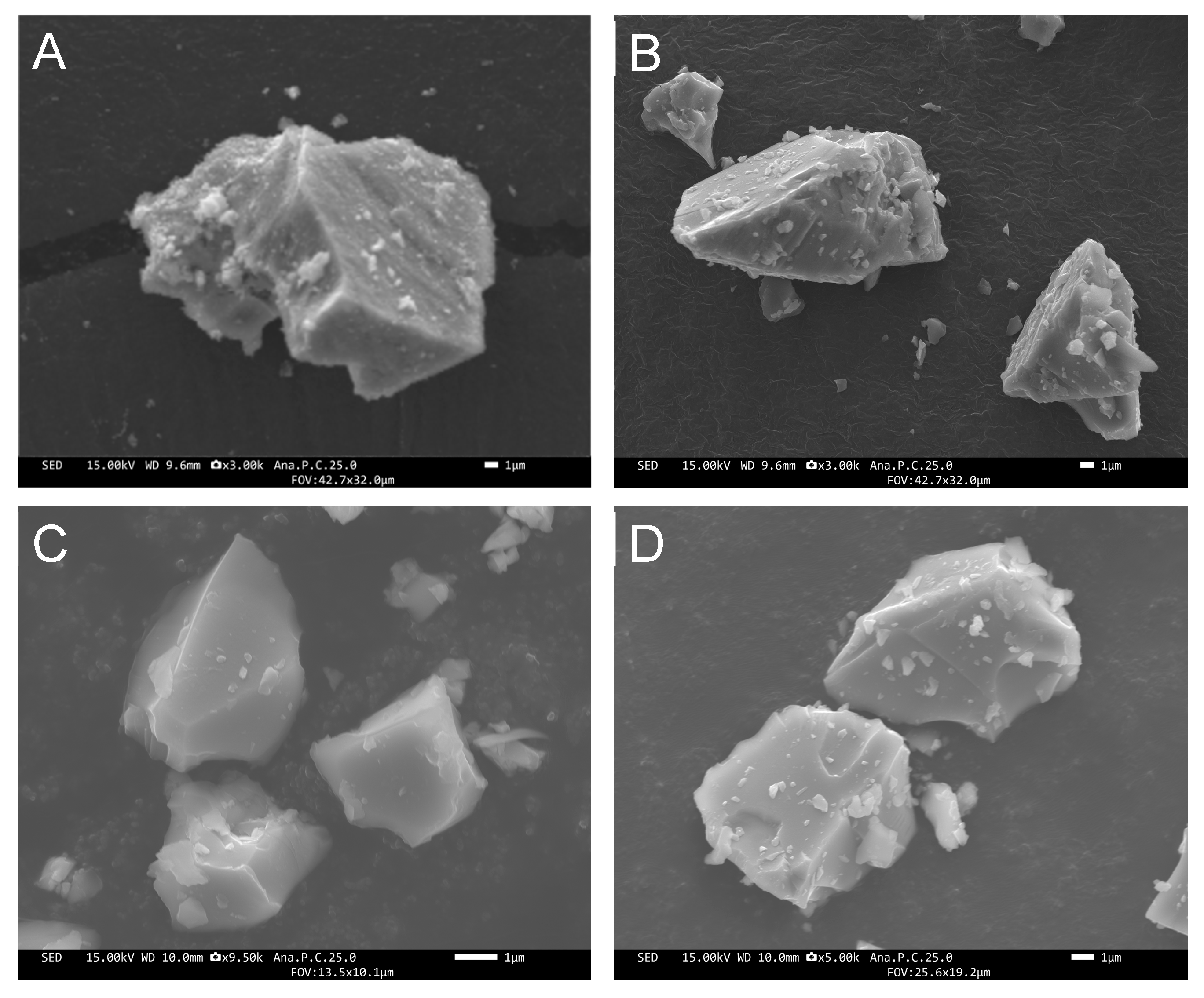
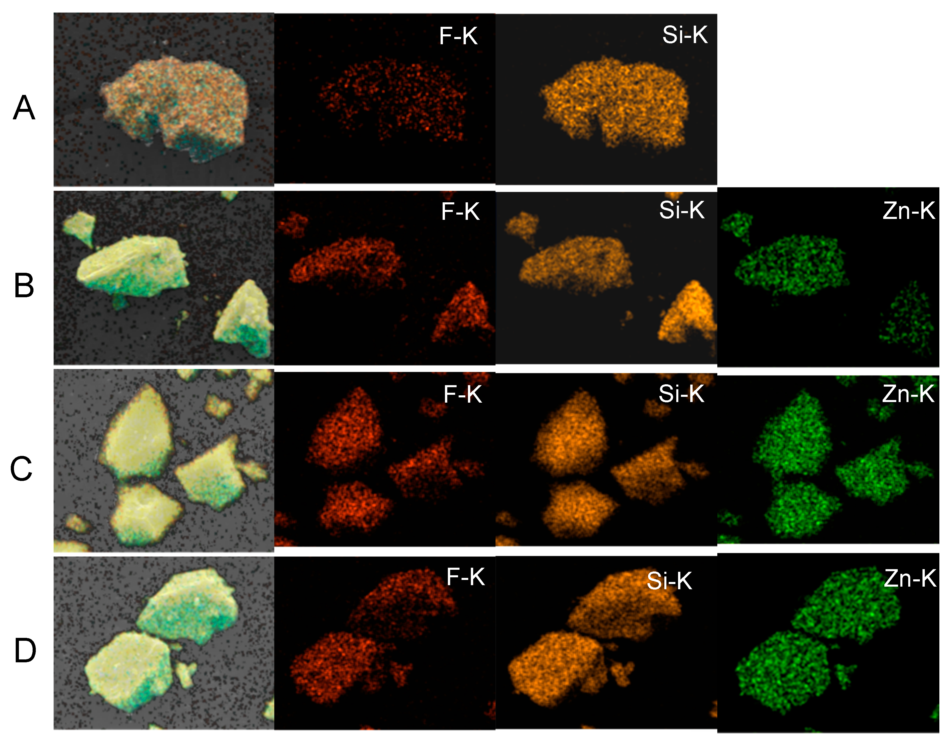
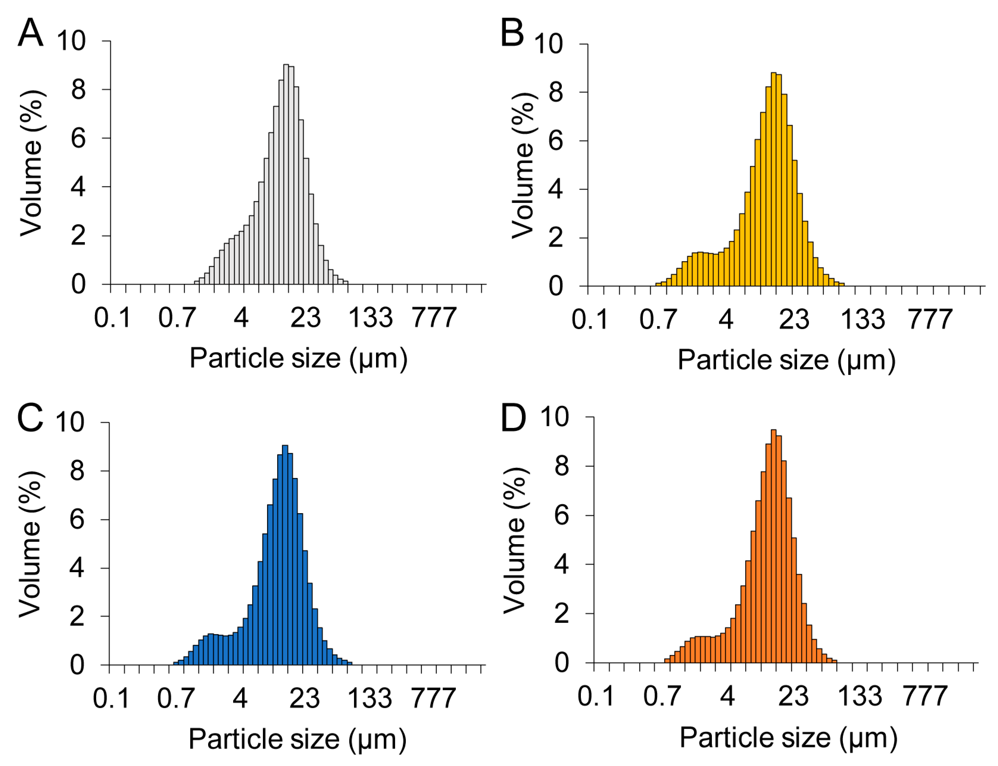
All of the prepared glass particles were of irregular shapes (Figure 1). The median diameter of Cont, PG-1, PG-2, and PG-3 was 11.2, 11.3, 10.9, and 11.3 µm, respectively. As shown in Figure 2, the elemental mapping images revealed that the elements were evenly dispersed in the four glass particles. Their elemental composition is shown in Table 1. According to XRF analysis, Si, Zn, and F were detected in Cont with a mol fraction of 70.8, 0, and 15.1%, respectively; in PG-1 with 42.9, 25.3, and 17.0%, respectively; in PG-2 with 33.4, 34.6, and 16.0%, respectively; and in PG-3 with 29.2, 42.7, and 10.5%, respectively. No remarkable difference in composition was observed between the preparation and result of the XRF analysis. The particle size distribution of each glass particle is shown in Figure 3. No significant difference was observed in the particle size range and distribution among the four groups.
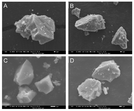
Figure 1.
FE-SEM images of the glass particle surfaces of (A) Cont, (B) PG-1, (C) PG-2, and (D) PG-3.
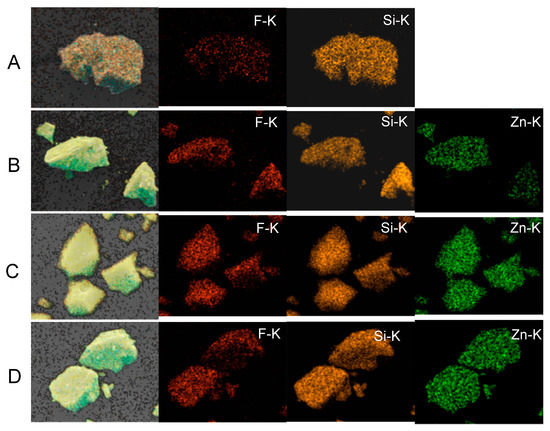
Figure 2.
Elemental mapping images (EDS) of the four glass particles. (A) Cont, (B) PG-1, (C) PG-2, and (D) PG-3.

Table 1.
Elemental compositions of Cont, PG-1, PG-2, and PG-3 glasses (mol%) via XRF analysis.
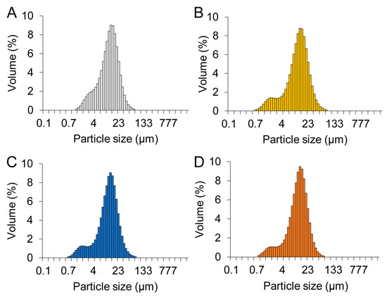
Figure 3.
Particle size distribution (PSD) of the four glass particles. (A) Cont, (B) PG-1, (C) PG-2, and (D) PG-3.
2.2. Solubility and Ion Release Property of Glass Particles in pH-Adjusted Media
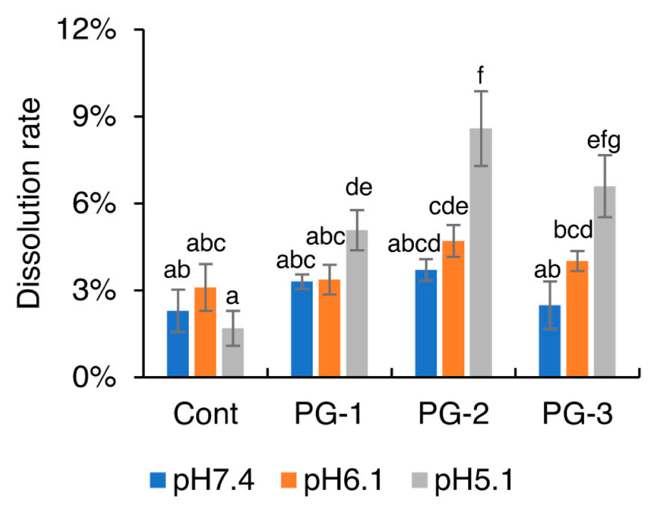
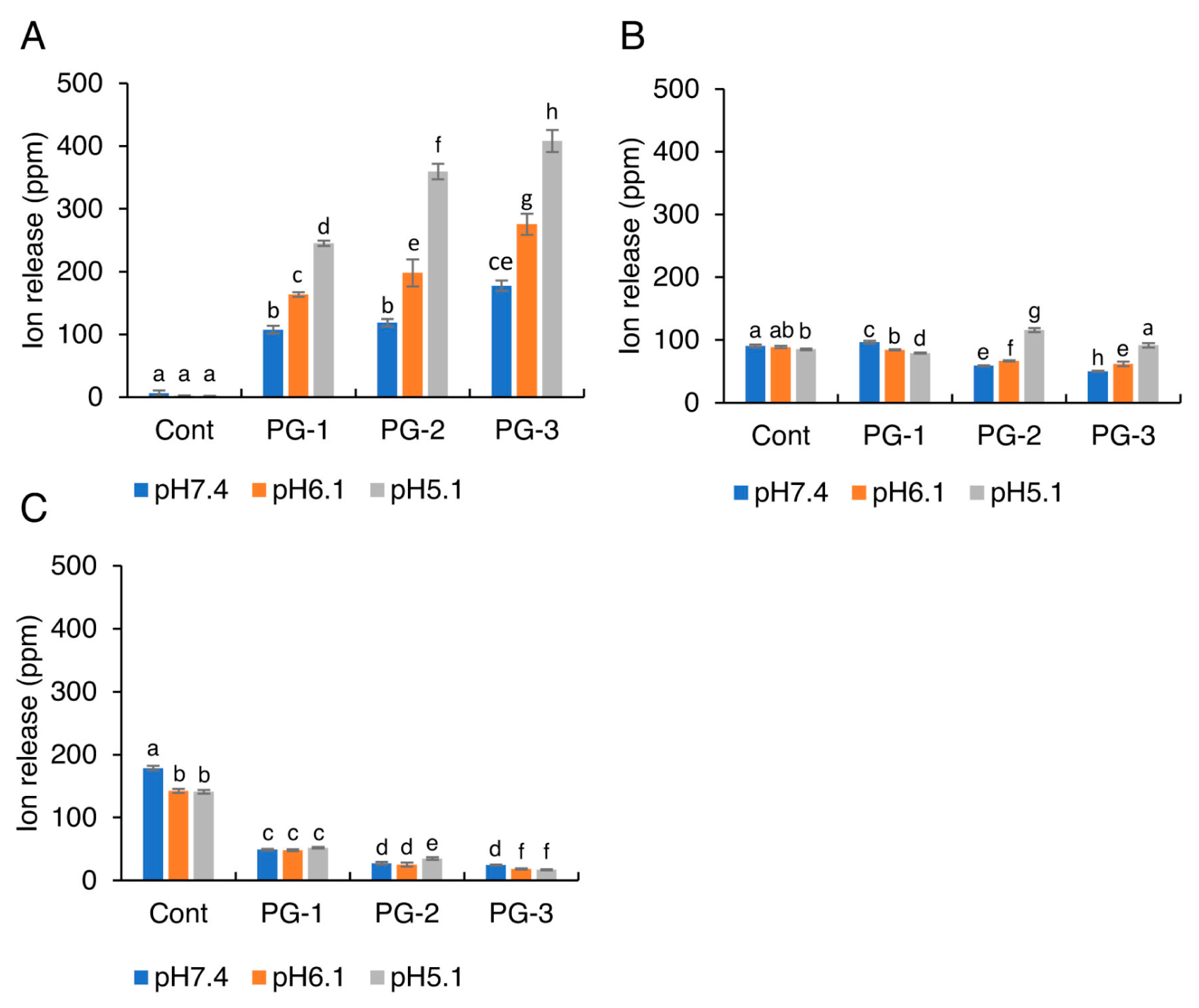
Figure 4 shows the solubility of each glass particle in pH-adjusted BHI broth (pH 7.4, 6.1, or 5.1). When the pH value was at 6.1 or 7.4, no significant difference among the glass particles was observed. When the pH decreased to 5.1, the solubility of the glass particles containing Zn showed a significant increase. Furthermore, no significant difference in the solubility was observed between PG-2 and PG-3 under each pH condition. The profiles of Zn2+, SiO32−, and F− released from glass particles into the pH-adjusted BHI broth are shown in Figure 5. With a decrease in pH values, the release of Zn2+ from Cont., PG-1, PG-2, and PG-3 increased significantly. With an increase in Zn content in Cont., PG-1, PG-2, and PG-3, the release of Zn2+ increased at each pH value, whereas the release of F− significantly decreased.
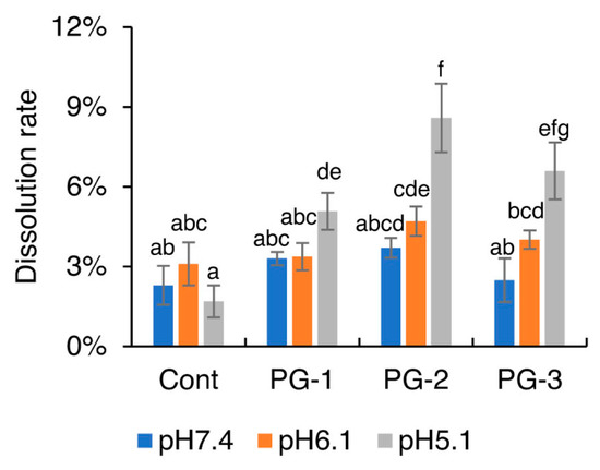
Figure 4.
Solubilities of the four glass particles: Cont., PG-1, PG-2, and PG-3 in pH-adjusted BHI broth. Bars represent the standard deviation of the three replicates. a–g: different letters indicate significant differences (p < 0.05, ANOVA, Tukey’s HSD test).
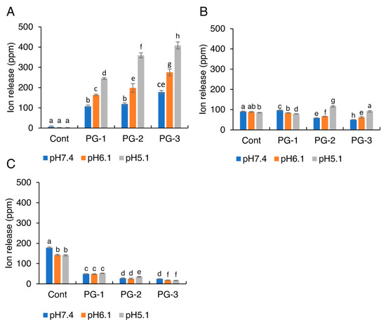
Figure 5.
The concentrations of (A) Zn2+, (B) SiO32−, and (C) F− released from the glass particles into the pH-adjusted BHI broth. Bars represent the standard deviations of the six replicates. a–h: letters indicate significant differences (p < 0.05, ANOVA, Tukey’s HSD test).
2.3. MICs and MBCs of Zn2+, SiO32−, and F− for S. mutans
The MICs and MBCs of Zn2+, SiO32−, and F− against S. mutans are shown in Table 2. For Zn2+, the MIC and MBC for S. mutans were 125 and 250 ppm, respectively. For SiO32−, the MIC and MBC for S. mutans were greater than 500 ppm. Additionally, for F−, the MIC for S. mutans was 125 ppm, whereas the MBC was greater than 500 ppm.

Table 2.
Minimum inhibitory concentrations (MICs) and minimal bactericidal concentrations (MBCs) of Zn2+, F−, and SiO32− on S. mutans (in ppm).
2.4. Antibacterial Activity of Glass Particles against S. mutans
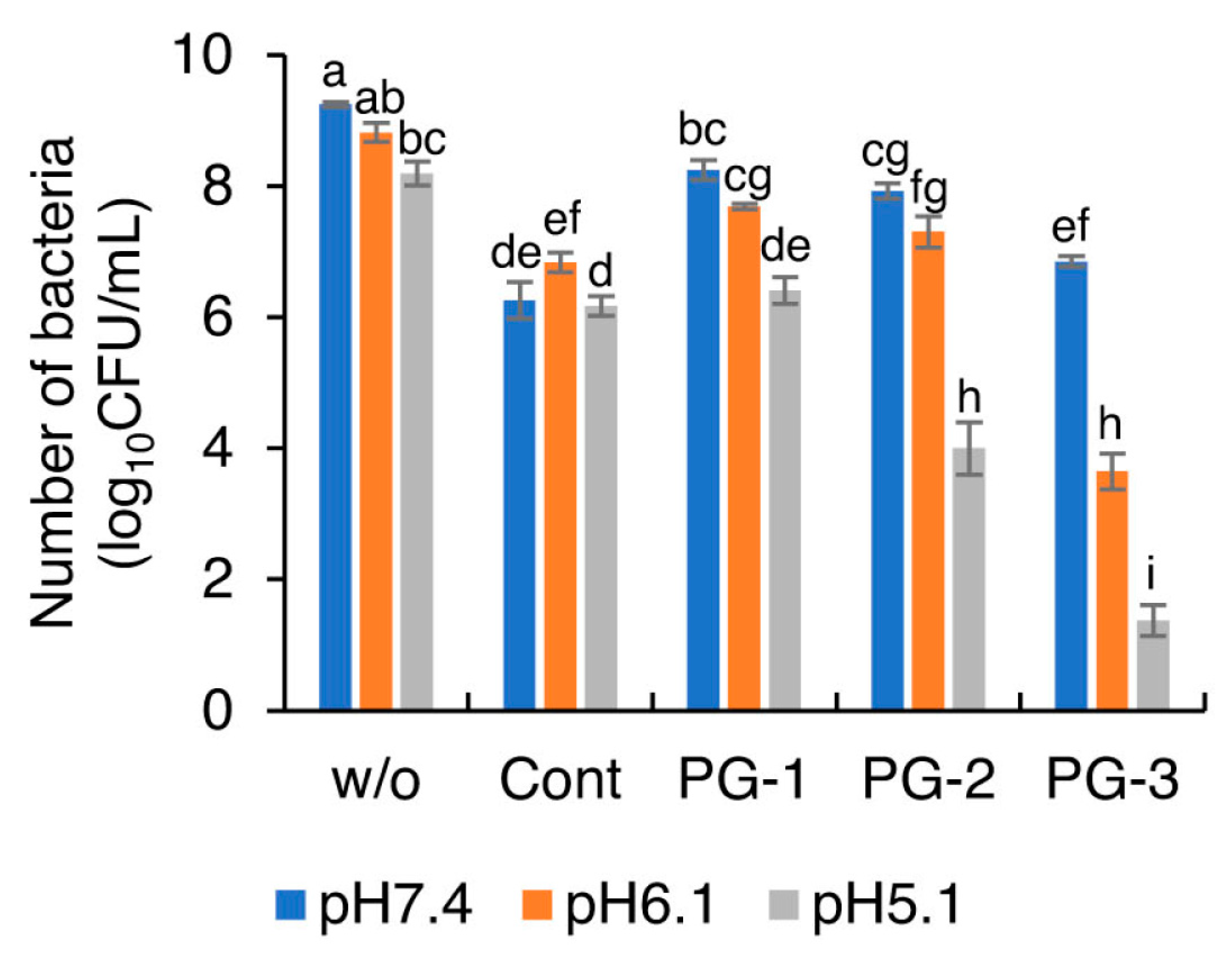
Figure 6 shows the number of viable bacteria after incubation after immersing Cont, PG-1, PG-2, and PG-3 in the pH-adjusted BHI broth. After 24 h of anaerobic incubation, the colony counts of viable S. mutans in the presence of glass particles were significantly lesser than tho without any particles at each pH value. No significant difference was found in the number of surviving cells in the presence of Cont (approximately 6.2 log10CFU/mL) between pH 7.4 and 6.1. With a decrease in the pH value, the viable number of S. mutans significantly decreased in the presence of PG-1, PG-2, and PG-3. The number of viable cells in the presence of PG-1 (pH 5.1), PG-2 (pH 5.1), and PG-3 (pH 6.1 and pH 5.1) was significantly smaller than the initial number of bacteria (approximately 7.0 log10CFU/mL), indicating that they exhibited bactericidal effects at the corresponding pH values.
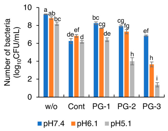
Figure 6.
Number of viable S. mutans after 24 h incubation at pH 5.1, 6.1, and 7.4 with four glass particles. Bars represent the standard deviations of six replicates. a–i: letters indicate significant differences (p < 0.05, ANOVA, Tukey’s HSD test). w/o, without any powders.
3. Discussion
Zn2+ has multiple inhibitory effects on intact bacterial cell activities such as glycolysis, glucosyltransferase production and polysaccharide synthesis, transmembrane proton translocation, and acid tolerance [23]. It can enhance the proton permeabilities of bacterial cell membranes, reduce acid production, and inhibit cell metabolism [17,24]. Fluoride is assumed to be anticariogenic via various mechanisms including the reduction in demineralization, enhancement in remineralization, interference of pellicle and plaque formation, and inhibition of microbial growth and metabolism [25,26]. As described above, the BioUnion filler is composed of SiO2, ZnO, CaO, and F. Ca2+ and F− enhance remineralization and inhibit demineralization [27]. However, the inhibitory effect of Ca2+ on the growth of oral bacteria is not as strong as that of Zn2+ and F− [17]. Therefore, in this paper, three types of experimental glasses containing Zn and F (i.e., without the addition of Ca) were studied to strengthen their antibacterial activity compared with the BioUnion filler.
As intermediates contributing to field strength, Zn2+ behaves either as a modifier or a network-former in glass [28]. As a modifier, Zn2+ exists as charged single ions in the cross-linked glass network. It disrupts the regular bonding between the glass-forming components and oxygen through non-bridging oxygen (NBO; Si-O−M+, where M+ is a modifier ion) [29]. This decreases the relative quantity of strong bonds in glass. As a network-former, Zn2+ enters the silicate network by forming Si-O-Zn bonds. The mechanism of glass dissolution involves the ion exchange (protons for modifier ions) and acid hydrolysis of Si-O-Zn bonds. Zinc-releasing silicate glasses are stable at physiological pH, but show accelerated dissolution under acidic conditions [30]. This suggests that zinc enters the silicate network to a much greater extent, with a majority or potentially all zinc forming Si-O-Zn bonds [29]. By adding zinc oxide to silicate glasses, modifying the ion-releasing behavior in response to changes in the pH environment is possible. Consequently, the glasses are more stable at neutral pH levels, but quickly dissolve in acidic conditions. In this study, the solubility of glass particles in acids increased with an increase in zinc content. According to the ICP results in our study, the release of Zn2+ increased with an increase in the Zn content of glass. Additionally, all glasses containing Zn released a greater amount of Zn2+ with a decrease in pH. These results can be attributed to the degradation mechanisms discussed above. A higher amount of zinc content was related to higher NBO and Si-O-Zn bonds in the glass structure, thus releasing a larger amount of Zn2+ in aqueous solution, particularly in acidic conditions. The control glass showed the greatest amount of fluoride release among the four groups despite an equal fluoride content. With the increase in Zn in the glass (PG-1 < PG-2 < PG-3), the release of F− from the glass decreased (PG-1 > PG-2 > PG-3). This may be explained by the complexing reaction: Zn2+ + HF = ZnF+ + H+, which occurs in the aqueous solution [31]. Most of the fluoride released from glass ionomer cements is known to be in the complexed form rather than as “free” F− [32]. Thus, it can be assumed that the existence of Zn in glass particles interferes with the release of F.
To determine the inhibitory effect of the components in the glass particles, the MICs and MBCs of Zn2+, SiO32−, and F− against S. mutans were measured via a microdilution assay. An MBC of 4.8 mg/mL (480 ppm) for NaF against S. mutans has been reported [33]. Our previous study reported an MIC of 128 ppm and MBC of 2048 ppm for F− against S. mutans [17]. The MIC of NaF against S. mutans in this study was 125 ppm, while the MBC was over 500 ppm. All of the concentrations of fluoride released from the four groups were much lower than the MBC of F−. The concentrations of fluoride released from control glass at the three pH levels were higher than the MIC of F−, while the concentrations of fluoride released from PG-1, PG-2, and PG-3 were lower than the MIC of F−. Therefore, control glass exhibited inhibitory effects against bacterial growth (the number of bacteria was the same/similar to the initial concentration of bacteria) as fluoride has a direct inhibitory effect on the metabolic activity of cariogenic bacteria. Hernández-Sierra et al. reported an MIC range of 500 ± 306.18 µg/mL (500 ± 306.18 ppm) and MBC of 500 μg/mL (500 ppm) for ZnO-NPs against S. mutans [34]. Yu et al. reported an MIC of 0.156 mg/mL (156 ppm) and MBC of 0.312 mg/mL (312 ppm) for ZnO-NPs against S. mutans [35]. Salts (zinc chloride and zinc gluconate) and zinc oxide nanoparticles (ZnO-NPs) are the most common forms used in the evaluation of MICs and MBCs [36], while zinc nitrate (Zn(NO3)2) was rarely reported. In this study, the MIC of Zn(NO3)2 for S. mutans was 125 ppm, while the MBC was 250 ppm. The concentrations of Zn2+ released at pH 5.1 and 6.1 for PG-1, PG-2, and PG-3 were greater than the MIC (125 ppm) against S. mutans, whereas the concentrations of Zn2+ released at pH 7.4 of PG-1 and PG-2 were lower than this value. The concentrations of Zn2+ released from PG-1 (pH 5.1), PG-2 (pH 5.1), and PG-3 (pH 5.1, 6.1) were higher than the MBC (250 ppm) against S. mutans, whereas the concentrations of Zn2+ released from PG-1 (pH 6.1 and 7.4), PG-2 (pH 6.1 and 7.4), and PG-3 (pH 7.4) were lower than this value. Liu et al. reported that the concentration of Zn2+ released from the BioUnion filler in acetic acid at pH 5.5 was 166.9 ± 11.2 ppm [15]. In this study, the concentration of Zn2+ released from PG-3 in the medium adjusted to pH 6.1 was 275.4 ± 17.0 ppm, higher than that of Zn2+ released from BioUnion filler at pH 5.5. As supported by these results, PG-3 demonstrated bactericidal effects (the number of bacteria was lower than the initial concentration of bacteria) under pH of 5.1, as well as 6.1, owing to the effective release of Zn2+ even under slightly acidic conditions. This result suggests that the release of Zn2+ from PG-3 would prevent caries on root surfaces with low acid resistance.
When the restorative materials incorporating the glass particles are exposed to acids, the increased dissolution might adversely affect the physical properties of the materials. An example is GICs wherein glass particles can be incorporated. The chemical composition of the GIC powder has been substantially modified to improve the handling characteristics and mechanical properties. These modifications are based on the hypothesis that a higher number of poly-salt bridges can be formed in the GIC matrix owing to the increased chemical affinity between the filler particles and GIC matrix. This in turn enhances the mechanical properties of the material, making it suitable for posterior dental restoration. In addition, the solubility of GICs is closely related to the ion-releasing behavior, and thus has considerable influence on the antibacterial action. Hence, the particle size and composition of the glass particles influence the antibacterial and physical properties of GICs [37]. The median diameter of glass particles in this study was approximately 10 μm. A previous study reported that the compressive strengths of the GICs incorporating Zn2+-released glass particles with a median diameter of 4.5–5.5 µm did not decrease even after 28-day aging [15]. Some studies indicated that GICs continued the maturation of the acid–base reaction after mixing over time [38] and, consequently, the compressive strength slightly increased. Therefore, we cannot assume that the adverse effects on physical properties are negligible. Furthermore, in this study, the ion release and antibacterial effect against S. mutans were evaluated only for the first 24 h. Further investigations are required to evaluate ion release over longer periods and assess the antibacterial/anti-plaque effects against multi-species bacteria under simulated oral environments.
4. Materials and Methods
4.1. Fabrication of Glass Particles
Three types of silicate-based glasses containing zinc were fabricated and named PG-1, PG-2, and PG-3. A glass particle without zinc was used as the control group (Cont.). The glass particles were prepared by melting SiO2, ZnO, F, and other raw materials in a platinum crucible for 1 h at 1300–1500 °C followed by immediate quenching in distilled water. A sieve was used for classification and particles with a median diameter of approximately 10 µm were obtained. The prospective compositions of Cont., PG-1, PG-2, and PG-3 during preparation are listed in Table 3. All of the glass particles were sterilized with ethylene oxide gas.

Table 3.
Prospective compositions of Cont, PG-1, PG-2, and PG-3 glasses (mol%) during preparation.
4.2. Characterization of Glass Particles
The morphology and elemental compositions of each glass particle were observed and analyzed via field emission scanning electron microscopy and energy-dispersive spectroscopy (FE-SEM/EDS; JSM-F100, JEOL Ltd., Tokyo, Japan). The chemical compositions were also analyzed via X-ray fluorescence (XRF) analysis. Further, the particle size distribution (PSD) of each glass particle was observed via a particle size analyzer (Partica LA-960V2, HORIBA, Kyoto, Japan).
4.3. Solubility and Ion Release Evaluation of Glass Particles in pH-Adjusted Media
The solubility and ion release property of each glass particle in the pH-adjusted media were evaluated. Here, 50 mg of each particle was immersed in 10 mL of brain heart infusion (BHI) broth (Becton Dickinson, Sparks, MD, USA), whose pH was adjusted by hydrochloride acid (Kanto Chemical Co., Inc, Tokyo, Japan) at 7.4, 6.1, and 5.1. After storage at 37 °C for 24 h and gyratory shaking at 100 rpm, the particles were filtered through a 0.22 µm syringe filter (Merck Millipore Ltd., Carrigtwohill, Ireland), which was weighed beforehand (W1 mg) and dried at 110 °C for 48 h. The total weight (W2 mg) of the undissolved particles and the syringe filter was then measured, and the solubility of each particle was calculated according to the following equation:
Solubility (%) = (50 + W1 − W2)/50 × 100
To determine the profiles of Zn2+, SiO32−, and F− released from the glass particles into pH-adjusted BHI broth, 20 mg of each glass particle was placed in one well of a 96-well microplate. The particles in the wells were immersed in 200 μL of pH-adjusted BHI broth. After storage at 37 °C for 24 h and gyratory shaking at 100 rpm, the suspensions were diluted with 9.8 mL distilled water, after which the concentrations of Zn2+ and SiO32− were measured using an inductively coupled plasma-optical emission spectrometer (ICP-OES; iCAP7200 ICP-OES Duo, Thermo Fisher Scientific, Cambridge, UK) and the concentration of F− was determined using a fluoride ion electrode (FIE; 6561S-10C, HORIBA, Kyoto, Japan). The experiments were repeated six times.
4.4. Measurement of MICs and MBCs of Zn2+, SiO32−, and F− for Streptococcus mutans NCTC10449
Streptococcus mutans NCTC10449 from a stock culture was incubated in BHI broth and on BHI agar plates (Becton Dickinson) at 37 °C for 24 h under anaerobic conditions. To evaluate the concentrations of Zn2+, SiO32−, and F− that could effectively inhibit the growth of S. mutans, the minimum inhibitory concentrations (MICs) and minimum bactericidal concentrations (MBCs) of each ion against S. mutans were measured with a microdilution assay. Briefly, 1 mg/mL of Zn(NO3)2, Na2SiO3, and NaF (FUJIFILM Wako Pure Chemical Corporation, Osaka, Japan) were used as standard solutions for Zn2+, SiO32−, and F−, respectively. Here, 50 μL of standard solutions with a concentration of 0.49–1000 ppm obtained after serial twofold dilutions was dropped into the wells of a 96-well microplate. Next, 50 μL of S. mutans suspension, adjusted to 2.0 × 10⁷ colony-forming units (CFU)/mL with 2× BHI broth, was added into each well with the standard solutions. Then, the microplates were anaerobically incubated at 37 °C for 24 h. The turbidity of the suspensions was observed visually and the MIC, which is the lowest concentration that prevents visible bacterial growth, was determined. Then, 20 μL of the clear samples was inoculated on agar plates. After anaerobic sub-culturing for 24 h at 37 °C, the MBC, which is the lowest concentration of the antimicrobial agent that kills the bacterium, was determined using the agar plates with no bacterial colonies. These experiments were repeated five times.
4.5. Evaluation of Antibacterial Activity of Glass Particles
The S. mutans NCTC10449 suspension was adjusted to approximately 1.0 × 10⁸ CFU/mL in BHI broth. A total of 20 mg of each glass particle was placed in a well of a 96-well microplate. Then, 180 μL of pH-adjusted BHI broth (pH 7.4, 6.1, and 5.1) and 20 μL of S. mutans suspension (approximately 1.0 × 10⁸ CFU/mL) were added to it. After anaerobic incubation at 37 °C for 24 h with gyratory shaking at 100 rpm, 100 μL of the suspension was collected and diluted with 9.9 mL of BHI broth. The suspension was then serially diluted with BHI broth and inoculated on BHI agar plates. The plates were incubated anaerobically at 37 °C for 24 h, after which the number of formed colonies was determined. This experiment was repeated six times.
4.6. Statistical Analysis
Statistical analyses were performed using SPSS Statistics 25 (IBM, Chicago, IL, USA). The homogeneity of variances was confirmed initially. The results of solubility, ion release, and bacterial growth were statistically analyzed using analysis of variance (ANOVA) and Tukey’s honestly significant difference (HSD) test. p < 0.05 was considered to indicate significance in this study.
5. Conclusions
Glass particles with a pH-responsive Zn2+-releasing property were successfully fabricated to inhibit S. mutans growth under acidic conditions. The glass particles with 25.3 mol% Zn exhibited bactericidal effects at pH 5.1, whereas glass particles with 42.7 mol% Zn demonstrated killing effects at pH 5.1 and 6.1 due to the effective release of Zn2+ even under slightly acidic conditions. The glass particle with 42.7 mol% Zn may also be useful for developing smart antibacterial restoratives that contribute to the prevention of diseases such as root surface caries.
Author Contributions
Conceptualization, S.I. and H.K.; writing—original draft preparation, F.D.; writing—review and editing, H.K. and S.I.; solubility calculation, H.S. and Y.L.; ICP analysis, T.K.; investigation, P.T.; validation, R.K., G.L.A. and J.-i.S. All authors have read and agreed to the published version of the manuscript.
Funding
This work was supported in part by the Grants-in-Aid for Scientific Research (Nos. JP 20K09937 and JP 20H03871) from the Japan Society for the Promotion of Science.
Institutional Review Board Statement
Not applicable.
Informed Consent Statement
Not applicable.
Data Availability Statement
Not applicable.
Acknowledgments
The authors thank Ayaka Fujimoto-Akiyama, Katsuhito Kato, and Tomohiro Kumagai for their assistance in glass preparation and ion release test.
Conflicts of Interest
The authors declare no conflict of interest.
Sample Availability
Not available.
References
- Paiva, L.; Fidalgo, T.K.S.; da Costa, L.P.; Maia, L.C.; Balan, L.; Anselme, K.; Ploux, L.; Thiré, R.M.S. Antibacterial properties and compressive strength of new one-step preparation silver nanoparticles in glass ionomer cements (NanoAg-GIC). J. Dent. 2018, 69, 102–109. [Google Scholar] [CrossRef] [PubMed]
- Xiao, S.; Wang, H.; Liang, K.; Tay, F.; Weir, M.D.; Melo, M.; Wang, L.; Wu, Y.; Oates, T.W.; Ding, Y.; et al. Novel multifunctional nanocomposite for root caries restorations to inhibit periodontitis-related pathogens. J. Dent. 2018, 81, 17–26. [Google Scholar] [CrossRef]
- Ozer, F.; Patel, R.; Yip, J.; Yakymiv, O.; Saleh, N.; Blatz, M.B. Five-year clinical performance of two fluoride-releasing giomer resin materials in occlusal restorations. J. Esthet. Restor. Dent. 2022. [Google Scholar] [CrossRef]
- Kitagawa, H.; Miki-Oka, S.; Mayanagi, G.; Abiko, Y.; Takahashi, N.; Imazato, S. Inhibitory effect of resin composite containing S-PRG filler on Streptococcus mutans glucose metabolism. J. Dent. 2018, 70, 92–96. [Google Scholar] [CrossRef] [PubMed]
- Aponso, S.; Ummadi, J.; Davis, H.; Ferracane, J.; Koley, D. A Chemical Approach to Optimizing Bioactive Glass Dental Composites. J. Dent. Res. 2018, 98, 194–199. [Google Scholar] [CrossRef]
- Kitagawa, H.; Takeda, K.; Kitagawa, R.; Izutani, N.; Miki, S.; Hirose, N.; Hayashi, M.; Imazato, S. Development of sustained antimicrobial-release systems using poly(2-hydroxyethyl methacrylate)/trimethylolpropane trimethacrylate hydrogels. Acta Biomater. 2014, 10, 4285–4295. [Google Scholar] [CrossRef] [PubMed]
- Imazato, S.; Kitagawa, H.; Tsuboi, R.; Kitagawa, R.; Thongthai, P.; Sasaki, J.-I. Non-biodegradable polymer particles for drug delivery: A new technology for “bio-active” restorative materials. Dent. Mater. J. 2017, 36, 524–532. [Google Scholar] [CrossRef] [PubMed]
- Kitagawa, H.; Kitagawa, R.; Tsuboi, R.; Hirose, N.; Thongthai, P.; Sakai, H.; Ueda, M.; Ono, S.; Sasaki, J.-I.; Ooya, T.; et al. Development of endodontic sealers containing antimicrobial-loaded polymer particles with long-term antibacterial effects. Dent. Mater. 2021, 37, 1248–1259. [Google Scholar] [CrossRef] [PubMed]
- Imazato, S.; Kohno, T.; Tsuboi, R.; Thongthai, P.; Xu, H.H.; Kitagawa, H. Cutting-edge filler technologies to release bio-active components for restorative and preventive dentistry. Dent. Mater. J. 2020, 39, 69–79. [Google Scholar] [CrossRef] [PubMed]
- Lamont, R.J.; Koo, H.; Hajishengallis, G. The oral microbiota: Dynamic communities and host interactions. Nat. Rev. Microbiol. 2018, 16, 745–759. [Google Scholar] [CrossRef]
- Radaic, A.; Kapila, Y.L. The oralome and its dysbiosis: New insights into oral microbiome-host interactions. Comput. Struct. Biotechnol. J. 2021, 19, 1335–1360. [Google Scholar] [CrossRef] [PubMed]
- Ibrahim, M.S.; Balhaddad, A.A.; Garcia, I.M.; Collares, F.M.; Weir, M.D.; Xu, H.H.; Melo, M.A.S. pH-responsive calcium and phosphate-ion releasing antibacterial sealants on carious enamel lesions In Vitro. J. Dent. 2020, 97, 103323. [Google Scholar] [CrossRef] [PubMed]
- Liang, Y.; Song, J.; Dong, H.; Huo, Z.; Gao, Y.; Zhou, Z.; Tian, Y.; Li, Y.; Cao, Y. Fabrication of pH-responsive nanoparticles for high efficiency pyraclostrobin delivery and reducing environmental impact. Sci. Total Environ. 2021, 787, 147422. [Google Scholar] [CrossRef] [PubMed]
- Namen, F.M.; Galan, J.; De Deus, G.; Cabreira, R.D.; Filho, F.C.E.S. Effect of pH on the Wettability and Fluoride Release of an Ion-releasing Resin Composite. Oper. Dent. 2008, 33, 571–578. [Google Scholar] [CrossRef] [PubMed]
- Liu, Y.; Kohno, T.; Tsuboi, R.; Kitagawa, H.; Imazato, S. Acidity-induced release of zinc ion from BioUnionTM filler and its inhibitory effects against Streptococcus mutans. Dent. Mater. J. 2020, 39, 547–553. [Google Scholar] [CrossRef]
- Kohno, T.; Liu, Y.; Tsuboi, R.; Kitagawa, H.; Imazato, S. Evaluation of ion release and the recharge ability of glass-ionomer cement containing BioUnion filler using an In Vitro saliva-drop setting assembly. Dent. Mater. 2021, 37, 882–893. [Google Scholar] [CrossRef] [PubMed]
- Liu, Y.; Kohno, T.; Tsuboi, R.; Thongthai, P.; Fan, D.; Sakai, H.; Kitagawa, H.; Imazato, S. Antibacterial effects and physical properties of a glass ionomer cement containing BioUnion filler with acidity-induced ability to release zinc ion. Dent. Mater. J. 2021, 40, 1418–1427. [Google Scholar] [CrossRef] [PubMed]
- Dawes, C. What Is the Critical pH and Why Does a Tooth Dissolve in Acid? J. Can. Dent. Assoc. 2003, 69, 722–724. [Google Scholar]
- Bowen, W.H. The Stephan Curve revisited. Odontology 2012, 101, 2–8. [Google Scholar] [CrossRef]
- Golub, L.M.; Borden, S.M.; Kleinberg, I. Urea content of gingival crevicular fluid and its relation to periodonal disease in humans. J. Periodontal Res. 1971, 6, 243–251. [Google Scholar] [CrossRef] [PubMed]
- Kaneshiro, A.V.; Imazato, S.; Ebisu, S.; Tanaka, S.; Tanaka, Y.; Sano, H. Effects of a self-etching resin coating system to prevent demineralization of root surfaces. Dent. Mater. 2008, 24, 1420–1427. [Google Scholar] [CrossRef] [PubMed]
- Burne, R.A.; Marquis, R.E. Alkali production by oral bacteria and protection against dental caries. FEMS Microbiol. Lett. 2000, 193, 1–6. [Google Scholar] [CrossRef] [PubMed]
- Phan, T.-N.; Buckner, T.; Sheng, J.; Baldeck, J.D.; Marquis, R.E. Physiologic actions of zinc related to inhibition of acid and alkali production by oral streptococci in suspensions and biofilms. Oral Microbiol. Immunol. 2004, 19, 31–38. [Google Scholar] [CrossRef] [PubMed]
- Koo, H.; Sheng, J.; Nguyen, P.T.M.; Marquis, R.E. Co-operative inhibition by fluoride and zinc of glucosyl transferase production and polysaccharide synthesis by mutans streptococci in suspension cultures and biofilms. FEMS Microbiol. Lett. 2006, 254, 134–140. [Google Scholar] [CrossRef]
- Wiegand, A.; Buchalla, W.; Attin, T. Review on fluoride-releasing restorative materials—Fluoride release and uptake characteristics, antibacterial activity and influence on caries formation. Dent. Mater. 2007, 23, 343–362. [Google Scholar] [CrossRef]
- Takahashi, N.; Washio, J. Metabolomic Effects of Xylitol and Fluoride on Plaque Biofilm In Vivo. J. Dent. Res. 2011, 90, 1463–1468. [Google Scholar] [CrossRef]
- Neel, E.A.A.; Aljabo, A.; Strange, A.; Ibrahim, S.; Coathup, M.; Young, A.M.; Bozec, L.; Mudera, V. Demineralization—Remineralization dynamics in teeth and bone. Int. J. Nanomed. 2016, 11, 4743–4763. [Google Scholar] [CrossRef]
- Dietzel, A. Strukturchemie des Glases. Die Naturwissenschaften 1941, 29, 537–547. [Google Scholar] [CrossRef]
- Blochberger, M.; Hupa, L.; Brauer, D.S. Influence of zinc and magnesium substitution on ion release from Bioglass 45S5 at physiological and acidic pH. Biomed. Glas. 2015, 1, 93–107. [Google Scholar] [CrossRef]
- Chen, X.; Brauer, D.; Karpukhina, N.; Waite, R.; Barry, M.; McKay, I.; Hill, R. ‘Smart’ acid-degradable zinc-releasing silicate glasses. Mater. Lett. 2014, 126, 278–280. [Google Scholar] [CrossRef]
- Connick, R.E.; Paul, A.D. The Fluoride Complexes of Zinc, Copper and Lead Ions in Aqueous Solution. J. Am. Chem. Soc. 1958, 80, 2069–2071. [Google Scholar] [CrossRef]
- Billington, R.; Hadley, P.; Williams, J.; Pearson, G. Kinetics of fluoride release from zinc oxide-based cements. Biomaterials 2001, 22, 2507–2513. [Google Scholar] [CrossRef]
- Pradiptama, Y.; Purwanta, M.; Notopuro, H. Antibacterial effects of fluoride in Streptococcus mutans growth in vitro. Biomol. Health Sci. J. 2019, 2, 1–3. [Google Scholar] [CrossRef]
- Hernández-Sierra, J.F.; Ruiz, F.; Pena, D.C.C.; Martínez-Gutiérrez, F.; Martínez, A.E.; Guillén, A.D.J.P.; Tapia-Pérez, H.; Castañón, G.M. The antimicrobial sensitivity of Streptococcus mutans to nanoparticles of silver, zinc oxide, and gold. Nanomed. Nanotechnol. Biol. Med. 2008, 4, 237–240. [Google Scholar] [CrossRef] [PubMed]
- Yu, J.; Zhang, W.; Li, Y.; Wang, G.; Yang, L.; Jin, J.; Chen, Q.; Huang, M. Synthesis, characterization, antimicrobial activity and mechanism of a novel hydroxyapatite whisker/nano zinc oxide biomaterial. Biomed. Mater. 2014, 10, 015001. [Google Scholar] [CrossRef]
- Almoudi, M.M.; Hussein, A.S.; Abu Hassan, M.I.; Zain, N. A systematic review on antibacterial activity of zinc against Streptococcus mutans. Saudi Dent. J. 2018, 30, 283–291. [Google Scholar] [CrossRef] [PubMed]
- Sawa, N. The Effect of Filler Shape and Size of Resin Composite on Fracture Toughness. Jpn. J. Conserv. Dent. 1993, 36, 507–518. [Google Scholar]
- Crisp, S.; Lewis, B.; Wilson, A. Characterization of glass-ionomer cements 1. Long term hardness and compressive strength. J. Dent. 1976, 4, 162–166. [Google Scholar] [CrossRef]
Publisher’s Note: MDPI stays neutral with regard to jurisdictional claims in published maps and institutional affiliations. |
© 2022 by the authors. Licensee MDPI, Basel, Switzerland. This article is an open access article distributed under the terms and conditions of the Creative Commons Attribution (CC BY) license (https://creativecommons.org/licenses/by/4.0/).

