Review

Seaweeds in the Oncology Arena: Anti-Cancer Potential of Fucoidan as a Drug—A Review

1
Shanghai Public Health Clinical Center & Institutes of Biomedical Sciences, Shanghai Medical College, Fudan University, Shanghai 201508, China
2
Department of Microbiology, University of Ulsan College of Medicine, Seoul 05505, Korea
3
Department of Life Sciences, Yeungnam University, Gyeongsan 38541, Korea
4
Department of Microbiology, BETS Science College, Sabar Kantha 385535, India
5
Department of Biochemistry & Genetics, Barkatullah University, Bhopal 462026, India
6
Department of Pathology, Stanford School of Medicine, Stanford University Medical Center, Stanford, CA 94305, USA
7
Department of Medicine, Multispeciality, Trauma and ICCU Center, Sardar Hospital, Ahmedabad 382350, India
*
Authors to whom correspondence should be addressed.
These authors contributed equally to this work.
Molecules 2022, 27(18), 6032; https://doi.org/10.3390/molecules27186032
Submission received: 27 July 2022 / Revised: 6 September 2022 / Accepted: 8 September 2022 / Published: 16 September 2022

Abstract

:
Marine natural products are a discerning arena to search for the future generation of medications to treat a spectrum of ailments. Meanwhile, cancer is becoming more ubiquitous over the world, and the likelihood of dying from it is rising. Surgery, radiation, and chemotherapy are the mainstays of cancer treatment worldwide, but their extensive side effects limit their curative effect. The quest for low-toxicity marine drugs to prevent and treat cancer is one of the current research priorities of researchers. Fucoidan, an algal sulfated polysaccharide, is a potent therapeutic lead candidate against cancer, signifying that far more research is needed. Fucoidan is a versatile, nontoxic marine-origin heteropolysaccharide that has received much attention due to its beneficial biological properties and safety. Fucoidan has been demonstrated to exhibit a variety of conventional bioactivities, such as antiviral, antioxidant, and immune-modulatory characteristics, and anticancer activity against a wide range of malignancies has also recently been discovered. Fucoidan inhibits tumorigenesis by prompting cell cycle arrest and apoptosis, blocking metastasis and angiogenesis, and modulating physiological signaling molecules. This review compiles the molecular and cellular aspects, immunomodulatory and anticancer actions of fucoidan as a natural marine anticancer agent. Specific fucoidan and membranaceous polysaccharides from Ecklonia cava, Laminaria japonica, Fucus vesiculosus, Astragalus, Ascophyllum nodosum, Codium fragile serving as potential anticancer marine drugs are discussed in this review.

1. Introduction

Modern pharmaceutical science is based on natural products (NPs) derived from cells and tissues of microorganisms, plants, and animals of both aquatic and terrestrial ecosystems. NPs are secondary or specialized metabolites employed as traditional medicines and are now regarded as pillars of conventional pharmacology. Secondary metabolites are critical prerequisites for competitiveness, stress tolerance, anti-predator adaption, anti-microbial immunity, anti-radiation, and marine anti-biofouling activities [1,2,3]. In order to overcome stress circumstances produced by fluctuating or variable environmental factors, for example, light intensity, temperature, moisture, and mechanical wounds, NPs are deemed important [4]. Moreover, NPs are critical modulators of complex interspecies interactions that have an impact on the survival and function of organisms [5]. As a result of the survival and environmental adaptation processes, very complex and diversified compounds that are extraordinarily potential therapeutics that synthetic small molecules cannot match are encouraged to be developed.
Although oceans cover more than three-quarters of the Earth’s surface and marine natural products (MNPs) have a pronounced cytotoxic activity, most bioactive secondary metabolites have been acquired from terrestrial organisms [6]. The adaptation to distinct environmental conditions has resulted in diversification and sophisticated development of marine creatures from their terrestrial counterparts. Marine flora is taxonomically diverse, chemically distinct, biologically efficient, and highly specialized. Most sessile marine invertebrates have been found to produce active natural chemical products for a variety of ecological functions, such as defense against predators, parasites, illnesses, and competition, making aquatic ecosystems an important potential source of bioactive chemical compounds [7]. Hundreds of novel MNPs and over 36,000 marine compounds are identified each year from micro and macro-organisms such as fungus, bacteria, microalgae, macroalgae, sponges, corals, and tunicates. Many of these compounds interact with receptors and enzymes, highlighting their pharmacologic activity and constantly attracting enormous attention in the biomedical area [8]. Several marine compounds have demonstrated the ability to intervene in complex inhibitory actions and regulate several physiological functions such as cell signaling, permeability, angiogenesis, and apoptosis [9]. In general, the marine environment has created several highly effective marine-derived compounds with potential anticancer effects confirmed in-vitro, in-vivo, and in clinical trials.
Cancer remains a major cause of morbidity and mortality in industrial nations despite decades of basic and clinical research on promising new medicines. Cancer development is a dynamic and long-term process involving many complicated components and follows a step-by-step progression that eventually leads to metastasis. Initiation, promotion, and progression are the three crucial steps in creating various forms of human cancer. After cardiac disorders, cancer is the second biggest cause of death in industrialized countries, accounting for one out of every four deaths. Cancer is a multifaceted disease caused primarily by acquired genetic changes that provide tumor cells an edge in terms of survival or proliferation [10]. It is a complex multi-step process involving multiple factors such as smoking, occupational exposure, environmental pollution, unreasonable diet, genetics, viral infection, etc. [11]. Self-sufficiency in growth signals, evading apoptosis, resistance to anti-growth signals, unbounded replicative potential, genome instability, unceasing angiogenesis, tissue invasion, and metastasis are the hallmarks of cancer. Research must therefore continue to improve existing therapies and create new cures. Chemotherapy, radiation therapy, surgery, and combinations of these therapies have all been used to treat cancer. Regrettably, this therapeutics has various negative effects and could be replaced by MNPs [12]. In MNPs, particularly marine drugs, have been at the forefront of the issue as a chemotherapeutic substitute due to several advantages such as abundancy, inertness, high structural flexibility, renewable nature, biodegradability, biocompatibility, bioavailability, non-toxicity, easy processing and extractability [13,14]. Epidemiological studies have recently revealed that marine chemicals significantly impact this proclivity. Some natural extracts have been discovered that target specific signaling pathways to prevent or slow down the carcinogenesis process at various stages and exhibit properties such as specificity, easy stimulation of cancer cell apoptosis and minimal cytotoxicity [15]. Hence, the search for minimal toxicity natural substances is the demand of current research priorities. The anticancer therapeutic aspect of fucoidan as a natural marine drug is discussed in this review.

1.1. Anti-Cancer Marine Drugs

Since cancer is one of the world’s deadliest diseases, the discovery of new anticancer drugs is extremely challenging. Recent improvements in marine bioprospecting have resulted in the identification of a large number of marine chemicals with anticancer potential [16,17]. However, whereas most marine compounds are obtained from shallow fauna, the latest anticancer compounds identified from deep sea species demonstrate how modern technology and deep sea sampling enable unequaled access to a previously untapped reservoir of biochemical diversity [18]. Only a few innovative synthetic compounds have become commercial treatments due to toxicity. Five anticancer drugs based on MNPs have previously been licensed for human use [19]. Most research into the anticancer capabilities of chemicals originating from marine invertebrates has focused on sponges, corals, and algae [20,21]. Complex ecosystems like coral reefs are characterized by severe competition for space and feeding pressure, which contain a high amount of bioactive metabolites. These metabolites have harmful or deterring effects and could be potent drugs against cancer. Various anticancer drugs are derivatives of these marine compounds that have been approved for clinical use, including cytarabine, vidarabine, nelarabine (prodrug of ara-G), fludarabine phosphate (prodrug of ara-A), trabectedin, eribulin mesylate, brentuximab vedotin, polatuzumab vedotin, enfortumab vedotin [22].
Marine plants are becoming more popular because of their ability to produce natural goods with outstanding health advantages. Marine algae is a rich source of vitamins, minerals, unsaturated fatty acid, polysaccharides and well-known bioactive compounds, including sulfated polysaccharides, phlorotannins and glycoproteins. Division Phaeophyta (known as brown seaweeds) includes the most studied species of marine plants, namely Ecklonia, Laminaria, Undaria, Fucus, Astragalus, Codium, Asconodulum and Himanthalia [23]. Seaweeds are abundant in active chemicals, like multi-ring sulphurouscyclics, macrolides and trace elements, as well as in polysaccharides, terpenoids, proteins, polyphenols, and sterols [24]. Brown algae, rich in sulfated polysaccharides and secondary plant metabolites, including fucoidans, phlorotannins, and fucoxanthin, can be used to find new medicinal agents [25,26,27,28]. These compounds have been proven to have anticancer capabilities in addition to antioxidant and immunomodulatory activities and favorable effects on chemotherapeutic side effects such as cardiovascular disease [29,30]. Antiproliferative and pro-apoptotic actions of fucoxanthin and phlorotannins have been documented [31,32]. However, the specific mechanisms have not been dismantled yet [33]. The latest research implies that the pure sulfated polysaccharides can be used as an anticancer fucoidan, which could aid in developing natural anticancer medications. Numerous studies have been undertaken to better understand the potential mechanism of fucoidan as a cancer-preventive drug in vitro and in experimental animal models and humans. There has been good documentation on the therapeutic potential of natural biodiversity along with its natural bioactive agents like as marine polysaccharides, especially fucoidan is present and this activity will enable us to develop new generations of cancer therapies [34].

1.2. Fucoidan

Fucoidan is a natural heteropolysaccharide that was isolated in 1913 by Kylin from sea brown algae, and he dubbed it “fucoidin” [35]. According to IUPAC standards, it is now known as “fucoidan”; however, it is also known as fucan, fucosan, or sulfated fucan [36]. The evolving term fucoidan covers a wide range of sulfated polysaccharides (FCSP) with a backbone of fucose (fucans), heterogeneous compositions (i.e., fucogalactans, xylofucoglucuromannan) and diverse origins [37]. Fucoidan has long been recognized as a therapeutic nutritional supplement in Asia due to its medicinal properties, including anticancer properties. Fucoidan has been a well-explored anticancer marine drug, with the first research findings appearing in the 1980s [38].
Fucoidan has been found to have several intriguing pharmacological properties, including antithrombotic, antitumoral, antiviral, and anti-inflammatory action (dos Santos Marlise A.) Animal research exploiting its numerous pharmacological characteristics has revealed anticancer and antimetastatic benefits [39]. Fucoidan delays cancer growth, exerts a cytotoxic effect, and manifests synergistic impact with anticancer chemotherapeutic drugs [40]. Over the last three years, published research on fucoidans has progressed, and a wide range of fucoidan extract products is now accessible. Fucoidans have recently been assessed, and their potential application as oncology treatments has been expanded [41]. The mechanism through which fucoidans can have a direct or indirect anticancer impact is becoming clearer. Anticancer activities of fucoidans include immune-modulatory and anti-inflammatory characteristics. Fucoidans’ capacity to bind to Toll-like receptors and intervene with the action of vascular endothelial growth factors (VEGF) and matrix metalloproteinases (MMPs) could explain their anti-neoplastic properties [42].
During the development of anticancer medications, it is critical to understand the mechanisms by which certain chemicals exert their effects. Angiogenesis suppression, cell cycle arrest, apoptosis and/or necrosis induction, and immunological activation are all tactics used by anticancer medications to limit tumor growth [43]. The mode of action of algae bioactive is largely determined by their composition and chemical characteristics. However, various brown algae species have different fucoidan structures and contents.

Fucoidan Sources and Structure

Fucoidan is a water-soluble, sulfated, 1,2- or 1, 3- or 1,4-α-l-fucos polymer, found in brown marine algae as structural polysaccharides, accounting for 5–20% of dry algal weight [44,45]. Fucoidan is structurally diverse, containing fucose, uronic acid, galactose, xylose, arabinose, mannose, and glucose residues and various degrees of branching sulfate concentration, polydispersity, and irregular patterns of monomers [34,46]. The major sources of fucoidans are sea cucumber and brown algae. Brown seaweeds are a big selection of marine plants, including Sargassum thunbergi, Ascophyllum nodosum, Fucus vesiculosus, Laminaria japonica, Fucus evanescens, and Laminaria cichorioides, and so on, that are commonly found in diverse cold water sea areas (Saadaoui, Imen). Fucoidan is an anionic polysaccharide with a high concentration of l-fucose and sulfate ester groups. It is mostly derived from brown seaweed, but other algae species, such as Fucus vesiculosus, Ascophyllum nodosum, Laminaria japonica, and Macrocystis pyrifera, can be employed as sources, resulting in a variety of polymeric compositions [36,45].
Fucoidan molecular weight (MW) has been recorded in a wide range of sizes, ranging from 5–7 kDa to several hundred kDa. Low molecular weight fractions (LMWF), in particular, are thought to be more biocompatible [47]. The sugar content, glycosidic bonds, branching, and degree of sulfation and acetylation of fucoidan vary depending on the algal species [48]. Laminaria japonica, Saccharina latissima, Sargassum polycystum, and Saccharina longicruris predominantly contain galactose, while Ascophyllum nodosum, Fucus serratus, Fucus vesiculosus comprise glucose, mannose, xylose, and uronic acids as other monosaccharides [49,50,51]. The backbone structures of Fucus vesiculosus, Fucus serratus, and Ascophyllum nodosum are dominated by linear (1→3) and (1→4) glycosidic linkages, while Chorda filum has a complex backbone structure due to the addition of a few branched fucose residues to the main chains, and further Analipus japonicus and Undaria pinnatifida have highly complex and heavily branched backbone structures [51,52]. Exceptionally invertebrate fucans rarely have regular, repetitive patterns regarding sugar units, glycosidic linkages, branching points, sulfation, or acetylation [37]. However, sulfate content, degree of sulfation, and molecular weight are typically cited as factors influencing the bioactivities of fucoidan. Thus, due to structural variability and lack of regularity in fucoidan molecules, it is frequently difficult to formulate distinct conclusions about the chemical structures of fucoidans.
Fucoidan is a mucilaginous component from the brown algal surface. Different kinds of fucoidan can be obtained, and extraction procedures vary depending upon its source. In general, the extraction of fucoidan using dilute acid or alkalis takes a long time and a big volume of reagents. Chemical, physical, and/or enzymatic treatments and other purifying and fractionation techniques can be used to extract and purify fucoidans from seaweeds [27,53]. Moreover, microwave or ultrasound is utilized to create vibrations in cellular water molecules, breaking apart cells and enhancing the efficiency of the traditional water extraction process. Enzymatic extraction is a highly efficient and specific approach involving cell wall dissolution and cell content removal [26]. According to in vivo studies, anticancer activity exhibited by fucoidan is dependent on source, dosage, frequency, and mode of administration. As per current research, the metabolic rate of fucoidan is subjective to different methods of administration and concentration, which has typical effects on tumors [37].

2. Pharmacokinetics of Fucoidan

Several experimental activities have been carried out to address the in-vivo bioavailability so-called ADME studies of fucoidan, i.e., absorption, distribution, metabolism, and excretion. These fundamental pharmacokinetic factors aid in the development of dosing regimens. Experiments in lab rodents may reveal the absorption and bioavailability of fucoidan. In intestinal absorption studies on rats, Fucus vesiculosus fucoidan (737 kDa) was utilized. After treatment, a maximum concentration of fucoidan in serum was reached after 4 h, with residual absorbed fucoidan accumulating in the kidney. In addition, absorption of fucoidan from C. okamuranus in rats has also confirmed the accumulation of fucoidan in organs [54]. Furthermore, comparative bioavailability tests between LMWF and (moderate molecular weight fucoidan) MMWF have shown that LMWF has a better absorption rate and bioavailability; therefore, its biological potential is higher. Furthermore, in rats, following topical administration of fucoidan (MW 750 kDa) from Fucus vesiculosus demonstrated fine skin-penetrating characteristics. In comparison to an intravenous supply of the same dose, the topical use of the 100 mg/kg body weight resulted in a long half-life of the substance. Hence, fucoidan is taken up by endocytosis and has been detectable in human serum and urine during topical or oral fucoidan therapy [55]. Moreover, recent pharmacokinetic research of Fucus vesiculosus [56] confirmed that the MW of fucoidan plays a crucial role in its absorption and deposition, which could be linked to its prolonged half-life following topical application [57]. In addition, the pharmacokinetics of fucoidan may also be affected by the type of pharmaceutical formulation. According to Kimura et al., encapsulation of fucoidan in nanoparticles can boost at least partial cytotoxicity due to higher permeability. However, fucoidan’s pharmacokinetics with respect to toxicity may still be favorable; data on its biodistribution in humans is currently lacking. Few clinical trials are presently ongoing focusing on the bio-distribution and tolerance of fucoidan. The bio-distribution, safety, and dosimetry of a tagged fucoidan are investigated in healthy persons or volunteers (ClinicalTrials.gov, Identifier: NCT03422055). Patients with stage III-IV non-small cell lung cancer (NSCLC) are being evaluated in a placebo-controlled trial. Fucoidan is added to their chemotherapy treatment to see how it affects their malignancy and quality of life (ClinicalTrials.gov, Identifier: NCT03130829). The outcomes of these clinical trials are crucial in understanding ADME and fucoidan toxicity in humans. Although more research on absorption across the intestinal tract is needed, it’s fascinating to see scientists working on different aspects of fucoidan absorption. As a result of the massive amount of knowledge that is gradually becoming available, future developments of fucoidan as a drug will rely on well-informed decisions [58].

2.1. Fucoidan and Cancer

The anticancer mechanisms of fucoidan are multifaceted. Fucoidan has a broad range of impacts on cellular functions, including cell cycle regulation, RNA metabolism, protein metabolism, carbohydrate metabolism, bioenergetics, mitochondrial maintenance, and DNA repair pathways. Induction/inhibition of reactive oxygen species (ROS), mitochondrial instability, and caspase and poly (ADP-ribose) polymerase (PARP) cleavage are all aspects of it [59,60]. Increased radiation susceptibility [61], prolonged cell cycle arrest, improved immunological clearance, enhanced apoptosis, decreased DNA damage, and metastatic suppression seems to be direct and indirect anti-neoplastic actions of fucoidans. Fucoidans are independent checkpoint modulators that can be used with modern checkpoint inhibitors to treat cancer [62].

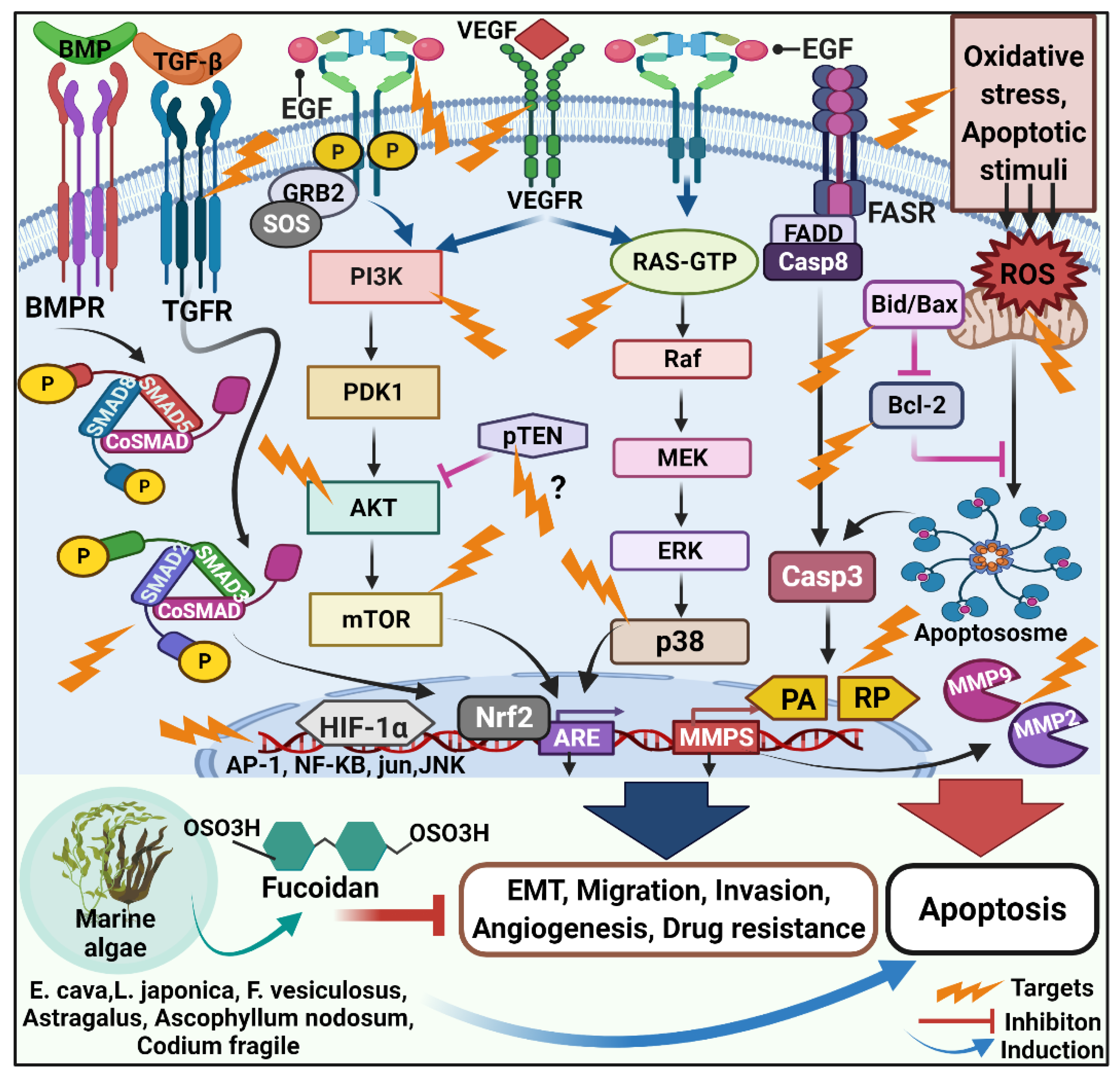
2.1.1. Anticarcinogenic Mechanism of Fucoidan

According to previous studies, the anticancer mechanism of fucoidan largely covers four components. They are (i) cell cycle arrest, (ii) induction of apoptosis, (iii) anti-angiogenesis and (iv) anti-inflammatory activities.

2.1.2. Role of Fucoidan in Cell Cycle Arrest and Apoptosis

Fucoidan inhibits cancer cell proliferation by regulating the cell cycle and blocking uncontrolled mitosis. Alekseyenko et al., 2007, performed Lewis lung cancer transplantation in C57 mice and found that tumor mass and lung metastasization were significantly reduced in fucoidan-treated mice, showing that fucoidan successfully suppressed tumor cell metastasis and proliferation in vivo [63]. Fucoidan (22.5 mg/mL) therapy-induced cell cycle arrest in the G2/M phase and inhibited cell growth in HCC cell lines [64]. Fucoidan boosted the number of G1/S cells in HUT-102 and NSCLC-N6 cell lines, while fucoidan promoted apoptosis and cell cycle arrest in HTLV-1 infected T cells and MCF-7 cells [65,66].
Programmed cell death, such as apoptosis, necroptosis and autophagy, is normally the result of an imposed form of cell cycle arrest. Apoptosis is essential for the survival of multicellular organisms. In general, apoptosis is required for homeostasis and is frequently deregulated in human diseases such as cancer [67]. In contrast to necrosis, apoptotic cells experience chromatin condensation, DNA fragmentation, cleavage of specific proteins, cellular DNA damage, morphological alterations such as blebbing and the budding of apoptotic bodies [68], as well as distinct energy-dependent biochemical mechanisms [69]. Fucoidan promotes apoptosis in cancer cells by aggressively activating apoptotic signals and their underlying downstream pathways. Eun et al., 2010 cocultured human colon cancer cells HT-29 and HCT116 with fucoidan derived from Fucus vesiculosus [70]. Fucoidan caused caspase-3, -7, -8, -9 activation, chromatin condensation, and PARP cleavage. According to the findings, fucoidan can effectively cause apoptosis via caspase-8 and -9-dependent pathways. In human lymphoma HS-Sultan cell lines, fucoidan suppresses tumor growth and promotes apoptosis [71]. Similarly, in MCF-7 cells, fucoidan extract enhanced mitochondrial depolarization by upregulating proapoptotic proteins Bax and Bad and downregulating antiapoptotic proteins Bcl-2 and Bcl-xl expression [72]. Furthermore, fucoidan treatment causes PARP cleavage and caspase-3/7 activation in MCF-7 cells, which are hallmarks of apoptosis [73]. Interestingly, Miyamoto et al., 2009 reported that MCF-7 cells require caspase-7, whereas activation of caspase-3 is not necessary for fucoidan-induced apoptosis [74].
Fucoidan can also show an antitumor effect through simultaneous cell cycle inhibition and induction of cancer cell apoptosis. In the human hepatoma SMMC-7721 cells, fucoidan therapy caused noteworthy growth inhibition and ROS-mediated apoptosis involving several distinctive features such as chromatin condensation or marginalization, vacuolization, lower glutathione consumption (GSH), mitochondrial swelling, and depolarization of the mitochondrial membrane potential [75]. Fucoidan inhibits PI3K, suppressing ERK and activates MAPK, limiting cancer cell proliferation and decreasing Bcl-2 to Bax ratio, inducing caspase-dependent apoptosis in BEL-7402 and LM3 cell lines [76]. Fucoidan inhibited cell growth in hepatocellular, cholangiocarcinoma, and gallbladder carcinoma cell lines by inducing apoptosis and inhibiting cell cycle progression in a dose- and time-dependent manner [64].

2.1.3. Fucoidan and Angiogenesis

Cancer cells rely on an adequate supply of oxygen and nutrients for cell proliferation and metastatic dissemination, highlighting angiogenesis and lymph angiogenesis are critical processes for tumorigenesis. Antiangiogenic therapy has proven to be an effective way to slow tumor growth; consequently, angiogenesis inhibitors are a popular target for cancer treatment [77]. Surplus sulfated fucoidan promotes antitumor and antiangiogenic actions in cancer cells. Fucoidan suppresses vascular endothelial growth factor (VEGF) expression, restricts angiogenesis, cuts off the neoplastic cells’ nutrients and oxygen, lowers its volume, and impedes invasion and metastasis. Using blocking VEGF165 from attaching to its cell surface receptor vascular endothelial growth factor receptor-2 (VEGFR-2), Koyanagi et al., 2003 observed that both natural and oversulfated fucoidans dramatically reduced the mitogenic and chemotactic activities of VEGF165 on HUVEC. They also claimed that increasing the number of sulfate groups in the fucoidan molecule increases its antiangiogenic and antitumor properties [78]. Tse-hung et al., 2015 supplied fucoidan to Lewis lung cancer cell implanted mice, resulting in reduced serum and lung tissue VEGF levels when compared to controls [79]. Accordingly, in mice implanted with Sarcoma-180 cells, both native and oversulfated fucoidans suppressed neovascularization [78]. Moreover, the experimental angiogenesis test showed that the expression of the angiogenesis factor VEGF-A was dramatically reduced by 100 mg/mL fucoidan [80]. Fucoidan-mediated in vivo inhibition of neovascularization is also reported [78]. These findings clearly state that fucoidan’s antitumor activity is attributable to its antiangiogenic effect.
Fucoidans exhibit entirely diverse effects on angiogenesis depending on their molecular weight and sulfate content. Natural fucoidan with a high molecular weight (30 kDa) has antiangiogenic properties, inhibiting proliferation, migration, endothelic tube formation, and vascular network formation, whereas low (4–9 kDa) and intermediate (15–20 kDa) molecular weight fucoidan stimulate angiogenesis and enhance HUVEC migration, respectively. Contrastingly, over-sulfated fucoidan seems to have a more potent antiangiogenic property than natural or desulphated fucoidan [81].

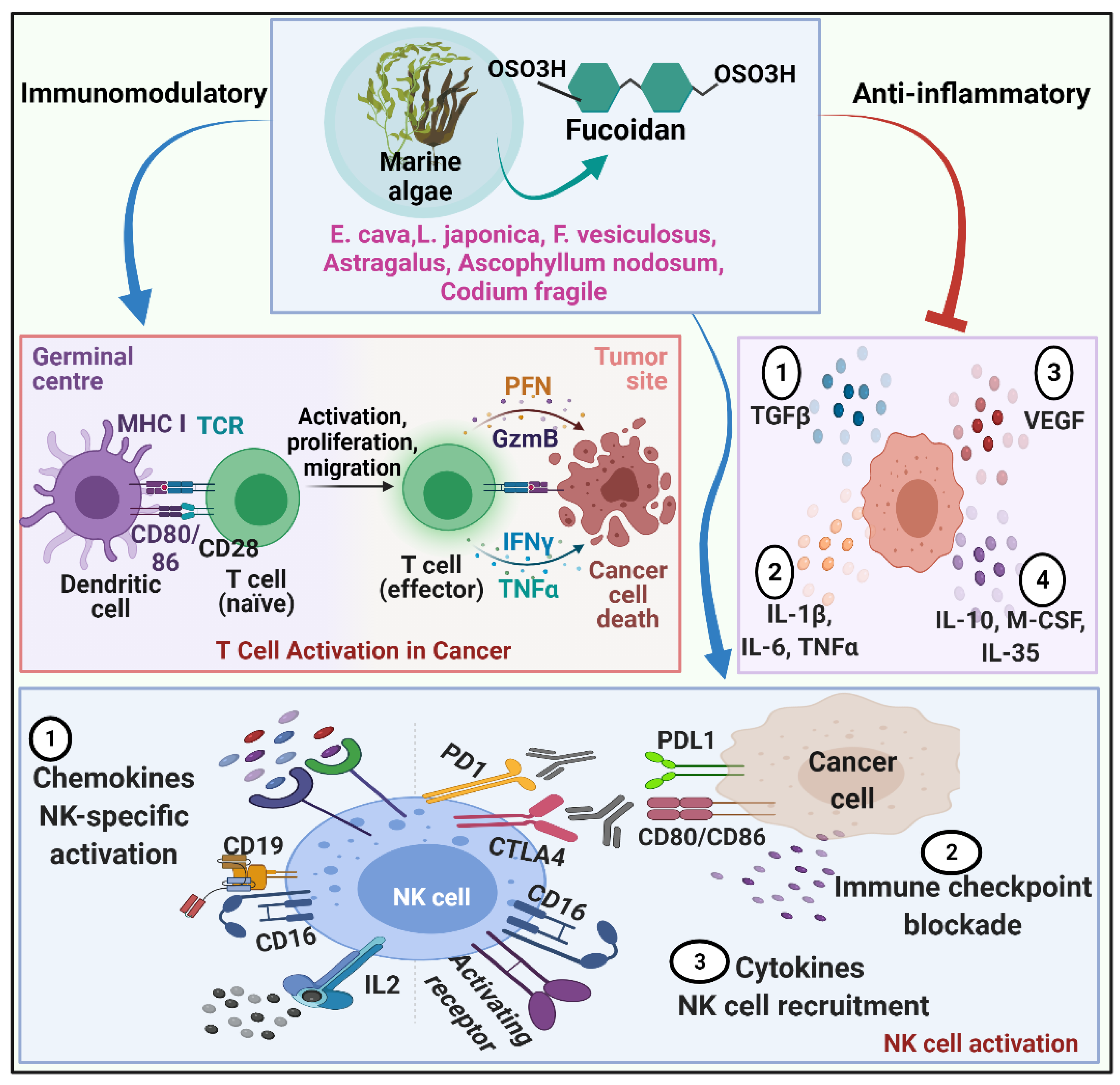
2.1.4. Fucoidan and Immunomodulation

Fucoidan remodels the immune system to improve the sustainability of cancer-specific T cells and NK cells and results in the elimination of cancer cells. Crude and modified fucoidans have been proven to have immunopotentiating effects in tumor-bearing mice, resulting in antitumor efficacy [27,53]. Several studies have confirmed crude fucoidan’s profound cell survival and natural killer activity in vitro and in vivo [82]. Farzaneh et al., 2015 found that fucoidan limits the development of promyelocytic leukemia cells in vitro and in vivo by boosting the cytotoxic activity of NK cells and delaying tumor formation [40]. In line with this, fucoidan reduced cell proliferation in Huh7 human liver cancer cells more effectively by inhibiting the chemotaxin CXCL12 and CXCR4 pathway, an effective target for cancer therapy [54]. Fucoidan has a strong anti-inflammatory activity; however, the underlying mechanisms exerted by fucoidan have not been fully elucidated. Fucoidan has been shown to impede lymphocyte adhesion and invasion, enzyme inhibition, and induce apoptosis at various phases of the inflammatory process. The downregulation of MAPK and NF-B signaling pathways, as well as selectins and the concomitant decrease in the generation of proinflammatory cytokines, is the most often debated putative mode of action for fucoidan [83,84].

2.1.5. Fucoidan and Metastasis

Tumor cell proliferation and survival are frequently linked with deregulated intracellular signal transduction and persistent activation of cellular pathways [38]. Metastasis is a leading cause of cancer-related fatalities, accounting for up to 90% of all cases due to its systemic and pervasive nature and higher drug resistance. Metastasis is the multi-stage process through which cancer cells spread to different body areas through circulation, lymphatic system, or direct extension. The antimetastatic property of fucoidans is evidenced in both in-vitro and in-vivo studies. Fucoidan had moderate anticancer and antimetastatic effects following single and recurrent administration at a dose of 10 mg/kg [63]. Fucoidans from L. saccharina, L. digitata, Fucus serratus, Fucus distichus, and Fucus vesiculosus effectively inhibited MDA-MB-231 breast carcinoma cell adhesion, underlining anti-metastasis outcomes [84]. During metastasis, malignant cells break away from the primary tumor and invade nearby tissues by destroying the extracellular matrix (ECM) with MMPs. In a mice model of lung cancer, increased levels of fucoidan exhibited diminished MMP-2 activity and altered expression of adhesive and migratory proteins, such as integrin, VEGF-1 and -2, P-selectin and neuropilin-1 [85,86]. In the 4T1 xenograft model, fucoidan greatly lowers tumor volume and the number of metastatic lung nodules by minimizing cell migration and invasion while suppressing cell proliferation, colony formation, and expression of epithelial to mesenchymal transition (EMT) biomarkers in vitro [59,60]. An important function in the modulation of EMT in cancer cells is the molecular network of transforming growth factor-β (TGF-β) receptors (TGFRs). Fucoidan lowers the amount of TGF-RI and TGF-RII proteins, including Smad5/8 and Smad2/3 phosphorylation and Smad4 expression, affecting downstream molecular signaling. Fucoidan inhibits ECM degradation and reduces invasiveness in hepatoma HCC cells by downregulating the TGFR and SMAD signals [87]. Fucoidan also can efficiently reverse TGFRs, elicit EMT, stimulate the expression of epithelial markers, depress the expression of interstitial markers, and diminish the expression of transcriptional repressors, including Twist, Snail, and Slug. MAPK/ERK/PI3K/Akt/mTOR pathways regulate cell proliferation, differentiation, and death, and thereby the number of malignancies exploits all of them to be invasive or migratory. Antimetastatic effect of fucoidan is owing to its ability to block MAPK cascade, inactivate ERK1/2 pathway, inhibit phosphorylation of PI3K-Akt, and suppress mTOR and its downstream 4E-BP1 and p70S6K, and downregulate the expression of MMP-2 in time- and concentration-dependent manner. Remarkably, the highest concentration at which fucoidan has the most inhibitory effect is 200 mg/mL [88]. Fucoidan preferentially suppresses lung cancer by promoting Smurf2-dependent ubiquitination of TGF receptors [89], downregulation of MMPs, VEGF, and activation of tumor apoptotic signaling molecules. Besides this, fucoidan therapy also suppresses NF-kB and activator protein-1 (AP-1) transcription factors that are deregulated in cancer, inhibiting tumorigenesis, promotion, and metastasis [90,91]. AP-1 binds to the promoters of several cellular genes and starts their immediate gene expression as these genes are involved in the transcriptional regulation process. Fucoidan suppresses the AP-1 activation by inhibiting JunD expression in an HTLV-1-infected T-cell line, thereby inhibiting HTLV-1-infected T-cell proliferation [66]. Fucoidan inhibits Ik-Ba phosphorylation and raises total p65 in the cytoplasm while decreasing it in the nucleus, modulating inflammation, immunity, differentiation, cell growth, tumorigenesis and apoptosis [88]. Fucoidan inhibits the transcription of c-fos, c-jun, and AP-1 as well as decreases phosphorylation of ERK and JNK via inhibiting EGF-induced EGFR phosphorylation [92], enhancing antimetastatic activities (Figure 1).
Furthermore, post-transcriptional mechanisms have been linked to TGF signaling via microRNAs (miRs) and have been involved in regulating EMT. Apparently, dose-dependent overexpression of microRNA-29b (miR-29b) in human HCC cells was demonstrated by blocking its downstream DNA methyltransferase 3B target (DNMT3B) and enhancing tumor metastasis suppressor gene 1 (MTSS1) expression [87]. Fucoidans are promising candidates being used as functional foods and pharmaceuticals for cancer prevention, chemotherapeutic agonists, and nanotechnology-based targeted therapy due to their extensive bioactivities in cancer, notably in metastasis.

2.1.6. Fucoidan and Gut Flora

Due to their commercial abundance, brown seaweeds are frequently used as food additives. Fermentation of high molecular Fucoidan extracted from Ascophyllum nodosum and Laminaria japonica lowers inflammatory response while promoting Ruminococcaceae and Lactobacillus, respectively [93]. Meanwhile, dietary fucoidan progressively restores intestinal villi by upregulating the expression of tight junction proteins such as ZO-1, Occludin, Claudin-1, and Claudin-8 via p38 MAPK and ERK1/2 activation. Eventually, fucoidan supplementation improves intestinal barrier function by enhancing intestinal microbiota diversity and causing changes in microbial composition with a higher Bacteroidetes/Firmicutes phylum ratio. Fucoidan induces the enrichment of short-chain fatty acids producer Prevotella at the genus level [94]. A study revealed that fucoidan could be used as a gut flora modulator for breast cancer prevention. More detailed research is needed to better understand the association between cancer and intestinal flora and the anticancer mechanism of fucoidan [93]. As a result, the improvement of gut microbiota, as well as antiobesogenic, anti-diabetic, anti-microbial, and anticancer bioactivities offered by Fucoidan, underlines the need for more research to analyze the structure-dependent fermentation of fucoidan to assign a prebiotic effect. Table 1, Table 2, and Table 3 illustrates preclinical and clinical studies of fucoidan and its anticancer action mechanism.

3. Fucoidan from Different Sources of Marine Sea Weeds

3.1. Ecklonia cava

Ecklonia cava (E. cava) is a brown alga widely distributed in China and Korea. E. Cava comprises abundant bio-active sulfated polysaccharide complexes. According to recent marine investigations, E. cava possess noteworthy antioxidant activity and is consequently competent in reducing the development of apoptotic bodies. Sulfated polysaccharide isolated from E. cava substantially promotes apoptosis via regulating pro-apoptotic caspases-7 and -8, Bax, and Bcl-xL [130]. According to Ahn et al., 2015 protamex extract (PCP) of E. cava, which is rich in fucose and sulfate components, strongly correlates with inhibitory growth sequels rather than antiproliferative effects involving enhanced apoptotic sub-G1 DNA contents, partial DNA fragmentation, hypo diploid sub-G0/1-peak as well as dosage dependent activated PARP and caspase 9 by dramatically modulating the Bcl-2/Bax signal pathway, indicating anticancer impact on colorectal carcinoma cells [68]. Additionally, U937 leukemic cells showed similar growth inhibitory results with sulfated polysaccharides of E. cava [131,132]. Furthermore, numerous researchers have discovered that fucoidan extracted from E. cava induces death in MCF-7 and HCT-15 cells via the activation of caspases and the regulation of MAPK, including ERK, p38 kinase, and the Akt pathway, as well as alterations in Bcl-2 and Bax [71,74,133]. Yet, it remains to be seen whether PCP is linked to MAPK signaling pathways such as ERK, p38 kinase, and the Akt pathway in CT 26 cells [68]. However, the efficiency of fucoidan in inducing apoptosis varied between different types of colon cancer cells.
Several preclinical trials have recently reported that E. cava fucoidan (ECF) has significant immunomodulatory effects. In view of this, ECF-mediated potent immunostimulative effects and cytotoxic effects on B16 and CT-26 carcinoma growth were reported in the mice model. However, the immunostimulatory effects of ECF on DC activation and anticancer immunity have been thoroughly investigated. Accordingly, ECF suppresses tumor growth by boosting DC activation and anti-inflammatory cytokine production. However, ECF promotes immune activation but has little effect on cancer cells since it is not an Ag-specific immune response. Because of this, cancer is treated differently from a bacterial infection in the body. ECF requires cancer antigens to stimulate an Ag-specific immunosuppressive response that targets tumors directly and promotes apoptosis to limit tumor growth. As a matter of fact, currently, cytotoxic T cell (CTL) activation is extensively exploited in cancer immunotherapy. Cancer antigen-specified CTL activation causes identification and apoptosis of antigen-presenting cells (APCs) through production of inflammatory cytokine and cytotoxic proteins such as perforins and granzyme B. CD8+ DCs activate antigen-specific CTLs in mice when antigens are presented on major histocompatibility complex (MHC) class I, which interacts with TCRs and CD8 on T cells [134,135]. Apart from antigenic presentation, CD8+ DCs produce a lot of co-stimulators and proinflammatory cytokines, which aid CD8 T cells in being differentiated into CTLs [136]. Antigen presentation, cytokine production, and co-stimulation are all signs of DC maturation. Moreover, in vitro activation of CTLs mediated by bone marrow-derived dendritic cells (BMDCs) is challenging to explain in mice, as BMDCs show distinct phenotypes and functions than in vivo DCs. In a mouse model, the combination of ECF and ovalbumin (OVA) administration resulted in ECF-mediated splenic CD8+ and CD8-DCs activation, enhanced OVA-specific T cell proliferation and IFN-γ production. These findings show that ECF, as an immunostimulatory molecule, significantly enhances antigen-specific immune responses in rodents [134]. Thus, ECF may be effective as an adjuvant to improve immune response against human malignancies. In a previous study, we compared the effects of four fucoidans isolated from Ecklonia cava, Macrocystis pyrifera, Undaria pinnatifida, and Fucus vesiculosus on human peripheral blood dendritic cells (PBDCs). In contrast to the other three fucoidans, we found that ECF significantly upregulated co-stimulatory molecules, MHC class I and II, and the production of proinflammatory cytokines in monocyte-derived DCs (MODCs) and PBDCs. Based on these findings, the study proposed that ECF may have the ability to increase human immunological activation [137].

3.2. Laminaria japonica

Laminaria japonica (L. japonica) is uncommon edible seaweed in Asia. L. japonica is a type of edible seaweed that is widely consumed in Asia. Laminarin is a polysaccharide composed of (13)-beta-d-glucan and (16)-beta-linkage that is isolated from L. japonica [138]. Laminaria has been shown to have antioxidant, antibacterial, anti-inflammatory and hypolipidemic properties. It has also been used as herbal medicine to treat cancer, gastrointestinal issues, diabetes, and arsenic poisoning. Preliminary trials suggested that L. japonica polysaccharides serve as a potent immunomodulator playing a pivotal role in combating cancer. In this context, polysaccharides from L. japonica can stimulate immune cells of both innate and adaptive immune systems, increasing the release of cytokines such as IL-10, IL-6, and IL-1 α, rendering them promising anticancer immunotherapy. It stimulates macrophages that directly release anticancer inflammatory molecules like NO and TNF- α while promoting bone marrow dendritic cells (BMDCs) maturation to activate naive T lymphocytes, thereby resulting in tumor cytotoxicity. Meng et al., 2014 confirmed declining phagocytosis, decreased lysosomal count, upregulated expression of BMDC’s major membrane components, including CD80, CD83, CD86, CD40, and MHC II, and increased BMDC-released production of IL-12 and TNF- α, all of which have been shown to significantly increase BMDC maturation by L. japonica polysaccharides [139]. Therefore, investigation of the immunological significance of laminarian indicated considerable BMDC maturation through increased expression levels of co-stimulatory molecules, IFN-γ and TNF-α cytokines production, and consequent cytotoxic T lymphocyte activation. As a result, it is reasonable to conclude that L. japonica polysaccharides have a high potential to boost BMDC maturation and can be employed as an immunological stimulant as well as a therapeutic adjuvant in immune-compromised individuals [139]. Furthermore, Lin et al., 2016 revealed that optimal fermentation products of L. japonica enhance anti-inflammatory effects by lowering ROS production, prostaglandin E2, NF-kB (p65) phosphorylation, and macrophagic NO production [140]. Fucoidan is a more powerful free radical scavenging antioxidant than laminarin due to the presence of the sulfate groups and the anionic charge [14]. As a result of these findings, laminarin has the potential to be employed as a novel immune stimulatory molecule for cancer immunotherapy.

3.3. Fucus vesiculosus

Fucoidan from Fucus vesiculosus (F. vesiculosus) induced apoptosis in HCT-15 cells by activating ERKs, p38, and inhibiting the PI3K/Akt signaling pathway [133]. Another study reported that F. vesiculosus fucoidans deplete cytosolic and nuclear NF-KB localization to suppress the invasion and migration of human lung cancer cells [88]. According to Ulf et al., 2015 treatment with F. vesiculosus resulted in a strong inhibition of viability in various pancreatic cancer cell lines. The fucoidan extract arrested the cell cycle at the S/G2 phase in cancerous proliferating cells due to the up-regulation of cell cycle inhibitors such as p21, p27, p57 and TP53INP1 while unaffecting non-malignant resting cells [141]. Hence, as a side effect of F. vesiculosus therapy, apoptosis was induced by PARP-cleavage in tumor cells rather than mitochondrial pathways. Intriguingly, in combination with autophagy inhibitors, enhanced cytotoxicity was reported. Overall, F. vesiculosus is a powerful cancer cell growth inhibitor; however, it neither causes mitochondrial inflammation nor causes caspase-dependent apoptosis, or necroptosis in normal cells, underlining the non-toxicity and antioxidant properties of fucoidan [141].
Researchers have been led to study the synergistic effects of fucoidan with the existing anticancer medicines. Doxorubicin (DOX) is renowned for its powerful dose-dependent anticancer efficacy, but a slew of significant side effects constrains it. Acute cardiotoxicity is a major side effect; hence, doxorubicin-loaded fucoidan nanoparticles are currently being investigated as a viable and safer option. Fucoidan, derived from F. vesiculosus, reduces doxorubicin-induced acute cardiotoxicity through modulating JAK2/STAT3-mediated apoptosis and autophagy [142]. By virtue of unique accumulation and preferential location in tumors, doxorubicin-loaded fucoidan nanoparticles demonstrated stronger anticancer activity in mice [143] or lower inhibitory concentration in cancer cells [144] compared to the free DOX model. Besides this, F. vesiculosus combined with paclitaxel had a potential antagonistic effect in breast cancer models, according to Burney et al., 2018 [145]. Moreover, recent in vivo human ovarian cancer murine model experiments illustrated no change in paclitaxel activity when given in combination with F. vesiculosus, contrary to prior in-vitro investigations. Additional studies are reasonable to delimitate mechanisms contributing to a discrepancy in the in vivo activity when given in combination with paclitaxel. As a preliminary stage, pharmacokinetic clinical research is presently underway to appraise the impact of fucoidan in conjunction with chemotherapy for solid tumor patients [145]. In vitro and in-vivo studies imply that F. vesiculosus could be a valuable marine drug that warrants additional research and development for clinical use.

3.4. Astragalus membranaceus

Astragalus membranaceus, a popular immunomodulatory herb used in China, has a long history in traditional Chinese medicine. Currently, it is commonly deployed in mixed herbal decoctions as an immunomodulating agent. Astragalus polysaccharides have been investigated extensively, especially for their immuno-potentiating features, such as induction of DCs, efficient vaccine adjuvant, and immuno-regulated anticancer activities. A. membranaceus polysaccharide raises IL-2, IL-12, and TNF-secretion while lowering IL-10 levels in the blood, conferring anticancer activity in vivo, improving host immune responses, and appears to be safe and effective for antitumor therapy [146]. According to cell viability detection data, Astragalus polysaccharides can reduce the proliferation of human lung cancer cell lines A549 and NCI-H358 by suppressing NF-KB transcription activity and down-regulating expression of p65, p50, CyclinD1, and Bcl-xL proteins. Similarly, Astragalus polysaccharide has shown up-regulation of p53 and PTEN expression through regulating p53/MDM2 feedback loops, signifying one of the alleged antiproliferative mechanisms [147]. APS activates macrophages and enhances the level of nitric oxide (NO) and tumor necrosis factor-α (TNF-α), both of which serve as pro-apoptotic inducers in MCF- 7 cells [148]. Notably, Astragalus polysaccharides have cytotoxic and growth-inhibitory effects against breast cancer by regulating novel molecular therapeutic targets such as EGFR and ANXA1 [149]. In addition, clinical trials have shown that Astragalus polysaccharides can improve the well-being of advanced non-small cell lung cancer (NSCLC) patients by increasing the efficiency of platinum-based chemotherapy [150]. Astragalus polysaccharides, an AKT signaling pathway blocker, can be used in conjunction with Apatinib to treat gastric cancer [151]. Intranasal administration of Astragalus polysaccharide upregulated CCR7 expression, resulting in expansion of CD11C+ DCs population and activation NK and T cells in the mesenteric lymph nodes along with growth-inhibitory activity against pulmonary metastatic melanoma in mice. According to in vivo studies, combining Astragalus polysaccharide and anti-PD-L1 antibody, immune checkpoint blockade exhibits anti-invasive and antimetastatic effects. These findings suggest the application of Astragalus polysaccharide as a topical mucosal adjuvant to boost the anticancer efficacy of immune checkpoint inhibitors [152]. Antioxidant, anti-inflammatory, and proapoptotic Astragalus polysaccharides improve the cardiotoxicity of doxorubicin by modulating PI3k/Akt and p38MAPK pathway for high antitumor and antiglomerulonephritis efficacy [153]. Furthermore, Astragalus polysaccharide modulates the M1/M2 macrophage pool, enabling DC maturation and synergistically augmenting the anticancer effect of a conventional chemotherapeutic agent, cisplatin. These exploratory investigations set the groundwork for additional research into the curative potential of marine drugs as surrogate immunotherapy and/or the therapeutic viability of using them to treat cancer [154]. Astragalus polysaccharide also effectively induced erythroid differentiation in K562 cells by regulating the LMO2, Klf1, Klf3, Runx1, EphB4, and Sp1 genes, promoting γ-globin mRNA expression and fetal hemoglobin synthesis, thereby proving its potential clinical utility for leukemia treatment [155]. Furthermore, in patients with chronic myelogenous leukemia, Astragalus polysaccharide improved the immunological activity of plasmacytoid dendritic cells (pDCs) and accelerated their development and maturation [156]. In comparison to 5-fluorouracil (5-FU), Astragalus polysaccharides have high anticancer and immuno-enhancement actions in vivo, involving macrophagic pinocytosis, NK cell killing activity, tumor suppression, and T cell subsets expansion [157]. In line with this, Astragalusx polysaccharide alleviates immunosuppression of 5-FU by up-regulating the expression of IL-2, IFN-γ and TNF-α, ensuring adequacy as an immune adjuvant for chemotherapy [158]. Thus, Astragalus polysaccharides can be employed for cancer management in combination with conventional chemotherapies to enhance anticancer effects.

3.5. Ascophyllum nodosum

Ascophyllum nodosum comprises ascophyllan, fucoidan and alginate. Fucose and xylose are nearly equimolecular in ascophyllan. At the same time, fucoidan had a significantly greater ratio of fucose to xylose, and the concentrations of these monosaccharides in the alginate were extremely low. Regarding in vitro studies, ascophyllan exerts antiproliferative effects by induction of DNA fragmentation, cleavage of PARP, activation of caspases-9 and -3, altered the mitochondrial membrane permeability and typical apoptotic nuclear morphological changes while manifests immune-modulation by stimulating the secretion of TNF-α and granulocyte colony-stimulating factor (G-CSF) [159]. In mice models, intraperitoneal administration of ascophyllan exhibits antimetastatic effects via involving the immune system, specifically enhanced NK cell activity, and inhibiting ERK-mediated MMP-9 activity [160]. Ascophyllan stimulates the ERK, p38, and JNK signaling pathways and acts as an immunostimulant by inducing human monocyte-derived DCs (MDDCs), PBDCs, and blood dendritic cells antigen-BDCA1 and BDCA3 expression [161]. Ascophyllan therapy triggered IFN- γ production and CD69 overexpression and improved NK cell cytotoxicity, but cell proliferation remained unaffected. These findings show that ascophyllan stimulates NK cell activation in vitro and in vivo, and its effect is greater than fucoidan [162]. Nevertheless, several in vitro and in vivo studies imply that ascophyllan exhibit anti-inflammatory effects on stimulated macrophages primarily by reducing NO and ROS generation and activating NK cells in a concentration-dependent manner [163,164]. In general, ascophyllan exerts potential anticancer effects by enhancing immune-enhancing events involving DC activation, Th1 immune response and antibody generation. Hence, ascophyllan can be used as an adjuvant to develop therapeutic and preventive tumor vaccines [165].

3.6. Codium fragile

Codium fragile (C. fragile) polysaccharides are linear homopolymers comprising ß-1.4-linked D-mannose residues. According to research, the anticancer properties of C. fragile sulfated polysaccharides are based on the antioxidant and immunomodulatory effects. The antioxidant effects of C. fragile sulfated polysaccharides against hydrogen peroxide-mediated oxidative stress in vitro and in vivo models have been extensively reported. In Vero cells, C. fragile polysaccharide has shown cytoprotective benefits by reducing intracellular ROS, enhancing cell survival, and regulating apoptosis dose-dependently. In H2O2-stimulated zebrafish, C. fragile polysaccharides enhanced survival and restored heartbeat while diminishing ROS, cell mortality, and lipid peroxidation [166]. C. fragile polysaccharide improves anticancer immunity by stimulating human monocyte-derived dendritic cells (MDDCs) to secrete more proinflammatory cytokines and overexpress CD80, CD83, and CD86, as well as MHC class I and II. C. fragile polysaccharide also induced BDCA1+ and BDCA3+ subsets of PBDCs, promoting syngeneic helper and cytotoxic T cell activation and proliferation [152]. The polysaccharide from C. fragile induces bone marrow-derived dendritic cells (BMDCs) to activate NK cells and infiltrate T lymphocytes that produce IFN and TNF cytokines. C. fragile polysaccharide stimulated NK cells, resulting in elevation of cytotoxic mediators such as IFN-γ, perforin, IL-12, granzyme B, and CD69 overexpression, ensuing anticancer immune responses [167]. Furthermore, in the mouse model, combining C. fragile polysaccharide and cancer self-antigen therapy effectively reduced B16 tumor growth. C. fragile polysaccharide administration strengthened anti-PD-L1 antibody-mediated anticancer immunity in CT-26 carcinoma-bearing BALB/c mice. These data suggest that C. fragile polysaccharide, through increasing immune activation, could be employed as an adjuvant in cancer treatment [167] (Figure 2).

4. Challenges and Future Prospects

The battle for life often leads a species to evolve its own distinctive weapons, such as speed, power, or even chemical toxins. These toxins have biologically powerful activity and distinct mechanisms of action, making them potent compounds for further therapeutic study [168,169]. Marine natural products have recently been praised for their exceptional bioactivity in extremely diluted circumstances [170,171]. Consequently, to find and develop new drugs further, it is reasonable to use marine natural products as a hit or lead compound [2,172]. One such strategy was the use of heparanase, a known anticancer drug target for developing anticancer therapy. Heparan sulfate, an essential part of the extracellular matrix, can only be broken down by heparanase, a mammalian enzyme. As a result, the extracellular matrix is remodeled, and growth factors and cytokines bound to heparan sulfate are released. Angiogenesis, immune cell migration, inflammation, wound healing, and metastasis is a few examples of physiological and pathological processes that are subsequently promoted [173]. Numerous studies have shown conclusively that heparanase plays a critical role in the advancement of cancer by degrading HSPGs, which in turn promotes neovascularization processes that support the invasion of metastatic cells [174,175]. Substances with a high level of heparanase inhibition and a low level of extracellular matrix-bound growth factor release or potentiation may hold hope for their potential as antiangiogenic and antimetastatic therapies [176]. Heparin and its derivatives, as well as other sulfated polysaccharides like fucoidan, have heparanase inhibitory activity, which enables them to block cancer cell invasion or prevent metastasis in in vitro experiments [177,178]. Although marine natural products offer a high potential for drug discovery, there are a few challenges associated with them. Despite extensive studies on fucoidan’s anticancer potential and targeting capabilities, the use of fucoidan as a cancer treatment molecule or a drug delivery system has hit certain roadblocks. Firstly, pure fucoidan is indeed a costly and limited biomolecule as it is a natural polymer obtained from brown seaweed. It is difficult to obtain a sufficient quantity of these chemicals for future research [179]. Also, extraction and purification operations are sophisticated and involve several painstaking steps. Fucus vesiculosus, Macrocystis pyrifera, Laminaria japonica and Undaria pinnatifida have all been commercially marketed fucoidans. Due to the apparent expensive cost, fucoidan applications for research may have been limited, and its industrial application may have been held back. Secondly, another challenge is structural complexity. The monosaccharide content, molecular weight, and sulfation degree of fucoidan from various sources differ [180]. Due to its intricate structure, the manipulation or synthesis of marine natural products and the development of a less expensive synthetic mimic of fucoidan have been challenged [181,182]. Biological activity and its selectivity is the third obstacle. Since marine natural products are originated from marine species rather than mammals, their administration may cause undesirable biological processes in humans [183]. Fourthly, safety analysis and biodegradability testing are underway. Fucoidan has been eventually a food ingredient consumed for a long period by Asians. Even though fucoidan has been used as a food and dietary supplement for decades, studies have been undertaken to determine the safety and estimate hepatotoxicity and nephrotoxicity of pure extract and LMW fucoidan. The safety profile of fucoidan has been evaluated in vivo acute toxicity trials (4 weeks), and no significant adverse effects have been observed. Furthermore, in murine models, fucoidan from F. vesiculosus, Laminaria japonica, and Undaria pinnatifida exhibited no toxicological manifestations [184,185,186]. However, fucoidan is considered to be eliminated intact in urine since it is not a biodegradable polymer. Researchers must develop a consensus on the various families of medications that can be derived from marine algae, their mechanisms of action, and the possibilities of offering these phytochemical substances as dietary supplements to cancer patients [187]. Fifthly, the safety of fucoidan, whether utilized as nanoparticles or as a coating material for various nanosystems, has yet to be determined. In order to confirm its use as a safe and effective anticancer agent, more studies should be conducted to test the long-term safety of fucoidan nanoparticles.
Nowadays, studies are urged to support the potential evaluation of fucoidan in prospective clinical trials in combination with chemotherapy to improve cancer patient outcomes. Several preclinical investigations have already been conducted to evaluate the potential hepatic metabolism-mediated chemo-drug interactions with fucoidan. Fucoidan from F. vesiculosus demonstrated considerable synergistic influence when combined with paclitaxel and tamoxifen [145], whereas it had additive efficacy with topotecan and no antagonistic activity with letrozole [188]. Hence, F. vesiculosus fucoidan may have minimal drug-supplement interactions with either the CYP450 or the COMT hepatic metabolic pathways. Notably, fucoidan does not support tumor growth, and its immunomodulatory effect may inhibit tumor growth; however, this evidence needs to be validated in future clinical trials. Future research is needed to corroborate the findings of fucoidans in combination with chemotherapy [188,189].
Fucoidan is a potential biopolymer with vivid applications in cancer treatment. The polymer contains inherent anticancer qualities, as well as excellent drug delivery characteristics, and active targeting capabilities. An excellent application for fucoidan is cancer-targeted therapy. Fucoidan can be conjugated to cancer-specific target molecules or chemo drugs or encased in liposomes that release their payload exclusively in neoplastic cells. However, before clinical trials, the inclusive structure and other chemical features of fucoidan must be determined. Most studies on polysaccharide nanoparticles in the literature employ quite a common carrier material such as Chitosan (CS), polyethyleneimine (PEI), polyamidoamine (PAMAM), and poly-l-lysine (PLL), epirubicin (EPI)-carrageenan oligosaccharide (CAO)-gold (Au) nanoparticles for nano delivery. Furthermore, well-designed clinical trials are required to examine the effectiveness and safety of marine drugs in cancer patients.

5. Conclusions

Marine drugs are key in cancer care and can be regarded as future frontiers in pharmaceutical research. The anticancer mechanism shown by marine drugs commonly involves regulation of signal transduction, cell cycle arrest, cell apoptosis, and inhibition of migration and neo-angiogenesis, and also stimulates the immune responses and antioxidant system to prevent cancer. Maintenance of immune homeostasis and regulation are significant in cancer treatment, and according to recent research, immunomodulatory marine drugs have a high potential to reduce the negative effects of immunosuppressive chemotherapies. The pathophysiological pathways which play an imperative role in the therapeutic and preventative activities of marine medications against oncogenesis are VEGF/VEGFR2, TLR4/ROS/ER, TGFR/Smad7/Smurf2, PI3K/AKT/mTOR, p38/MAPK/ERK/JNK, TGFR/Smad/Snail, β-catenin/wnt, CXCL12/ CXCR4, and PBK/topk. Marine polysaccharides primarily produce antitumor effects by direct cytotoxicity, immune stimulation, and synergistic effects with conventional anticancer medicines. Since fucoidan is a natural polymer, it requires extensive processing before medicinal use, and the structure of the extracted polymer is highly reliant on the source. The considerations above may limit its use on a large basis. As a result, more research is needed to establish strong structure-activity connections. Further exploration is therefore recommended to develop a solid structural activity correlation that paves the way for a polymer synthesis with expected activity and possible safety. Conclusively, fucoidan is an invaluable multifunctional therapeutic drug against cancer.

Author Contributions

Conceptualization, J.-O.J., D.Y. and V.C.; writing—original draft preparation, D.Y., K.M. and V.C.; writing—review and editing, N.P., D.Y., J.-O.J., V.C. and M.S.; supervision, J.-O.J. and M.S. All authors have read and agreed to the published version of the manuscript.

Funding

This work was supported by the National Research Foundation (NRF) of Korea (NRF-2021R1I1A3055750) and the National Institute of Biological Resources (NIBR202219101). JOJ was supported by the Basic Research Program through the NRF funded by the MSIT (NRF-2020R1A4A1016029).

Conflicts of Interest

The authors declare no conflict of interest.

References

  1. Debbab, A.; Aly, A.H.; Lin, W.H.; Proksch, P. Bioactive compounds from marine bacteria and fungi. Microb. Biotechnol. 2010, 3, 544–563. [Google Scholar] [CrossRef]
  2. Gerwick, W.H.; Moore, B.S. Lessons from the past and charting the future of marine natural products drug discovery and chemical biology. Chem. Biol. 2012, 19, 85–98. [Google Scholar] [CrossRef]
  3. Reen, F.J.; Gutiérrez-Barranquero, J.A.; Dobson, A.D.W.; Adams, C.; Gara, F. Emerging concepts promising new horizons for marine biodiscovery and synthetic biology. Mar. Drugs 2015, 13, 2924–2954. [Google Scholar] [CrossRef]
  4. Jacobo-Velázquez, D.A.; González-Agüero, M.; Cisneros-Zevallos, L. Cross-talk between signaling pathways: The link between plant secondary metabolite production and wounding stress response. Sci. Rep. 2015, 5, 8608. [Google Scholar] [CrossRef]
  5. Parmentier, E.; Michel, L. Boundary lines in symbiosis forms. Symbiosis 2013, 60, 1–5. [Google Scholar] [CrossRef]
  6. Hill, R.T.; Fenical, W. Pharmaceuticals from marine natural products: Surge or ebb? Curr. Opin. Biotechnol. 2010, 21, 777–779. [Google Scholar] [CrossRef]
  7. Jimenez, P.C.; Wilke, D.V.; Branco, P.C.; Bauermeister, A.; Rezende-Teixeira, P.; Gaudêncio, S.P.; Costa-Lotufo, L.V. Enriching cancer pharmacology with drugs of marine origin. Br. J. Pharmacol. 2020, 177, 3–27. [Google Scholar] [CrossRef]
  8. Molinski, T.F.; Dalisay, D.S.; Lievens, S.L.; Saludes, J.P. Drug development from marine natural products. Nat. Rev. Drug Discov. 2009, 8, 69–85. [Google Scholar] [CrossRef]
  9. Fuentes, B. Antidiabetic drugs for stroke prevention in patients with type-2 diabetes. The neurologist’s point of view. Med. Clínica 2018, 150, 275–281. [Google Scholar] [CrossRef]
  10. Friedman, A. Cancer as multifaceted disease. Math. Model. Nat. Phenom. 2012, 7, 3–28. [Google Scholar] [CrossRef] [Green Version]
  11. Stein, C.J.; Colditz, G.A. Modifiable risk factors for cancer. Br. J. Cancer 2004, 90, 299–303. [Google Scholar] [CrossRef]
  12. Iwamoto, T. Clinical application of drug delivery systems in cancer chemotherapy: Review of the efficacy and side effects of approved drugs. Biol. Pharm. Bull. 2013, 36, 715–718. [Google Scholar] [CrossRef]
  13. Bilal, M.; Iqbal, H.M.N. Marine seaweed polysaccharides-based engineered cues for the modern biomedical sector. Mar. Drugs 2020, 18, 7. [Google Scholar] [CrossRef]
  14. Moroney, N.C.; O’Grady, M.N.; Lordan, S.; Stanton, C.; Kerry, J.P. Seaweed polysaccharides (laminarin and fucoidan) as functional ingredients in pork meat: An evaluation of anti-oxidative potential, thermal stability and bioaccessibility. Mar. Drugs 2015, 13, 2447. [Google Scholar] [CrossRef]
  15. Senthilkumar, R.; Chen, B.-A.; Cai, X.-H.; Fu, R. Anticancer and multidrug-resistance reversing potential of traditional medicinal plants and their bioactive compounds in leukemia cell lines. Chin. J. Nat. Med. 2014, 12, 881–894. [Google Scholar] [CrossRef]
  16. Schwartsmann, G.; da Rocha, A.B.; Berlinck, R.G.S.; Jimeno, J. Marine organisms as a source of new anticancer agents. Lancet Oncol. 2001, 2, 221–225. [Google Scholar] [CrossRef]
  17. Blunt, J.W.; Carroll, A.R.; Copp, B.R.; Davis, R.A.; Keyzers, R.A.; Prinsep, M.R. Marine natural products. Nat. Prod. Rep. 2018, 35, 8–53. [Google Scholar] [CrossRef]
  18. Li, K.; Chung-Davidson, Y.-W.; Bussy, U.; Li, W. Recent advances and applications of experimental technologies in marine natural product research. Mar. Drugs 2015, 13, 2694–2713. [Google Scholar] [CrossRef]
  19. Ruiz-Torres, V.; Encinar, J.A.; Herranz-López, M.; Pérez-Sánchez, A.; Galiano, V.; Barrajón-Catalán, E.; Micol, V. An updated review on marine anticancer compounds: The use of virtual screening for the discovery of small-molecule cancer drugs. Molecules 2017, 22, 1037. [Google Scholar] [CrossRef]
  20. Anjum, K.; Abbas, S.Q.; Shah, S.A.A.; Akhter, N.; Batool, S.; Hassan, S.S.U. Marine sponges as a drug treasure. Biomol. Ther. 2016, 24, 347–362. [Google Scholar] [CrossRef] [Green Version]
  21. Cooper, E.L.; Hirabayashi, K.; Strychar, K.B.; Sammarco, P.W. Corals and their potential applications to integrative medicine. Evid. Based Complementary Altern. Med. 2014, 2014, 184959. [Google Scholar] [CrossRef]
  22. Barreca, M.; Spanò, V.; Montalbano, A.; Cueto, M.; Díaz Marrero, A.R.; Deniz, I.; Erdoğan, A.; Bilela, L.L.; Moulin, C.; Taffin-De-Givenchy, E.; et al. Marine anticancer agents: An overview with a particular focus on their chemical classes. Mar. Drugs 2020, 18, 619. [Google Scholar] [CrossRef]
  23. Bae, M.J.; Karadeniz, F.; Ahn, B.N.; Kong, C.S. Evaluation of effective mmp inhibitors from eight different brown algae in human fibrosarcoma ht1080 cells. Prev. Nutr. Food Sci. 2015, 20, 153–161. [Google Scholar] [CrossRef]
  24. Kusaykin, M.; Bakunina, I.; Sovo, V.; Ermakova, S.; Kuznetsova, T.; Besednova, N.; Zaporozhets, T.; Zvyagintseva, T. Structure, biological activity, and enzymatic transformation of fucoidans from the brown seaweeds. Biotechnol. J. 2008, 3, 904–915. [Google Scholar] [CrossRef]
  25. Kumar, S.R.; Hosokawa, M.; Miyashita, K. Fucoxanthin: A marine carotenoid exerting anti-cancer effects by affecting multiple mechanisms. Mar. Drugs 2013, 11, 5130–5147. [Google Scholar] [CrossRef]
  26. Jin, J.-O.; Chauhan, P.S.; Arukha, A.P.; Chavda, V.; Dubey, A.; Yadav, D. The therapeutic potential of the anticancer activity of fucoidan: Current advances and hurdles. Mar. Drugs 2021, 19, 265. [Google Scholar] [CrossRef]
  27. Ale, M.T.; Maruyama, H.; Tamauchi, H.; Mikkelsen, J.D.; Meyer, A.S. Fucoidan from Sargassum sp. And Fucus vesiculosus reduces cell viability of lung carcinoma and melanoma cells in vitro and activates natural killer cells in mice in vivo. Int. J. Biol. Macromol. 2011, 49, 331–336. [Google Scholar] [CrossRef]
  28. Glombitza, K.W.; Hauperich, S.; Keusgen, M. Phlorotannins from the brown algae cystophora torulosa and Sargassum spinuligerum. Nat. Toxins 1997, 5, 58–63. [Google Scholar] [CrossRef]
  29. Park, H.S.; Kim, G.Y.; Nam, T.J.; Deuk Kim, N.; Hyun Choi, Y. Antiproliferative activity of fucoidan was associated with the induction of apoptosis and autophagy in ags human gastric cancer cells. J. Food Sci. 2011, 76, T77–T83. [Google Scholar] [CrossRef]
  30. Wang, S.K.; Li, Y.; White, W.L.; Lu, J. Extracts from new zealand Undaria pinnatifida containing fucoxanthin as potential functional biomaterials against cancer in vitro. J. Funct. Biomater. 2014, 5, 29–42. [Google Scholar] [CrossRef] [Green Version]
  31. Hye, S.K.; Hae, Y.C.; Ji, Y.K.; Byeng, W.S.; Hyun, A.J.; Jae, S.C. Inhibitory phlorotannins from the edible brown alga ecklonia stolonifera on total reactive oxygen species (ros) generation. Arch. Pharmacal Res. 2004, 27, 194–198. [Google Scholar] [CrossRef]
  32. Peng, J.; Yuan, J.-P.; Wu, C.-F.; Wang, J.-H. Fucoxanthin, a Marine Carotenoid Present in Brown Seaweeds and Diatoms: Metabolism and Bioactivities Relevant to Human Health. Mar. Drugs 2011, 9, 1806–1828. [Google Scholar] [CrossRef]
  33. Chauhan, P.S.; Yadav, D.; Jin, J.O. Therapeutic potential of algal nanoparticles: A brief review. Comb. Chem. High Throughput Screen 2021, 25, 2443–2451. [Google Scholar] [CrossRef]
  34. Jiao, G.; Yu, G.; Zhang, J.; Ewart, H.S. Chemical structures and bioactivities of sulfated polysaccharides from marine algae. Mar. Drugs 2011, 9, 196–223. [Google Scholar] [CrossRef]
  35. Kylin, H. Zur biochemie der meeresalgen. Hoppe-Seyler. Z. Physiol. Chem. 1913, 83, 171–197. [Google Scholar] [CrossRef]
  36. Berteau, O.; Mulloy, B. Sulfated fucans, fresh perspectives: Structures, functions, and biological properties of sulfated fucans and an overview of enzymes active toward this class of polysaccharide. Glycobiology 2003, 13, 29R–40R. [Google Scholar] [CrossRef]
  37. Kopplin, G.; Rokstad, A.M.; Mélida, H.; Bulone, V.; Skjåk-Bræk, G.; Aachmann, F.L. Structural characterization of fucoidan from Laminaria hyperborea: Assessment of coagulation and inflammatory properties and their structure–function relationship. ACS Appl. Bio Mater. 2018, 1, 1880–1892. [Google Scholar] [CrossRef]
  38. Lin, Y.; Qi, X.; Liu, H.; Xue, K.; Xu, S.; Tian, Z. The anti-cancer effects of fucoidan: A review of both in vivo and in vitro investigations. Cancer Cell Int. 2020, 20, 154. [Google Scholar] [CrossRef]
  39. Bittkau, K.S.; Dörschmann, P.; Blümel, M.; Tasdemir, D.; Roider, J.; Klettner, A.; Alban, S. Comparison of the effects of fucoidans on the cell viability of tumor and non-tumor cell lines. Mar. Drugs 2019, 17, 441. [Google Scholar] [CrossRef]
  40. Atashrazm, F.; Lowenthal, R.M.; Woods, G.M.; Holloway, A.F.; Dickinson, J.L. Fucoidan and cancer: A multifunctional molecule with anti-tumor potential. Mar. Drugs 2015, 13, 2327–2346. [Google Scholar] [CrossRef] [Green Version]
  41. Sanjeewa, K.K.A.; Lee, J.-S.; Kim, W.-S.; Jeon, Y.-J. The potential of brown-algae polysaccharides for the development of anticancer agents: An update on anticancer effects reported for fucoidan and laminaran. Carbohydr. Polym. 2017, 177, 451–459. [Google Scholar] [CrossRef]
  42. Dutot, M.; Grassin-Delyle, S.; Salvator, H.; Brollo, M.; Rat, P.; Fagon, R.; Naline, E.; Devillier, P. A marine-sourced fucoidan solution inhibits toll-like-receptor-3-induced cytokine release by human bronchial epithelial cells. Int. J. Biol. Macromol. 2019, 130, 429–436. [Google Scholar] [CrossRef]
  43. Saadaoui, I.; Rasheed, R.; Abdulrahman, N.; Bounnit, T.; Cherif, M.; Al Jabri, H.; Mraiche, F. Algae-derived bioactive compounds with anti-lung cancer potential. Mar. Drugs 2020, 18, 197. [Google Scholar] [CrossRef]
  44. Jankowski, V.; Vanholder, R.; Van Der Giet, M.; Henning, L.; Tölle, M.; Schönfelder, G.; Krakow, A.; Karadogan, S.; Gustavsson, N.; Gobom, J.; et al. Detection of angiotensin ii in supernatants of stimulated mononuclear leukocytes by matrix-assisted laser desorption ionization time-of-flight/time- of-flight mass analysis. Hypertension 2005, 46, 591–597. [Google Scholar] [CrossRef]
  45. Li, B.; Lu, F.; Wei, X.; Zhao, R. Fucoidan: Structure and bioactivity. Molecules 2008, 13, 1671–1695. [Google Scholar] [CrossRef]
  46. García-Ríos, V.; Ríos-Leal, E.; Robledo, D.; Freile-Pelegrin, Y. Polysaccharides composition from tropical brown seaweeds. Phycol. Res. 2012, 60, 305–315. [Google Scholar] [CrossRef]
  47. Balboa, E.M.; Conde, E.; Moure, A.; Falqué, E.; Domínguez, H. In vitro antioxidant properties of crude extracts and compounds from brown algae. Food Chem. 2013, 138, 1764–1785. [Google Scholar] [CrossRef]
  48. Ustyuzhanina, N.E.; Bilan, M.I.; Ushakova, N.A.; Usov, A.I.; Kiselevskiy, M.V.; Nifantiev, N.E. Fucoidans: Pro- or antiangiogenic agents? Glycobiology 2014, 24, 1265–1274. [Google Scholar] [CrossRef]
  49. Zvyagintseva, T.N.; Shevchenko, N.M.; Chizhov, A.O.; Krupnova, T.N.; Sundukova, E.V.; Isakov, V.V. Water-soluble polysaccharides of some far-eastern brown seaweeds. Distribution, structure, and their dependence on the developmental conditions. J. Exp. Mar. Biol. Ecol. 2003, 294, 1–13. [Google Scholar] [CrossRef]
  50. Rioux, L.-E.; Turgeon, S.L.; Beaulieu, M. Structural characterization of laminaran and galactofucan extracted from the brown seaweed Saccharina longicruris. Phytochemistry 2010, 71, 1586–1595. [Google Scholar] [CrossRef]
  51. Bilan, M.I.; Grachev, A.A.; Shashkov, A.S.; Nifantiev, N.E.; Usov, A.I. Structure of a fucoidan from the brown seaweed Fucus serratus L. Carbohydr. Res. 2006, 341, 238–245. [Google Scholar] [CrossRef]
  52. Chevolot, L.; Mulloy, B.; Ratiskol, J.; Foucault, A.; Colliec-Jouault, S. A disaccharide repeat unit is the major structure in fucoidans from two species of brown algae. Carbohydr. Res. 2001, 330, 529–535. [Google Scholar] [CrossRef]
  53. Ale, M.T.; Meyer, A.S. Fucoidans from brown seaweeds: An update on structures, extraction techniques and use of enzymes as tools for structural elucidation. RSC Adv. 2013, 3, 8131–8141. [Google Scholar] [CrossRef]
  54. Nagamine, T.; Hayakawa, K.; Kusakabe, T.; Takada, H.; Nakazato, K.; Hisanaga, E.; Iha, M. Inhibitory effect of fucoidan on huh7 hepatoma cells through downregulation of cxcl12. Nutr. Cancer 2009, 61, 340–347. [Google Scholar] [CrossRef]
  55. Tokita, Y.; Nakajima, K.; Mochida, H.; Iha, M.; Nagamine, T. Development of a fucoidan-specific antibody and measurement of fucoidan in serum and urine by sandwich elisa. Biosci. Biotechnol. Biochem. 2010, 74, 350–357. [Google Scholar] [CrossRef]
  56. Pozharitskaya, O.N.; Shikov, A.N.; Faustova, N.M.; Obluchinskaya, E.D.; Kosman, V.M.; Vuorela, H.; Makarov, V.G. Pharmacokinetic and tissue distribution of fucoidan from Fucus vesiculosus after oral administration to rats. Mar. Drugs 2018, 16, 132. [Google Scholar] [CrossRef]
  57. Pozharitskaya, O.N.; Shikov, A.N.; Obluchinskaya, E.D.; Vuorela, H. The pharmacokinetics of fucoidan after topical application to rats. Mar. Drugs 2019, 17, 687. [Google Scholar] [CrossRef]
  58. Luthuli, S.; Wu, S.; Cheng, Y.; Zheng, X.; Wu, M.; Tong, H. Therapeutic effects of fucoidan: A review on recent studies. Mar. Drugs 2019, 17, 487. [Google Scholar] [CrossRef]
  59. Hsu, H.-Y.; Lin, T.-Y.; Hwang, P.-A.; Tseng, L.-M.; Chen, R.-H.; Tsao, S.-M.; Hsu, J. Fucoidan induces changes in the epithelial to mesenchymal transition and decreases metastasis by enhancing ubiquitin-dependent tgfβ receptor degradation in breast cancer. Carcinogenesis 2013, 34, 874–884. [Google Scholar] [CrossRef]
  60. Hsu, H.-Y.; Lin, T.-Y.; Hu, C.-H.; Shu, D.T.F.; Lu, M.-K. Fucoidan upregulates tlr4/chop-mediated caspase-3 and parp activation to enhance cisplatin-induced cytotoxicity in human lung cancer cells. Cancer Lett. 2018, 432, 112–120. [Google Scholar] [CrossRef]
  61. Malyarenko, O.S.; Zdobnova, E.V.; Silchenko, A.S.; Kusaykin, M.I.; Ermakova, S.P. Radiosensitizing effect of the fucoidan from brown alga fucus evanescens and its derivative in human cancer cells. Carbohydr. Polym. 2019, 205, 465–471. [Google Scholar] [CrossRef]
  62. Fitton, J.H.; Stringer, D.N.; Park, A.Y.; Karpiniec, S.S. Therapies from fucoidan: New developments. Mar. Drugs 2019, 17, 571. [Google Scholar] [CrossRef]
  63. Alekseyenko, T.V.; Zhanayeva, S.Y.; Venediktova, A.A.; Zvyagintseva, T.N.; Kuznetsova, T.A.; Besednova, N.N.; Korolenko, T.A. Antitumor and antimetastatic activity of fucoidan, a sulfated polysaccharide isolated from the okhotsk sea fucus evanescens brown alga. Bull. Exp. Biol. Med. 2007, 143, 730–732. [Google Scholar] [CrossRef]
  64. Fukahori, S.; Yano, H.; Akiba, J.; Ogasawara, S.; Momosaki, S.; Sanada, S.; Kuratomi, K.; Ishizaki, Y.; Moriya, F.; Yagi, M.; et al. Fucoidan, a major component of brown seaweed, prohibits the growth of human cancer cell lines in vitro. Mol. Med. Rep. 2008, 1, 537–542. [Google Scholar] [CrossRef]
  65. Banafa, A.M.; Roshan, S.; Liu, Y.-Y.; Chen, H.-J.; Chen, M.-J.; Yang, G.-X.; He, G.-Y. Fucoidan induces g1 phase arrest and apoptosis through caspases-dependent pathway and ros induction in human breast cancer mcf-7 cells. J. Huazhong Univ. Sci. Technol. [Med. Sci.] 2013, 33, 717–724. [Google Scholar] [CrossRef]
  66. Haneji, K.; Matsuda, T.; Tomita, M.; Kawakami, H.; Ohshiro, K.; Uchihara, J.-N.; Masuda, M.; Takasu, N.; Tanaka, Y.; Ohta, T.; et al. Fucoidan extracted from Cladosiphon okamuranus tokida induces apoptosis of human t-cell leukemia virus type 1-infected t-cell lines and primary adult t-cell leukemia cells. Nutr. Cancer 2005, 52, 189–201. [Google Scholar] [CrossRef]
  67. Burz, C.; Berindan-Neagoe, I.; Balacescu, O.; Irimie, A. Apoptosis in cancer: Key molecular signaling pathways and therapy targets. Acta Oncol. 2009, 48, 811–821. [Google Scholar] [CrossRef]
  68. Ahn, G.; Lee, W.; Kim, K.N.; Lee, J.H.; Heo, S.J.; Kang, N.; Lee, S.H.; Ahn, C.B.; Jeon, Y.J. A sulfated polysaccharide of Ecklonia cava inhibits the growth of colon cancer cells by inducing apoptosis. EXCLI J. 2015, 14, 294–306. [Google Scholar] [CrossRef]
  69. Johnstone, R.W.; Ruefli, A.A.; Lowe, S.W. Apoptosis: A link between cancer genetics and chemotherapy. Cell 2002, 108, 153–164. [Google Scholar] [CrossRef]
  70. Kim, E.J.; Park, S.Y.; Lee, J.-Y.; Park, J.H.Y. Fucoidan present in brown algae induces apoptosis of human colon cancer cells. BMC Gastroenterol. 2010, 10, 96. [Google Scholar] [CrossRef] [Green Version]
  71. Aisa, Y.; Miyakawa, Y.; Nakazato, T.; Shibata, H.; Saito, K.; Ikeda, Y.; Kizaki, M. Fucoidan induces apoptosis of human hs-sultan cells accompanied by activation of caspase-3 and down-regulation of erk pathways. Am. J. Hematol. 2005, 78, 7–14. [Google Scholar] [CrossRef] [PubMed]
  72. Zhang, Z.; Alonzo, R.; Athitsos, V. Experiments with Computer Vision Methods for Hand Detection. In Proceedings of the 4th International Conference on PErvasive Technologies Related to Assistive Environments, Crete, Greece, 25–27 May 2011; Association for Computing Machinery: New York, NY, USA, 2011; p. 21. [Google Scholar]
  73. Teruya, T.; Konishi, T.; Uechi, S.; Tamaki, H.; Tako, M. Anti-proliferative activity of oversulfated fucoidan from commercially cultured Cladosiphon okamuranus tokida in u937 cells. Int. J. Biol. Macromol. 2007, 41, 221–226. [Google Scholar] [CrossRef] [PubMed]
  74. Yamasaki-Miyamoto, Y.; Yamasaki, M.; Tachibana, H.; Yamada, K. Fucoidan induces apoptosis through activation of caspase-8 on human breast cancer mcf-7 cells. J. Agric. Food Chem. 2009, 57, 8677–8682. [Google Scholar] [CrossRef]
  75. Yang, L.; Wang, P.; Wang, H.; Li, Q.; Teng, H.; Liu, Z.; Yang, W.; Hou, L.; Zou, X. Fucoidan derived from Undaria pinnatifida induces apoptosis in human hepatocellular carcinoma smmc-7721 cells via the ros-mediated mitochondrial pathway. Mar. Drugs 2013, 11, 1961–1976. [Google Scholar] [CrossRef]
  76. Duan, Y.; Li, J.; Jing, X.; Ding, X.; Yu, Y.; Zhao, Q. Fucoidan induces apoptosis and inhibits proliferation of hepatocellular carcinoma via the p38 mapk/erk and pi3k/akt signal pathways. Cancer Manag. Res. 2020, 12, 1713–1723. [Google Scholar] [CrossRef]
  77. Nishida, N.; Yano, H.; Nishida, T.; Kamura, T.; Kojiro, M. Angiogenesis in cancer. Vasc. Health Risk Manag. 2006, 2, 213–219. [Google Scholar] [CrossRef]
  78. Koyanagi, S.; Tanigawa, N.; Nakagawa, H.; Soeda, S.; Shimeno, H. Oversulfation of fucoidan enhances its anti-angiogenic and antitumor activities. Biochem. Pharmacol. 2003, 65, 173–179. [Google Scholar] [CrossRef]
  79. Huang, T.-H.; Chiu, Y.-H.; Chan, Y.-L.; Chiu, Y.-H.; Wang, H.; Huang, K.-C.; Li, T.-L.; Hsu, K.-H.; Wu, C.-J. Prophylactic administration of fucoidan represses cancer metastasis by inhibiting vascular endothelial growth factor (vegf) and matrix metalloproteinases (mmps) in lewis tumor-bearing mice. Mar. Drugs 2015, 13, 1882–1900. [Google Scholar] [CrossRef]
  80. Liu, F.; Wang, J.; Chang, A.K.; Liu, B.; Yang, L.; Li, Q.; Wang, P.; Zou, X. Fucoidan extract derived from Undaria pinnatifida inhibits angiogenesis by human umbilical vein endothelial cells. Phytomedicine 2012, 19, 797–803. [Google Scholar] [CrossRef]
  81. Wang, Y.Q.; Miao, Z.H. Marine-derived angiogenesis inhibitors for cancer therapy. Mar. Drugs 2013, 11, 903–933. [Google Scholar] [CrossRef] [Green Version]
  82. Ale, M.T.; Maruyama, H.; Tamauchi, H.; Mikkelsen, J.D.; Meyer, A.S. Fucose-containing sulfated polysaccharides from brown seaweeds inhibit proliferation of melanoma cells and induce apoptosis by activation of caspase-3 in vitro. Mar. Drugs 2011, 9, 2605–2621. [Google Scholar] [CrossRef] [PubMed]
  83. Zhang, X.W.; Liu, Q.; Thorlacius, H. Inhibition of selectin function and leukocyte rolling protects against dextran sodium sulfate-induced murine colitis. Scand. J. Gastroenterol. 2001, 36, 270–275. [Google Scholar] [CrossRef] [PubMed]
  84. Cumashi, A.; Ushakova, N.A.; Preobrazhenskaya, M.E.; D’Incecco, A.; Piccoli, A.; Totani, L.; Tinari, N.; Morozevich, G.E.; Berman, A.E.; Bilan, M.I.; et al. A comparative study of the anti-inflammatory, anticoagulant, antiangiogenic, and antiadhesive activities of nine different fucoidans from brown seaweeds. Glycobiology 2007, 17, 541–552. [Google Scholar] [CrossRef] [PubMed]
  85. Bachelet, L.; Bertholon, I.; Lavigne, D.; Vassy, R.; Jandrot-Perrus, M.; Chaubet, F.; Letourneur, D. Affinity of low molecular weight fucoidan for p-selectin triggers its binding to activated human platelets. Biochim. Biophys. Acta (BBA) Gen. Subj. 2009, 1790, 141–146. [Google Scholar] [CrossRef] [PubMed]
  86. Rouzet, F.; Bachelet-Violette, L.; Alsac, J.-M.; Suzuki, M.; Meulemans, A.; Louedec, L.; Petiet, A.; Jandrot-Perrus, M.; Chaubet, F.; Michel, J.-B.; et al. Radiolabeled fucoidan as a p-selectin targeting agent for in vivo imaging of platelet-rich thrombus and endothelial activation. J. Nucl. Med. 2011, 52, 1433–1440. [Google Scholar] [CrossRef] [PubMed]
  87. Yan, M.-D.; Yao, C.-J.; Chow, J.-M.; Chang, C.-L.; Hwang, P.-A.; Chuang, S.-E.; Whang-Peng, J.; Lai, G.-M. Fucoidan elevates microrna-29b to regulate dnmt3b-mtss1 axis and inhibit emt in human hepatocellular carcinoma cells. Mar. Drugs 2015, 13, 6099–6116. [Google Scholar] [CrossRef]
  88. Lee, H.; Kim, J.S.; Kim, E. Fucoidan from seaweed Fucus vesiculosus inhibits migration and invasion of human lung cancer cell via pi3k-akt-mtor pathways. PLoS ONE 2012, 7, e50624. [Google Scholar] [CrossRef]
  89. Kang, Y.; Wang, Z.-J.; Xie, D.; Sun, X.; Yang, W.; Zhao, X.; Xu, N. Characterization and potential antitumor activity of polysaccharide from gracilariopsis lemaneiformis. Mar. Drugs 2017, 15, 100. [Google Scholar] [CrossRef]
  90. Busch, S.; Renaud, S.J.; Schleussner, E.; Graham, C.H.; Markert, U.R. Mtor mediates human trophoblast invasion through regulation of matrix-remodeling enzymes and is associated with serine phosphorylation of stat3. Exp. Cell Res. 2009, 315, 1724–1733. [Google Scholar] [CrossRef]
  91. Wu, W.-S.; Wu, J.-R.; Hu, C.-T. Signal cross talks for sustained mapk activation and cell migration: The potential role of reactive oxygen species. Cancer Metastasis Rev. 2008, 27, 303–314. [Google Scholar] [CrossRef]
  92. Lee, N.Y.; Ermakova, S.P.; Choi, H.K.; Kusaykin, M.I.; Shevchenko, N.M.; Zvyagintseva, T.N.; Choi, H.S. Fucoidan from laminaria cichorioides inhibits ap-1 transactivation and cell transformation in the mouse epidermal jb6 cells. Mol. Carcinog. 2008, 47, 629–637. [Google Scholar] [CrossRef] [PubMed]
  93. Kang, Y.M.; Lee, B.J.; Kim, J.I.; Nam, B.H.; Cha, J.Y.; Kim, Y.M.; Ahn, C.B.; Choi, J.S.; Choi, I.S.; Je, J.Y. Antioxidant effects of fermented sea tangle (Laminaria japonica) by lactobacillus brevis bj20 in individuals with high level of γ-gt: A randomized, double-blind, and placebo-controlled clinical study. Food Chem. Toxicol. 2012, 50, 1166–1169. [Google Scholar] [CrossRef] [PubMed]
  94. Cherry, P.; Yadav, S.; Strain, C.R.; Allsopp, P.J.; McSorley, E.M.; Ross, R.P.; Stanton, C. Prebiotics from seaweeds: An ocean of opportunity? Mar. Drugs 2019, 17, 327. [Google Scholar] [CrossRef] [PubMed]
  95. Tocaciu, S.; Oliver, L.J.; Lowenthal, R.M.; Peterson, G.M.; Patel, R.; Shastri, M.; McGuinness, G.; Olesen, I.; Fitton, J.H. The effect of Undaria pinnatifida fucoidan on the pharmacokinetics of letrozole and tamoxifen in patients with breast cancer. Integr. Cancer Ther. 2018, 17, 99–105. [Google Scholar] [CrossRef]
  96. Tsai, H.-L.; Tai, C.-J.; Huang, C.-W.; Chang, F.-R.; Wang, J.-Y. Efficacy of low-molecular-weight fucoidan as a supplemental therapy in metastatic colorectal cancer patients: A double-blind randomized controlled trial. Mar. Drugs 2017, 15, 122. [Google Scholar] [CrossRef]
  97. Ikeguchi, M.; Yamamoto, M.; Arai, Y.; Maeta, Y.; Ashida, K.; Katano, K.; Miki, Y.; Kimura, T. Fucoidan reduces the toxicities of chemotherapy for patients with unresectable advanced or recurrent colorectal cancer. Oncol. Lett. 2011, 2, 319–322. [Google Scholar] [CrossRef]
  98. Tobiume, K.; Matsuzawa, A.; Takahashi, T.; Nishitoh, H.; Morita, K.-I.; Takeda, K.; Minowa, O.; Miyazono, K.; Noda, T.; Ichijo, H. Ask1 is required for sustained activations of jnk/p38 map kinases and apoptosis. EMBO Rep. 2001, 2, 222–228. [Google Scholar] [CrossRef]
  99. Azuma, K.; Ishihara, T.; Nakamoto, H.; Amaha, T.; Osaki, T.; Tsuka, T.; Imagawa, T.; Minami, S.; Takashima, O.; Ifuku, S. Effects of oral administration of fucoidan extracted from Cladosiphon okamuranus on tumor growth and survival time in a tumor-bearing mouse model. Mar. Drugs 2012, 10, 2337–2348. [Google Scholar] [CrossRef]
  100. Suresh, V.; Anbazhagan, C.; Thangam, R.; Senthilkumar, D.; Senthilkumar, N.; Kannan, S.; Rengasamy, R.; Palani, P. Stabilization of mitochondrial and microsomal function of fucoidan from Sargassum plagiophyllum in diethylnitrosamine induced hepatocarcinogenesis. Carbohydr. Polym. 2013, 92, 1377–1385. [Google Scholar]
  101. Takeda, K.; Tomimori, K.; Kimura, R.; Ishikawa, C.; Nowling, T.K.; Mori, N. Anti-tumor activity of fucoidan is mediated by nitric oxide released from macrophages. Int. J. Oncol. 2012, 40, 251–260. [Google Scholar]
  102. Maruyama, H.; Tamauchi, H.; Iizuka, M.; Nakano, T. The role of nk cells in antitumor activity of dietary fucoidan from Undaria pinnatifida sporophylls (mekabu). Planta Med. 2006, 72, 1415–1417. [Google Scholar] [CrossRef] [PubMed]
  103. Bogen, B.; Fauskanger, M.; Haabeth, O.A.; Tveita, A. Cd4+ t cells indirectly kill tumor cells via induction of cytotoxic macrophages in mouse models. Cancer Immunol. Immunother. 2019, 68, 1865–1873. [Google Scholar] [CrossRef] [PubMed]
  104. Thinh, P.D.; Menshova, R.V.; Ermakova, S.P.; Anastyuk, S.D.; Ly, B.M.; Zvyagintseva, T.N. Structural characteristics and anticancer activity of fucoidan from the brown alga Sargassum mcclurei. Mar. Drugs 2013, 11, 1456–1476. [Google Scholar] [CrossRef] [PubMed]
  105. Ahn, J.-H.; Yang, Y.-I.; Lee, K.-T.; Choi, J.-H. Dieckol, isolated from the edible brown algae Ecklonia cava, induces apoptosis of ovarian cancer cells and inhibits tumor xenograft growth. J. Cancer Res. Clin. Oncol. 2015, 141, 255–268. [Google Scholar] [CrossRef]
  106. Zhang, W.; An, E.-K.; Park, H.-B.; Hwang, J.; Dhananjay, Y.; Kim, S.-J.; Eom, H.-Y.; Oda, T.; Kwak, M.; Lee, P.C.-W. Ecklonia cava fucoidan has potential to stimulate natural killer cells in vivo. Int. J. Biol. Macromol. 2021, 185, 111–121. [Google Scholar] [CrossRef]
  107. Wang, H.; Chiu, L.C.; Ooi, V.E.; Ang, P.O., Jr. A potent antitumor polysaccharide from the edible brown seaweed Hydroclathrus clathratus. Bot. Mar. 2010, 53, 265–274. [Google Scholar] [CrossRef]
  108. Dias, P.F.; Siqueira, J.M.; Vendruscolo, L.F.; Neiva, T.D.J.; Gagliardi, A.N.R.; Maraschin, M.; Ribeiro-do-Valle, R.M. Antiangiogenic and antitumoral properties of a polysaccharide isolated from the seaweed Sargassum stenophyllum. Cancer Chemother. Pharmacol. 2005, 56, 436–446. [Google Scholar] [CrossRef]
  109. Vinayak, R.; Puttananjaiah, S.; Chatterji, A.; Salimath, B. Anti-proliferative and angio-suppressive effect of stoechospermum marginatum (c. Agardh) kutzing extract using various experimental models. Nutr. Res. Pract. 2014, 8, 377–385. [Google Scholar] [CrossRef]
  110. Chen, X.; Nie, W.; Yu, G.; Li, Y.; Hu, Y.; Lu, J.; Jin, L. Antitumor and immunomodulatory activity of polysaccharides from Sargassum fusiforme. Food Chem. Toxicol. 2012, 50, 695–700. [Google Scholar] [CrossRef]
  111. Chen, H.; Zhang, L.; Long, X.; Li, P.; Chen, S.; Kuang, W.; Guo, J. Sargassum fusiforme polysaccharides inhibit vegf-a-related angiogenesis and proliferation of lung cancer in vitro and in vivo. Biomed. Pharmacother. 2017, 85, 22–27. [Google Scholar] [CrossRef]
  112. Ye, H.; Wang, K.; Zhou, C.; Liu, J.; Zeng, X. Purification, antitumor and antioxidant activities in vitro of polysaccharides from the brown seaweed Sargassum pallidum. Food Chem. 2008, 111, 428–432. [Google Scholar] [CrossRef] [PubMed]
  113. Numata, A.; Kanbara, S.; Takahashi, C.; Fujiki, R.; Yoneda, M.; Usami, Y.; Fujita, E. A cytotoxic principle of the brown alga Sargassum tortile and structures of chromenes. Phytochemistry 1992, 31, 1209–1213. [Google Scholar] [CrossRef]
  114. Ahn, M.H.; Shin, J.A.; Yang, S.O.; Choi, W.S.; Jang, S.; Kang, S.C.; Cho, S.D. Metabolite profiling of a Sargassum micracanthum methanol extract with in vitro efficacy against human head and neck squamous cell carcinoma aggressiveness. Arch. Oral Biol. 2022, 137, 105386. [Google Scholar] [CrossRef] [PubMed]
  115. Boo, H.J.; Hyun, J.H.; Kim, S.C.; Kang, J.I.; Kim, M.K.; Kim, S.Y.; Cho, H.; Yoo, E.S.; Kang, H.K. Fucoidan from Undaria pinnatifida induces apoptosis in a549 human lung carcinoma cells. Phytother. Res. 2011, 25, 1082–1086. [Google Scholar] [CrossRef] [PubMed]
  116. Rui, X.; Pan, H.F.; Shao, S.L.; Xu, X.M. Anti-tumor and anti-angiogenic effects of fucoidan on prostate cancer: Possible jak-stat3 pathway. BMC Complement. Altern. Med. 2017, 17, 378. [Google Scholar] [CrossRef] [PubMed]
  117. Teng, H.; Yang, Y.; Wei, H.; Liu, Z.; Liu, Z.; Ma, Y.; Gao, Z.; Hou, L.; Zou, X. Fucoidan suppresses hypoxia-induced lymphangiogenesis and lymphatic metastasis in mouse hepatocarcinoma. Mar. Drugs 2015, 13, 3514–3530. [Google Scholar] [CrossRef]
  118. Choi, Y.K.; Kim, J.; Lee, K.M.; Choi, Y.J.; Ye, B.R.; Kim, M.S.; Ko, S.G.; Lee, S.H.; Kang, D.H.; Heo, S.J. Tuberatolide b suppresses cancer progression by promoting ros-mediated inhibition of stat3 signaling. Mar. Drugs 2017, 15, 55. [Google Scholar] [CrossRef]
  119. Namvar, F.; Mohamad, R.; Baharara, J.; Zafar-Balanejad, S.; Fargahi, F.; Rahman, H.S. Antioxidant, antiproliferative, and antiangiogenesis effects of polyphenol-rich seaweed (Sargassum muticum). Biomed. Res. Int. 2013, 2013, 604787. [Google Scholar] [CrossRef]
  120. Vaseghi, G.; Sharifi, M.; Dana, N.; Ghasemi, A.; Yegdaneh, A. Cytotoxicity of Sargassum angustifolium partitions against breast and cervical cancer cell lines. Adv. Biomed. Res. 2018, 7, 43. [Google Scholar] [CrossRef]
  121. Narayani, S.S.; Saravanan, S.; Ravindran, J.; Ramasamy, M.S.; Chitra, J. In vitro anticancer activity of fucoidan extracted from Sargassum cinereum against caco-2 cells. Int. J. Biol. Macromol. 2019, 138, 618–628. [Google Scholar] [CrossRef]
  122. Somasundaram, S.N.; Shanmugam, S.; Subramanian, B.; Jaganathan, R. Cytotoxic effect of fucoidan extracted from Sargassum cinereum on colon cancer cell line hct-15. Int. J. Biol. Macromol. 2016, 91, 1215–1223. [Google Scholar] [CrossRef] [PubMed]
  123. Silva Costa, L.; Silva Telles, C.B.; Medeiros Oliveira, R.; Duarte Barreto Nobre, L.T.; Dantas-Santos, N.; Barros Gomes Camara, R.; Santana Santos Pereira Costa, M.; Almeida-Lima, J.; Melo-Silveira, R.F.; Lopes Albuquerque, I.R.; et al. Heterofucan from Sargassum filipendula induces apoptosis in hela cells. Mar. Drugs 2011, 9, 603–614. [Google Scholar] [CrossRef] [PubMed]
  124. Menshova, R.V.; Ermakova, S.P.; Anastyuk, S.D.; Isakov, V.V.; Dubrovskaya, Y.V.; Kusaykin, M.I.; Um, B.-H.; Zvyagintseva, T.N. Structure, enzymatic transformation and anticancer activity of branched high molecular weight laminaran from brown alga Eisenia bicyclis. Carbohydr. Polym. 2014, 99, 101–109. [Google Scholar] [CrossRef] [PubMed]
  125. Ermakova, S.; Men’shova, R.; Vishchuk, O.; Kim, S.-M.; Um, B.-H.; Isakov, V.; Zvyagintseva, T. Water-soluble polysaccharides from the brown alga Eisenia bicyclis: Structural characteristics and antitumor activity. Algal Res. 2013, 2, 51–58. [Google Scholar] [CrossRef]
  126. Choi, E.O.; Lee, H.; Park, C.; Kim, G.-Y.; Cha, H.-J.; Kim, S.; Kim, H.-S.; Jeon, Y.-J.; Hwang, H.J.; Choi, Y.H. Ethanol extracts of hizikia fusiforme induce apoptosis in human prostate cancer pc3 cells via modulating a ros-dependent pathway. Asian Pac. J. Trop. Biomed. 2020, 10, 78. [Google Scholar] [CrossRef]
  127. Wang, H.; Ooi, E.V.; Ang, P.O., Jr. Antiviral polysaccharides isolated from hong kong brown seaweed Hydroclathrus clathratus. Sci. China C Life Sci. 2007, 50, 611–618. [Google Scholar] [CrossRef] [PubMed]
  128. Gamal-Eldeen, A.M.; Ahmed, E.F.; Abo-Zeid, M.A. In vitro cancer chemopreventive properties of polysaccharide extract from the brown alga, Sargassum latifolium. Food Chem. Toxicol. 2009, 47, 1378–1384. [Google Scholar] [CrossRef]
  129. Taheri, A.; Ghaffari, M.; Bavi, Z.; Sohili, F. Cytotoxic effect of the extract of seaweed Sargassum glaucescens against breast (mcf-7) and colorectal (ht-29) cancer cell lines. KAUMS J. (FEYZ) 2018, 22, 292–301. [Google Scholar]
  130. Fedorov, S.N.; Ermakova, S.P.; Zvyagintseva, T.N.; Stonik, V.A. Anticancer and cancer preventive properties of marine polysaccharides: Some results and prospects. Mar. Drugs 2013, 11, 4876–4901. [Google Scholar] [CrossRef]
  131. Athukorala, Y.; Kim, K.-N.; Jeon, Y.-J. Antiproliferative and antioxidant properties of an enzymatic hydrolysate from brown alga, Ecklonia cava. Food Chem. Toxicol. 2006, 44, 1065–1074. [Google Scholar] [CrossRef]
  132. Athukorala, Y.; Ahn, G.N.; Jee, Y.H.; Kim, G.Y.; Kim, S.H.; Ha, J.H.; Kang, J.S.; Lee, K.W.; Jeon, Y.J. Antiproliferative activity of sulfated polysaccharide isolated from an enzymatic digest of Ecklonia cava on the u-937 cell line. J. Appl. Phycol. 2009, 21, 307–314. [Google Scholar] [CrossRef]
  133. Hyun, J.H.; Kim, S.C.; Kang, J.I.; Kim, M.K.; Boo, H.J.; Kwon, J.M.; Koh, Y.S.; Hyun, J.W.; Park, D.B.; Yoo, E.S.; et al. Apoptosis inducing activity of fucoidan in hct-15 colon carcinoma cells. Biol. Pharm. Bull. 2009, 32, 1760–1764. [Google Scholar] [CrossRef] [PubMed]
  134. Park, H.B.; Hwang, J.; Lim, S.M.; Zhang, W.; Jin, J.O. Dendritic cell-mediated cancer immunotherapy with Ecklonia cava fucoidan. Int. J. Biol. Macromol. 2020, 159, 941–947. [Google Scholar] [CrossRef] [PubMed]
  135. Cohn, L.; Delamarre, L. Dendritic cell-targeted vaccines. Front. Immunol. 2014, 5, 255. [Google Scholar] [CrossRef]
  136. Banchereau, J.; Palucka, A.K. Dendritic cells as therapeutic vaccines against cancer. Nat. Rev. Immunol. 2005, 5, 296–306. [Google Scholar] [CrossRef]
  137. Zhang, W.; Park, H.B.; Yadav, D.; Hwang, J.; An, E.K.; Eom, H.Y.; Kim, S.J.; Kwak, M.; Lee, P.C.; Jin, J.O. Comparison of human peripheral blood dendritic cell activation by four fucoidans. Int. J. Biol. Macromol. 2021, 174, 477–484. [Google Scholar] [CrossRef]
  138. Song, K.; Xu, L.; Zhang, W.; Cai, Y.; Jang, B.; Oh, J.; Jin, J.-O. Laminarin promotes anti-cancer immunity by the maturation of dendritic cells. Oncotarget 2017, 8, 38554–38567. [Google Scholar] [CrossRef]
  139. Meng, J.; Cao, Y.; Meng, Y.; Luo, H.; Gao, X.; Shan, F. Maturation of mouse bone marrow dendritic cells (bmdcs) induced by Laminaria japonica polysaccharides (ljp). Int. J. Biol. Macromol. 2014, 69, 388–392. [Google Scholar] [CrossRef]
  140. Lin, H.T.V.; Lu, W.J.; Tsai, G.J.; Chou, C.T.; Hsiao, H.I.; Hwang, P.A. Enhanced anti-inflammatory activity of brown seaweed Laminaria japonica by fermentation using bacillus subtilis. Process. Biochem. 2016, 51, 1945–1953. [Google Scholar] [CrossRef]
  141. Geisen, U.; Zenthoefer, M.; Peipp, M.; Kerber, J.; Plenge, J.; Managò, A.; Fuhrmann, M.; Geyer, R.; Hennig, S.; Adam, D. Molecular mechanisms by which a Fucus vesiculosus extract mediates cell cycle inhibition and cell death in pancreatic cancer cells. Mar. Drugs 2015, 13, 4470–4491. [Google Scholar] [CrossRef]
  142. Zhang, J.; Sun, Z.; Lin, N.; Lu, W.; Huang, X.; Weng, J.; Sun, S.; Zhang, C.; Yang, Q.; Zhou, G.; et al. Fucoidan from Fucus vesiculosus attenuates doxorubicin-induced acute cardiotoxicity by regulating jak2/stat3-mediated apoptosis and autophagy. Biomed. Pharmacother. 2020, 130, 110534. [Google Scholar] [CrossRef] [PubMed]
  143. Pawar, V.K.; Singh, Y.; Sharma, K.; Shrivastav, A.; Sharma, A.; Singh, A.; Meher, J.G.; Singh, P.; Raval, K.; Kumar, A.; et al. Improved chemotherapy against breast cancer through immunotherapeutic activity of fucoidan decorated electrostatically assembled nanoparticles bearing doxorubicin. Int. J. Biol. Macromol. 2019, 122, 1100–1114. [Google Scholar] [CrossRef] [PubMed]
  144. Lee, K.W.; Jeong, D.; Na, K. Doxorubicin loading fucoidan acetate nanoparticles for immune and chemotherapy in cancer treatment. Carbohydr. Polym. 2013, 94, 850–856. [Google Scholar] [CrossRef] [PubMed]
  145. Burney, M.; Mathew, L.; Gaikwad, A.; Nugent, E.K.; Gonzalez, A.O.; Smith, J.A. Evaluation fucoidan extracts from Undaria pinnatifida and Fucus vesiculosus in combination with anticancer drugs in human cancer orthotopic mouse models. Integr. Cancer Ther. 2018, 17, 755–761. [Google Scholar] [CrossRef]
  146. Yang, B.; Xiao, B.; Sun, T. Antitumor and immunomodulatory activity of Astragalus membranaceus polysaccharides in h22 tumor-bearing mice. Int. J. Biol. Macromol. 2013, 62, 287–290. [Google Scholar] [CrossRef]
  147. Zong, A.; Cao, H.; Wang, F. Anticancer polysaccharides from natural resources: A review of recent research. Carbohydr. Polym. 2012, 90, 1395–1410. [Google Scholar] [CrossRef]
  148. Li, W.; Song, K.; Wang, S.; Zhang, C.; Zhuang, M.; Wang, Y.; Liu, T. Anti-tumor potential of Astragalus polysaccharides on breast cancer cell line mediated by macrophage activation. Mater. Sci. Eng. C 2019, 98, 685–695. [Google Scholar] [CrossRef]
  149. Li, W.; Hu, X.; Li, Y.; Song, K. Cytotoxicity and growth-inhibiting activity of Astragalus polysaccharides against breast cancer via the regulation of egfr and anxa1. J. Nat. Med. 2021, 75, 854–870. [Google Scholar] [CrossRef]
  150. Wu, C.-Y.; Ke, Y.; Zeng, Y.-F.; Zhang, Y.-W.; Yu, H.-J. Anticancer activity of Astragalus polysaccharide in human non-small cell lung cancer cells. Cancer Cell Int. 2017, 17, 115. [Google Scholar] [CrossRef]
  151. Wu, J.; Yu, J.; Wang, J.; Zhang, C.; Shang, K.; Yao, X.; Cao, B. Astragalus polysaccharide enhanced antitumor effects of apatinib in gastric cancer ags cells by inhibiting akt signalling pathway. Biomed. Pharmacother. 2018, 100, 176–183. [Google Scholar] [CrossRef]
  152. Zhang, W.; Hwang, J.; Park, H.-B.; Lim, S.-M.; Go, S.; Kim, J.; Choi, I.; You, S.; Jin, J.-O. Human peripheral blood dendritic cell and t cell activation by Codium fragile polysaccharide. Mar. Drugs 2020, 18, 535. [Google Scholar] [CrossRef] [PubMed]
  153. Cao, Y.; Ruan, Y.; Shen, T.; Huang, X.; Li, M.; Yu, W.; Zhu, Y.; Man, Y.; Wang, S.; Li, J. Astragalus polysaccharide suppresses doxorubicin-induced cardiotoxicity by regulating the pi3k/akt and p38mapk pathways. Oxidative Med. Cell. Longev. 2014, 2014, 674219. [Google Scholar] [CrossRef] [PubMed]
  154. Bamodu, O.A.; Kuo, K.-T.; Wang, C.-H.; Huang, W.-C.; Wu, A.T.H.; Tsai, J.-T.; Lee, K.-Y.; Yeh, C.-T.; Wang, L.-S. Astragalus polysaccharides (pg2) enhances the m1 polarization of macrophages, functional maturation of dendritic cells, and t cell-mediated anticancer immune responses in patients with lung cancer. Nutrients 2019, 11, 2264. [Google Scholar] [CrossRef] [PubMed] [Green Version]
  155. Yang, M.; Qian, X.-H.; Zhao, D.-H.; Fu, S.-Z. Effects of Astragalus polysaccharide on the erythroid lineage and microarray analysis in k562 cells. J. Ethnopharmacol. 2010, 127, 242–250. [Google Scholar] [CrossRef] [PubMed]
  156. Liu, L.-M.; Zhang, L.-S. effect of Astragalus polysaccharide on the function and maturation of plasmacytoid dendritic cells from chronic myelogenous leukemia before and after treatment. Zhonghua Xue Ye Xue Za Zhi = Zhonghua Xueyexue Zazhi 2010, 31, 740–743. [Google Scholar]
  157. Liu, A.-J.; Yu, J.; Ji, H.-Y.; Zhang, H.-C.; Zhang, Y.; Liu, H.-P. Extraction of a novel cold-water-soluble polysaccharide from Astragalus membranaceus and its antitumor and immunological activities. Molecules 2018, 23, 62. [Google Scholar] [CrossRef]
  158. Li, W.; Hu, X.; Wang, S.; Jiao, Z.; Sun, T.; Liu, T.; Song, K. Characterization and anti-tumor bioactivity of Astragalus polysaccharides by immunomodulation. Int. J. Biol. Macromol. 2020, 145, 985–997. [Google Scholar] [CrossRef]
  159. Nakayasu, S.; Soegima, R.; Yamaguchi, K.; Oda, T. Biological activities of fucose-containing polysaccharide ascophyllan isolated from the brown alga Ascophyllum nodosum. Biosci. Biotechnol. Biochem. 2009, 73, 961–964. [Google Scholar] [CrossRef]
  160. Abu, R.; Jiang, Z.; Ueno, M.; Isaka, S.; Nakazono, S.; Okimura, T.; Cho, K.; Yamaguchi, K.; Kim, D.; Oda, T. Anti-metastatic effects of the sulfated polysaccharide ascophyllan isolated from Ascophyllum nodosum on b16 melanoma. Biochem. Biophys. Res. Commun. 2015, 458, 727–732. [Google Scholar] [CrossRef]
  161. Zhang, W.; Kwak, M.; Park, H.B.; Okimura, T.; Oda, T.; Lee, P.C.; Jin, J.-O. Activation of human dendritic cells by ascophyllan purified from Ascophyllum nodosum. Mar. Drugs 2019, 17, 66. [Google Scholar] [CrossRef]
  162. Zhang, W.; Okimura, T.; Oda, T.; Jin, J.O. Ascophyllan induces activation of natural killer cells in mice in vivo and in vitro. Mar. Drugs 2019, 17, 197. [Google Scholar] [CrossRef] [PubMed]
  163. Nakano, K.; Kim, D.; Jiang, Z.; Ueno, M.; Okimura, T.; Yamaguchi, K.; Oda, T. Immunostimulatory activities of the sulfated polysaccharide ascophyllan from Ascophyllum nodosum in in vivo and in vitro systems. Biosci. Biotechnol. Biochem. 2012, 76, 1573–1576. [Google Scholar] [CrossRef] [PubMed]
  164. Liang, Y.; Zha, S.; Tentaku, M.; Okimura, T.; Jiang, Z.; Ueno, M.; Hirasaka, K.; Yamaguchi, K.; Oda, T. Suppressive effects of sulfated polysaccharide ascophyllan isolated from Ascophyllum nodosum on the production of no and ros in lps-stimulated raw264.7 cells. Biosci. Biotechnol. Biochem. 2021, 85, 882–889. [Google Scholar] [CrossRef] [PubMed]
  165. Zhang, W.; Okimura, T.; Xu, L.; Zhang, L.; Oda, T.; Kwak, M.; Yu, Q.; Jin, J.-O. Ascophyllan functions as an adjuvant to promote anti-cancer effect by dendritic cell activation. Oncotarget 2016, 7, 19284–19298. [Google Scholar] [CrossRef] [Green Version]
  166. Wang, L.; Oh, J.Y.; Je, J.G.; Jayawardena, T.U.; Kim, Y.-S.; Ko, J.Y.; Fu, X.; Jeon, Y.-J. Protective effects of sulfated polysaccharides isolated from the enzymatic digest of Codium fragile against hydrogen peroxide-induced oxidative stress in in vitro and in vivo models. Algal Res. 2020, 48, 101891. [Google Scholar] [CrossRef]
  167. Park, H.B.; Hwang, J.; Zhang, W.; Go, S.; Kim, J.; Choi, I.; You, S.; Jin, J.-O. Polysaccharide from Codium fragile induces anti-cancer immunity by activating natural killer cells. Mar. Drugs 2020, 18, 626. [Google Scholar] [CrossRef]
  168. Harvey, A.L. Toxins and drug discovery. Toxicon 2014, 92, 193–200. [Google Scholar] [CrossRef]
  169. De Souza, J.M.; Goncalves, B.D.C.; Gomez, M.V.; Vieira, L.B.; Ribeiro, F.M. Animal toxins as therapeutic tools to treat neurodegenerative diseases. Front. Pharmacol. 2018, 9, 145. [Google Scholar] [CrossRef]
  170. Kiuru, P.; D’Auria, M.V.; Muller, C.D.; Tammela, P.; Vuorela, H.; Yli-Kauhaluoma, J. Exploring marine resources for bioactive compounds. Planta Med. 2014, 80, 1234–1246. [Google Scholar] [CrossRef]
  171. Mayer, A.M.S.; Guerrero, A.J.; Rodríguez, A.D.; Taglialatela-Scafati, O.; Nakamura, F.; Fusetani, N. Marine pharmacology in 2014–2015: Marine compounds with antibacterial, antidiabetic, antifungal, anti-inflammatory, antiprotozoal, antituberculosis, antiviral, and anthelmintic activities; affecting the immune and nervous systems, and othermiscellaneousme. Mar. Drugs 2020, 18, 5. [Google Scholar] [CrossRef]
  172. Huang, L.-G.; Li, J.-P.; Pang, X.-M.; Chen, C.-Y.; Xiang, H.-Y.; Feng, L.-B.; Su, S.-Y.; Li, S.-H.; Zhang, L.; Liu, J.-L. Microrna-29c correlates with neuroprotection induced by fns by targeting both birc2 and bak1 in rat brain after stroke. CNS Neurosci. Ther. 2015, 21, 496–503. [Google Scholar] [CrossRef] [PubMed]
  173. Jayatilleke, K.M.; Hulett, M.D. Heparanase and the hallmarks of cancer. J. Transl. Med. 2020, 18, 453. [Google Scholar] [CrossRef] [PubMed]
  174. Rivara, S.; Milazzo, F.M.; Giannini, G. Heparanase: A rainbow pharmacological target associated to multiple pathologies including rare diseases. Future Med. Chem. 2016, 8, 647–680. [Google Scholar] [CrossRef] [PubMed]
  175. Hammond, E.; Khurana, A.; Shridhar, V.; Dredge, K. The role of heparanase and sulfatases in the modification of heparan sulfate proteoglycans within the tumor microenvironment and opportunities for novel cancer therapeutics. Front. Oncol. 2014, 4, 195. [Google Scholar] [CrossRef] [PubMed]
  176. Zhao, H.; Liu, H.; Chen, Y.; Xin, X.; Li, J.; Hou, Y.; Zhang, Z.; Zhang, X.; Xie, C.; Geng, M.; et al. Oligomannurarate sulfate, a novel heparanase inhibitor simultaneously targeting basic fibroblast growth factor, combats tumor angiogenesis and metastasis. Cancer Res. 2006, 66, 8779–8787. [Google Scholar] [CrossRef]
  177. Parish, C.R.; Coombe, D.R.; Jakobsen, K.B.; Bennett, F.A.; Underwood, P.A. Evidence that sulphated polysaccharides inhibit tumour metastasis by blocking tumour-cell-derived heparanases. Int. J. Cancer 1987, 40, 511–518. [Google Scholar] [CrossRef]
  178. Soeda, S.; Ishida, S.; Shimeno, H.; Nagamatsu, A. Inhibitory effect of oversulfated fucoidan on invasion through reconstituted basement membrane by murine Lewis lung carcinoma. Jpn. J. Cancer Res. 1994, 85, 1144–1150. [Google Scholar]
  179. Montaser, R.; Luesch, H. Marine natural products: A new wave of drugs? Future Med. Chem. 2011, 3, 1475–1489. [Google Scholar] [CrossRef]
  180. Bilan, M.I.; Usov, A.I. Structural analysis of fucoidans. Nat. Prod. Commun. 2008, 3, 1639–1648. [Google Scholar] [CrossRef]
  181. Choudhary, A.; Naughton, L.M.; Montánchez, I.; Dobson, A.D.W.; Rai, D.K. Current status and future prospects of marine natural products (mnps) as antimicrobials. Mar. Drugs 2017, 15, 272. [Google Scholar] [CrossRef]
  182. Lear, M.J.; Hirai, K.; Ogawa, K.; Yamashita, S.; Hirama, M. A convergent total synthesis of the kedarcidin chromophore: 20-years in the making. J. Antibiot. 2019, 72, 350–363. [Google Scholar] [CrossRef] [PubMed]
  183. Lindequist, U. Marine-derived pharmaceuticals–challenges and opportunities. Biomol. Ther. 2016, 24, 561–571. [Google Scholar] [CrossRef] [PubMed]
  184. Hwang, P.-A.; Lin, H.-T.V.; Lin, H.-Y.; Lo, S.-K. Dietary supplementation with low-molecular-weight fucoidan enhances innate and adaptive immune responses and protects against mycoplasma pneumoniae antigen stimulation. Mar. Drugs 2019, 17, 175. [Google Scholar] [CrossRef] [PubMed]
  185. Kim, K.-J.; Lee, O.-H.; Lee, H.-H.; Lee, B.-Y. A 4-week repeated oral dose toxicity study of fucoidan from the sporophyll of Undaria pinnatifida in sprague–dawley rats. Toxicology 2010, 267, 154–158. [Google Scholar] [CrossRef]
  186. Etman, S.M.; Elnaggar, Y.S.R.; Abdallah, O.Y. Fucoidan, a natural biopolymer in cancer combating: From edible algae to nanocarrier tailoring. Int. J. Biol. Macromol. 2020, 147, 799–808. [Google Scholar] [CrossRef]
  187. Kiruba, N.J.M.; Pradeep, M.A.; Thatheyus, A.J. Discovering promising anti-cancer drug candidates from marine algae. Sci. Int. 2018, 6, 44–50. [Google Scholar] [CrossRef]
  188. Mathew, L.; Burney, M.; Gaikwad, A.; Nyshadham, P.; Nugent, E.K.; Gonzalez, A.; Smith, J.A. Preclinical evaluation of safety of fucoidan extracts from Undaria pinnatifida and Fucus vesiculosus for use in cancer treatment. Integr. Cancer Ther. 2017, 16, 572–584. [Google Scholar] [CrossRef]
  189. Bovet, L.; Samer, C.; Daali, Y. Preclinical evaluation of safety of fucoidan extracts from Undaria pinnatifida and Fucus vesiculosus for use in cancer treatment. Integr. Cancer Ther. 2019, 18, 1–2. [Google Scholar] [CrossRef] [Green Version]
Figure 1. Schematic representation of following signaling pathways and molecules targeted by fucoidan to exert anticancer activities. Fucoidan from marine algae modulates TGF-β, PI3K/Akt/MAPK and extrinsic and intrinsic apoptosis pathways to enhance cytotoxic effects. The aforementioned pathways are deregulated in cancer, resulting in Epithelial-mesenchymal transition (EMT), migration, adhesion, drug resistance and metastasis. Fucoidan enhances antimetastatic effects, induces apoptosis, and serves as a potential marine anticancer drug.
Molecules 27 06032 g001
Figure 2. Depicts immunomodulatory features of fucoidan serve as a potential approach to managing cancer metastasis. Fucoidan from marine algae enhances immune cell proliferation, activation and migration. Fucoidan significantly activates NK cells and recruits them into the tumor microenvironment. It also stimulates dendritic cells to activate T cells and modulates immune responses against tumor cells. Fucoidan illustrates anti-inflammatory effects by suppressing the secretion of proinflammatory mediators at the tumor site.
Molecules 27 06032 g002
Table 1. Case studies of fucoidan in alternative medicine and complementary therapy: human clinical trials.
Fucoidan SourceCancer TypeResultsRisk of Clinically Significant InteractionsReference
U. pinnatifida-Breast cancerNo significant changesAbsent[95]
Low-molecular-weight fucoidan (LMF)Metastatic colorectal cancer (mcrc)Improved disease control rateAbsent[96]
HiQ-fucoidan from Laminaria japonicaLung cancerSurvival rates increased by approx. 50%Reduced the occurrence of general fatigue[59]
Cladosiphon okamuranusColorectal cancerEndure prolonged chemotherapy without fatigue-Suppressed general fatigue
-No suppression of diarrhea and neurotoxicity
[97]
Mozuku, Cladosiphon novae-caledoniae KylinAdvanced cancerDecreased level of proinflammatory cytokinesInsignificant quality of life score[98]
Table 2. In-vivo effect of fucoidan on tumor growth in tumor-bearing mice.
Fucoidan SourceCancer Cell TypeAction MechanismReference
Cladosiphon okamuranusColon 26tumor growth↓[99]
(LMWF)
IMWF and HMWF↑survival time
Oral administration↑NK cells in the spleen
Fucus vesiculosus4T1
Lewis lung cancer cells
B16
-Inhibition of angiogenesis
-Induction of apoptosis
-Prevention of metastasis
[59,78,79]
Fucus evanescenceLewis lung cancer cellsAntitumor and antimetastatic activities[63]
Sargassum plagiophyllumDiethylnitrosamine-induced hepatocellularInhibition of carcinogen metabolism[100]
Cladosiphon okamuranus TokidaSarcoma 180 (S-180)-xenograft↑cytotoxicity via NO production by fucoidan-stimulated macrophages[101]
Undaria pinnatifidaA20Cytolytic activity by Th1 and NK cell activation[102]
Ascophyllum nodosumMOPC-315 plasma cell tumorAnti-angiogenesis[103]
Sargassum mcclureicolon cancer DLD-1 cellsAnti-tumorigenesis[104]
Ecklonia cavaSKOV3 tumor xenograft-↑ROS-mediated apoptosis
-antitumoral
[105]
CT-26 carcinoma xenograft↑NK cell-mediated anticancer immunity[106]
Hydroclathrus clathratusSarcoma 180 xenograftSuppressed tumor growth[107]
Sargassum stenophyllumB16F10 cellsAntiangiogenic and antitumoral[108]
Stoechospermum marginatumEhrlich ascites tumor (EAT) cellsAngio-suppressive and antiproliferative activities[109]
Sargassum fusiformeA549Immunomodulatory activity[110]
SPC-A-1Anti-angiogenesis[111]
Table 3. In-vitro effect of fucoidan on different cancer cell types.
Fucoidan SourceCell TypeAction MechanismAction CharacteristicReference
Sargassum pallidumHepG2, A549, and MGC-803Antitumor activity-Antioxidant[112]
Sargassum tortileP-388Increases cytotoxicity-[113]
Sargassum micracanthumHuman head and neck squamous cell carcinoma (HNSCC)Anticancer efficacy-[114]
Undaria pinnatifidaA549
SMMC-7721
NB4, KG1a, HL60, and K562
-Induces apoptosis
-Inhibit cell proliferation
Down-regulation of p38, PI3K/Akt, and the activation of the ERK1/2 MAPK pathway
-NK-cell ↑
-Livin, XIAP mRNA ↓
Caspase-3,8,9 ↑
Bax-to-Bcl-2 ratio↑
Cytochrome c ↑
-ROS↑
[40,115]
Fucus vesiculosusHT-29
MCF-7
MDA-MB-231
Lewis lung
A549
H1975
Huh-7
SNU-761
SNU-3085
HL-60
NB4
THP-1
SUDHL-4
OCI-LY8
NU-DUL-1
TMD8
U293
DB
-Inhibit cell proliferation
-Induce cell apoptosis
-Inhibit metastasis
-IRS-1/PI3K/AKT↓
-Ras/Raf/ERK↓
-Caspase-7,8,9 activation, cytochrome c, Bax ↑
Bcl-2↓
-Smad2/3,Smad4↓
-NF-κB↓
-Inhibit VEGF,MMPs
-Caspase-3↑
PARP cleavage
-ERK1/2,
MEK1/2, JNK ↑
[59,70,116,117]
Sargassum macrocarpumMDA-MB-231, A549, and HCT116Induces ROS-mediated apoptosisInhibits STAT3 Signaling
[118]
Sargassum muticumMCF-7 and MDA-MB-231Induce apoptosis, antioxidant, and antiangiogenesis effects-[119]
Sargassum angustifoliumHeLa and MCF-7Cytotoxic activity-[120]
Sargassum cinereumCaco-2 and HCT-15Anticancer and apoptotic effectEnhances ROS production[121,122]
Sargassum filipendulaHeLInduces apoptosisDown-regulates Bcl-2[123]
Ecklonia cavaCT-26Induces apoptosisBcl-2/Bax signal pathway[68]
Eisenia bicyclisSK-MEL-28, DLD-1Inhibited the colony formation-[124,125]
Hizikia fusiformisPC3Induces ROS-dependent apoptosisElevated expression of Fas, FasL, Bax and tBid, and decreased expression of Bcl-2
-reduced c-Flip expression and activated caspase-8, -9 and -3, leading to an increment of poly (ADP-ribose) polymerase (PARP) cleavage
[126]
Hydroclathrus clathratusHL-60, MCF-7Antiproliferative activityInduced sub-G1 arrest[127]
SaccharinaDLD-1Inhibit cell proliferationInhibit the binding of EGF receptor with EGF[38]
T-47D
SargassumDLD-1
Huh6
Huh7
SK-Hep1
HepG2
Inhibit cell proliferation-Colony formation inhibition
-TGF-β R1, 2↓
Phospho-Smad2/3↓
Smad 4 protein↓
[38]
CladosiphonMCF-7Induce cell apoptosisPARP cleavage
Caspase-7,8,9 ↑
Cytochrome C, Bax,
Bid↑
[38]
Bifurcaria bifurcataNSCLC-N6Inhibit cell proliferationThe growth arrest is
irreversible
[38]
Turbinaria conoidesA549-Inhibit cell proliferation
-Induce cell apoptosis
G0/G1 phase arrest[38]
Sargassum latifoliumleukemia (1301 cells)Chemopreventive activityAntioxidant capacity[128]
Sargassum glaucescensMCF-7
HT-29
Induce apoptosisFragmented the DNA of cancer cells[129]
Upwards arrow shows an increase (↑), a downwards arrow a decrease (↓).
Publisher’s Note: MDPI stays neutral with regard to jurisdictional claims in published maps and institutional affiliations.

