Review

Genus Smenospongia: Untapped Treasure of Biometabolites—Biosynthesis, Synthesis, and Bioactivities

1
Preparatory Year Program, Department of Chemistry, Batterjee Medical College, Jeddah 21442, Saudi Arabia
2
Department of Pharmacognosy, Faculty of Pharmacy, Assiut University, Assiut 71526, Egypt
3
Department of Natural Products and Alternative Medicine, Faculty of Pharmacy, King Abdulaziz University, Jeddah 21589, Saudi Arabia
4
Department of Clinical and Hospital Pharmacy, Faculty of Pharmacy, Taibah University, Almadinah Almunawarah 30078, Saudi Arabia
5
Department of Pharmacology and Toxicology, Faculty of Pharmacy, King Abdulaziz University, Jeddah 21589, Saudi Arabia
6
Department of Pharmacognosy, Faculty of Pharmacy, Cairo University, Cairo 11562, Egypt
*
Author to whom correspondence should be addressed.
Molecules 2022, 27(18), 5969; https://doi.org/10.3390/molecules27185969
Submission received: 12 August 2022 / Revised: 10 September 2022 / Accepted: 10 September 2022 / Published: 14 September 2022
(This article belongs to the Special Issue Structural Characterization of Marine-Derived Compounds)

Abstract

:
Marine sponges continue to attract remarkable attention as one of the richest pools of bioactive metabolites in the marine environment. The genus Smenospongia (order Dictyoceratida, family Thorectidae) sponges can produce diverse classes of metabolites with unique and unusual chemical skeletons, including terpenoids (sesqui-, di-, and sesterterpenoids), indole alkaloids, aplysinopsins, bisspiroimidazolidinones, chromenes, γ-pyrones, phenyl alkenes, naphthoquinones, and polyketides that possessed diversified bioactivities. This review provided an overview of the reported metabolites from Smenospongia sponges, including their biosynthesis, synthesis, and bioactivities in the period from 1980 to June 2022. The structural characteristics and diverse bioactivities of these metabolites could attract a great deal of attention from natural-product chemists and pharmaceuticals seeking to develop these metabolites into medicine for the treatment and prevention of certain health concerns.

1. Introduction

Marine organisms are renowned as a prosperous pool of diverse metabolites with unparalleled structural features and prominent bioactivities that display potential advantages as lead candidates for developing new pharmaceuticals [1,2]. The rate of discovery of marine metabolites has dramatically increased in the last decades [3]. It has been estimated that, since 2010, more than 15,000 marine natural metabolites have been reported [3,4].
Marine sponges (phylum Porifera) are a substantial part of the benthic biomass and have many essential ecosystem functions such as food, shelter, or regulation of substrate settlement [5]. They are sessile invertebrates that developed an efficacious chemical system based on secondary biometabolite production for communication and defense purposes [6]. In addition, the metabolites produced by sponges and their associated microorganisms are particularly beneficial to repel their surface colonization by harmful biofouling and to fight diseases [6,7]. These metabolites displayed not only unique chemical structures but also interesting bioactivities and made sponges a potential pool of lead compounds for drug discovery [8,9,10,11,12].
The genus Smenospongia (order Dictyoceratida, family Thorectidae) comprises 19 species [13]. The sponges of this genus have the capacity to produce diverse classes of secondary metabolites including phenyl alkenes, indole alkaloids, terpenoids (sesqui-, di-, and sesterterpenoids), aplysinopsins, bisspiroimidazolidinones, chromenes, γ-pyrones, naphthoquinones, and polyketides. Many reported metabolites from this genus possess unique and unusual chemical skeletons, often having halogen atoms. Several studies are reported on the isolation, structural characterization, and bioevaluation of Smenospongia sponges’ metabolites. In this current work, the potential of Smenospongia sponges as a natural metabolite source was highlighted. It provides an overview of the literature records upon Smenospongia secondary metabolites including their structures, biosynthesis, activities, and synthesis, in addition to isolation, structural characterization, and structure–activity studies, starting from 1980 until June 2022. This review could be considered as a comprehensive reference for future research on Smenospongia sponges and their metabolites. Additionally, the reported data here magnified the relevance of these sponges in the field of marine metabolite production and their significance in the discovery of naturally derived biometabolites.

2. Methodology

The literature survey for this work was accomplished by collecting the data on the performed studies on this genus from the websites of various journals and scientific databases, including Scopus, ACS (American Chemical Society), Google Scholar, PubMed, MDPI, Thieme, Science-Direct, Wiley Online Library, Bentham, Taylor & Francis, and Springer Link.

3. Smenospongia Metabolites and Their Bioactivities

3.1. Indole Derivatives

Several studies reported the isolation of a variety of indole alkaloids as well as closely related brominated derivatives from this genus, which are summarized in the current work along with their characterization, isolation, and bioactivities.
The cytotoxic activity assessment of compounds 410, 15, 16, and 1820 reported from Thai Smenospongia sp. versus MOLT-3, HepG2, A549, HuCCA-1, HeLa, HL-60, and MDA-MB-231 in the MTT assay revealed that 15 possessed cytotoxic capacity (IC50 5.4 and 9.4 μM, respectively) versus MOLT-3 and HeLa cells compared with etoposide (IC50 0.03 μM for MOLT-3) and doxorubicin (IC50 0.38 μM for HeLa) [14]. In addition, 10 and 15 exhibited moderate activity (HeLa, IC50 13.0 μM for 10 and HepG2 and HuCCA-1, IC50 23.1 and 23.6 μM for 15) [14] (Figure 1).
Djura et al. and Tymiak et al. reported the isolation of 5-bromo N,N-dimethyltryptamine (14) and 5,6-dibromo-N,N-dimethyltryptamine (17) from S. aurea and S. echina from the EtOH extract by SiO2 CC, prep-GC, as well as crystallization from MeOH that were characterized by NMR and chemical methods [15,16] (Supplementary Materials Table S1). Interestingly, the dibromo derivative had more antimicrobial potential versus B. subtilis, P. atrovenetum, E. coli, and S. cerevisiae than the mono-bromo analog [16] (Supplementary Materials Table S2). Furthermore, they were assessed for antidepressant potential utilizing Porsolt FST (forced swim test) and the chick anxiety-depression continuum model. Compound 17 displayed a notable antidepressant effect in the rodent FST model, whereas 14 significantly reduced the locomotor potential, revealing potential sedative influence [17].
Makaluvamine O (24), a new pyrroloiminoquinone, along with 3-carboxylindole (2) and N,N-dimethyltryptamine (13) were obtained from S. aurea. Makaluvamine O (24) displayed moderate antimalarial potential versus P. falciparum (D6 Clone I) (IC50 0.94 µg/mL, selectivity index > 5.1) [18]. A detailed analysis of the Philippine Smenospongia sp. using C-18 flash and Sephadex LH-20 column chromatography yielded four new metabolites: 5-bromo-1H-indole-3-carboxylic acid (5), 5-bromo-l-tryptophan (21), 5-bromoabrine (22), and 5,6-dibromoabrine (23), along with 15 and 24 (Figure 2).
Their cytotoxic potential was assessed versus a panel of isogenic HCT-116 cell lines consisting of p21 and p53 knockouts (p21−/− and p53−/−) as well as the parental cell line of each (p21+/+ and p53+/+) in the MTT assay [19]. A p53 is a tumor suppressor gene that primarily acts via its downstream p21 protein mediator. The p53 and p21 alterations are the most known mutations occurring in human tumors. Only 15 possessed potential versus HCT-116 cell lines [19].
In 2002, McKay et al. purified a symmetric bisindole dimer: 1,2-Bis(1H-indol-3-yl)ethane-1,2-dione (25), in addition to 1, 6, and 12 from Smenospongia sp. MeOH extract using DIOL-bonded SiO2 CC (i-PrOH:hexane) and HPLC [20]. Compound 25 featured a conjugated diketo group that is related to hyrotisin B previously isolated from Hyrtios erecta [21]. Compounds 8 and 11 had antibacterial potential (MICs 6.25 and 50 µg/mL, respectively) versus S. epidermidis ATCC12228 in the microbroth dilution test [22].

3.2. Aplysinopsin Derivatives

Aplysinopsins are tryptophan-derived metabolites that vary in the indole moiety bromination pattern and variation in the C ring structure, including the position and number of N-methyl, oxidation state, absence and presence of the C-1′–C-8 double bond, and stereochemistry [23].
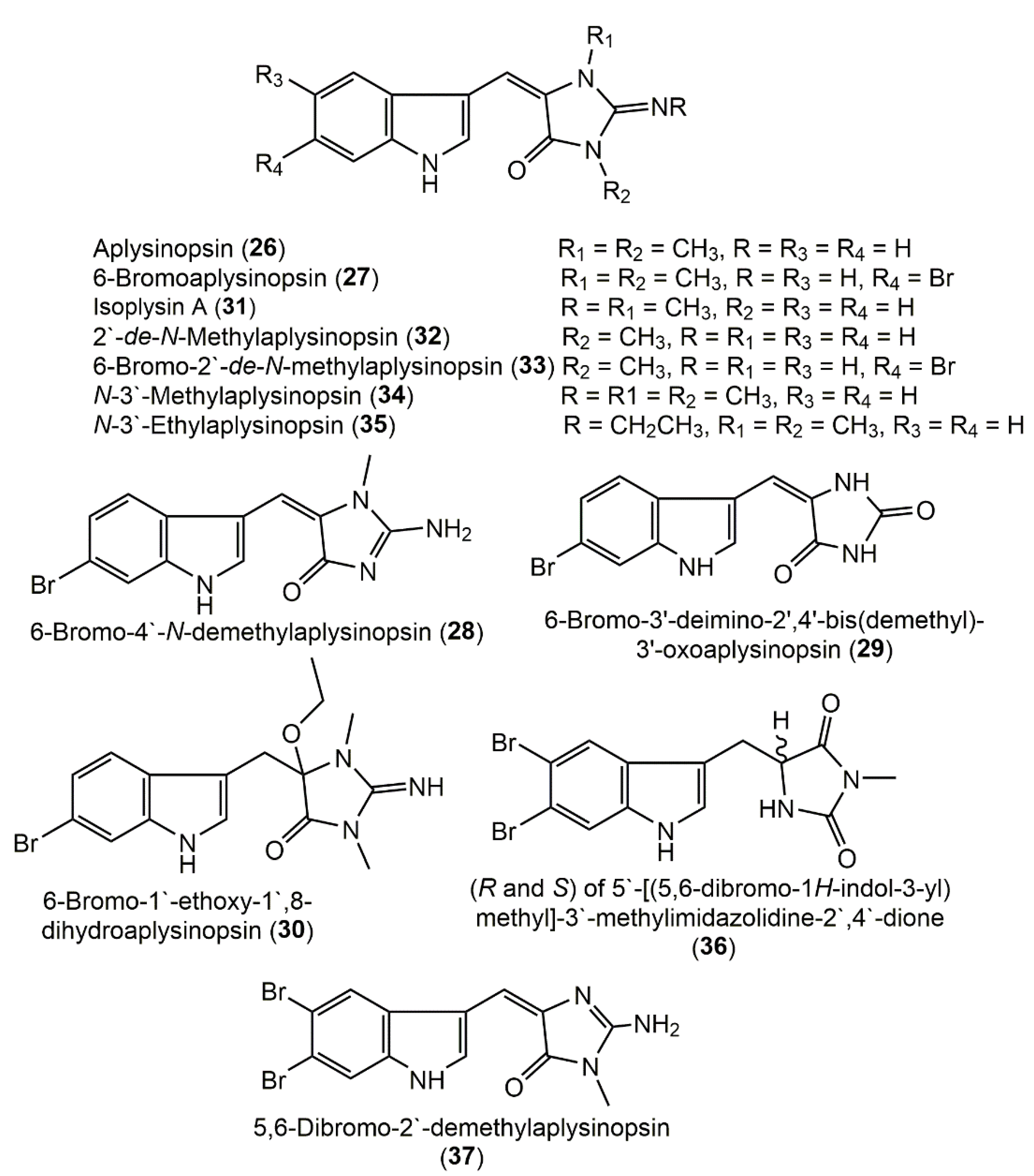
Two new aplysinopsin derivatives, 6-bromo-aplysinopsin (27) and 6-bromo-4′-N-demethylaplysinopsin (28), were purified from the Caribbean S. aurea extract (toluene: MeOH 1:3) using SiO2 CC and crystallization from MeOH: H2O and elucidated using NMR, MS, and chemical synthesis (Supplementary Materials Table S3) [16]. In addition, aplysinopsins 2735 separated from S. aurea were assessed for their in vitro antimycobacterial and antimalarial activity, as well as human 5-HT2 receptor binding antagonists in FP and FRET assays [18] (Figure 3).
5-HT2C organizes food intake in mammals, whereas the 5-HT2A receptor displays a role in depression’s pathophysiology. Hence, these receptors’ modulators could be potential antiobesity and antidepressant agents, respectively. Only 27 exhibited antimalarial activity at endpoint 0.34 µg/mL with a selectivity index of 14, whereas 31 and 33 were moderately active (conc. 0.97 and 1.1 µg/mL, respectively, with selectivity indexes of >4.9 and >4.3, respectively). Compounds 27, 33, and 35 displaced high-affinity [3H] antagonist binding from cloned 5-HT2C receptors, whereas 27 and 35 also displaced high-affinity [3H] antagonist binding from the 5-HT2A receptor subtype, and 33 had only partial displacement at the highest concentration at the 5-HT2A subtype. None of the others (31, 33, and 34) had displacement effectiveness at the highest concentration tested. Among the tested metabolites, 27 had the highest overall affinity, which was like that of endogenous serotonin at the 5-HT2C receptor subtype, while 33 and 35 had a 5- to 27-fold lower affinity than serotonin. The structure–activity study revealed the role of functional groups at C-6, C-2′, and C-3′, in the binding to 5-HT2 human serotonin receptors. The C-3′ alkyl chain length displayed a substantial role in binding to the 5-HT receptors, and the presence of the ethyl group (as in 35) enhanced the binding potential compared with the methyl group (as in 34). Additionally, the Br atom at C-6 in the absence of C-3′-ethyl increased the binding affinity (as in 27 and 33 versus 32) and selectivity to the 5-HT2C receptor subtype over the 5-HT2A receptor subtype. Additionally, C-2′-methylation facilitated the binding to the 5-HT2A receptor subtype (as in 27 versus 33) [20]. Chemical investigation of Smenospongia sp. CH2Cl2 fraction by SiO2 CC and HPLC yielded brominated alkaloids derivatives 26 and 30 that revealed antibacterial capacity (MICs 25 and 12.5 µg/mL) versus S. epidermidis ATCC12228 compared with vancomycin (MIC 0.625 µg/mL) in the microbroth dilution assay [22].

3.3. Bisspiroimidazolidinone Alkaloids

Imidazolidinones are a class of heterocyclic metabolites characterized by a nitrogen-containing five-membered ring that have received remarkable attention from medicinal chemists, as many derivatives of this scaffold class possessed variable bioactivities and are used as chiral catalysts by organic chemists [24].
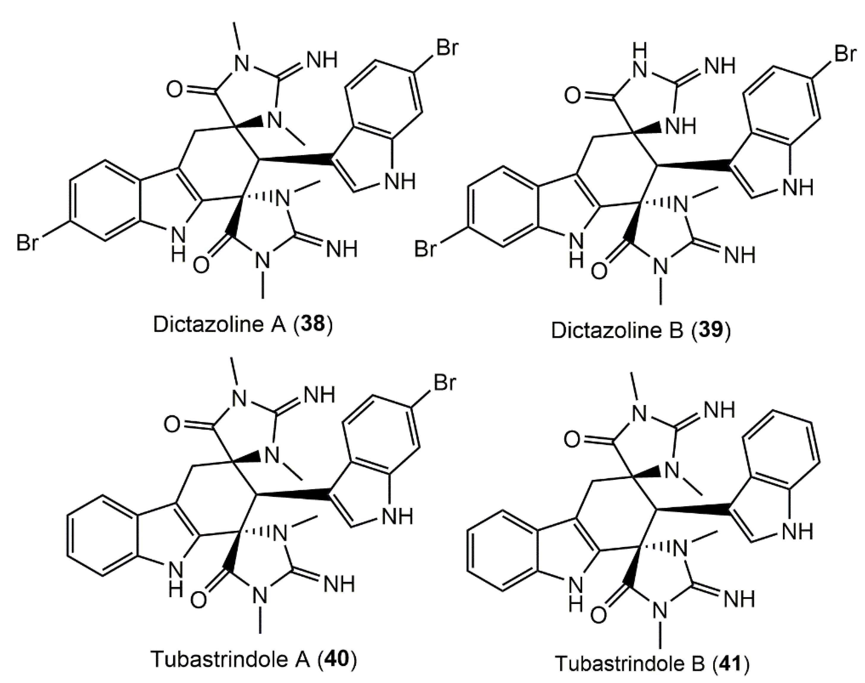
Two new bisspiroimidazolidinone alkaloids, dictazolines A (38) and B (39), along with related metabolites tubastrindoles A (40) and B (41) were obtained from S. cerebriformis using Sephadex LH20/SiO2 flash column and HPLC and elucidated by NMR analyses (Supplementary Materials Table S4). Only 40 and 41 were evaluated for the inhibition of the serine and threonine kinase PKC. Initially, 41 displayed weak inhibition of PKCδ, whereas 40 and 41 were inactive at reducing the β-secretase-proteolytic cleavage of the amyloid precursor protein, an assay of Alzheimer’s disease treatment [25] (Figure 4).

3.4. Polyketides

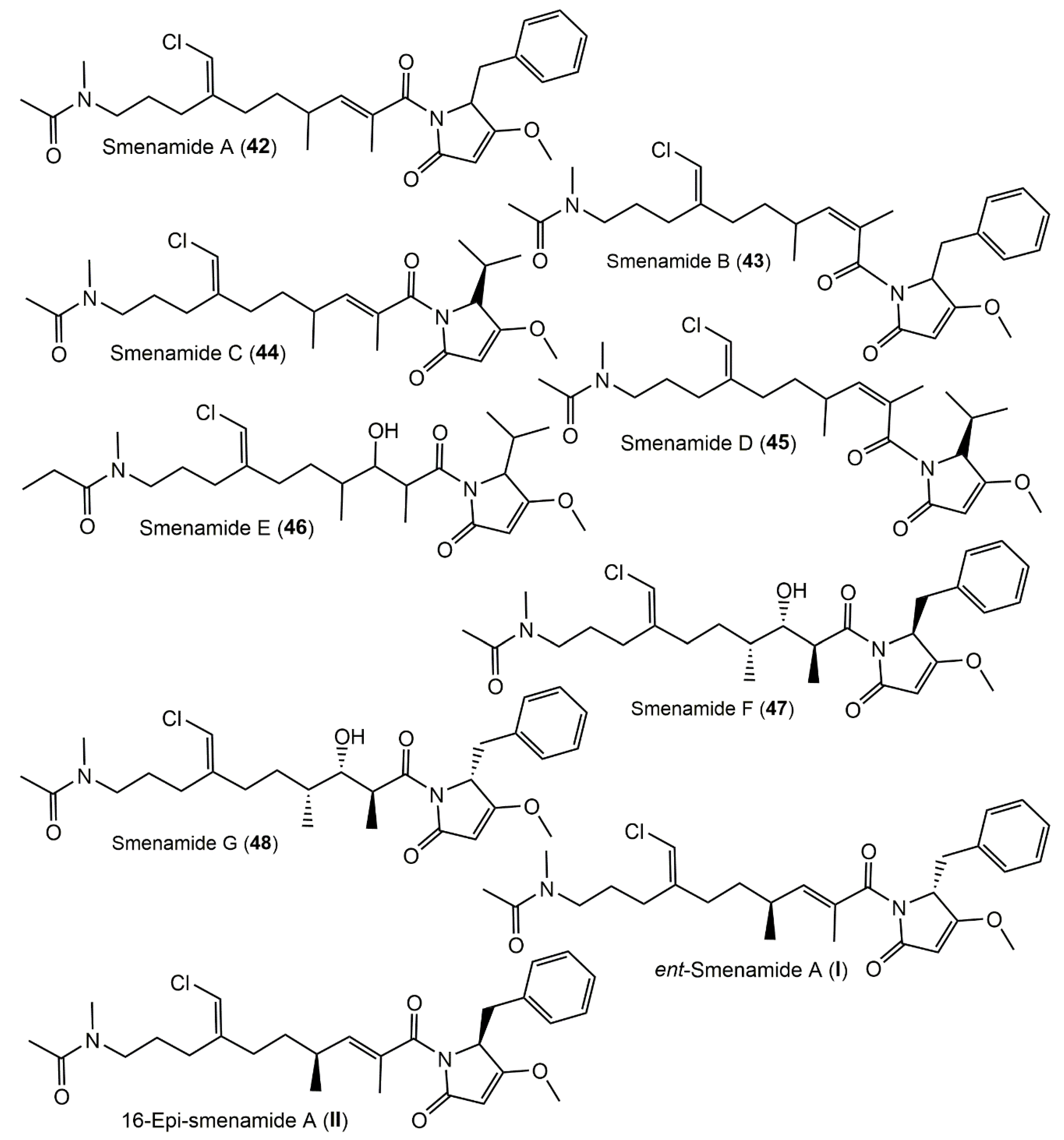
Smenamides A (42) and B (43) are hybrid polyketide–peptide metabolites having dolapyrrolidinone, N-methylacetamide, and chlorovinyl moieties that were separated from Caribbean S. aurea and assigned using HRESIMS/MS and NMR experiments. They demonstrated no notable cytotoxic potential (conc. 30 nM) on Calu-1 in the MTT assay after 27 h of treatment; however, they (conc. 50 nM) had remarkable effectiveness (IC50 48 nM for 42 and 49 nM for 43) (Figure 5). Compound 42 exerted its powerful cytotoxic potential via a proapoptotic mechanism with increasing apoptotic cells and no or little necrotic cells, whereas in 43, the apoptotic cells’ percentage was much less, and the necrotic cells’ percentage was high (47%, conc. 100 nM) in the Annexin-V PI/FITC kit assays. These different mechanisms could be attributed to the difference in the C13–C15 double-bond configuration.
These metabolites could be promising leads for designing antitumor agents [26]. Caso et al. reported the synthesis of ent-smenamide (I) and 16-epi-smenamide A (II) and established smenamide A’s 16-R configuration. The carboxylic acid moiety was created starting from S-citronellene via a Grignard process and two Wittig reactions. Furthermore, the Andrus protocol was used for carboxylic acid moiety coupling with either (R)- or (S)-dolapyrrolidinone [27]. In 2018, Caso et al. attempted to study if the C-16 configuration influenced the bioactivities. They synthesized 16-epi-smenamide A and eight analogs of the 16-epi-series that were tested for antiproliferative capacity versus MM (multiple myeloma) cell lines. It was found that the C-16 configuration had a slight influence on the activity since the 16-epi-analogs were active at nanomolar concentrations [28]. Interestingly, 44 and 46 revealed moderate neurotoxicity versus neuro-2A cells, whereas 45, the geometric isomer of 44, had no potential [29]. Using a molecular-networking dereplication strategy, two new members of smenamides, 47 and 48, were separated through Rp-HPLC separations and elucidated by NMR, ECD, and Marfey’s analyses (Supplementary Materials Table S5). Compound 47 was a hydrated analog of 42 with 8S/13S/15S/16R/20Z-configuration [30], whereas 48 is a C-8-epimer of 47 having 8S/13S/15S/16R/20Z-configuration. Their cytotoxic potential versus MCF-7, MDA-MB-231, and MG-63 was estimated in the xCELLIgence assays. It is noteworthy that they exerted (conc. 5 μM) a moderate selective antiproliferative capacity on MDA-MB-231 and MCF-7; however, they had no effectiveness versus MG-63. It was found that the dolapyrrolinone C-8 absolute configuration did not affect the activity [30]. From the same Caribbean S. aurea using RP-18 CC and HPLC, smenothiazoles A (49) and B (50), which are hybrid peptide–polyketide compounds, were separated; these are biogenetically related and structurally vary from smenamides (Figure 6).
They were tested versus Calu-1, LC31, A2780, and MCF7 in the MTT assay. These metabolites displayed a potent cytotoxic potential at low nanomolar concentrations with selectivity versus ovarian cancer cells. They showed a comparable effect on LC31, Calu-1, and A2780 cell lines and a lower effect on MCF7 in the MTT assay, whereas in Annexin-V FITC/PI assays, smenothiazoles showed a strong apoptotic effect on A2780 (ovarian cancer cells) that is accompanied with an S phase decrease resulting in blocking the cellular cell-cycle G0G1 phase [31].
Four chlorinated polyketides, smenolactones A–D (5154), and trichophycin B (55) were separated from S. aurea and characterized by NMR, ECD, and Mosher analyses. The cytotoxic capacity of 51 and 5355 was evaluated versus MCF-7, PANC-1, and BxPC-3 tumor cell lines using the MTS assay and an xCELLigence System Real-Time Cell Analyzer (RTCA). They displayed cytotoxic activity at low- or submicromolar concentrations [32]. Smenolactones were found (conc. 250 and 500 nM and 1 and 2 µM for 48 h) to reduce the cell viability and increase the tumor cell doubling time, revealing their antiproliferative potential on MCF-7 cells. Notably, 53 (conc. 1 and 2 μM) was the most potent one (IC50 918 and 652 nM, respectively); however, 51 and 54 (conc. 2 μM) caused less notable MCF-7 growth inhibition. For in vitro antiproliferative selectivity assessment, PANC-1 experienced induced or unaffected proliferation with 51 and 54 (conc. 500 nM and 1 μM, 48 h), while BXPC-3 displayed a delayed growth at 1 μM. The results demonstrated that the polyketide chain length and flexibility influence the activity as in 53, which had no double bonds, and a longer chain was the most bioactive. Furthermore, lactone moiety C-5R configuration led to strong activity as in 51 versus 54 with C-5-S stereochemistry that had a weaker inhibition, whereas chlorovinylidene configuration had no dramatic influence on the activity [32].
Phytochemical investigation of the Caribbean S. conulosa led to the separation of two new chlorinated thiazole-involving metabolites, conulothiazoles A (56) and B (57), along with 42, 43, 49, and 50 that were elucidated by NMR and MS analyses, and their absolute configuration was assigned by chemical degradation and Marfy’s and HRLCM analyses [33]. These metabolites possessed molecular features of several cyanobacterial metabolites, including a terminal thiazole ring as in barbamide and middle vinyl chloride as in jamaicamides [33].

3.5. Terpenoids

Terpenoids of variable carbon skeletons have been reported from this genus, including sesqui-, sester-, and diterpenoids (Supplementary Materials Table S6). These metabolites along with their reported bioactivities and synthesis were summarized here.

3.5.1. Hydroquinone and Quinone Sesquiterpenoids

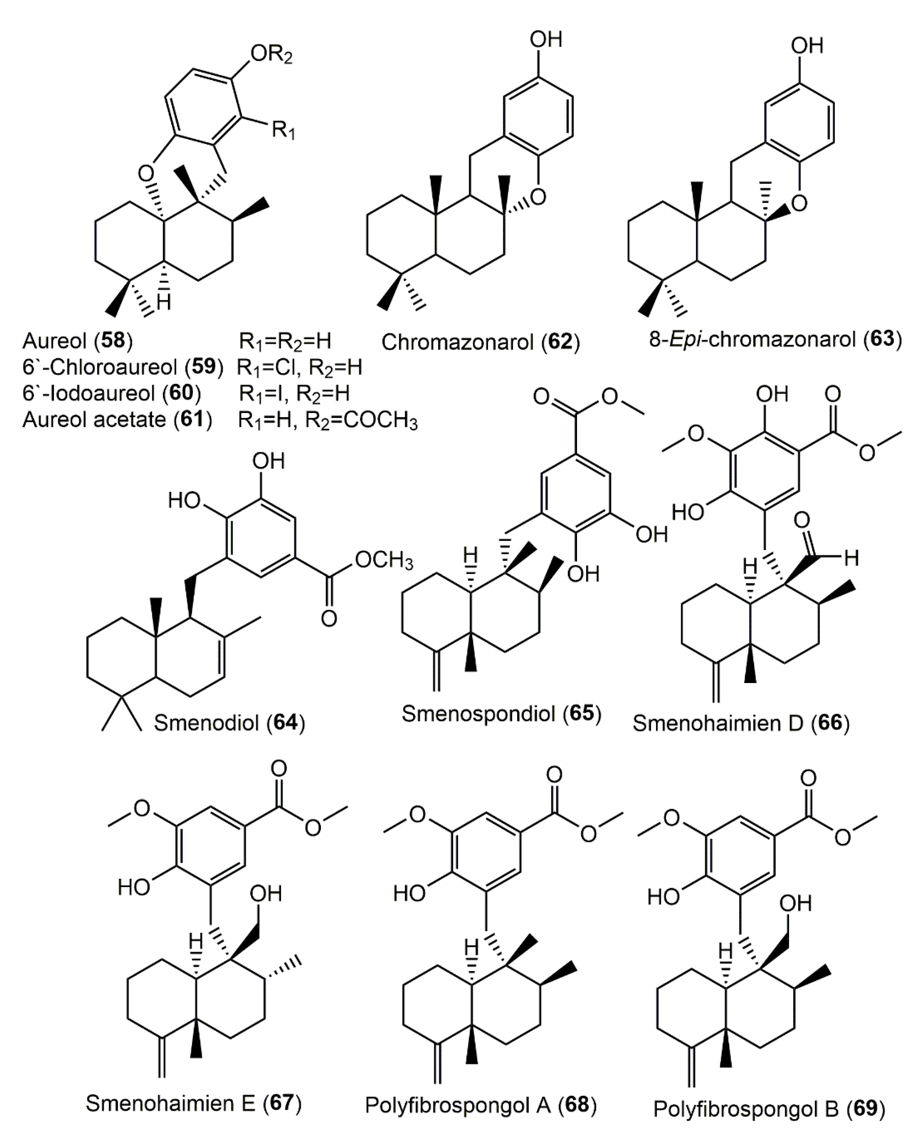
Compounds 58 and 63 were purified from the less-polar fraction of S. aurea and characterized by NMR and chemical interconversion. The 5S/8S/9R/10S configuration of 58 was established based on X-ray analysis [15]. In addition, the EtOH extract of the Jamaican S. aurea afforded sesquiterpenes 58, 59, and 61 using SiO2 and Sephadex LH-20 (Figure 7).
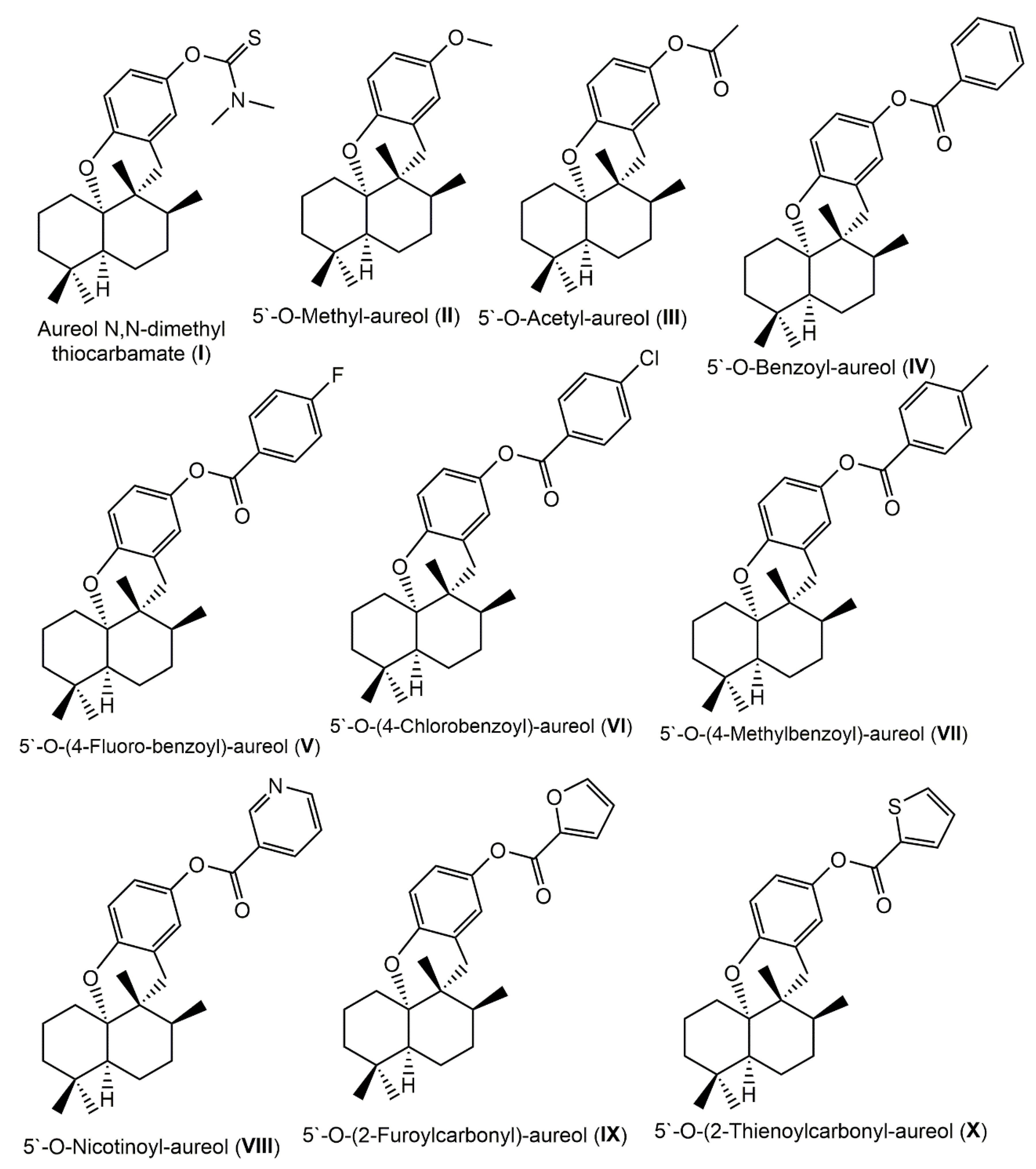
Additionally, two derivatives, aureol N,N-dimethyl thiocarbamate (I) and 5′-O-methyl-aureol (II), were semisynthesized from aureol (58) (Figure 8). Aureol N,N-dimethyl thiocarbamate (I) exhibited in vitro antimalarial potential versus the P. falciparum D6 clone with a selectivity index of 55, whereas it had no in vivo activity. Compounds I and 58 (MIC < 6.25 and >6.25 µg/mL with 100% and 31% inhibition, respectively) displayed in vitro potential versus M. tuberculosis H37Rv, indicating that thiocarbamate moiety highly improved the antimycobacterial activity [18]. Moreover, the EtOAc extract of S. aurea collected from the San Salvador Island coasts afforded 58, 59, and 61. Compounds 58 and 59 were assessed for their antibacterial potential versus B. subtilis 6633ATCC, B. cereus var. microides 213PCI, Staph. aureus 6538ATCC, S. epidermidis 12228ATCC, S. subflava 7468ATCC, S. faecalis, E. coli, and S. typhi 19430ATCC. They showed antibacterial capacity with MICs ranging from 0.0008 to 0.125 for 58 and 0.5 to 0.008 mg/mL for 59 toward the tested strain [34].
Additionally, aureol (58) was separated by Shen et al. from the Smenospongia sp. nonpolar extract [36]. Its methylation yielded II, whereas the acylation using different acid chlorides or acetic anhydride produced I and IIIX (Figure 8) [36]. These derivatives were evaluated in vitro for cytotoxic capacities versus VGH/Hepa59T, KB, and Hela cell lines in the MTT assay. Compound 58 had cytotoxic effectiveness (IC50 values of 5.77, 4.94, and 7.65 µg/mL, respectively) versus the three tested cancer cell lines compared with mitomycin (IC50 0.1, 0.1, and 0.11 µg/mL, respectively). Derivatives I, III, IX, and X revealed a cytotoxic potential (IC50s ranging from 3.08 to 10.93 µg/mL), where X (IC50 3.08, 3.73, and 4.15 µg/mL, respectively) was the most potent compound. The inactivity of II and IVVIII indicated that 5-OHmethylation of aureol or its acylation with benzoyl, nicotinoyl, or other derivatives abolished the activity [36]. Additionally, Wang et al. reported a concise synthesis of 58 in six steps, including a BF3 (boron trifluoride)-catalyzed 1,2-methyl and domino 1,2-H shifts and a Ni+2 (nickel II)-catalyzed cross-coupling between an aryl Grignard reagent and alkyl iodide as key steps [37]. From the EtOAc-soluble fraction of Smenospongia sp. using SiO2 and HPLC, 5862 were separated. Compound 60 represented the first report of iodo-sesquiterpene hydroquinone. Only 58 had moderate and weak cytotoxic (IC50 14.6 and 76.4 μM, respectively) potential versus LH-60 and A549, respectively [14].
Smenospongine (82) is a drimane sesquiterpene-aminoquinone that was isolated as red crystals from the Smenospongia sp. CH2Cl2 fraction of the MeOH extract using SiO2 and Sephadex LH20 and characterized using NMR and CD analyses. It displayed cytotoxic potential versus L1210 (leukemia cells, LD50 1.0 μg/mL) by prohibiting DNA synthesis and antibacterial capacity versus Staph. aureus (MIC < 5 μg/mL) and P. aeruginosa (strains: PYO9 and 8203S, MIC 25 μg/mL), but weak activity against E. coli (MIC 70 μg/mL) [38].
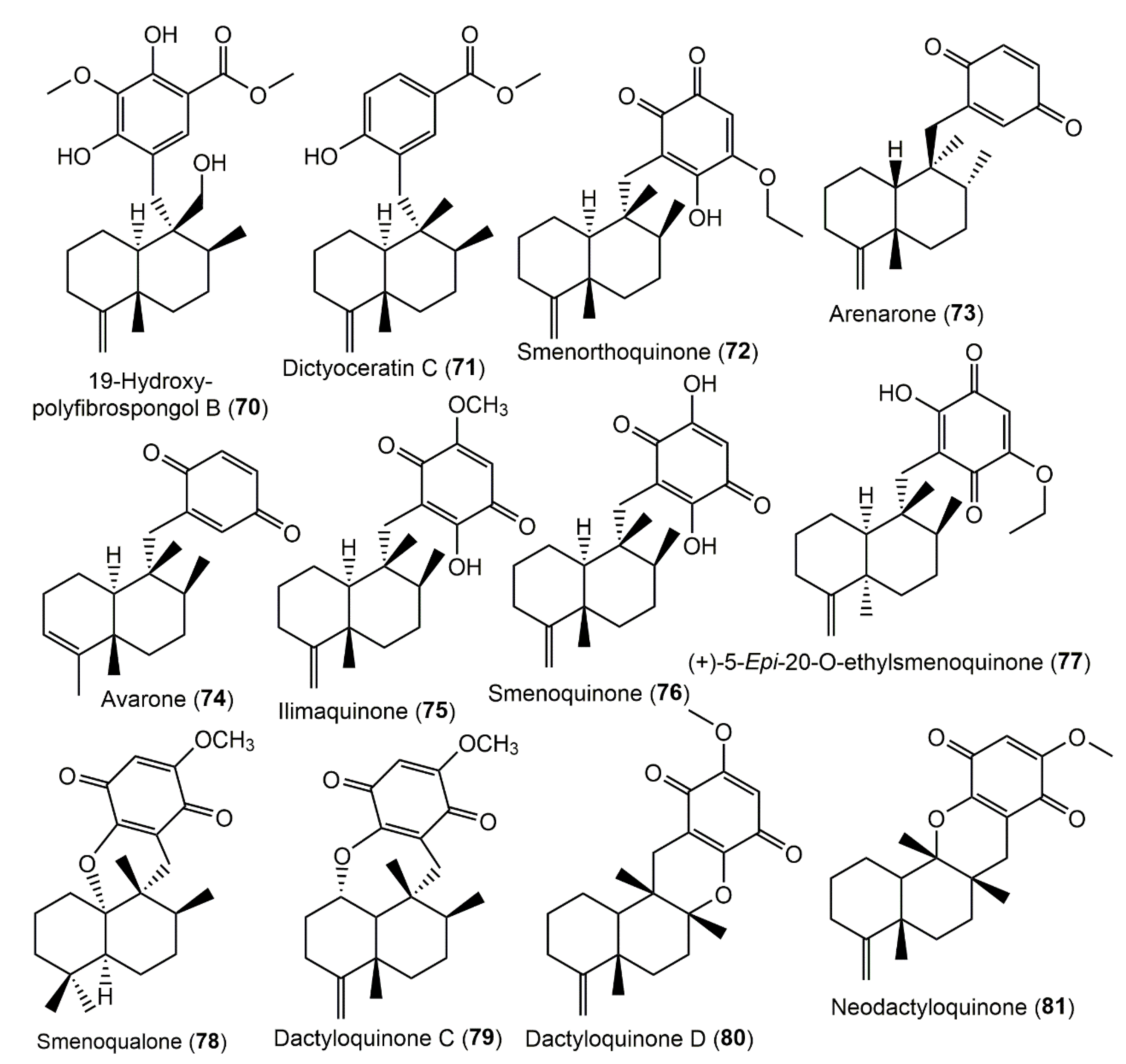
Various sesquiterpenoids, 65, 7276, 82, and 8486, were separated from the Snenospongia sp. obtained from the Gulf of Aden, Red Sea, near Djibouti. The CH2Cl2 fraction utilizing SiO2/Sephadex LH-20 CC and preparative TLC was established by various spectral analyses (Figure 9).
Compounds 65, 72, 75, and 82 revealed a significant potential versus Staph. aureus (MICs 2, 10, 25, and 7 μg/mL respectively); additionally, 82 (MIC 25 μg/mL) had a potential on antibiotic-resistant P. morganii [38]. Furthermore, 72, 76, and 82 were the most powerful compounds (LD50 1.5, 2.5, and 1.5 μg/mL, respectively), whereas 65, 75, and 85 had an LD50 of 4 μg/mL versus L1210 cells by assessing the inhibition of DNA synthesis estimated by 3H thymidine incorporation [39].
From Snenospongia sp., smenoqualone (78) was purified, which is structurally related to strongylin A separated from Strongylophora hartmani [40]. It had no antimicrobial and cytotoxic potential, suggesting that the free OH group on the quinone ring is substantial for the activity [41].
Do et al. reported that the 75 pretreatment remarkably boosted TRAIL-produced apoptosis in HCT-116 cells and stimulated TRAIL-caused apoptosis on colon cancer cells via increased caspase-8 and -3 activation, DNA damage, and PARP cleavage. It also lessened Bcl-xL and Bcl2 cell survival proteins, while it strongly upregulated death receptors’ DR4 and DR5 expression through the upregulation of CCAAT/CHOP (enhancer-binding protein homologous protein). The DR4, CHOP, and DR5 expression upregulation had occurred through the activation of ERK (extracellular-signal-regulated kinase) and p38 MAPK (mitogen-activated protein kinase) signaling pathways, as well as ROS generation. Therefore, 75 boosted the human colon cancer cells’ sensitivity to TRAIL-caused apoptosis via the ERK-ROS/CHOP-p38 MAPK–mediated upregulation of DR4 and DR5 expression, indicating that 75 could be developed into a chemotherapeutic agent [42].
Aerobic glycolysis is preferred more in cancer cells than the oxidative phosphorylation for ATP production. In different cancers, the upregulation of PDK1 (pyruvate dehydrogenase kinase 1) minimizes the PDH (pyruvate dehydrogenase) activity via the induction of its E1α subunit (PDHA1) phosphorylation and, subsequently, turns the energy metabolism from oxidative phosphorylation to aerobic glycolysis [43]. Therefore, PDK1 is regarded as a target for anticancer therapy. Compound 75 decreased the viability of A549, DLD-1, RKO, HEK293T, Detroit-551, and LLC cells (GI50 10.5, 8.61, 50.16, 37.30, and >100 µM respectively) in the MTT sassy. It reduced the PDHA1 phosphorylation in the A549 cells by suppressing the PDK activity. It also increased oxygen consumption and decreased secretory lactate levels. Thus, it increased the oxidative phosphorylation and PDH activity while subsequently reducing cell viability via the suppression of the PDK activity. It could be a candidate for anticancer agents that acted via the PDK1 activity inhibition [44].
nAMD (neovascular age-related macular degeneration) is a common reason for irreversible vision loss in the elderly. Son et al. stated that 75 topical and oral administrations in mice and rabbits caused the inhibition and regression of laser-induced CNV (choroidal neovascularization) by β-catenin downregulation in RPE (retinal pigment epithelial) cells ((hTERT-RPE1 and ARPE-19), and it prohibited p53-mediated apoptosis induction in HUVECs (human umbilical vein endothelial cells). It repressed the expression of inflammatory and angiogenic factors and restored the E-cadherin expression in RPE cells by prohibiting the Wnt–β-catenin pathway. Therefore, it functioned through the p53–Wnt–β-catenin pathway regulation with advantages over the available cytokine-targeted anti-angiogenic therapies. It has a unique mechanism by suppressing the expression of proinflammatory and angiogenic factors and prohibiting the growth of vascular endothelial cells; it could be administered more safely, cost-effectively, and conveniently in the form of eye drops or oral drugs to patients than the currently used intravitreal drugs [45].
From S. aurea and S. cerebriformis77, 8790, and 9799 were purified and assigned based on various NMR data (Figure 10). Compounds 89 and 90 have an unprecedented 2,2-dimethylbenzo[d]oxazol-6(2H)-one moiety. GIAO (gauge-invariant atomic orbital) NMR chemical shift calculations along with the application of CP3 and DP4 advanced statistics were utilized for stereochemistry determination.
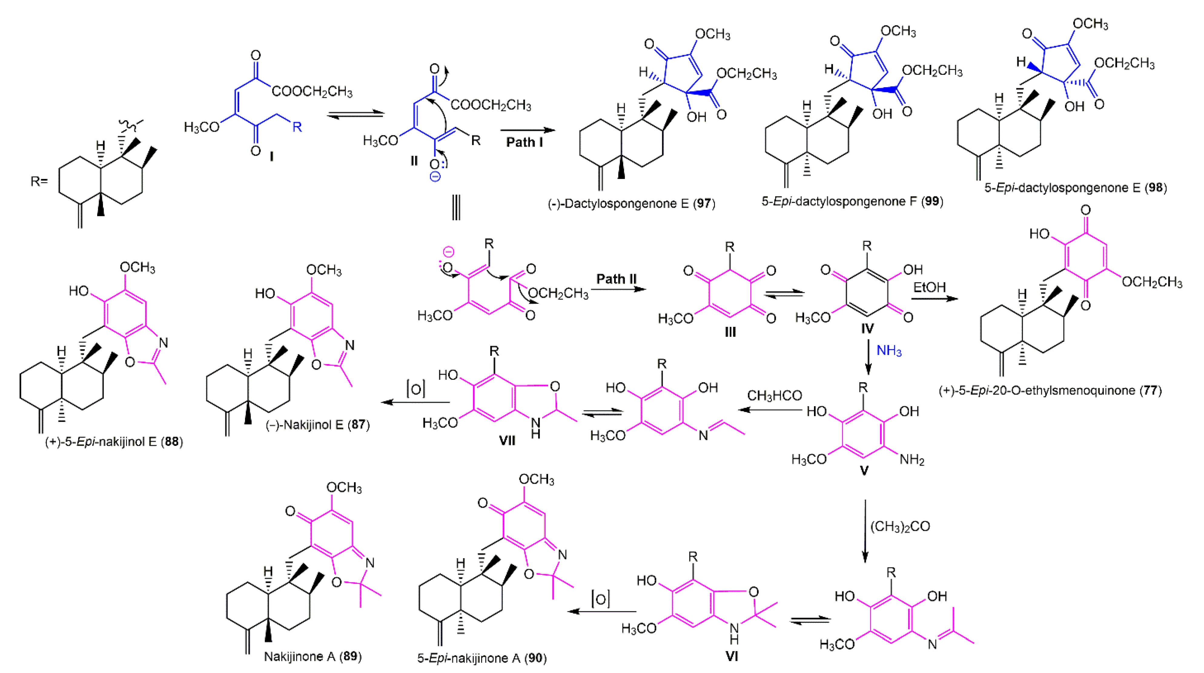
The downregulation of β-catenin expression has been considered a promising approach for cytotoxic drug formulation. Compounds 77 and 88 and the mixture of 89 and 90 suppressed CRT (β-catenin response transcription) via degrading β-catenin and exhibited cytotoxic potential versus colon cancer cells through their anti-CRT potential using a CellTiter-Glo assay kit [46]. Compounds 88 and 89/90 mixture started to produce antiproliferative potential (conc. > 20 μM), whereas 77 began to inhibit tumor growth (conc. 1.5 and 0.75 μM) versus HCT-116 cells (IC50 2.95 μM) and SW4 (IC50 3.24 μM), revealing that 77 had more potent cytotoxic potential than the other compounds [46]. It was reported that a competitive intramolecular Michael addition might be involved in these metabolites’ formation. The intramolecular addition of enolate II onto the C-20 carbonyl would result in the generation of 97 and 99 or 98, relying on si- or re-face addition (path I). Alternatively, the addition of enolate onto the C-21 carbonyl group and tautomerization followed by trans-etherification would result in 77 formation (path II). Additionally, reductive amination of IV followed by Schiff’s base formation with acetone or acetaldehyde and consecutive isomerization generates VI and VII, respectively (Scheme 1). Lastly, the oxidation of these two intermediates leads to the formation of benzoxazole moieties in 8790 [46].
New sesquiterpenes, 66, 67, 91, 100, and 101, along with 6870, 71, and 75 were separated from S. cerebriformis and assigned using NMR, MS, and ECD spectra. Compounds 100 and 101 featured cyclopentenone and 4,9-friedodrim-4(11)-ene sesquiterpene skeleton with 16R/20R and 16S/20R, respectively. Compound 91 had C-15 benzoxazole moiety instead of cyclopentenone in 100 and 101; however, 67 was structurally similar to 70 and 69 with a different C-8-configuration. Their inhibitory potential versus NO production in BV2 microglia cells stimulated with LPS in the immune-fluorescence assay using the Griess reaction was estimated [47]. Compounds 67, 69, 70, 75, 100, and 101 (conc. 10, 20, and 40 µM) noticeably prohibited (IC50 ranged from 10.40 to 30.43 µM) LPS-induced NO production in BV2 cells, whereas 75 had a significant activity (IC50 10.40 µM) compared with L-NMMA (IC50 22.1 µM). The structure–activity study suggested that the C-14-OH group had an important role in the NO production inhibition. Thus, 75 could be a marked anti-inflammation constituent of S. cerebriformis [47].
Smenohaimien F (83), a new sesquiterpene with dialkoxy-1,4-benzoquinone moiety, in addition to 7981 were reported from S. cerebriformis collected from Vietnam. Compound 80 showed significant cytotoxic activities on LU-1, HL-60, SK-Mel-2, HepG-2, and MCF-7 (IC50 ranging from 0.7 to 1.6 µg/mL) compared with ellipticine (IC50 0.4 to 0.6 µg/mL), while 79 and 83 (IC50 ranging from 10.0 to 63.1 µg/mL) were moderately active and 81 had a weak potential in the MTT assay [48].

3.5.2. Tetronic Acid and Cyclopentenone Sesquiterpenoids

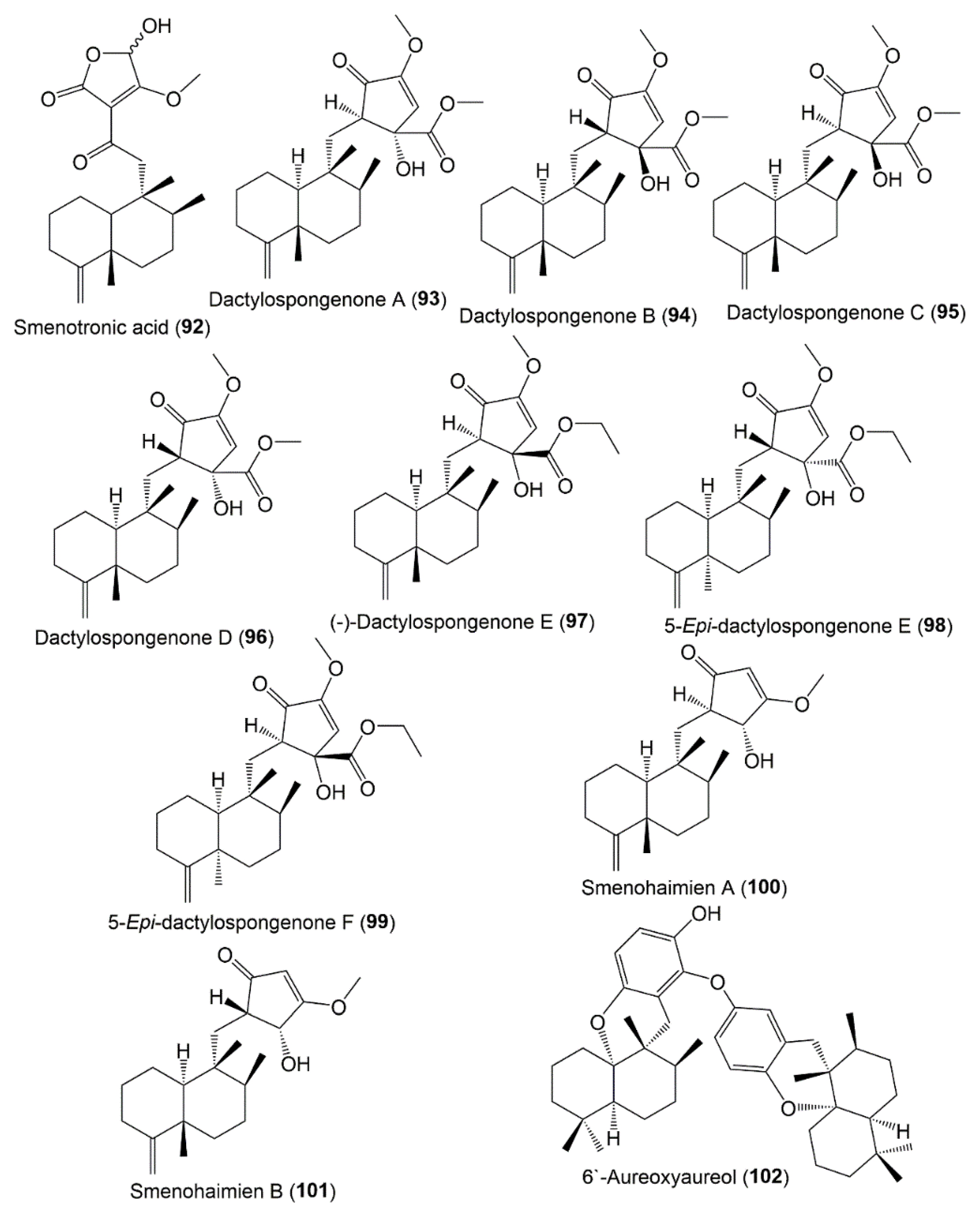
In 1999, Bourguet-Kondracki and Guyot reported the characterization of smenotronic acid (92) from Smenospongia sp. collected from the Red Sea using NMR, IR, and UV tools [49]. It featured a rearranged drimane core with tetronic acid moiety. During storage in C5D5N, the epimerization of this compound took place to produce a mixture (1:1) of two C19 epimers that are the isomers of a dactyltronic acid, the previously reported Dactylospongia elegans [50]. Dactylospongenones A–D (9396) were isolated from the S. cerebriformis CH2Cl2 fraction. They are sesquiterpene cyclopentenones that were formerly separated from D. elegans [2] (Figure 11). They were assigned by NMR analyses, and cyclopentenone’s C-16 and C-17 configuration was assigned by X-ray and CD analyses as 16R/17S, 16S/17R, 16R/17R, and 16S/17S, respectively. In the MTT assay, they revealed mild or no effectiveness against LU-1, HepG-2, HL-60, MCF-7, and SK-Mel-2 [51].

3.5.3. Diterpenoids

Compounds 103 and 104 are diterpenoids reported from Vietnamese S. cerebriformis that had a weak cytotoxic potential versus LU-1, HL-60, SK-Mel-2, HepG-2, and MCF-7 in the MTT assay [48].

3.5.4. Sesterterpenoids

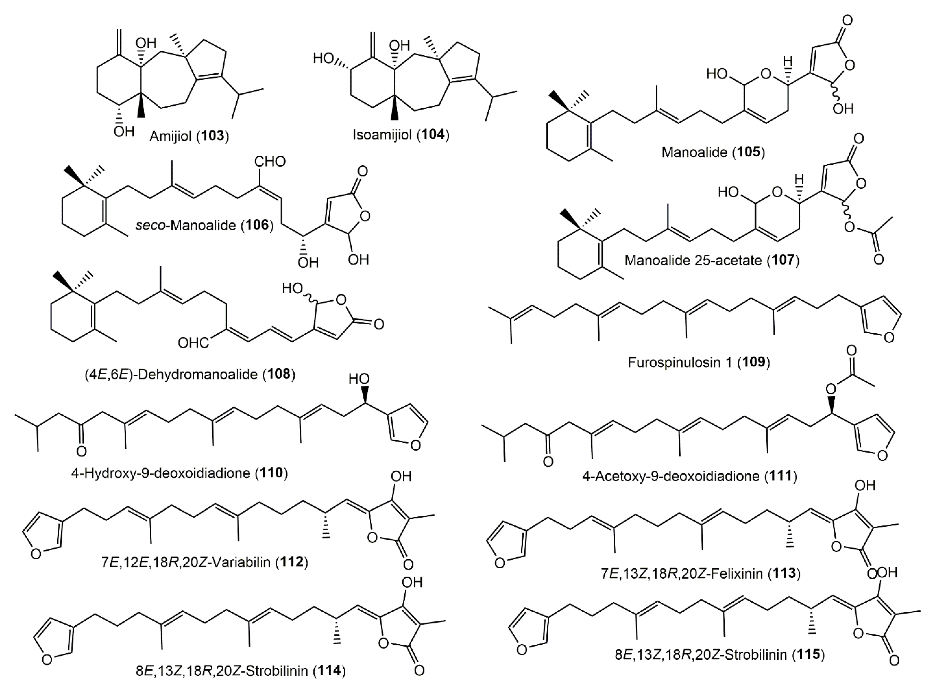
Four terpenoids 104108 were separated using SiO2/Sephadex LH-20 CC and HPLC from the EtOAc extract of Smenospongia sp. (Figure 12). The metabolites showed an antifouling potential but no toxicity against the barnacle Balamis Amphitrite cypris larvae (EC50 0.24, 0.80, 0.53, and 2.7 μg/mL, respectively) in the 24-well polystyrene plates assay [52]. Furthermore, two new linear furanosesterterpenes, 110 and 111, and three new scalaranes, 116, 117, and 120, were purified from Smenospongia sp. obtained from Gagu-Do, Korea, using RP-flash chromatography and HPLC and were characterized by NMR spectral data; 108 configuration was assigned using Mosher’s method. These metabolites revealed a cytotoxic potential (LC50 31.6, 3.0, 4.9, 11.2, and 0.02 µg/mL, respectively) versus K562 (human leukemia cell line). It is noteworthy that the changes in scalarane-sesterterpenoid functional groups led to marked differences in the activity [35].
A new scalarane sesterterpenoid, 124, and formerly reported 116, 120, 121, 126, and 128 were separated from the Korean Smenospongia sp. by reversed-phase HPLC (Figure 13).
Their activation of AMPK (5′ adenosine monophosphate-activated protein kinase) in L6 myoblast cells was tested utilizing an AMPK phosphorylation assay. Only 128 displayed a dark band that indicated the creation of phosphorylated AMPK at a concentration of 10 mM [53].
Ten new sesterterpenoids, 118, 119, 121123, and 125129, and known metabolites 110, 112117, and 120 were biosynthesized by Smenospongia sp. obtained from Korean coastal waters and assigned by spectroscopic analyses. Compounds 123 and 127 featured unprecedented 23-aldehyde and 20-carboxylic acid, respectively, among scalarane sesterterpenoids. The isolated metabolites were evaluated for their antibacterial activity toward Staph. aureus (ATCC65389), B. subtilis (ATCC6633), M. leuteus (IFC12708), P. vulgaris (ATCC3851), S. typhimurium (ATCC14028), and E. coli (ATCC25922) in the microdilution assay, and also for the cytotoxic activity in the SRB assay against the K562 cell line and the inhibitory activity against isocitrate lyase in the colorimetric assay [54]. Compounds 112115 and 129 were active versus Staph. aureus (MICs ranging from 25.0 to 6.25 μM), whereas 110, 112115, 117, 118, 120, 122, 125, 126, and 129 had a potential versus B. subtilis (MICs 0.78–6.25 μM) compared with ampicillin (MIC 1.56 μM). On the other side, 112115, 118, 125, and 129 were effective versus M. leuteus (MICs ranging from 12.5 to 3.12 μM), whereas 110 and 112115 had activity versus P. vulgaris (MICs 3.12–6.25 μM) compared with ampicillin (MIC 3.12 μM). In the cytotoxic assay, these metabolites showed potent cytotoxic effectiveness versus the K562 cell line (LC50s ranging from 0.11 to 43.7 μM) compared with doxorubicin (LC50 4.9 μM). Moreover, 112116, 119, 125, and 129 had inhibition capacity (IC50 ranged from 24.1 to 67.2 μM) toward microbial enzyme ICL (isocitrate lyase) compared with 3-nitropropinate (IC50 6.05 μM) [54].

3.6. Chromene Derivatives

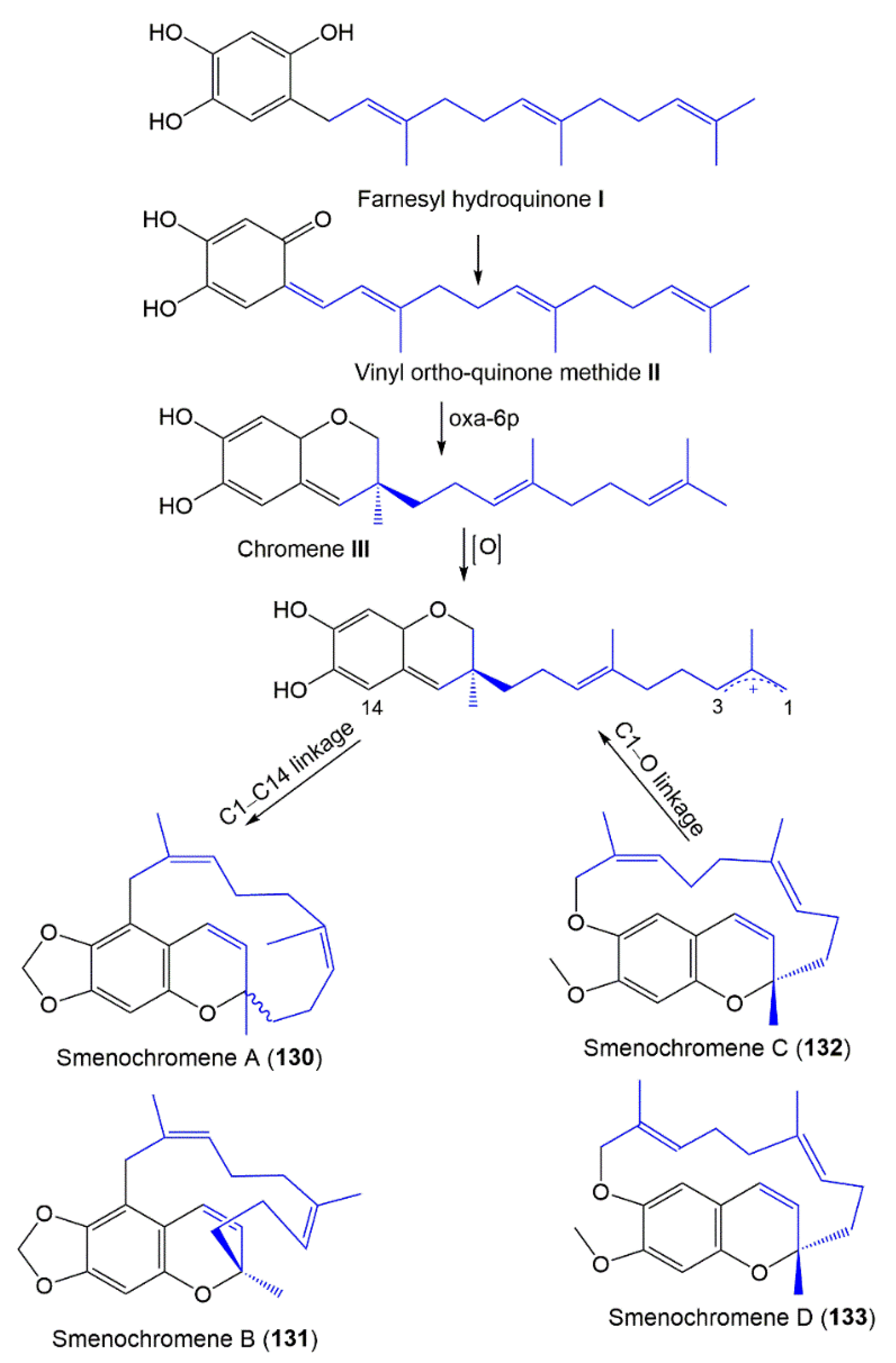
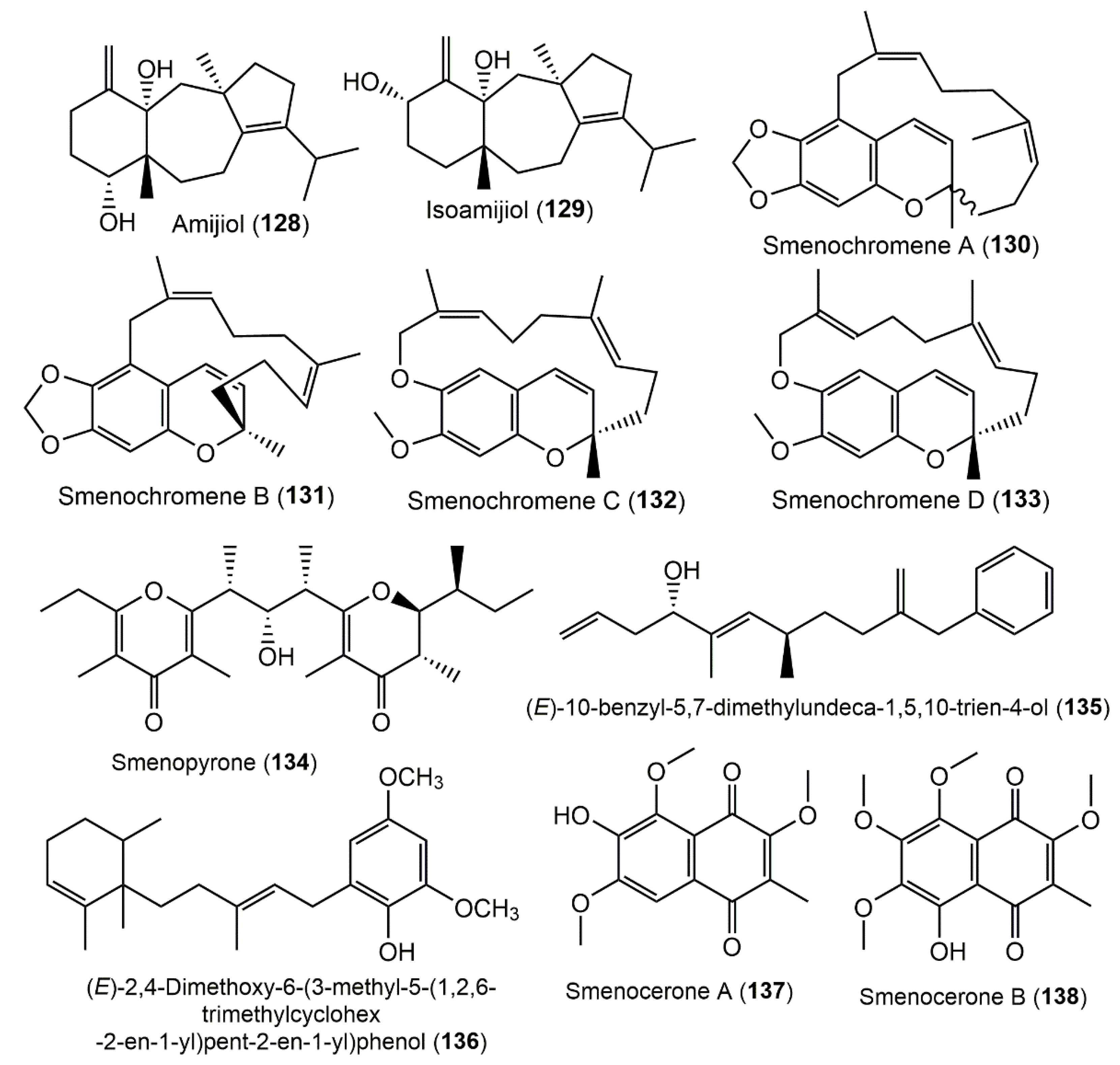
Unusual macrocyclic chromenes, smenochromenes A–D (130133), were purified from the Smenospongia sp. EtOAc fraction by Sephadex LH-20 and HPLC and characterized by NMR and X-ray tools [55] (Supplementary Materials Table S7). Compound 130 was a racemate, whereas 131133 were optically active. These metabolites possessed a chromene core fused to a 14-membered ring system that could be derived from farnesyl hydroquinone I (Scheme 2) that undergoes dehydrogenation to produce II. Subsequently, the oxa-6π electrocyclization of II affords chromene III featuring the hydroxy-chromene core. Furthermore, an allylic cation IV is formed by the terminal allylic position oxidation that undergoes various cyclizations between C1 and C14 giving the 14-membered carbocyclic 130 after that Δ6,7-double-bond isomerization produces 131. On the other side, the formation of a bond between C1 and the phenolic oxygen yields 132’s and 133′s 16-membered heterocyclic system (Scheme 2).
Rosa et al. reported the synthesis of 131 via a 133 unusual rearrangement with concomitant double-bond isomerization and ring contraction (Scheme 3) [56].

3.7. γ-Pyrone Derivatives

The Caribbean S. aurea MeOH/CHCl3 extract afforded 134 by RP-18 CC and HPLC that featured a γ-pyrone polypropionate framework. It was assigned by NMR, MS, and ECD [57]. It is noteworthy that pyrone polypropionates are uncommon in sponges; however, they are commonly encountered in marine mollusks and bacteria [58,59].

3.8. Phenyl Alkenes

From Florida sponges S. aurea and S. cerebriformi, a novel phenyl alkene, 135, with unprecedented linear phenyl alkene skeleton was separated (Supplementary Materials Table S8). Its 4R absolute configuration was established by a modified Mosher’s method. It showed in vitro cytotoxic activity versus HL-60 (IC50 8.1 µM). The molecular docking study suggested that 135 produced its cytotoxic potential through the inhibition of microtubule activity [60].

3.9. Naphthoquinones

Huyen et al. purified new naphthoquinones, smenocerones A (137) and B (138), from the CH2Cl2 fraction of S. cerebriformis obtained from the Eastern Sea of Vietnam by SiO2 and RP-18 CC (Figure 14). Compound 138 (Conc. 20 µg/mL) significantly produced over 80% death of HepG-2, LU-1, MCF-7, HL-60, and SK-Mel-2 in the MTT assay with IC50 ranging from 3.2 to 5.7 µg/mL compared with ellipticine (IC50 0.4–0.6 µg/mL) [51].

3.10. Fatty Acids, Sterols, and Phthalates

The chemical investigation of Caribbean sponge S. aurea revealed the separation of six novel branched α-hydroxy fatty acids, 139144, with an anteiso or iso terminal methyl, along with a sterols mixture consisting of 147 (61%), 149 (10%), 150 (5%), 151 (2%), and 152 (22%) [61]. They were characterized by NMR, GC-MS and retention times, and chemical degradation. Additionally, from the Smenospongia sp. CH2Cl2–MeOH extract collected from El-Gouna–Hurghada-coasts–Red Sea–Egypt, 145, 147, 148, 153, and 154 were separated by SiO2 CC that were identified by NMR and GCMS analyses [62] (Figure 15, Supplementary Materials Table S9).

4. Biological Activity of Extracts

4.1. Antimicrobial Activity

Smenospongia sp. extracts were found to exhibit noticeable antimicrobial capacity. The CH2Cl2 extract appeared the most active versus Staph. aureus and E. coli (inhibition zone diameters (IZDs) 23 and 11 mm, respectively at conc. 500 μg/disk), whereas the MeOH extract had weaker activity (IZD 20 mm/disk versus Staph. aureus), and the aqueous extract displayed no activity [39].

4.2. Cytotoxic Activity

The CH2Cl2 extract of Smenospongia sp. was cytotoxic (LD50 3 μg/mL) versus the L1210 cell line, whereas the MeOH and aqueous extracts were not cytotoxic toward L1210 leukemia cells until 8 μg/mL [38]; the MeOH, on the other hand, exhibited moderate cytotoxic (LC50 47 µg/mL versus K562 cell line) and brine shrimp lethality (LC50 160 ppm) [35].

5. Conclusions

Marine sponges are a wealth of biometabolites with chemical diversity that have been proven to be beneficial sources for novel drug target discovery. Among the marine sponges, Smenospongia sponges have been reported as a reservoir of diverse biometabolites. In the current work, Smenospongia, one of the most interesting sponge genera, was highlighted. The species of this genus have been collected from various regions (Figure 16). The bigger number of metabolites has been reported from the Smenospongia species obtained from Thailand (21 compounds), whereas the least number (4 compounds) was isolated from sponge samples that were collected from both the Milne Bay region, Papua New Guinea, and the Ninamijima and Nichinan-oshima Islands.
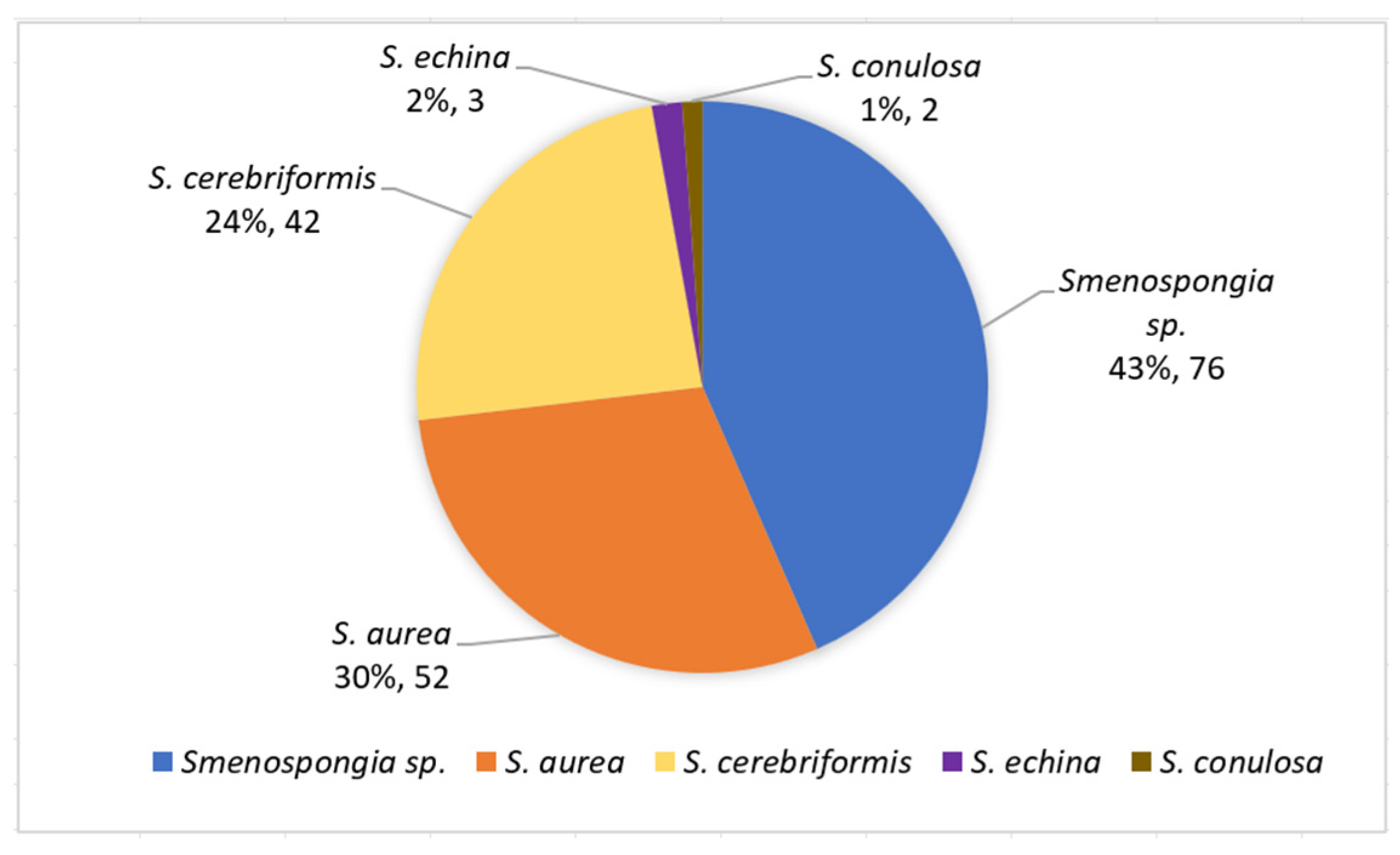
A total of 154 metabolites have been purified and characterized from various Smenospongia species over the last 42 years from four species: S. aurea (56 compounds), S. cerebriformis (42 compounds), S. echina (3 compounds), and S. conulosa (2 compounds), as well as unidentified Smenospongia species (76 compounds) (Figure 17).
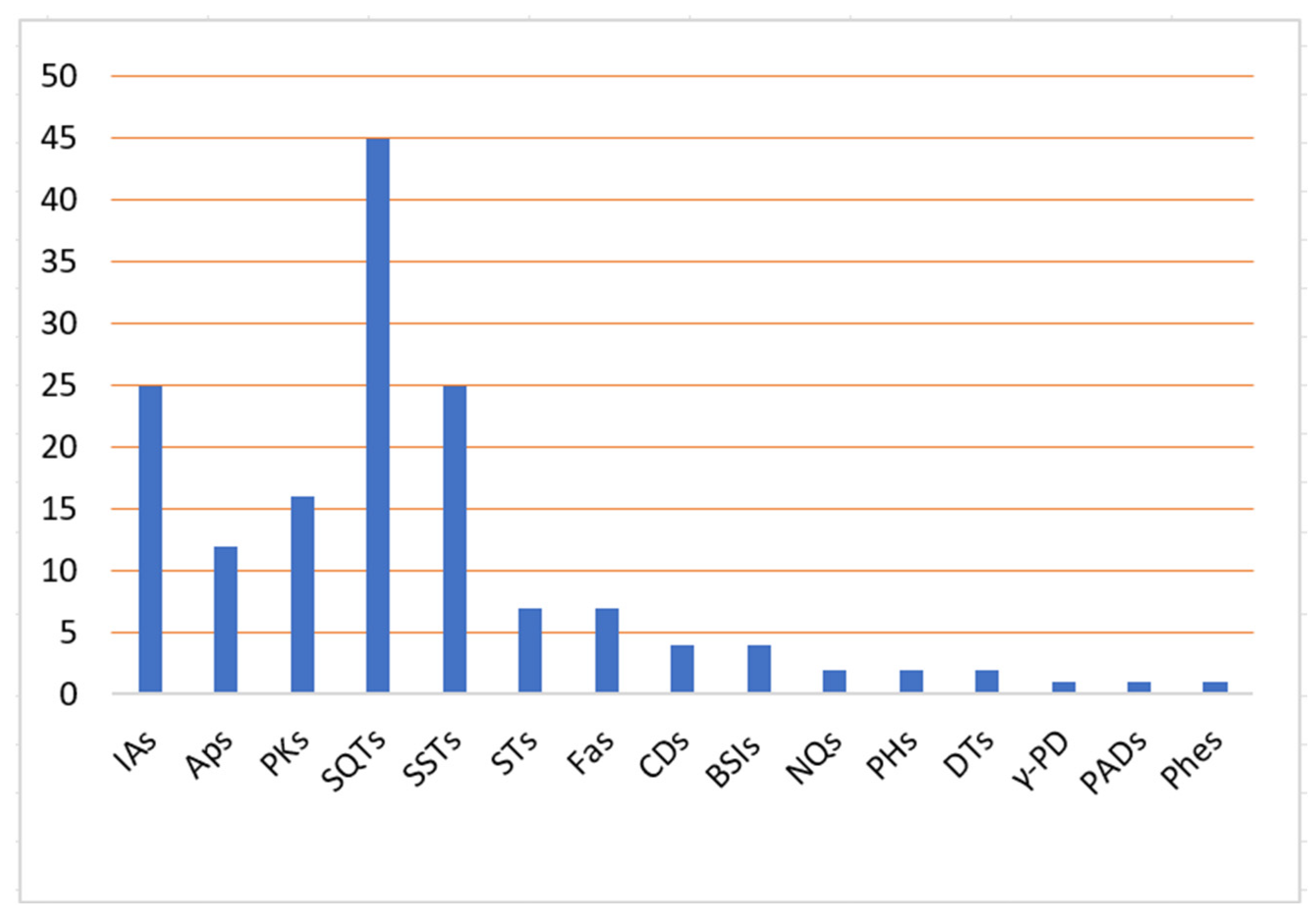
The results revealed that sesqui- and sesterterpenoids and indole derivatives are the major metabolites of this genus. Additionally, few studies reported the isolation of aplysinopsins, bisspiroimidazolidinones, chromenes, γ-pyrones, phenyl alkenes, naphthoquinones, polyketides, fatty acids, sterols, and phthalates (Figure 18).
These metabolites have been assessed for diverse bioactivities, including antimalarial, antimicrobial, cytotoxic, 5-HT2 receptor antagonistic, antifouling, and anti-inflammation. Some metabolites displayed moderated to potent cytotoxic, anti-inflammation, and antimicrobial capacities (Figure 19). Among these metabolites, 80, 117, 119, 120, 126, and 128 exhibited more potent cytotoxic capacity than standard anticancer drugs, whereas 114, 115, 117, 118, 122, 126, and 129 demonstrated marked antimicrobial activity versus some microbial strains.
Interestingly, Smenospongia genus sponges have been reported to biosynthesize chlorinated mixed polyketide–peptide compounds, including smenamides, smenothiazoles, and conulothiazoles, that shared chlorovinylidene, dolapyrrolidone ring, and terminal alkyne that are characteristics of cyanobacterial metabolites. Some of these compounds possessed potential antitumor capacity that could be promising leads for antitumor drug design. Structure–activity assessments of some reported metabolites from this genus revealed that the chemical skeletons’ nature of these metabolites and substituent patterns greatly affected the bioactivities. In addition, reported synthetic work established that the modifications of structures and replacement by some functional groups resulted in more potential and useful tags for further functionalization through click chemistry, which is a new area for drug-like molecule synthesis that can boost the drug discovery process. Unfortunately, the bioassays of some reported metabolites demonstrated no notable effectiveness, suggesting a more potential for searching and running other bioevaluations. For enriching the metabolite discovery from this genus, advanced techniques, including LC–MS–NMR, metabolomics, and UPLC–MS, could be utilized. This work aimed to be beneficial for the Smenospongia genus’s bioprospecting process and to bring attention to the chemical diversity of their metabolites. Finally, we think that the genus and its metabolites still warrant considerable research attention.

Supplementary Materials

The following supporting information can be downloaded at: https://www.mdpi.com/article/10.3390/molecules27185969/s1, Tables S1 and S3–S9: List of reported metabolites from genus Smenospongia (source and place), Table S2: Biological activity of reported metabolites from genus Smenospongia. References [14,15,16,17,18,19,20,22,25,26,30,31,32,33,34,35,37,38,40,43,45,46,47,48,50,51,52,53,54,55,57,60,61,62,63,64] are cited in the Supplementary Materials.

Author Contributions

Conceptualization, S.R.M.I., H.M.A. and G.A.M.; resources, S.A.F., H.A.F., and R.H.H.; data curation, S.A.F., H.A.F. and R.H.H.; writing—original draft preparation, S.R.M.I., H.M.A. and G.A.M.; writing—review and editing, S.A.F., H.A.F., R.H.H., S.R.M.I. and G.A.M. All authors have read and agreed to the published version of the manuscript.

Funding

This research received no external funding.

Institutional Review Board Statement

Not applicable.

Informed Consent Statement

Not applicable.

Data Availability Statement

Not applicable.

Conflicts of Interest

The authors declare no conflict of interest.

Abbreviations

A549Human lung adenocarcinoma epithelial cell line
A2780Human ovarian cancer cells line
AMPKAMP-Activated protein kinase
ARPE-19Retinal pigment epithelial cell line
ATRAdenosine triphosphate
BV2Microglia cells
BxPC-3Human pancreas adenocarcinoma cell line
Calu-1Human nonsmall-cell lung cancer cell line
CDCircular dichroism
CH2Cl2Dichloromethane
CHCl3Chloroform
CHOP, CCAATEnhancer-binding protein homologous protein
CNV Choroidal neovascularization
CRT β-catenin response transcription
DLD-1 Human colorectal cancer cell line
DR4 Death receptor 4
DR5 Death receptor 5
DPPH 1,1-Diphenyl-2-picrylhydrazyl
EC50 Half-maximal effective concentration
ERK Extracellular signal-regulated kinase
EtOH Ethanol
EtOAc Ethyl acetate
FST Porsolt forced swim test
FRET Fluorescence resonance energy transfer
FP Fluorescence polarization
GCMS Gas chromatography mass spectrometry
GIAO Gauge-invariant atomic orbital
GI50 The concentration for 50% of maximal inhibition of cell
HCT-116 Human colon cancer cell line
HEK293 Human embryonic kidney cell
HepG2 Human liver cancer cell line
Hepa59T/VGH Human liver carcinoma cell line
HeLa Human cervical epitheloid carcinoma cell line
HL-60 Human promyelocytic leukemia cell line
HPLC High-performance liquid chromatography
HT-29 Human colon cancer cell line
hTERT-RPE1 Retinal pigment epithelial cell lines
HuCCA-1 Human cholangiocarcinoma cell line
HUVECs Human umbilical vein endothelial cell line
HUVSMCs Human umbilical vein smooth muscle cells line
IC50 Half-maximal inhibitory concentration
ICL Microbial enzyme isocitrate lyase
K562 Human immortalized myelogenous leukemia cell line
KB Human oral epidermoid carcinoma cell line
L1210 Mouse lymphocytic leukemia cell line
LC31 Human lung squamous adenocarcinoma cell line
LC50 Lethal concentration 50
LC–MS–NMRLiquid chromatography–mass spectrometry–nuclear magnetic resonance
LD50 Half maximal lethal concentration
LPS Lipopolysaccharide
LLC Murine Lewis lung carcinoma
L-NMMA Nitric oxide synthase inhibitor NG-monomethyl-L-arginine
LU-1 Human lung carcinoma cell line
MCF-7 Human breast cancer cell line
MDA-MB-231 Human breast cancer cell line
MeOH Methanol
MG-63 Human osteosarcoma cell line
MIC Minimum inhibitory concentration
MOLT-3 Human T lymphoblast cell line
MTS(3-(4,5-Dimethylthiazol-2-yl)-5-(3-carboxymethoxyphenyl)-2-(4-sulfophenyl)-2H-tetrazoliuminner salt)
MTT 3-(4,5-Dimethylthiazol-2-yl)-2,5-diphenyltetrazolium bromide
nAMD Neovascular age-related macular degeneration
n-BuOH n-Butanol
NMR Nuclear magnetic resonance
NO Nitric oxide
p38 MAPK p38 Mitogen-activated protein kinase
P 388 Human leukemia cell line
PANC-1 Human pancreas ductal carcinoma cell line
PARP Poly-AD Pribose polymerase
PDK1 Pyruvate dehydrogenase kinase 1
PDHA1 Phosphorylation of its E1α subunit
RKO Human colon cancer cell line
ROS Reactive oxygen species
RTCA xCELLigence system real-time cell analyzer
RP-18 Reversed phase-18
SRB Sulforhodamine B
SiO2 CC Silica gel column chromatography
SK-MEL-2 Human melanoma cell line
SRB Sulforhodamine B
SW480 Human colorectal cancer cell line
TLC Thin layer chromatography
TRAILTumor necrosis factor-related apoptosis-inducing ligand
UPLC–MSUltra performance liquid chromatography–tandem mass spectrometer

References

  1. Ibrahim, S.R.; Mohamed, G.A. Marine pyridoacridine alkaloids: Biosynthesis and biological activities. Chem. Biodiv. 2016, 13, 37–47. [Google Scholar] [CrossRef]
  2. Ibrahim, S.R.; Fadil, S.A.; Fadil, H.A.; Hareeri, R.H.; Alolayan, S.O.; Abdallah, H.M.; Mohamed, G.A. Dactylospongia elegans—A promising drug source: Metabolites, bioactivities, biosynthesis, synthesis, and structural-activity relationship. Mar. Drugs 2022, 20, 221. [Google Scholar] [CrossRef] [PubMed]
  3. Esposito, R.; Federico, S.; Bertolino, M.; Zupo, V.; Costantini, M. Marine Demospongiae: A challenging treasure of bioactive compounds. Mar. Drugs 2022, 20, 244. [Google Scholar] [CrossRef] [PubMed]
  4. Perdicaris, S.; Vlachogianni, T.; Valavanidis, A. Bioactive natural substances from marine sponges: New developments and prospects for future pharmaceuticals. Nat. Prod. Chem. Res. 2013, 1, 1–8. [Google Scholar] [CrossRef]
  5. Pawlik, J.R.; McMurray, S.E. The emerging ecological and biogeochemical importance of sponges on coral reefs. Ann. Rev. Mar. Sci. 2020, 12, 315–337. [Google Scholar] [CrossRef]
  6. Maslin, M.; Gaertner-Mazouni, N.; Debitus, C.; Joy, N.; Ho, R. Marine sponge aquaculture towards drug development: An ongoing history of technical, ecological, chemical considerations and challenges. Aquacult. Rep. 2021, 21, 100813. [Google Scholar] [CrossRef]
  7. Aguila-Ramírez, R.N.; Hernández-Guerrero, C.J.; González-Acosta, B.; Id-Daoud, G.; Hewitt, S.; Pope, J.; Hellio, C. Antifouling activity of symbiotic bacteria from sponge Aplysina gerardogreeni. Int. Biodeterior. Biodegrad. 2014, 90, 64–70. [Google Scholar] [CrossRef]
  8. Ibrahim, S.R.; Edrada-Ebel, R.; Mohamed, G.A.; Youssef, D.T.; Wray, V.; Proksch, P. Callyaerin G, a new cytotoxic cyclic peptide from the marine sponge Callyspongia aerizusa. Arkivoc 2008, 12, 164–171. [Google Scholar] [CrossRef]
  9. Ibrahim, S.R.; Min, C.C.; Teuscher, F.; Ebel, R.; Kakoschke, C.; Lin, W.; Wray, V.; Edrada-Ebel, R.; Proksch, P. Callyaerins A–F and H, new cytotoxic cyclic peptides from the indonesian marine sponge Callyspongia aerizusa. Bioorg. Med. Chem. 2010, 18, 4947–4956. [Google Scholar] [CrossRef]
  10. Ibrahim, S.R.; Mohamed, G.A.; Zayed, M.F.; Sayed, H.M. Ingenines A and B. Two new alkaloids from the indonesian sponge Acanthostrongylophora ingens. Drug Res. 2015, 65, 361–365. [Google Scholar] [CrossRef] [Green Version]
  11. Ibrahim, S.R.; Mohamed, G.A. Ingenines C and D, new cytotoxic pyrimidine-β-carboline alkaloids from the indonesian sponge Acanthostrongylophora ingens. Phytochem. Lett. 2016, 18, 168–171. [Google Scholar] [CrossRef]
  12. Ibrahim, S.R.; Mohamed, G.A. Pyridoacridine alkaloids from deep-water marine organisms: Structural elucidation. Bull. Fac. Pharm. Cairo Univ. 2016, 54, 107–135. [Google Scholar] [CrossRef]
  13. Sim, C.J.; Lee, K.J.; Kim, Y.A. twelve new species of two genera Smenospongia and Cacospongia (Demospongia: Dictyoceratida: Thorectidae) from Korea. J. Spec. Res. 2016, 5, 31–48. [Google Scholar] [CrossRef]
  14. McKay, M.J.; Carroll, A.R.; Quinn, R.J.; Hooper, J.N. 1,2-Bis (1H-indol-3-Yl) ethane-1,2-dione, an indole alkaloid from the marine sponge Smenospongia sp. J. Nat. Prod. 2002, 65, 595–597. [Google Scholar] [CrossRef]
  15. Hang, D.T.T.; Nhiem, N.X.; Tai, B.H.; Anh, H.L.T.; Yen, P.H.; Van Dau, N.; Van Minh, C.; Van Kiem, P. Merosesquiterpenes from marine sponge Smenospongia Cerebriformis. Vietnam J. Chem. 2017, 55, 153. [Google Scholar]
  16. Hu, J.; Schetz, J.A.; Kelly, M.; Peng, J.; Ang, K.K.; Flotow, H.; Leong, C.Y.; Ng, S.B.; Buss, A.D.; Wilkins, S.P. New antiinfective and human 5-HT2 receptor binding natural and semisynthetic compounds from the Jamaican sponge Smenospongia aurea. J. Nat. Prod. 2002, 65, 476–480. [Google Scholar] [CrossRef]
  17. Prawat, H.; Mahidol, C.; Kaweetripob, W.; Wittayalai, S.; Ruchirawat, S. Iodo–sesquiterpene hydroquinone and brominated indole alkaloids from the Thai sponge Smenospongia sp. Tetrahedron 2012, 68, 6881–6886. [Google Scholar] [CrossRef]
  18. Djura, P.; Stierle, D.B.; Sullivan, B.; Faulkner, D.J.; Arnold, E.V.; Clardy, J. Some metabolites of the marine sponges Smenospongia aurea and Smenospongia (Ident. Polyfibrospongia) echina. J. Org. Chem. 1980, 45, 1435–1441. [Google Scholar] [CrossRef]
  19. Tymiak, A.A.; Rinehart Jr, K.L.; Bakus, G.J. Constituents of morphologically similar sponges: Aplysina and Smenospongia species. Tetrahedron 1985, 41, 1039–1047. [Google Scholar] [CrossRef]
  20. Tasdemir, D.; Bugni, T.S.; Mangalindan, G.C.; Concepción, G.P.; Harper, M.K.; Ireland, C.M. Cytotoxic bromoindole derivatives and terpenes from the Philippine marine sponge Smenospongia sp. Z. Naturforsch. C 2002, 57, 914–922. [Google Scholar] [CrossRef]
  21. Teta, R.; Della Sala, G.; Esposito, G.; Via, C.W.; Mazzoccoli, C.; Piccoli, C.; Bertin, M.J.; Costantino, V.; Mangoni, A. A joint molecular networking study of a Smenospongia sponge and a cyanobacterial bloom revealed new antiproliferative chlorinated polyketides. Org. Chem. Front. 2019, 6, 1762–1774. [Google Scholar] [CrossRef] [PubMed]
  22. Aiello, A.; Fattorusso, E.; Menna, M. A new antibiotic chloro-sesquiterpene from the Caribbean sponge Smenospongia aurea. Z. Naturforsch. B 1993, 48, 209–212. [Google Scholar] [CrossRef]
  23. Esposito, G.; Della Sala, G.; Teta, R.; Caso, A.; Bourguet-Kondracki, M.; Pawlik, J.R.; Mangoni, A.; Costantino, V. Chlorinated thiazole-containing polyketide-peptides from the Caribbean sponge Smenospongia conulosa: Structure elucidation on microgram scale. Eur. J. Org. Chem. 2016, 2016, 2871–2875. [Google Scholar] [CrossRef]
  24. Shen, Y.; Liaw, C.; Ho, J.; Khalil, A.T.; Kuo, Y. Isolation of aureol from Smenospongia sp. and cytotoxic activity of some aureol derivatives. Nat. Prod. Res. 2006, 20, 578–585. [Google Scholar] [CrossRef] [PubMed]
  25. Venkateswarlu, Y.; Faulkner, D.J.; Steiner, J.L.R.; Corcoran, E.; Clardy, J. Smenochromenes, unusual macrocyclic sesquiterpene hydroquinone derivatives from a Seychelles sponge of the genus Smenospongia. J. Org. Chem. 1991, 56, 6271–6274. [Google Scholar] [CrossRef]
  26. Kondracki, M.; Guyot, M. Biologically active quinone and hydroquinone sesquiterpenoids from the sponge Smenospongia sp. Tetrahedron 1989, 45, 1995–2004. [Google Scholar] [CrossRef]
  27. Van Kiem, P.; Hang, D.T.; Nhiem, N.X.; Tai, B.H.; Anh, H.L.T.; Van Cuong, P.; Quang, T.H.; Van Minh, C.; Van Dau, N.; Kim, Y. Sesquiterpene derivatives from marine sponge Smenospongia cerebriformis and their anti-inflammatory activity. Bioorg. Med. Chem. Lett. 2017, 27, 1525–1529. [Google Scholar] [CrossRef]
  28. Hang, D.T.T.; Nhiem, N.X.; Tai, B.H.; Anh, H.L.T.; Yen, P.H.; Van Dau, N.; Van Minh, C.; Van Kiem, P. Sesquiterpene phenols from marine sponge Smenospongia cerebriformis. Vietnam J. Chem. 2017, 55, 148. [Google Scholar]
  29. Bourguet-Kondracki, M.; Guyot, M. A new sesquiterpene tetronic acid derivative from the marine sponge Smenospongia sp. Tetrahedron Lett. 1999, 40, 3149–3150. [Google Scholar] [CrossRef]
  30. Kwak, C.; Jin, L.; Han, J.H.; Han, C.W.; Kim, E.; Cho, M.; Chung, T.; Bae, S.; Jang, S.B.; Ha, K. Ilimaquinone induces the apoptotic cell death of cancer cells by reducing pyruvate dehydrogenase kinase 1 activity. Int. J. Mol. Sci. 2020, 21, 6021. [Google Scholar] [CrossRef]
  31. Hwang, I.H.; Oh, J.; Zhou, W.; Park, S.; Kim, J.; Chittiboyina, A.G.; Ferreira, D.; Song, G.Y.; Oh, S.; Na, M. Cytotoxic activity of rearranged drimane meroterpenoids against colon cancer cells via down-regulation of β-catenin expression. J. Nat. Prod. 2015, 78, 453–461. [Google Scholar] [CrossRef] [PubMed]
  32. Bourguet-Kondracki, M.; Martin, M.; Guyot, M. Smenoqualone a novel sesquiterpenoid from the marine sponge Smenospongia sp. Tetrahedron Lett. 1992, 33, 8079–8080. [Google Scholar] [CrossRef]
  33. Huyen, L.T.; Hang, D.T.; Nhiem, N.X.; Tai, B.H.; Anh, H.L.T.; Quang, T.H.; Yen, P.H.; Van Minh, C.; Van Dau, N.; Van Kiem, P. Sesquiterpene quinones and diterpenes from Smenospongia cerebriformis and their cytotoxic activity. Nat. Prod. Commun. 2017, 12, 1934578X1701200402. [Google Scholar] [CrossRef]
  34. Kochanowska, A.J.; Rao, K.V.; Childress, S.; El-Alfy, A.; Matsumoto, R.R.; Kelly, M.; Stewart, G.S.; Sufka, K.J.; Hamann, M.T. Secondary metabolites from three Florida sponges with antidepressant activity. J. Nat. Prod. 2008, 71, 186–189. [Google Scholar] [CrossRef] [PubMed]
  35. Kobayashi, J.; Murayama, T.; Ishibashi, M.; Kosuge, S.; Takamatsu, M.; Ohizumi, Y.; Kobayashi, H.; Ohta, T.; Nozoe, S.; Takuma, S. Hyrtiosins A and B, new indole alkaloids from the okinawan marine sponge Hyrtios erecta. Tetrahedron 1990, 46, 7699–7702. [Google Scholar] [CrossRef]
  36. Swain, S.P.; Mohanty, S. Imidazolidinones and imidazolidine-2,4-diones as antiviral agents. ChemMedChem 2019, 14, 291–302. [Google Scholar] [CrossRef]
  37. Dai, J.; Jiménez, J.I.; Kelly, M.; Barnes, S.; Lorenzo, P.; Williams, P. Dictazolines A and B, bisspiroimidazolidinones from the marine sponge Smenospongia cerebriformis. J. Nat. Prod. 2008, 71, 1287–1290. [Google Scholar] [CrossRef]
  38. Hwang, I.H.; Oh, J.; Kochanowska-Karamyan, A.; Doerksen, R.J.; Na, M.; Hamann, M.T. A novel natural phenyl alkene with cytotoxic activity. Tetrahedron Lett. 2013, 54, 3872–3876. [Google Scholar] [CrossRef]
  39. Le, T.H.; Hang, D.T.T.; Nhiem, N.X.; Yen, P.H.; Anh, H.L.T.; Quang, T.H.; Tai, B.H.; Van Dau, N.; Van Kiem, P. Naphtoquinones and sesquiterpene cyclopentenones from the sponge Smenospongia cerebriformis with their cytotoxic activity. Chem. Pharm. Bull. 2017, 65, 589–592. [Google Scholar]
  40. Caso, A.; Mangoni, A.; Piccialli, G.; Costantino, V.; Piccialli, V. Studies toward the synthesis of smenamide a, an antiproliferative metabolite from smenospongia aurea: Total synthesis of ent-smenamide A and 16-epi-smenamide A. Acs Omega 2017, 2, 1477–1488. [Google Scholar] [CrossRef]
  41. Esposito, G.; Teta, R.; Della Sala, G.; Pawlik, J.R.; Mangoni, A.; Costantino, V. Isolation of smenopyrone, a bis-γ-pyrone polypropionate from the Caribbean sponge Smenospongia aurea. Mar. Drugs 2018, 16, 285. [Google Scholar] [CrossRef] [PubMed]
  42. Caso, A.; Laurenzana, I.; Lamorte, D.; Trino, S.; Esposito, G.; Piccialli, V.; Costantino, V. Smenamide A analogues. synthesis and biological activity on multiple myeloma cells. Mar. Drugs 2018, 16, 206. [Google Scholar] [CrossRef] [PubMed]
  43. Via, C.W.; Glukhov, E.; Costa, S.; Zimba, P.V.; Moeller, P.D.; Gerwick, W.H.; Bertin, M.J. The metabolome of a cyanobacterial bloom visualized by MS/MS-based molecular networking reveals new neurotoxic smenamide analogs (C. D. and E). Front. Chem. 2018, 6, 316. [Google Scholar] [CrossRef] [PubMed] [Green Version]
  44. Song, J.; Jeong, W.; Wang, N.; Lee, H.; Sim, C.J.; Oh, K.; Shin, J. Scalarane sesterterpenes from the sponge Smenospongia sp. J. Nat. Prod. 2008, 71, 1866–1871. [Google Scholar] [CrossRef]
  45. Wang, J.; Li, H.; Wang, M.; Wang, J.; Wu, Y. A six-step synthetic approach to marine natural product (-)-aureol. Tetrahedron Lett. 2018, 59, 945–948. [Google Scholar] [CrossRef]
  46. Hwang, B.; Rho, J. Scalaran-type sesterterpenes from a marine sponge Smenospongia species showing the AMPK sctivation. J. Korean Magn. Reson. Soc. 2012, 16, 1–10. [Google Scholar] [CrossRef]
  47. Teta, R.; Irollo, E.; Della Sala, G.; Pirozzi, G.; Mangoni, A.; Costantino, V. Smenamides A and B, chlorinated peptide/polyketide hybrids containing a dolapyrrolidinone unit from the Caribbean sponge Smenospongia aurea. evaluation of their role as leads in antitumor drug research. Mar. Drugs 2013, 11, 4451–4463. [Google Scholar] [CrossRef]
  48. Esposito, G.; Teta, R.; Miceli, R.; Ceccarelli, L.S.; Della Sala, G.; Camerlingo, R.; Irollo, E.; Mangoni, A.; Pirozzi, G.; Costantino, V. Isolation and assessment of the in vitro anti-tumor activity of smenothiazole A and B, chlorinated thiazole-containing peptide/polyketides from the Caribbean sponge, Smenospongia aurea. Mar. Drugs 2015, 13, 444–459. [Google Scholar] [CrossRef]
  49. Rho, J.; Lee, H.; Shin, H.J.; Ahn, J.; Kim, J.; Sim, C.J.; Shin, J. New sesterterpenes from the sponge Smenospongia sp. J. Nat. Prod. 2004, 67, 1748–1751. [Google Scholar] [CrossRef]
  50. Wright, A.E.; Rueth, S.A.; Cross, S.S. An antiviral sesquiterpene hydroquinone from the marine sponge Strongylophora hartmani. J. Nat. Prod. 1991, 54, 1108–1111. [Google Scholar] [CrossRef]
  51. Carballelra, N.M.; Emiliano, A.; Rodriguez, J.; Reyes, E.D. Isolation and characterization of novel 2-hydroxy fatty acids from the phospholipids of the sponge Smenospongia aurea. Lipids 1992, 27, 681–685. [Google Scholar] [CrossRef] [PubMed]
  52. Shaaban, M.; Abd-Alla, H.I.; Hassan, A.Z.; Aly, H.F.; Ghani, M.A. Chemical characterization, antioxidant and inhibitory effects of some marine sponges against carbohydrate metabolizing enzymes. Org. Med. Chem. Lett. 2012, 2, 30. [Google Scholar] [CrossRef] [PubMed]
  53. Bialonska, D.; Zjawiony, J.K. Aplysinopsins-marine indole alkaloids: Chemistry, bioactivity and ecological significance. Mar. Drugs 2009, 7, 166–183. [Google Scholar] [CrossRef] [Green Version]
  54. Caso, A.; Esposito, G.; Della Sala, G.; Pawlik, J.R.; Teta, R.; Mangoni, A.; Costantino, V. Fast detection of two smenamide family members using molecular networking. Mar. Drugs 2019, 17, 618. [Google Scholar] [CrossRef] [PubMed]
  55. Kondracki, M.; Guyot, M. Smenospongine: A cytotoxic and antimicrobial aminoquinone isolated from Smenospongia sp. Tetrahedron Lett. 1987, 28, 5815–5818. [Google Scholar] [CrossRef]
  56. Do, M.T.; Na, M.; Kim, H.G.; Khanal, T.; Choi, J.H.; Jin, S.W.; Oh, S.H.; Hwang, I.H.; Chung, Y.C.; Kim, H.S. Ilimaquinone induces death receptor expression and sensitizes human colon cancer cells to trail-induced apoptosis through activation of ROS-ERK/p38 MAPK–CHOP signaling pathways. Food Chem. Toxicol. 2014, 71, 51–59. [Google Scholar] [CrossRef] [PubMed]
  57. Atas, E.; Oberhuber, M.; Kenner, L. The implications of PDK1–4 on tumor energy metabolism, aggressiveness and therapy resistance. Front. Oncol. 2020, 10, 583217. [Google Scholar] [CrossRef]
  58. Son, Y.; Lim, D.; Park, S.; Song, I.; Kim, J.; Shin, S.; Jang, H.; Liu, K.; Yuseok, O.; Song, G. Ilimaquinone inhibits neovascular age-related macular degeneration through modulation of Wnt/Β-catenin and p53 pathways. Pharmacol. Res. 2020, 161, 105146. [Google Scholar] [CrossRef]
  59. López, M.D.; Quiñoá, E.; Riguera, R.; Omar, S. Dactyltronic acids from the sponge Dactylospongia elegans. J. Nat. Prod. 1994, 57, 992–996. [Google Scholar] [CrossRef]
  60. Rosa, C.P.; Kienzler, M.A.; Olson, B.S.; Liang, G.; Trauner, D. Total synthesis of smenochromene B through ring contraction. Tetrahedron 2007, 63, 6529–6534. [Google Scholar] [CrossRef]
  61. Lin, Z.; Torres, J.P.; Ammon, M.A.; Marett, L.; Teichert, R.W.; Reilly, C.A.; Kwan, J.C.; Hughen, R.W.; Flores, M.; Tianero, M.D. A bacterial source for mollusk pyrone polyketides. Chem. Biol. 2013, 20, 73–81. [Google Scholar] [CrossRef] [PubMed]
  62. Zhou, Z.; Li, X.; Yao, L.; Li, J.; Gavagnin, M.; Guo, Y. Marine bis-Γ-pyrone polypropionates of onchidione family and their effects on the XBP1 gene expression. Bioorg. Med. Chem. Lett. 2018, 28, 1093–1096. [Google Scholar] [CrossRef] [PubMed]
  63. Segraves, N.L.; Crews, P. Investigation of brominated tryptophan alkaloids from two Thorectidae sponges: Thorectandra and Smenospongia. J. Nat. Prod. 2005, 68, 1484–1488. [Google Scholar] [CrossRef] [PubMed]
  64. Tsukamoto, S.; Kato, H.; Hirota, H.; Fusetani, N. Antifouling terpenes and steroids against barnacle larvae from marine sponges. Biofouling 1997, 11, 283–291. [Google Scholar] [CrossRef]
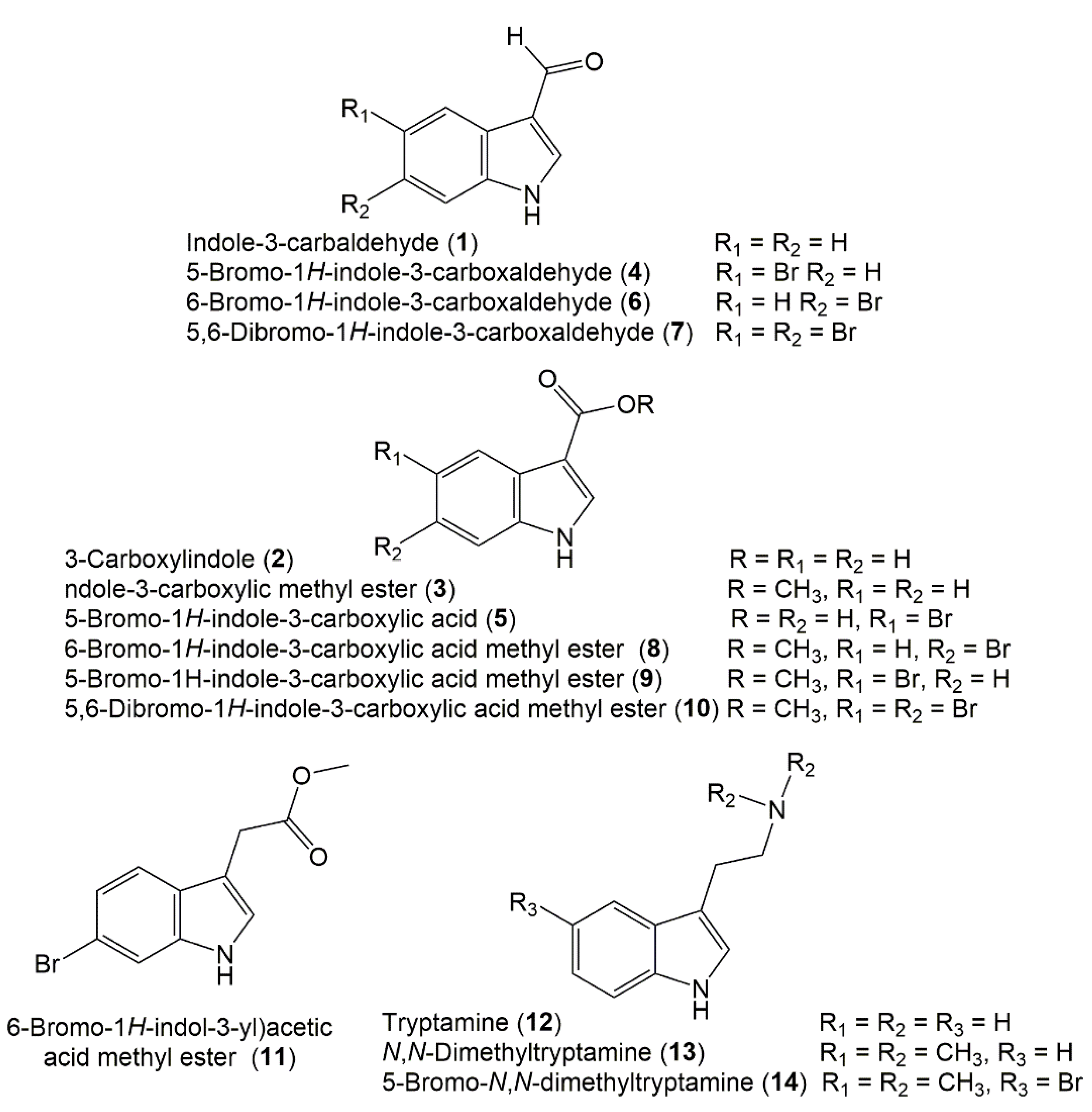
Figure 1. Chemical structures of indole (114) derivatives reported from Smenospongia species.
Molecules 27 05969 g001
Figure 2. Chemical structures of indole (1525) derivatives reported from Smenospongia species.
Molecules 27 05969 g002
Figure 3. Chemical structures of aplysinopsin (2637) derivatives reported from Smenospongia species.
Molecules 27 05969 g003
Figure 4. Chemical structures of bisspiroimidazolidinone (3841) alkaloids reported from Smenospongia species.
Molecules 27 05969 g004
Figure 5. Chemical structures of smenamides (4248) reported from Smenospongia species and synthetic analogs;16-epi-smenamide A and ent-smenamide.
Molecules 27 05969 g005
Figure 6. Chemical structures of smenothiazoles A (49) and B (50), smenolactones A–D (5154), and conulothiazoles A (56) and B (57) reported from Smenospongia species.
Molecules 27 05969 g006
Figure 7. Chemical structures of hydroquinone sesquiterpenoids (5869) reported from Smenospongia species.
Molecules 27 05969 g007
Figure 8. Synthetic derivatives of aureol (IX) [20,35].
Molecules 27 05969 g008
Figure 9. Chemical structures of hydroquinone (70 and 71) and quinone (7281) sesquiterpenoids reported from Smenospongia species.
Molecules 27 05969 g009
Figure 10. Chemical structures of amino- (8286) and oxazole-containing (8791) sesquiterpenoids reported from Smenospongia species.
Molecules 27 05969 g010
Scheme 1. Biosynthetic pathways of 77, 8790, and 9799 [46].
Molecules 27 05969 sch001
Figure 11. Chemical structures of tetronic acid (92), cyclopentenone (93101), and dimer hydroquinone (102). Sesquiterpenoids reported from Smenospongia species.
Molecules 27 05969 g011
Figure 12. Chemical structures of diterpenoids (103 and 104) and sesterterpenoids (105115) reported from Smenospongia species.
Molecules 27 05969 g012
Figure 13. Chemical structures of scalarane sesterterpenoids (116129) reported from Smenospongia species.
Molecules 27 05969 g013
Scheme 2. Biosynthetic pathways of smenochromenes A–D (130133) from farnesyl hydroquinone precursor [56].
Molecules 27 05969 sch002
Scheme 3. Synthesis of smenochromene B (131) from smenochromene D (133) [56].
Molecules 27 05969 sch003
Figure 14. Chemical structures of chromenes (130133), γ-pyrone (134), phenyl alkene (135), phenolic (136), and naphthoquinones (137 and 138) reported from Smenospongia species.
Molecules 27 05969 g014
Figure 15. Chemical structures of fatty acids (139145), sterols (146152), and phthalate (153 and 154) reported from Smenospongia species.
Molecules 27 05969 g015
Figure 16. Number of reported metabolites and geographical distribution of Smenospongia species. The darker blue color indicated large number of metabolites reported from sponge species collected from this region; 4–21 is the range of metabolites.
Molecules 27 05969 g016
Figure 17. Metabolites reported from various Smenospongia species (% and number).
Molecules 27 05969 g017
Figure 18. Various chemical classes of metabolites reported from various Smenospongia species. IAs: indole alkaloids; PKs: polyketides; APs: aplysinopsins; SQTs: sesquiterpenoids; SSTs: sesterterpenoids; STs: sterols; FAs: fatty acids; CDs: chromene derivatives; BSIs: bisspiroimidazolidinones; NQs: naphthoquinones; PHs: phthalates; DTs: diterpenoids; γ-PD: γ-pyrone derivatives; PADs: phenyl alkene derivatives; Phe: phenolics.
Molecules 27 05969 g018
Figure 19. Number of compounds with potential bioactivity.
Molecules 27 05969 g019
Publisher’s Note: MDPI stays neutral with regard to jurisdictional claims in published maps and institutional affiliations.