
Oncolytic Newcastle Disease Virus Co-Delivered with Modified PLGA Nanoparticles Encapsulating Temozolomide against Glioblastoma Cells: Developing an Effective Treatment Strategy

 ,
,  , , and
Abstract
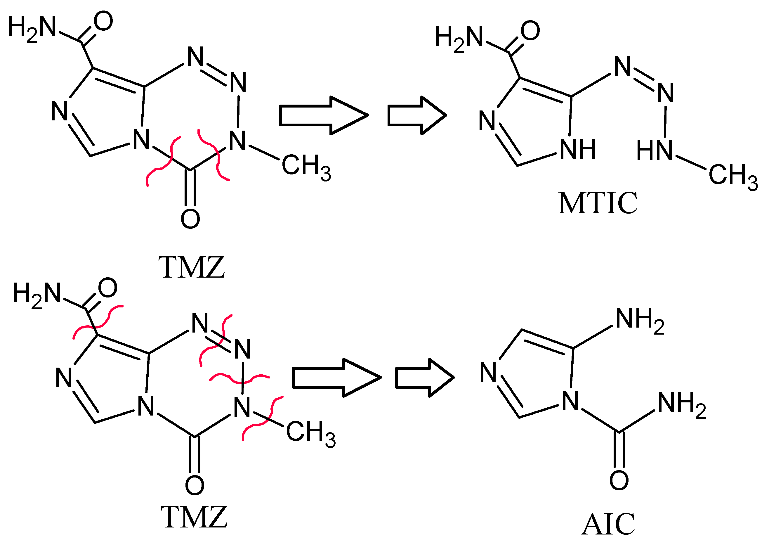
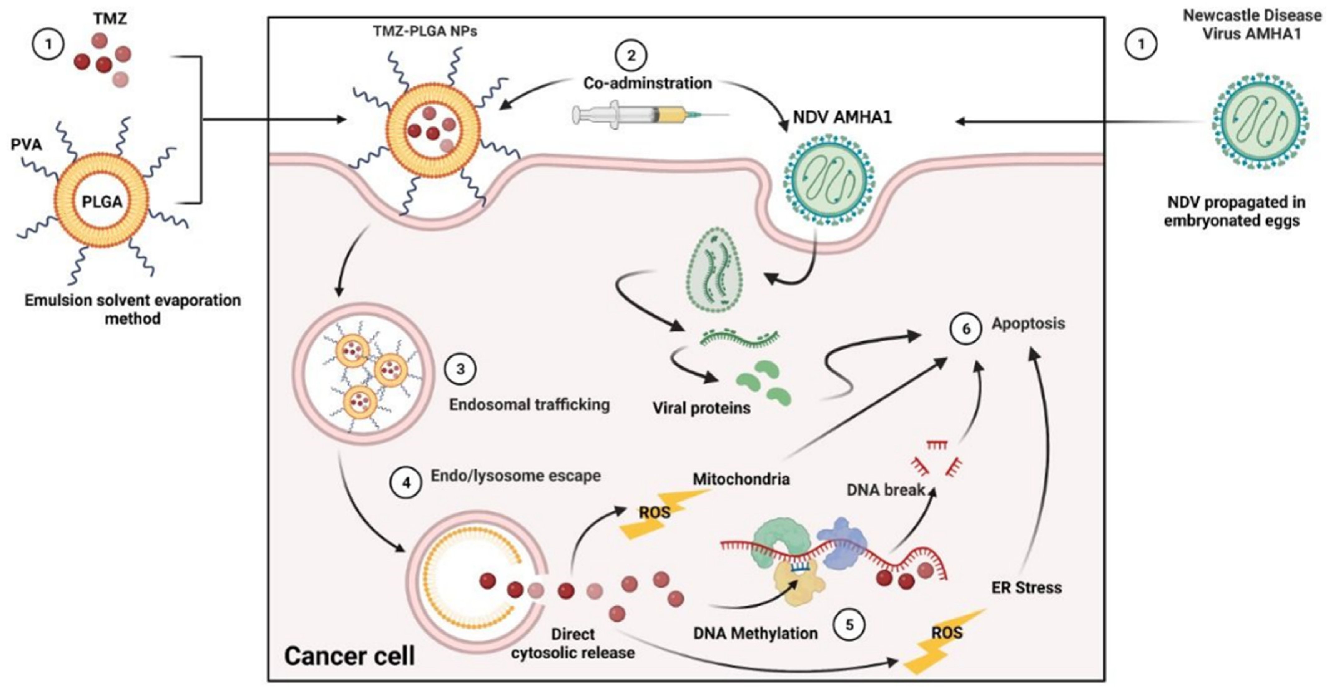
1. Introduction
2. Results and Discussion
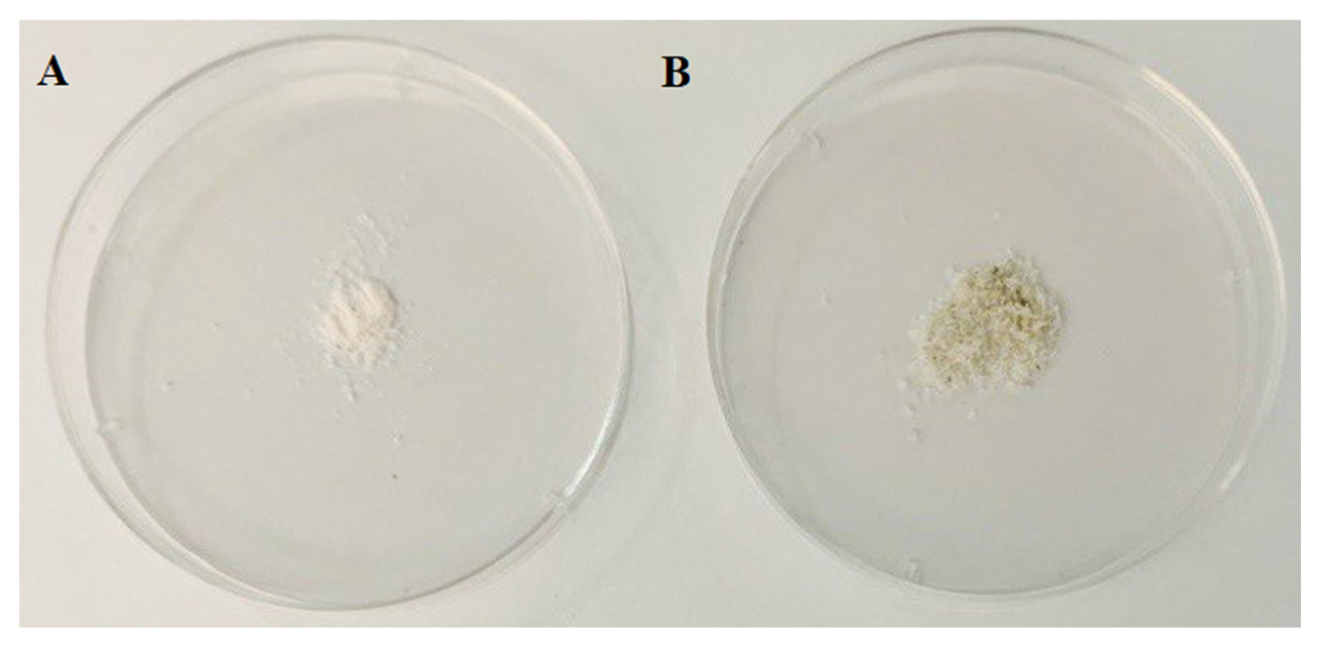
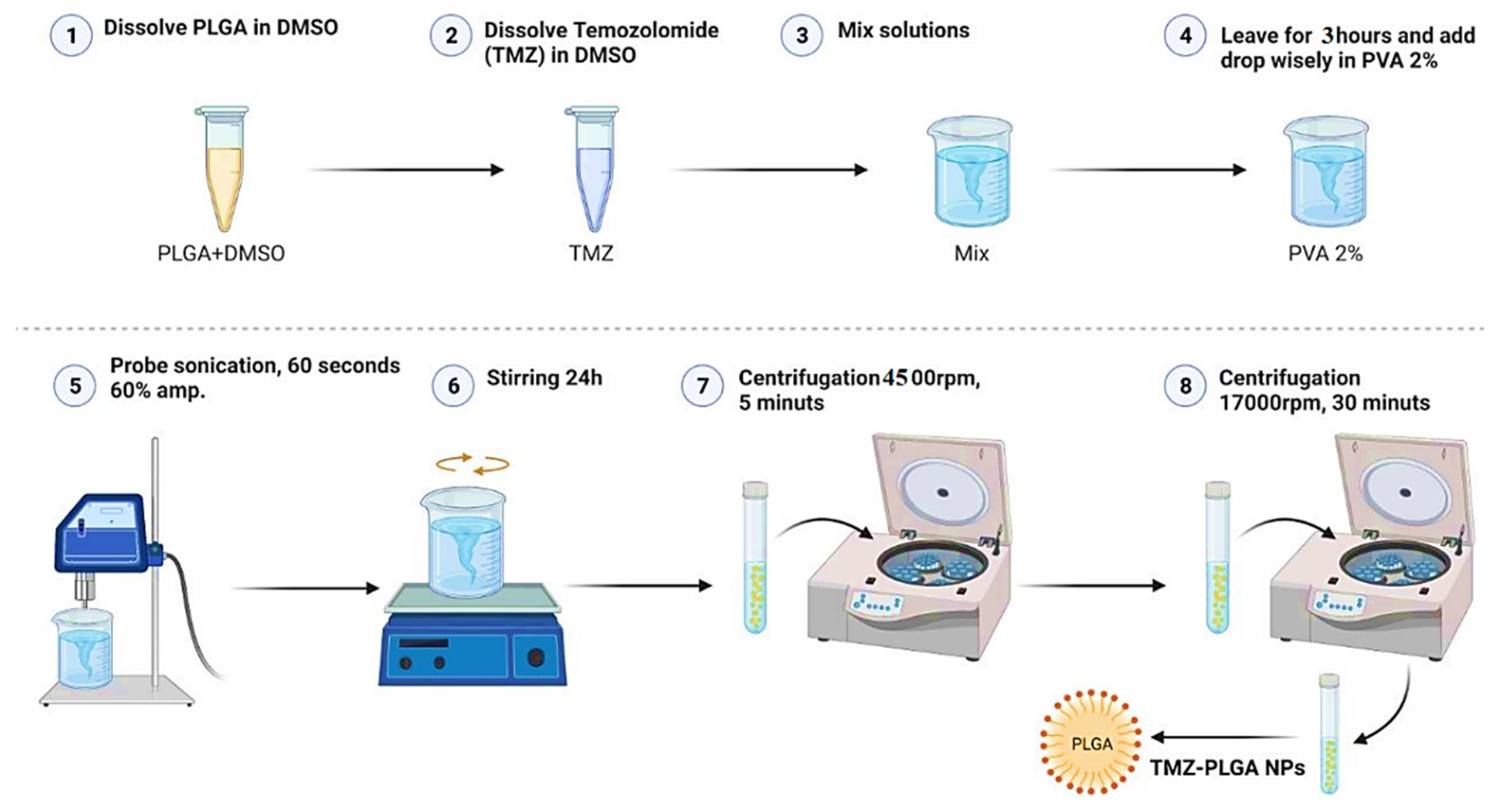
2.1. Preparation of TMZ-PLGA Nanoparticles
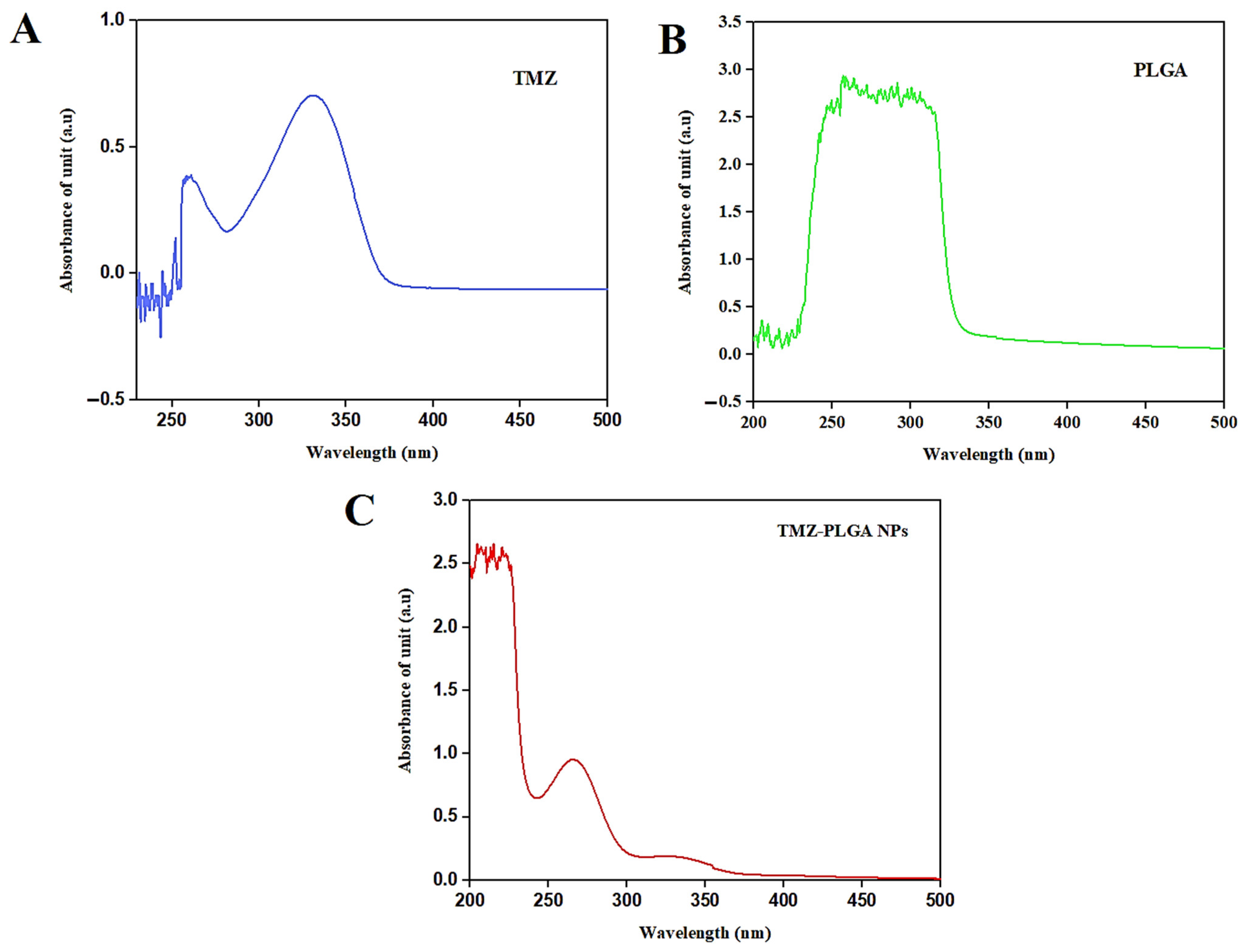
2.2. UV-Vis Spectral Analyses
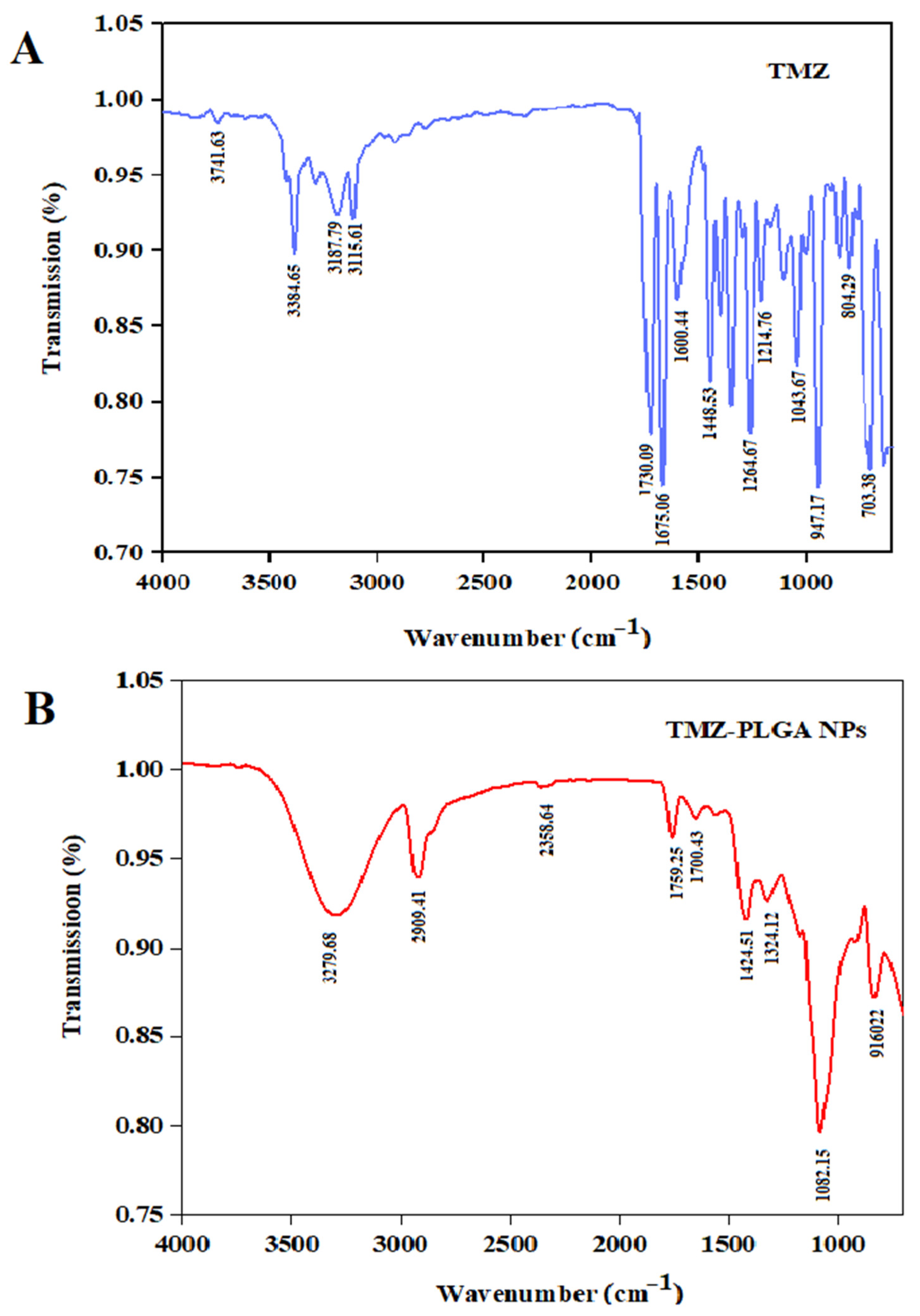
2.3. Fourier Transform Infrared Spectroscopic (FT-IR) Analyses
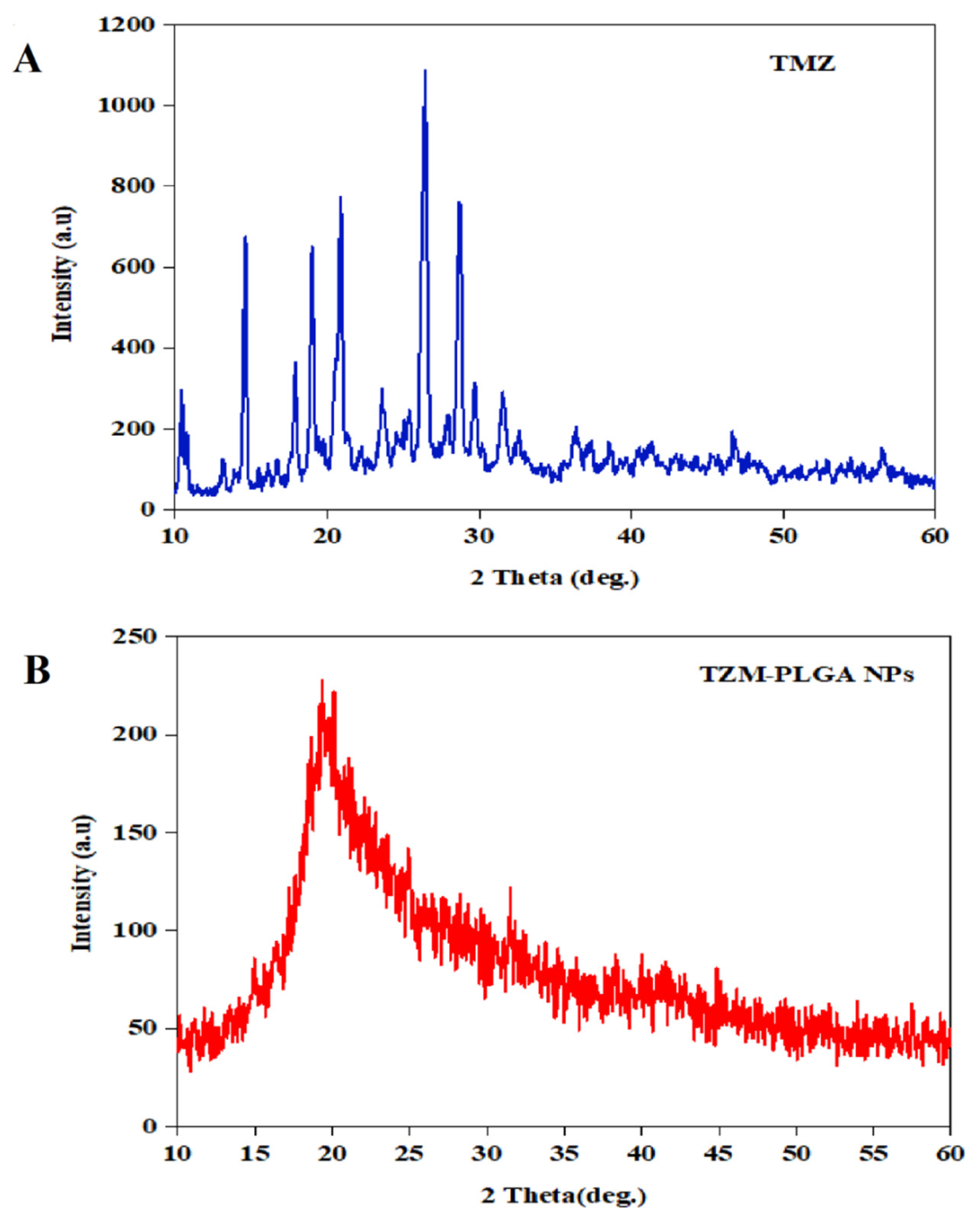
2.4. X-ray Diffraction Characterizations
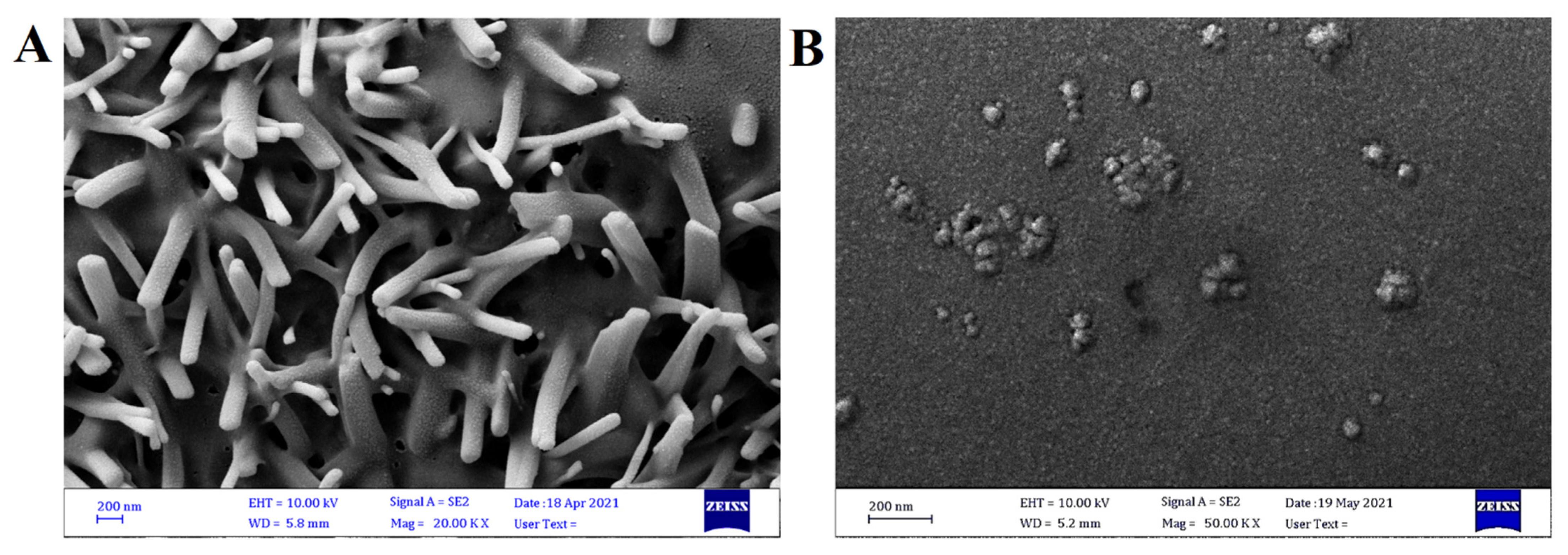
2.5. Field Emission Scanning Electron Microscopy (FE-SEM)
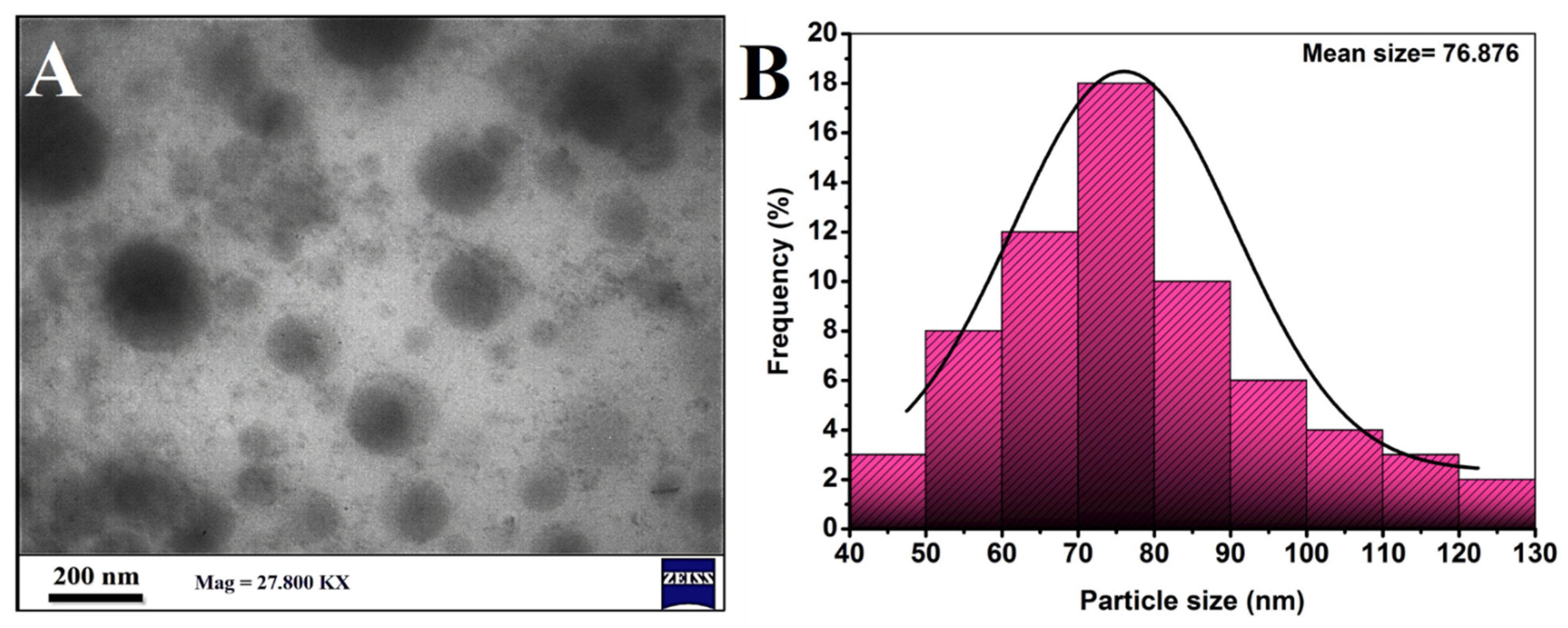
2.6. Transmission Electron Microscopy (TEM)
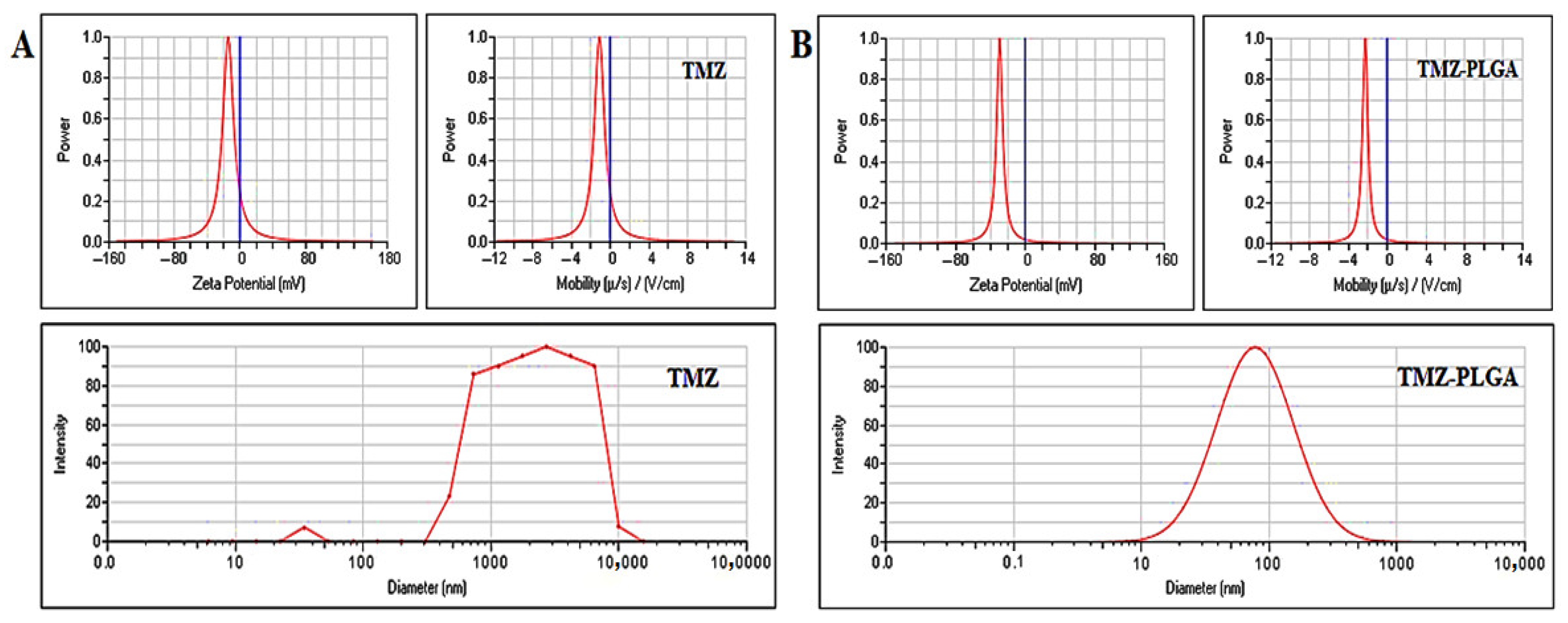
2.7. Zeta Potential Analysis
2.8. Particle Size Analysis
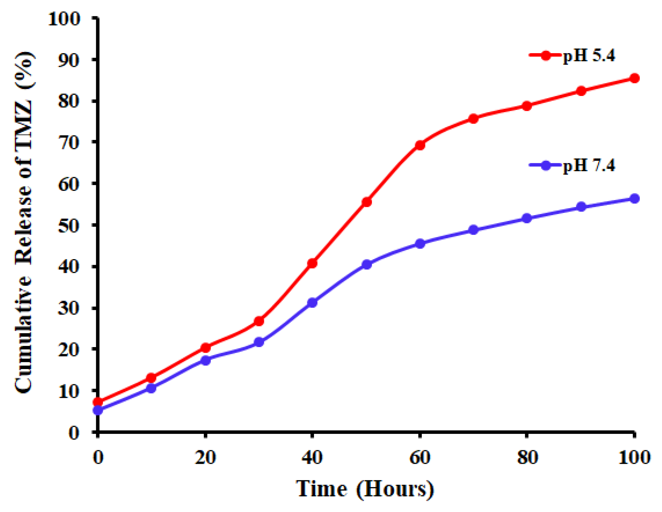
2.9. In Vitro Drug Release
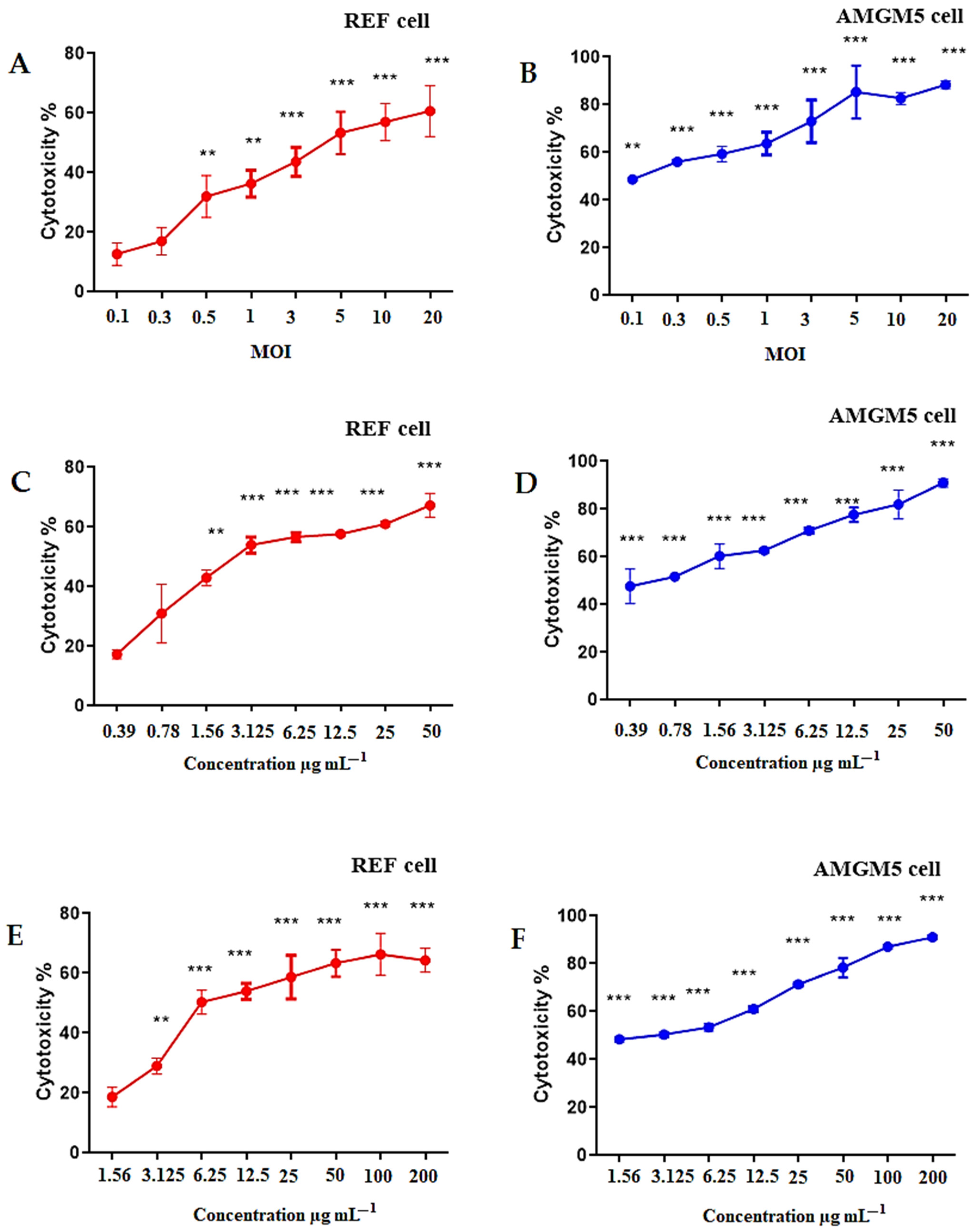
2.10. Cytotoxicity Testings against the AMGM5 and REF Cell Lines
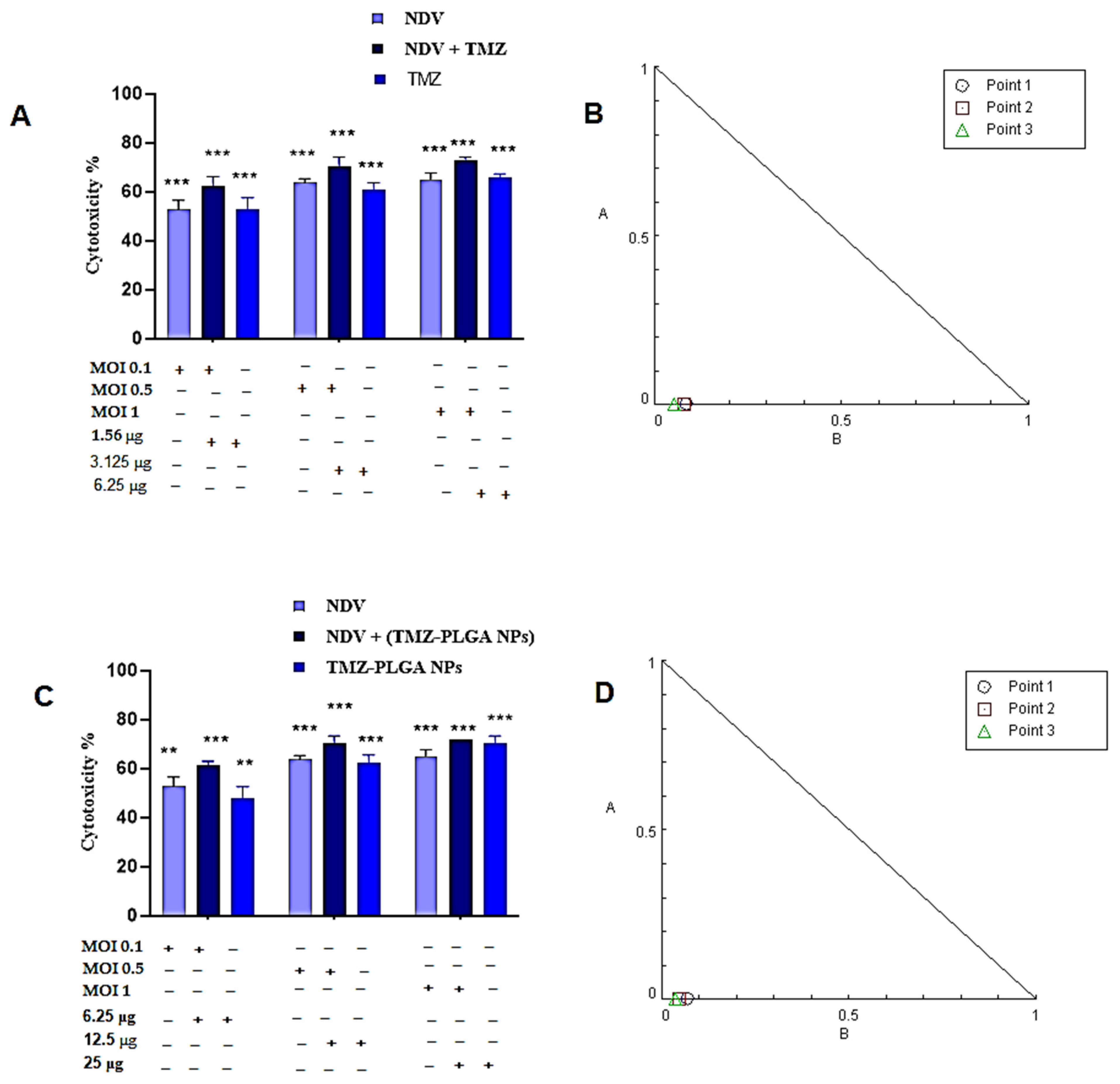
2.11. Synergistic Effects of the TMZ, NDV, and TMZ-PLGA-NPs Combinations Utilizing Cytotoxicity Assay and Chou–Talalay Analysis of the AMGM5 Cell Lines
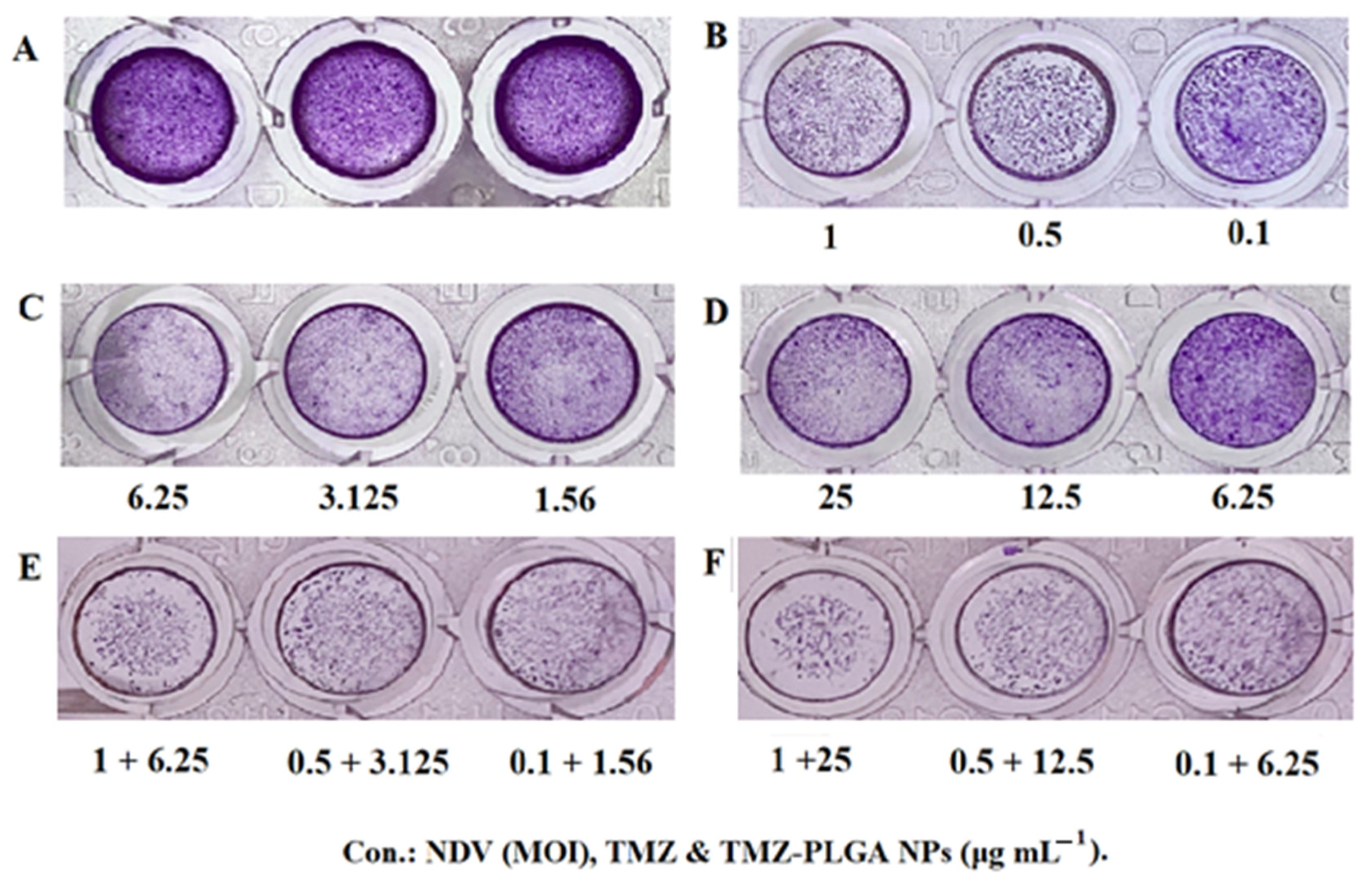
2.12. Colony Formation Assay
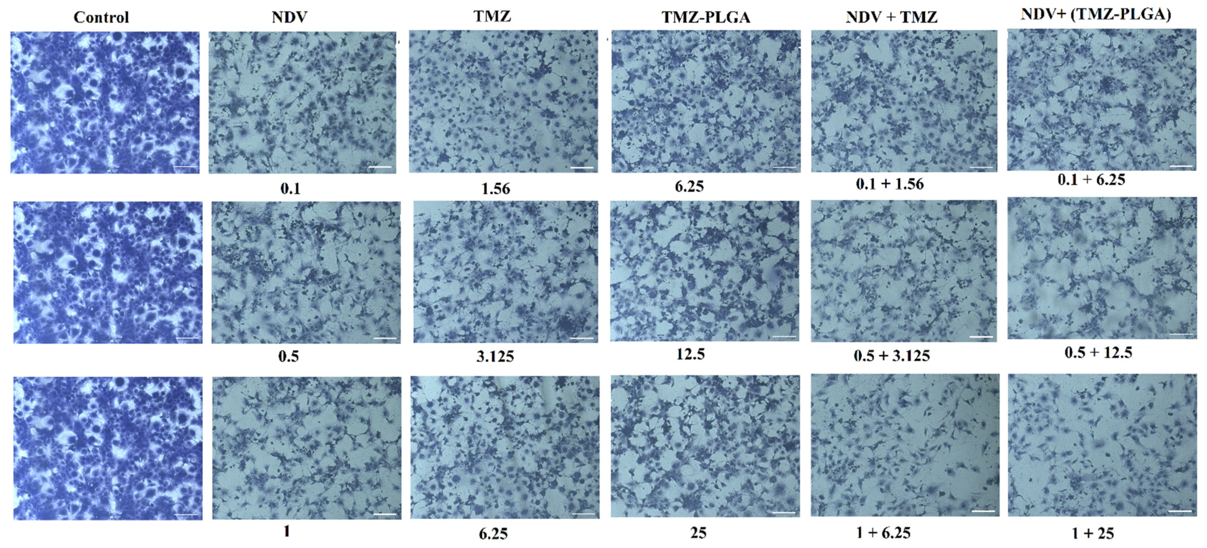
2.13. Morphology and Quantitative Image Analyses Using Crystal Violet
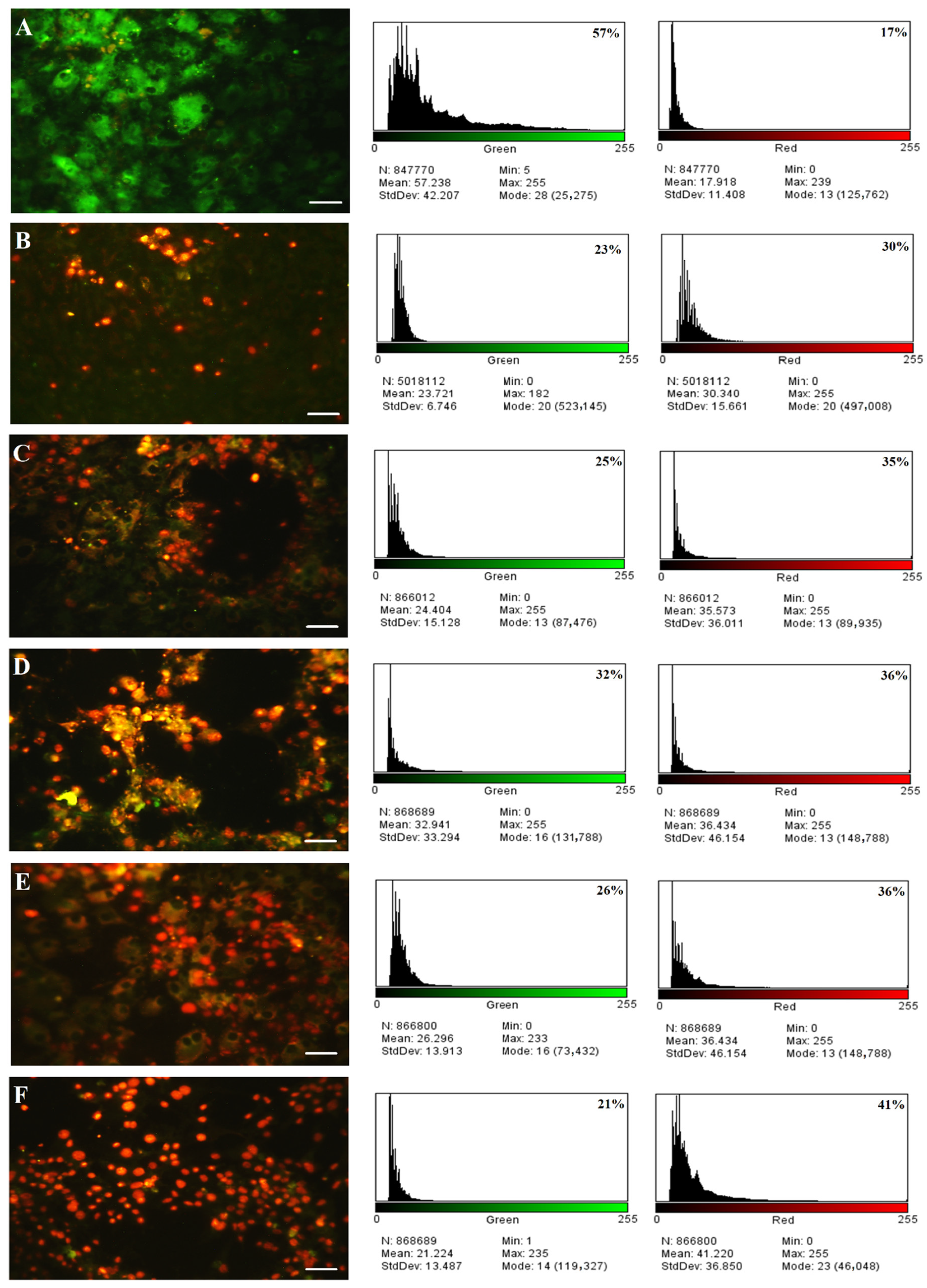
2.14. Acridine Orange–Propidium Iodide (AO/PI) Dual-Staining Assay
3. Materials and Methods
3.1. Materials and Reagents
3.2. Cell Line Cultures
3.3. Preparation of the TMZ-Loaded PLGA Nanoparticles
3.4. Characterization of the Temozolomide Nanoparticles
3.5. In Vitro Release of TMZ
3.6. NDV Propagation
3.7. Cytotoxicity against the REF Normal Cell Line and the AMGM5 Cancer Cell Line
3.8. Combined Cytotoxicity Assays and Chou–Talalay Analysis
3.9. Clonogenicity Survival Assay
3.10. Morphology Analysis
3.11. Assessment of Apoptosis
3.12. Statistical Analysis
4. Conclusions
Author Contributions
Funding
Institutional Review Board Statement
Informed Consent Statement
Data Availability Statement
Acknowledgments
Conflicts of Interest
References
- Bower, J.E.; Asher, A.; Garet, D.; Petersen, L.; Ganz, P.A.; Irwin, M.R.; Cole, S.W.; Hurvitz, S.A.; Crespi, C.M. Testing a biobehavioral model of fatigue before adjuvant therapy in women with breast cancer. Cancer 2019, 125, 633–641. [Google Scholar] [CrossRef]
- Mula-Hussain, L.; Alabedi, H.; Al-Alloosh, F.; Alharganee, A. Cancer in war-torn countries: Iraq as an example. In Handbook of Healthcare in the Arab World; Springer: Berlin/Heidelberg, Germany, 2021; pp. 481–494. [Google Scholar]
- Al Alwan, N.A.S. General Oncology Care in Iraq. In Cancer in the Arab World; Springer: Berlin/Heidelberg, Germany, 2022; pp. 63–82. [Google Scholar]
- Mutar, M.T.; Goyani, M.S.; Had, A.M.; Mahmood, A.S. Pattern of presentation of patients with breast cancer in Iraq in 2018: A cross-sectional study. J. Glob. Oncol. 2019, 5, 1–6. [Google Scholar] [CrossRef] [PubMed]
- Bai, H. Integrative Omic Analysis of IDH1-Mutant Glioma Malignant Progression; Yale University: New Haven, CT, USA, 2016; ISBN 1369053436. [Google Scholar]
- Ramalho, M.J.; Andrade, S.; Coelho, M.Á.N.; Loureiro, J.A.; Pereira, M.C. Biophysical interaction of temozolomide and its active metabolite with biomembrane models: The relevance of drug-membrane interaction for Glioblastoma Multiforme therapy. Eur. J. Pharm. Biopharm. 2019, 136, 156–163. [Google Scholar] [CrossRef] [PubMed]
- Shergalis, A.; Bankhead, A.; Luesakul, U.; Muangsin, N.; Neamati, N. Current challenges and opportunities in treating glioblastoma. Pharmacol. Rev. 2018, 70, 412–445. [Google Scholar] [CrossRef] [PubMed]
- Bahjat, H.H.; Ismail, R.A.; Sulaiman, G.M.; Mohammed, H.A.; Al-Omar, M.; Mohammed, S.A.A.; Khan, R.A. Preparation of iron oxide and titania-based composite, core-shell populated, nanoparticulates material by two-step LASER ablation in aqueous media as antimicrobial and anticancer agents. Bioinorg. Chem. Appl. 2022, 2022, 1854473. [Google Scholar] [CrossRef]
- Patra, J.K.; Das, G.; Fraceto, L.F.; Campos, E.V.R.; Rodriguez-Torres, M.d.P.; Acosta-Torres, L.S.; Diaz-Torres, L.A.; Grillo, R.; Swamy, M.K.; Sharma, S. Nano based drug delivery systems: Recent developments and future prospects. J. Nanobiotechnology 2018, 16, 71. [Google Scholar] [CrossRef]
- Ramalho, M.J.; Pereira, M.C. Preparation and characterization of polymeric nanoparticles: An interdisciplinary experiment. J. Chem. Educ. 2016, 93, 1446–1451. [Google Scholar] [CrossRef]
- Tang, C.-M.; Tian, Y.-H.; Hsu, S.-H. Poly (vinyl alcohol) nanocomposites reinforced with bamboo charcoal nanoparticles: Mineralization behavior and characterization. Materials 2015, 8, 4895–4911. [Google Scholar] [CrossRef]
- Mai, P.L.; Best, A.F.; Peters, J.A.; DeCastro, R.M.; Khincha, P.P.; Loud, J.T.; Bremer, R.C.; Rosenberg, P.S.; Savage, S.A. Risks of first and subsequent cancers among TP53 mutation carriers in the National Cancer Institute Li-Fraumeni syndrome cohort. Cancer 2016, 122, 3673–3681. [Google Scholar] [CrossRef]
- Shi, J.; Kantoff, P.W.; Wooster, R.; Farokhzad, O.C. Cancer nanomedicine: Progress, challenges and opportunities. Nat. Rev. Cancer 2017, 17, 20–37. [Google Scholar] [CrossRef]
- Al-Shammari, A.M.; Al-Nassrawei, H.A.; Kadhim, A.M.A. Isolation and sero-diagnosis of Newcastle disease virus infection in human and chicken poultry flocks in three cities of middle Euphrates. Kufa J. Vet. Med. Sci. 2014, 5, 16–21. [Google Scholar]
- Paul, R.; Kulkarni, P.; Ganesh, N. Avocado fruit (Persea americana Mill) exhibits chemo-protective potentiality against cyclophosphamide induced genotoxicity in human lymphocyte culture. J. Exp. Ther. Oncol. 2011, 9, 221–230. [Google Scholar] [PubMed]
- Kaufman, H.L.; Kohlhapp, F.J.; Zloza, A. Oncolytic viruses: A new class of immunotherapy drugs. Nat. Rev. Drug Discov. 2015, 14, 642–662. [Google Scholar] [CrossRef] [PubMed]
- Schirrmacher, V. From chemotherapy to biological therapy: A review of novel concepts to reduce the side effects of systemic cancer treatment. Int. J. Oncol. 2019, 54, 407–419. [Google Scholar] [PubMed]
- Bai, Y.; Hui, P.; Du, X.; Su, X. Updates to the antitumor mechanism of oncolytic virus. Thorac. Cancer 2019, 10, 1031–1035. [Google Scholar] [CrossRef]
- Prinsloo, S.; Novy, D.; Driver, L.; Lyle, R.; Ramondetta, L.; Eng, C.; Lopez, G.; Li, Y.; Cohen, L. The long-term impact of neurofeedback on symptom burden and interference in patients with chronic chemotherapy-induced neuropathy: Analysis of a randomized controlled trial. J. Pain Symptom Manag. 2018, 55, 1276–1285. [Google Scholar] [CrossRef] [PubMed]
- Al-Shammari, A.M.; Ismaeel, F.E.; Salih, S.M.; Yaseen, N.Y. Live attenuated measles virus vaccine therapy for locally established malignant glioblastoma tumor cells. Oncolytic Virotherapy 2014, 3, 57. [Google Scholar] [CrossRef]
- Al-Shammari, A.M.; Rameez, H.; Al-Taee, M.F. Newcastle disease virus, rituximab, and doxorubicin combination as anti-hematological malignancy therapy. Oncolytic Virotherapy 2016, 5, 27. [Google Scholar] [CrossRef]
- Parsons, D.W.; Roy, A.; Yang, Y.; Wang, T.; Scollon, S.; Bergstrom, K.; Kerstein, R.A.; Gutierrez, S.; Petersen, A.K.; Bavle, A. Diagnostic yield of clinical tumor and germline whole-exome sequencing for children with solid tumors. JAMA Oncol. 2016, 2, 616–624. [Google Scholar] [CrossRef]
- Martin, C. Oncolytic viruses: Treatment and implications for patients with gliomas. Clin. J. Oncol. Nurs. 2017, 21, 60–64. [Google Scholar] [CrossRef]
- Bai, Y.; Chen, Y.; Hong, X.; Liu, X.; Su, X.; Li, S.; Dong, X.; Zhao, G.; Li, Y. Newcastle disease virus enhances the growth-inhibiting and proapoptotic effects of temozolomide on glioblastoma cells in vitro and in vivo. Scintific Rep. 2018, 8, 11470. [Google Scholar] [CrossRef] [PubMed]
- Dimitrov, K.M.; Abolnik, C.; Afonso, C.L.; Albina, E.; Bahl, J.; Berg, M.; Briand, F.-X.; Brown, I.H.; Choi, K.-S.; Chvala, I. Updated unified phylogenetic classification system and revised nomenclature for Newcastle disease virus. Infect. Genet. Evol. 2019, 74, 103917. [Google Scholar] [CrossRef] [PubMed]
- Tsai, Y.-M.; Chien, C.-F.; Lin, L.-C.; Tsai, T.-H. Curcumin and its nano-formulation: The kinetics of tissue distribution and blood brain barrier penetration. Int. J. Pharm. 2011, 416, 331–338. [Google Scholar] [CrossRef] [PubMed]
- Patil, R.; Portilla-Arias, J.; Ding, H.; Inoue, S.; Konda, B.; Hu, J.; Wawrowsky, K.A.; Shin, P.K.; Black, K.L.; Holler, E. Temozolomide delivery to tumor cells by a multifunctional nano vehicle based on poly (β-L-malic acid). Pharm. Res. 2010, 27, 2317–2329. [Google Scholar] [CrossRef]
- del Burgo, L.S.; Hernández, R.M.; Orive, G.; Pedraz, J.L. Nanotherapeutic approaches for brain cancer management. Nanomed. Nanotechnol. Biol. Med. 2014, 10, e905–e919. [Google Scholar] [CrossRef]
- Zhang, D.; Tian, A.; Xue, X.; Wang, M.; Qiu, B.; Wu, A. The effect of temozolomide/poly (lactide-co-glycolide)(PLGA)/nano-hydroxyapatite microspheres on glioma U87 cells behavior. Int. J. Mol. Sci. 2012, 13, 1109–1125. [Google Scholar] [CrossRef]
- Huang, G.; Zhang, N.; Bi, X.; Dou, M. Solid lipid nanoparticles of temozolomide: Potential reduction of cardial and nephric toxicity. Int. J. Pharm. 2008, 355, 314–320. [Google Scholar] [CrossRef] [PubMed]
- Jain, D.S.; Athawale, R.B.; Bajaj, A.N.; Shrikhande, S.S.; Goel, P.N.; Nikam, Y.; Gude, R.P. Unraveling the cytotoxic potential of Temozolomide loaded into PLGA nanoparticles. DARU J. Pharm. Sci. 2014, 22, 1–9. [Google Scholar] [CrossRef][Green Version]
- Schwendy, M.; Unger, R.E.; Bonn, M.; Parekh, S.H. Automated cell segmentation in FIJI® using the DRAQ5 nuclear dye. BMC Bioinform. 2019, 20, 1–9. [Google Scholar] [CrossRef]
- Vergara-Jaque, A.; Comer, J.; Monsalve, L.; Gonzalez-Nilo, F.D.; Sandoval, C. Computationally efficient methodology for atomic-level characterization of dendrimer–drug complexes: A comparison of amine-and acetyl-terminated PAMAM. J. Phys. Chem. B 2013, 117, 6801–6813. [Google Scholar] [CrossRef]
- Gelperina, S.; Maksimenko, O.; Khalansky, A.; Vanchugova, L.; Shipulo, E.; Abbasova, K.; Berdiev, R.; Wohlfart, S.; Chepurnova, N.; Kreuter, J. Drug delivery to the brain using surfactant-coated poly (lactide-co-glycolide) nanoparticles: Influence of the formulation parameters. Eur. J. Pharm. Biopharm. 2010, 74, 157–163. [Google Scholar] [CrossRef] [PubMed]
- Rodriguez de Anda, D.A.; Ohannesian, N.; Martirosyan, K.S.; Chew, S.A. Effects of solvent used for fabrication on drug loading and release kinetics of electrosprayed temozolomide-loaded PLGA microparticles for the treatment of glioblastoma. J. Biomed. Mater. Res. Part B Appl. Biomater. 2019, 107, 2317–2324. [Google Scholar] [CrossRef] [PubMed]
- Lopes, I.C.; de Oliveira, S.C.B.; Oliveira-Brett, A.M. Temozolomide chemical degradation to 5-aminoimidazole-4-carboxamide–electrochemical study. J. Electroanal. Chem. 2013, 704, 183–189. [Google Scholar] [CrossRef]
- Sahu, S.K.; Mallick, S.K.; Santra, S.; Maiti, T.K.; Ghosh, S.K.; Pramanik, P. In vitro evaluation of folic acid modified carboxymethyl chitosan nanoparticles loaded with doxorubicin for targeted delivery. J. Mater. Sci. Mater. Med. 2010, 21, 1587–1597. [Google Scholar] [CrossRef]
- Dhas, D.A. Vibrational spectra and natural bond orbital analysis of temozolomide acid. J. Appl. Sci. Eng. Methodol. 2016, 2, 322–325. [Google Scholar]
- Arasoglu, T.; Derman, S.; Mansuroglu, B. Comparative evaluation of antibacterial activity of caffeic acid phenethyl ester and PLGA nanoparticle formulation by different methods. Nanotechnology 2015, 27, 25103. [Google Scholar] [CrossRef]
- Barone, G.; Bartoli, L.; Belfiore, C.M.; Crupi, V.; Longo, F.; Majolino, D.; Mazzoleni, P.; Venuti, V. Comparison between TOF-ND and XRD quantitative phase analysis of ancient potteries. J. Anal. At. Spectrom. 2011, 26, 1060–1067. [Google Scholar] [CrossRef]
- Gu, W.; Fan, R.; Quan, J.; Cheng, Y.; Wang, S.; Zhang, H.; Zheng, A.; Song, S. Intracranial In Situ Thermosensitive Hydrogel Delivery of Temozolomide Accomplished by PLGA–PEG–PLGA Triblock Copolymer Blending for GBM Treatment. Polymers 2022, 14, 3368. [Google Scholar] [CrossRef]
- Assefi, M.; Onsory, K.; Iranbakhsh, A. Understanding the toxicity of nanotubes and nanoparticles in the environment: Are nanotubes and nanoparticles safe. Asian J. Od Pharm. Technol. Innoviation 2021, 9, 5–10. [Google Scholar]
- Jain, D.; Bajaj, A.; Athawale, R.; Shrikhande, S.; Goel, P.N.; Nikam, Y.; Gude, R.; Patil, S.; Prashant Raut, P. Surface-coated PLA nanoparticles loaded with temozolomide for improved brain deposition and potential treatment of gliomas: Development, characterization and in vivo studies. Drug Deliv. 2016, 23, 989–1006. [Google Scholar] [CrossRef]
- Win, K.Y.; Feng, S.-S. Effects of particle size and surface coating on cellular uptake of polymeric nanoparticles for oral delivery of anticancer drugs. Biomaterials 2005, 26, 2713–2722. [Google Scholar] [CrossRef] [PubMed]
- Tang, X.; Cai, S.; Zhang, R.; Liu, P.; Chen, H.; Zheng, Y.; Sun, L. Paclitaxel-loaded nanoparticles of star-shaped cholic acid-core PLA-TPGS copolymer for breast cancer treatment. Nanoscale Res. Lett. 2013, 8, 420. [Google Scholar] [CrossRef]
- Saraiva, C.; Praça, C.; Ferreira, R.; Santos, T.; Ferreira, L.; Bernardino, L. Nanoparticle-mediated brain drug delivery: Overcoming blood–brain barrier to treat neurodegenerative diseases. J. Control. Release 2016, 235, 34–47. [Google Scholar] [CrossRef] [PubMed]
- Dembek, M.; Bocian, S.; Buszewski, B. Solvent Influence on Zeta Potential of Stationary Phase—Mobile Phase Interface. Molecules 2022, 27, 968. [Google Scholar] [CrossRef]
- Sahana, D.K.; Mittal, G.; Bhardwaj, V.; Kumar, M.N.V.R. PLGA nanoparticles for oral delivery of hydrophobic drugs: Influence of organic solvent on nanoparticle formation and release behavior in vitro and in vivo using estradiol as a model drug. J. Pharm. Sci. 2008, 97, 1530–1542. [Google Scholar] [CrossRef]
- Derman, S. Caffeic acid phenethyl ester loaded PLGA nanoparticles: Effect of various process parameters on reaction yield, encapsulation efficiency, and particle size. J. Nanomater. 2015, 2015, 341848. [Google Scholar] [CrossRef]
- Wohlfart, S.; Gelperina, S.; Kreuter, J. Transport of drugs across the blood–brain barrier by nanoparticles. J. Control. Release 2012, 161, 264–273. [Google Scholar] [CrossRef] [PubMed]
- Fornaguera, C.; Solans, C. Characterization of polymeric nanoparticle dispersions for biomedical applications: Size, surface charge and stability. Pharm. Nanotechnol. 2018, 6, 147–164. [Google Scholar] [CrossRef] [PubMed]
- Suvarna, S.; Das, U.; Kc, S.; Mishra, S.; Sudarshan, M.; Saha, K.D.; Dey, S.; Chakraborty, A.; Narayana, Y. Synthesis of a novel glucose capped gold nanoparticle as a better theranostic candidate. PLoS ONE 2017, 12, e0178202. [Google Scholar] [CrossRef]
- Dangi, R.S.; Shakya, S. Preparation, optimization and characterization of PLGA nanoparticle. Int. J. Pharm. Life Sci. 2013, 4, 2810–2818. [Google Scholar]
- Lu, B.; Lv, X.; Le, Y. Chitosan-modified PLGA nanoparticles for control-released drug delivery. Polymers 2019, 11, 304. [Google Scholar] [CrossRef] [PubMed]
- Souza, T.G.F.; Ciminelli, V.S.T.; Mohallem, N.D.S. A comparison of TEM and DLS methods to characterize size distribution of ceramic nanoparticles. J. Phys. Conf. Ser. 2016, 733, 12039. [Google Scholar] [CrossRef]
- Prüger, B.; Eppmann, P.; Donath, E.; Gimsa, J. Measurement of inherent particle properties by dynamic light scattering: Introducing electrorotational light scattering. Biophys. J. 1997, 72, 1414–1424. [Google Scholar] [CrossRef]
- Sánchez-López, E.; Ettcheto, M.; Egea, M.A.; Espina, M.; Cano, A.; Calpena, A.C.; Camins, A.; Carmona, N.; Silva, A.M.; Souto, E.B.; et al. Memantine loaded PLGA PEGylated nanoparticles for Alzheimer’s disease: In vitro and in vivo characterization. J. Nanobiotechnology 2018, 16, 32. [Google Scholar] [CrossRef] [PubMed]
- Lam, F.C.; Morton, S.W.; Wyckoff, J.; Vu Han, T.L.; Hwang, M.K.; Maffa, A.; Balkanska-Sinclair, E.; Yaffe, M.B.; Floyd, S.R.; Hammond, P.T. Enhanced efficacy of combined temozolomide and bromodomain inhibitor therapy for gliomas using targeted nanoparticles. Nat. Commun. 2018, 9, 1991. [Google Scholar] [CrossRef]
- Kumari, S.; Ahsan, S.M.; Kumar, J.M.; Kondapi, A.K.; Rao, N.M. Overcoming blood brain barrier with a dual purpose Temozolomide loaded Lactoferrin nanoparticles for combating glioma (SERP-17-12433). Sci. Rep. 2017, 7, 6602. [Google Scholar] [CrossRef]
- Pham, T.T.; Nguyen, T.T.; Pathak, S.; Regmi, S.; Nguyen, H.T.; Tran, T.H.; Yong, C.S.; Kim, J.O.; Park, P.; Park, M.H. Tissue adhesive FK506–loaded polymeric nanoparticles for multi–layered nano–shielding of pancreatic islets to enhance xenograft survival in a diabetic mouse model. Biomaterials 2018, 154, 182–196. [Google Scholar] [CrossRef]
- Duwa, R.; Banstola, A.; Emami, F.; Jeong, J.-H.; Lee, S.; Yook, S. Cetuximab conjugated temozolomide-loaded poly (lactic-co-glycolic acid) nanoparticles for targeted nanomedicine in EGFR overexpressing cancer cells. J. Drug Deliv. Sci. Technol. 2020, 60, 101928. [Google Scholar] [CrossRef]
- Holy, C.E.; Dang, S.M.; Davies, J.E.; Shoichet, M.S. In vitro degradation of a novel poly (lactide-co-glycolide) 75/25 foam. Biomaterials 1999, 20, 1177–1185. [Google Scholar] [CrossRef]
- Baiti, R.N.; Ardhyananta, H.; El Kirat, K. Effect of acidic and basic environment to the degradation behavior of PLGA nanocapsules for biomedical application. Adv. Mater. Res. 2015, 1123, 213–216. [Google Scholar] [CrossRef]
- Al-Shamery, A.; Yaseen, N.; Alwan, M.J. Study the Antigenic modification of tumor cell surface by NDV infection. Iraqi J. Cancer 2009, 2, 95–100. [Google Scholar]
- Al-Shammari, A.M.; Al-Hili, Z.A.; Yaseen, N.Y. 647. Iraqi Newcastle disease virus virulent strain as cancer antiangiogenic agent. Molicular Ther. 2013, 21, S247. [Google Scholar] [CrossRef]
- Schirrmacher, V. Oncolytic Newcastle disease virus as a prospective anti-cancer therapy. A biologic agent with potential to break therapy resistance. Expert Opin. Biol. Ther. 2015, 15, 1757–1771. [Google Scholar] [CrossRef] [PubMed]
- Choi, A.H.; O’Leary, M.P.; Fong, Y.; Chen, N.G. From benchtop to bedside: A review of oncolytic virotherapy. Biomedicines 2016, 4, 18. [Google Scholar] [CrossRef]
- Othman, F.; Ideris, A.; Motaleb, G.H.R.; Eshak, Z.B.T.; Rahmat, A. Oncolytic effect of Newcastle disease virus AF2240 strain on the MCF-7 breast cancer cell line. Yakhteh Med. J. 2010, 12, 17–24. [Google Scholar]
- Kwan, A.; Winder, N.; Muthana, M. Oncolytic Virotherapy Treatment of Breast Cancer: Barriers and Recent Advances. Viruses 2021, 13, 1128. [Google Scholar] [CrossRef]
- Suffness, M. Assays related to cancer drug discovery. Methods plant Biochem. Assays Bioactivity 1990, 6, 71–133. [Google Scholar]
- Al-Shammari, A.M.; Yaseen, N.; Alwan, M.J. Immunology study for NDV treatment in mice bearing mammary adenocarcinoma tumor. J. Cancer Med. Genet. 2011, 4, 11–21. [Google Scholar]
- Alonso, M.M.; Gomez-Manzano, C.; Bekele, B.N.; Yung, W.K.A.; Fueyo, J. Adenovirus-based strategies overcome temozolomide resistance by silencing the O6-methylguanine-DNA methyltransferase promoter. Cancer Res. 2007, 67, 11499–11504. [Google Scholar] [CrossRef]
- Ciccia, A.; Elledge, S.J. The DNA damage response: Making it safe to play with knives. Molecular Cell 2010, 40, 179–204. [Google Scholar] [CrossRef]
- Borges, H.L.; Linden, R.; Wang, J.Y.J. DNA damage-induced cell death: Lessons from the central nervous system. Cell Res. 2008, 18, 17–26. [Google Scholar] [CrossRef] [PubMed]
- Wang, J.Y.J.; Cho, S.K. Coordination of repair, checkpoint, and cell death responses to DNA damage. Adv. Protein Chem. 2004, 69, 101–135. [Google Scholar] [PubMed]
- Di Martino, A.; Kucharczyk, P.; Capakova, Z.; Humpolicek, P.; Sedlarik, V. Enhancement of temozolomide stability by loading in chitosan-carboxylated polylactide-based nanoparticles. J. Nanoparticle Res. 2017, 19, 71. [Google Scholar] [CrossRef] [PubMed]
- Foucquier, J.; Guedj, M. Analysis of drug combinations: Current methodological landscape. Pharmacol. Res. Perspect. 2015, 3, e00149. [Google Scholar] [CrossRef]
- Al-Qubaisi, M.S.; Rasedee, A.; Flaifel, M.H.; Ahmad, S.H.J.; Hussein-Al-Ali, S.; Hussein, M.Z.; Eid, E.E.M.; Zainal, Z.; Saeed, M.; Ilowefah, M. Cytotoxicity of nickel zinc ferrite nanoparticles on cancer cells of epithelial origin. Int. Joural Nanomed. 2013, 8, 2497. [Google Scholar] [CrossRef]
- Hegi, M.E.; Liu, L.; Herman, J.G.; Stupp, R.; Wick, W.; Weller, M.; Mehta, M.P.; Gilbert, M.R. Correlation of O6-methylguanine methyltransferase (MGMT) promoter methylation with clinical outcomes in glioblastoma and clinical strategies to modulate MGMT activity. J. Clin. Oncol. 2008, 26, 4189–4199. [Google Scholar] [CrossRef]
- Zhang, J.; Stevens, M.F.G.; Laughton, C.A.; Madhusudan, S.; Bradshaw, T.D. Acquired resistance to temozolomide in glioma cell lines: Molecular mechanisms and potential translational applications. Oncology 2010, 78, 103–114. [Google Scholar] [CrossRef]
- Felsberg, J.; Thon, N.; Eigenbrod, S.; Hentschel, B.; Sabel, M.C.; Westphal, M.; Schackert, G.; Kreth, F.W.; Pietsch, T.; Löffler, M. Promoter methylation and expression of MGMT and the DNA mismatch repair genes MLH1, MSH2, MSH6 and PMS2 in paired primary and recurrent glioblastomas. Int. J. Cancer 2011, 129, 659–670. [Google Scholar] [CrossRef]
- Oike, T.; Suzuki, Y.; Sugawara, K.; Shirai, K.; Noda, S.; Tamaki, T.; Nagaishi, M.; Yokoo, H.; Nakazato, Y.; Nakano, T. Radiotherapy plus concomitant adjuvant temozolomide for glioblastoma: Japanese mono-institutional results. PLoS ONE 2013, 8, e78943. [Google Scholar] [CrossRef]
- Meng, G.; Xia, M.; Wang, D.; Chen, A.; Wang, Y.; Wang, H.; Yu, D.; Wei, J. Mitophagy promotes replication of oncolytic Newcastle disease virus by blocking intrinsic apoptosis in lung cancer cells. Oncotarget 2014, 5, 6365. [Google Scholar] [CrossRef]
- Szeberényi, J.; Fábián, Z.; Töröcsik, B.; Kiss, K.; Csatary, L.K. Newcastle disease virus-induced apoptosis in PC12 pheochromocytoma cells. Am. J. Ther. 2003, 10, 282–288. [Google Scholar] [CrossRef]
- Sulaiman, G.M.; Jabir, M.S.; Hameed, A.H. Nanoscale modification of chrysin for improved of therapeutic efficiency and cytotoxicity. Artif. Cells Nanomed. Biotechnol. 2018, 46, 708–720. [Google Scholar] [CrossRef] [PubMed]
- Yin, H.; Zhou, Y.; Wen, C.; Zhou, C.; Zhang, W.; Hu, X.; Wang, L.; You, C.; Shao, J. Curcumin sensitizes glioblastoma to temozolomide by simultaneously generating ROS and disrupting AKT/mTOR signaling. Oncol. Rep. 2014, 32, 1610–1616. [Google Scholar] [CrossRef] [PubMed]
- Zhang, J.; FG Stevens, M.; Bradshaw, T.D. Temozolomide: Mechanisms of action, repair and resistance. Curr. Mol. Pharmacol. 2012, 5, 102–114. [Google Scholar] [CrossRef]
- Agarwala, S.S.; Kirkwood, J.M. Temozolomide, a novel alkylating agent with activity in the central nervous system, may improve the treatment of advanced metastatic melanoma. Oncologist 2000, 5, 144–151. [Google Scholar] [CrossRef]
- Sharma, A.; Singh, K.; Almasan, A. Histone H2AX phosphorylation: A marker for DNA damage. In DNA Repair Protocols; Humana Press: Totowa, NJ, USA, 2012; Volume 920, pp. 613–626. [Google Scholar] [CrossRef]
- Podhorecka, M.; Skladanowski, A.; Bozko, P. H2AX phosphorylation: Its role in DNA damage response and cancer therapy. J. Nucleic Acids 2010, 2010, 920161. [Google Scholar] [CrossRef] [PubMed]
- Mohammed, H.A.; Sulaiman, G.M.; Anwar, S.S.; Tawfeeq, A.T.; Khan, R.A.; Mohammed, S.A.A.; Al-Omar, M.S.; Alsharidah, M.; Rugaie, O.A.; Al-Amiery, A.A. Quercetin against MCF7 and CAL51 breast cancer cell lines: Apoptosis, gene expression and cytotoxicity of nano-quercetin. Nanomedicine 2021, 16, 1937–1961. [Google Scholar] [CrossRef] [PubMed]
- Salih, R.H.; Odisho, S.M.; Al-Shammari, A.M.; Ibrahim, O.M.S. Antiviral effects of olea europaea leaves extract and interferon-beta on gene expression of newcastle disease virus. Adv. Anim. Vet. Sci. 2017, 5, 436–445. [Google Scholar] [CrossRef]
- Gao, S.; Yu, B.-P.; Li, Y.; Dong, W.-G.; Luo, H.-S. Antiproliferative effect of octreotide on gastric cancer cells mediated by inhibition of Akt/PKB and telomerase. World J. Gastroenterol. 2003, 9, 2362. [Google Scholar] [CrossRef]
- Freshney, R.I. Culture of Animal Cells: A Manual of Basic Technique and specialized Applications; John Wiley & Sons: Hoboken, NJ, USA, 2015; ISBN 1118873378. [Google Scholar]
- Al-Shammari, A.M.; Salman, M.I.; Saihood, Y.D.; Yaseen, N.Y.; Raed, K.; Shaker, H.K.; Ahmed, A.; Khalid, A.; Duiach, A. In vitro synergistic enhancement of Newcastle Disease Virus to 5-fluorouracil cytotoxicity against tumor cells. Biomedicines 2016, 4, 3. [Google Scholar] [CrossRef]
- Mullerad, M.; Bochner, B.H.; Adusumilli, P.S.; Bhargava, A.; Kikuchi, E.; Hui-Ni, C.; Kattan, M.W.; Chou, T.-C.; Fong, Y. Herpes simplex virus based gene therapy enhances the efficacy of mitomycin C for the treatment of human bladder transitional cell carcinoma. J. Urol. 2005, 174, 741–746. [Google Scholar] [CrossRef] [PubMed]
- Franken, N.A.P.; Rodermond, H.M.; Stap, J.; Haveman, J.; Van Bree, C. Clonogenic assay of cells in vitro. Nat. Protoc. 2006, 1, 2315–2319. [Google Scholar] [CrossRef] [PubMed]


| A | MOI Value NDV | Concentration, μgmL−1 TMZ | Growth Inhibition (G.I., %) | CI | Effects |
|---|---|---|---|---|---|
| 1 | 0.1 | 1.56 | 0.68 | 0.05547 | Synergism |
| 2 | 0.5 | 3.125 | 0.69 | 0.08220 | Synergism |
| 3 | 1 | 6.25 | 0.71 | 0.08633 | Synergism |
| B | NDV | TMZ-PLGA-NPs | G.I. | CI | Effects |
| 1 | 0.1 | 6.25 | 0.67 | 0.07167 | Synergism |
| 2 | 0.5 | 12.5 | 0.69 | 0.04749 | Synergism |
| 3 | 1 | 25 | 0.70 | 0.04033 | Synergism |
Publisher’s Note: MDPI stays neutral with regard to jurisdictional claims in published maps and institutional affiliations. |
© 2022 by the authors. Licensee MDPI, Basel, Switzerland. This article is an open access article distributed under the terms and conditions of the Creative Commons Attribution (CC BY) license (https://creativecommons.org/licenses/by/4.0/).
Share and Cite
Kadhim, Z.A.; Sulaiman, G.M.; Al-Shammari, A.M.; Khan, R.A.; Al Rugaie, O.; Mohammed, H.A. Oncolytic Newcastle Disease Virus Co-Delivered with Modified PLGA Nanoparticles Encapsulating Temozolomide against Glioblastoma Cells: Developing an Effective Treatment Strategy. Molecules 2022, 27, 5757. https://doi.org/10.3390/molecules27185757
Kadhim ZA, Sulaiman GM, Al-Shammari AM, Khan RA, Al Rugaie O, Mohammed HA. Oncolytic Newcastle Disease Virus Co-Delivered with Modified PLGA Nanoparticles Encapsulating Temozolomide against Glioblastoma Cells: Developing an Effective Treatment Strategy. Molecules. 2022; 27(18):5757. https://doi.org/10.3390/molecules27185757
Chicago/Turabian StyleKadhim, Zahraa A., Ghassan M. Sulaiman, Ahmed M. Al-Shammari, Riaz A. Khan, Osamah Al Rugaie, and Hamdoon A. Mohammed. 2022. "Oncolytic Newcastle Disease Virus Co-Delivered with Modified PLGA Nanoparticles Encapsulating Temozolomide against Glioblastoma Cells: Developing an Effective Treatment Strategy" Molecules 27, no. 18: 5757. https://doi.org/10.3390/molecules27185757
APA StyleKadhim, Z. A., Sulaiman, G. M., Al-Shammari, A. M., Khan, R. A., Al Rugaie, O., & Mohammed, H. A. (2022). Oncolytic Newcastle Disease Virus Co-Delivered with Modified PLGA Nanoparticles Encapsulating Temozolomide against Glioblastoma Cells: Developing an Effective Treatment Strategy. Molecules, 27(18), 5757. https://doi.org/10.3390/molecules27185757