Article

Multivariate Imaging for Fast Evaluation of In Situ Dark Field Microscopy Hyperspectral Data
1
Department of Chemistry, Humboldt-Universität zu Berlin, Brook-Taylor-Str. 2, 12489 Berlin, Germany
2
Institute of Chemistry, University of Potsdam, Karl-Liebknecht-Str. 24-25, 14476 Potsdam, Germany
*
Author to whom correspondence should be addressed.
Molecules 2022, 27(16), 5146; https://doi.org/10.3390/molecules27165146

Submission received: 21 July 2022 / Revised: 9 August 2022 / Accepted: 10 August 2022 / Published: 12 August 2022
(This article belongs to the Special Issue Women’s Special Issue Series: Analytical Chemistry)

Abstract

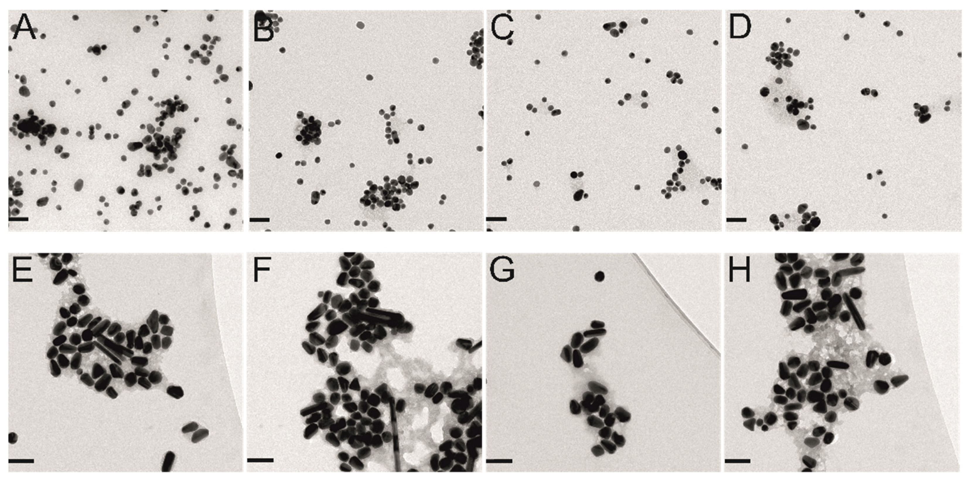
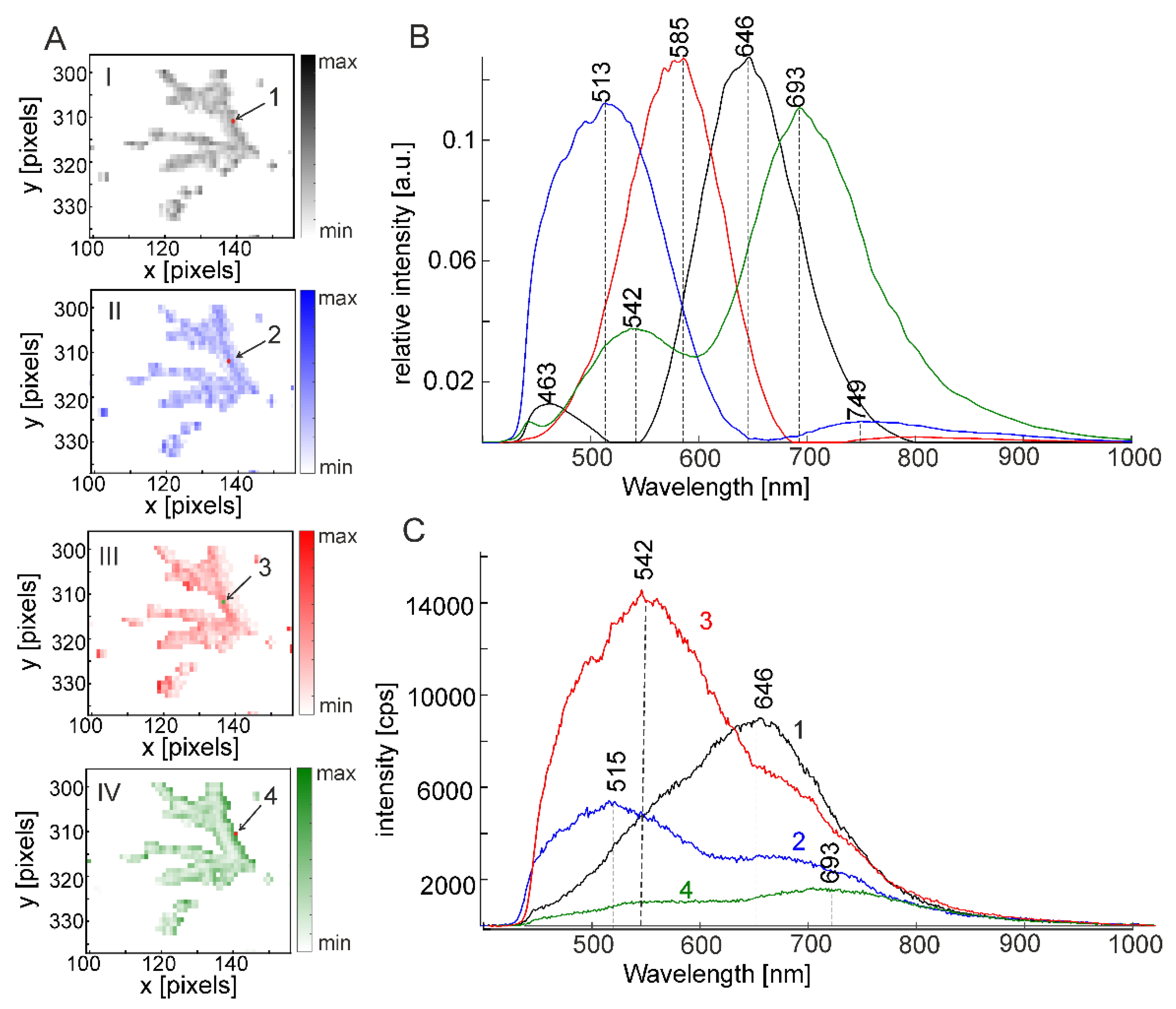
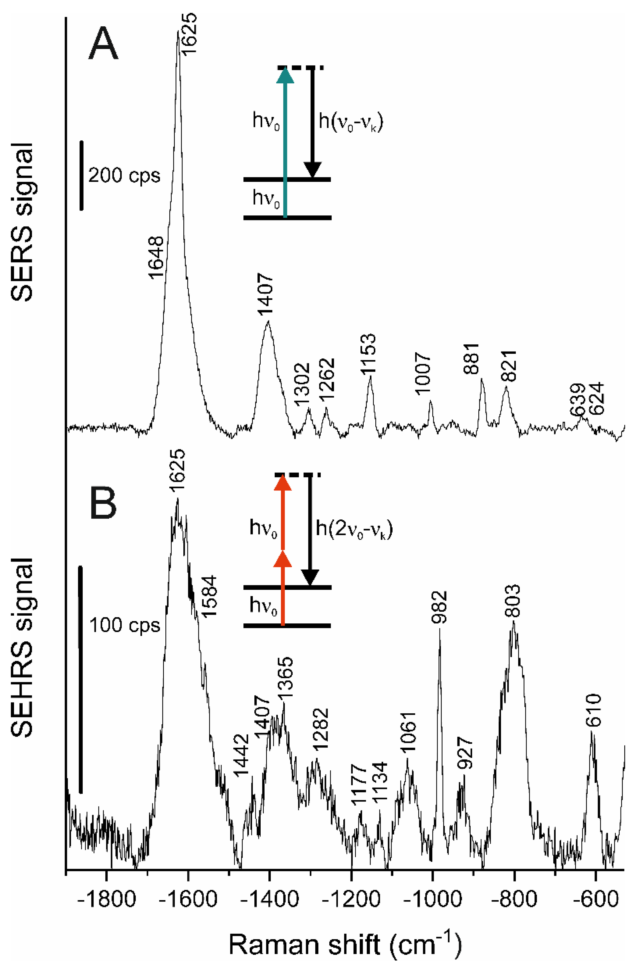
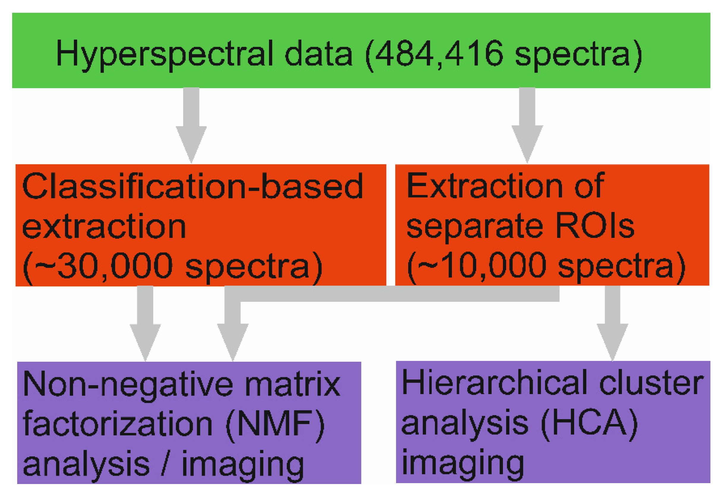
Dark field scattering microscopy can create large hyperspectral data sets that contain a wealth of information on the properties and the molecular environment of noble metal nanoparticles. For a quick screening of samples of microscopic dimensions that contain many different types of plasmonic nanostructures, we propose a multivariate analysis of data sets of thousands to several hundreds of thousands of scattering spectra. By using non-negative matrix factorization for decomposing the spectra, components are identified that represent individual plasmon resonances and relative contributions of these resonances to particular microscopic focal volumes in the mapping data sets. Using data from silver and gold nanoparticles in the presence of different molecules, including gold nanoparticle-protein agglomerates or silver nanoparticles forming aggregates in the presence of acrylamide, plasmonic properties are observed that differ from those of the original nanoparticles. For the case of acrylamide, we show that the plasmon resonances of the silver nanoparticles are ideally suited to support surface enhanced Raman scattering (SERS) and the two-photon excited process of surface enhanced hyper Raman scattering (SEHRS). Both vibrational tools give complementary information on the in situ formed polyacrylamide and the molecular composition at the nanoparticle surface.
Keywords: localized surface plasmon resonances; gold nanoparticles; silver nanoparticles; dark field microscopy; acrylamide; hyperspectral imaging; non-negative matrix factorization; surface-enhanced Raman scattering (SERS); surface-enhanced hyper Raman scattering (SEHRS) localized surface plasmon resonances; gold nanoparticles; silver nanoparticles; dark field microscopy; acrylamide; hyperspectral imaging; non-negative matrix factorization; surface-enhanced Raman scattering (SERS); surface-enhanced hyper Raman scattering (SEHRS)