Article

Antimicrobial and Cytotoxic Activities of Constituents from the Fruit of Albizia lebbeck L. Benth (Fabaceae)

by
Bosco Peron Leutcha
1,2,
Jean Paul Dzoyem
3,
Jean-Bosco Jouda
4,
Denis Kehdinga Sema
1,
Virginie Flaure Tsague Tankeu
1,
Gabin Thierry Mbahbou Bitchagno
5,6,
Billy Toussie Tchegnitegni
2,5,6,
Flaure Rosette Ehawa Essoung
7,
Bruno Ndjakou Lenta
2,
Siméon Fogue Kouam
2,
Florence Delie
8,
Alain Meli Lannang
4,9,* and
Norbert Sewald
6,*
1
Department of Chemistry, Faculty of Science, University of Maroua, Maroua P.O. Box 814, Cameroon
2
Department of Chemistry, Higher Teacher Training College, University of Yaoundé I, Yaoundé P.O. Box 47, Cameroon
3
Department of Biochemistry, Faculty of Science, University of Dschang, Dschang P.O. Box 67, Cameroon
4
Department of Chemical Engineering, School of Chemical Engineering and Mineral Industries, University of Ngaoundere, Ngaoundéré P.O. Box 454, Cameroon
5
Department of Chemistry, Faculty of Science, University of Dschang, Dschang P.O. Box 67, Cameroon
6
Department of Chemistry, Organic and Bioorganic Chemistry, Bielefeld University, 33501 Bielefeld, Germany
7
University Institute of Technology, University of Ngaoundere, Ngaoundere P.O. Box 455, Cameroon
8
Institute of Pharmaceutical Sciences of Western Switzerland, University of Geneva, Rue Michel-Servet 1, 1211 Geneva, Switzerland
9
Department of Chemistry, Higher Teacher Training College, University of Maroua, Maroua P.O. Box 55, Cameroon
*
Authors to whom correspondence should be addressed.
Molecules 2022, 27(15), 4823; https://doi.org/10.3390/molecules27154823
Submission received: 27 June 2022 / Revised: 16 July 2022 / Accepted: 27 July 2022 / Published: 28 July 2022

Abstract

:
Twenty-two compounds were isolated from the fruit of Albizia lebbeck including one unprecedented, rare amino acid-derived zwitterionic and one new flavone derivative. The isolation was performed on repeated column chromatography over silica gel and their structures were determined by 1D-, 2D-NMR and HR-ESI-MS spectra together with reported data in the literature. The chemophenetic significance is also discussed. Some isolated compounds were reported for the first time to be found in the species. Additionally, compound 2 showed antibacterial activity and compounds 1 and 2 revealed moderate cytotoxic activity against the Raw 264.7 cancer cell line with IC50 values of 37.19 µM and 29.36 µM, respectively. Furthermore, a proposed biosynthetic pathway of compound 1 is described.

1. Introduction

Albizia lebbeck (or Acacia lebbeck) is a tree found in the Fabaceae family, mostly reported in tropical areas and in rainfall forests and usually grows in lateritic and sandy soils [1]. During our ethnobotanical survey in Northern Cameroon, Albizia lebbeck was claimed by the local population to treat dysentery, asthma, hemorrhoids, bronchitis, eczema, leprosy, human fertility, and diarrhea, meanwhile some constituents of A. lebbeck are reported for their antimicrobial, antioxidant, anti-inflammatory, and toxicity properties [2,3,4,5,6,7]. In light of our study and as part of our ongoing research on bioactive metabolites from Cameroonian medicinal plants [8,9], we isolated a new amino acid-derived zwitterionic (1) and a new flavone flavonoid derivative (2) together with twenty known compounds from the fruit of A. lebbeck. Eugenol (5) is reported for the first time from the Fabaceae family while 29-hydroxyhopane (8), spinassterol (16), spinasterol-3-O-β-D-glucopyranoside (17), D-mannitol (20), octacosanoic acid (21), and bis ((S)-2, 3-dihydroxypropyl) (22) are reported for the first time from A. lebbeck. Herein, we report the isolation, structure elucidation, antimicrobial potential, as well as cytotoxic properties of the extracts and isolated compounds.

2. Results

Column chromatography (CC) of the CH2Cl2/MeOH (1/1, v/v) extract of the seeds and pods of A. lebbeck led to the isolation and structure elucidation of one unprecedented amino acid-derived zwitterionic named chiakine (1), one new flavone derivative, lebbeckisoetin A (2), and twenty known compounds (322) (Figure 1).

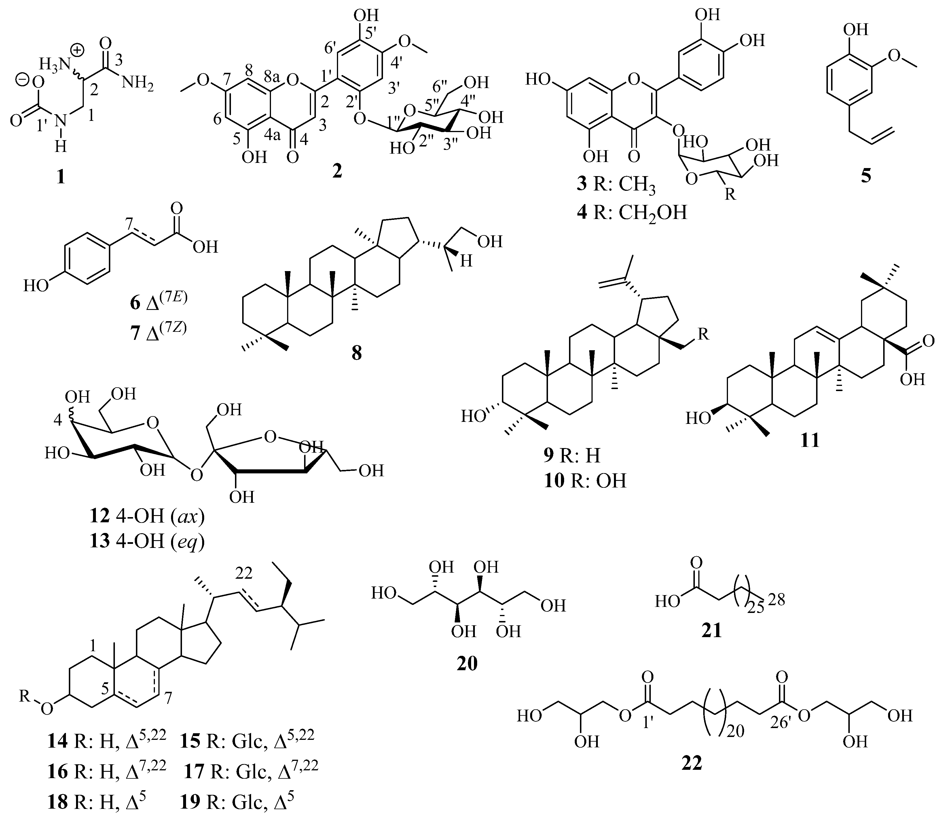
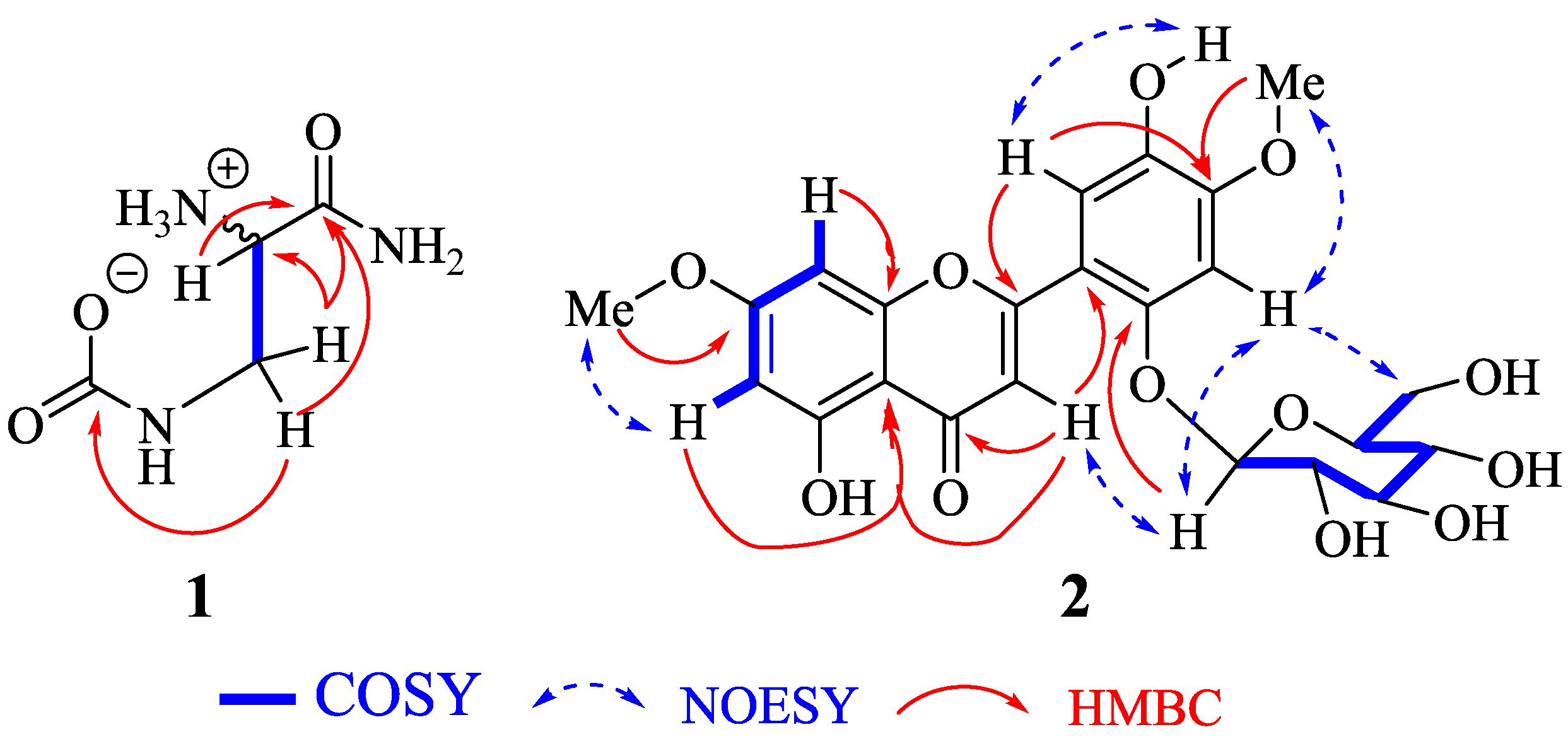
2.1. Structure Elucidation of Compounds 1 and 2

Compound 1 was obtained as an optically white powder which exhibits a levorotatory activity with [ α ] D 20 = −1.4 (c 0.5, MeOH). Its HR-ESI-MS showed a pseudomolecular ion peak [M+Na]+ at m/z 170.0542 (Figure S1) (calcd. m/z 170.0536 for C4H9N3O3Na) indicating two double-bond equivalents. The IR spectrum displayed absorption bands of OH (3281 cm−1), NH (2929 cm−1), ester/amide carbonyl (1634 cm−1) and C-O/C-N (1326/1157 cm−1). Its 13C-NMR data (Table 1) together with the HSQC spectrum (Figure S4) led to the identification of four carbon atoms including two carbonyls at δC 172.4 and 161.6, one sp3 methine at δC 55.4, and one sp3 methylene at δC 40.2. The above data (Table 1) suggested that compound 1 had aminomethine and aminomethylene groups [10]. Its 1H-NMR spectrum (Figure S2) exhibits signals of three groups of protons at δH 3.55 (ddd, J = 15.3, 3.6, 1.3 Hz, H-1′a), 3.35 (ddd, J = 15.2, 6.5, 1.4 Hz, H-1′b) and 3.70 (ddd, J = 6.5, 3.5, 1.3 Hz, H-2′). The COSY spectrum (Figure S5), led to the location of the vicinal as well as geminal correlations on the structure: H-1′a (δH 3.55) and H-1′b (δH 3.35) (gem), and H-2′ (δH 3.70), H-1′a (δH 3.55) and H-1′b (δH 3.35) (vic) (Figure 2). The HMBC spectrum (Figure S6) led to position both quaternary carbons through the correlation between H-1′a/H1′b (δH 3.55/3.35) with C-1 (δC 161.6) and C-3′ (δC 172.4) (Figure 2). Thus, compound 1 was identified as a new amino acid-derived zwitterionic natural product trivially named chiakine. Compound 1 would be derived from the hypothetical precursors carbamic acid (23) and phosphoseriamide (27). In this proposed pathway, the phosphoseriamide could be formed from the nucleotide precursor glycoaldehyde (24) [11] and glycolaldehyde-2-phosphate (25) via the intermediate glycolaldehyde phosphate aminonitrile (26) [12].
Compound 2 was obtained as an optically yellow amorphous powder which exhibits a dextrorotatory activity with [ α ] D 20 = +164.7 (c 0.5, MeOH). Its molecular formula C23H24O12 was established by the positive ion mode HR-ESI-MS which showed the pseudomolecular ion peak [M+H]+ at m/z 493.1336 (Figure S8) (calcd. m/z 493.1346 for C23H25O12) containing 12 double-bond equivalents. The IR spectrum displayed absorption bands of the hydroxyl group (3852 cm−1), carbonyl group (1733 cm−1), and C-O group (1206 cm−1). Its 1H-NMR spectrum (Figure S9) reveals the signals of a chelated proton at δH 13.05 (s) and a phenolic proton at δH 9.09 (s) both attributed to polyphenols. The signals of a singlet at δH 7.17 (1H) together with two doublets of a metasystem of a proton at δH 6.38 (d, J = 2.2 Hz) and 6.71 (d, J = 2.2 Hz) are characteristic of a tetrasubstituted ring A of a flavone derivative [13]. Moreover, the metasystem of the previously described protons is supported by the COSY spectrum (Figure S12) with correlation peaks between H-6 (δH 6.38) and H-8 (δH 6.71). In addition, two singlet signals are observed at δH 6.99 (1H, s, H-3′) and δH 7.37 (1H, s, H-6′) suggesting a pair of paraprotons in ring B, two singlets of methoxy protons at δH 3.87 (3H, s, MeO-7) and δH 3.86 (3H, s, MeO-4′). The 1H-NMR signal at δH 5.05 (d, J = 7.2 Hz) was assigned to the β-anomeric proton characteristic of a sugar moiety [14]. The 13C-NMR (Figure S10) together with DEPT-135 spectra reveal the signals of 23 carbon atoms, where the signals of a sugar moiety were identified as glucopyranosyl at δC 101.6 (C-1″), 73.9 (C-2″), 77.9 (C-3″), 70.5 (C-4″), 77.5 (C-5″), and 61.3 (C-6″) [8] together with those of two methoxy groups at δC 56.6 (MeO-7) and 56.5 (MeO-4′). The aglycone was identified from the aforementioned observation as a flavone flavonoid derivative with characteristic signals at δC 161.5 (C-2), 109.6 (C-3), 182.6 (C-4), 161.6 (C-5) 165.6 (C-7), 157.8 (C-8a), 150.6 (C-2′), and 151.9 (C-4′) [15,16]. Extensive analysis of COSY, HSQC, and HMBC (Figure 3) spectra allowed the connection of the functional groups to be assigned and the structure of compound 2 to be built. Furthermore, the cross correlation observed in the HMBC spectrum (Figure S13) between the methoxy protons at δH 3.87 (MeO-7) and δH 3.86 (MeO-4′) with carbon at δC 165.6 (C-7) and 151.9 (C-4′), respectively showed that there were linked at (C-7) and (C-4′). The linkage of the sugar moiety in the aglycone was assumed by the correlation between the anomeric proton at δH 5.05 (H-1″) and carbon at δC 150.6 (C-2′) and this finding was further confirmed by the NOESY spectrum. Indeed, extensive analysis of the NOESY spectrum showed a strong correlation between H-1″ (δH 5.05) with H-3 (δH 7.17) and H-3′ (δH 6.99), then between H-3′ (δH 6.99) and H-6″ (δH 3.74/3.43) (Figure 3), which unambiguously supports the position of sugar moiety at C-2′. Therefore, the structure of compound 2 was characterized as a new flavone derivative trivially named Lebbeckisoetin A.
The known compounds were identified based on the interpretation of their NMR (1D and 2D) spectra and confirmed by comparing the data to those described so far in the literature as: quericitrin (3) [9], quercetin-3-O-β-D-glucopyranoside (4) [16], eugenol (5) [3], (E)-p-coumaric acid (6) [17], (Z)-p-coumaric acid (7) [18], 29-hydroxyhopane (8) [19], lupeol (9) [20], betulin (10) [21], oleanolic acid (11) [22], lactulose or α-D-galactopyranoside-(1→2′)-β-D-fructofuranoside (12) [23], saccharose or α-D-glucopyranosyl-(1→2′)-β-D-fructofuranoside (13) [24], stigmasterol (14) [25], stigmasitosterol-3-O-β-D-glucopyranoside (15) [26], spinassterol (16) (Kamboj and Saluja, 2011), spinasterol-3-O-β-D-glucopyranoside (17) [27], β-sitosterol (18), β-sitosterol-3-O-β-D-glucopyranoside (19) [4,27], D-mannitol (20) [28], octacosanoic acid (21) [29], and hexacosan-1′,26′-dioate of bis ((S)-2, 3-dihydroxypropyl) (22) [30].

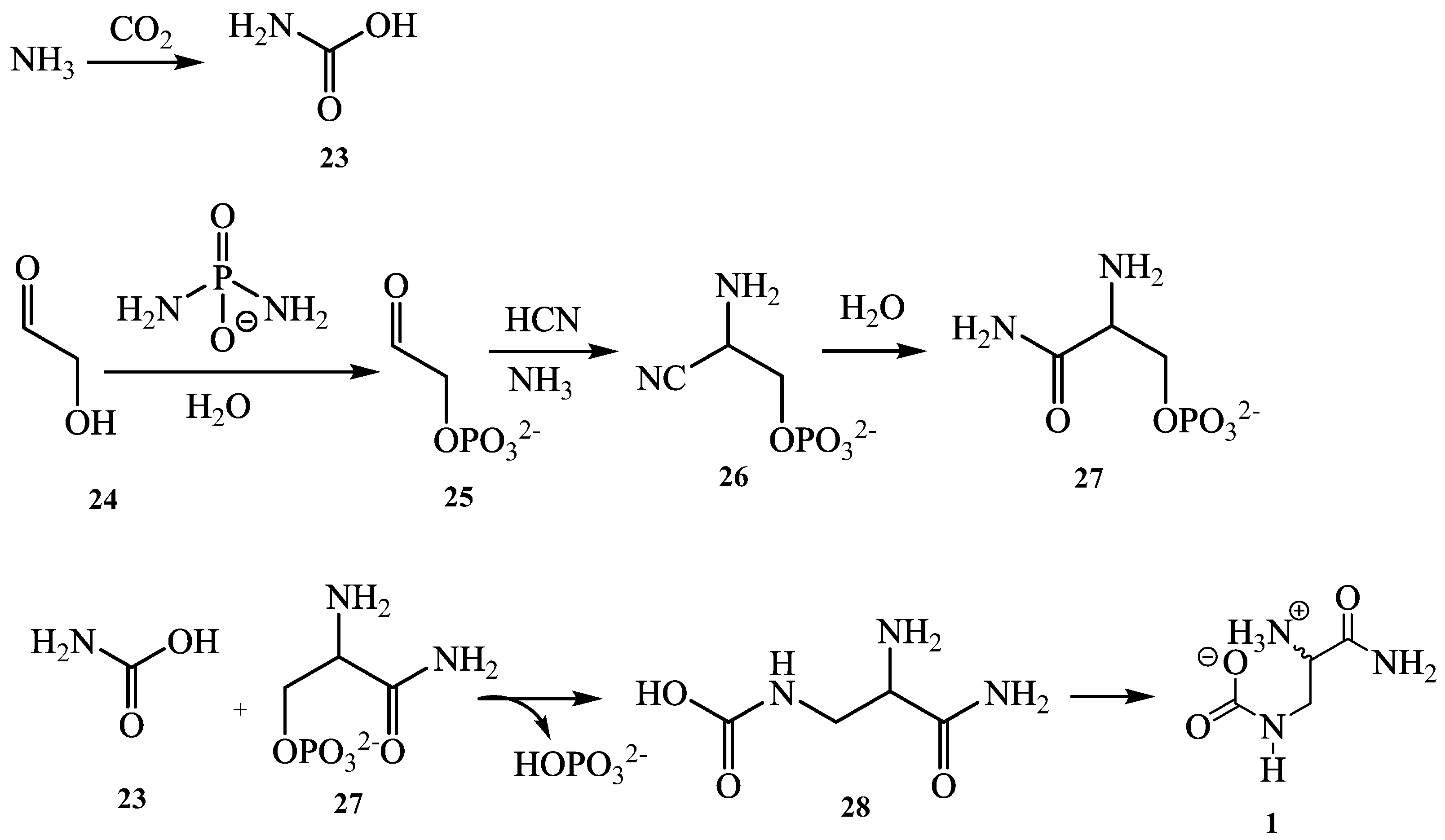
2.2. Physicochemical Characteristics of Compounds 1 and 2

Chiakine (1) white powder, 1H-NMR, and 13C-NMR (D2O, 600 MHz), see Table 1; HR-ESI-MS at m/z 170.0542 [M + Na]+ and m/z 148.0728 [M + H]+ (calcd. m/z 170.0536 for C4H9N3O3Na+ and m/z 148.0722 for C4H10N3O3+).
Lebbeckisoetin A (2) yellow amorphous powder, 1H-NMR, and 13C-NMR (DMSO-d6, 600 MHz), see Table 2; HR-ESI-MS at [M + H]+ at m/z 493.1336 (calcd. m/z 493.1346 for C23H25O12) and [M + Na]+ at m/z 515.1156 (calcd. m/z 515.1165 for C23H24O12Na+).

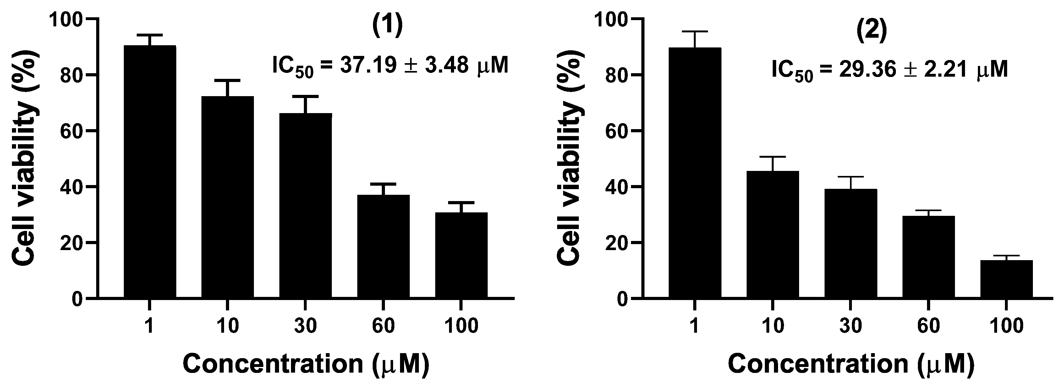
2.3. Biological Assays

The antimicrobial activities were performed on the pod crude extract and several isolated compounds using ketoconazole and ciprofloxacin as antifungal and antibacterial drug references. The interpretation of the result was based on the positive controls [31]. The CH2Cl2-MeOH (1:1, v/v) crude extract, n-hexane, ethyl acetate, and n-butanol extracts together with compounds 1, 2, 12, and 13 were tested against five microbial strains (Table 2). All the tested samples showed antimicrobial activity against at least one microbial strain, except the n-butanol extract which did not show any antimicrobial activity. Compounds 1 and 2 showed the most potent antifungal activity with MIC values of 32 µg/mL against Candida albicans while compound 2 further showed similar antibacterial activity against Escherichia coli. Compounds 12 and 13 were the least active in both bacteria and yeast strains. It clearly appears that the antimicrobial activity of Albizia lebbeck pods against Candida albicans and Escherichia coli increased with fractionation, from the CH2Cl2-MeOH (1:1, v/v) crude extract (MIC values of 512 µg/mL and 256 µg/mL) to the ethyl acetate extract (MIC values of 128 µg/mL) and finally to compounds 1 and 2, which were the most active (MIC values of 32 µg/mL). The above results are in accordance with previous reports of antimicrobial assays on the seeds, pods, and flowers of A. lebbeck [10,32] and could support the local uses of the fruit of this plant for the treatment of child and adult diarrhea. The mouse macrophages raw 264.7 cancer cell line was used to investigate the in vitro cytotoxicity of compounds 1 and 2 using the WST-1 assay. The concentration–response graph (Figure 4) showed that compounds 1 and 2 inhibited the proliferation of Raw 264.7 cancer cells in a dose-dependent manner. The IC50 values were determined based on the percentage of viable cells less than 50%. The two compounds showed moderate cytotoxic activity against Raw 264.7 with IC50 values of 37.19 µM and 29.36 µM, respectively. Raw 264.7 cell line has been exhaustively used for the screening of secondary metabolites for their cytotoxic activities [33,34]. However, the high sensitivity of Raw 264.7 compared to other cancer cell lines, such as human leukemia monocyte cell line THP-1 and the human lung adenocarcinoma A549 cell lines, has been previously reported [8]. To the best of the author’s knowledge, the anticancer properties of an isolated compound from the seeds of A. lebbeck have never been reported. The obtained activity of chiakine (1) and lebbeckisoetin A (2) on Raw 264.7 cells indicated that these compounds might be potential candidates for new anticancer agents.

3. Discussion

Twenty-two (122) compounds were isolated using chromatographic techniques from the crude extracts of the fruit of Albizia lebbeck L. Benth, including a new carbamide (1) and flavone (2) derivatives together with twenty known compounds grouped into two flavonoids (3, 4), three alkylphenols (57), four triterpenoids (811), three sugars (12, 13, 20), three sterols (14, 16, 18), three saponins (15, 17, 18), and two fatty acids (21, 22). To the best of our knowledge, compounds (1, 2, 5, 8, 16, 17, 20, 21, and 22) are herein reported for the first time from A. lebbeck. This is the first report of compound 5 in either the Mimosaceae subfamily or the Fabaceae family. Compound 5 is considered a chemomarker of Syzygium aromaticum, well known as “Clou de Girolf” [3]. However, compounds 1 and 2 were found to be new and are reported for the first time from the Fabaceae family as their derivatives were reported from A. lebbeck [10,35]. This is the first report of compounds 8, 16, 17, 20, 21, and 22 from Albizia lebbeck, although they have already been reported from the Albizia genus as 8 (Albizia chinensis) [36], 1617 (Albizia dealbata and Albizia melanoxylon) [28] and 2022 (Albizia Amara and Albizia julibrissin) [37,38]. This paper has then strengthened the chemophenetic survey on the Albizia genus. These observations give new insights into the occurrence of flavonoids and saponins in the genus Albizia and the family Fabaceae. The flavone flavonoids might represent a new significant chemophenetic finding in the Albizia genus, specifically in the Albizia lebbeck plant. However, saponins in general, could be considered chemotaxonomic markers for the genus Albizia due to the soapy aspect of all the species. The study of the fruit of A. lebbeck L. Benth led to the isolation of a chemical constituent which enriches the information on the phytochemistry of the plant and provides further knowledge in regard to the possible chemotaxonomic markers present in the A. lebbeck species, the Albizia genus, as well as the Fabaceae family. It is notable that compounds 1, 2, and 5 have not yet been reported either from the Albizia genus or the Fabaceae family and suggest a new observation of chemotaxonomic knowledge of the Albizia and more diverse chemical constituents from A. lebbeck L. Benth.

4. Materials and Methods

4.1. General Experimental Procedures

The melting points were measured on a Gallenkamp melting point apparatus. Optical rotations were recorded with a JASCO DIP-360 polarimeter. The IR spectrum was measured in MeOH on a JASCO A-302 spectrophotometer. The proton and carbon (1D and 2D)-NMR (600/500 MHz) spectra were measured on a Bruker AMX machine. The chemical shifts of proton and carbon were recorded based on the internal reference TMS (Tetramethylsilane) in δ (ppm). Moreover, coupling constants (J) were measured in Hz. The ESIMS was recorded on a double-focusing mass spectrometer (Varian MAT 311A), while HREIMS was recorded on a JEOL HX 110 mass spectrometer. Silica gel 60 (0.2–0.5 mm and 0.2–0.063 mm) (70–230 and 240–300 mesh sizes, E. Merck) was used as a stationary phase for CC (Column Chromatography). The purity of compounds and the monitoring of fractions were based on precoated silica gel TLC (Thin Layer Chromatography) plates supported on either plastic or aluminum sheets (E. Merck, F254). Spots were visualized on TLC with UV light (254 nm and 365 nm) on a CN-6 UV spectrometer (made in France) then sprayed with ceric sulphate and heated at about 90 °C.

4.2. Plant Material

The fruit of A. lebbeck was harvested on 20 April 2018 in Maroua town, headquarters of the Far-North Region of Cameroon and identified by Mr Tadjouteu Flubert by comparing it with a specimen available at the National Herbarium at Yaoundé with a Voucher specimen 58964/NHC.

4.3. Extraction and Isolation

The plant material used for this work was the mature and dry fruit of Albizia lebbeck. The pale-yellow seeds of A. lebbeck were powdered (1.3 kg) and extracted for 72 h with CH2Cl2/MeOH (1/1, v/v) at room temperature. The mixture was filtered and taken to dryness under reduced pressure at about 60 °C to afford a crude extract of 120.3 g. Compound 1 (2.1 g) was precipitated in the crude extract and was obtained by filtration. Then, 100.0 g of this extract were subjected to open CC over silica gel and eluted with a gradient of n-hexane/CH2Cl2/MeOH to afford four major fractions: A (1.2 g), B (1.9 g), C (60.5 g), and D (10.8 g). Fraction A (1.2 g) was purified using CC (isocratic, 15 % n-hexane/CH2Cl2) to afford 21 (15 mg) and 8 (11 mg). Fraction B (1.9 g) was also subjected to open CC (gradient, CH2Cl2/EtOAc/MeOH) which led to 9 (19 mg), 5 (89 mg), 10 (10 mg), 11 (20 mg), and 20 (17 mg). Fraction C (60.5 g) was submitted to open CC (gradient, EtOAc/MeOH) to yield 6 (150 mg), 7 (56 mg), 12 (705 mg), and 13 (969 mg). Fraction D (10.8 g) was purified using open CC (silica gel, gradient, EtOAc/MeOH) which led to 19 (704 mg), 15 (908 mg), and 17 (431 mg).
Furthermore, 10.3 kg of powdered pods of A. lebbeck were extracted for 72 h with CH2Cl2-MeOH (1/1, v/v). The mixture was filtered and taken to dryness under 300 mbar at 40 °C to afford 500.6 g of crude extract. Then 480.0 g of crude extract were further successively extracted with n-hexane, ethyl acetate, and n-butanol to afford 203.8 g, 23.5 g, and 109.9 g of residues, respectively. A portion of the n-hexane extract (200.9 g) was subjected to CC (gradient, n-hexane/CH2Cl2) to afford 18 (90.2 mg), a mixture of 14 and 16 (105.1 mg), and 22 (80.2 mg). Next 20.0 g of ethyl acetate extract was subjected to CC (n-hexane/CH2Cl2/EtOAc/MeOH) to yield four fractions F (2.0 g), G (2.9 g), H (6.5 g), and I (3.8 g). Finally, fraction I (3.8 g) was run over open CC (EtOAc/MeOH) to afford 2 (5 mg), 17 (5 mg), 3 (7 mg), and 4 (4.3 mg).

4.4. Antimicrobial Assay

Five microorganisms from American Type Culture Collection including four bacterial (two Gram-positive and two Gram-negative) and one fungal strain were used: Candida albicans ATCC 9028, Staphylococcus aureus (ATCC 1026), Enterococcus faecalis (ATCC 29212), Escherichia coli (ATCC 25922), and Pseudomonas aeruginosa (ATCC 74117). The antibacterial activity of the crude extract, fractions, and compounds was assessed by determining the minimum inhibitory concentration (MIC) and minimum bactericidal and fungicidal concentration (CBC and MFC) using the broth microdilution method, as previously described [39]. Briefly, the MIC was performed by broth microdilution method, with Mueller–Hinton broth for bacteria and Sabouraud dextrose broth for yeast. Stock solutions of samples were prepared in 100% dimethyl sulfoxide and twofold serial dilutions were prepared in media in amounts of 100 μL per well in a 96-well. Microbial suspensions were prepared in culture media, and 100 μL of this inoculum was added to each well of the plate, resulting in a final inoculum of 1.5 × 10⁶ CFU/mL for bacteria and 2 × 10⁴ CFU/mL for yeast. The final concentration of samples ranged from 2 μg/mL to 256 μg/mL and from 8 μg/mL to 1024 μg/mL for extracts. The medium without the agents was used as a growth control and the blank control used contained only the medium. Ciprofloxacin and ketoconazole served as the standard drug controls. The MIC of samples was detected after 24 h (for bacteria) and 48 h (for fungi) of incubation at 37 °C, following an addition (40 µL) of 0.2 mg/mL of p-iodonitrotetrazolium (INT) chloride and incubation at 37 °C for 30 min, as the lowest sample concentration that prevented the color change of the medium and exhibited complete inhibition of microbial growth. The MBC or MFC was determined by adding 50 µL aliquots of the preparations, which did not show any growth after incubation during MIC assays to 150 µL of adequate broth. These preparations were incubated at 37 °C for 48 h. The MBC or MFC was regarded as the lowest concentration of a sample which did not produce a color change after an addition of INT, as mentioned above.

4.5. Cytotoxicity Assay

The cytotoxicity of unprecedented carbamide (1) and flavone (2) derivatives were evaluated on the Raw 264.7 cells (ATCC) cancer cell line. Cells were cultured in DMEM (Dulbecco’s Modified Eagle’s Medium) culture media supplemented with 10% fetal calf serum and 1% antibiotics (100 IU/mL penicillin and 100 μL/mL streptomycin) and maintained at 37 °C in a humidified atmosphere containing 5% CO2. The WST-1 assay was used to quantify the cell viability and the cytotoxicity was evaluated by determining the concentration inhibiting 50% of viable cells (IC50), as previously described [8].

Supplementary Materials

The following supporting information can be downloaded at: https://www.mdpi.com/article/10.3390/molecules27154823/s1, Figure S1: HR-ESI-MS (+) spectra of compound 1, Figure S2: 1H-NMR spectrum (D2O, 600 MHz) of compound 1, Figure S3: 13C-NMR spectrum (D2O, 150 MHz) of compound 1, Figure S4: HSQC spectrum of compound 1, Figure S5: COSY 1H-1H spectrum of compound 1, Figure S6: HMBC spectrum of compound 1, Figure S7: NOESY spectrum of compound 1, Figure S8: HR-ESI-MS (+) spectrum of compound 2, Figure S9: 1H-NMR spectra (600 MHz, DMSO-d6) of compound 2, Figure S10: 13C-NMR and DEPT-135 spectra (150 MHz, DMSO-d6) of compound 2, Figure S11: HMQC spectrum of compound 2, Figure S12: COSY 1H-1H spectrum of compound 2, Figure S13: HMBC spectrum of compound 2, Figure S14: NOESY spectrum of compound 2, Figure S15: 1H-NMR spectrum (500 MHz, MeOD) of compound 3, Figure S16: 13C-NMR and DEPT-135 spectra (125 MHz, MeOD) of compound 3, Figure S17: 1H-NMR spectrum (500 MHz, MeOD) of compound 4, Figure S18: 13C-NMR and DEPT135 spectra (125 MHz, MeOD) of compound 4, Figure S19: 1H-NMR spectrum (MeOD, 500 MHz) of compound 5, Figure S20: 13C-NMR and DEPT-135 spectra (MeOD, 125 MHz) of compound 5, Figure S21: 1H-NMR spectrum (CDCl3; 500 MHz) of compounds 6 and 7, Figure S22: 13C-NMR and DEPT135 spectra (CDCl3; 125 MHz) of compounds 6 and 7, Figure S23: 1H-NMR spectrum (CDCl3, 500 MHz) of compound 8, Figure S24: 13C-NMR and DEPT-135 spectra (CDCl3, 125 MHz) of compound 8, Figure S25: 1H-NMR spectrum (CDCl3, 400 MHz) of compound 9, Figure S26: 1H-NMR spectrum (CDCl3, 500 MHz) of compound 10, Figure S27: 13C-NMR and DEPT-135 spectra (CDCl3, 125 MHz) of compound 10, Figure S28: 1H-NMR spectrum (MeOD; 600 MHz) of compound 11, Figure S29: 13C-NMR and DEPT-135 spectra (MeOD; 600 MHz) of compound 11, Figure S30: 1H-NMR spectrum (D2O, 600 MHz) of compound 12, Figure S31: 13C-NMR spectrum (D2O, 125 MHz) of compound 12, Figure S32: 1H-NMR spectrum (DMSO-d6, 600 MHz) of compound 13, Figure S33: 13C-NMR spectrum (DMSO-d6, 125 MHz) of compound 13, Figure S34: 1H-NMR spectrum (CDCl3, 500 MHz) of compounds 14 and 16, Figure S35: 1H-NMR spectrum (DMSO-d6, 500 MHz) of compounds 15 and 17, Figure S36: 13C-NMR spectrum (DMSO-d6, 125 MHz) of compound 17, Figure S37: 1H-NMR spectrum (CDCl3, 500 MHz) of compound 18, Figure S38: 1H-NMR spectrum (MeOD, 500 MHz) of compound 19, Figure S39: 13C-NMR spectrum (CDCl3/MeOD, 125 MHz) of compound 19, Figure S40: 1H-NMR spectrum (DMSO-d6, 500 MHz) of compound 20, Figure S41: 13C-NMR spectrum (DMSO-d6, 500 MHz) of compound 20, Figure S42: 1H-NMR spectrum (Pyridine-d5, 500 MHz) of compound 21, Figure S43: 13C-NMR spectrum (Pyridine-d5, 125 MHz) of compound 21, Figure S44: 1H-NMR spectrum (DMSO-d6, 500 MHz) of compound 22, Figure S45: 13C-NMR and DEPT-135 spectra (DMSO-d6, 125 MHz) of compound 22.

Author Contributions

Conceptualization, B.P.L.; investigation B.P.L. and J.P.D.; methodology, B.P.L., D.K.S. and V.F.T.T.; writing—original draft, B.P.L. and F.R.E.E.; formal analysis, G.T.M.B., B.T.T. and J.-B.J.; writing—review & editing, J.-B.J. and A.M.L.; resources, B.N.L., F.D. and N.S.; funding acquisition, N.S.; supervision, S.F.K., F.D. and A.M.L. All authors have read and agreed to the published version of the manuscript.

Funding

This research was funded by the Alexander von Humboldt–Stiftung Foundation through the Georg Forster Fellowship for Experienced Researchers (ID N° 1137675) granted to A.M.L., the Yaoundé–Bielefield Graduate School of Natural Products with Antiparasite and Antibacterial Activities (YaBiNaPA) which is supported by the German Academic Exchange Service (DAAD project N° 57316173), and the Swiss National Foundation for providing financial support to J.P.D through the project n° IZSEZO_180383/1.

Institutional Review Board Statement

Not applicable.

Informed Consent Statement

Not applicable.

Data Availability Statement

The raw data supporting the conclusions of this article will be made available by the authors without undue reservation to any qualified researcher.

Acknowledgments

We acknowledge support for the publication costs by the Open Access Publication Fund of Bielefeld University and the Deutsche Forschungsgemeinschaft (DFG). AML thank the Alexander von Humboldt–Stiftung Foundation through the Georg Forster Fellowship for Experienced Researchers (ID N° 1137675) at the University of Bielefeld. They are also grateful to the German Academic Exchange Service (DAAD) for the financial support and facilities of the Yaoundé–Bielefield Graduate School of Natural Products with Antiparasite and Antibacterial Activities.

Conflicts of Interest

The authors declare that there is no conflict of interest.

Sample Availability

Samples of the compounds are not available from the authors.

References

  1. Al-Massarani, S.M.; El Gamal, A.A.; Abd El Halim, M.F.; Al-Said, M.S.; Abdel-Kader, M.S.; Basudan, O.A.; Alqasoumi, S.I. New acyclic secondary metabolites from the biologically active fraction of Albizia lebbeck flowers. Saudi Pharm. J. 2017, 25, 110–119. [Google Scholar] [CrossRef] [PubMed] [Green Version]
  2. Liu, H.; Mou, Y.; Zhao, J.; Wang, J.; Zhou, L.; Wang, M.; Yang, F. Flavonoids from Halostachys caspica and their antimicrobial and antioxidant activities. Molecules 2010, 15, 7933–7945. [Google Scholar] [CrossRef] [PubMed]
  3. Yang, C.J.; Gao, Y.; Du, K.Y.; Luo, X.Y. Screening of 17 Chinese medicine plants against phytopathogenic fungi and active component in Syzygium aromaticum. J. Plant Dis. Prot. 2020, 127, 237–244. [Google Scholar] [CrossRef]
  4. Abd El-Ghany, A.E.S.; Dora, G.; Abdallah, R.H.; Hassan, W.; El-Salam, E.A. Phytochemical and biological study of Albizia lebbeck stem bark. J. Chem. Pharma. Res. 2015, 7, 29–43. [Google Scholar]
  5. Tchinda, C.F.; Sonfack, G.; Simo, I.K.; Çelik, İ.; Voukeng, I.K.; Nganou, B.K.; Kuete, V. Antibacterial and antibiotic-modifying activities of fractions and compounds from Albizia adianthifolia against MDR Gram-negative enteric bacteria. BMC Complement. Altern. Med. 2019, 19, 120. [Google Scholar] [CrossRef] [PubMed] [Green Version]
  6. Ragasa, C.Y.; Lim, K. Sterols from Cucurbita maxima. Philipp. J. Sci. 2005, 134, 83. [Google Scholar]
  7. Dzoyem, J.P.; Melong, R.; Tsamo, A.T.; Tchinda, A.T.; Kapche, D.G.; Ngadjui, B.T.; Eloff, J.N. Cytotoxicity, antimicrobial and antioxidant activity of eight compounds isolated from Entada abyssinica (Fabaceae). BMC Res. Notes 2017, 10, 1–6. [Google Scholar] [CrossRef] [PubMed] [Green Version]
  8. Leutcha, B.P.; Sema, D.K.; Dzoyem, J.P.; Ayimele, G.A.; Nyongbela, K.D.; Delie, F.; Alléman, E.; Sewald, N.; Lannang, A.M. Cytotoxicity of a new tirucallane derivative isolated from Stereospermum acuminatissimum K. Schum stem bark. Nat. Prod. Res. 2020, 35, 4417–4422. [Google Scholar] [CrossRef] [PubMed]
  9. Lannang, A.M.; Sema, D.K.; Tatsimo, S.J.; Tankeu, V.T.; Tegha, H.F.; Wansi, J.D.; Shiono, Y.; Sewald, N. A new depsidone derivative from the leaves of Garcinia polyantha. Nat. Prod. Res. 2018, 32, 1033–1038. [Google Scholar] [CrossRef] [PubMed]
  10. Misra, L.N.; Dixit, A.K.; Wagner, H. N-demethylbudmunchiamines from Albizzia lebbeck seeds. Phytochemistry 1995, 39, 247–249. [Google Scholar] [CrossRef]
  11. Schaefer, H.W. Reaction of primary and secondary amines to form carbamic acid glucuronides. Curr. Drug Metab. 2006, 7, 873–881. [Google Scholar] [CrossRef] [PubMed]
  12. Coggins, J.A.; Powner, W.M. Prebiotic synthesis of phosphoenol pyruvate by α-phosphorylation-controlled triose glycolysis. Nat. Chem. 2017, 9, 310–317. [Google Scholar] [CrossRef] [PubMed]
  13. Koagne, R.R.; Annang, F.; Cautain, B.; Martín, J.; Pérez-Moreno, G.; Bitchagno, G.T.M.; González-Pacanowska, D.; Vicente, F.; Simo, I.K.; Reyes, F.; et al. Cytotoxycity and antiplasmodial activity of phenolic derivatives from Albizia zygia (DC.) J.F. Macbr. (Mimosaceae). BMC Complement. Med. Ther. 2020, 20, 1–8. [Google Scholar] [CrossRef] [PubMed]
  14. Agrawal, P.K. NMR spectroscopy in the structural elucidation of oligosaccharides and glycosides. Phytochemistry 1992, 31, 3307–3330. [Google Scholar] [CrossRef]
  15. Kosuge, T.; Ishida, H.; Satoh, T. Studies on Antihemorrhagic Substances in Herbs Classified as Hemostatics in Chinese Medicine. V. On Antihemorrhagic Principle in Biota orientalis (L.) ENDL. Chem. Pharm. Bull. 1985, 33, 206–209. [Google Scholar] [CrossRef] [PubMed] [Green Version]
  16. Islam, M.; Al-Amin, M.D.; Siddiqi, M.M.A.; Akter, S.; Haque, M.M.; Sultana, N.; Chowdhury, A.S. Isolation of Quercetin-3-O-β-D-glucopyranoside from the Leaves of AzadirachtaIndica and Antimicrobial and Cytotoxic screening of the Crude Extracts. Dhaka Univ. J. Sci. 2012, 60, 11–14. [Google Scholar] [CrossRef] [Green Version]
  17. Taewoong, R.; Kee, D.Y. Chemical Constituents of Nelumbonucifera Seeds. Nat. Prod. Sci. 2017, 23, 253–257. [Google Scholar]
  18. Hu, J.; Feng, X. Triterpenoids, p-coumaric acid esters and flavonoids from Artemisia igniaria. Planta Med. 2000, 66, 684–686. [Google Scholar]
  19. Kamaya, R.; Tanaka, Y.; Hiyama, R.; Ageta, H. Fern constituents: Triterpenoids isolated from the leaves of Cheiropleuria bicuspis. Chem. Pharm. Bull. 1990, 38, 2130–2132. [Google Scholar] [CrossRef] [Green Version]
  20. You, Y.J.; Nam, N.H.; Kim, Y.; Bae, K.H.; Ahn, B.Z. Antiangiogenic activity of lupeol from Bombax ceiba. Phytotherapy Res. 2003, 17, 341–344. [Google Scholar] [CrossRef] [PubMed]
  21. Tijjani, A.; Ndukwe, I.G.; Ayo, R.G. Isolation and characterization of lup-20(29)-ene-3, 28-diol (Betulin) from the stem-bark of Adeniumobesum (Apocynaceae). Trop. J. Pharm. Res. 2012, 11, 259–262. [Google Scholar] [CrossRef] [Green Version]
  22. Seebacher, W.; Simic, N.; Weis, R.; Saf, R.; Kunert, O. Complete assignments of 1H and 13C-NMR resonances of oleanolic acid, 18α-oleanolic acid, ursolic acid and their 11-oxo derivatives. Magn. Reason. Chem. 2003, 41, 636–638. [Google Scholar] [CrossRef]
  23. Edwards, J.C.; Hunter, J.M.; Nemzer, B.V. Multinuclear NMR of calcium fructoborate complex-structure, stability, and quantitation in the presence of other ingredients, excipients or adulterants. J. Food Res. 2014, 3, 115–131. [Google Scholar] [CrossRef] [Green Version]
  24. Jones, A.J.; Hanisch, P.; McPhail, A.K. Sucrose: An assignment of the 13C-NMR parameters by selective decoupling. Aust. J. Chem. 1979, 32, 2763–2766. [Google Scholar] [CrossRef]
  25. Kamboj, A.; Saluja, A.K. Isolation of stigmasterol and β-sitosterol from petroleum ether extract of aerial parts of Ageratum conyzoides (Asteraceae). Int. J. Pharm. Pharm. Sci. 2011, 3, 94–96. [Google Scholar]
  26. Ramaiarantsoa, H.; Koffi, A.B.; Assi, A.M.; Djakoure, L.A. Les 3-O-β-D-glucoside du β-sitostérol isolés des feuilles de Ravenala madagascariensis. J. Soc. Ouest-Afr. Chim. 2008, 26, 99–103. [Google Scholar]
  27. Freire, C.S.; Silvestre, A.J.; Neto, C.P. Demonstration of long-chain n-alkyl caffeates and Δ7-steryl glucosides in the bark of Acacia species by gas chromatography-mass spectrometry. Phytochem. Anal. Int. J. Plant. Chem. Biochem. Tech. 2007, 18, 151–156. [Google Scholar] [CrossRef] [PubMed]
  28. Hawkes, G.E.; Lewis, D. 1H nuclear magnetic resonance spectra and conformations of alditols in deuterium oxide. J. Chem. Soc. Perkin Trans. 1984, 2, 2073–2078. [Google Scholar] [CrossRef]
  29. Koay, Y.C.; Wong, K.C.; Osman, H.; Eldeen, I.; Asmawi, M.Z. Chemical constituents and biological activities of Strobilanthescrispus L. Rec. Nat. Prod. 2013, 7, 59–64. [Google Scholar]
  30. Melong, R.; Kapche, D.G.; Feussi, M.T.; Laatsch, H. A new aggreceride analogue and a peltogynoid isolated from the stem bark of Entadaabyssinica (Fabaceae). Nat. Prod. Commun. 2014, 9, 1934578X1400901024. [Google Scholar]
  31. Kuete, V. Potential of Cameroonian plants and derived products against microbial infections: A review. Planta Med. 2010, 76, 1479–1491. [Google Scholar] [CrossRef] [Green Version]
  32. Venkatesh, H.N.; Mohana, D.C. Antimicrobial activities of successive solvent extracts of Albizialebbeck and Solanumseaforthianum against some human pathogenic microorganisms. J. Pharm. Clin. Res. 2019, 12, 294–296. [Google Scholar]
  33. Tegha, H.F.; Jouda, J.B.; Dzoyem, J.P.; Sema, D.K.; Leutcha, B.P.; Allémann, E.; Delie, F.; Shiono, Y.; Sewald, N.; Lannang, A.M. A New Chromene Derivative and a New Polyalcohol Isolated from the Fungus Xylaria sp. 111A Associated with Garciniapolyantha Leaves. Nat. Prod. Commun. 2021, 16, 1934578X20987334. [Google Scholar]
  34. Kaoke, M.D.; Sema, D.K.; Dzoyem, J.P.; Leutcha, P.B.; Delie, F.; Allemann, E. A new cytotoxic glycocerebroside from RauvolfiamacrophyllaStapf. Nat. Prod. Chem. Res. 2020, 8, 1–5. [Google Scholar]
  35. Mishra, S.S.; Gothecha, V.K.; Sharma, A. Albizia lebbeck: A short review. J. Herb. Med. Toxicol. 2010, 4, 9–15. [Google Scholar]
  36. Tam, S.W. Flavonoid and Triterpenoid Constituents of the Ericaceae of Hong-Kong. Master’s Thesis, The University of Hong Kong, Hong Kong, China, 1961. [Google Scholar]
  37. Abdurrahman, I.; Cai-Xiab, Y. Isolation and characterization of fatty acid derivatives from the stem barks of Albizia Amara (Fabaceae), Sudanese Medicinal Plant. Chem. Methodol. 2020, 4, 369–377. [Google Scholar]
  38. Nehdi, I. Characteristics, chemical composition and utilisation of Albizia julibrissin seed oil. Ind. Crops Prod. 2011, 33, 30–34. [Google Scholar] [CrossRef]
  39. Dzoyem, J.P.; Tchamgoue, J.; Tchouankeu, J.C.; Kouam, S.F.; Choudhary, M.I.; Bakowsky, U. Antibacterial activity and cytotoxicity of flavonoids compounds isolated from Pseudarthriahookeri Wight &Arn. (Fabaceae). S. Afr. J. Bot. 2018, 114, 100–103. [Google Scholar]
Figure 1. Structures of compounds 122.
Molecules 27 04823 g001
Figure 2. COSY, HMBC, and NOESY correlations of compounds 1 and 2.
Molecules 27 04823 g002
Figure 3. Proposed biosynthetic pathway of compound 1.
Molecules 27 04823 g003
Figure 4. Cytotoxicity of compounds (1) and (2) against Raw 264.7 cancer cell line.
Molecules 27 04823 g004
Table 1. 13C and 1H-NMR spectroscopic data (1H 600 MHZ, 13C 150 MHZ for compounds 1 (in D2O) and 2 (in DMSO-d6).
12
δC,δH (m, J in Hz)HMBCδC,δH (m, J in Hz)HMBC
1′161.6 1
140.23.55 (ddd, 15.3, 3.6, 1.3)1, 2′, 3′2161.5
3.35 (ddd, 15.2, 6.5, 1.4)
255.43.70 (ddd, 6.5, 3.5, 1.3)1′, 3′3109.67.17 (s)2, 4, 4a, 1′
3172.4 4182.6
4a105.1
5161.6
698.36.38 (d, 2.2)4a, 5, 7, 8
7165.6
892.86.71 (d, 2.2)4a, 6, 7, 8a
8a157.8
1′111.6
2′150.6
3′101.16.99 (s)1′, 2′, 4′, 5′
4′151.9
5′141.5
6′114.47.37 (s)2, 2′, 4′, 5′
7-OMe56.63.87 (s)7
4′-OMe56.53.86 (s)4′
1″101.65.05 (d, 7.2)2′
2″73.93.35 (m)
3″77.93.43 (dd, 9.6, 4.0)
4″70.53.14 (td, 8.6, 4.8)
5″77.53.29 (m)
6″61.33.74 (dd, 9.7, 5.4)
3.43 (dd, 9.6, 4.0)
4′-OH 9.09 (s)4′
5-OH 13.05 (s)
d: doublet, dd: doublet of doublets, ddd: doublet of doublet of doublets, m: multiplet, s: singlet, td: triplet of doublets.
Table 2. Minimum Inhibitory Concentration (MIC), Minimum Bactericidal Concentration (MBC) and Minimal Fungicidal Concentration (MFC) values (in µg/mL) of extracts, fractions, and compounds from Albizia lebbeck fruit.
SamplesMicroorganisms
Candida albicansEscherichia coliStaphylococcus aureusPseudomonas aeruginosaEnterococcus faecalis
MICMFCMICMBCMICMBCMICMBCMICMBC
CH2Cl2-MeOH (1:1, v/v) extract512-128-512-256-2561024
n-hexane extract----512---512-
ethyl acetate extract128-1281024256-512-2561024
n-butanol extract----------
132256256-128-128---
2322563225625625612825664-
12256---256---256-
13----256-----
Ketoconazole0.564--------
Ciprofloxacin--251215122844
- = >1024 µg/mL for crude extracts and fractions and >256 µg/mL for compounds. Four bacteria strains and one yeast strain from American Type Culture Collection were used: Candida albicans ATCC 9028, Staphylococcus aureus (ATCC 1026), Enterococcus faecalis (ATCC 29212), Escherichia coli (ATCC 25922), and Pseudomonas aeruginosa (ATCC74117).
Publisher’s Note: MDPI stays neutral with regard to jurisdictional claims in published maps and institutional affiliations.

