Article

Conformational Properties and Putative Bioactive Targets for Novel Thiosemicarbazone Derivatives

1
Laboratory of Organic Chemistry, Department of Chemistry, National and Kapodistrian University of Athens, Panepistimioupolis Zografou, 11571 Athens, Greece
2
Department of Pharmacognosy and Natural Products Chemistry, Faculty of Pharmacy, National and Kapodistrian University of Athens, Panepistimiopolis Zografou, 15771 Athens, Greece
3
Slovenian NMR Centre, National Institute of Chemistry, SI-1001 Ljubljana, Slovenia
4
Laboratory of Physical Chemistry, Department of Chemistry, National and Kapodistrian University of Athens, Panepistimioupolis Zografou, 11571 Athens, Greece
5
Theoretical and Physical Chemistry Institute, National Hellenic Research Foundation, 48 Vassileos Constantinou Ave., 11635 Athens, Greece
*
Authors to whom correspondence should be addressed.
Molecules 2022, 27(14), 4548; https://doi.org/10.3390/molecules27144548
Submission received: 22 June 2022 / Revised: 7 July 2022 / Accepted: 14 July 2022 / Published: 16 July 2022

Abstract

:
The structure assignment and conformational analysis of the thiosemicarbazones, DKI21 and DKI24, were performed through homonuclear and heteronuclear 2D Nuclear Magnetic Resonance (NMR) spectroscopy (2D-COSY, 2D-NOESY, 2D-ROESY, 2D-HSQC, and 2D-HMBC) and quantum mechanics (QM) calculations, using Functional Density Theory (DFT). In addition, utilizing a combination of 2D-NOESY and 2D-ROESY spectra an exo structure was established for both of the analogs. This experimental results were confirmed by theoretical mechanistic studies, as the lowest minima conformations derived through DFT calculations were compatible with the spatial correlations observed in the 2D-NOESY and 2D-ROESY spectra. Finally, molecular binding experiments were performed to detect the potential targets for DKI21 and DKI24, derived from SwissAdme. In silico molecular binding experiments showed favorable binding energy values for the most of the enzymes studied. The ADMET calculations, using the preADMET and pKCSm software, showed that the two molecules appear as possible drug leads.

1. Introduction

Thiosemicarbazones represent a class of small molecules with various pharmacological properties [1], including antiviral [2], antibacterial [3], and antitumor activities [4]. Moreover, they represent key intermediates for a great variety of heterocyclic products, such as thiazolidine-4-ones.
Recently, the interest in thiazolidine-4-one derivatives has been increased among scientists due to their broad spectrum of biological activities, including antidiabetic, antibacterial, antifungal, anticancer, and anti-inflammatory, confirmed by numerous reviews on the activity and mechanisms of action of thiazolidine-4-ones [5,6,7,8,9].
The main synthetic route to thiazolidine-4-ones is the reaction between thiosemicarbazones and α-halo carboxylic esters, with several interesting structures obtained recently with this method [10,11,12,13].
However, the regio-outcome of this reaction has never been studied in detail, to the best of our knowledge, with the exception of Qian [14] who, apart from the expected thiazolidine-4-one as a major product, observed its regio-isomeric thiazolone and isolated the two regio-isomers as N-benzyl derivatives. This study triggered our research interest to perform a detailed study towards the refinement of this reaction regio-selectivity. In addition, a main target of our research was to unambiguously assign the regio-isomeric product through 2D-NOESY and 2D-ROESY spectroscopy methodologies and explain the product by theoretical means.
Thiosemicarbazones DKI reacted with methyl chloroacetate in ethanol in the presence of fused sodium acetate to give thiazolone/thiazolidinone derivatives, DKI21 and DKI24, as a mixture of regio-isomers I and II (Scheme 1). The geometric configuration of the double bond and the regio-chemistry of the thiazolylhydrazones are the topic of this research work.

2. Results

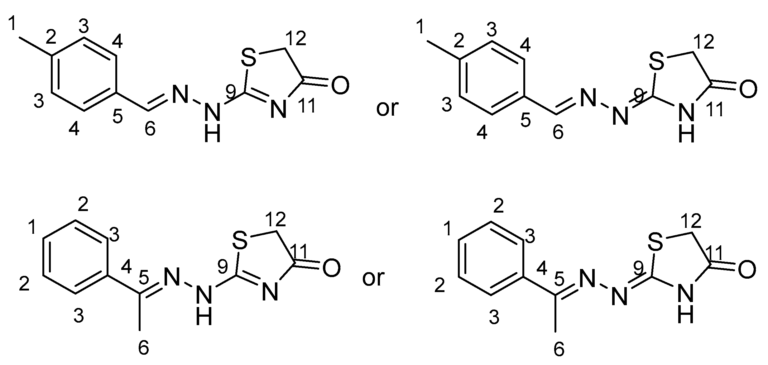
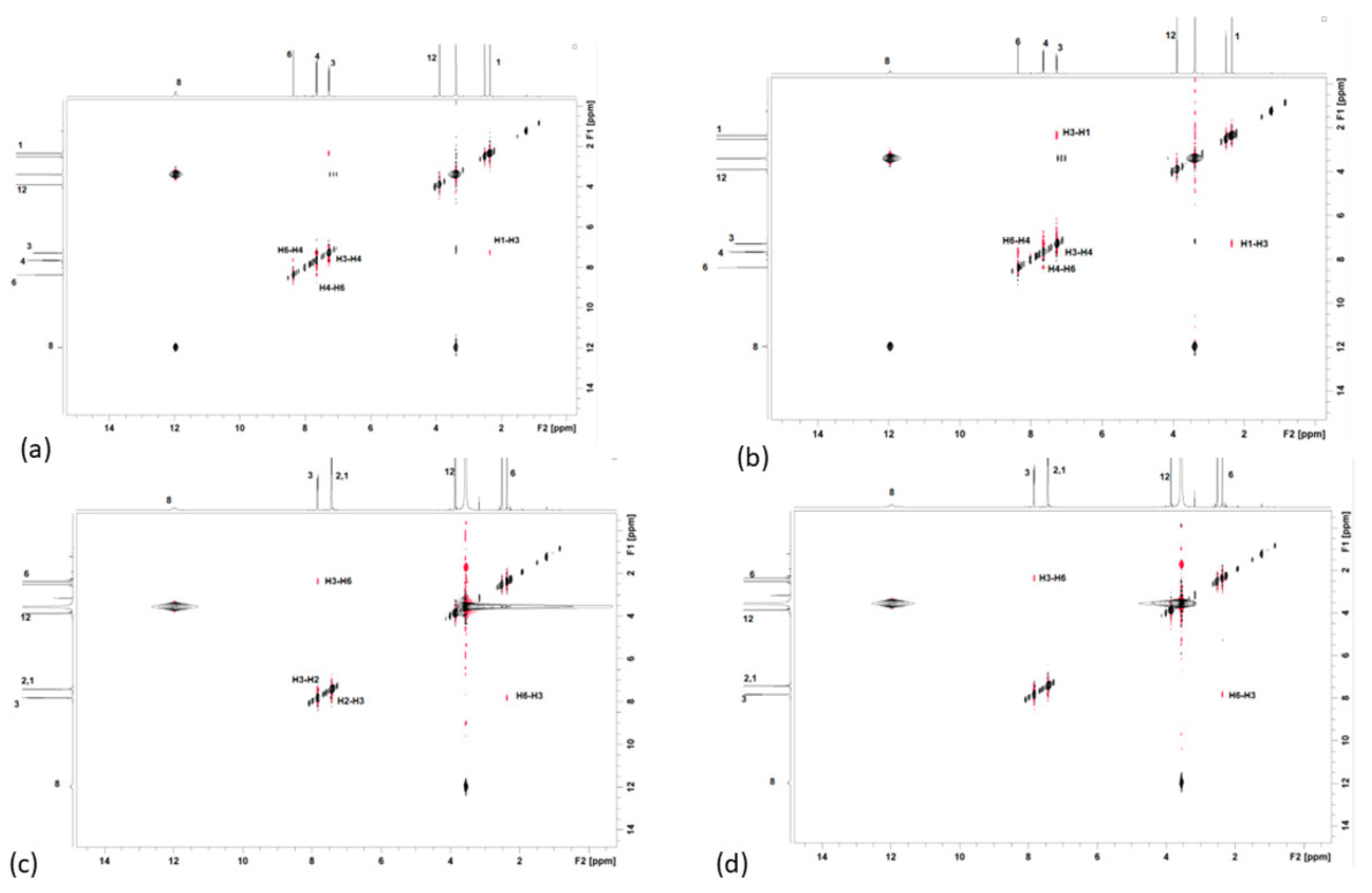
2.1. Structure Assignment

The possible structures of DKI21 and DKI24 are shown below in Figure 1.
The methyl group H-1 is a convenient starting point for the structure assignment of DKI21, which resonates at 2.35 ppm. Through 2D-NOESY and 2D-COSY, H-3 and H-4 are identified. Through 2D HSQC, the H-3 and H-4 show 1JC-H coupling with the C3 and C4, correspondingly, and, therefore, C3 and C4 are assigned unambiguously at 129.9 and 128.10 ppm, respectively. Through 2D-NOESY, H-6 is identified, because it shows spatial correlation with the H-4. H-12 is identified, as the remaining unidentified proton. Through 2D-HSQC, all of the carbons were identified, except for the quaternary and carbonyl ones. These carbons were identified through 2D-HMBC. Specifically, H-1 shows 2JC-H with C-2, H-4 shows 2JC-H with C-5, H-12 shows 2JC-H with C-11, and finally, H-12 shows 3JC-H with C-9. Based on this strategy, the complete identification of all of the proton and carbon atoms of the DKI21 molecule was achieved. The same procedure was achieved for the isomer DKI24 molecule. The two identification strategies, with all of the spectra obtained, are shown in detail in the Supplementary Materials.
Table 1 shows the proton chemical shifts of these compounds, their bond and spatial correlations, and heteronuclear correlations.

2.2. Conformational Analysis

DFT was used to predict the lowest energy conformations for DKI21 and DKI24. Various structures were used as the initial guess for optimization. The lowest conformations for DKI21 and DKI24 are shown below. Considering the predicted energy values, the structures of the compounds, and the correlations that were observed in the 2D-NOESY and 2D-ROESY spectra, the structures shown in (a) and (b) are taken as the most probable conformations for DKI21 and DKI24, respectively (Figure 2).
The correlations between the protons in 2D-NOESY, using mixing times 0.3 and 0.15 s, are shown below. These correlations were confirmed, using other mixing times and 2D-ROESY experiments (Figure 3).
According to the results, the exo compound is the one that matches with the experimental results. The reason for this is that it does not observe the correlation between the amino group (NH) and the H-6 in each compound.
Furthermore, the exo intermediate has a low energy barrier, due to the extended resonance forms that exo can undergo. These extending resonance forms cannot be applied for the higher energy endo analogue. In addition, in the DKI24 exo intermediate there is a hydrogen bond between proton 6 and nitrogen, while it does not favor in endo. Finally, in the DKI24 exo conformation the atoms 3, 4, 5, and 6 are at the same level, in contrast with the endo conformation. On the other hand, in DKI21, the exo intermediate is the most favorable because there is a repulsion between the amino proton and proton in carbon 6, that does not exist in exo.
Regarding the molecular orbitals of both the endo and exo conformations of both of the compounds, to derive a more quantitative answer, the HOMO molecular orbital is localized in the heterocyclic ring, while the LUMO molecular orbital is mainly localized in the aromatic ring and in the nitrogen atoms. The frontiers’ molecular orbitals are depicted in Figures S19 and S20 (Supplementary Materials).
Finally, the hardness (η), chemical potential (μ), and softness (S) of the lowest in the energy conformers were calculated from the energies of frontier HOMOs and LUMOs [15,16]. The following equations are used: η = ((LUMO) − ε(HOMO))/2;μ = (ε(LUMO) + ε(HOMO))/2; S = 1/η. It is found that the exo conformers present a lower H–L energy gap than the endo ones, and, thus, lower hardness and higher chemical softness, see Table S1 (Supplementary Materials). That shows that the exo intermediates are more reactive than endo intermediates. The energetics are given in the Supplementary Materials.

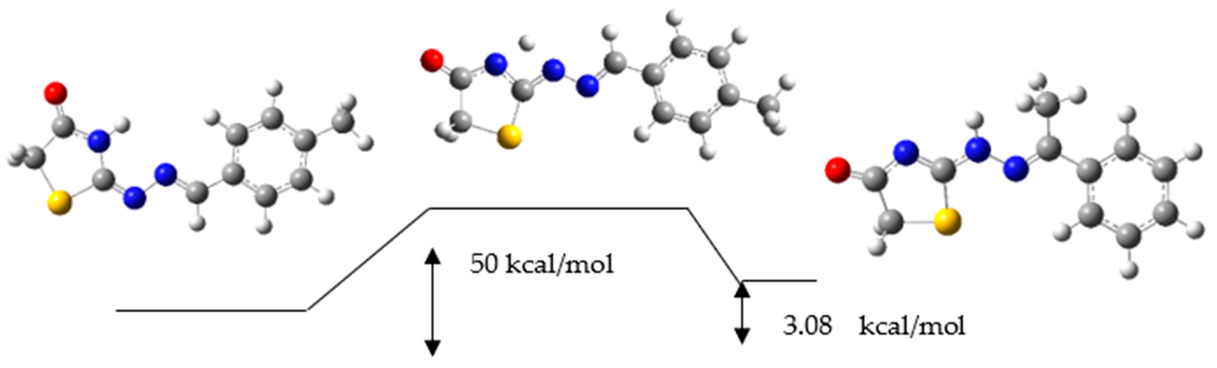
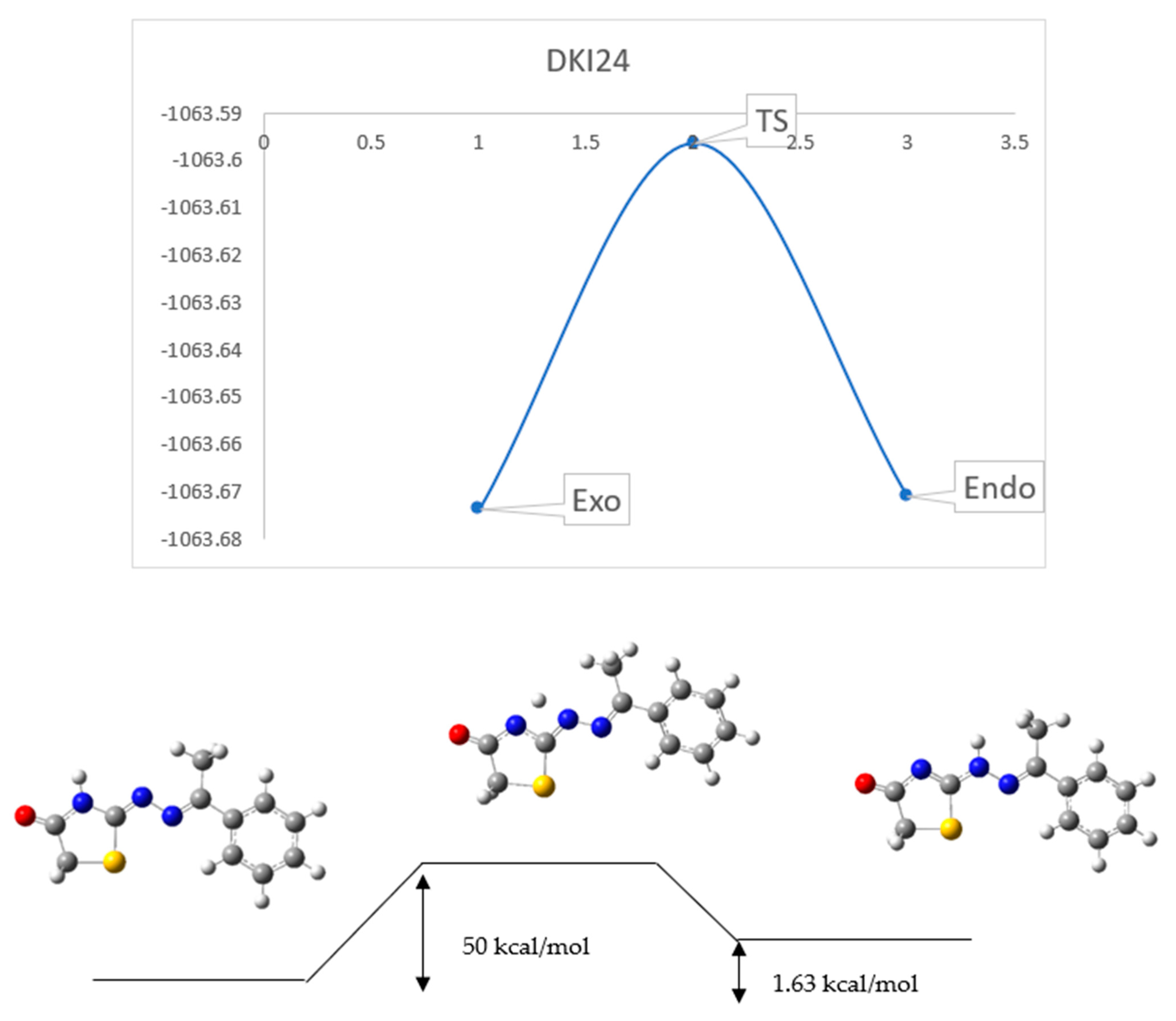
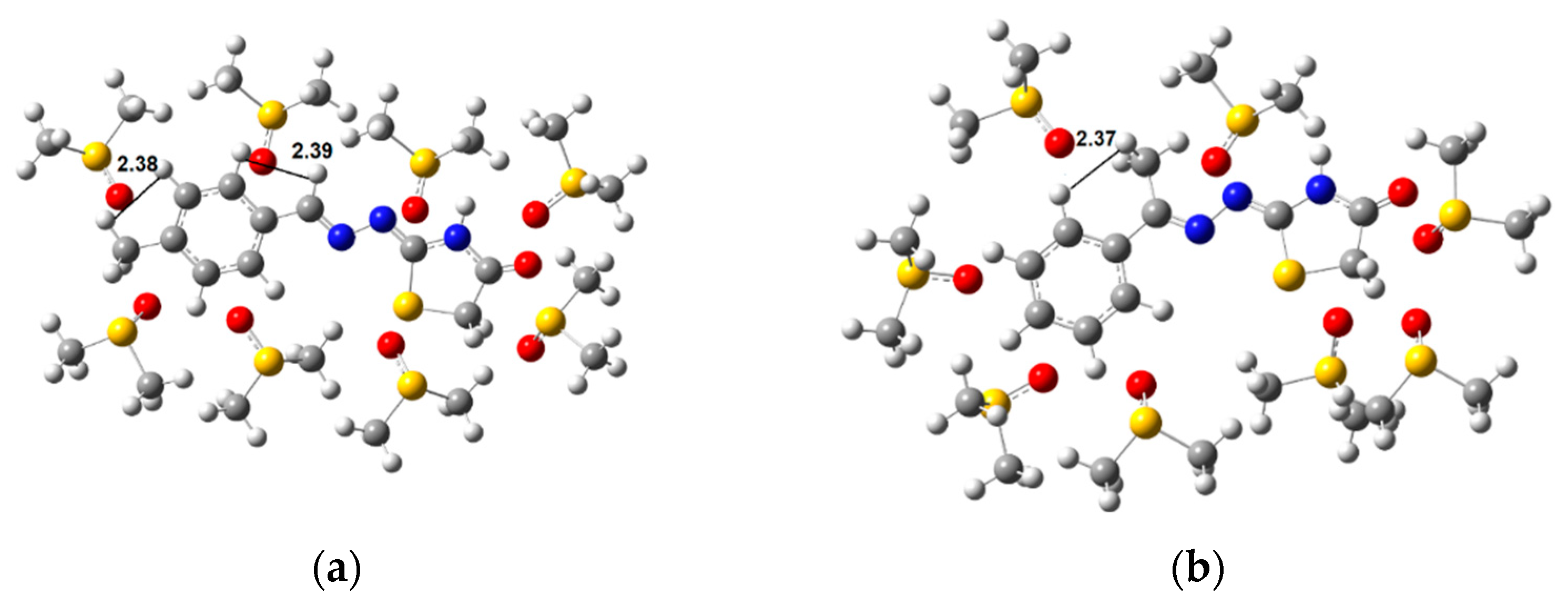
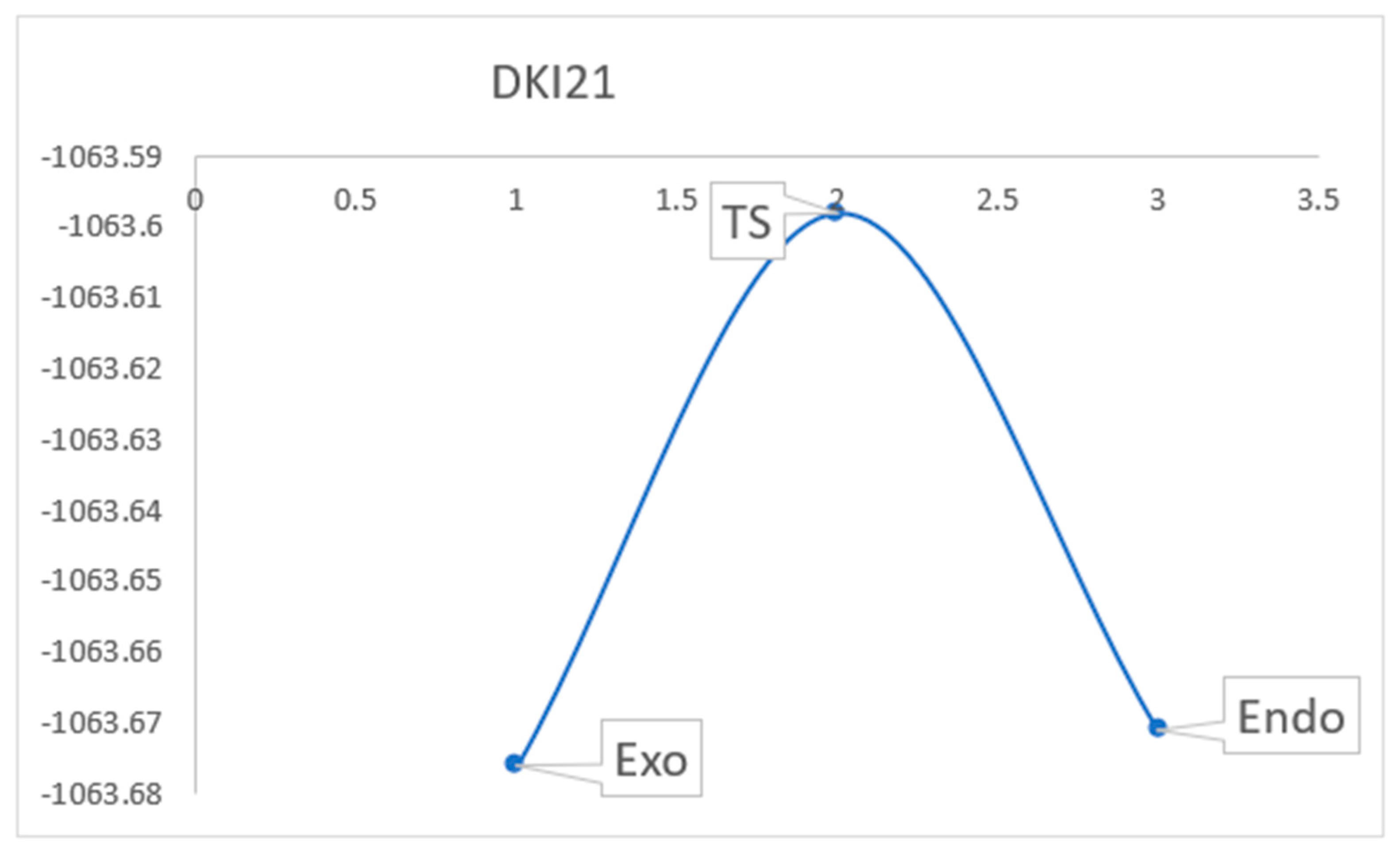
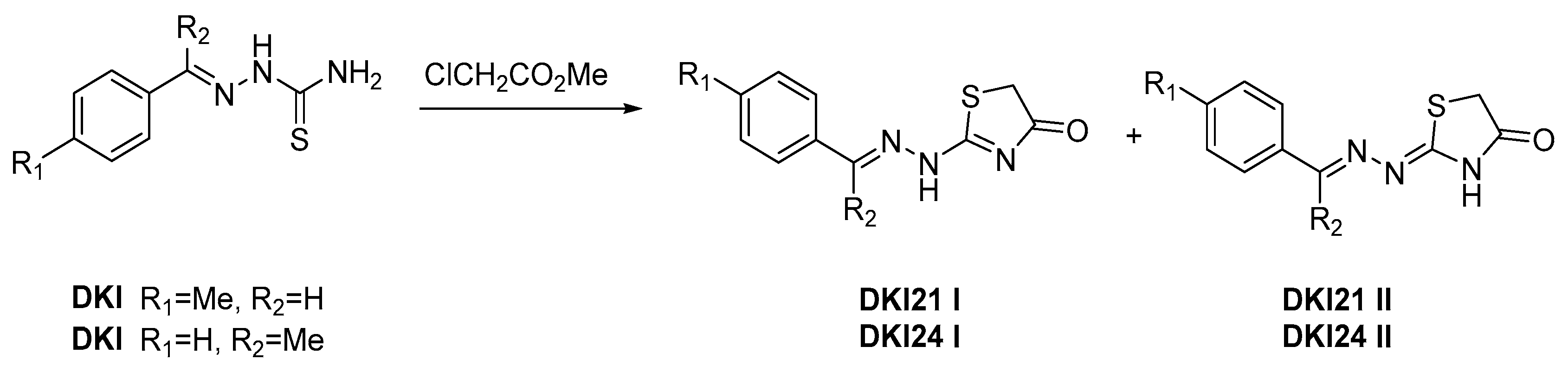
2.3. Reaction Mechanism

Using the DFT calculations, we have studied the reaction mechanism to confirm the experimental results. The first intermediate to study was the formation of the endo and exo double bond in the DKI21 compound.
An energy diagram of the formation between the endo and exo is shown below (Figure 4).
The next intermediate to study was the formation of the endo and exo double bond in the DKI24 compound (Figure 5).
An energy diagram of the formation between endo and exo is shown below.
It becomes clear from the above Figures that the exo intermediate compound has a very low energy barrier and it is therefore a preferable pathway. This comes in agreement with the NMR experiments.

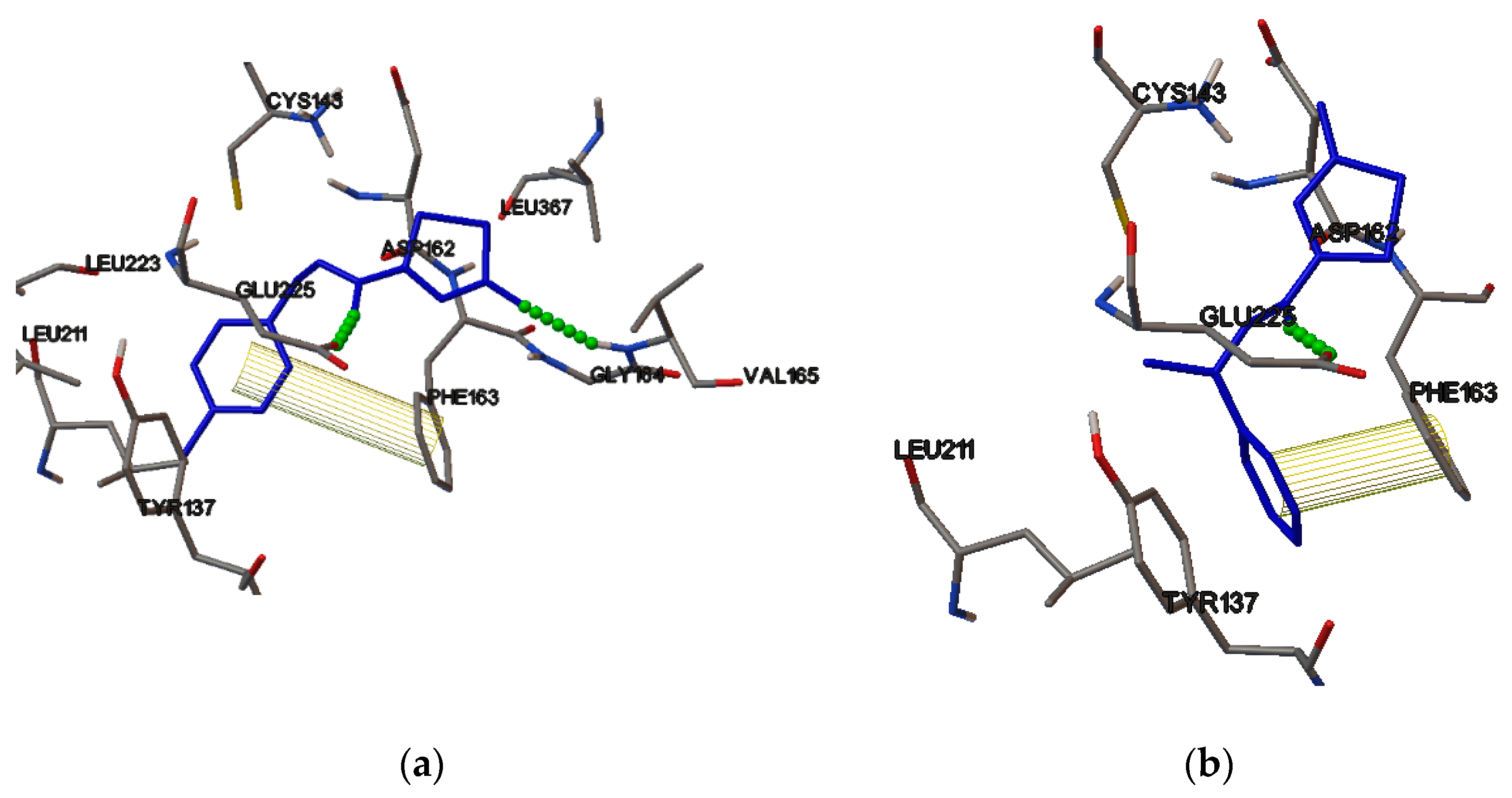
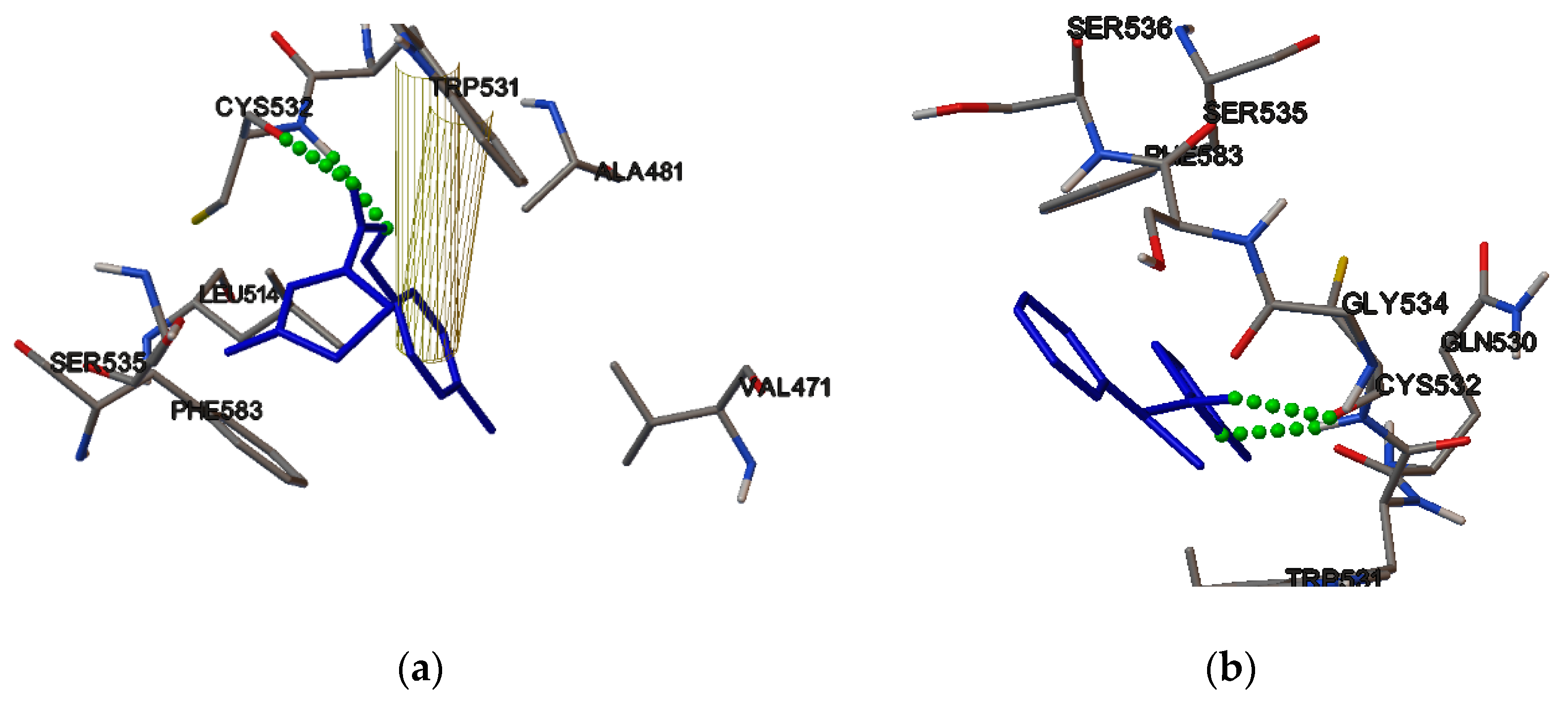
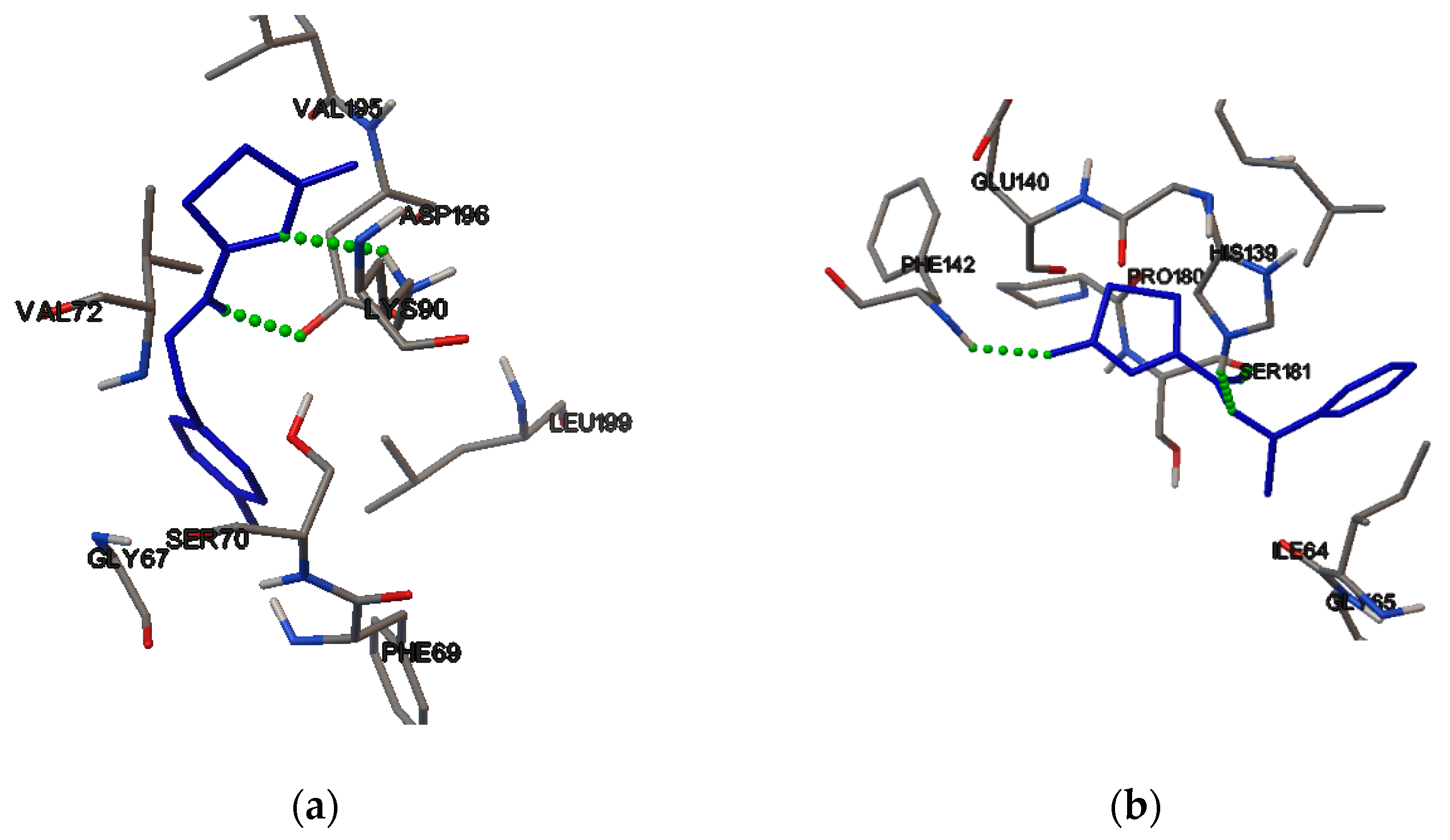
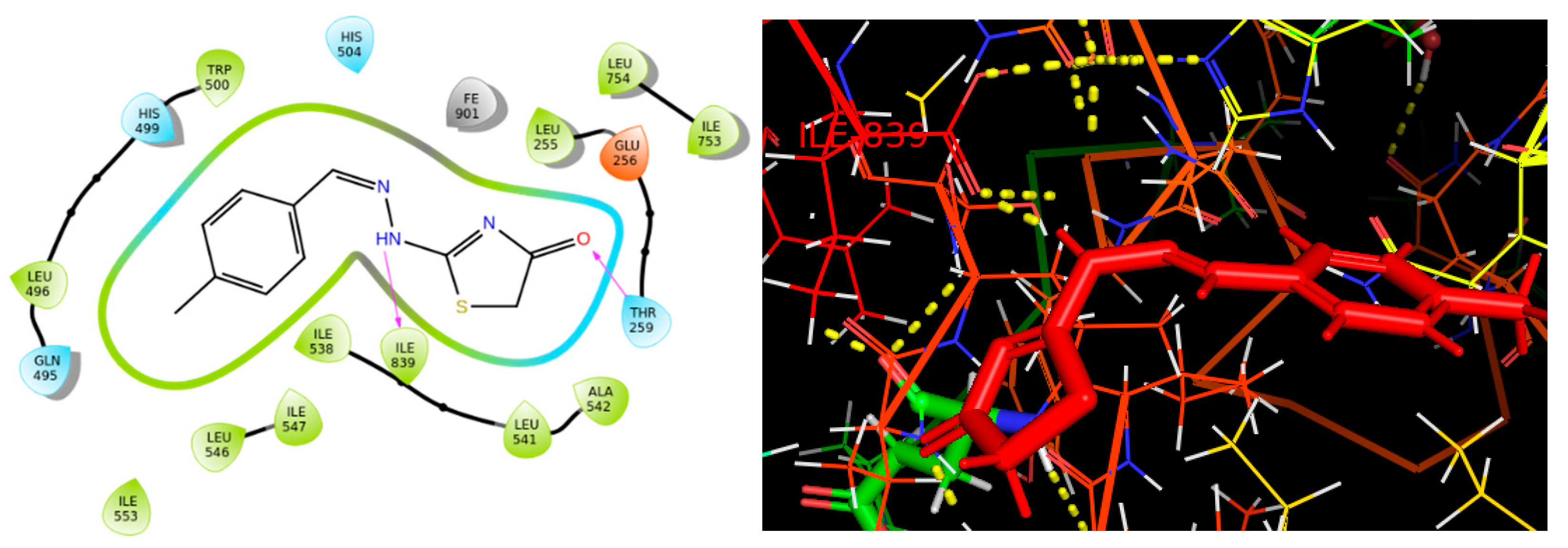
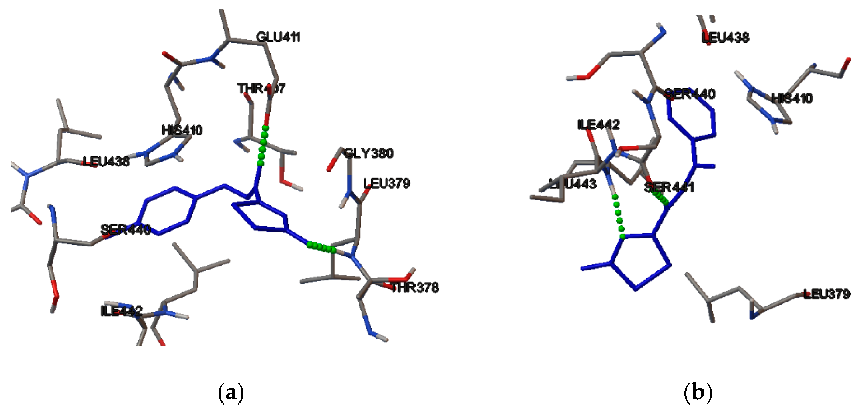
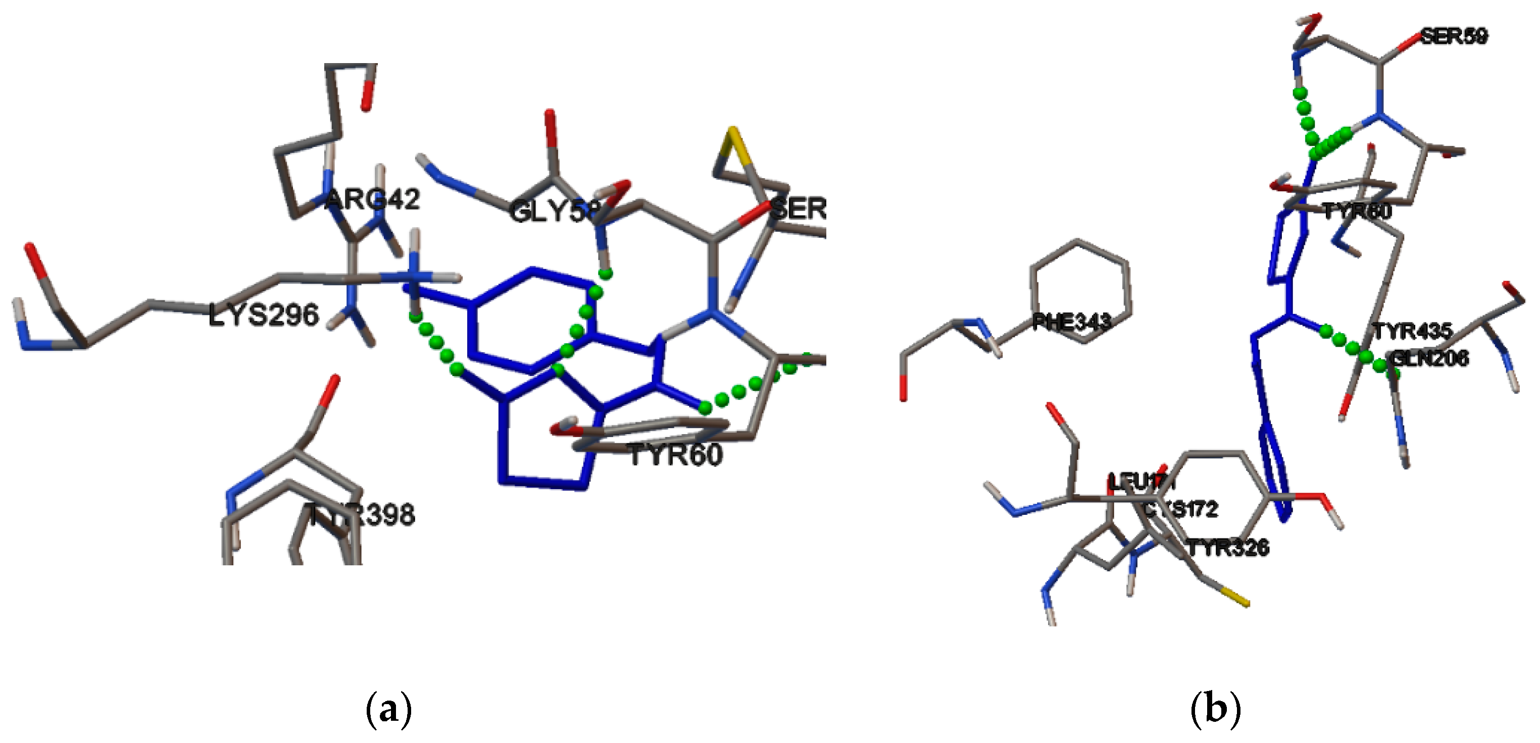
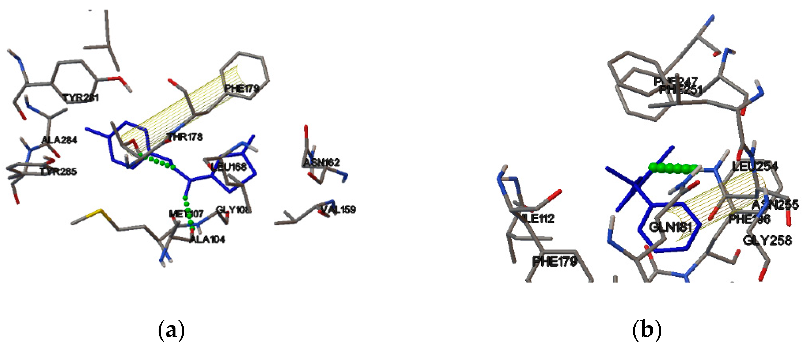
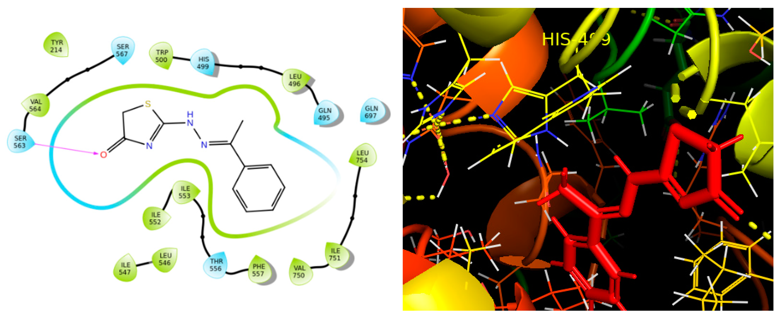
2.4. Molecular Binding

SwissTarget Prediction was used to predict some of the possible macromolecules for the in silico experiments. Eight targets were detected, specifically, the monoamine oxidase B (MAO B), the Melatonin Receptor MT1, the Human acid Ceramidase, the Kinase Domain, the ADAMTS5, the Cell division cycle 7-related protein kinase/Activator of S phase kinase, the Phosphodiesterase 4D, and the Lipoxygenase-1. The grid parameters used were the same for all of the substrates.: X = 40; Y = 40; Z = 40 (default); and the distance of the dots: 0.375 Å (default).
Then, the coordinates from the co-crystallized ligand, which were used for the active center of each macromolecule, were: 2VRM [17]: X = 53.541; Y = 151.302; Z = 23.411 6ME2 [18]: X = 24.378; Y = −21.359; Z= 46.386, 6MHM [19]: X = −7.583; Y = 4.843, Z = 28.586; 5HIE [20] X = 71.977; Y = 12.669, Z = 131.026, 3G4L [21]: X = 20.794; Y = −4.795; Z = 28.848; 4F9C [22]: X = 17.309; Y = 21.398; Z = 58.968 and 2RJQ [23]: X = −42.728; Y = −22.678; Z = 6.105.
The results are shown in Table 2.
The most favorable target was found to be the ADAMTS5 enzyme. The binding energy and the inhibition constants were calculated computationally via the AutoDock program. Thus, these compounds can serve as potential leads for these targets. The interactions of these two molecules with ADAMTS5 are shown in Figure 6. Both of the compounds form two hydrogen bonds. Specifically, DKI21 forms one hydrogen bond with GLU411 amino acid and one hydrogen bond with THR378. In addition, DKI24 forms one hydrogen bond with SER441 amino acid and one hydrogen bond with ILE442. Both of the compounds bind favorably to the active center of the enzyme.
The interactions of these two molecules with monoamine oxidase B are shown in Figure 7. Specifically, DKI21 forms three hydrogen bonds with the TYR60, GLY68, and ARG42 amino acids. In addition, the DKI24 forms two hydrogen bonds with SER206 and SER50 amino acids. Both of the compounds bind to the active center of the enzyme and the DKI24 binds more favorably than the DKI21.
The interactions of these two molecules with the Melatonin receptor, MT1, are shown in Figure 8. Specifically, the DKI21 forms two hydrogen bonds with the TYR175 and ALA104 amino acids and one pi–pi interaction with the PHE179 amino acid. In addition, the DKI24 forms one hydrogen bond with LEU254 amino acid and one pi–pi interaction with the PHE196 amino acid. Both compounds bind favorably to the active center of the enzyme.
The interactions of these two molecules with the Human acid ceramidase are shown in Figure 9. Specifically, the DKI21 forms two hydrogen bonds with GLU225 amino acid and with GLY164, and one pi–pi interaction with PHE163 amino acid. In addition, the DKI24 forms one hydrogen bond with the GLU225 amino acid and one pi–pi interaction with PHE163 amino acid. Both of the compounds bind favorably to the active center of the enzyme.
The interactions of these two molecules with the Kinase domain are shown in Figure 10. Specifically, the DKI21 forms two hydrogen bonds with the CYS532 amino acid and two pi–pi interactions with TRP531. In addition, DKI24 forms two hydrogen bonds with the CYS532 amino acid. Both of the compounds bind favorably to the active center of the enzyme.
The interactions of these two molecules with Phosphodiesterase 4D are shown in Figure 11. Specifically, the DKI21 forms two hydrogen bonds with TYR325 and HIS326 amino acids and one pi–pi interaction with TYR325. In addition, the DKI24 forms two hydrogen bonds with HIS326 and ASP484 amino acids and one pi–pi interaction with TYR326 amino acid. Both of the enzymes bind favorably to the active center of the enzyme.
The interactions of these two molecules with the Cell division cycle 7-related protein kinase/Activator of S phase kinase are shown in Figure 12. Specifically, the DKI21 forms two hydrogen bonds with the ASP196 and LYS90 amino acids. In addition, the DKI24 forms two hydrogen bonds with the PHE142 and SER181 amino acids. Both of the compounds bind favorably to the active center of the enzyme.
Induced Fit docking was applied, in order to find how strong these two compounds bind to the active center of LOX-1. The DKI21 binds favorably to the active site of LOX-1 with a binding score of ΔG= −7.52 kcal/mol. Specifically, the DKI21 forms two hydrogen bonds with the ILE839 and THR259 amino acids (Figure 13).
In addition, DKI24 binds favorably to the active site of LOX-1 with a binding energy of −7.54 kcal/mol. Specifically, it forms one hydrogen bond with the SER563 amino acid (Figure 14).

2.5. Results of the Pharmacokinetics and Toxicity Properties of the Two Compounds

Both of the compounds obey the Lipinski’s Rule of Five [24] and Veber’s Rule [25], because they have less than seven rotable bonds. Compound DKI21 is more soluble than DKI24 (see Table 3).
According to preADMET, the BBB [26] value is less than one. As a result, both of the compounds are classified as inactive in the central nervous system (CNS) (Table 4). The values for human intestinal absorption is high for both of the compounds, and this signifies that these compounds might be better absorbed from the intestinal tract on oral administration. Both of the compounds are not inhibitors of the CP isoenzymes, and therefore are not toxic (Table 4).
According to pkCSm (Table 5), both of the compounds have been predicted to be hepatotoxic. Compound DKI24 has negative AMES [27] toxicity and, as a result, it is not mutagenic, in contrast to DKI21.

3. Materials and Methods

3.1. Synthesis

The reagents were purchased with the highest commercial quality from Aldrich, Acros, and Fluka and were used without further purification. The reactions were monitored by thin-layer chromatography (TLC), carried out on 0.25 mm silica gel plates (E. Merck silica gel 60F254), and components were visualized by UV light absorbance. Purification of compounds by column chromatography was carried out on silica gel (Merck, 70−230 mesh) and the indicated solvents. The 1H and 13C NMR spectra were recorded on Bruker 400 and 500 MHz Avance spectrometers. The 1H and 13C spectra are referenced according to the residual peak of the solvent, based on the literature data. The 13C NMR spectra are fully proton-decoupled. The electrospray ionization (ESI) mass spectral analyses were performed on a mass spectrometer, MSQ Surveyor, Finnigan, using direct sample injection. The negative or positive ion ESI spectra were acquired by adjusting the needle and cone voltages accordingly.
Synthesis: to a stirred solution of thiosemicarbazone (1 mmol) in methanol, sodium acetate (2 mmol) was added, followed by methyl-3-chloroacetate (1 mmol) and the mixture was refluxed for 2 h. A new portion of methyl-3-chloroacetate (1 mmol) was added every two hours, (2×). The reaction mixture was left to stand at room temperature, the solid was filtered, washed with methanol, to give the DKI21 as an off-white solid in 72% yield.
Following the same procedure, the DKI24 was obtained as an off-white solid in 77% yield.

3.2. Structure Assignment

The two molecules under study were structurally identified using 400 and 500 MHz spectrometers (Bruker Avance Spectrometer, Billerica, MA, USA), installed in the National and Kapodistrian University of Athens using 1D and 2D homonuclear and heteronuclear experiments. Various mixing times (d8) for 2D-NOESY were applied (0.15, 0.30, 0.5, and 0.8). The pulse sequences were obtained from the library of the spectrometer. The spectra were processed and analyzed using the MestreNova (Santiago de Compostela, Spain) and TopSpin softwares.

3.3. Conformational Analysis

The onformational analysis was performed to find the most stable conformation of these two compounds. All of the calculations were performed using B3LYP [29,30] as functional and 6-311G(d,p) [31]. This methodology is suitable for these organic compounds [32]. All of the calculations were performed using DMSO as a solvent, so the experimental results would be similar, and employing the polarizable continuum model (PCM) [33]. All of the conformers were fully optimized and their frequencies were calculated. Finally, all of the theoretical calculations were compared with the experimental ones. All of the calculations were performed with Gaussian 16 [34].

3.4. Reaction Mechanism

The mechanism of the reaction was calculated to find pit which intermediate will decide to go and which compound is most favorable.

3.5. Molecular Binding

AutoDock [35] software was used for the molecular binding [36] calculations and, more specifically, the Lamarckian Genetic algorithm. The crystal structures of the proteins were used by the online database “Protein Data Bank—PDB” and downloaded directly to the AutoDock program for study. The compounds used as ligands were designed with the help of the ChemOffice program, and, using the same program, their energy was minimized with an MM2 force field.

3.6. Induced Fit Docking

In the next stage, DKI21 and DKI24 were investigated for their potent binding to major targets revealed from the Swiss Target tool (http://www.swisstargetprediction.ch/, accessed on 25 June 2022) and bibliography [37,38], through molecular docking calculations. The crystal structures used for the in silico studies carried the following PDB IDs: 5T5V [39]. The protein preparation wizard, a module available in Schrödinger Suites, was used to prepare the crystal structure for the in silico calculations. Since LOX is a metalloprotein, it has been considered that there are several computational challenges that need to be addressed. In order to take account of the quantum effects associated with the presence of a Fe3+ cation in LOX’s active site, we used the “create zero-order bonds to metals” module of the Schrödinger’s Maestro molecular modeling platform. This module breaks the existing bonds to metals—since they cannot be examined through the over-simplified model of a spring attached to a hard sphere—and adds new zero-order bonds between the metals and nearby atoms and corrects their formal charges accordingly, to constrain the X-ray-acquired coordination geometry.
Both of the compounds were sketched in the Schrodinger’s Maestro [40] molecular modeling platform, and it was initially minimized using Macromodel [41] and DFT calculations. LigPrep was used to prepare the 3D models, restricted to the specific stereochemistry of each molecule. During the ligand preparation, the “add metal binding states” option of the Epik module of LigPrep was chosen, in order to create ligand binding states that are suitable for metal binding that would have been rejected otherwise, due to high energy state penalties. The geometries were optimized with MacroModel in order to relax the structures, while the chiral centers retained the proper chiralities. The force field used for minimization was OPLS2005 [42]. All of the the compounds were subjected to proper treatment of their protonation states at physiological pH (~7.4). Hammett and Taft methods were implemented, in conjunction with an ionization tool to generate chemically sensible 3D models. The three-dimensional ligands’ structures were further minimized, more rigorously, by MacroModel, with water as the solvent and OPLS2005 as the force field, using a conjugate gradient (CG) method with a threshold of 0.01 kcal/mol. The minimized structure was further used as input to a mixed-torsional/low-sampling conformational search forced to keep the input chiralities. The conformational search generated a number of conformers for each molecule under study and the conformers were energetically ranked. The most favored conformation was used as input for the following docking calculations.
The docking calculations were performed to reveal the possible binding of compounds to LOX-1. The calculations were performed with the Induced Fit Docking (IFD) method. The ligand was docked in the 10 energetically favored conformations generated by Macromodel. The protein preparation constrained refinement was applied in the Glide docking stage. Trimming the side chains automatically (based on B—factor) and a Prime refinement of the protein side chains were applied and the docking process was accomplished by Glide/XP. Finally, the binding energy was calculated. The active site was described, using a dielectric constant of 80 and all of the crystallographic waters of the active site were preserved.

3.7. ADMET Calculations

Both of the compounds were sketched in ChemDraw, in order to find their SMILES. After their SMILES were found, they were imported in SwissADME [43], pkCSM [28], and pro-TOX to find their pharmacological and toxicological results. This procedure is very important for computational drug design, because the vast majority of drugs that do not reach the market are either due to limited effectiveness or due to side effects.

4. Conclusions

This study focuses on structure assignment and conformational analysis of two promising bioactive thiosemicarbazone adducts, DKI21 and DKI24, using a combination of NMR spectroscopy and computational studies (QM methods). The NMR experiments were performed, using a combination of 2D-ROESY and 2D-NOESY at different mixing times to assign unambiguously the exo structure of the two molecules under study. The calculation of the mechanistic pathway of the reaction proves that the molecules obtain exo configuration. In silico experiments were performed to find some possible biological targets for the two molecules under study. The results showed that the compounds bind favorably to the revealed enzyme targets, using Swissadme software. Both of the derivatives obey Lipinski’s Rule of Five and Veber’s Rule. It appears that both of the molecules are predicted not to have toxic properties and to be bioactive for various biological targets. Thus, these molecules can be promising leads for these targets. In addition, the derivatives of these compounds can be synthesized by medicinal chemists to optimize their biological targeting.

Supplementary Materials

The following supporting information can be downloaded at https://www.mdpi.com/article/10.3390/molecules27144548/s1, Figure S1: 1H-NMR spectra. The spectra were recorded in DMSO-d6 on a Bruker AC 400 MHz spectrometer at 25 °C; Figure S2: 2D-NOESY-NMR spectra. The spectra were recorded in DMSO-d6 on a Bruker AC 400 MHz spectrometer at 25 °C; Figure S3: 13C-NMR spectra. The spectra were recorded in DMSO-d6 on a Bruker AC 400 MHz spectrometer at 25 °C; Figure S4: 2D-HSQC-NMR spectra. The spectra were recorded in DMSO-d6 on a Bruker AC 400 MHz spectrometer at 25 °C; Figure S5: 2D-HMBC-NMR spectra. The spectra were recorded in DMSO-d6 on a Bruker AC 400 MHz spectrometer at 25 °C; Figure S6: 1H-NMR spectra. The spectra were recorded in DMSO-d6 on a Bruker AC 400 MHz spectrometer at 25 °C; Figure S7: 2D-NOESY-NMR spectra. The spectra were recorded in DMSO-d6 on a Bruker AC 400 MHz spectrometer at 25 °C; Figure S8: 13C-NMR spectra. The spectra were recorded in DMSO-d6 on a Bruker AC 400MHz spectrometer at 25 °C; Figure S9: 2D-HSQC-NMR spectra. The spectra were recorded in DMSO-d6 on a Bruker AC 400 MHz spectrometer at 25 °C; Figure S10: 2D-HMBC-NMR spectra. The spectra were recorded in DMSO-d6 on a Bruker AC 400 MHz spectrometer at 25 °C; Figure S11: 2D-ROESY-NMR spectra. The spectra were recorded in DMSO-d6 on a Bruker AC 500 MHz spectrometer at 25 °C using P15 = 150.000 us; Figure S12: 2D-ROESY-NMR spectra. The spectra were recorded in DMSO-d6 on a Bruker AC 500 MHz spectrometer at 25 °C using P15 = 300.000 us; Figure S13: 2D-ROESY-NMR spectra. The spectra were recorded in DMSO-d6 on a Bruker AC 500 MHz spectrometer at 25 °C using P15 = 500.000 us; Figure S14: 2D-ROESY-NMR spectra. The spectra were recorded in DMSO-d6 on a Bruker AC 500 MHz spectrometer at 25 °C using P15 = 150.000 us; Figure S15: 2D-ROESY-NMR spectra. The spectra were recorded in DMSO-d6 on a Bruker AC 500 MHz spectrometer at 25 °C using P15 = 300.000 us; Figure S16: 2D-ROESY-NMR spectra. The spectra were recorded in DMSO-d6 on a Bruker AC 500 MHz spectrometer at 25 °C using P15 = 500.000 us; Figure S17: Overall diagram showing the identification strategy of the DKI21 compound in DMSO; Figure S18: Overall diagram showing the identification strategy of the DKI24 compound in DMSO; Figure S19: Orbitals in HOMO (top) and LUMO (bottom) in DKI21 exo conformation; Figure S20: Orbitals in HOMO (top) and LUMO (bottom) in DKI24 exo conformation; Figure S21: Orbitals in HOMO (top) and LUMO (bottom) in DKI21 endo conformation; Figure S22: Orbitals in HOMO (top) and LUMO (bottom) in DKI24 endo conformation; Table S1: HOMO-LUMO gap, hardness, and softness of lowest in energy endo and exo conformers.

Author Contributions

N.G., Investigation, Formal analysis, Methodology, Writing—original draft; A.C., NMR experiments; D.K., Synthesis; E.C. Docking experiments; J.P., review and editing; D.T., Resources, Supervision, Methodology, Writing—review and editing; S.V., Resources, Synthesis Supervision, Methodology, Writing—review and editing; T.M. Conceptualization, Resources, Supervision, Writing—original draft, Writing—review and editing. All authors have read and agreed to the published version of the manuscript.

Funding

J.P. acknowledges financial support of the Slovenian Research Agency [Grants Nos. P1-0242 and J1-1704]. The authors acknowledge the CERIC-ERIC 20217107 consortium for the access to experimental facilities and financial support.

Institutional Review Board Statement

Not applicable.

Informed Consent Statement

Not applicable.

Data Availability Statement

The data presented in this paper are available in the Supplementary Materials.

Acknowledgments

NMR studies were performed at the National and Kapodistrian University of Athens. Materials were supported by Special Account for Research Grants (SARG), National Kapodistrian University of Athens (NKUA).

Conflicts of Interest

The authors declare no conflict of interest.

Sample Availability

Samples of the compounds DKI21 and DKI24 are available from the authors.

References

  1. Parrilha, G.L.; dos Santos, R.G.; Beraldo, H. Applications of Radiocomplexes with Thiosemicarbazones and Bis(Thiosemicarbazones) in Diagnostic and Therapeutic Nuclear Medicine. Coord. Chem. Rev. 2022, 458, 214418. [Google Scholar] [CrossRef]
  2. Sevinçli, Z.Ş.; Duran, G.N.; Özbil, M.; Karalı, N. Synthesis, Molecular Modeling and Antiviral Activity of Novel 5-Fluoro-1H-Indole-2,3-Dione 3-Thiosemicarbazones. Bioorg. Chem. 2020, 104, 104202. [Google Scholar] [CrossRef] [PubMed]
  3. Ebenezer, O.; Singh-Pillay, A.; Koorbanally, N.A.; Singh, P. Antibacterial Evaluation and Molecular Docking Studies of Pyrazole–Thiosemicarbazones and Their Pyrazole–Thiazolidinone Conjugates. Mol. Divers. 2021, 25, 191–204. [Google Scholar] [CrossRef] [PubMed]
  4. Mashayekhi, V.; Haj Mohammad Ebrahim Tehrani, K.; Azerang, P.; Sardari, S.; Kobarfard, F. Synthesis, Antimycobacterial and Anticancer Activity of Novel Indole-Based Thiosemicarbazones. Arch. Pharm. Res. 2021, 44, 1–13. [Google Scholar] [CrossRef] [PubMed]
  5. Kaminskyy, D.; Kryshchyshyn, A.; Lesyk, R. 5-Ene-4-Thiazolidinones—An Efficient Tool in Medicinal Chemistry. Eur. J. Med. Chem. 2017, 140, 542–594. [Google Scholar] [CrossRef]
  6. Havrylyuk, D.; Roman, O.; Lesyk, R. Synthetic Approaches, Structure Activity Relationship and Biological Applications for Pharmacologically Attractive Pyrazole/Pyrazoline–Thiazolidine-Based Hybrids. Eur. J. Med. Chem. 2016, 113, 145–166. [Google Scholar] [CrossRef]
  7. Jain, V.S.; Vora, D.K.; Ramaa, C.S. Thiazolidine-2,4-Diones: Progress towards Multifarious Applications. Bioorg. Med. Chem. 2013, 21, 1599–1620. [Google Scholar] [CrossRef]
  8. Verma, A.; Saraf, S.K. 4-Thiazolidinone—A Biologically Active Scaffold. Eur. J. Med. Chem. 2008, 43, 897–905. [Google Scholar] [CrossRef]
  9. Tripathi, A.C.; Gupta, S.J.; Fatima, G.N.; Sonar, P.K.; Verma, A.; Saraf, S.K. 4-Thiazolidinones: The Advances Continue…. Eur. J. Med. Chem. 2014, 72, 52–77. [Google Scholar] [CrossRef]
  10. Benmohammed, A.; Rekiba, N.; Sehanine, Y.; Louail, A.A.; Khoumeri, O.; Kadiri, M.; Djafri, A.; Terme, T.; Vanelle, P. Synthesis and Antimicrobial Activities of New Thiosemicarbazones and Thiazolidinones in Indole Series. Chem. Mon. 2021, 152, 977–986. [Google Scholar] [CrossRef]
  11. Nechak, R.; Bouzroura, S.A.; Benmalek, Y.; Salhi, L.; Martini, S.P.; Morizur, V.; Dunach, E.; Kolli, B.N. Synthesis and Antimicrobial Activity Evaluation of Novel 4-Thiazolidinones Containing a Pyrone Moiety. Synth. Commun. 2015, 45, 262–272. [Google Scholar] [CrossRef]
  12. Salem, M.A.; Abbas, S.Y.; El-Sharief MA, M.S.; Alzahrani, A.Y.; Helal, M.H.; Thabet, H.K. Synthesis and Antimicrobial Activity of 4-Methylthiazole and 4-Thiazolidinone Derivatives Derived from 5-(Aryldiazo)Salicylaldehyde Thiosemicarbazones. Synth. Commun. 2021, 51, 3325–3331. [Google Scholar] [CrossRef]
  13. Trotsko, N.; Bekier, A.; Paneth, A.; Wujec, M.; Dzitko, K. Synthesis and In Vitro Anti-Toxoplasma Gondii Activity of Novel Thiazolidin-4-One Derivatives. Molecules 2019, 24, 3029. [Google Scholar] [CrossRef] [PubMed] [Green Version]
  14. Yang, H.; Qi, H.; Hao, Z.; Shao, X.; Liu, T.; Yang, Q.; Qian, X. Thiazolylhydrazone Dervatives as Inhibitors for Insect N-Acetyl-β-d-Hexosaminidase and Chitinase. Chin. Chem. Lett. 2020, 31, 1271–1275. [Google Scholar] [CrossRef]
  15. Hoque, M.J.; Ahsan, A.; Hossain, B. Molecular Docking, Pharmacokinetic, and DFT Calculation of Naproxen and Its Degradants. Biomed. J. Sci. Tech. Res. 2018, 9, 7360–7365. [Google Scholar] [CrossRef] [Green Version]
  16. De Proft, F.; Geerlings, P. Conceptual and Computational DFT in the Study of Aromaticity. Chem. Rev. 2001, 101, 1451–1464. [Google Scholar] [CrossRef]
  17. Binda, C.; Wang, J.; Li, M.; Hubalek, F.; Mattevi, A.; Edmondson, D.E. Structural and Mechanistic Studies of Arylalkylhydrazine Inhibition of Human Monoamine Oxidases A and B. Biochemistry 2008, 47, 5616–5625. [Google Scholar] [CrossRef]
  18. Stauch, B.; Johansson, L.C.; McCorvy, J.D.; Patel, N.; Han, G.W.; Huang, X.-P.; Gati, C.; Batyuk, A.; Slocum, S.T.; Ishchenko, A.; et al. Structural Basis of Ligand Recognition at the Human MT1 Melatonin Receptor. Nature 2019, 569, 284–288. [Google Scholar] [CrossRef]
  19. Dementiev, A.; Joachimiak, A.; Nguyen, H.; Gorelik, A.; Illes, K.; Shabani, S.; Gelsomino, M.; Ahn, E.-Y.E.; Nagar, B.; Doan, N. Molecular Mechanism of Inhibition of Acid Ceramidase by Carmofur. J. Med. Chem. 2019, 62, 987–992. [Google Scholar] [CrossRef]
  20. Foster, S.A.; Whalen, D.M.; Özen, A.; Wongchenko, M.J.; Yin, J.; Yen, I.; Schaefer, G.; Mayfield, J.D.; Chmielecki, J.; Stephens, P.J.; et al. Activation Mechanism of Oncogenic Deletion Mutations in BRAF, EGFR, and HER2. Cancer Cell 2016, 29, 477–493. [Google Scholar] [CrossRef] [Green Version]
  21. Burgin, A.B.; Magnusson, O.T.; Singh, J.; Witte, P.; Staker, B.L.; Bjornsson, J.M.; Thorsteinsdottir, M.; Hrafnsdottir, S.; Hagen, T.; Kiselyov, A.S.; et al. Design of Phosphodiesterase 4D (PDE4D) Allosteric Modulators for Enhancing Cognition with Improved Safety. Nat. Biotechnol. 2010, 28, 63–70. [Google Scholar] [CrossRef] [PubMed]
  22. Hughes, S.; Elustondo, F.; Di Fonzo, A.; Leroux, F.G.; Wong, A.C.; Snijders, A.P.; Matthews, S.J.; Cherepanov, P. Crystal Structure of Human CDC7 Kinase in Complex with Its Activator DBF4. Nat. Struct. Mol. Biol. 2012, 19, 1101–1107. [Google Scholar] [CrossRef] [PubMed]
  23. Mosyak, L.; Georgiadis, K.; Shane, T.; Svenson, K.; Hebert, T.; McDonagh, T.; Mackie, S.; Olland, S.; Lin, L.; Zhong, X.; et al. Crystal Structures of the Two Major Aggrecan Degrading Enzymes, ADAMTS4 and ADAMTS5. Protein Sci. 2008, 17, 16–21. [Google Scholar] [CrossRef] [PubMed] [Green Version]
  24. Benet, L.Z.; Hosey, C.M.; Ursu, O.; Oprea, T.I. BDDCS, the Rule of 5 and Drugability. Adv. Drug Deliv. Rev. 2016, 101, 89–98. [Google Scholar] [CrossRef] [PubMed] [Green Version]
  25. Veber, D.F.; Johnson, S.R.; Cheng, H.-Y.; Smith, B.R.; Ward, K.W.; Kopple, K.D. Molecular Properties That Influence the Oral Bioavailability of Drug Candidates. J. Med. Chem. 2002, 45, 2615–2623. [Google Scholar] [CrossRef]
  26. Daneman, R.; Prat, A. The Blood-Brain Barrier. Cold Spring Harb. Perspect. Biol. 2015, 7, a020412. [Google Scholar] [CrossRef] [Green Version]
  27. Guy, R.C. Ames Test. In Encyclopedia of Toxicology; Elsevier: Amsterdam, The Netherlands, 2005; pp. 88–91. [Google Scholar] [CrossRef]
  28. Pires DE, V.; Blundell, T.L.; Ascher, D.B. PkCSM: Predicting Small-Molecule Pharmacokinetic and Toxicity Properties Using Graph-Based Signatures. J. Med. Chem. 2015, 58, 4066–4072. [Google Scholar] [CrossRef] [PubMed]
  29. Lee, C.; Yang, W.; Parr, R.G. Development of the Colle-Salvetti Correlation-Energy Formula into a Functional of the Electron Density. Phys. Rev. B 1988, 37, 785–789. [Google Scholar] [CrossRef] [Green Version]
  30. Becke, A.D. A New Mixing of Hartree–Fock and Local Density-functional Theories. J. Chem. Phys. 1993, 98, 1372–1377. [Google Scholar] [CrossRef]
  31. Curtiss, L.A.; McGrath, M.P.; Blaudeau, J.; Davis, N.E.; Binning, R.C.; Radom, L. Extension of Gaussian-2 Theory to Molecules Containing Third-row Atoms Ga–Kr. J. Chem. Phys. 1995, 103, 6104–6113. [Google Scholar] [CrossRef]
  32. Tzeli, D.; Tsoungas, P.G.; Petsalakis, I.D.; Kozielewicz, P.; Zloh, M. Intramolecular Cyclization of β-Nitroso-o-Quinone Methides. A Theoretical Endoscopy of a Potentially Useful Innate ‘Reclusive’ Reaction. Tetrahedron 2015, 71, 359–369. [Google Scholar] [CrossRef]
  33. Cossi, M.; Scalmani, G.; Rega, N.; Barone, V. New Developments in the Polarizable Continuum Model for Quantum Mechanical and Classical Calculations on Molecules in Solution. J. Chem. Phys. 2002, 117, 43–54. [Google Scholar] [CrossRef]
  34. Frisch, M.J.; Trucks, G.W.; Schlegel, H.B.; Scuseria, G.E.; Robb, M.A.; Cheeseman, J.R.; Scalmani, G.; Barone, V.; Petersson, G.A.; Nakatsuji, H.; et al. Gaussian 16, Revision B.01; Gaussian, Inc.: Wallingford, CT, USA, 2016.
  35. Rizvi SM, D.; Shakil, S.; Haneef, M. A Simple Click by Click Protocol to Perform Docking: Autodock 4.2 Made Easy for Non-Bioinformaticians. EXCLI J. 2013, 12, 830–857. [Google Scholar] [CrossRef]
  36. Schleinkofer, K.; Wang, T.; Wade, R.C. Molecular Docking. In Encyclopedic Reference of Genomics and Proteomics in Molecular Medicine; Springer: Berlin/Heidelberg, Germany, 2006; Volume 443, pp. 1149–1153. [Google Scholar] [CrossRef]
  37. Ghafary, S.; Ghobadian, R.; Mahdavi, M.; Nadri, H.; Moradi, A.; Akbarzadeh, T.; Najafi, Z.; Sharifzadeh, M.; Edraki, N.; Moghadam, F.H.; et al. Design, Synthesis, and Evaluation of Novel Cinnamic Acid-Tryptamine Hybrid for Inhibition of Acetylcholinesterase and Butyrylcholinesterase. DARU J. Pharm. Sci. 2020, 28, 463–477. [Google Scholar] [CrossRef]
  38. Peperidou, A.; Pontiki, E.; Hadjipavlou-Litina, D.; Voulgari, E.; Avgoustakis, K. Multifunctional Cinnamic Acid Derivatives. Molecules 2017, 22, 1247. [Google Scholar] [CrossRef] [PubMed]
  39. Offenbacher, A.R.; Hu, S.; Poss, E.M.; Carr, C.A.M.; Scouras, A.D.; Prigozhin, D.M.; Iavarone, A.T.; Palla, A.; Alber, T.; Fraser, J.S.; et al. Hydrogen-Deuterium Exchange of Lipoxygenase Uncovers a Relationship between Distal, Solvent Exposed Protein Motions and the Thermal Activation Barrier for Catalytic Proton-Coupled Electron Tunneling. ACS Cent. Sci. 2017, 3, 570–579. [Google Scholar] [CrossRef] [PubMed] [Green Version]
  40. MacroModel, Version 10; Schrodinger, LLC: New York, NY, USA, 2013.
  41. MacroModel, Version 10.2; Schrodinger, LLC: New York, NY, USA, 2013.
  42. Jorgensen, W.L.; Maxwell, D.S.; Tirado-Rives, J. Development and Testing of the OPLS All-Atom Force Field on Conformational Energetics and Properties of Organic Liquids. J. Am. Chem. Soc. 1996, 118, 11225–11236. [Google Scholar] [CrossRef]
  43. Daina, A.; Michielin, O.; Zoete, V. SwissADME: A Free Web Tool to Evaluate Pharmacokinetics, Drug-Likeness and Medicinal Chemistry Friendliness of Small Molecules. Sci. Rep. 2017, 7, 42717. [Google Scholar] [CrossRef] [Green Version]
Scheme 1. These molecules underwent structure elucidation, utilizing 2D-NOESY and ROESY experiments, to establish their exo or endo products. In addition, molecular docking experiments were performed to establish the putative bioactive targets.
Molecules 27 04548 sch001
Figure 1. Possible Structures of DKI21 and DKI24.
Molecules 27 04548 g001
Figure 2. The lowest in energy conformations for DKI21 (a) and DKI24 (b) and their critical spatial correlations that determine their lowest energy conformations.
Molecules 27 04548 g002
Figure 3. 2D–NOESY spectra for DKI21 (above) using d8 = 0.15 s (a) and 0.30 s (b) and DKI24 using d8 = 0.15 s (c) and d8 = 0.30 s (d) (bottom). The spectra were recorded in DMSO-d6 on a Bruker AC 500 MHz spectrometer at 25 °C.
Molecules 27 04548 g003
Figure 4. Energy diagram (in Hartree) of the endo and exo isomerization of the DKI21 compound.
Molecules 27 04548 g004aMolecules 27 04548 g004b
Figure 5. Energy diagram (in Hartree) of the endo and exo isomerization of the DKI24 compound.
Molecules 27 04548 g005
Figure 6. Interactions of DKI21 (a) and DKI24 (b) with ADAMTS5, which gave the most favorable results.
Molecules 27 04548 g006
Figure 7. Interactions of DKI21 (a) and DKI24 (b) with Monoamine oxidase B.
Molecules 27 04548 g007
Figure 8. Interactions of DKI21 (a) and DKI24 (b) with Melatonin receptor MT1.
Molecules 27 04548 g008
Figure 9. Interactions of DKI21 (a) and DKI24 (b) with Human acid ceramidase.
Molecules 27 04548 g009
Figure 10. Interactions of DKI21 (a) and DKI24 (b) with Kinase domain.
Molecules 27 04548 g010
Figure 11. Interactions of DKI21 (a) and DKI24 (b) with Phosphodiesterase 4D.
Molecules 27 04548 g011
Figure 12. Interactions of DKI21 (a) and DKI24 (b) with Cell division cycle 7-related protein kinase/Activator of S phase kinase.
Molecules 27 04548 g012
Figure 13. Interactions of DKI21 with LOX-1.
Molecules 27 04548 g013
Figure 14. Interactions of DKI24 with LOX-1.
Molecules 27 04548 g014
Table 1. Assignment of the experimental 1H-NMR spectra of DKI21 (top) and DKI24 (below) in DMSO-d6.
Position1HCOSYNOESYHSQCHMBC13C
12.35-H-3C-1C-2. C-321.51
2----H-1/H-3141.03
37.26H-4H-1/H-4C-3C-2/C-5129.90
47.66H-3H-3/H-6C-4C-5/C-3/C-6128.10
5----H-3/H-6/H-4131.98
68.36 H-4C-6H-5/H-4156.68
7
8
9----C-11/C-12/H-8165.20
10
11----H-12174.62
123.89--C-12C-11/C-933.45
Position1HCOSYNOESYHSQCHMBC13C
17.47H-2H-2C-1C-2. C-3121.87
27.47H-1/H-3H-1/H-3C-2H-1/H-3121.87
37.86H-2H-2/H-6C-3C-2/C-5/C-4126.84
4----H-3/H-2/H-6138.22
5----H-3/H-6/H-4160.83
62.37-H-3C-6H-5/H-415.04
7
8
9----H-12164.53
10
11----H-12174.42
123.85--C-12C-11/C-933.26
Table 2. Binding energies BE (kcal/mol) and inhibition constants between the compounds and macromolecules.
Binding EnergyInhibition Constant
2VRM (MAO B)
DKI21−7.64 ± 0.52.52 ± 0.5 μΜ/3.13 ± 0.5 μΜ
DKI24−7.55 ± 0.52.93 ± 0.5 μΜ
6ME2 (Melatonin receptor MT1)
DKI21−6.81 ± 0.510.17 ± 0.5 μΜ
DKI24−7.38 ± 0.53.92 ± 0.5 μΜ
6MHM (Human acid ceramidase)
DKI21−7.10 ± 0.56.29 ± 0.5 μΜ
DKI24−7.07 ± 0.56.58 ± 0.5 μΜ
5HIE (Kinase domain)
DKI21−6.41 ± 0.519.9 ± 0.5 μΜ
DKI24−6.36 ± 0.521.89 ± 0.5 μΜ
3G4L(Phosphodiesterase 4D)
DKI21−7.1 ± 0.56.3 ± 0.5 μΜ
DKI24−7.76 ± 0.52.05 ± 0.5 μΜ
2RJQ (ADAMTS5)
DKI21−7.65 ± 0.52.48 ± 0.5 μΜ
DKI24−7.7 ± 0.54.47 ± 0.5 μΜ
CDC7/DBF4 (Cell division cycle 7-related protein kinase/Activator of S phase kinase) (4F9C)
DKI21−6.07 ± 0.535.5 ± 0.5 μΜ
DKI24−6.20 ± 0.528.43 ± 0.5 μΜ
Table 3. The physicochemical parameters for compounds DKI21 and DKI24.
PropertiesCompound DK121Compound DΚΙ24
Molecular Weight233.296233.296
LogP1.547921.6296
Rotable bonds22
Hydrogen Bond Acceptors44
Hydrogen Bond Donors11
Surface Area97.846 (Å2)97.846 (Å2)
Water solubility−2.855 (logmolL−1)−2.702 (logmolL−1)
Table 4. The ADME results of DKI21 and DKI24 according to preADMET.
Compound DKI21Compound DKI24
BBB0.1218710.370766
Buffer_solubility_mg_L101.47811192.1
Caco28.610837.52339
CYP_2C19_inhibitionNonNon
CYP_2C9_inhibitionNonNon
CYP_2D6_inhibitionNonNon
CYP_2D6_substrateNonNon
CYP_3A4_inhibitionNonNon
CYP_3A4_substrateNonWeakly
HIA96.09691896.096626
MDCK23.466822.523
Pgp_inhibitionNonNon
Plasma_Protein_Binding73.33695870.164297
Pure_water_solubility_mg_L12.662820.7683
Skin_Permeability−3.1538−3.40889
Table 5. Toxicity results of DKI21 and DKI24 according to pKCSm [28].
PropertiesCompound DK121Compound DKI24
Toxicity
AMES toxicityYesNo
Max. tolerated dose (human)0.399 (log mg/kg/day)0.269 (log mg/kg/day)
Herg I inhibitorNoNo
Herg II inhibitorNoNo
Oral Rat Acute Toxicity (LD50)2.699 (mol/kg)2.878 (mol/kg)
Oral Rat Chronic Toxicity1.417 (log mg/kg_bw/day)1.386 (log mg/kg_bw/day)
HepatotoxicityYesYes
Skin SensitizationYesYes
Publisher’s Note: MDPI stays neutral with regard to jurisdictional claims in published maps and institutional affiliations.

