Review

Anti-Inflammatory Effects of Phytochemical Components of Clinacanthus nutans

1
Department of Anatomy and Neurobiology Research Programme, National University of Singapore, Singapore 119260, Singapore
2
Department of Pharmacology, National University of Singapore, Singapore 119260, Singapore
3
Department of Biochemistry, University of Missouri, Columbia, MO 65211, USA
4
Institute of Biomedical Sciences, Academia Sinica, Taipei 11529, Taiwan
*
Author to whom correspondence should be addressed.
Molecules 2022, 27(11), 3607; https://doi.org/10.3390/molecules27113607
Submission received: 5 May 2022 / Revised: 2 June 2022 / Accepted: 2 June 2022 / Published: 4 June 2022
(This article belongs to the Special Issue Phytochemistry and Biological Properties of Medicinal Plants)

Abstract

:
Recent studies on the ethnomedicinal use of Clinacanthus nutans suggest promising anti-inflammatory, anti-tumorigenic, and antiviral properties for this plant. Extraction of the leaves with polar and nonpolar solvents has yielded many C-glycosyl flavones, including schaftoside, isoorientin, orientin, isovitexin, and vitexin. Aside from studies with different extracts, there is increasing interest to understand the properties of these components, especially regarding their ability to exert anti-inflammatory effects on cells and tissues. A major focus for this review is to obtain information on the effects of C. nutans extracts and its phytochemical components on inflammatory signaling pathways in the peripheral and central nervous system. Particular emphasis is placed on their role to target the Toll-like receptor 4 (TLR4)-NF-kB pathway and pro-inflammatory cytokines, the antioxidant defense pathway involving nuclear factor erythroid-2-related factor 2 (NRF2) and heme oxygenase 1 (HO-1); and the phospholipase A2 (PLA2) pathway linking to cyclooxygenase-2 (COX-2) and production of eicosanoids. The ability to provide a better understanding of the molecular targets and mechanism of action of C. nutans extracts and their phytochemical components should encourage future studies to develop new therapeutic strategies for better use of this herb to combat inflammatory diseases.

1. Introduction

Clinacanthus nutans Lindau is a perennial shrub in the Acanthaceae family that is also known by its common names belalai gajah (Malay), phaya yo (Thai), Sabah snake grass, ki tajam (Sunda), dandang gendis (Jawa), and you dun cao (Mandarin). This plant is found largely in China, Vietnam, Thailand, Malaysia, and Indonesia, and has upright, drooping branches that grow to 1–3 m tall. Its name is derived from the Greek “klinh” meaning prostrate, and “Acanthus”, referring to Acantha, a nymph loved by the Greek god Apollo who was changed into an Acanthus plant. The Latin “nutans” means nodding, referring to the drooping form of the branches and leaves. The leaves of this plant are simple, opposite, stalked, and are linear-lanceolate in shape, 4–12 cm long by 1–4 cm wide [1] (Figure 1).
Clinacanthus nutans has diverse uses as a traditional herbal medicine for treating skin rashes, insect and snake bites, lesions caused by herpes simplex virus, diabetes, and gout in Malaysia, Indonesia, Thailand, Singapore, and China [2,3]. In addition to its antioxidant and anti-cancer effects, a recent review of the pharmacological properties of C. nutans leaves also highlighted potential anti-hyperlipidemia, vasorelaxation, and renoprotective activities [4]. Early harvesting of C. nutans (6 months old) yielded the highest concentrations of secondary metabolites, which are potent antioxidant compounds. A key enzyme for flavonoid synthesis, chalcone synthase, is present in C. nutans leaves and buds, and its activity is correlated with the production of phenolic acids, which increases from 1 to 6 months, but decreases from 6 months to 1 year [5]. The antioxidant activity and phytochemical contents of C. nutans gradually increase with the age of the plant and peak at week 16. The content of bioactive compounds of C. nutans is significantly influenced by harvesting frequencies, especially regarding levels of schaftoside, isoorientin, and orientin. Among these constituents, schaftoside is the major compound at different harvesting ages and frequencies [6]. A study of crude methanol extract of C. nutans leaves from 11 different locations with diverse environmental conditions in Malaysia, Thailand, and Vietnam, showed that extracts from plants grown at higher elevations and lower air temperatures were more cytotoxic to D24 melanoma cells, than those grown at lower elevations and higher air temperatures [7].
The median lethal dose (LD50) of aqueous extract of C. nutans leaves was reported to be >5000 mg/kg, and in one study, no adverse effect was observed in rats after oral administration of 2000 mg/kg/day for 90 days [8]. In another study, a single dose of 5000 mg/kg of C. nutans water extract was administered to Sprague Dawley rats, and no sign of overt toxicity was found on routine toxicity evaluation for 14 days [9]. Serum ¹H-NMR metabolomic analyses also revealed no significant metabolic difference between untreated and treated groups. Urinary ¹H-NMR analysis however, showed alterations in carbohydrate metabolism, energy metabolism, and amino acid metabolism 2 h after extract administration, but these changes reverted to normal at 24 h post-administration [9]. Long term oral administration of C. nutans ethanolic leaf extract of 1000 mg/kg for 28 days in female ICR mice resulted in significant increases in creatinine, alanine transaminase, and moderate hepatic and renal necrosis [10]. Together, these results suggest that although a single dose of C. nutans extract may not have lasting cytotoxic effects, repeated oral administration of high doses of extracts (1 g/kg for 1 month) could potentially lead to toxic effects in the liver and kidneys.
In our previous work on C. nutans, we noticed significant effect of its ethanolic and methanolic extracts in inhibiting cytosolic phospholipase A2 (cPLA2), which is a key enzyme for producing arachidonic acid (ARA) and pro-inflammatory eicosanoids. The aim of this paper is the review of existing literature, not only about the anti-inflammatory effects of C. nutans, but also its prominent constituent phytochemicals in relation to the anti-inflammatory and antioxidant signaling pathways involving NF-kB, Nrf2, and PLA2, with the hope to seek a better mechanistic understanding of the herb on disease processes.

2. Effects of Different Extraction Methods on Biological Properties

2.1. Aqueous, Polar, and Semi-Polar Extractions

Recent reviews of the ethnomedicinal use of C. nutans suggest that the plant has promising anti-inflammatory, anti-tumorigenic, and antiviral properties. Different extraction methods have been shown to yield different compounds with anti-inflammatory, antioxidant, anti-diabetic, and antiviral effects (Table 1). Several classes of phytochemicals such as flavonoids, glycosides, cerebrosides, lignans, and phytosterols have been isolated and identified from different parts of the plant, and these components may produce specific biological effects [11]. A comprehensive review of the phytochemicals in C. nutans by Khoo et al. in 2018 reported that (1) the polar extract could have anti-inflammatory, antiviral, anticancer, wound healing, immune, and neuromodulating effects, (2) the semi-polar extract could be promising antiviral, anticancer, and wound healing agents, and (3) the nonpolar extract could have potential anticancer properties [12].
Previous studies on C. nutans revealed the presence of β-sitosterol, stigmasterol, and myricyl alcohol. Six known C-glycosyl flavones, namely, vitexin, isovitexin, schaftoside, isomollupentin 7-O-β-glucopyranoside, orientin, and isoorientin, have been isolated from the n-butanol and water-soluble portion of the methanolic extract of leaves and stems of C. nutans collected in Thailand [13,14]. Likewise, ethanol extract of the leaves yielded C-glycosyl flavones. Among these components, schaftoside, isovitexin, orientin, and vitexin were further identified and quantified, using a two-step thin-layer chromatography and high-performance liquid chromatography protocol. Schaftoside was found to be the major flavone present in samples (concentration ranges from 2.55 mmol/g to 17.43 mmol/g), whereas the other C-glycosyl flavones were present in lower amounts, ranging from isovitexin (0.00–2.01 mmol/g), followed by orientin (0.00–0.86 mmol/g), and vitexin being the lowest amount (0.00–0.91 mmol/g) [15]. As detailed in Section 3 below, these compounds have anti-inflammatory properties when tested in different cells and tissues. Four sulfur-containing compounds, namely clinamides A–C and 2-cis-entadamide A, were also isolated from the ethanol extract from the aerial parts of C. nutans [16]. An extraction temperature of 60 °C and an extraction time of 120 min in a water:ethanol solvent ratio of 90:10 v/v% resulted in maximal yield of the phenolic compounds and offered the highest anti-radical activity [17]. A mixture of nine cerebrosides and a monoacylmonogalactosylglycerol were also identified in the ethylacetate extract of C. nutans. The structures of the cerebrosides were characterized as 1-O-β-D-glucosides of phytosphingosines. However, these cerebrosides did not show anti-inflammatory (i.e., anti-cyclooxygenase 1 (COX-1) or anti-cyclooxygenase 2 (COX-2)) activities [13]. Comparison of different extraction techniques, i.e., ethanol, acetone, and chloroform extraction protocols showed that acetone-extracted C. nutans contained the highest level of antioxidant potential in terms of its total phenolic and flavonoid activity [18].
In addition to anti-inflammatory properties, C. nutans extracts may have anti-cancer effects. A study using triple-negative breast cancer cell line MDA-MB-231 showed no cytotoxic or anti-migratory effect of C. nutans ethanolic extracts, but instead, the extract provided significant effects reducing the nuclear factor kB (NF-kB) pathway and expression of proinflammatory cytokines such as interleukin-1β (IL-1β), IL-6, and tumor necrosis factor-α (TNF-α) expression [19].
Direct cytotoxic effects of specific phytochemicals from polar extracts of C. nutans have also been found. A novel polysaccharide-peptide complex (CNP-1-2) was obtained from C. nutans leaves after hot water extraction followed by ethanol precipitation, and this extract showed a growth inhibitory effect on human gastric cancer cells [20]. One of the fractions of methanol extract of C. nutans leaves, identified as triterpenes, has been shown to have anti-proliferative activity on Hep-G2 liver cancer cells [21]. Clinamide D and entadamide C, which were isolated from the powdered bark of the plant via methanol extraction, also showed anticancer effects in two breast cancer cell lines [22].

2.2. Non-Polar Extraction

The n-hexane fraction of C. nutans was found to have cytotoxic effects in vitro and in vivo. This fraction contained palmitic acid, phytol, hexadecanoic acid, 1-monopalmitin, stigmast-5-ene, pentadecanoic acid, heptadecanoic acid, 1-linolenoylglycerol, and stigmasterol [23]. Three chlorophyll derivatives (phaeophytins) have also been isolated from the chloroform extract of C. nutans leaves, which showed inhibitory effect on the herpes simplex virus [24]. Four compounds have been isolated from the hexane fraction of C. nutans leaves and identified as schaftoside, stigmasterol, β-sitosterol, and a triterpenoid lupeol. Of these, β-sitosterol was found to have immunosuppressive potential by blocking murine T cell proliferation and secretion of cytokines from T helper 2 cells [25].

2.3. Microwave-Assisted and Carbon Dioxide-Assisted Extraction

Phytochemicals have also been extracted from C. nutans by a microwave-assisted extraction, pressurized microwave-assisted extraction, supercritical carbon dioxide extraction, and Soxhet method to investigate the best technique for the recovery of phenols, flavonoids, and phytosterols [26]. Results show that microwave-assisted extraction is the best technique to achieve a high yield of polyphenols and flavonoids, whereas supercritical carbon dioxide extraction is the best method for the extraction of phytosterols, including β-sitosterol [26].

3. Effects of Individual Phytochemical Components of C. nutans

3.1. Schaftoside

In the study by De Melo et al., schaftoside (6-C-beta-glucopyranosyl-8-C-alpha-arabinopyranosylapigenin) and vitexin (8-C-beta-glucopyranosylapigenin) isolated from Eleusine indica were found to inhibit lung neutrophil recruitment/influx after lipopolysaccharide (LPS)-induced aerosol injury in mice [33]. In another study, schaftoside from Desmodium styracifolium was shown to protect mice against acetaminophen-induced hepatotoxicity via regulation of oxidative stress and inflammation. In liver cells, schaftoside was shown to offer both anti-oxidative and anti-inflammatory properties due to its ability to activate the farnesoid X receptor which results in activation of phase II detoxifying enzymes, and regulation of oxidative stress and inflammation [34].
In a study by Zhou et al., it was shown to inhibit mRNA and expressions of pro-inflammatory cytokines IL-1β and IL-6 and TNF-α in microglia induced by subjecting the cells to oxygen glucose deprivation (OGD) [33]. These effects further demonstrated ability of schaftoside to inhibit Toll-like receptor 4 (TLR4) pathway in these cells [35]. Schaftoside was also found to modulate pentylenetetrazol-induced seizure-like behavior in a zebrafish model by suppressing NF-κB and inflammatory cytokine expression [36].

3.2. Vitexin

3.2.1. NF-kB and Nrf2 Pathways in Cells and Peripheral Tissues

Intraperitoneal treatment with vitexin was shown to dose-dependently inhibit acetic acid-induced writhing in mice. This could be due to an effect on prevention of decrease of reduced glutathione, ferric-reducing potential, and free-radical scavenger levels [35]. In addition, the sensitivity of vitexin to pain response is linked to its ability to inhibit the production of hyperalgesic cytokines such as IL-1β, IL-6, IL-33, and TNF-α, and upregulate the anti-inflammatory cytokine IL-10 [37]. Vitexin was found to reduce the apoptotic index and the expression of myocardial NF-κB/p65 protein in cardiac muscle cells in a rat model of ischemia/reperfusion injury [38]. Oral administration of vitexin at 30 mg/kg once daily could also inhibit doxorubicin-induced toxicity and increase in inflammatory genes such as NF-κB, IL-1β, IL-6, and TNF-α in the rat heart [39]. Vitexin was reported to protect pancreatic islet cells against LPS-induced inflammation and apoptosis in vivo and in vitro [40]. In a study with rat chondrocytes, vitexin could significantly inhibit the endoplasmic reticulum-activated NF-κB pathway and suppressed the expression of inflammatory cytokines (IL-6 and TNF-α) [41]. It can also modulate the increase in oxidative stress and inflammatory cytokine production after acute LPS-induced lung injury in wild type mice, but not nuclear factor erythroid-2-related factor 2 (Nrf2) knockout mice. These results suggest that the effects of vitexin are dependent on the Nrf2-pathway [42]. Vitexin inhibited the expression of pro-inflammatory mediators IL-1β, IL-6, TNF-α, and matrix metalloproteinases MMP-1, MMP-3, and MMP-13 in chondrocytes derived from osteoarthritis patients. These effects may be due to an effect of vitexin on inhibiting the hypoxia-inducible factor 1α (HIF-1α) signaling pathway [43]. Vitexin decreased the levels of pro-inflammatory cytokines IL-1β and IL-6 and TNF-α in a 4% dextran sodium sulfate-induced model of acute ulcerative colitis [44]. This phytochemical could also reduce LPS-induced liver injury in mice by inhibiting the NF-κB pathway [45]. Vitexin reduced chronic stress and high fat diet-induced hepatic fat deposition and liver inflammation in mice. This could occur via inhibition of the TLR4-NF-κB signaling pathway [46]. Studies on the azoxymethane and dextran sodium sulfate-induced mouse model of ulcerative colitis-associated colorectal cancer showed that oral administration of vitexin significantly improved the clinical signs and symptoms with decreased cytokine production and reduced numbers of macrophages with the M1 pro-inflammatory phenotype in the adjacent non-cancerous tissue [47] These results suggest that the anticancer effect of vitexin in chronic colitis-associated carcinogenesis may be due to its ability to alter the tumor microenvironment [47].
Vitexin was found to inhibit cell-cycle progression in nasopharyngeal carcinoma cells in culture [48]. These effects could be due to an ability of vitexin to inhibit the NF-kB pathway. In this study using a cell-free system, the activity of inhibitor of nuclear factor kappa-B kinase subunit beta (IKKβ) could be suppressed by vitexin, and overexpression of IKKβ could attenuate the inhibitory effect of vitexin [48].
In addition to the NF-kB pathway and production of pro-inflammatory cytokines, vitexin can also interact with the antioxidant pathway involving the nuclear factor erythroid-2-related factor 2 (Nrf2). Here, many phytochemicals can interact the Nrf2/Keap1 complex, and allowing Nrf2 to translocate to the nucleus and interact with the Antioxidant Response Element (ARE). Activation of ARE is responsible for transcription activation of heme oxgenase-1 (HO-1) and other phase II enzymes [49]. In a study by Li et al., vitexin was shown to protect melanocytes from oxidative stress induced by H2O2 through activating the Nrf2/ARE signaling pathway, and knockdown of Nrf2 reversed the protective effect of vitexin on H2O2-induced melanocytes [50].

3.2.2. Cells and Vessels in the CNS

Vitexin pretreatment could dose-dependently reduce brain infarct volume in a model of neonatal hypoxia-ischemia, together with an increase in Bcl-2/Bax protein ratio and decrease in the number of TUNEL-positive cells [51]. Vitexin could inhibit oxidative damage in Neuro-2a neurons exposed to glutamate excitotoxicity by augmenting the Nrf2/heme oxygenase-1 (HO-1) pathway, upregulation of glutamate transporters, and downregulation of calpain and NMDA receptors [52]. Vitexin could also offer protective effects on Neuro-2a cells treated with the cytotoxic Aβ25-35, by suppressing ROS-mediated lipid peroxidation and modulating the expression of genes involved in antioxidant response mechanisms (Nrf2, HO-1), cholesterol metabolism (LXR-α, APOE, ABCA-1, Seladin-1), and endoplasmic reticulum stress (Grp78, Gadd153) [53]. Vitexin reduced cellular injury and increased the expression of tight junction protein ZO-1 in rat brain microvascular endothelial cells after oxygen-glucose deprivation [54]. These effects could be attributed to alteration of the Na+-K+-Cl co-transporter 1 and F actin expression. Moreover, the ability of vitexin to suppress excessive brain electrical activity in the neonatal rat brain after carotid artery occlusion led to the suggestion that this compound could be a potential therapeutic for treatment of neonatal epilepsy [54]. In a study on ischemic stroke in rats using the middle cerebral artery occlusion (MCAO) model, vitexin treatment reduced brain infarct volume and decreased oxidative damage and pro-inflammatory cytokine IL-6 and TNF-α secretion [54]. Vitexin could also inhibit MCAO-induced decreased expression of peroxisome proliferator-activated receptor γ (PPARγ) and p62 [55]. In a study with zebrafish embryos treated with acrylamide, a pro-inflammatory agent, vitexin could reduce behavioral changes and modulate the release of pro-inflammatory mediators [56]. Vitexin also reduced septicemia-induced downregulation of chemokines CXCL1 and CX3CL1, and expression of pro-inflammatory mediators such as NF-κB p65, MCP-1, IL-6, IL-8. and TNF-α in endothelial cells [57]. These effects could reduce neutrophil recruitment and inflammation during septic encephalopathy [57]. Vitexin could suppress inflammatory changes and endothelial damage in mouse carotid arteries subjected to disturbed blood flow [58]. These results were thought to occur via an interaction between vitexin and apurinic/apyrimidinic endonuclease 1 that prevents the acetylation and translocation of the endonuclease to the nucleus and subsequently initiating inflammation via the NF-kB pathway [58]. In mice fed with a high-fat diet, vitexin was shown to upregulate antioxidant enzymes such as superoxide dismutase and catalase and decrease the levels of malondialdehyde, an oxidative stress marker, as well as expression of pro-inflammatory mediators IL-1β and TNF-α in the brain [59]. Furthermore, there is evidence that vitexin could modulate cognitive defects and suppress neuronal injury after chronic cerebral hypoperfusion in rats [60]. It could also reduce NLRP3-mediated inflammation and improve cell viability in HT22 mouse hippocampal neurons subjected to oxygen-glucose deprivation (OGD) [60].

3.3. Isovitexin

3.3.1. Peripheral Organs

Isovitexin has antioxidant activity that is reported to be on par with that of α-tocopherol. This phytochemical was found to reduce TNF-α, prostaglandin E2 (PGE2), and COX-2 expression in LPS-activated RAW 264.7 macrophages [61]. Isovitexin also attenuated histopathological changes, infiltration of polymorphonuclear granulocytes, and endothelial cell activation in an LPS-induced model of acute lung injury. Isovitexin could inhibit MAPK phosphorylation, reduce NF-κB nuclear translocation, and upregulate Nrf2 and HO-1 expression in RAW 264.7 cells [62,63]. The phytochemical also protected against LPS/D-Gal-induced liver injury by inhibiting oxidative stress and inflammatory responses [64], and decreased cisplatin-induced kidney injury [65]. These effects are thought to occur via a decrease in the NF-κB pathway and an increase in the Nrf2-HO-1 pathway [65]. Isovitexin also inhibited TNF-α expression in LPS-incubated RAW 264.7 monocyte/macrophage-like cells [66].

3.3.2. Blood Vessels and the CNS

Isovitexin was found to suppress TNF-α induced expression of ICAM-1 and VCAM-1 in human umbilical vein endothelial cells [67]. This phytochemical could also reduce the expression of M1 pro-inflammatory markers from BV-2 microglia and increase the expression of M2 anti-inflammatory markers such as IL-10 from these cells. These effects were thought to be associated with the ability for vitexin to induce PPAR-γ activation and the CaMKKβ/AMPK-PGC-1α pathway [68].

3.4. Orientin

3.4.1. Peripheral Organs

Orientin was shown to reduce LPS-induced upregulation of pro-inflammatory markers including IL-1β, IL-6, IL-18, and TNF-α and increase in prostaglandin E2 (PGE2) and nitric oxide (NO), in RAW 264.7 macrophage cells. This phytochemical could also reduce the expression of COX-2 and inducible nitric oxide synthase (iNOS) as well as the NF-κB pathway and nucleotide-binding domain-like receptor protein 3 (NLRP3) inflammasome activation [69]. Orientin was also shown to alleviate hydrogen peroxide-stimulated cytotoxicity, inhibit reactive oxygen species (ROS) generation, and prevent glutathione depletion in RAW 264.7 cells. These effects were mostly abolished after siRNA knockdown of Nrf2 and HO-1, suggesting the involvement of the Nrf2-HO-1 signaling pathway [70]. Orientin could reduce inflammation by inhibiting mast cell degranulation and release of histamine and pro-inflammatory cytokines [71]. It was found to protect against acute lung injury by suppressing inflammation and oxidative stress. These effects could occur via suppression of the NF-kB pathway and activation of the Nrf2-HO-1 pathway [72]. Orientin also downregulated the expression of inflammatory genes, IL-6, TNF-α, NF-kB, and COX-2, as well as iNOS in the 1,2-dimethyl hydrazine-induced colorectal cancer model in rats [73].

3.4.2. Blood Vessels and the CNS

Orientin and isoorientin showed vascular barrier protective effects by reducing the shedding of endothelial cell protein C receptor from the cell surface of endothelial cells [74]. Orientin was also found to inhibit high glucose-induced vascular inflammation in vitro and in vivo, suggesting that it may have beneficial effects in the treatment of diabetic vascular complications [75]. Orientin-2″-O-galactopyranoside significantly reduced the production of NO and TNF-α in LPS-stimulated BV-2 microglial cells. This compound could also inhibit LPS-induced expression of NF-kB, IL-1β, COX-2, and iNOS, and upregulate the Nrf2/HO-1 antioxidant pathway in these cells [76]. Part of the anti-inflammatory effect of orientin on blood vessels may be due to its ability to inhibit high mobility group box-1 protein. The latter could induce cytoskeletal rearrangements in endothelial cells and result in increased vessel permeability during systemic inflammation [77]. Orientin could inhibit the expression and/or activity of two of the major isoforms of phospholipase A2, i.e., cytosolic phospholipase A2 (cPLA2) and secretory phospholipase A2 (sPLA2) in LPS-treated human umbilical vein endothelial cells [78]. Intraperitoneal injection of orientin (5 mg/kg) was found to ameliorate cognitive deficits in mice induced with the Alzheimer’s disease peptide, Aβ1-42. These effects were associated with nuclear translocation of Nrf2, enhanced expression of HO-1, attenuation of mitochondrial dysfunction, and reduced oxidative stress markers such as 4-hydroxynonenal (4-HNE) and 8-hydroxy-2’-deoxyguanosine (8-OHdG) [79]. Orientin was also reported to improve neuron morphology and decrease expression of aquaporin-4 and inflammatory molecules as well as infarct volume in the brain, after cerebral ischemia-reperfusion injury in rats [80]. The phytochemical was found to alleviate thermal and mechanical allodynia in rats after spinal nerve ligation, a condition correlated with modulation of microglia and astrocyte activation, and inhibition of the TLR4/NF-κB signaling pathway [81]. Orientin could also inhibit oxidized LDL-induced increases in NF-κB, IL-1β, IL-6, and TNF-α expression in RAW 264.7 macrophages. This effect may be partly due to downregulation of the oxidized low-density lipoprotein receptor CD36 [82]. Orientin and isoorientin were found to produce strong inhibition (≥70%), while isovitexin and vitexin moderate inhibition (50–69%) on IL-1β, IL-6, COX-2, and PGE2 expression/activities at 25 and 50 μM concentrations [83].

3.5. Isoorientin

3.5.1. Peripheral Organs

Isoorientin has significant anti-inflammatory properties at a dose of 30 mg/kg in the carrageenan-induced hind paw edema model without inducing any apparent acute toxicity [84]. Isoorientin obtained from the leaves of Sasa borealis was found to upregulate and activate Nrf2 and offer protective effects against reactive oxygen intermediates in HepG2 human liver cancer cell line. Isoorientin could also induce increases in levels of antioxidant enzyme proteins. The cytoprotective and antioxidative effects of isoorientin appear to involve the PI3K/Akt pathway [85]. Isovitexin could alter lipid metabolism and reduce oxidative stress and inflammatory mediators such as IL-1, IL-6, and TNF-α in the liver of high-fructose treated hyperlipidemic mice [86]. The phytochemical also reduced the expression of IL-1β, IL-6, and TNF-α via inhibiting NF-κB activation after carbon tetrachloride-induced liver injury in rats [87]. Isoorientin was found to reduce tissue edema in the mouse carrageenan injection model of inflammation. This effect could be due to its ability to decrease the levels of pro-inflammatory mediators such as IL-1β, IL-6, and TNF-α and COX-2, and increase levels of antioxidant enzymes, including catalase and glutathione S-transferase [88]. The phytochemical was also found to upregulate the expression of sirtuin 1 and sirtuin 6 in vivo and in vitro and had a protective effect on cisplatin-induced nephrotoxicity. These effects of isoorientin were abolished in Nrf2 knockout mice [89]. JAK2/STAT3 pathway inhibitors were also shown to inhibit the anti-inflammatory effect of isoorientin in a model of sepsis-induced acute lung injury [90].

3.5.2. Blood Vessels and the CNS

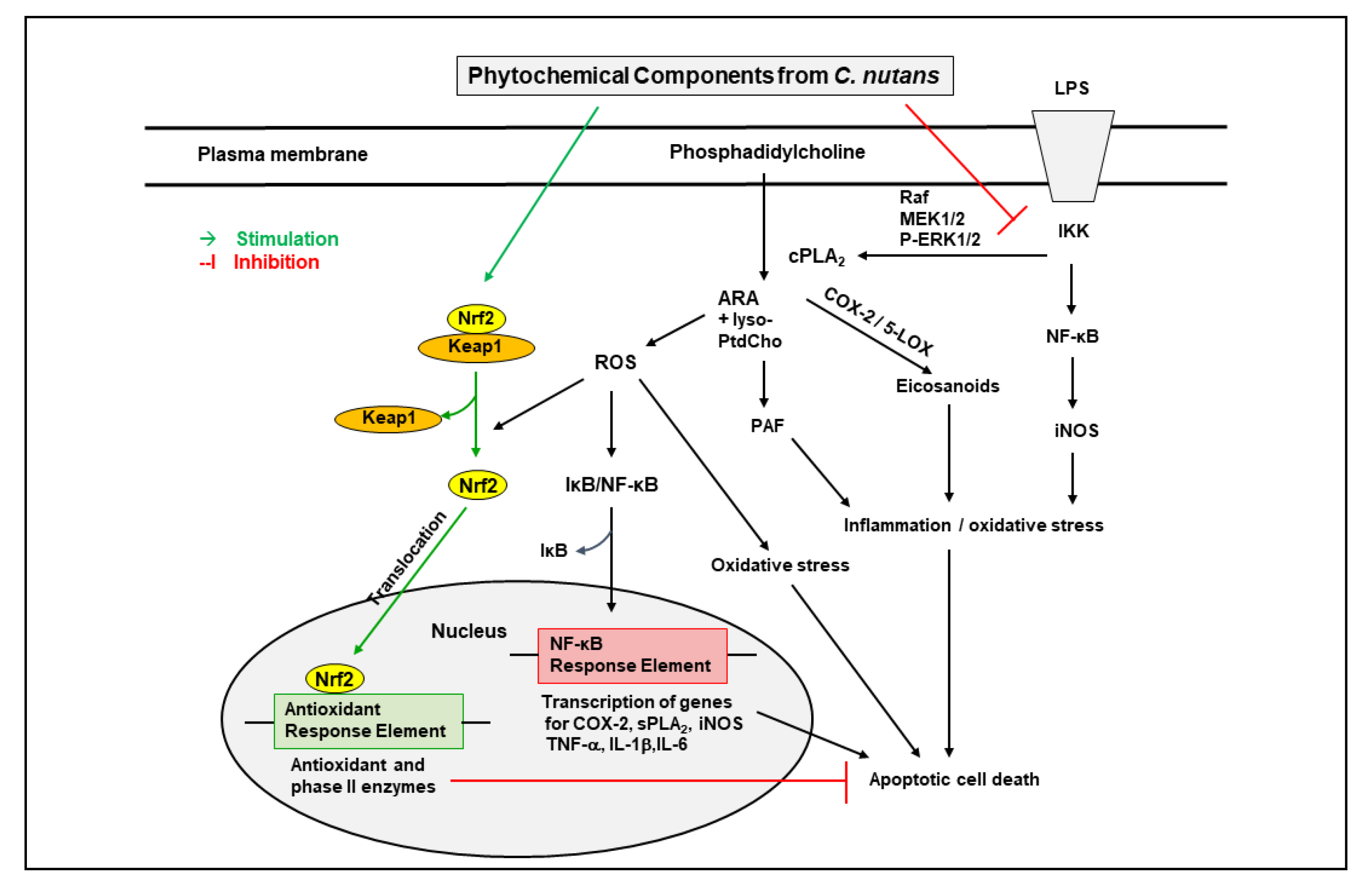
Isoorientin could attenuate LPS-induced pro-inflammatory responses including expression of iNOS, COX-2, and pro-inflammatory cytokines such as TNF-α and IL-1β, through down-regulation of ROS-related MAPK/NF-κB signaling pathway in BV-2 microglial cells [91]. Treatment with isoorientin (5 and 10 mg/kg, p.o.) significantly improved cognitive impairment, and decreased acetylcholinesterase and thiobarbituric acid reactive substance activities in the hippocampus and frontal cortex of scopolamine injected mice. These results suggest an antioxidant and memory-enhancing effect of isoorientin [92]. Isoorientin increased the expression of tight junction proteins occludin and ZO-1, in the brain of endotoxemic mice. The phytochemical also decreased activation of ERK and NF-κB and expression of COX-2, and increased activation of Nrf2/HO-1, in LPS-induced RAW264.7 cells. These results suggest an effect of isoorientin in reducing inflammation and protecting the integrity of the blood–brain barrier in endotoxemic mice [93]. Isoorientin could also modulate Aβ25-35-induced increase in pro-inflammatory mediators such as IL-6, TNF-α, and COX-2, as well as ROS production in BV-2 microglial cells [94]. The inflammatory and oxidative stress pathways targeted by C. nutans extracts and their phytochemical components are indicated in Figure 2.

4. Clinacanthus nutans Extracts on Inflammatory Pathways in Cerebral Ischemia and Neurovascular Systems

4.1. Clinacanthus nutans on Cerebral Ischemia

Clinacanthus nutans extracts have been shown to affect the expression and activity of cPLA2, an important enzyme in inflammation. This enzyme is known for hydrolysis of glycerophospholipids to produce arachidonic acid and lysophospholipids, and in turn, metabolism of arachidonic acid produces pro-inflammatory mediators and eicosanoids, as well generation of free radicals [95,96,97]. The expression cPLA2 mRNA is regulated by histone acetylation. In a study by Tan et al., C. nutans extract as well as NU9056, a Tip60 histone acetylase (HAT) inhibitor, could suppress cPLA2 mRNA expression in neurons due to OGD [98]. C. nutans extracts also suppressed histone deacetylase 1 (HDAC1) and HDAC6 transcription and ameliorated neuronal death and astrocyte/endothelial damage due to OGD [99]. In another study, C. nutans extract was found to protect neurons and ameliorate ischemic injury by promoting the anti-apoptotic activity of peroxisome proliferator-activated receptor-gamma (PPAR-γ), a stress-induced transcription factor. The augmentation of transcription by C. nutans extract appeared to occur through selective increase of CCAAT/enhancer binding protein (C/EBP) β binding to a specific C/EBP binding site on the PPAR-γ promoter [100]. PPAR-γ stimulation is known to downregulate the NF-kB pathway, thus inhibiting the expression of a wide array of pro-inflammatory mediators in microglia and macrophages, including IL-1β, IL-6, IL-12, TNF-α, COX-2, and iNOS [101,102]. Transient OGD led to induction of IL-1β, IL-6, and TNFα, and pretreatment with C. nutans extract suppressed the production of these inflammatory cytokines in primary neurons. C. nutans extract also inhibited IL-1β transcription through NF-κB/p65 nuclear translocation, and siRNA knockdown of either p65 or IL-1β mitigated OGD-mediated neuronal death. Post-ischemic treatment of C. nutans attenuated IκBα degradation and decreased IL-1β, IL-6, and TNFα production in the ischemic brain [103].

4.2. Clinacanthus nutans on Neurovascular Systems

Clinacanthus nutans extracts could also show potential effects against vascular dementia. Oral administration of C. nutans leaf water extract (500 and 1000 mg/kg) was found to reduce sickness behavior and brain inflammatory markers in rats after intracerebral injection of LPS [104]. Clinacanthus nutans ethanolic extract was found to have anti-apoptotic effects against LPS-induced damage in bovine endothelial cells. This could be due to an effect of its components, glyceryl 1,3-disterate, kaempferol 3-O-feruloyl-sophoroside 7-O-glucoside, and hydroxypthioceranic acid, which showed pro-surviving Bcl-2 and pro-apoptotic Bax expression activities [30]. Clinacanthus nutans aqueous extracts could reduce LPS-induced increase in proinflammatory IL-1β expression but increase anti-inflammatory IL-2 and IL-4 expression in brain tissue after oral administration in rats [105]. A recent study showed that C. nutans leaf, but not stem, extracts had a significant effect in reducing 7-kecoholesterol-induced brain endothelial cell death and suppressed inflammatory responses in endothelial cells, including reducing IL-1β, IL-6, IL-8, TNF-α, and COX-2 expression [106]. Together, the above results suggest a protective effect of C. nutans in neurovascular injury by reducing the expression of pro-inflammatory enzymes such as cPLA2 and increasing the expression of cytoprotective enzymes.

5. Conclusions and Future Directions

Studies with polar and nonpolar extraction procedures for leaves of C. nutans indicated different extracts yielded components with anti-inflammatory, anti-tumorigenic, and antiviral properties. There are increasing studies linking these extracts against disorders in cells and tissues in the central and peripheral systems. Future studies to standardize the extraction protocols and to associate them with treatment of specific disorders will help to direct better use of the products for health and diseases.
Analyses of extracts from C. nutans leaves have led to identification of C-glycosyl flavones including schaftoside, isovitexin, orientin, and vitexin. A review of studies on individual phytochemicals indicated anti-inflammatory and antioxidative properties to different extents. In general, anti-inflammatory properties are linked to their ability to suppress the NF-kB pathway and production of iNOS as well as pro-inflammatory cytokines, such as IL-1β and TNF-α. In addition, anti-inflammatory properties are also linked to ability to suppress the cPLA2 pathway associated with increase of COX-2 and pro-inflammatory eicosanoids. To some extent, individual phytochemicals also exhibit antioxidative properties as demonstrated by their ability to upregulate the antioxidative stress pathway involving Nrf2 and production of HO-1. The ability for the C. nutans extracts and their components to modulate these pathways under different inflammatory conditions has not been quantitatively defined. Therefore, more studies are needed to understand the molecular mechanism of action. Development of reporter gene assays targeting the transcription factors will be useful to better define properties of the pathways and aid development of therapeutic potential for treatment of diseases.

Author Contributions

Conceptualization, W.-Y.O., G.Y.S. and T.-N.L.; writing—review and editing, W.-Y.O., D.R.H., G.Y.S. and T.-N.L. All authors have read and agreed to the published version of the manuscript.

Funding

This research was funded by grants from the Ministry of Education (NUHSRO/2019/051/T1/Seed-Mar/04) and the National Medical Research Council of Singapore (HLCA21Jan-0019).

Institutional Review Board Statement

Not applicable.

Informed Consent Statement

Not applicable.

Conflicts of Interest

The authors declare no conflict of interest.

References

  1. Flora and Fauna Web. Clinacanthus nutans (Burm. F.) Lindau. National Parks: Singapore, 2022. Available online: https://www.nparks.gov.sg/florafaunaweb/flora/5/6/5655 (accessed on 4 May 2022).
  2. Alam, A.; Ferdosh, S.; Ghafoor, K.; Hakim, A.; Juraimi, A.S.; Khatib, A.; Sarker, Z.I. Clinacanthus nutans: A review of the medicinal uses, pharmacology and phytochemistry. Asian Pac. J. Trop. Med. 2016, 9, 402–409. [Google Scholar] [CrossRef] [PubMed] [Green Version]
  3. Siew, Y.Y.; Zareisedehizadeh, S.; Seetoh, W.G.; Neo, S.Y.; Tan, C.H.; Koh, H.L. Ethnobotanical survey of usage of fresh medicinal plants in Singapore. J. Ethnopharmacol. 2014, 155, 1450–1466. [Google Scholar] [CrossRef] [PubMed]
  4. Chia, T.Y.; Gan, C.Y.; Murugaiyah, V.; Hashmi, S.F.; Fatima, T.; Ibrahim, L.; Abdulla, M.H.; Alswailmi, F.K.; Johns, E.J.; Ahmad, A.A. Narrative Review on the Phytochemistry, Pharmacology and Therapeutic Potentials of Clinacanthus nutans (Burm. f.) Lindau Leaves as an Alternative Source of Future Medicine. Molecules 2021, 27, 139. [Google Scholar] [CrossRef] [PubMed]
  5. Ghasemzadeh, A.; Nasiri, A.; Jaafar, H.Z.; Baghdadi, A.; Ahmad, I. Changes in phytochemical synthesis, chalcone synthase activity and pharmaceutical qualities of sabah snake grass (Clinacanthus nutans L.) in relation to plant age. Molecules 2014, 19, 17632–17648. [Google Scholar] [CrossRef]
  6. Abd Samat, N.M.A.; Ahmad, S.; Awang, Y.; Bakar, R.A.H.; Hakiman, M. Alterations in Herbage Yield, Antioxidant Activities, Phytochemical Contents, and Bioactive Compounds of Sabah Snake Grass (Clinacanthus Nutans L.) with Regards to Harvesting Age and Harvesting Frequency. Molecules 2020, 25, 2833. [Google Scholar] [CrossRef]
  7. Fong, S.Y.; Piva, T.; Dekiwadia, C.; Urban, S.; Huynh, T. Comparison of cytotoxicity between extracts of Clinacanthus nutans (Burm. f.) Lindau leaves from different locations and the induction of apoptosis by the crude methanol leaf extract in D24 human melanoma cells. BMC Complementary Altern. Med. 2016, 16, 368. [Google Scholar] [CrossRef] [Green Version]
  8. Farsi, E.; Esmailli, K.; Shafaei, A.; Moradi Khaniabadi, P.; Al Hindi, B.; Khadeer Ahamed, M.B.; Sandai, D.; Abdul Sattar, M.; Ismail, Z.; Abdul Majid, A.M.; et al. Mutagenicity and preclinical safety assessment of the aqueous extract of Clinacanthus nutans leaves. Drug Chem. Toxicol. 2016, 39, 461–473. [Google Scholar] [CrossRef]
  9. Khoo, L.W.; Foong Kow, A.S.; Maulidiani, M.; Lee, M.T.; Tan, C.P.; Shaari, K.; Tham, C.L.; Abas, F. Hematological, Biochemical, Histopathological and (1)H-NMR Metabolomics Application in Acute Toxicity Evaluation of Clinacanthus nutans Water Leaf Extract. Molecules 2018, 23, 2172. [Google Scholar] [CrossRef] [Green Version]
  10. Aliyu, A.; Shaari, M.R.; Ahmad Sayuti, N.S.; Reduan, M.F.H.; Sithambaram, S.; Noordin, M.M.; Shaari, K.; Hamzah, H. Subacute Oral Administration of Clinacanthus nutans Ethanolic Leaf Extract Induced Liver and Kidney Toxicities in ICR Mice. Molecules 2020, 25, 2631. [Google Scholar] [CrossRef]
  11. Tan, L.T.-H.; Khaw, K.Y.; Ong, Y.S.; Khan, T.M.; Lee, L.-H.; Lee, W.-L.; Goh, B.-H. An Overview of Clinacanthus nutans (Burm. f.) Lindau as a Medicinal Plant with Diverse Pharmacological Values. Plant Deriv. Bioact. 2020, 461–491. [Google Scholar]
  12. Khoo, L.W.; Audrey Kow, S.; Lee, M.T.; Tan, C.P.; Shaari, K.; Tham, C.L.; Abas, F. A Comprehensive Review on Phytochemistry and Pharmacological Activities of Clinacanthus nutans (Burm.f.) Lindau. Evid. Based Complementary Altern. Med. eCAM 2018, 2018, 9276260. [Google Scholar] [CrossRef] [PubMed] [Green Version]
  13. Tuntiwachwuttikul, P.; Pootaeng-On, Y.; Phansa, P.; Taylor, W.C. Cerebrosides and a monoacylmonogalactosylglycerol from Clinacanthus nutans. Chem. Pharm. Bull. 2004, 52, 27–32. [Google Scholar] [CrossRef] [PubMed] [Green Version]
  14. Teshima, K.-I.; Kaneko, T.; Ohtani, K. C-glycosyl flavones from Clinacanthus nutans. Nat. Med. 1997, 51, 557. [Google Scholar]
  15. Chelyn, J.L.; Omar, M.H.; Mohd Yousof, N.S.; Ranggasamy, R.; Wasiman, M.I.; Ismail, Z. Analysis of flavone C-glycosides in the leaves of Clinacanthus nutans (Burm. f.) Lindau by HPTLC and HPLC-UV/DAD. Sci. World J. 2014, 2014, 724267. [Google Scholar] [CrossRef] [PubMed] [Green Version]
  16. Tu, S.F.; Liu, R.H.; Cheng, Y.B.; Hsu, Y.M.; Du, Y.C.; El-Shazly, M.; Wu, Y.C.; Chang, F.R. Chemical constituents and bioactivities of Clinacanthus nutans aerial parts. Molecules 2014, 19, 20382–20390. [Google Scholar] [CrossRef]
  17. Che Sulaiman, I.S.; Basri, M.; Fard Masoumi, H.R.; Chee, W.J.; Ashari, S.E.; Ismail, M. Effects of temperature, time, and solvent ratio on the extraction of phenolic compounds and the anti-radical activity of Clinacanthus nutans Lindau leaves by response surface methodology. Chem. Cent. J. 2017, 11, 54. [Google Scholar] [CrossRef]
  18. Ban, W.K.; Fong, I.L.; Khong, H.Y.; Phung, J.H.Y. Wound Healing, Antimicrobial and Antioxidant Properties of Clinacanthus nutans (Burm.f.) Lindau and Strobilanthes crispus (L.) Blume Extracts. Molecules 2022, 27, 1722. [Google Scholar] [CrossRef]
  19. Nordin, F.J.; Pearanpan, L.; Chan, K.M.; Kumolosasi, E.; Yong, Y.K.; Shaari, K.; Rajab, N.F. Immunomodulatory potential of Clinacanthus nutans extracts in the co-culture of triple-negative breast cancer cells, MDA-MB-231, and THP-1 macrophages. PLoS ONE 2021, 16, e0256012. [Google Scholar] [CrossRef]
  20. Huang, D.; Li, Y.; Cui, F.; Chen, J.; Sun, J. Purification and characterization of a novel polysaccharide-peptide complex from Clinacanthus nutans Lindau leaves. Carbohydr. Polym. 2016, 137, 701–708. [Google Scholar] [CrossRef]
  21. Zakaria, K.N.; Amid, A.; Zakaria, Z.; Jamal, P.; Ismail, A. Anti-Proliferative Activity of Triterpenes Isolated from Clinicanthus nutans on Hep-G2 Liver Cancer Cells. Asian Pac. J. Cancer Prev. APJCP 2019, 20, 563–567. [Google Scholar] [CrossRef] [Green Version]
  22. Mutazah, R.; Hamid, H.A.; Mazila Ramli, A.N.; Fasihi Mohd Aluwi, M.F.; Yusoff, M.M. In vitro cytotoxicity of Clinacanthus nutans fractions on breast cancer cells and molecular docking study of sulphur containing compounds against caspase-3. Food Chem. Toxicol. 2020, 135, 110869. [Google Scholar] [CrossRef] [PubMed]
  23. Murugesu, S.; Khatib, A.; Ahmed, Q.U.; Ibrahim, Z.; Uzir, B.F.; Benchoula, K.; Yusoff, N.I.N.; Perumal, V.; Alajmi, M.F.; Salamah, S.; et al. Toxicity study on Clinacanthus nutans leaf hexane fraction using Danio rerio embryos. Toxicol. Rep. 2019, 6, 1148–1154. [Google Scholar] [CrossRef] [PubMed]
  24. Sakdarat, S.; Shuyprom, A.; Pientong, C.; Ekalaksananan, T.; Thongchai, S. Bioactive constituents from the leaves of Clinacanthus nutans Lindau. Bioorg. Med. Chem. 2009, 17, 1857–1860. [Google Scholar] [CrossRef] [PubMed]
  25. Le, C.F.; Kailaivasan, T.H.; Chow, S.C.; Abdullah, Z.; Ling, S.K.; Fang, C.M. Phytosterols isolated from Clinacanthus nutans induce immunosuppressive activity in murine cells. Int. Immunopharmacol. 2017, 44, 203–210. [Google Scholar] [CrossRef] [PubMed]
  26. Mustapa, A.N.; Martin, A.; Mato, R.B.; Cocero, M.J. Extraction of phytocompounds from the medicinal plant Clinacanthus nutans Lindau by microwave-assisted extraction and supercritical carbon dioxide extraction. Ind. Crops Prod. 2015, 74, 83–94. [Google Scholar] [CrossRef]
  27. Khoo, L.W.; Kow, A.S.F.; Maulidiani, M.; Ang, M.Y.; Chew, W.Y.; Lee, M.T.; Tan, C.P.; Shaari, K.; Tham, C.L.; Abas, F. (1) H-NMR metabolomics for evaluating the protective effect of Clinacanthus nutans (Burm. f) Lindau water extract against nitric oxide production in LPS-IFN-gamma activated RAW 264.7 macrophages. Phytochemic. Anal. PCA 2019, 30, 46–61. [Google Scholar] [CrossRef] [PubMed] [Green Version]
  28. Murugesu, S.; Ibrahim, Z.; Ahmed, Q.U.; Nik Yusoff, N.I.; Uzir, B.F.; Perumal, V.; Abas, F.; Saari, K.; El-Seedi, H.; Khatib, A. Characterization of alpha-Glucosidase Inhibitors from Clinacanthus nutans Lindau Leaves by Gas Chromatography-Mass Spectrometry-Based Metabolomics and Molecular Docking Simulation. Molecules 2018, 23, 2402. [Google Scholar] [CrossRef] [Green Version]
  29. Teoh, P.L.; Cheng, A.Y.; Liau, M.; Lem, F.F.; Kaling, G.P.; Chua, F.N.; Cheong, B.E. Chemical composition and cytotoxic properties of Clinacanthus nutans root extracts. Pharm. Biol. 2017, 55, 394–401. [Google Scholar] [CrossRef] [Green Version]
  30. Panya, A.; Pundith, H.; Thongyim, S.; Kaewkod, T.; Chitov, T.; Bovonsombut, S.; Tragoolpua, Y. Antibiotic-Antiapoptotic Dual Function of Clinacanthus nutans (Burm. f.) Lindau Leaf Extracts against Bovine Mastitis. Antibiotics 2020, 9, 429. [Google Scholar] [CrossRef]
  31. Roeslan, M.O.; Ayudhya, T.D.N.; Yingyongnarongkul, B.E.; Koontongkaew, S. Anti-biofilm, nitric oxide inhibition and wound healing potential of purpurin-18 phytyl ester isolated from Clinacanthus nutans leaves. Biomed. Pharmacother. 2019, 113, 108724. [Google Scholar] [CrossRef]
  32. Ismail, N.Z.; Md Toha, Z.; Muhamad, M.; Nik Mohamed Kamal, N.N.S.; Mohamad Zain, N.N.; Arsad, H. Antioxidant Effects, Antiproliferative Effects, and Molecular Docking of Clinacanthus nutans Leaf Extracts. Molecules 2020, 25, 2067. [Google Scholar] [CrossRef] [PubMed]
  33. De Melo, G.O.; Muzitano, M.F.; Legora-Machado, A.; Almeida, T.A.; De Oliveira, D.B.; Kaiser, C.R.; Koatz, V.L.; Costa, S.S. C-glycosylflavones from the aerial parts of Eleusine indica inhibit LPS-induced mouse lung inflammation. Planta Med. 2005, 4, 362–363. [Google Scholar] [CrossRef] [PubMed]
  34. Liu, M.; Zhang, G.; Song, M.; Wang, J.; Shen, C.; Chen, Z.; Huang, X.; Gao, Y.; Zhu, C.; Lin, C.; et al. Activation of Farnesoid X Receptor by Schaftoside Ameliorates Acetaminophen-Induced Hepatotoxicity by Modulating Oxidative Stress and Inflammation. Antioxid. Redox Signal. 2020, 33, 87–116. [Google Scholar] [CrossRef] [PubMed]
  35. Zhou, K.; Wu, J.; Chen, J.; Zhou, Y.; Chen, X.; Wu, Q.; Xu, Y.; Tu, W.; Lou, X.; Yang, G.; et al. Schaftoside ameliorates oxygen glucose deprivation-induced inflammation associated with the TLR4/Myd88/Drp1-related mitochondrial fission in BV2 microglia cells. J. Pharmacol. Sci. 2019, 139, 15–22. [Google Scholar] [CrossRef]
  36. Dang, J.; Paudel, Y.N.; Yang, X.; Ren, Q.; Zhang, S.; Ji, X.; Liu, K.; Jin, M. Schaftoside Suppresses Pentylenetetrazol-Induced Seizures in Zebrafish via Suppressing Apoptosis, Modulating Inflammation, and Oxidative Stress. ACS Chem. Neurosci. 2021, 12, 2542–2552. [Google Scholar] [CrossRef]
  37. Borghi, S.M.; Carvalho, T.T.; Staurengo-Ferrari, L.; Hohmann, M.S.; Pinge-Filho, P.; Casagrande, R.; Verri, W.A., Jr. Vitexin inhibits inflammatory pain in mice by targeting TRPV1, oxidative stress, and cytokines. J. Nat. Prod. 2013, 76, 1141–1149. [Google Scholar] [CrossRef]
  38. Dong, L.Y.; Li, S.; Zhen, Y.L.; Wang, Y.N.; Shao, X.; Luo, Z.G. Cardioprotection of vitexin on myocardial ischemia/reperfusion injury in rat via regulating inflammatory cytokines and MAPK pathway. Am. J. Chin. Med. 2013, 41, 1251–1266. [Google Scholar] [CrossRef]
  39. Sun, Z.; Yan, B.; Yu, W.Y.; Yao, X.; Ma, X.; Sheng, G.; Ma, Q. Vitexin attenuates acute doxorubicin cardiotoxicity in rats via the suppression of oxidative stress, inflammation and apoptosis and the activation of FOXO3a. Exp. Ther. Med. 2016, 12, 1879–1884. [Google Scholar] [CrossRef] [Green Version]
  40. Wang, F.; Yin, J.; Ma, Y.; Jiang, H.; Li, Y. Vitexin alleviates lipopolysaccharide-induced islet cell injury by inhibiting HMGB1 release. Mol. Med. Rep. 2017, 15, 1079–1086. [Google Scholar] [CrossRef]
  41. Xie, C.L.; Li, J.L.; Xue, E.X.; Dou, H.C.; Lin, J.T.; Chen, K.; Wu, H.Q.; Wu, L.; Xuan, J.; Huang, Q.S. Vitexin alleviates ER-stress-activated apoptosis and the related inflammation in chondrocytes and inhibits the degeneration of cartilage in rats. Food Funct. 2018, 9, 5740–5749. [Google Scholar] [CrossRef]
  42. Lu, Y.; Yu, T.; Liu, J.; Gu, L. Vitexin attenuates lipopolysaccharide-induced acute lung injury by controlling the Nrf2 pathway. PLoS ONE 2018, 13, e0196405. [Google Scholar] [CrossRef]
  43. Yang, H.; Huang, J.; Mao, Y.; Wang, L.; Li, R.; Ha, C. Vitexin alleviates interleukin-1β-induced inflammatory responses in chondrocytes from osteoarthritis patients: Involvement of HIF-1α pathway. Scand. J. Immunol. 2019, 90, e12773. [Google Scholar] [CrossRef] [PubMed]
  44. Duan, S.; Du, X.; Chen, S.; Liang, J.; Huang, S.; Hou, S.; Gao, J.; Ding, P. Effect of vitexin on alleviating liver inflammation in a dextran sulfate sodium (DSS)-induced colitis model. Biomed. Pharmacother. 2020, 121, 109683. [Google Scholar] [CrossRef] [PubMed]
  45. Sae-Tan, S.; Kumrungsee, T.; Yanaka, N. Mungbean seed coat water extract inhibits inflammation in LPS-induced acute liver injury mice and LPS-stimulated RAW 246.7 macrophages via the inhibition of TAK1/IkappaBalpha/NF-kappaB. Journal Food Sci. Technol. 2020, 57, 2659–2668. [Google Scholar] [CrossRef] [PubMed]
  46. Li, C.; Chen, Y.; Yuan, X.; He, L.; Li, X.; Huang, S.; Hou, S.; Liang, J. Vitexin ameliorates chronic stress plub high fat diet-induced nonalcoholic fatty liver disease by inhibiting inflammation. Eur. J. Pharmacol. 2020, 882, 173264. [Google Scholar] [CrossRef]
  47. Chen, Y.; Wang, B.; Yuan, X.; Lu, Y.; Hu, J.; Gao, J.; Lin, J.; Liang, J.; Hou, S.; Chen, S. Vitexin prevents colitis-associated carcinogenesis in mice through regulating macrophage polarization. Phytomed. Int. J. Phytother. Phytopharm. 2021, 83, 153489. [Google Scholar] [CrossRef]
  48. Wang, W.; Cheng, H.; Gu, X.; Yin, X. The natural flavonoid glycoside vitexin displays preclinical antitumor activity by suppressing NF-κB signaling in nasopharyngeal carcinoma. OncoTarget Ther. 2019, 12, 4461–4468. [Google Scholar] [CrossRef] [Green Version]
  49. Saha, S.; Buttari, B.; Panieri, E.; Profumo, E.; Saso, L. An Overview of Nrf2 Signaling Pathway and Its Role in Inflammation. Molecules 2020, 25, 5474. [Google Scholar] [CrossRef]
  50. Li, X.S.; Tang, X.Y.; Su, W.; Li, X. Vitexin protects melanocytes from oxidative stress via activating MAPK-Nrf2/ARE pathway. Immunopharmacol. Immunotoxicol. 2020, 42, 594–603. [Google Scholar] [CrossRef]
  51. Min, J.W.; Kong, W.L.; Han, S.; Bsoul, N.; Liu, W.H.; He, X.H.; Sanchez, R.M.; Peng, B.W. Vitexin protects against hypoxic-ischemic injury via inhibiting Ca2+/Calmodulin-dependent protein kinase II and apoptosis signaling in the neonatal mouse brain. Oncotarget 2017, 8, 25513–25524. [Google Scholar] [CrossRef] [Green Version]
  52. Malar, D.S.; Prasanth, M.I.; Shafreen, R.B.; Balamurugan, K.; Devi, K.P. Grewia tiliaefolia and its active compound vitexin regulate the expression of glutamate transporters and protect Neuro-2a cells from glutamate toxicity. Life Sci. 2018, 203, 233–241. [Google Scholar] [CrossRef] [PubMed]
  53. Malar, D.S.; Suryanarayanan, V.; Prasanth, M.I.; Singh, S.K.; Balamurugan, K.; Devi, K.P. Vitexin inhibits Aβ25-35 induced toxicity in Neuro-2a cells by augmenting Nrf-2/HO-1 dependent antioxidant pathway and regulating lipid homeostasis by the activation of LXR-α. Toxicol. Vitr. 2018, 50, 160–171. [Google Scholar] [CrossRef] [PubMed]
  54. Luo, W.D.; Min, J.W.; Huang, W.X.; Wang, X.; Peng, Y.Y.; Han, S.; Yin, J.; Liu, W.H.; He, X.H.; Peng, B.W. Vitexin reduces epilepsy after hypoxic ischemia in the neonatal brain via inhibition of NKCC1. J. Neuroinflamm. 2018, 15, 186. [Google Scholar] [CrossRef] [PubMed] [Green Version]
  55. Jiang, J.; Dai, J.; Cui, H. Vitexin reverses the autophagy dysfunction to attenuate MCAO-induced cerebral ischemic stroke via mTOR/Ulk1 pathway. Biomed. Pharmacother. 2018, 99, 583–590. [Google Scholar] [CrossRef] [PubMed]
  56. Krishnan, M.; Kang, S.C. Vitexin inhibits acrylamide-induced neuroinflammation and improves behavioral changes in zebrafish larvae. Neurotoxicol. Teratol. 2019, 74, 106811. [Google Scholar] [CrossRef]
  57. Cao, H.; Wang, X.; Zhang, B.; Ren, M. The protective effect of vitexinin septic encephalopathy by reducing leukocyte-endothelial adhesion and inflammatory response. Ann. Palliat. Med. 2020, 9, 2079–2089. [Google Scholar] [CrossRef]
  58. Zhao, C.R.; Yang, F.F.; Cui, Q.; Wang, D.; Zhou, Y.; Li, Y.S.; Zhang, Y.P.; Tang, R.Z.; Yao, W.J.; Wang, X.; et al. Vitexin inhibits APEX1 to counteract the flow-induced endothelial inflammation. Proc. Natl. Acad. Sci. USA 2021, 118, 48. [Google Scholar] [CrossRef]
  59. Li, S.; Liang, T.; Zhang, Y.; Huang, K.; Yang, S.; Lv, H.; Chen, Y.; Zhang, C.; Guan, X. Vitexin alleviates high-fat diet induced brain oxidative stress and inflammation via anti-oxidant, anti-inflammatory and gut microbiota modulating properties. Free Radic. Biol. Med. 2021, 171, 332–344. [Google Scholar] [CrossRef]
  60. Zhang, Q.; Fan, Z.; Xue, W.; Sun, F.; Zhu, H.; Huang, D.; Wang, Z.; Dong, L. Vitexin regulates Epac and NLRP3 and ameliorates chronic cerebral hypoperfusion injury. Can. J. Physiol. Pharmacol. 2021, 99, 1079–1087. [Google Scholar] [CrossRef]
  61. Huang, S.T.; Chen, C.T.; Chieng, K.T.; Huang, S.H.; Chiang, B.H.; Wang, L.F.; Kuo, H.S.; Lin, C.M. Inhibitory effects of a rice hull constituent on tumor necrosis factor alpha, prostaglandin E2, and cyclooxygenase-2 production in lipopolysaccharide-activated mouse macrophages. Ann. N. Y. Acad. Sci. 2005, 1042, 387–395. [Google Scholar] [CrossRef] [Green Version]
  62. Lv, H.; Yu, Z.; Zheng, Y.; Wang, L.; Qin, X.; Cheng, G.; Ci, X. Isovitexin Exerts Anti-Inflammatory and Anti-Oxidant Activities on Lipopolysaccharide-Induced Acute Lung Injury by Inhibiting MAPK and NF-kappaB and Activating HO-1/Nrf2 Pathways. Int. J. Biol. Sci. 2016, 12, 72–86. [Google Scholar] [CrossRef] [PubMed] [Green Version]
  63. Lin, C.M.; Huang, S.T.; Liang, Y.C.; Lin, M.S.; Shih, C.M.; Chang, Y.C.; Chen, T.Y.; Chen, C.T. Isovitexin suppresses lipopolysaccharide-mediated inducible nitric oxide synthase through inhibition of NF-kappa B in mouse macrophages. Planta Med. 2005, 71, 748–753. [Google Scholar] [CrossRef] [PubMed]
  64. Hu, J.J.; Wang, H.; Pan, C.W.; Lin, M.X. Isovitexin alleviates liver injury induced by lipopolysaccharide/d-galactosamine by activating Nrf2 and inhibiting NF-kappaB activation. Microb. Pathog. 2018, 119, 86–92. [Google Scholar] [CrossRef] [PubMed]
  65. Liu, S.; Zhang, X.; Wang, J. Isovitexin protects against cisplatin-induced kidney injury in mice through inhibiting inflammatory and oxidative responses. Int. Immunopharmacol. 2020, 83, 106437. [Google Scholar] [CrossRef] [PubMed]
  66. Deng, J.; Ma, Y.; He, Y.; Yang, H.; Chen, Y.; Wang, L.; Huang, D.; Qiu, S.; Tao, X.; Chen, W. A Network Pharmacology-Based Investigation to the Pharmacodynamic Material Basis and Mechanisms of the Anti-Inflammatory and Anti-Viral Effect of Isatis indigotica. Drug Des. Dev. Ther. 2021, 15, 3193–3206. [Google Scholar] [CrossRef] [PubMed]
  67. Kesavan, R.; Chandel, S.; Upadhyay, S.; Bendre, R.; Ganugula, R.; Potunuru, U.R.; Giri, H.; Sahu, G.; Kumar, P.U.; Reddy, G.B.; et al. Gentiana lutea exerts anti-atherosclerotic effects by preventing endothelial inflammation and smooth muscle cell migration. Nutr. Metab. Cardiovasc. Dis. NMCD 2016, 26, 293–301. [Google Scholar] [CrossRef]
  68. Liu, B.; Huang, B.; Hu, G.; He, D.; Li, Y.; Ran, X.; Du, J.; Fu, S.; Liu, D. Isovitexin-Mediated Regulation of Microglial Polarization in Lipopolysaccharide-Induced Neuroinflammation via Activation of the CaMKKβ/AMPK-PGC-1α Signaling Axis. Front. Immunol. 2019, 10, 2650. [Google Scholar] [CrossRef]
  69. Xiao, Q.; Qu, Z.; Zhao, Y.; Yang, L.; Gao, P. Orientin Ameliorates LPS-Induced Inflammatory Responses through the Inhibitory of the NF-κB Pathway and NLRP3 Inflammasome. Evid. Based Complementary Altern. Med. eCAM 2017, 2017, 2495496. [Google Scholar] [CrossRef] [Green Version]
  70. Xiao, Q.; Piao, R.; Wang, H.; Li, C.; Song, L. Orientin-mediated Nrf2/HO-1 signal alleviates H2O2-induced oxidative damage via induction of JNK and PI3K/AKT activation. Int. J. Biol. Macromol. 2018, 118, 747–755. [Google Scholar] [CrossRef]
  71. Dhakal, H.; Lee, S.; Choi, J.K.; Kwon, T.K.; Khang, D.; Kim, S.H. Inhibitory effects of orientin in mast cell-mediated allergic inflammation. Pharmacol. Rep. PR 2020, 72, 1002–1010. [Google Scholar] [CrossRef]
  72. Xiao, Q.; Cui, Y.; Zhao, Y.; Liu, L.; Wang, H.; Yang, L. Orientin relieves lipopolysaccharide-induced acute lung injury in mice: The involvement of its anti-inflammatory and anti-oxidant properties. Int. Immunopharmacol. 2021, 90, 107189. [Google Scholar] [CrossRef] [PubMed]
  73. Thangaraj, K.; Vaiyapuri, M. Orientin, a C-glycosyl dietary flavone, suppresses colonic cell proliferation and mitigates NF-κB mediated inflammatory response in 1,2-dimethylhydrazine induced colorectal carcinogenesis. Biomed. Pharmacother. 2017, 96, 1253–1266. [Google Scholar] [CrossRef] [PubMed]
  74. Lee, W.; Ku, S.K.; Bae, J.S. Vascular barrier protective effects of orientin and isoorientin in LPS-induced inflammation in vitro and in vivo. Vasc. Pharmacol. 2014, 62, 3–14. [Google Scholar] [CrossRef] [PubMed]
  75. Ku, S.K.; Kwak, S.; Bae, J.S. Orientin inhibits high glucose-induced vascular inflammation in vitro and in vivo. Inflammation 2014, 37, 2164–2173. [Google Scholar] [CrossRef]
  76. Zhou, X.; Gan, P.; Hao, L.; Tao, L.; Jia, J.; Gao, B.; Liu, J.Y.; Zheng, L.T.; Zhen, X. Antiinflammatory effects of orientin-2′′-O-galactopyranoside on lipopolysaccharide-stimulated microglia. Biol. Pharm. Bull. 2014, 37, 1282–1294. [Google Scholar] [CrossRef] [Green Version]
  77. Yoo, H.; Ku, S.K.; Lee, T.; Bae, J.S. Orientin inhibits HMGB1-induced inflammatory responses in HUVECs and in murine polymicrobial sepsis. Inflammation 2014, 37, 1705–1717. [Google Scholar] [CrossRef]
  78. Bae, J.S. Inhibitory Effect of Orientin on Secretory Group IIA Phospholipase A2. Inflammation 2015, 38, 1631–1638. [Google Scholar] [CrossRef]
  79. Yu, L.; Wang, S.; Chen, X.; Yang, H.; Li, X.; Xu, Y.; Zhu, X. Orientin alleviates cognitive deficits and oxidative stress in Aβ1-42-induced mouse model of Alzheimer’s disease. Life Sci. 2015, 121, 104–109. [Google Scholar] [CrossRef]
  80. Wang, X.; An, F.; Wang, S.; An, Z.; Wang, S. Orientin Attenuates Cerebral Ischemia/Reperfusion Injury in Rat Model through the AQP-4 and TLR4/NF-κB/TNF-α Signaling Pathway. J. Stroke Cerebrovasc. Dis. 2017, 26, 2199–2214. [Google Scholar] [CrossRef]
  81. Guo, D.; Hu, X.; Zhang, H.; Lu, C.; Cui, G.; Luo, X. Orientin and neuropathic pain in rats with spinal nerve ligation. Int. Immunopharmacol. 2018, 58, 72–79. [Google Scholar] [CrossRef]
  82. Li, C.; Cai, C.; Zheng, X.; Sun, J.; Ye, L. Orientin suppresses oxidized low-density lipoproteins induced inflammation and oxidative stress of macrophages in atherosclerosis. Biosci. Biotechnol. Biochem. 2020, 84, 774–779. [Google Scholar] [CrossRef] [PubMed]
  83. Attiq, A.; Jalil, J.; Husain, K.; Mohamad, H.F.; Ahmad, A. Luteolin and apigenin derived glycosides from Alphonsea elliptica abrogate LPS-induced inflammatory responses in human plasma. J. Ethnopharmacol. 2021, 275, 114120. [Google Scholar] [CrossRef] [PubMed]
  84. Küpeli, E.; Aslan, M.; Gürbüz, I.; Yesilada, E. Evaluation of in vivo biological activity profile of isoorientin. Z. Naturforschung. C J. Biosci. 2004, 59, 787–790. [Google Scholar] [CrossRef] [PubMed]
  85. Lim, J.H.; Park, H.S.; Choi, J.K.; Lee, I.S.; Choi, H.J. Isoorientin induces Nrf2 pathway-driven antioxidant response through phosphatidylinositol 3-kinase signaling. Arch. Pharm. Res. 2007, 30, 1590–1598. [Google Scholar] [CrossRef] [PubMed]
  86. Yuan, L.; Han, X.; Li, W.; Ren, D.; Yang, X. Isoorientin Prevents Hyperlipidemia and Liver Injury by Regulating Lipid Metabolism, Antioxidant Capability, and Inflammatory Cytokine Release in High-Fructose-Fed Mice. J. Agric. Food Chem. 2016, 64, 2682–2689. [Google Scholar] [CrossRef]
  87. Lin, X.; Chen, Y.; Lv, S.; Tan, S.; Zhang, S.; Huang, R.; Zhuo, L.; Liang, S.; Lu, Z.; Huang, Q. Gypsophila elegans isoorientin attenuates CCl(4)-induced hepatic fibrosis in rats via modulation of NF-kappaB and TGF-β1/Smad signaling pathways. Int. Immunopharmacol. 2015, 28, 305–312. [Google Scholar] [CrossRef]
  88. Anilkumar, K.; Reddy, G.V.; Azad, R.; Yarla, N.S.; Dharmapuri, G.; Srivastava, A.; Kamal, M.A.; Pallu, R. Evaluation of Anti-Inflammatory Properties of Isoorientin Isolated from Tubers of Pueraria tuberosa. Oxidative Med. Cell. Longev. 2017, 2017, 5498054. [Google Scholar] [CrossRef] [Green Version]
  89. Fan, X.; Wei, W.; Huang, J.; Liu, X.; Ci, X. Isoorientin Attenuates Cisplatin-Induced Nephrotoxicity Through the Inhibition of Oxidative Stress and Apoptosis via Activating the SIRT1/SIRT6/Nrf-2 Pathway. Front. Pharmacol. 2020, 11, 264. [Google Scholar] [CrossRef] [Green Version]
  90. Hu, M.; Yang, J.; Xu, Y. Isoorientin suppresses sepsis-induced acute lung injury in mice by activating an EPCR-dependent JAK2/STAT3 pathway. J. Mol. Histol. 2021, 53, 97–109. [Google Scholar] [CrossRef]
  91. Yuan, L.; Wu, Y.; Ren, X.; Liu, Q.; Wang, J.; Liu, X. Isoorientin attenuates lipopolysaccharide-induced pro-inflammatory responses through down-regulation of ROS-related MAPK/NF-kappaB signaling pathway in BV-2 microglia. Mol. Cell. Biochem. 2014, 386, 153–165. [Google Scholar] [CrossRef]
  92. Ko, Y.H.; Kwon, S.H.; Lee, S.Y.; Jang, C.G. Isoorientin improves scopolamine-induced cognitive impairments by restoring the cholinergic system, antioxidant defense, and p-CREB/BDNF signaling in the hippocampus and frontal cortex. Arch. Pharm. Res. 2019, 42, 722–731. [Google Scholar] [CrossRef] [PubMed]
  93. Li, Y.; Zhao, Y.; Tan, X.; Liu, J.; Zhi, Y.; Yi, L.; Bai, S.; Du, Q.; Li, Q.X.; Dong, Y. Isoorientin Inhibits Inflammation in Macrophages and Endotoxemia Mice by Regulating Glycogen Synthase Kinase 3β. Mediat. Inflamm. 2020, 2020, 8704146. [Google Scholar] [CrossRef] [PubMed]
  94. Kim, B.; Lee, K.Y.; Park, B. Isoorientin Inhibits Amyloid β(25-35)-Induced Neuronal Inflammation in BV2 Cells by Blocking the NF-κB Signaling Pathway. Molecules 2021, 26, 7056. [Google Scholar] [CrossRef] [PubMed]
  95. Sun, G.Y.; Geng, X.; Teng, T.; Yang, B.; Appenteng, M.K.; Greenlief, C.M.; Lee, J.C. Dynamic Role of Phospholipases A2 in Health and Diseases in the Central Nervous System. Cells 2021, 10, 2693. [Google Scholar] [CrossRef]
  96. Ong, W.Y.; Farooqui, T.; Ho, C.F.Y.; Ng, Y.K.; Farooqui, A.A. Use of Phytochemicals against Neuroinflammation. In Neuroprotective Effects of Phytochemicals in Neurological Disorders; Wiley: Hoboken, NJ, USA, 2017; p. 4. [Google Scholar]
  97. Yang, B.; Fritsche, K.L.; Beversdorf, D.Q.; Gu, Z.; Lee, J.C.; Folk, W.R.; Greenlief, C.M.; Sun, G.Y. Yin-Yang Mechanisms Regulating Lipid Peroxidation of Docosahexaenoic Acid and Arachidonic Acid in the Central Nervous System. Front. Neurol. 2019, 10, 642. [Google Scholar] [CrossRef]
  98. Tan, C.S.; Ho, C.F.; Heng, S.S.; Wu, J.S.; Tan, B.K.; Ng, Y.K.; Sun, G.Y.; Lin, T.N.; Ong, W.Y. Clinacanthus nutans Extracts Modulate Epigenetic Link to Cytosolic Phospholipase A2 Expression in SH-SY5Y Cells and Primary Cortical Neurons. Neuromol. Med. 2016, 18, 441–452. [Google Scholar] [CrossRef]
  99. Tsai, H.D.; Wu, J.S.; Kao, M.H.; Chen, J.J.; Sun, G.Y.; Ong, W.Y.; Lin, T.N. Clinacanthus nutans Protects Cortical Neurons Against Hypoxia-Induced Toxicity by Downregulating HDAC1/6. Neuromol. Med. 2016, 18, 274–282. [Google Scholar] [CrossRef]
  100. Wu, J.S.; Kao, M.H.; Tsai, H.D.; Cheung, W.M.; Chen, J.J.; Ong, W.Y.; Sun, G.Y.; Lin, T.N. Clinacanthus nutans Mitigates Neuronal Apoptosis and Ischemic Brain Damage Through Augmenting the C/EBPβ-Driven PPAR-gamma Transcription. Mol. Neurobiol. 2018, 55, 5425–5438. [Google Scholar] [CrossRef]
  101. Chen, Y.C.; Wu, J.S.; Tsai, H.D.; Huang, C.Y.; Chen, J.J.; Sun, G.Y.; Lin, T.N. Peroxisome proliferator-activated receptor gamma (PPAR-gamma) and neurodegenerative disorders. Mol. Neurobiol. 2012, 46, 114–124. [Google Scholar] [CrossRef]
  102. Cai, W.; Yang, T.; Liu, H.; Han, L.; Zhang, K.; Hu, X.; Zhang, X.; Yin, K.J.; Gao, Y.; Bennett, M.V.L.; et al. Peroxisome proliferator-activated receptor gamma (PPARgamma): A master gatekeeper in CNS injury and repair. Prog. Neurobiol. 2018, 163, 27–58. [Google Scholar] [CrossRef]
  103. Kao, M.-H.; Wu, J.-S.; Cheung, W.-M.; Chen, J.-J.; Sun, G.Y.; Ong, W.-Y.; Herr, D.R.; Lin, T.-N. Clinacanthus nutans mitigates neuronal death and reduces ischemic brain injury: Role of NF-κB-driven IL-1β transcription. Neuromolecular Med. 2020, 23, 199–210. [Google Scholar] [CrossRef] [PubMed]
  104. Ahmad Azam, A.; Ismail, I.S.; Shaikh, M.F.; Shaari, K.; Abas, F. Effects of Clinacanthus nutans leaf extract on lipopolysaccharide-induced neuroinflammation in rats: A behavioral and (1)H NMR-based metabolomics study. Avicenna J. Phytomed. 2019, 9, 164–186. [Google Scholar]
  105. Ahmad Azam, A.; Ismail, I.S.; Kumari, Y.; Shaikh, M.F.; Abas, F.; Shaari, K. The anti-neuroinflammatory effects of Clinacanthus nutans leaf extract on metabolism elucidated through 1H NMR in correlation with cytokines microarray. PLoS ONE 2020, 15, e0238503. [Google Scholar] [CrossRef] [PubMed]
  106. Kuo, X.; Herr, D.R.; Ong, W.-Y. Anti-inflammatory and Cytoprotective Effect of Clinacanthus nutans Leaf but not Stem Extracts on 7-Ketocholesterol Induced Brain Endothelial Cell Injury. Neuromol. Med. 2021, 23, 176–183. [Google Scholar] [CrossRef] [PubMed]
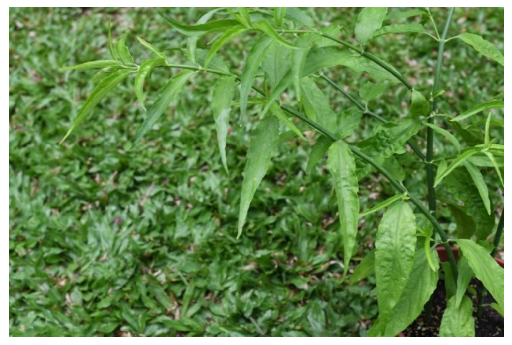
Figure 1. Clinacanthus nutans plant (Photo by W.Y. Ong).
Molecules 27 03607 g001
Figure 2. Inflammatory and oxidative stress pathways targeted by C. nutans and its phytochemical components. Abbreviations: LPS: lipopolysaccharide; IKK: inhibitor of kappaB kinase; Raf: rapidly activated sarcoma; MEK1/2: mitogen activated protein kinase 1/2; P-ERK1/2: phosphorylated extracellular signal related kinase 1/2; NF-kB: nuclear factor kappa-light-chain-enhancer of activated B cells; iNOS: inducible nitric oxide synthase; cPLA2: cytosolic phospholipase A2; COX-2: cyclooxygenase 2; 5-LOX: 5-lipoxygenase; ARA: arachidonic acid; lyso-PtdCho: lysophosphatidylcholine; PAF: platelet activating factor; sPLA2: secretory phopholipase A2, TNF-α: tumor necrosis factor-alpha; IL-1β: interleukin-1β; Nrf-2: nuclear factor erythroid 2–related factor 2; KEAP1: Kelch-like ECH associated protein 1.
Molecules 27 03607 g002
Table 1. Solvents used in extraction and phytochemicals obtained from C. nutans.
SolventPhytochemical ComponentsPotential Health EffectsReferences
Distilled waterGlucoside
Sulfur-containing compounds
Phytosterols
Triterpenoids
Flavones
Amino acids
Anti-inflammatory[27]
MethanolSchaftoside
Isoorientin
Orientin
Isovitexin
Vitexin
Anti-inflammatory[6]
MethanolEntadamide C
Clinamide D
Anticancer[22]
MethanolPalmitic acid
Phytol
1-Monopalmitin
Stigmast-5-ene
Pentadecanoic acid
Heptadecanoic acid
1-linolenoylglycerol
Glycerol monostearate
Alpha-tocospiro B
Stigmasterol
Anti-diabetic[28]
MethanolBetulin
Stigmasterol
Sitosterol
β-Amyrin
Vitamin E
Campesterol
Anticancer[29]
MethanolSchaftoside
Isomollupentin
7-O-β-glucopyranoside
Orientin
Isoorientin
Vitexin
Isovitexin
[14]
EthanolGlyceryl 1,3-disterate ester (C39H76O5),
Kaempferol
3-O-feruloyl-sophoroside 7-O-glucoside (C43H48O24)
Hydroxypthioceranic acid (C46H92O3)
Antiapoptotic[30]
EthanolMyricetin
Orientin
Isoorientin
Vitexin
Isovitexin
Apigenin
Ferulic acid
[10]
EthanolClinamides
2-cis-entadamide
[16]
EthanolSchaftoside
Orientin
Isovitexin
Vitexin
Anti-inflammatory[15]
ChloroformPurpurin-18 phytyl esterAnti-inflammatory[31]
DichloromethanePalmitic acid
Linolenyl alcohol
Anticancer[32]
HexanePalmitic acid
Phytol
Hexadecanoic acid
1-Monopalmitin
Stigmast-5-ene
Pentadecanoic acid
Heptadecanoic acid
1-Linolenoylglycerol
Stigmasterol
[23]
HexaneSchaftoside
Stigmasterol
β-sitosterol
Triterpenoid lupeol
Immuno-modulatory[25]
Hexane13(2)-hydroxy-(13(2)-R)-phaeophytin b,
13(2)-hydroxy-(13(2)-S)-phaeophytin a
13(2)-hydroxy-(13(2)-R)-phaeophytin a.
Antiviral[24]
Ethyl acetateLupeol
Lup-20(29)-en-3-one
Lup-20(29)-en-ol acetate
Stigmasterol
Sitosterol
Betulin
Campesterol
Squalene
Vitamin E
Oleic acid
Anticancer[29]
Microwave Assisted ExtractionPolyphenols
Flavonoids
[26]
Supercritical carbon dioxidePhytosterols
β-Sitosterol
[26]
Publisher’s Note: MDPI stays neutral with regard to jurisdictional claims in published maps and institutional affiliations.