Article

Antimicrobial Polyketide Metabolites from Penicillium bissettii and P. glabrum

1
School of Chemical Sciences, University of Auckland, Waipapa Taumata Rau, Private Bag 92019, Auckland 1142, New Zealand
2
Bioluminescent Superbugs Lab, School of Medical Sciences, University of Auckland, Waipapa Taumata Rau, Private Bag 92019, Auckland 1142, New Zealand
3
Manaaki Whenua, Landcare Research, Private Bag 92170, Auckland 1142, New Zealand
*
Authors to whom correspondence should be addressed.
These authors contributed equally to this work.
Molecules 2022, 27(1), 240; https://doi.org/10.3390/molecules27010240
Submission received: 24 November 2021 / Revised: 23 December 2021 / Accepted: 27 December 2021 / Published: 31 December 2021

Abstract

:
Screening of several fungi from the New Zealand International Collection of Microorganisms from Plants identified two strains of Penicillium, P. bissettii and P. glabrum, which exhibited antimicrobial activity against Escherichia coli, Klebsiella pneumoniae, and Staphylococcus aureus. Further investigation into the natural products of the fungi, through extraction and fractionation, led to the isolation of five known polyketide metabolites, penicillic acid (1), citromycetin (2), penialdin A (3), penialdin F (4), and myxotrichin B (5). Semi-synthetic derivatization of 1 led to the discovery of a novel dihydro (1a) derivative that provided evidence for the existence of the much-speculated open-chained form of 1. Upon investigation of the antimicrobial activities of the natural products and derivatives, both penicillic acid (1) and penialdin F (4) were found to inhibit the growth of Methicillin-resistant S. aureus. Penialdin F (4) was also found to have some inhibitory activity against Mycobacterium abscessus and M. marinum along with citromycetin (2).

1. Introduction

Manaaki Whenua’s International Collection of Microorganisms from Plants (ICMP) contains over 10,000 fungal cultures, most of which originate from Aotearoa New Zealand and the South Pacific [1]. These fungal cultures include a diverse range of fungal species, many of which are unique to Aotearoa New Zealand and most of which have not been rigorously tested for antimicrobial activity or investigated for novel natural products. The most well-known and significant example of antibiotic discovery from microbes was the discovery of penicillin from a Penicillium rubens culture by Alexander Fleming in 1928 [2,3]. The structure of Fleming’s penicillin was proposed in 1942 by Edward Abraham and confirmed by Dorothy Hodgkin in 1945. While the original penicillins were active mostly against Gram-positive bacteria, numerous semisynthetic antibiotics were produced in the subsequent years with an improved spectrum of activity [2].
The Penicillium genus is large with more than 350 currently accepted species divided into 28 sections [4]. Section Aspergilloides contains many common cosmopolitan species isolated from soil, food, and plants, including Penicillium glabrum ICMP 5686 isolated from sheep dung in New Zealand [5]. Section Lanata-Divaricata contains many common soil-inhabiting fungi, including Penicillium bissetti ICMP 21333 isolated from tree roots in New Zealand [6]. Investigation of ICMP 5685 and ICMP 21333 led to the isolation of five known polyketide secondary metabolites. Structure elucidation of secondary metabolites 15 with NMR spectroscopic and mass spectrometric data identified the natural products as penicillic acid (1) [7,8], citromycetin (2) [9], penialdin A (3) [10], penialdin F (4) [11], and myxotrichin B (5) [12] (Figure 1). The antimicrobial activities of 24 have been previously investigated and were found to be not active when tested against S. aureus, E. coli, Bacillus subtilis, and Acinetobacter sp. at concentrations less than 10 µg/mL [9,10,13]. However, 3 has been reported to exhibit anti-inflammatory activity [14], antioxidant activity [15], and moderate anti-diabetic activities [16]. Semisynthetic derivatization of penicillic acid (1), including hydrogenation and acetylation, resulted in the discovery of a novel keto acid derivative 1a and two other known derivatives, 1b and 1c. The structure of keto acid derivative 1a was suspected to be a hydrogenation product of the open chain tautomer of penicillic acid (1), the existence of which has somewhat been disagreed upon. Herein, we report the isolation, derivatization, structure elucidation, and bioactivities of polyketide secondary metabolites from Penicillium and provide evidence for the existence of the open-chain tautomer of penicillic acid (1).

2. Results and Discussion

2.1. Evaluation of Extract Bioactivity

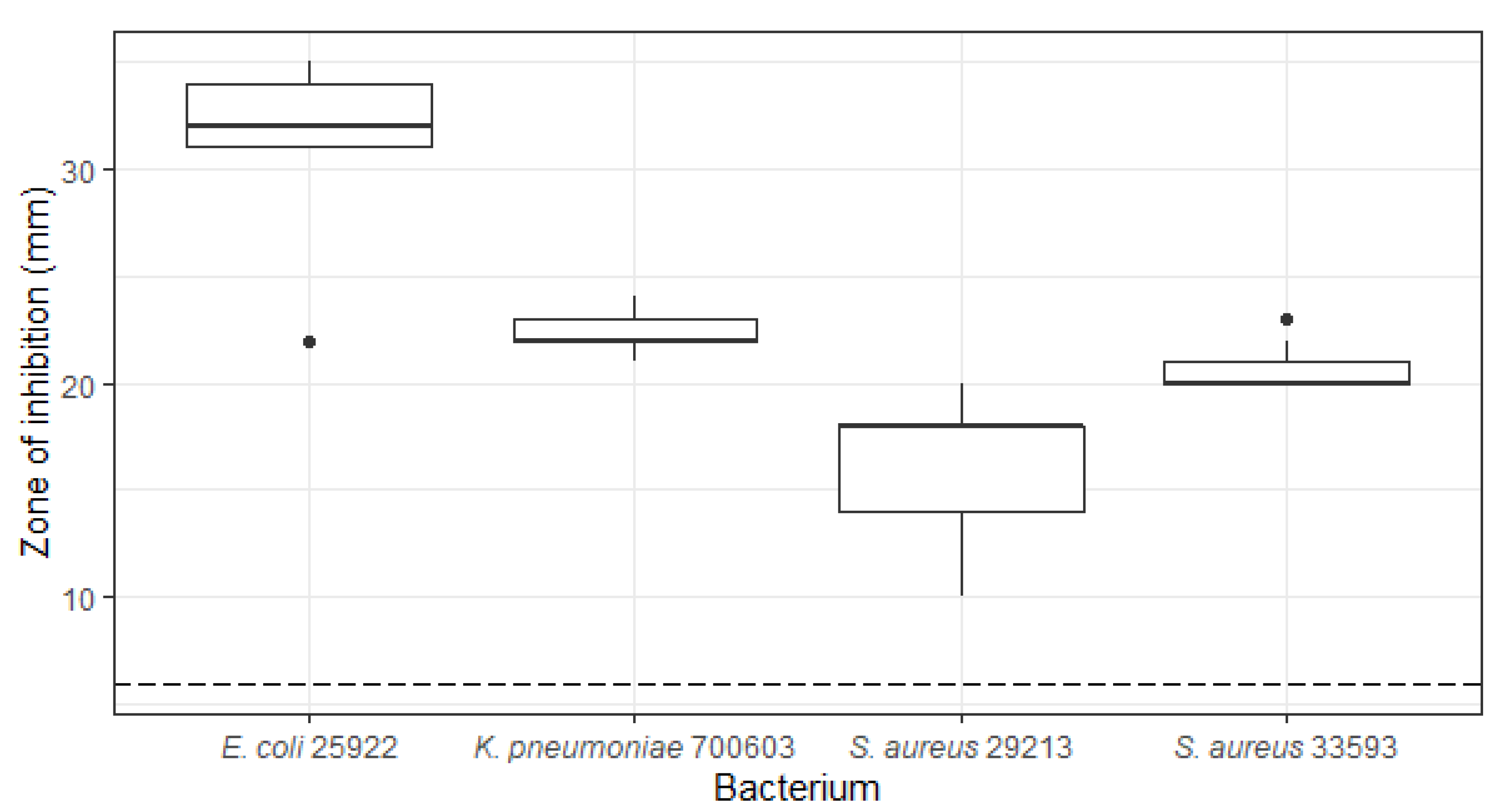
During initial whole cell screening, Penicillium bissettii (ICMP 21333) showed antibacterial activity against E. coli, K. pneumoniae, and antibiotic-sensitive and resistant S. aureus, with zones of inhibition of up to 35 mm in diameter (Figure 2).
From the crude extract of Penicillium bissettii ICMP 21333, a polyketide metabolite 1 was isolated via flash column chromatography. HRESI mass spectrometric data for 1 identified a sodiated adduct observed at m/z 193.0494 [M + Na]+ corresponding to a molecular formula of C8H11O4. The 1H NMR spectrum of 1 exhibited one broad exchangeable singlet (δH 7.87, OH), one sharp olefinic singlet (δH 5.41), two broad olefinic signals (δH 5.11 and 5.30), a methoxy singlet (δH 3.85), and a methyl singlet (δH 1.65). The 13C NMR spectrum showed eight signals at δC 179.4, 170.0, 140.2, 115.3, 102.4, 89.4, 59.8, and 17.1. HSQC NMR data connected the two olefinic signals (δH 5.11 and 5.30, H2-6) to a carbon signal at δC 115.3 (C-6), giving evidence for the presence of a terminal alkene. The HMBC spectrum identified correlations between these terminal alkene protons (δH 5.11 and 5.30, H2-6) and the methyl carbon at δC 17.1 (C-7) as well as the quaternary carbons at δC 102.4 (C-4) and 140.2 (C-5), suggesting the presence of a fragment with a methyl group adjacent to the terminal alkene. HMBC correlations between the other olefinic proton (δH 5.41, H-2) and both the carbonyl carbon at δC 179.4 (C-1) and quaternary carbon at δC 102.4 (C-4), and the correlation between the methoxy singlet (δH 3.85) and the other quaternary carbon at δC 170.0 (C-3), identified the presence of an α,β-unsaturated γ-lactone ring. Additional HMBC correlations between both the methyl (δH 1.65, H3-7) and olefinic (δH 5.41, H-2) protons and the quaternary carbon signal at δC 102.4 (C-4) connected the lactone ring to the terminal alkene fragment. The mass spectrometric and 1H NMR data were consistent with data previously reported for penicillic acid (1) (Figure 1) [7,8].
From the crude extracts of P. glabrum, four metabolites were isolated after purification by flash chromatography. Mass spectrometric analysis of metabolite 2 afforded a molecular formula of C14H10O7 from a sodiated adduct at m/z 313.0328 [M + Na]+. The 1H NMR spectrum showed the presence of four singlets corresponding to a methyl group (δH 2.35), a methylene group (δH 5.03), and two olefinic protons (δH 6.22 and 6.53). Analysis of the HMBC spectrum identified correlations between the olefinic proton at δH 6.53 (H-7) and four quaternary carbons at δC 104.7 (C-10a), 141.9 (C-9), 153.3 (C-8), and 158.1 (C-6a) and between the methylene protons at δH 5.03 (H2-5) and three quaternary carbons at δC 111.1 (C-4a), 152.5 (C-10b), and 158.1 (C-6a), indicating the presence of a chromene. The other olefinic proton at δH 6.22 (H-3) exhibited HMBC correlations to the methyl carbon at δC 19.1 (Me), a carbonyl carbon at δC 177.2 (C-4), and two quaternary carbons at δC 166.2 (C-2) and 111.1 (C-4a), indicating the presence of a γ-pyrone fused to the chromene ring. Substructure searching through the literature led to the identification of this metabolite as citromycetin (2) [9]. The other three metabolites showed similar 1H and 13C NMR spectra to 2. Comparison of mass spectrometric and NMR data to the literature led to the identification of these metabolites as penialdin A (3) [10], penialdin F (4) [11], and myxotrichin B (5) [12].

2.2. Derivatisation of Penicillic Acid

Penicillic acid (1) was first reported in 1913 during an investigation between the incidence of pellagra and mould spoiled corn where they isolated an unknown fungal metabolite [17]. The structure of this metabolite was not determined until 1936 when Birkinshaw, Oxford, and Raistrick, who had been working on Alsberg and Black’s original cultures of Penicillium puberculum, were able to isolate the same compound in significantly higher yields from cultures of P. cyclopium [18]. They used this material to complete their investigation on the structure, which they reported as a substituted γ-keto acid that tautomerized to a γ-hydroxy lactone (Figure 3) [10].
There has been some disagreement on the existence of the open chain γ-keto acid tautomer [18,19,20,21]. The chemical studies carried out by Birkinshaw et al. provided evidence for the open-chain form [18]. They observed esterification of anhydrous penicillic acid (1) with diazomethane and the reaction of a cold aqueous solution of penicillic acid with one mole equivalent of free hydroxylamine, both of which they suggested provided evidence for the presence of the carboxyl group. However, a study by Raphael presented UV absorption spectra of both penicillic acid (1) and the methyl ester in acid and alkaline aqueous solutions in which a maximal absorbance was observed at ~220 nm for both compounds, which was consistent with the closed-ring lactone but not with the open-chain keto acid/keto ester [20]. Similar data for dihydropenicillic acid (1b), a derivative formed via hydrogenation of the natural product, were also obtained suggesting that penicillic acid exists purely as the lactone and that the keto acid tautomer is not required to explain the chemical properties observed by Birkinshaw et al. [18,20]. A study by Shaw also presented UV absorbance data for penicillic acid (1) in both acid and alkaline solutions [21]. Results comparable to Raphael’s were observed for the acid solution, but a shift in the absorption maxima to ~295 nm was observed in 0.1N NaOH solution, consistent with a change to the keto acid structure [20,21].
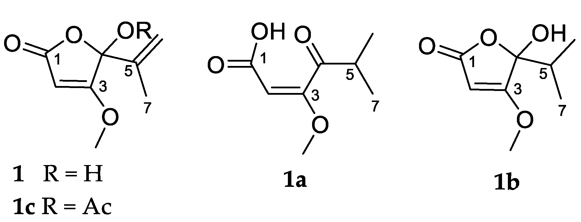
To investigate the existence of the open chain γ-keto acid tautomer, in the present study, we proceeded to prepare dihydropenicillic acid (1b) (Figure 4) via catalytic hydrogenation of penicillic acid (1) in methanol, using palladium on carbon under a hydrogen atmosphere. The crude product was characterized via mass spectrometry and NMR. The sodiated adduct observed at m/z 195.0622 for [M + Na]+ in the (+)-HRESI mass spectrum of the crude product 1a confirmed the addition of one mole of H2 to penicillic acid (1), giving a molecular formula of C8H13O4. The 1H NMR spectrum (Figure S4) of 1a exhibited one sharp olefinic singlet (δH 5.19), one methoxy singlet (δH 3.72), and one dimethyl singlet (δH 1.07) (Table 1). The absence of the two olefinic signals of the terminal alkene in penicillic acid (1) and the presence of a dimethyl signal in the 1H NMR spectrum, suggested successful hydrogenation of the alkene at C-5/C-6. Of particular note was the absence of the expected septet signal of the H-5 proton and the expected splitting of the dimethyl signal into a doublet was also not observed. The HMBC NMR data of the crude product identified a correlation between the dimethyl singlet and a carbon signal at δc 39.3, consistent with an alkyl carbon at the C-5 position. Also present in the HMBC data was a correlation to a carbon signal at δc 217.1, consistent with the presence of a ketone, suggesting that 1a was the ring opened keto acid structure as shown. This explained the absence of the 1H NMR signal for H-5 (α-keto proton) and the lack of splitting of the dimethyl signal, as it was speculated that the acidic α-keto proton had exchanged with deuterium from the CD3OD NMR solvent. This was confirmed by the 1H NMR spectrum recorded in non-exchangeable solvent CDCl3, which showed all expected signals: one sharp olefinic singlet (δH 5.12), one methoxy singlet (δH 3.82), one septet (δH 2.39, J = 7.0 Hz), and one dimethyl doublet (δH 1.01, J = 7.0 Hz) (Figure S6). Purification of 1a was achieved using silica-gel-column chromatography. Changes were observed in the 1H NMR spectrum of the purified product (Figure S7) when compared to the crude product, necessitating further NMR characterization of the purified product 1b.
The 1H NMR spectrum of the purified hydrogenation product 1b showed one sharp olefinic singlet (δH 5.20), one methoxy singlet (δH 3.93), one septet (δH 2.12, J = 6.9 Hz), and two broad methyl doublets (δH 1.04, J = 6.8 Hz and δH 0.88, J = 6.8 Hz). The presence of the septet and the splitting of the methyl signals into doublets indicated that H-5 (δH 2.12, J = 6.9 Hz) of the purified product 1b was not exchangeable with the NMR solvent, suggesting a change to the ring-closed lactone structure. The presence of two nonequivalent methyl signals was also consistent with the ring-closed form, with the chiral center at C-4 (δC 107.2) inducing non-equivalence in the pendant gem dimethyl groups. A change to the ring-closed structure was confirmed by HMBC data of the purified material, which exhibited a correlation between the two methyl proton signals and a carbon signal at δc 107.2 as well a correlation between the H-5 (δH 2.12, J = 6.9 Hz) proton signal and the same carbon signal at δc 107.2. The shift in this carbon signal was consistent with the shift observed for the hemiacetal carbon of penicillic acid (1) (δc 106.8). The HMBC data of the purified product also identified a second weaker set of correlations from a proton signal partially obscured by the broad methyl signal at δH 1.04, to carbons at δc 17.5, 39.3, and 217.1 (data not shown). This set of correlations suggested that some of the keto acid tautomer was present as the correlations were consistent with those observed in HMBC of the crude product. However, the expected second methoxy signal at δH 3.72 in the 1H NMR spectrum was absent. The NMR data for the crude and purified products (1a and 1b) clearly show the existence of the two forms.
Acetylated penicillic acid (1c) was prepared via acetylation with acetic anhydride in pyridine over 18 h, under a nitrogen atmosphere. The crude material was purified by diol-bonded silica-gel-column chromatography. The sodiated adduct observed at m/z 235.0578 [M + Na]+ in the (+)-HRESI mass spectrum was consistent with the loss of a proton and the addition of an acetyl group, giving a molecular formula of C10H12O5. The 1H NMR spectrum (Figure S9) identified the introduction of new methyl protons (δH 2.12, s), which in turn exhibited a correlation in the HMBC spectrum to a carbon at δc 167.7, which is consistent with the introduction of an acetyl group. The mass spectrometric values and 1H NMR data obtained were in agreement with the literature [22].

2.3. 13C NMR Study of Penicillic Acid (1) Biosynthesis

A sodium [1-13C] acetate labelling study was conducted to confirm the biosynthetic origin of penicillic acid (1). Cultures of P. bissettii ICMP 21333 were grown on PDA agar supplemented with 1g/L 1-13C sodium acetate. Extraction and fractionation as conducted previously afforded penicillic acid (1) (132 mg). The 13C NMR spectrum (Figure S3) of the 13C-labelled penicillic acid (1) was acquired and compared to the spectrum observed for non-labelled compound. The signals observed for C-1, C-3, and C-5 showed increased intensity, consistent with 13C enrichment. An indication of the extent of 13C enrichment was given by the relative peak integrals when compared to the spectrum of unlabeled penicillic acid (1). The relative peak integrals for labelled penicillic acid (1) clearly show enhancement for peaks at δc 181.2 (C-1, 12.3×), 173.2 (C-3, 10.1×), and 141.7 (C-5, 8.5×) (Table 2). The observed labelling pattern (Figure 5) was consistent with previous studies utilizing 14C-labelled acetate [23,24].

2.4. Bioactivity

Penicillic acid (1) and its derivatives 1b and 1c, as well as citromycetin (2) and penialdin A (3) and F (4), were screened for antimicrobial activity. Compounds 14 were tested in growth-inhibition assays against Methicillin-resistant S. aureus (MRSA), E. coli, K. pneumoniae (multi-drug resistant strain), Acinetobacter baumannii, Pseudomonas aeruginosa, Candida albicans, and Cryptococcus neoformans by the Community for Open Antimicrobial Drug Discovery (COADD) at The University of Queensland. The COADD test compound activity as an inhibition of microbial growth at a single concentration of 32 µg/mL is presented in Table 3.
At the concentration tested, penicillic acid (1) showed some inhibition of the growth of MRSA (53%) and E. coli (26%), as expected based on previous literature [25,26]. However, neither of the penicillic acid derivatives (1b and 1c) showed any activity. Loss of activity on hydrogenation or acetylation of 1 suggests that the unsaturation at C-5 to C-6 is of importance as is the ability to convert to the open-chain form. Penialdin F (4) exhibited the strongest antimicrobial activity, inhibiting the growth of MRSA by over 71% (Table 3) at the concentration tested. This was surprising as 4 has been previously reported to be not active against wild-type and multidrug-resistant strains of E. faecalis, E. faecium, and S. aureus [13]. However, the inhibition was not complete, suggesting an MIC >32 µg/mL, which is considerably higher than antibiotics used for clinical treatment of antibiotic-sensitive S. aureus infections, which range from <0.001-~4 µg/mL [27] depending on the antibiotic. None of the other natural products showed strong enough inhibitory activity against any of the organisms, as expected. Citromycetin (2) has been previously investigated for antimicrobial activity (E. coli, Bacillus subtilis, and Septoria nodurum) and antiparasitic activity (Haemonchus contortus) and was found not active while also being non-cytotoxic against NS-1 cells [12]. Penialdin A (3) has also been reported as not active at a concentration of 10 µg/mL against S. aureus, E. coli, B. subtilis, and Acinetobacter sp. [10].
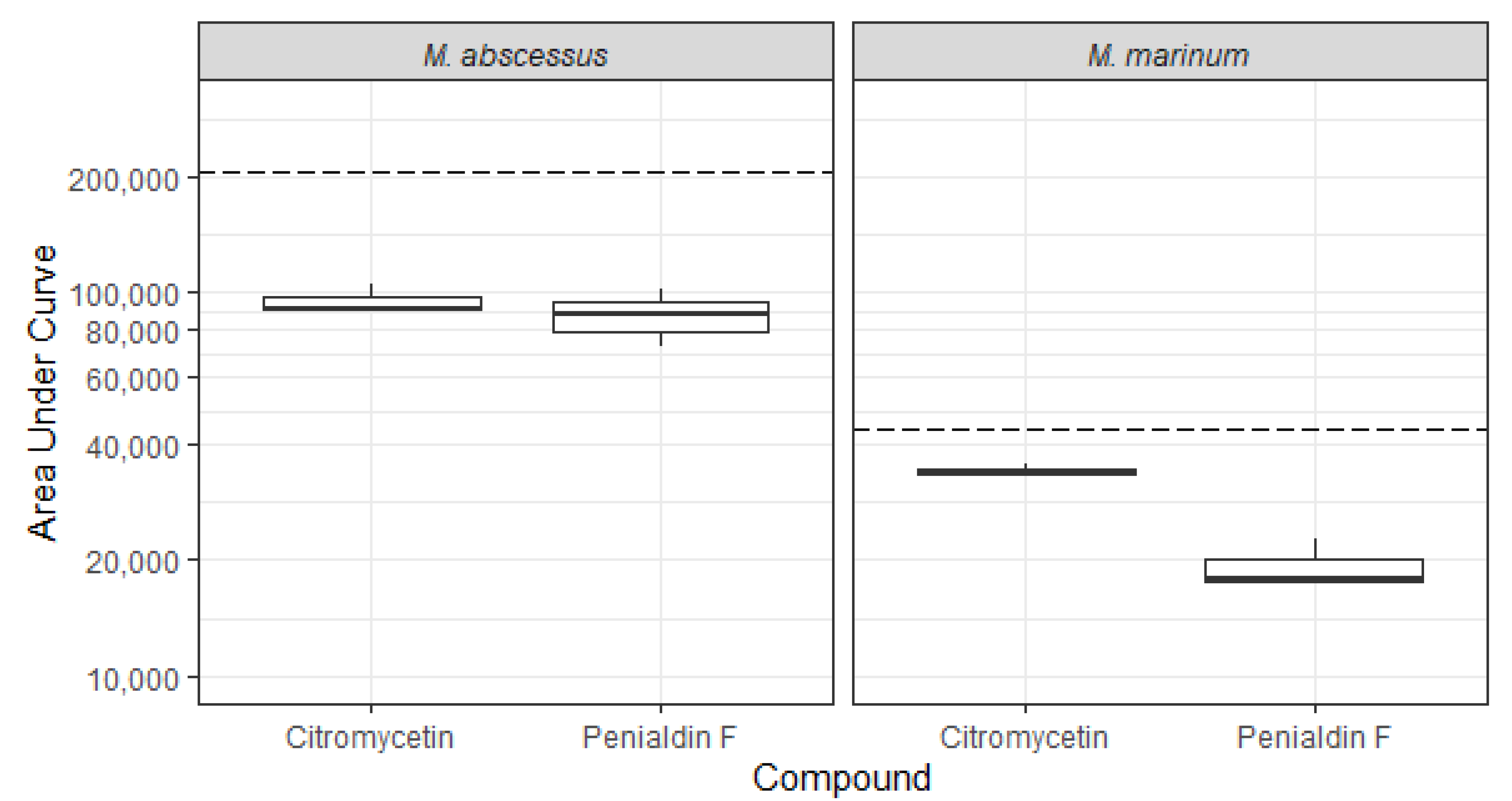
In addition to the assays performed by COADD, the compounds citromycetin (2) and penialdin F (4) were evaluated for their bioactivity against luminescent derivatives of Mycobacterium abscessus and M. marinum at a maximum concentration of 64 µg/mL using inhouse assays. Luminescence is frequently used as rapid readout of potential antimycobacterial activity [28,29,30]. Citromycetin and another compound of the penialdin family, penialdin C, have previously been shown to have activity against the Mycobacterial species M. smegmatis, with minimum inhibitory concentrations (MIC) of 31.2 and 15.6 μg/mL, respectively [31]. We found that citromycetin (2) and penialdin F (4) both displayed some activity against M. abscessus BSG301 and M. marinum BSG101, decreasing bacterial light output compared to the no-compound control (Figure 6). However, at the concentration tested, neither compound caused a reduction in light production of at least 1 log, which is how we define the MIC indicating their MIC against M. abscessus and M. marinum is greater than 64 μg/mL.

3. Materials and Methods

3.1. General Experimental Procedures

Infrared spectra were recorded on a Perkin–Elmer spectrometer 100 Fourier Transform infrared spectrometer equipped with a universal ATR accessory. Mass spectra were acquired on a Bruker micrOTOF Q II spectrometer (Bruker Daltonics, Bremen, Germany). Melting points were recorded on an electrothermal melting point apparatus and were uncorrected. 1H and 13C NMR spectra were recorded at 298K on a Bruker AVANCE 400 spectrometer (Bruker, Karlsruhe, Germany) at 400 and 100 MHz, respectively, using standard pulse sequences. Proto–deutero solvent signals were used as internal references (CD3OD: δH 3.31, δC 49.0; CDCl3: δH 0.00 (TMS), δC 77.16; DMSO-d6: δH 2.50, δC 39.52). For 1H NMR, the data are quoted as position (δ), relative integral, multiplicity (s = singlet, d = doublet, t = triplet, q = quartet, p = pentet, m = multiplet, dd = doublet of doublets, ddd = doublet of doublets of doublets, td = triplet of doublets, and br = broad), coupling constant (J, Hz), and assignment to the atom. The 13C NMR data are quoted as position (δ) and assignment to the atom. Flash column chromatography was carried out using Kieselgel 60 (40-63 μm) (Merck, Munich, Germany) silica gel or Merck Diol bonded silica (40–63 μm), with Merck C8 reversed–phase (40–63 μm) solid support. Gel filtration was carried out on Sephadex LH–20 (Pharmacia, Uppsala, Sweden). Thin-layer chromatography was conducted on Merck DC–plastikfolien Kieselgel 60 F254 plates. All solvents used were of analytical grade or better and/or purified according to standard procedures. Chemical reagents used were purchased from standard chemical suppliers and used as purchased.

3.2. Fungal Material

P. bissettii was described in 2016 from Canada [6] and later found to be present in South Korea [32] and New Zealand. The ecology of P. bissettii is unknown; however, it appears to be associated with tree soils or roots; the Canadian isolates were from Spruce forest soil and the Korean isolates from pine forest soil. Culture ICMP 21333 was isolated as an endophyte from the living roots of a kauri (Agathis australis) tree in Auckland, New Zealand in 2016. The identification of this isolate is supported by DNA sequences of the ITS (GENBANK# MW862794), BTUB (GENBANK# MZ393465), and CAL (GENBANK# MZ393460) genes. P. glabrum is a common cosmopolitan (GBIF 2021) filamentous ascomycete fungus. It has long been recognised as a cause of post-harvest fruit and vegetable rots since it was first described as Citromyces glaber in 1893. Culture ICMP 5686 was isolated from sheep dung in Palmerston North, New Zealand in July 1976. The identification of this isolate is supported by DNA sequences of the ITS (GENBANK# MW862782), BTUB (GENBANK# MZ393464), and CAL (GENBANK# MZ393462) genes. Cultures of P. bissettii and P. glabrum were grown at room temperature on Potato Dextrose Agar (PDA) (Fort Richard, New Zealand) plates. For extraction, the cultures were grown on 40 PDA plates until the fungus covered an area of 75% and were freeze dried for 5 days.

3.3. Antimicrobial Testing of Fungal Cultures

The ICMP fungal cultures were screened for antimicrobial activity after growth on PDA. Briefly, cultures of E. coli ATCC 25922, K. pneumoniae ATCC 700603, and S. aureus ATCC 29213 (antibiotic-sensitive (oxacillin)) and ATCC 33593 (antibiotic-resistant (gentamicin and methicillin)) were grown overnight in Tryptic Soy Broth (TSB) (Fort Richard, New Zealand) at 37 °C with shaking at 200 rpm. Cultures were diluted in TSB to an optical density at 600 nm of 0.1 and spread onto PDA plates for E. coli and K. pneumoniae and Mueller Hinton Agar (MHA) (Fort Richard, New Zealand) for S. aureus to produce a confluent bacterial lawn. Plugs were removed from fungal cultures using a 6 mm Paramount biopsy punch (Capes Medical, New Zealand) when the fungal cultures had grown to cover 75% of the plates. These were placed onto the inoculated PDA and MHA plates and incubated overnight at 37 °C. Any zones of inhibition were measured the next day using a ruler, and the diameter was recorded. Experiments were performed three times with three technical replicates per experiment.

3.4. Extraction and Isolation

Freeze-dried cultures of P. bissettii (17.49 g) were extracted with MeOH (1 L) for 4 h followed by CH2Cl2 (1 L) overnight. Concentration of the combined organic extracts afforded 2.46 g of crude extract. The crude extract was subjected to C8 flash column chromatography (H2O/MeOH), which afforded 5 fractions (A–E). Penicillic acid (1) was isolated from fraction C (1:1, H2O/MeOH) and further purified by Sephadex LH20 (MeOH) to give white crystals (25 mg).
The dry cultures of P. glabrum (20.86 g) were extracted with MeOH (1 L) for 4 h followed by CH2Cl2 (1 L) overnight. Concentration of the combined organic extracts afforded 2.03 g of crude extract. The crude extract was subjected to C8 flash column chromatography (H2O/MeOH), which afforded 5 fractions (A–E). Subsequent purification of fraction B (4:1, H2O/MeOH) by Sephadex LH20 (MeOH) afforded 5 fractions (B1–B5). Citromycetin (2) was isolated from fraction B4 after further purification by Diol-bonded silica column chromatography (n-Hexane/EtOAc, gradient) to give a yellow oil (2.9 mg). Purification of fraction C (1:1, H2O/MeOH) by Sephadex LH20 (MeOH) afforded 4 fractions (C1–C4). Penialdin A (3) (3.5 mg), penialdin F (4) (4.6 mg), and myxotrichin B (5) (0.75 mg) were isolated from fraction C2 after further purification by Diol-bonded silica column chromatography (n-Hexane/EtOAc, gradient).

3.4.1. Penicillic acid (1)

1H NMR (CD3OD, 400 MHz) δ 5.41 (1H, br s, H-6), 5.27 (1H, s, H-2), 5.17 (1H, br s, H-6), 3.92 (3H, s, OMe), 1.75 (3H, dd, J = 1.0, 1.0 Hz, H3-7); 13C NMR (CD3OD, 100 MHz) 181.2 (C-1), 173.0 (C-3), 141.5 (C-5), 116.6 (C-6), 106.8 (C-4), 90.0 (C-2), 60.5 (C-8), 17.5 (C-7); 1H NMR (DMSO-d6, 400 MHz) δ 7.87 (1H, br s, OH), 5.41 (1H, s, H-2), 5.30 (1H, br s, H-6), 5.11 (1H, br s, H-6), 3.85 (3H, s, OMe), 1.65 (3H, s, H3-7); 13C NMR (DMSO-d6, 100 MHz) δ 179.4 (C-1), 170.0 (C-3), 140.2 (C-5), 115.3 (C-6), 102.4 (C-4), 89.4 (C-2), 59.8 (C-8), 17.1 (C-7); (+)-HRESIMS m/z 193.0474 [M + Na]+ (calcd for C8H10NaO4, 193.0470).

3.4.2. Citromycetin (2)

1H NMR (CD3OD, 400 MHz) δ 6.53 (1H, s, H-7), 6.22 (1H, s, H-3), 5.03 (2H, s, H2-5), 2.35 (3H, s, Me); 13C NMR (CD3OD, 100 MHz) δ 177.2 (C-4), 167.0 (COOH), 166.1 (C-2), 158.1 (C-6a), 153.3 (C-8), 152.5 (C-10b), 141.9 (C-9), 120.0 (C-10), 113.3 (C-3), 111.1 (C-4a), 104.7 (C-10a), 104.5 (C-6a), 62.7 (C-5), 19.1 (Me); (+)-HRESIMS m/z 313.0328 [M + Na]+ (calcd for C14H10NaO7, 313.0319).

3.4.3. Penialdin A (3)

1H NMR (CD3OD, 400 MHz) δ 7.09 (1H, s, H-9), 4.65 (2H, s, H2-1), 2.99 (1H, d, J = 17.8 Hz, H2-4A), 2.77 (1H, d, J = 17.8 Hz, H2-4B), 1.57 (3H, s, Me); 13C NMR (CD3OD, 100 MHz) δ 175.2 (C-10), 168.0 (COOH), 160.8 (C-4a), 154.8 (C-8), 152.6 (C-5a), 144.1 (C-7), 119.1 (C-6), 114.6 (C-10a), 111.8 (C-9a), 105.6 (C-9), 94.3 (C-3), 56.1 (C-1), 37.2 (C-5), 27.2 (Me); (+)-HRESIMS m/z 287.0529 [M + Na]+ (calcd for C13H12NaO6, 287.0526).

3.4.4. Penialdin F (4)

1H NMR (CD3OD, 400 MHz) δ 7.36 (1H, s, H-9), 6.86 (1H, s, H-6), 4.62 (2H, s, H2-1), 2.91 (1H, d, J = 17.0 Hz, H2-4A), 2.68 (1H, d, J = 17.0 Hz, H2-4B), 1.55 (3H, s, Me); 13C NMR (CD3OD, 100 MHz) δ 174.9 (C-10), 160.8 (C-4a), 151.9 (C-5a, C-8), 144.3 (C-7), 115.7 (C-9a), 114.6 (C-10a), 106.7 (C-9), 101.9 (C-6), 94.3 (C-3), 46.1 (C-1), 37.1 (C-5), 27.2 (Me); (+)-HRESIMS m/z 331.0428 [M + Na]+ (calcd for C14H12NaO8, 331.0424).

3.4.5. Myxotrichin B (5)

1H NMR (CD3OD, 400 MHz) δ 7.09 (1H, s, H-6), 4.68 (1H, d, J = 15.0 Hz, H2-1A), 4.40 (1H, d, J = 15.0 Hz, H2-1B), 3.33 (3H, s, OMe), 3.02 (1H, d, J = 17.9 Hz, H2-4A), 2.81 (1H, d, J = 17.9 Hz, H2-4B), 1.54 (3H, s, Me); 13C NMR (CD3OD, 100 MHz) δ 175.5 (C-10), 167.8 (COOH), 160.4 (C-4a), 154.1 (C-7), 151.5 (C-5a), 144.4 (C-7), 118.4 (C-9), 113.8 (C-10a), 111.8 (C-9a), 105.1 (C-6), 98.2 (C-3), 57.0 (C-1), 48.3 (OMe), 37.2 (C-5), 21.3 (Me); (+)-HRESIMS m/z 345.0588 [M + Na]+ (calcd for C15H14NaO8, 345.0581).

3.4.6. (E)-3-Methoxy-5-methyl-4-oxohex-2-enoic acid (1a)

To a solution of penicillic acid (1) (5 mg, 0.03 mmol) in anhydrous MeOH (1 mL) was added Pd/C (1 mg, 0.003 mmol), and the reaction was stirred under an H2 atmosphere for 18 h. The reaction mixture was filtered through celite and washed with MeOH (50 mL). The solvent was removed under reduced pressure to give the crude product as a pale-yellow oil (3.43 mg, 66%).
1H NMR (CD3OD, 400 MHz) δ 5.19 (1H, s, H-2), 3.72 (3H, s, OMe), 3.31 (1H, obs by solvent, H-5), 1.07 (6H, s, H3-6, H3-7); 13C NMR (CD3OD, 100 MHz) δ 217.2 (C-4), 174.6 (C-1), 171.2 (C-3), 97.2 (C-2), 57.8 (OMe), 39.2 (C-5), 17.7 (C-6, C-7); 1H NMR (CDCl3, 400 MHz) δ 5.12 (1H, s, H-2), 3.82 (3H, s, OMe), 2.39 (1H, septet, J = 7.0 Hz, H-5) 1.01 (6H, d, J = 7.0 Hz, H3-6, H3-7); (+)-HRESIMS m/z 195.0622 [M + Na]+ (calcd for C8H12NaO4, 195.0630).

3.4.7. 5-Hydroxy-5-isopropyl-4-methoxyfuran-2(5H)-one (1b)

The product was purified by silica-gel column chromatography (10% MeOH:CH2Cl2) to give the product as white crystals (1.72 mg, 33%).
1H NMR (CD3OD, 400 MHz) δ 5.20 (1H, s, H-2), 3.93 (3H, s, OMe), 2.12 (1H, septet, J = 6.8 Hz, H-5), 1.04 (3H, d, J = 6.4 Hz H3-6), 0.88 (3H, d, J = 6.4 Hz, H3-7); 13C NMR (CD3OD, 100 MHz) δ 182.1 (C-3), 173.5 (C-1), 107.2 (C-4), 89.2 (C-2), 59.9 (OMe), 34.6 (C-5), 16.4 (C-7), 16.1 (C-6); 1H NMR (CDCl3, 400 MHz) δ 5.06 (1H, s, H-2), 3.91 (3H, s, OMe), 2.21 (1H, p, J = 6.8 Hz, H-5), 1.12–1.02 (3H, m, H3-6), 0.96–0.88 (3H, m, H3-7).

3.4.8. 3-Methoxy-5-oxo-2-(prop-1-en-2-yl)-2,5-dihydrofuran-2-yl acetate (1c)

To a solution of penicillic acid (1) (5 mg, 0.03 mmol) in pyridine (1 mL) was added acetic anhydride (0.05 mL, 0.52 mmol) dropwise, and the reaction was stirred for 18 hr under a nitrogen atmosphere. The reaction mixture was quenched with 10% HCl and extracted with CH2Cl2 (3 × 10 mL) and ethyl acetate (1 × 10 mL). The combined organic layers were dried with MgSO4 and the solvent removed under reduced pressure. The product was purified by diol-bonded silica-gel column chromatography, eluting with CH2Cl2, to give the product as a brown oil (1.96 mg, 30%).
1H NMR (CDCl3, 400 MHz) δ 5.38 (1H, br s, H-6), 5.17 (2H, br s, H-2, H-6), 3.91 (3H, s, OMe), 2.12 (3H, s, OAc), 1.83 (3H, d, J = 1.0 Hz, H3-7); 13C NMR (CDCl3, 100 MHz) δ 178.1 (C-1), 168.9 (C-3), 167.7 (OAc), 138.4 (C-5), 116.1 (C-6), 101.6 (C-4), 89.8 (C-2), 59.8 (OMe), 21.3 (OAc), 17.3 (C-7); (+)-HRESIMS m/z 235.0578 [M + Na]+ (calcd for C10H12NaO5, 235.0580).

3.5. Sodium [1-13C] Acetate Incorporated Acetate Fermentation of P. bissettii

Extraction and Isolation

Cultures of Penicillium bissettii were grown on 17 PDA plates supplemented with 1g/L 1-13C sodium acetate until the fungus covered 75% of the plate. The cultured plates were freeze dried (11.00 g) and extracted with methanol and dichloromethane to give 2.28 g of crude extract. The crude extract was purified via C8 reverse-phased column chromatography (H2O/MeOH), and penicillic acid (1) (132 mg) was isolated from fraction C (1:1, H2O/MeOH).

3.6. Antimicrobial Assays of Pure Compounds

Antimicrobial evaluation of the pure compounds against S. aureus ATCC 43300 (MRSA), E. coli ATCC 25922, Pseudomonas aeruginosa ATCC 27853, Klebsiella pneumoniae ATCC 700603, Acinetobacter baumannii ATCC 19606, Candida albicans ATCC 90028, and Cryptococcus neoformans ATCC 208821 was undertaken at the Community for Open Antimicrobial Drug Discovery at The University of Queensland (Queensland, Australia) according to their standard protocols [33]. For antimicrobial assays, the tested strains were cultured in either Luria broth (LB) (In Vitro Technologies, USB75852, Victoria, Australia), nutrient broth (NB) (Becton Dickson 234000, New South Wales, Australia), or MHB at 37 °C overnight. A sample of culture was then diluted 40-fold in fresh MHB and incubated at 37 °C for 1.5−2 h. The compounds were serially diluted 2-fold across the wells of 96-well plates (Corning 3641, nonbinding surface), with compound concentrations ranging from 0.015 to 64 μg/mL, plated in duplicate. The resultant mid-log-phase cultures were diluted to the final concentration of 1 × 10⁶ CFU/mL; then, 50 μL were added to each well of the compound containing plates giving a final compound concentration range of 0.008–32 μg/mL and a cell density of 5 × 10⁵ CFU/mL. All plates were then covered and incubated at 37 °C for 18 h. Inhibition of bacterial growth was determined measuring absorbance at 600 nm (OD600), using a Tecan M1000 Pro monochromator plate reader. The percentage of growth inhibition was calculated for each well, using the negative control (media only) and positive control (bacteria without inhibitors) on the same plate as references.
For the antifungal assay, fungi strains were cultured for 3 days on Yeast Extract Peptone Dextrose (YPD) agar at 30 °C. A yeast suspension of 1 × 10⁶ to 5 × 10⁶ CFU/mL was prepared from five colonies. These stock suspensions were diluted with yeast nitrogen base (YNB) (Becton Dickinson, 233520, New South Wales, Australia) broth to a final concentration of 2.5 × 10³ CFU/mL. The compounds were serially diluted 2-fold across the wells of 96-well plates (Corning 3641, nonbinding surface), with compound concentrations ranging from 0.015 to 64 μg/mL and final volumes of 50 μL, plated in duplicate. Then, 50 μL of the fungi suspension that was previously prepared in YNB broth to the final concentration of 2.5 × 10³ CFU/mL were added to each well of the compound-containing plates, giving a final compound concentration range of 0.008–32 μg/mL. Plates were covered and incubated at 35 °C for 36 h without shaking. C. albicans MICs were determined by measuring the optical density at 530 nm. For C. neoformans, resazurin was added at 0.006% final concentration to each well and incubated for a further 3 h before MICs were determined by measuring the optical density at 570–600 nm.
Colistin and vancomycin were used as positive bacterial inhibitor standards for Gram-negative and Gram-positive bacteria, respectively. Fluconazole was used as a positive fungal inhibitor standard for C. albicans and C. neoformans. The antibiotics were provided in 4 concentrations, with 2 above and 2 below its MIC value, and they were plated into the first 8 wells of column 23 of the 384-well NBS plates. The quality control (QC) of the assays was determined by the antimicrobial controls and the Z’-factor (using positive and negative controls). Each plate was deemed to fulfil the quality criteria (pass QC) if the Z’-factor was above 0.4, and the antimicrobial standards showed a full range of activity, with full growth inhibition at their highest concentration and no growth inhibition at their lowest concentration.
Antimicrobial evaluation against M. abscessus and M. marinum was undertaken using inhouse assays. M. abscessus BSG301 and M. marinum BSG101 [28] are stable bioluminescent derivatives transformed with the integrating plasmid pMV306G13ABCDE [34]. This allows light production to be used as a surrogate for bacterial viability [28,29,30]. Mycobacterial cultures were grown shaking at 200 rpm in Middlebrook 7H9 broth (Fort Richard, Auckland, New Zealand) supplemented with 10% Middlebrook ADC enrichment media (Fort Richard, Auckland, New Zealand), 0.4% glycerol (Sigma-Aldrich, St. Louis, MO, USA), and 0.05% tyloxapol (Sigma-Aldrich, St. Louis, MO, USA). M. abscessus was grown at 37 °C and M. marinum at 28 °C. Cultures were grown until they reached the stationary phase (approximately 3–5 days for M. abscessus BSG301 and 7–10 days for M. marinum BSG101) and then diluted in Mueller Hinton broth II (MHB) (Fort Richard, Auckland, NewZealand) supplemented with 10% Middlebrook ADC enrichment media and 0.05% tyloxapol to give an optical density at 600 nm (OD600) of 0.001, which is the equivalent of ~106 bacteria per mL. Pure compounds were dissolved in DMSO and added in duplicate to the wells of a black 96-well plate (Nunc, Thermo Scientific, Waltham, MA, USA) at doubling dilutions with a maximum concentration of 128 μg/mL. Then, 50 μL of diluted bacterial culture was added to each well of the compound containing plates giving final compound concentrations of 0–64 μg/mL and a cell density of ~5 × 10⁵ CFU/mL. Rifampicin (Sigma-Aldrich, St. Louis, MO, USA) was used as positive control at 1000 μg/mL for M. abscessus and 10 μg/mL for M. marinum. Between measurements, plates were covered, placed in a plastic box lined with damp paper towels, and incubated with shaking at 100 rpm at 37 °C for M. abscessus and 28 °C for M. marinum. Bacterial luminescence was measured at regular intervals over 72 h using a Victor X-3 luminescence plate reader (PerkinElmer, Boston, MA, USA) with an integration time of 1 s. We defined the MIC as causing a 1-log reduction in light production, as previously described [29]. More detailed protocols are available at protocols.io (accessed on 19 November 2021) [35,36].

3.7. Statistical Analysis

Statistical analysis was performed as described using GraphPad Prism version 8.4.3.

4. Conclusions

Investigation of the secondary metabolites of two strains of Penicillium led to the isolation of five known natural products. Antimicrobial screening of selected natural products identified penicillic acid (1) and penialdin F (4), which exhibited good activity against MRSA with the latter also exhibiting weak antimycobacterial activity against M. abscessus and M. marinum along with citromycetin (2). To the best of our knowledge, this is the first instance where these compounds have been evaluated for antimycobacterial activity. Derivatisation of 1 through hydrogenation led to the synthesis of a novel dihydro (1a) derivative, which provided evidence to the existence of an open-chained γ-keto acid tautomer, with purification attempts leading to ring closure of 1a, forming the lactone 1b. Although none of the natural products or their derivatives exhibited significant antimicrobial activity to be of interest as lead compounds for antibiotic development, the ICMP collection remains a vast untapped resource for novel bioactive natural products worthy of further exploration.

Supplementary Materials

The following are available online, Figures S1–S10: 1H and 13C NMR spectra of 1, 1a, and 1c and the 1H NMR spectrum of 1b as well as the 13C NMR spectrum of 1-13C labelled 1.

Author Contributions

Conceptualization and supervision, S.W. and B.R.C.; methodology and validation, M.M.C., N.S.L.N., S.G., S.J.v.d.P. and A.G.; formal analysis, investigation, and data curation, S.G., N.S.L.N. and M.M.C.; resources, B.S.W.; writing—original draft preparation, M.M.C.; writing—review and editing, M.M.C., B.R.C., and S.W.; funding acquisition, S.W. All authors have read and agreed to the published version of the manuscript.

Funding

This research was funded by Cure Kids, NZ Carbon Farming, the Maurice Wilkins Centre for Molecular Biodiscovery, and donations from the New Zealand public.

Institutional Review Board Statement

Not applicable.

Informed Consent Statement

Not applicable.

Acknowledgments

We gratefully acknowledge the iwi Te Kawerau ā Maki for permission to collect samples of kauri roots on their whenua (land). We would like to thank Michael Schmitz and Tony Chen for their assistance with the NMR and mass spectrometric data and the Community for Antimicrobial Drug Discovery (CO-ADD), funded by the Wellcome Trust (UK) and The University of Queensland (Australia), for carrying out the biological activity and toxicity testing of the pure compounds.

Conflicts of Interest

The authors declare no conflict of interest.

Sample Availability

Samples of compounds 14 are available from the authors.

References

  1. Johnston, P.R.; Weir, B.S.; Cooper, J.A. Open Data on Fungi and Bacterial Plant Pathogens in New Zealand. Mycology 2017, 8, 59–66. [Google Scholar] [CrossRef] [Green Version]
  2. Lobanovska, M.; Pilla, G. Penicillin’s Discovery and Antibiotic Resistance: Lessons for the Future? Yale J. Biol. Med. 2017, 90, 135–145. [Google Scholar] [PubMed]
  3. Houbraken, J. Fleming’s Penicillin Producing Strain Is Not Penicillium Chrysogenum but P. Rubens. Ima Fungus 2011, 2, 87–95. [Google Scholar] [CrossRef] [PubMed] [Green Version]
  4. Visagie, C.M.; Houbraken, J.; Frisvad, J.C.; Hong, S.-B.; Klaassen, C.H.W.; Perrone, G.; Seifert, K.A.; Varga, J.; Yaguchi, T.; Samson, R.A. Identification and Nomenclature of the Genus Penicillium. Stud. Mycol. 2014, 78, 343–371. [Google Scholar] [CrossRef] [Green Version]
  5. Houbraken, J.; Visagie, C.M.; Meijer, M.; Frisvad, J.C.; Busby, P.E.; Pitt, J.I.; Seifert, K.A.; Louis-Seize, G.; Demirel, R.; Yilmaz, N.; et al. A Taxonomic and Phylogenetic Revision of Penicillium Section Aspergilloides. Stud. Mycol. 2014, 78, 373–451. [Google Scholar] [CrossRef] [Green Version]
  6. Visagie, C.M.; Renaud, J.B.; Burgess, K.M.N.; Malloch, D.W.; Clark, D.; Ketch, L.; Urb, M.; Louis-Seize, G.; Assabgui, R.; Sumarah, M.W.; et al. Fifteen New Species of Penicillium. Pers. Int. Mycol. J. 2016, 36, 247–280. [Google Scholar] [CrossRef] [Green Version]
  7. Pohland, A.E.; Schuller, P.L.; Steyn, P.S.; Van Egmond, H.P. Physicochemical data for some selected mycotoxins. Pure Appl. Chem. 1982, 54, 2219–2284. [Google Scholar] [CrossRef]
  8. Li, H.-J.; Cai, Y.-T.; Chen, Y.-Y.; Lam, C.-K.; Lan, W.-J. Metabolites of Marine Fungus Aspergillus sp. Collected from Soft Coral Sarcophyton tortuosum. Chem. Res. Chin. U. 2010, 26, 415–419. [Google Scholar]
  9. Capon, R.J.; Stewart, M.; Ratnayake, R.; Lacey, E.; Gill, J.H. Citromycetins and Bilains A–C: New Aromatic Polyketides and Diketopiperazines from Australian Marine-Derived and Terrestrial Penicillium Spp. J. Nat. Prod. 2007, 70, 1746–1752. [Google Scholar] [CrossRef]
  10. Jouda, J.-B.; Kusari, S.; Lamshöft, M.; Mouafo Talontsi, F.; Douala Meli, C.; Wandji, J.; Spiteller, M. Penialidins A–C with Strong Antibacterial Activities from Penicillium Sp., an Endophytic Fungus Harboring Leaves of Garcinia nobilis. Fitoterapia 2014, 98, 209–214. [Google Scholar] [CrossRef]
  11. Cheng, X.; Yu, L.; Wang, Q.; Ding, W.; Chen, Z.; Ma, Z. New Brefeldins and Penialidins from Marine Fungus Penicillium Janthinellum DT-F29. Nat. Prod. Res. 2018, 32, 282–286. [Google Scholar] [CrossRef]
  12. Yuan, C.; Wang, H.-Y.; Wu, C.-S.; Jiao, Y.; Li, M.; Wang, Y.-Y.; Wang, S.-Q.; Zhao, Z.-T.; Lou, H.-X. Austdiol, Fulvic Acid and Citromycetin Derivatives from an Endolichenic Fungus, Myxotrichum Sp. Phytochem. Lett. 2013, 6, 662–666. [Google Scholar] [CrossRef]
  13. Kumla, D.; Pereira, J.A.; Dethoup, T.; Gales, L.; Freitas-Silva, J.; Costa, P.M.; Lee, M.; Silva, A.M.S.; Sekeroglu, N.; Pinto, M.M.M.; et al. Chromone Derivatives and Other Constituents from Cultures of the Marine Sponge-Associated Fungus Penicillium Erubescens KUFA0220 and Their Antibacterial Activity. Mar. Drugs 2018, 16, 289. [Google Scholar] [CrossRef] [PubMed] [Green Version]
  14. Liu, Y.; Ruan, Q.; Jiang, S.; Qu, Y.; Chen, J.; Zhao, M.; Yang, B.; Liu, Y.; Zhao, Z.; Cui, H. Cytochalasins and Polyketides from the Fungus Diaporthe Sp. GZU-1021 and Their Anti-Inflammatory Activity. Fitoterapia 2019, 137, 104187. [Google Scholar] [CrossRef]
  15. Fu, J.; Hu, L.; Shi, Z.; Sun, W.; Yue, D.; Wang, Y.; Ma, X.; Ren, Z.; Zuo, Z.; Peng, G.; et al. Two Metabolites Isolated from Endophytic Fungus Coniochaeta Sp. F-8 in Ageratina Adenophora Exhibit Antioxidative Activity and Cytotoxicity. Nat. Prod. Res. 2021, 35, 2840–2848. [Google Scholar] [CrossRef]
  16. Gou, X.; Tian, D.; Wei, J.; Ma, Y.; Zhang, Y.; Chen, M.; Ding, W.; Wu, B.; Tang, J. New Drimane Sesquiterpenes and Polyketides from Marine-Derived Fungus Penicillium Sp. TW58-16 and Their Anti-Inflammatory and α-Glucosidase Inhibitory Effects. Mar. Drugs 2021, 19, 416. [Google Scholar] [CrossRef] [PubMed]
  17. Alsberg, C.L.; Black, O.F. Contributions to the Study of Maize Deterioration: Biochemical and Toxicological Investigations of Penicillium Puberulum and Penicillium Stoloniferum; G.P.O.: Washington, DC, USA, 1913; Volume 270. [Google Scholar]
  18. Birkinshaw, J.H.; Oxford, A.E.; Raistrick, H. Studies in the Biochemistry of Micro-Organisms. Biochem. J. 1936, 30, 394–411. [Google Scholar] [CrossRef] [Green Version]
  19. Frisvad, J.C. A Critical Review of Producers of Small Lactone Mycotoxins: Patulin, Penicillic Acid and Moniliformin. World Mycotoxin, J. 2018, 11, 73–100. [Google Scholar] [CrossRef]
  20. Raphael, R.A. Compounds Related to Penicillic Acid; Synthesis of Dihydropenicillic Acid. J. Chem. Soc. 1947, 805–808. [Google Scholar] [CrossRef] [PubMed]
  21. Shaw, E. A Synthesis of Protoanemonin. The Tautomerism of Acetylacrylic Acid and of Penicillic Acid. J. Am. Chem. Soc. 1946, 68, 2510–2513. [Google Scholar] [CrossRef]
  22. Shan, R.; Stadler, M.; Anke, H.; Sterner, O. The Reactivity of the Fungal Toxin Papyracillic Acid. Tetrahedron 1997, 53, 6209–6214. [Google Scholar] [CrossRef]
  23. Birch, A.J.; Blance, G.E.; Smith, H. Studies in Relation to Biosynthesis. Part XVIII. Penicillic Acid. J. Chem. Soc. 1958, 4582–4583. [Google Scholar] [CrossRef]
  24. Bentley, R.; Keil, J.G. Tetronic Acid Biosynthesis in Molds. II. Formation of Penicillic Acid in Penicillium Cyclopium. J. Biol. Chem. 1962, 237, 867–873. [Google Scholar] [CrossRef]
  25. Olivigni, F.J.; Bullerman, L.B. A Microbiological Assay for Penicillic Acid. J. Food. Prot. 1978, 41, 432–434. [Google Scholar] [CrossRef]
  26. Geiger, W.B.; Conn, J.E. The Mechanism of the Antibiotic Action of Clavacin and Penicillic Acid. J. Am. Chem. Soc. 1945, 67, 112–116. [Google Scholar] [CrossRef]
  27. EUCAST: Clinical Breakpoints and Dosing of Antibiotics. Available online: https://www.eucast.org/clinical_breakpoints/ (accessed on 23 December 2021).
  28. Dalton, J.P.; Uy, B.; Okuda, K.S.; Hall, C.J.; Denny, W.A.; Crosier, P.S.; Swift, S.; Wiles, S. Screening of Anti-Mycobacterial Compounds in a Naturally Infected Zebrafish Larvae Model. J. Antimicrob. Chemother. 2017, 72, 421–427. [Google Scholar] [CrossRef] [PubMed] [Green Version]
  29. Dalton, J.P.; Uy, B.; Phummarin, N.; Copp, B.R.; Denny, W.A.; Swift, S.; Wiles, S. Effect of Common and Experimental Anti-Tuberculosis Treatments on Mycobacterium tuberculosis Growing as Biofilms. Peer J. 2016, 4, e2717. [Google Scholar] [CrossRef] [Green Version]
  30. Grey, A.B.J.; Cadelis, M.M.; Diao, Y.; Park, D.; Lumley, T.; Weir, B.S.; Copp, B.R.; Wiles, S. Screening of Fungi for Antimycobacterial Activity Using a Medium-Throughput Bioluminescence-Based Assay. Front. Microbiol. 2021, 12, 739995. [Google Scholar] [CrossRef]
  31. Jouda, J.B.; Mawabo, I.K.; Notedji, A.; Mbazoa, C.D.; Nkenfou, J.; Wandji, J.; Nkenfou, C.N. Anti-mycobacterial activity of polyketides from Penicillium sp. endophyte isolated from Garcinia nobilis against Mycobacteriumsmegmatis. Int. J. Mycobacteriol. 2016, 5, 192–196. [Google Scholar] [CrossRef] [Green Version]
  32. Oh, S.-Y.; Park, M.S.; Lim, Y.W. The Influence of Microfungi on the Mycelial Growth of Ectomycorrhizal Fungus Tricholoma Matsutake. Microorganisms 2019, 7, 169. [Google Scholar] [CrossRef] [Green Version]
  33. Blaskovich, M.A.T.; Zuegg, J.; Elliott, A.G.; Cooper, M.A. Helping Chemists Discover New Antibiotics. ACS Infect. Dis. 2015, 1, 285–287. [Google Scholar] [CrossRef] [PubMed]
  34. Andreu, N.; Zelmer, A.; Fletcher, T.; Elkington, P.T.; Ward, T.H.; Ripoll, J.; Parish, T.; Bancroft, G.J.; Schaible, U.; Robertson, B.D.; et al. Optimisation of Bioluminescent Reporters for Use with Mycobacteria. PLoS ONE 2010, 5, e10777. [Google Scholar] [CrossRef] [PubMed] [Green Version]
  35. Wiles, S.; Grey, A. Bioluminescence-Based Minimum Inhibitory Concentration (MIC) Testing of Pure Compounds Isolated from Fungi against Mycobacterium abscessus. 2021. Available online: Dx.doi.org/10.17504/protocols.io.bumcnu2w (accessed on 30 December 2021).
  36. Wiles, S.; Grey, A. Bioluminescence-Based Minimum Inhibitory Concentration (MIC) Testing of Pure Compounds Isolated from Fungi against Mycobacterium marinum. 2021. Available online: Dx.doi.org/10.17504/protocols.io.3x7gprn (accessed on 30 December 2021).
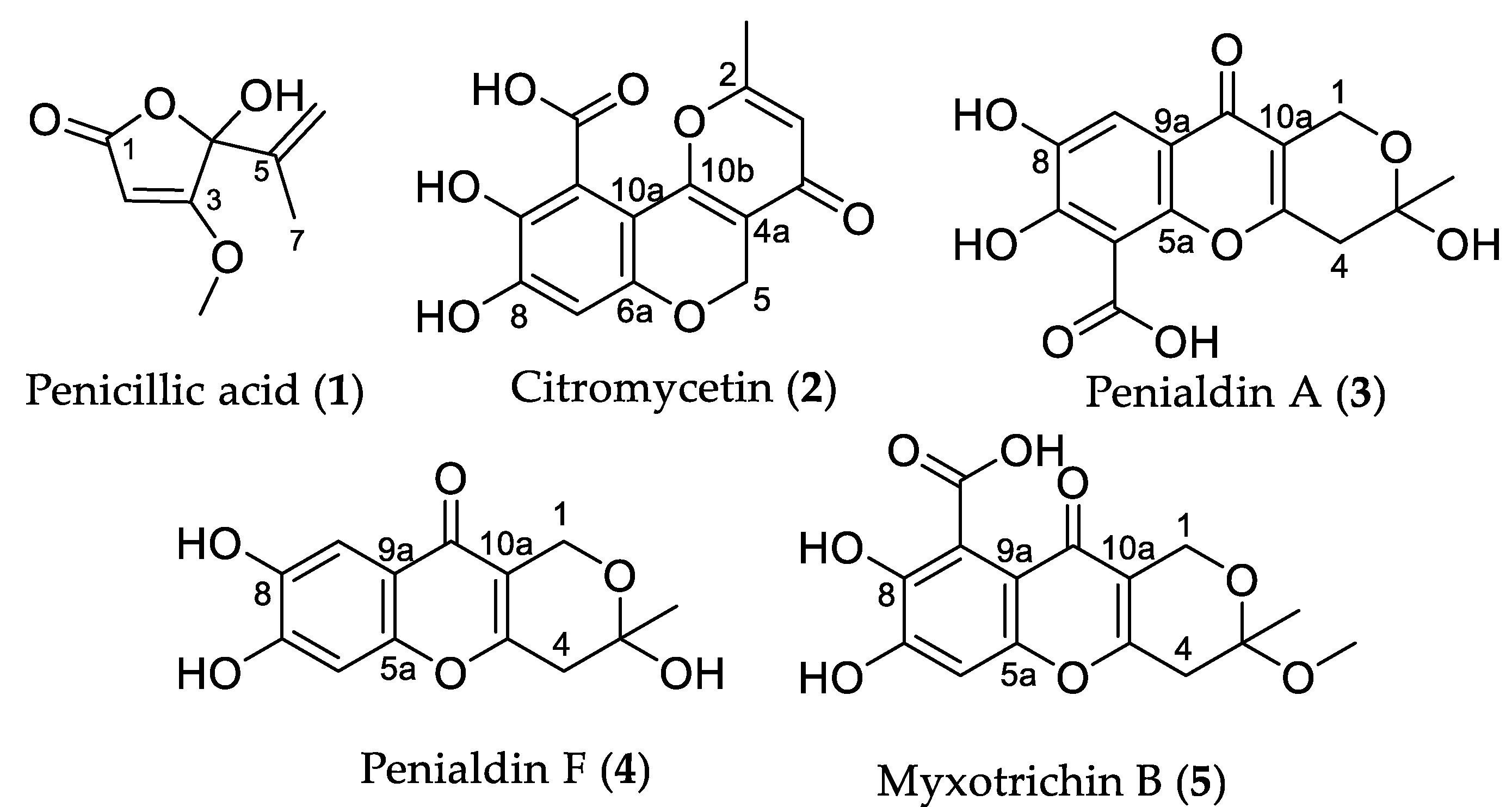
Figure 1. Structures of polyketide metabolites isolated from Penicillium.
Molecules 27 00240 g001
Figure 2. Antibacterial activity of Penicillium bissettii ICMP 21333 against Escherichia coli, Klebsiella pneumoniae, and Staphylococcus aureus. The antibacterial activity of P. bissettii ICMP 21333 was measured by the production of zones of inhibition (mm) when fungal plugs were incubated on lawns of E. coli ATCC 25922, K. pneumoniae ATCC 700603, S. aureus ATCC 29213 (antibiotic-sensitive), and S. aureus ATCC 33593 (antibiotic-resistant). Boxes are upper and lower quartiles with median shown. The whiskers extend up to 1.5× the inter-quartile range, and any dots beyond those bounds are outliers. The dotted line represents the diameter of the fungal plugs. Experiments were performed on three separate occasions with three technical replicates per experiment.
Molecules 27 00240 g002
Figure 3. Tautomerization of penicillic acid (1).
Molecules 27 00240 g003
Figure 4. Structures of penicillic acid (1) and its derivatives (1a1c).
Molecules 27 00240 g004
Figure 5. Labelling pattern observed in 13C NMR spectrum of penicillic acid (1) produced by cultures supplemented with 1-13C sodium acetate.
Molecules 27 00240 g005
Figure 6. Effect of citromycetin (2) and penialdin F (4) on the luminescence of Mycobacterium abscessus BSG301 and M. marinum BSG101. The antibacterial activity of citromycetin (2) and penialdin F (4) were measured by changes in bacterial luminescence. Data are presented as box and whisker plots of the area under the curve values calculated from luminescence readings taken over 72 h. Boxes are upper and lower quartiles with median shown. The whiskers extend up to 1.5 × the inter-quartile range. The dotted line represents the no-compound control.
Molecules 27 00240 g006
Table 1. 1H and 13C NMR (CD3OD) signals observed for crude (1a) and purified (1b) products.
Position1a1b
δHδCδHδC
1 174.6 173.5
25.1997.25.2089.2
3 171.2 182.1
4 217.1 107.2
5Not observed39.32.1234.6
61.0717.51.0416.1
6′1.0717.50.8816.4
73.7257.83.9359.9
Table 2. Relative Integral Values for 13C NMR Peaks Observed for 13C-labelled Penicillic Acid (1).
PositionΔcRelative Integral (Labelled/Unlabelled)
C-1181.212.3
C-290.11.0
C-3173.210.1
C-4Not observed
C-5141.78.5
C-6116.71.1
C-717.51.0
C-860.40.9
Table 3. Antimicrobial and antifungal activities of natural products 14 and derivatives 1b and 1c.
CompoundPercentage Inhibition at 32 µg/mL
S. aaE. cbK. pcP. adA. beC. afC. ng
153.07 ****26.11 ***18.06 *−1.156.126.502.15
1b−1.050.344.322.75−26.552.4918.33 **
1c17.41 *5.159.34−10.06−11.882.56−7.04
214.2211.1613.494.70−4.31−1.861.11
3−7.2313.60−7.65−4.08−24.24−2.2020.72 **
471.52 ****9.2915.62 *0.5212.558.846.73
All values are presented as the mean (n = 2). Negative values represent increases in growth compared to the no-compound controls. Data were analysed using a two-way ANOVA with Dunn’s multiple comparison test. The adjusted p values of those compounds that were significantly different to the relevant no inhibition control are given by * (* p < 0.05; ** p < 0.005; *** p < 0.0005; **** p < 0.0001). a Staphylococcus aureus ATCC 43300 (MRSA) with vancomycin (MIC 1 μg/mL) used as a positive control; b Escherichia coli ATCC 25922 with colistin (MIC 0.125 μg/mL) used as a positive control; c Klebsiella pneumoniae ATCC 700603 with colistin (MIC 0.25 μg/mL) as a positive control; d Pseudomonas aeruginosa ATCC 27853 with colistin (MIC 0.25 μg/mL); e Acinetobacter baumanii ATCC 19606 with colistin (MIC 0.25 μg/mL) as a positive control; f Candida albicans ATCC 90028 with fluconazole (MIC 0.125 μg/mL) as a positive control; g Cryptococcus neoformans ATCC 208821 with fluconazole (MIC 8 μg/mL) as a positive control.
Publisher’s Note: MDPI stays neutral with regard to jurisdictional claims in published maps and institutional affiliations.