
Green Synthesis and Characterization of Silver Nanoparticles Using Spondias mombin Extract and Their Antimicrobial Activity against Biofilm-Producing Bacteria


 ,
,  , ,
, ,  and
and
Abstract
1. Introduction
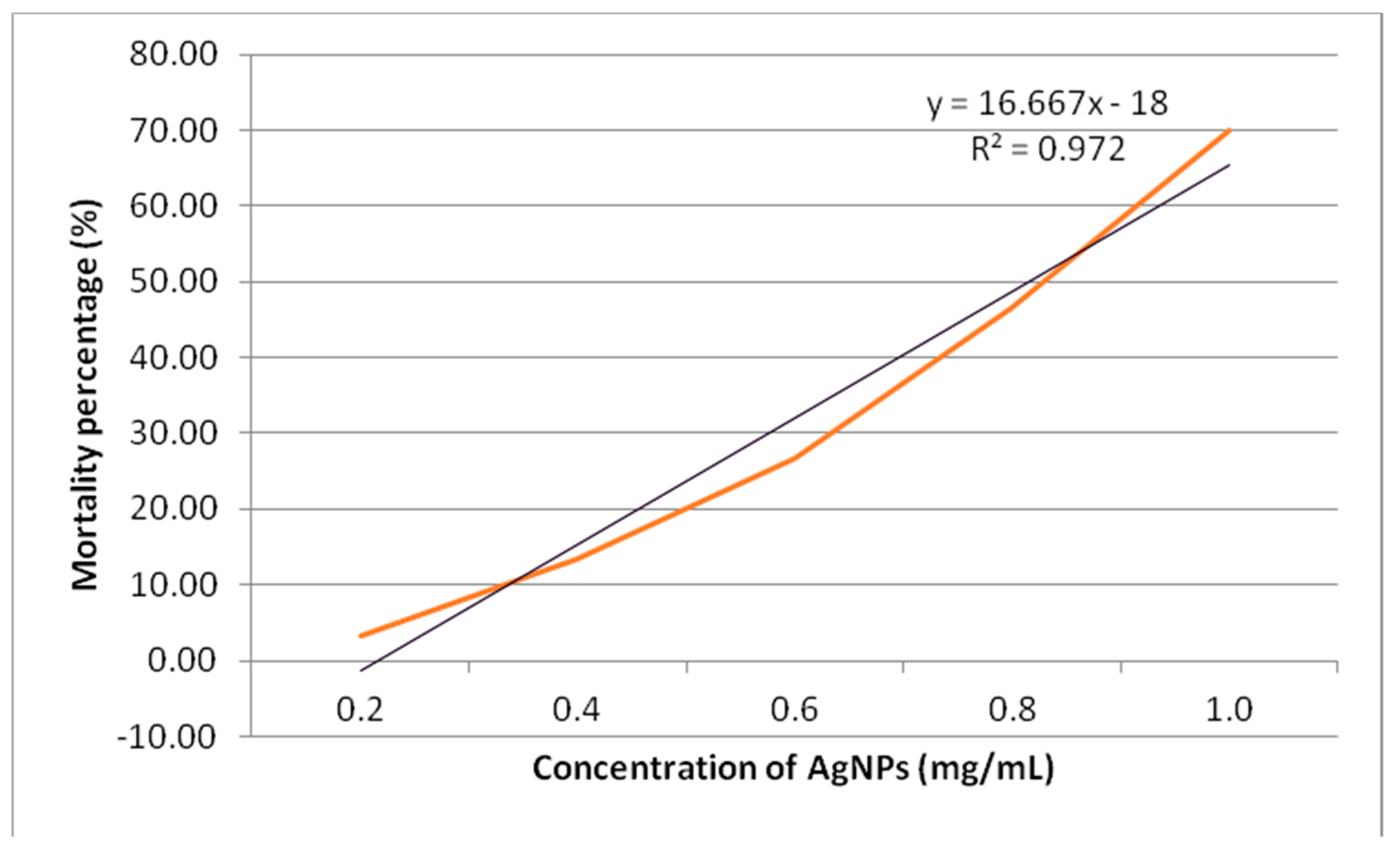
2. Results and Discussion
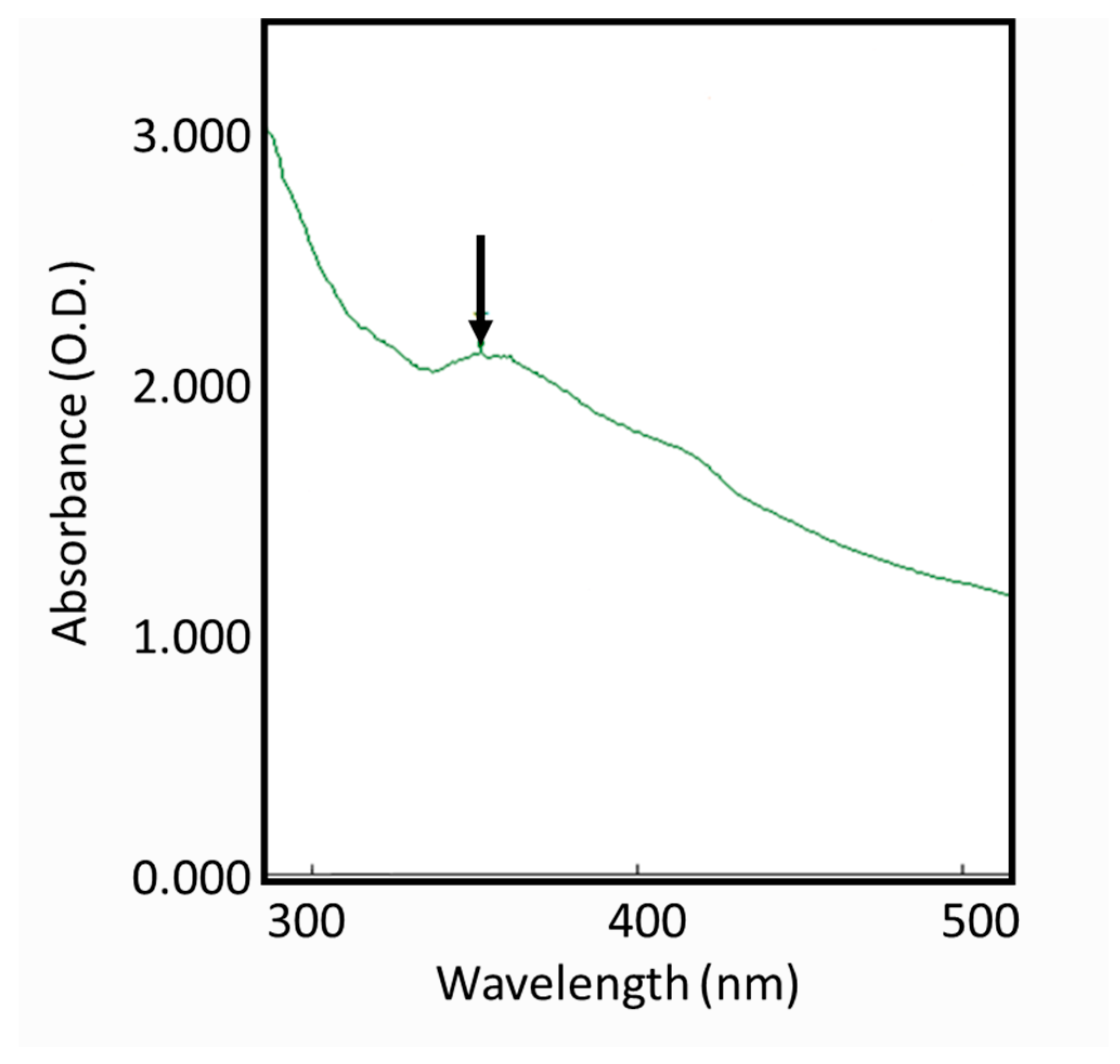
2.1. Silver Nanoparticle-Synthesis
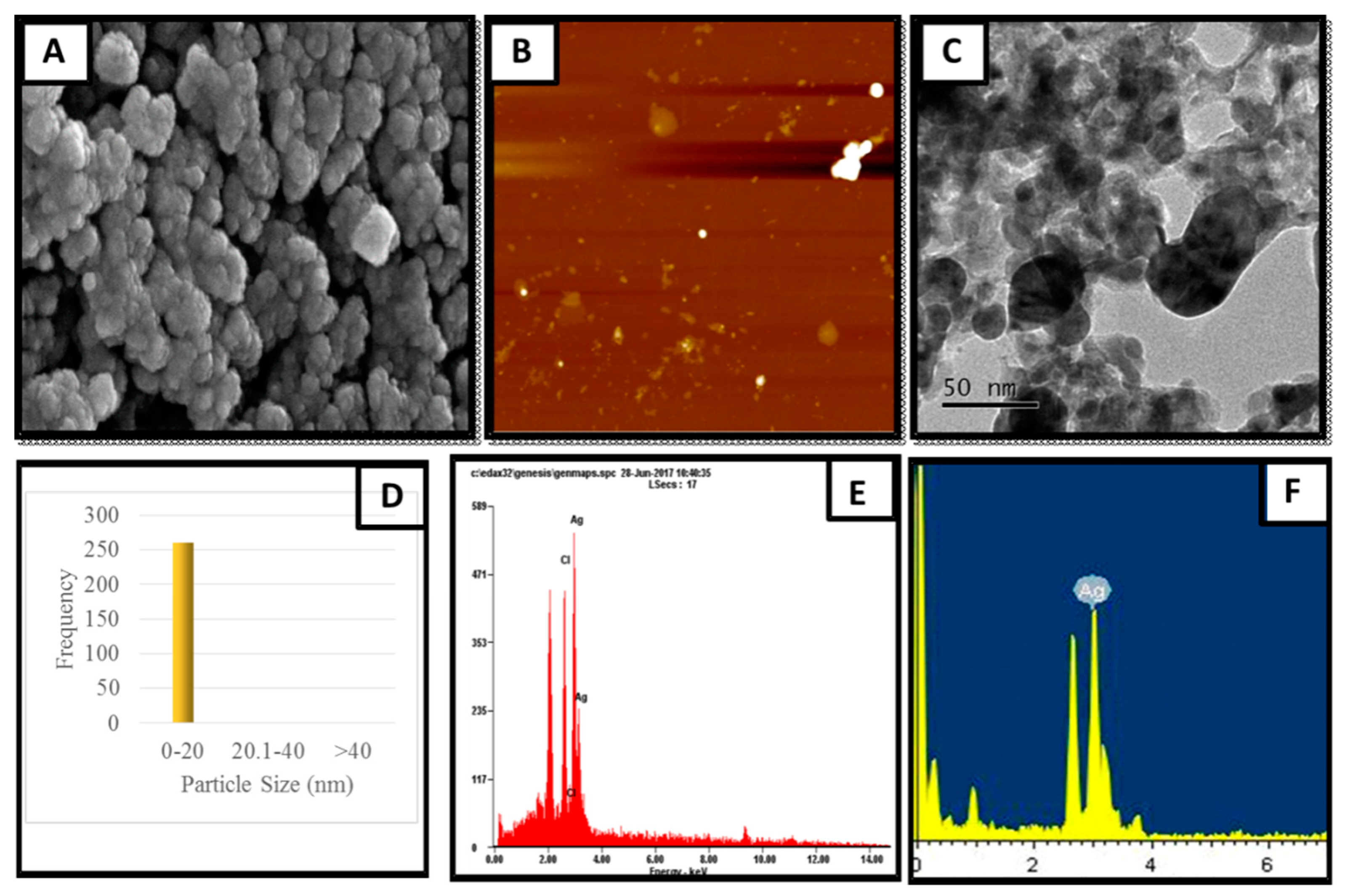
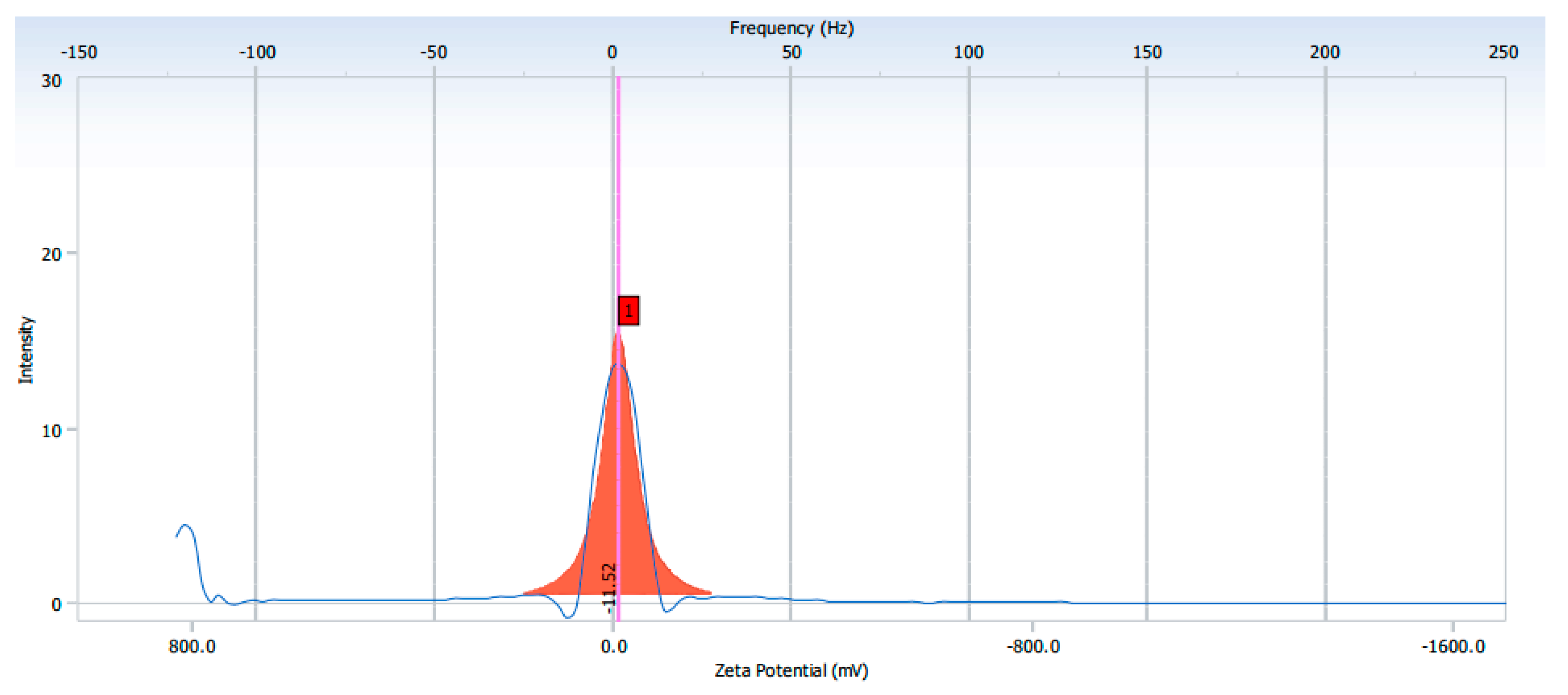
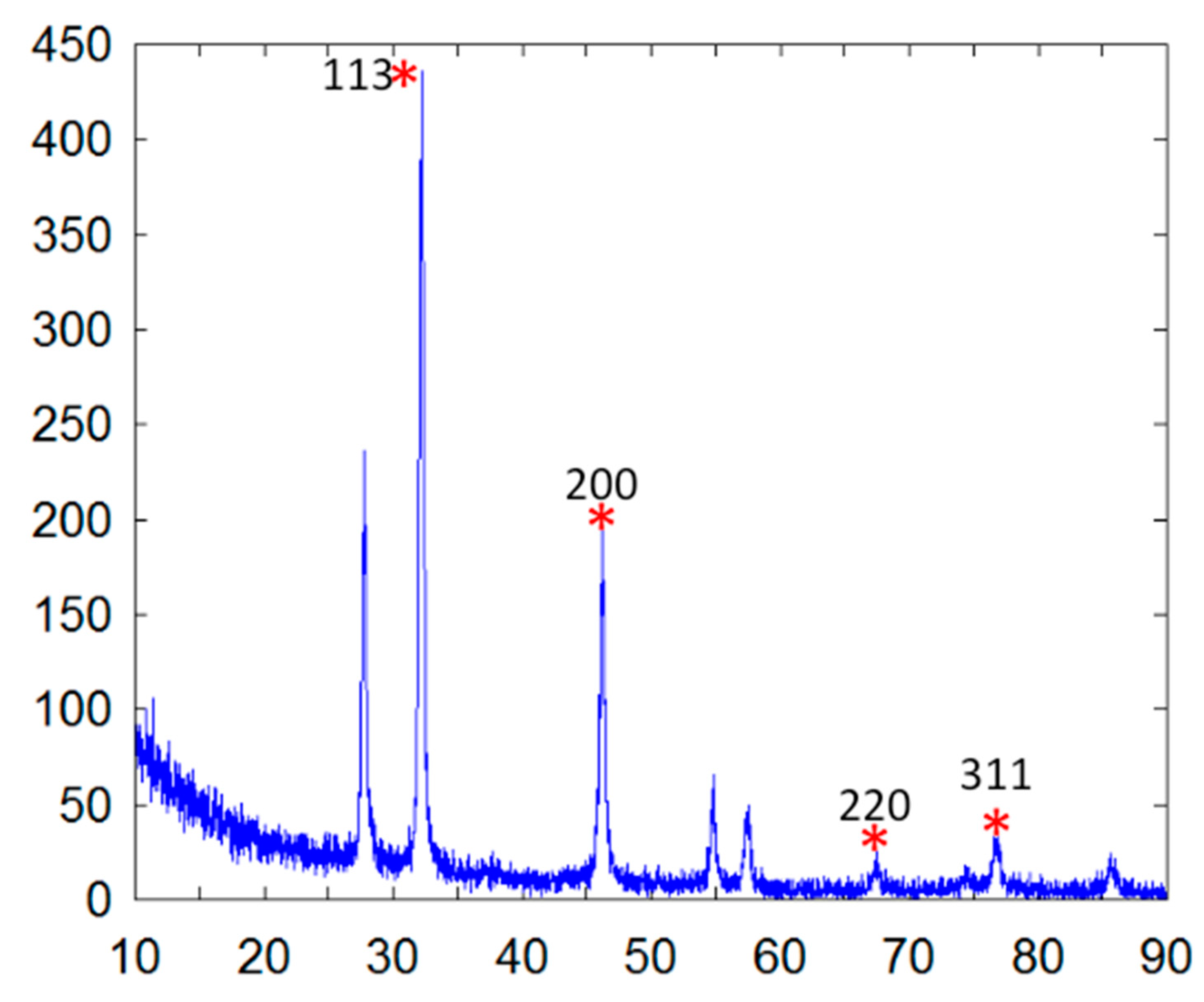
2.2. Characterization of Synthesized Silver Nanoparticles
2.3. Antibacterial Activity of Spondias Mombin Leaf Extract
2.4. Antibacterial Activity of Synthesized Silver Nanoparticles
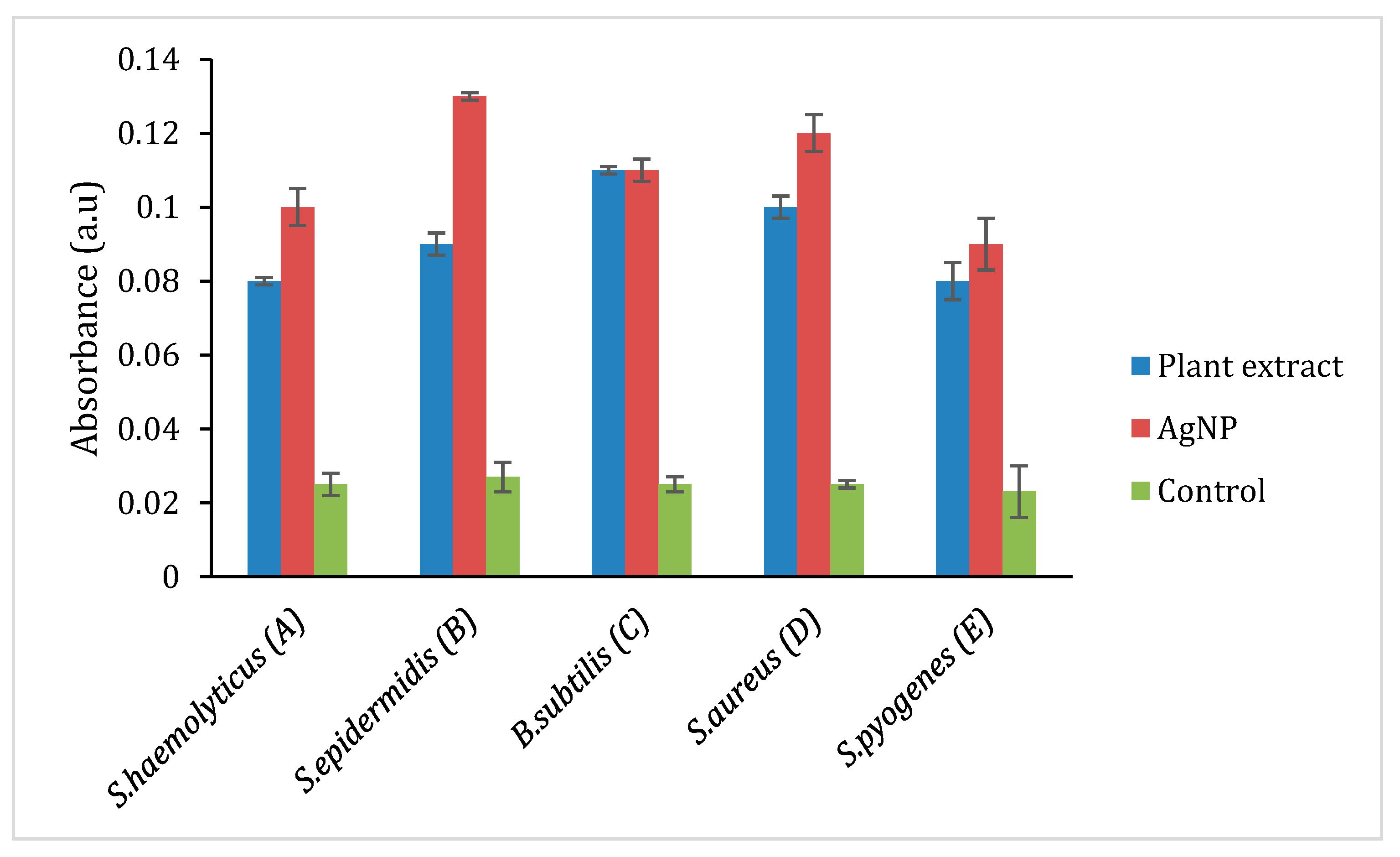
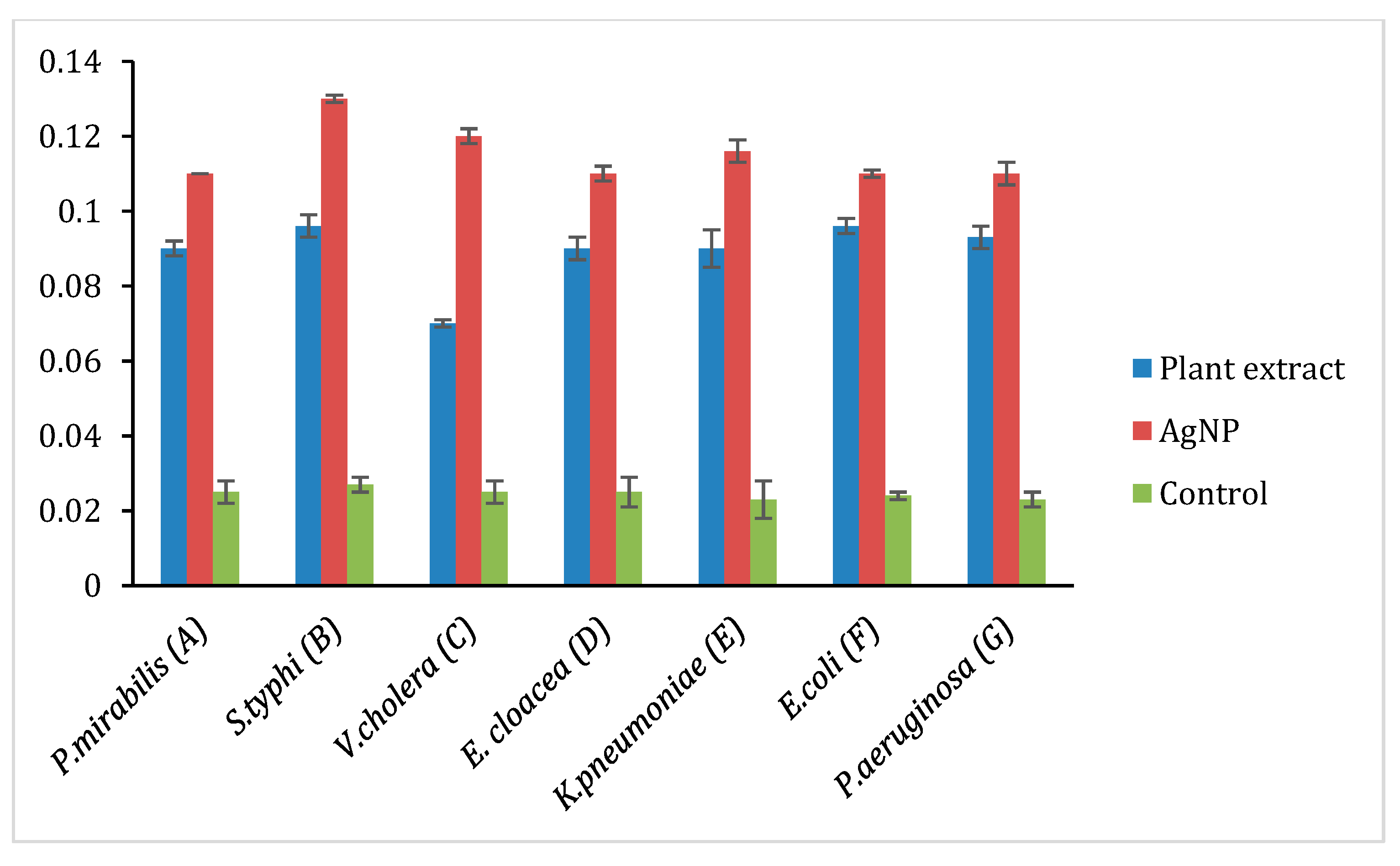
2.5. Cytotoxic Study
3. Materials and Methods
3.1. Collecting the Plant Samples
3.2. Preparation of S. mombin Leaf Extract
3.3. Biosynthesis of Silver Nanoparticles
3.4. Characterization of Synthesized Silver Nanoparticles
3.5. Verification of the Antibacterial Activity of Synthesized Silver Nanoparticles
3.6. Reactive Oxygen Species (ROS) Quantification
3.7. Cytotoxicity Study
4. Conclusions
Author Contributions
Funding
Institutional Review Board Statement
Informed Consent Statement
Data Availability Statement
Acknowledgments
Conflicts of Interest
Sample Availability
References
- Garibo, D.; Borbón-Nuñez, H.A.; De León, J.N.D.; Mendoza, E.G.; Estrada, I.; Toledano-Magaña, Y.; Tiznado, H.; Ovalle-Marroquin, M.; Soto-Ramos, A.G.; Blanco, A.; et al. Green synthesis of silver nanoparticles using Lysiloma acapulcensis exhibit high-antimicrobial activity. Sci. Rep. 2020, 10, 1–11. [Google Scholar] [CrossRef]
- Chen, H.; Roco, M.C.; Li, X.; Lin, Y.-L. Trends in nanotechnology patents. Nat. Nanotechnol. 2008, 3, 123–125. [Google Scholar] [CrossRef]
- Chinni, S.; Gopinath, S.; Anbu, P.; Fuloria, N.; Fuloria, S.; Mariappan, P.; Krusnamurthy, K.; Reddy, L.V.; Ramachawolran, G.; Sreeramanan, S.; et al. Characterization and Antibacterial Response of Silver Nanoparticles Biosynthesized Using an Ethanolic Extract of Coccinia indica Leaves. Crystals 2021, 11, 97. [Google Scholar] [CrossRef]
- Lashin, I.; Fouda, A.; Gobouri, A.; Azab, E.; Mohammedsaleh, Z.; Makharita, R. Antimicrobial and In Vitro Cytotoxic Efficacy of Biogenic Silver Nanoparticles (Ag-NPs) Fabricated by Callus Extract of Solanum incanum L. Biomolecules 2021, 11, 341. [Google Scholar] [CrossRef] [PubMed]
- Das, C.G.; Kumar, G.; Dhas, S.; Velu, K.; Govindaraju, K.; Joselin, J.; Baalamurugan, J. Antibacterial activity of silver nanoparticles (biosynthe-sis): A short review on recent advances. Biocatal. Agric. Biotechnol. 2020, 27, 101593. [Google Scholar] [CrossRef]
- Natsuki, J.; Natsuki, T.; Hashimoto, Y. A Review of Silver Nanoparticles: Synthesis Methods, Properties and Applications. Int. J. Mater. Sci. Appl. 2015, 4, 325. [Google Scholar] [CrossRef]
- Ahmed, S.; Ahmad, M.; Swami, B.L.; Ikram, S. A review on plants extract mediated synthesis of silver nanoparticles for antimi-crobial applications: A green expertise. J. Adv. Res. 2016, 7, 17–28. [Google Scholar] [CrossRef]
- Iravani, S.; Korbekandi, H.; Mirmohammadi, S.V.; Zolfaghari, B. Synthesis of silver nanoparticles: Chemical, physical and bio-logical methods. Res. Pharm. Sci. 2014, 9, 385–406. [Google Scholar]
- Rajeshkumar, S.; Bharath, L.V. Mechanism of plant-mediated synthesis of silver nanoparticles—A review on biomolecules involved, characterisation and antibacterial activity. Chem. Biol. Interact. 2017, 273, 219–227. [Google Scholar] [CrossRef] [PubMed]
- Pantidos, N.; Horsfall, L.E. Biological Synthesis of Metallic Nanoparticles by Bacteria, Fungi and Plants. J. Nanomed. Nanotechnol. 2000, 5. Available online: https://www.omicsonline.org/open-access/biological-synthesis-of-metallic-nanoparticles-by-bacteria-fungi-and-plants-2157-7439.1000233.php?aid=31363 (accessed on 18 June 2018). [CrossRef]
- Cowan, M.M. Plant Products as Antimicrobial Agents. Clin. Microbiol. Rev. 1999, 12, 564–582. [Google Scholar] [CrossRef]
- Siddiqi, K.S.; Husen, A.; Rao, R.A.K. A review on biosynthesis of silver nanoparticles and their biocidal properties. J. Nanobiotechnol. 2018, 16, 14. [Google Scholar] [CrossRef] [PubMed]
- WHO Publishes List of Bacteria for Which New Antibiotics Are Urgently Needed. Available online: https://www.who.int/news/item/27-02-2017-who-publishes-list-of-bacteria-for-which-new-antibiotics-are-urgently-needed (accessed on 27 March 2021).
- Sharma, D.; Misba, L.; Khan, A.U. Antibiotics versus biofilm: An emerging battleground in microbial communities. Antimicrob. Resist. Infect. Control. 2019, 8, 1–10. [Google Scholar] [CrossRef]
- Kuppusamy, P.; Yusoff, M.M.; Maniam, G.P.; Govindan, N. Biosynthesis of metallic nanoparticles using plant derivatives and their new avenues in pharmacological applications–An updated report. Saudi Pharm. J. 2016, 24, 473–484. [Google Scholar] [CrossRef] [PubMed]
- Islam, S.M.d.A.; Ahmed, K.T.; Manik, M.K.; Wahid, M.d.A.; Kamal, C.S.I. A comparative study of the antioxidant, antimicrobial, cyto-toxic and thrombolytic potential of the fruits and leaves of Spondias dulcis. Asian Pac J Trop Biomed. 2013, 3, 682–691. [Google Scholar] [CrossRef]
- Fernandes, F.A.; Salgado, H. Antimicrobial Activity of Spondias dulcis Parkinson Extract Leaves Using Microdilution and Agar Diffusion: A Comparative Study. EC Microbiol. 2018, 14, 9. [Google Scholar]
- Zhang, X.-F.; Liu, Z.-G.; Shen, W.; Gurunathan, S. Silver Nanoparticles: Synthesis, Characterization, Properties, Applications, and Therapeutic Approaches. Int. J. Mol. Sci. 2016, 17, 1534. [Google Scholar] [CrossRef] [PubMed]
- Mohanta, Y.K.; Panda, S.K.; Bastia, A.K.; Mohanta, T.K. Biosynthesis of Silver Nanoparticles from Protium serratum and Investi-gation of their Potential Impacts on Food Safety and Control. Front. Microbiol. 2017, 8. Available online: https://www.frontiersin.org/articles/10.3389/fmicb.2017.00626/full (accessed on 24 January 2021). [CrossRef]
- Shankar, S.S.; Ahmad, A.; Sastry, M. Geranium Leaf Assisted Biosynthesis of Silver Nanoparticles. Biotechnol. Prog. 2003, 19, 1627–1631. [Google Scholar] [CrossRef]
- Duraisamy, S.; Kasi, M.; Balakrishnan, S.; Al-Sohaibani, S.; Murugan, K.; Senthilkumar, B.; Senbagam, D. Biosynthesis of silver nanoparticles using Acacia leucophloea extract and their antibacterial activity. Int. J. Nanomed. 2014, 9, 2431. [Google Scholar] [CrossRef]
- Suvith, V.; Philip, D. Catalytic degradation of methylene blue using biosynthesized gold and silver nanoparticles. Spectrochim. Acta Part A Mol. Biomol. Spectrosc. 2014, 118, 526–532. [Google Scholar] [CrossRef] [PubMed]
- Georgieva, R.; Yocheva, L.; Tserovska, L.; Zhelezova, G.; Stefanova, N.; Atanasova, A.; Danguleva, A.; Ivanova, G.; Karapetkov, N.; Rumyan, N.; et al. Antimicrobial activity and antibiotic susceptibility of Lactobacillus and Bifidobacterium spp. intended for use as starter and probiotic cultures. Biotechnol. Biotechnol. Equip. 2015, 29, 84. [Google Scholar] [CrossRef] [PubMed]
- Kang, C.-G.; Hah, D.-S.; Kim, C.-H.; Kim, Y.-H.; Kim, E.-K.; Kim, J.-S. Evaluation of Antimicrobial Activity of the Methanol Extracts from 8 Traditional Medicinal Plants. Toxicol. Res. 2011, 27, 31–36. [Google Scholar] [CrossRef]
- Jeong, S.H.; Yeo, S.Y.; Yi, S.C. The effect of filler particle size on the antibacterial properties of compounded polymer/silver fi-bers. J. Mater Sci. 2005, 40, 5407–5411. [Google Scholar] [CrossRef]
- Mohamed, D.S.; El-Baky, R.M.A.; Sandle, T.; Mandour, S.A.; Ahmed, E.F. Antimicrobial Activity of Silver-Treated Bacteria against other Multi-Drug Resistant Pathogens in Their Environment. Antibiotics 2020, 9, 181. [Google Scholar] [CrossRef] [PubMed]
- Wang, L.; Hu, C.; Shao, L. The antimicrobial activity of nanoparticles: Present situation and prospects for the future. Int. J. Nanomed. 2017, 12, 1227–1249. [Google Scholar] [CrossRef] [PubMed]
- Sumitha, S.; Vasanthi, S.; Shalini, S.; Chinni, S.V.; Gopinath, S.C.B.; Anbu, P.; Bahari, M.B.B.; Harish, R.; Kathiresan, S.; Ravichandran, V. Phyto-Mediated Photo Catalysed Green Synthesis of Silver Nanoparticles Using Durio Zibethinus Seed Extract: Antimicrobial and Cytotoxic Activity and Photocatalytic Applications. Molecules 2018, 23, 3311. [Google Scholar] [CrossRef]
- Patil, P.S.; Kumbhar, S.T. Antioxidant, antibacterial and cytotoxic potential of silver nanoparticles synthesized using terpenes rich extract of Lantana camara L. leaves. Biochem. Biophys. Rep. 2017, 10, 76–81. [Google Scholar]
- Kittler, S.; Greulich, C.; Diendorf, J.; Koller, M.; Epple, M. Toxicity of Silver Nanoparticles Increases during Storage Because of Slow Dissolution under Release of Silver Ions. Chem. Mater. 2010, 22, 4548–4554. [Google Scholar] [CrossRef]


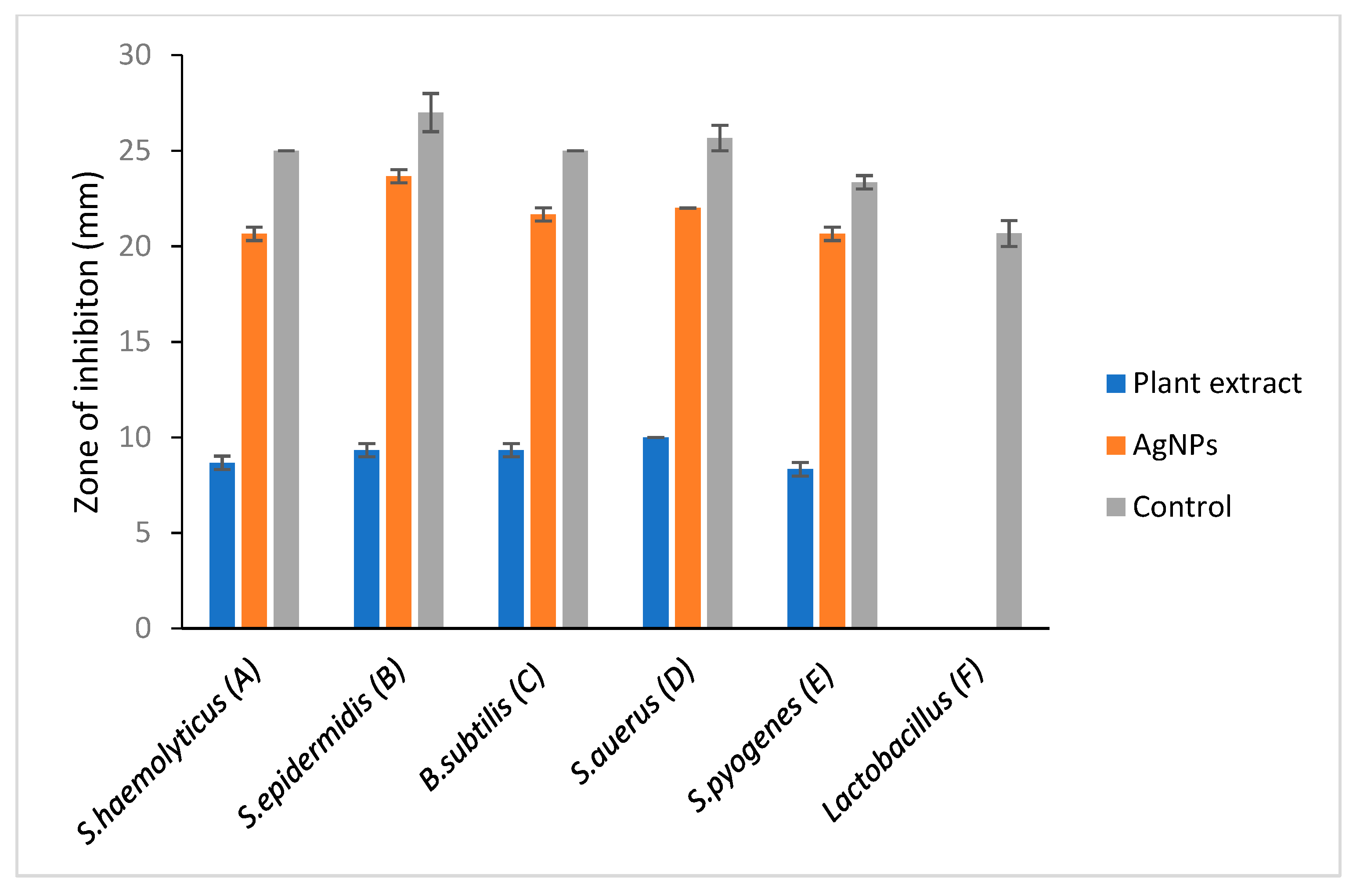
| Gram-Positive Bacteria | Zone of Inhibition (mm) | |||
|---|---|---|---|---|
| Plant Extract (Ethanolic) | Silver Nanoparticles (AgNPs) | Controls | ||
| Positive (Ciprofloxacin) | Negative | |||
| S.haemolyticus | 8.67 ± 0.35 | 20.65 ± 0.35 | 25 | - |
| S. epidermis | 9.35 ± 0.35 | 23.65 ± 0.35 | 27 ± 1.00 | - |
| B.subtilis | 9.35 ± 0.35 | 21.65 ± 0.35 | 25 | - |
| S. aurus | 10 | 22 | 25.67 ± 0.67 | - |
| S. pyogenes | 8.33 ± 0.35 | 20.65 ± 0.35 | 23.35 ± 0.35 | - |
| Lactobacillus | 0.00 | - | 20.67 ± 0.67 | - |
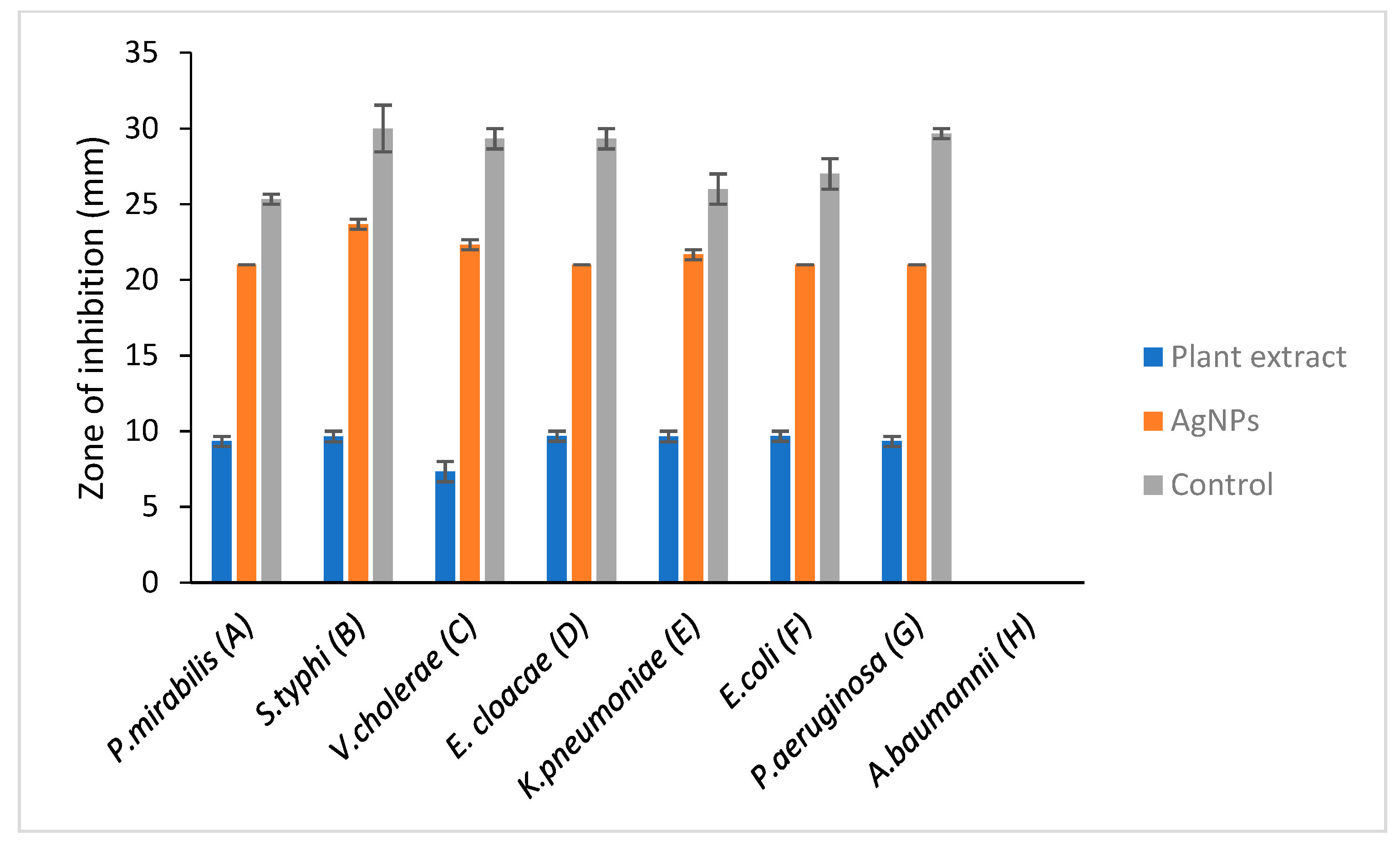
| Gram-Negative Bacteria | Zone of Inhibition (mm) | |||
|---|---|---|---|---|
| Plant Extract (Ethanolic) | Silver Nanoparticles (AgNPs) | Controls | ||
| Positive (Ciprofloxacin) | Negative | |||
| P. mirabilis | 9.33 ± 0.33 | 21 | 25.33 ± 0.33 | - |
| S. typhi | 9.65 ± 0.35 | 23.67 ± 0.33 | 30 ± 1.53 | - |
| V. cholera | 7.33 ± 0.67 | 22.33 ± 0.33 | ± 0.67 | - |
| E. cloacae | 9.67 ± 0.33 | 21 | ± 0.67 | - |
| K. pneumoniae | 9.65 ± 0.35 | 0.33 | 26 ± 1.00 | - |
| E. coli | 0.33 | 21 | 26 ± 1.00 | - |
| P.aeruginosa | 9.33 ± 0.33 | 21 | 29.7 ± 0.33 | - |
| A. baumannii | 0.00 | - | - | - |
Publisher’s Note: MDPI stays neutral with regard to jurisdictional claims in published maps and institutional affiliations. |
© 2021 by the authors. Licensee MDPI, Basel, Switzerland. This article is an open access article distributed under the terms and conditions of the Creative Commons Attribution (CC BY) license (https://creativecommons.org/licenses/by/4.0/).
Share and Cite
Samuggam, S.; Chinni, S.V.; Mutusamy, P.; Gopinath, S.C.B.; Anbu, P.; Venugopal, V.; Reddy, L.V.; Enugutti, B. Green Synthesis and Characterization of Silver Nanoparticles Using Spondias mombin Extract and Their Antimicrobial Activity against Biofilm-Producing Bacteria. Molecules 2021, 26, 2681. https://doi.org/10.3390/molecules26092681
Samuggam S, Chinni SV, Mutusamy P, Gopinath SCB, Anbu P, Venugopal V, Reddy LV, Enugutti B. Green Synthesis and Characterization of Silver Nanoparticles Using Spondias mombin Extract and Their Antimicrobial Activity against Biofilm-Producing Bacteria. Molecules. 2021; 26(9):2681. https://doi.org/10.3390/molecules26092681
Chicago/Turabian StyleSamuggam, Sumitha, Suresh V. Chinni, Prasanna Mutusamy, Subash C. B. Gopinath, Periasamy Anbu, Vijayan Venugopal, Lebaka Veeranjaneya Reddy, and Balaji Enugutti. 2021. "Green Synthesis and Characterization of Silver Nanoparticles Using Spondias mombin Extract and Their Antimicrobial Activity against Biofilm-Producing Bacteria" Molecules 26, no. 9: 2681. https://doi.org/10.3390/molecules26092681
APA StyleSamuggam, S., Chinni, S. V., Mutusamy, P., Gopinath, S. C. B., Anbu, P., Venugopal, V., Reddy, L. V., & Enugutti, B. (2021). Green Synthesis and Characterization of Silver Nanoparticles Using Spondias mombin Extract and Their Antimicrobial Activity against Biofilm-Producing Bacteria. Molecules, 26(9), 2681. https://doi.org/10.3390/molecules26092681