Article

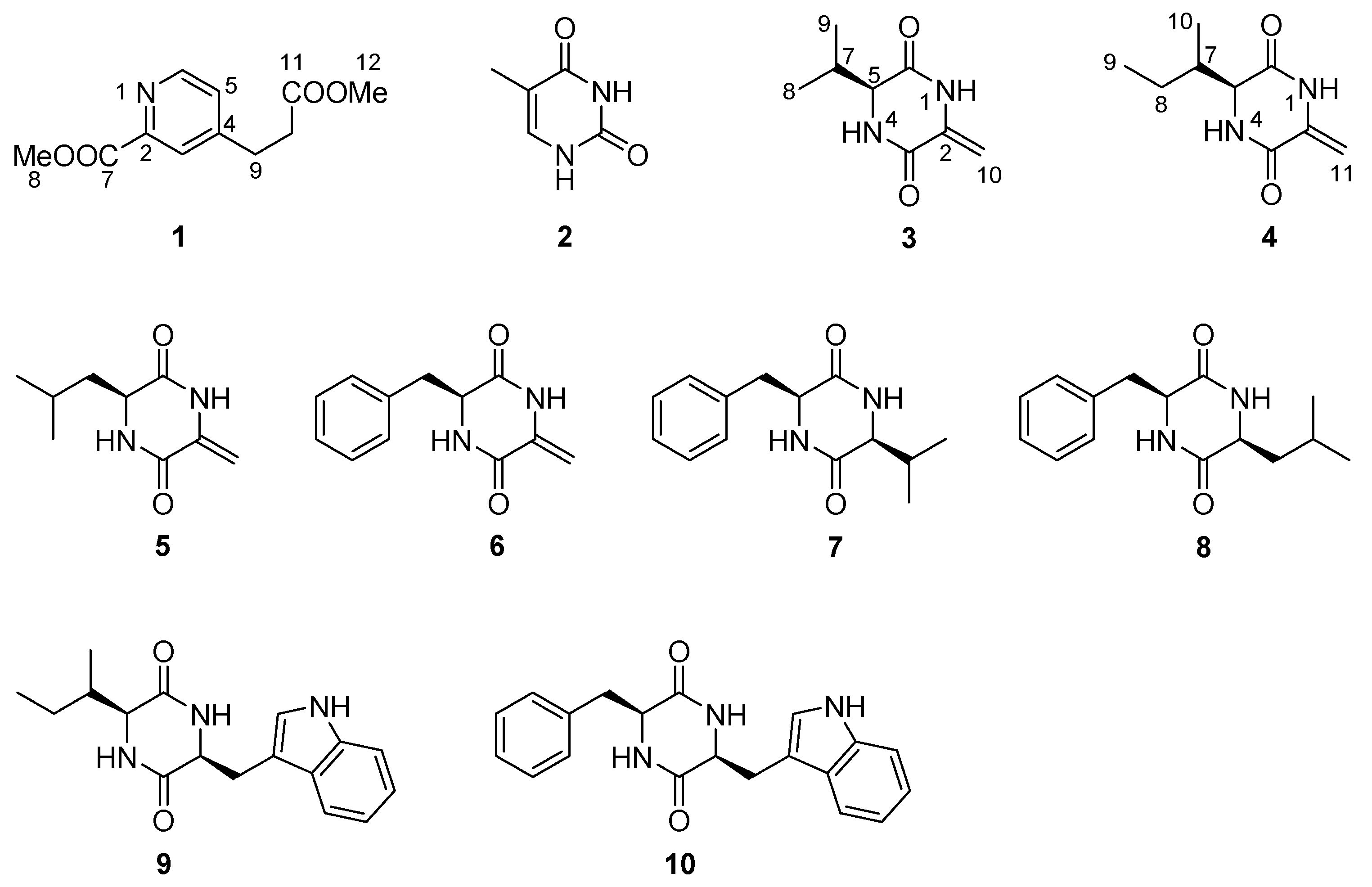
Nitrogenous Compounds from the Antarctic Fungus Pseudogymnoascus sp. HSX2#-11

1
State Key Laboratory of Microbial Technology, Institute of Microbial Technology, Shandong University, Qingdao 266200, China
2
Key Laboratory of Marine Eco-Environmental Science and Technology, First Institute of Oceanography, Ministry of Natural Resources, Qingdao 266061, China
3
Laboratory for Marine Ecology and Environmental Science, Qingdao Pilot National Laboratory for Marine Science and Technology, Qingdao 266071, China
4
Laboratory for Marine Drugs and Bioproducts of Qingdao National Laboratory for Marine Science and Technology, Qingdao 266071, China
*
Author to whom correspondence should be addressed.
Molecules 2021, 26(9), 2636; https://doi.org/10.3390/molecules26092636
Submission received: 23 March 2021 / Revised: 20 April 2021 / Accepted: 22 April 2021 / Published: 30 April 2021
(This article belongs to the Special Issue Microbial Natural Products in Drug Discovery Chemistry)

Abstract

:
The species Pseudogymnoascus is known as a psychrophilic pathogenic fungus which is ubiquitously distributed in Antarctica. While the studies of its secondary metabolites are infrequent. Systematic research of the metabolites of the Antarctic fungus Pseudogymnoascus sp. HSX2#-11 led to the isolation of one new pyridine derivative, 4-(2-methoxycarbonyl-ethyl)-pyridine-2-carboxylic acid methyl ester (1), together with one pyrimidine, thymine (2), and eight diketopiperazines, cyclo-(dehydroAla-l-Val) (3), cyclo-(dehydroAla-l-Ile) (4), cyclo-(dehydroAla-l-Leu) (5), cyclo-(dehydroAla-l-Phe) (6), cyclo-(l-Val-l-Phe) (7), cyclo-(l-Leu-l-Phe) (8), cyclo-(l-Trp-l-Ile) (9) and cyclo-(l-Trp-l-Phe) (10). The structures of these compounds were established by extensive spectroscopic investigation, as well as by detailed comparison with literature data. This is the first report to discover pyridine, pyrimidine and diketopiperazines from the genus of Pseudogymnoascus.

1. Introduction

Nitrogenous compounds represent one of the most momentous family of secondary metabolites which are widely distributed in different biological sources [1]. They have been proved to exhibit various biological activities including cytotoxic, anti-inflammatory, antimicrobial activities and so on [1,2,3,4]. For example, pegaharine D, a β-carboline alkaloid isolated from the seeds of Peganum harmala exhibited strong antiviral activity against herpes simplex virus-2 [5]. For another example, asperversiamides A–C, a kind of cycloheptapeptide, showed potent inhibitory activity against Mycobacterium marinum [6]. Antarctica as the southernmost point of the earth, has the most hostile environment including cold, dry climate and low level of nutrition [7]. Microbes, especially fungi, have been proved to have the potential capacity to produce abundant novel compounds to adapt the extreme habitat. There were more and more bioactive natural products with novel structures have been discovered from Antarctic fungi [8,9,10,11]. The species Pseudogymnoascus is known as a psychrophilic pathogenic fungus with a ubiquitous distribution in Antarctica. While the rare research about its secondary metabolites suggested the potentials to discover interesting compounds [12]. Pseudogymnoascus sp. HSX2#-11, an Antarctic fungus derived from a soil sample of the Fields Peninsula, which can produce various compounds according to our previous study [13], was further investigated to search for new secondary metabolites. As a result, one new pyridine derivative, 4-(2-methoxycarbonyl-ethyl)-pyridine-2-carboxylic acid methyl ester (1), together with one known pyrimidine, thymine (2) and eight known diketopiperazines (310) (Figure 1), were isolated and identified from the potato dextrose agar (PDA) culture broth fermentative extracts of this strain. This paper addresses the isolation, structure elucidation, and bioactivity evaluation of the isolated compounds.

2. Results

4-(2-Methoxycarbonyl-ethyl)-pyridine-2-carboxylic acid methyl ester (1) was obtained as a colorless oil. The molecular formula of C11H13O4N was determined by high resolution electrospray ionization mass spectroscopy (HRESIMS) that displayed the [M + Na]+ peak at m/z 246.0742 (calcd for C11H13O4NNa, 246.0737), indicating six degrees of unsaturation (Figure S8 from Supplementary Materials). The 1H-NMR, 13C-NMR and heteronuclear single quantum coherence (HSQC) spectra (Figures S1, S2 and S6 in the Supplementary Materials) exhibited two methoxyls, (δH 3.66 (3H, s), δC 52.0; δH 3.98 (3H, s), δC 53.1), two methylenes, (δH 2.69 (2H, t, 7.5 Hz), δC 34.0; δH 3.03 (2H, t, 7.5 Hz), δC 30.1), three aromatic methines, (δH 7.37 (1H, br s), δC 127.4; δH 8.00 (1H, br s), δC 125.4; δH 8.66 (1H, br s), δC 149.6), two aromatic quaternary carbon signals (δC 147.6, δC 151.8), and two carbonyls (δC 165.4, δC 172.4) (Table 1). The five aromatic carbon signals combined with the molecular formula of C11H13O4N indicated a pyridine substructure in 1. The splitting effects of 1H-NMR were undesirable in CDCl3 of the aromatic methines of 1. So the 1H-NMR spectrum was measured again in DMSO-d6. The coupling constants of 5.0 Hz (H-5/H-6), 1.7 Hz (H-3/H-5) and 0.8 Hz (H-3/H-6) revealed the ortho-position of H-5/H-6, meta-position of H-3/H-5 and para-position of H-3/H-6, respectively (Table 1). The 1H-1H chemical-shift correlation spectroscopy (COSY) cross peak of H-9/H-10, and key heteronuclear multiple-bond correlation (HMBC) correlations of H-9/C-3, H-9/C-5, H-10/C-4, H-10/C-11 and H-12/C-11 elucidated the methyl propionate substituent located at C-4 (Figure 2). The methyl formate group at C-2 was suggested by the key HMBC correlations from H-8 to C-7 and C-2, and H-3 to C-7 (Figure 2). Thus compound 1 was identified as 4-(2-methoxycarbonyl-ethyl)-pyridine-2-carboxylic acid methyl ester.
Cyclo-(dehydroAla-l-Ile) (4) was obtained as a white powder. The HRESIMS spectrum of 4 led to the molecular formula of C9H14O2N2 (m/z 205.0975, calcd for C9H14O2N2Na, 205.0948), indicating 4 degrees of unsaturation (Figure S16 in the Supplementary Materials). The 1H-NMR, 13C-NMR and HSQC spectra (Figures S11, S12 and S14 in the Supplementary Materials) exhibited two methyls, (δH 0.85 (3H, t, 7.4 Hz), δC 11.7; δH 0.88 (3H, d, 7.0 Hz), δC 14.8), two methylenes, (δH 1.20–1.12, (1H, m), 1.41–1.33, (1H, m), δC 24.1; δH 4.76, (1H, br s), 5.17, (1H, br s), δC 98.8), two methines, (δH 1.85–1.80 (1H, m), δC 40.7; δH 3.91 (1H, t, 2.8), δC 59.6), one aromatic quaternary carbon signals (δC 134.7), and two carbonyls (δC 158.6, δC 165.4) (Table 1). The two NH signals (δH 8.34, (1H, br s), δH 10.53, (1H, s)) combined with two carbonyls indicated the diketopiperazine structure of 4. The 1H-1H COSY correlations of H-5/H-7, H-7/H-10, H-7/H-8 and H-8/H-9 revealed the substitute group of 1-methyl-propyl. And the group was located at C-5 determined by the 1H-1H COSY cross peak of H-5 and N-4, and the HMBC correlation from H-10 to C-5. The dehydro-methyl substitute was located at C-2 by the HMBC signal from H-11 to C-2. Thus the planar structure of 4 was confirmed as cyclo-(dehydroAla-Ile) which was first isolated from marine bacteria Claviceps purpurea in 1994, while its detailed NMR data were not reported [14]. The absolute configuration of 4 was proposed to be same as 3 and 5 according to biogenetic perspective and their similar specific optical rotation (OR) data ([α ] D 20 −19.3 (c 0.18, CH3OH) of 4 vs ] D 20 −10.5 (c 0.18, CH3OH) of 3 and [α ] D 20 −78.1 (c 0.18, CH3OH) of 5, Table 2).
The structures of 2, 3, 510 were determined as thymine [15], cyclo-(dehydroAla-l-Val) [16,17], cyclo-(dehydroAla-l-Leu) [18], cyclo-(dehydroAla-l-Phe) [19], cyclo-(l-Val-l-Phe) [20], cyclo-(l-Leu-l-Phe) [20], cyclo-(l-Trp-l-Ile) [21] and cyclo-(l-Trp-l-Phe) [22], respectively, by comparing their NMR and specific OR data (Table 2) with those in the literature.
All the isolated compounds were evaluated for their antibacterial activities against a panel of bacteria, including four pathogenic bacteria, E. coli, S. aureus, P. aeruginosa and B. subtilis, and nine marine fouling bacteria, P. fulva, A. hydrophila, A. salmonicida, V. anguillarum, V. harveyi, P. halotolerans, P. angustum, E. cloacae and E. hormaechei, and cytotoxic activities against five human cancer cell lines A549, PANC-1, HCT116, HepG2 and MDA-MB-231. Unfortunately, none of the tested compounds showed any activity.

3. Materials and Methods

3.1. General Experimental Procedures

Optical rotations were measured on a JASCO P-1020 digital polarimeter (JASCO, Tokyo, Japan). UV spectra were recorded using an Implen Gmbh NanoPhotometer N50 Touch (Implen, Munich, Germany). NMR spectra were recorded on a Bruker AVANCE NEO (Bruker, Fällanden, Switzerland) at 600 MHz for 1H and 150 MHz for 13C in CDCl3 or DMSO-d6. Chemical shifts δ were recorded in ppm, using TMS as internal standard. HRESIMS spectra were measured on a Thermo Scientific LTQ Orbitrap XL spectrometer (Thermo Fisher Scientific, Bremen, Germany). HPLC separation was performed using a Hitachi Primaide Organizer Semi-HPLC system (Hitachi High Technologies, Tokyo, Japan) coupled with a Hitachi Primaide 1430 photodiodearray detector (Hitachi High Technologies). A Kromasil C18 semi-preparative HPLC column (250 × 10 mm, 5 µm) (Eka Nobel, Bohus, Sweden) was used. Silica gel (200–300 mesh; Qingdao Marine Chemical Group Co., Qingdao, China) and Sephadex LH-20 (Amersham Biosciences Inc., Piscataway, NJ, USA) were used for column chromatography. Precoated silica gel GF254 plates (Yantai Zifu Chemical Group Co., Yantai, China).

3.2. Fungal Materials

The fungus Pseudogymnoascus sp. HSX2#-11 was isolated from a soil sample of the Fields Peninsula at Chinese 35th Antarctic expedition in 2019. The strain was deposited in the State Key Laboratory of Microbial Technology, Institute of Microbial Technology, Shandong University, Qingdao, China, with the GenBank (NCBI) accession number MT367223.

3.3. Extraction and Isolation

The fungal strain Pseudogymnoascus sp. HSX2#-11 was fermented in a PDA liquid medium in 200 Erlenmeyer flasks (300 mL in each 1000 mL flask) at 16 °C for 45 days. The culture (60 L) was filtered to separate the broth from the mycelia. Then the mycelia were extracted three times with EtOAc (3 × 4000 mL) and then repeated extracted with CH2Cl2–MeOH (v/v, 1:1) three times (3 × 4000 mL). The broth was extracted repeatedly with EtOAc (3 × 60 L) to get the EtOAc layer. All the extracts were combined and were evaporated to dryness under reduced pressure to afford a residue (71.5 g). The residue was subjected to vacuum liquid chromatography (VLC) on silica gel using step gradient elution with EtOAc–petroleum ether (PE) (0–100%) and then with MeOH–EtOAc (0–100%) to afford eight fractions (Fr.1–Fr.8). Fr.3 was first subjected to gradient elution of octadecylsilyl silica gel (ODS) column chromatography (CC) with MeOH in H2O (10–100%), and then purified by using semi-preparative HPLC on an ODS column (Kromasil C18, 250 × 10 mm, 5 µm, 2 mL/min) eluted with 45% MeOH–H2O to give compounds 7 (5.5 mg), 8 (6.4 mg), 9 (3.5 mg) and 10 (4.4 mg). Fr.4 was isolated by CC on Sephadex LH-20 eluted with CH2Cl2–MeOH (v/v, 1:1) to afford two fractions (Fr.4.1, Fr.4.2). Fr.4.1 was first subjected to silica gel CC eluting with EtOAc–PE (0–50%), and then purified with HPLC eluted with 10% MeOH–H2O to give compound 2 (3.1 mg). Fr.7 was separated on CC on Sephadex LH-20 eluted with CH2Cl2–MeOH (v/v, 1:1) to afford three fractions (Fr.7.1–Fr.7.3). Fr. 7.2 was first subjected on HPLC eluted with 50% MeOH–H2O, and then purified with HPLC eluted with 20% MeOH–H2O to afford 3 (2.5 mg). Fr.7.3 was first isolated by HPLC eluted with 50% MeOH–H2O, and then purified with HPLC eluted with 30% MeOH–H2O to obtain 1 (2.1 mg), 4 (2.8 mg) and 5 (2.9 mg). Fr.8 was subjected to HPLC with 55% MeOH–H2O to gain 6 (7.6 mg).
4-(2-Methoxycarbonylethyl)-pyridine-2-carboxylic acid methyl ester (1): colorless oil; UV (MeOH) λmax (log ε): 200 (4.70), 264 (4.11); 1H- and 13C-NMR data, see Table 1; HRESIMS m/z 246.0742 [M + Na]+ (calcd for C11H13O4NNa, 246.0737).
Cyclo-(dehydroAla-l-Ile) (4): white powder; [α ] D 20 −19.3 (c 0.18, MeOH); UV (MeOH) λmax (log ε): 247 (4.30), 288 (4.01); 1H- and 13C-NMR data, see Table 1; HRESIMS m/z 205.0975 [M + Na]+ (calcd for C9H14O2N2Na, 205.0948).

3.4. Antibacterial and Cytotoxic Activity Assays

The antibacterial activities were evaluated by the conventional broth dilution assay [23]. Four pathogenic bacteria, Escherichia coli, Staphylococcus aureus, P. aeruginosa and Bacillus subtilis, and nine marine fouling bacteria, P. fulva, Aeromonas hydrophila, A. salmonicida, Vibrio anguillarum, V. harveyi, Photobacterium halotolerans, P. angustum, Enterobacter cloacae and E. hormaechei, were used, and cipofloxacin was used as a positive control.
The cytotoxicities against human breast cancer (MDA-MB-231, Shanghai Institute of Biological Sciences, Chinese Academy of Sciences, L-15), colorectal cancer (HCT116, Shanghai Institute of Biological Sciences, Chinese Academy of Sciences, McCOY’s 5A), lung carcinoma (A549, Shanghai Institute of Biological Sciences, Chinese Academy of Sciences, F-12), pancreatic carcinoma (PANC-1, Shanghai Institute of Biological Sciences, Chinese Academy of Sciences, DMEM) and hepatoma (HepG2, Shanghai Institute of Biological Sciences, Chinese Academy of Sciences, DMEM) cell lines were evaluated using the sulphorodamine B (SRB) method [24]. Adriamycin was used as a positive control.

4. Conclusions

In summary, one new pyridine derivative, 4-(2-methoxycarbonyl-ethyl)-pyridine-2-carboxylic acid methyl ester (1), together with one pyrimidine, thymine (2), and eight diketopiperazines 310, were isolated from the Antarctic fungus Pseudogymnoascus sp. HSX2#-11. All the isolated compounds showed no antibacterial or cytotoxic activities. More bioactivity evaluating models should be needed to find the effects of these secondary metabolites. This is the first time to find pyridine, pyrimidine and diketopiperazines from the genus of Pseudogymnoascus. Our chemical investigation of the Antarctic fungus Pseudogymnoascus sp. HSX2#-11 enriches the chemical diversity of this fungal species.

Supplementary Materials

The following are available online, NMR and HRESIMS spectra of the isolated compounds 110: Figure S1: 1H NMR spectrum of compound 1 (CDCl3); Figure S2: 13C NMR spectrum of compound 1 (CDCl3); Figure S3: 1H NMR spectrum of compound 1 (DMSO-d6); Figure S4: 13C NMR spectrum of compound 1 (DMSO-d6); Figure S5: COSY spectrum of compound 1 (CDCl3); Figure S6: HSQC spectrum of compound 1 (CDCl3); Figure S7: HMBC spectrum of compound 1 (CDCl3); Figure S8: HRESIMS spectrum of compound 1; Figure S9: 1H NMR spectrum of compound 2 (DMSO-d6); Figure S10: 1H NMR spectrum of compound 3 (DMSO-d6); Figure S11: 1H NMR spectrum of compound 4 (DMSO-d6); Figure S12: 13C NMR spectrum of compound 4 (DMSO-d6); Figure S13: COSY spectrum of compound 4 (DMSO-d6); Figure S14: HSQC spectrum of compound 4 (DMSO-d6); Figure S15: HMBC spectrum of compound 4 (DMSO-d6); Figure S16: HRESIMS spectrum of compound 4; Figure S17: 1H NMR spectrum of compound 5 (DMSO-d6); Figure S18: 1H NMR spectrum of compound 6 (DMSO-d6); Figure S19: 1H NMR spectrum of compound 7 (DMSO-d6); Figure S20: 1H NMR spectrum of compound 8 (DMSO-d6); Figure S21: 1H NMR spectrum of compound 9 (DMSO-d6); Figure S22: 1H NMR spectrum of compound 10 (DMSO-d6).

Author Contributions

T.S. contributed to experimental design and operation, data analysis and manuscript preparation; L.Z. supported the sample of the Antarctica soil; X.-Q.L., J.-J.D., Y.-T.Z., Y.-Y.Y. and W.-P.H. contributed to activities evaluation; D.-Y.S. was the project leader organizing and guiding the experiments. All authors have read and agreed to the published version of the manuscript.

Funding

This work was supported by the Natural Science Foundation of Shandong Province of China (No. ZR2020QD111); the China Postdoctoral Science Foundation (No. 2019M662378); the National Program for Support of Top-notch Young Professionals; the Fund of Taishan scholar project; The Shandong Provincial Natural Science Foundation for Distinguished Young Scholars (JQ201722); the Qingdao Science and Technology Benefit People Demonstration Guide Special Project (20-3-4-20-nsh); and the Fundamental Research Funds of Shandong University (2020GN033).

Institutional Review Board Statement

Not applicable.

Informed Consent Statement

Not applicable.

Data Availability Statement

The fungus Pseudogymnoascus sp. HSX2#-11’s ribosomal RNA gene, partial sequence can be found at https://www.ncbi.nlm.nih.gov/nuccore/MT367223.1/.

Acknowledgments

We would like to thank Antarctic Great Wall National Observation and Research Station of Polar Ecosystem for sample collection; Jing-Yao Qu, Jing Zhu and Zhi-Feng Li in MS, Hai-Yan Sui in NMR for help and guidance from State Key laboratory of Microbial Technology of Shandong University.

Conflicts of Interest

The authors declare no conflict of interest.

Sample Availability

Samples of the compounds 110 are available from the authors.

References

  1. Elissawy, A.; Dehkordi, E.S.; Mehdinezhad, N.; Ashour, M.; Pour, P.M. Cytotoxic Alkaloids Derived from Marine Sponges: A Comprehensive Review. Biomolecules 2021, 11, 258. [Google Scholar] [CrossRef]
  2. Lai, Y.-W.; Wang, S.-W.; Hu, Y.-Y.; Hwang, T.-L.; Cheng, M.-J.; Chen, I.-S.; Sung, P.-J.; Chen, J.-J. Anti-inflammatory alkaloids from the root bark of Hernandia nymphaeifolia. Phytochemistry 2020, 173, 112326. [Google Scholar] [CrossRef]
  3. Ballard, E.; Yucel, R.; Melchers, W.J.G.; Brown, A.J.P.; Verweij, P.E.; Warris, A. Antifungal Activity of Antimicrobial Peptides and Proteins against Aspergillus fumigatus. J. Fungi 2020, 6, 65. [Google Scholar] [CrossRef]
  4. Sittmann, J.; Bae, M.; Mevers, E.; Li, M.; Quinn, A.; Sriram, G.; Clardy, J.; Liu, Z. Bacterial diketopiperazines stimulate diatom growth and lipid accumulation. Plant Physiol. 2021. [Google Scholar] [CrossRef] [PubMed]
  5. Wu, Z.-N.; Chen, N.-H.; Tang, Q.; Chen, S.; Zhan, Z.-C.; Zhang, Y.-B.; Wang, G.-C.; Li, Y.-L.; Ye, W.-C. β-Carboline Alkaloids from the Seeds of Peganum harmala and Their Anti-HSV-2 Virus Activities. Org. Lett. 2020, 22, 7310–7314. [Google Scholar] [CrossRef]
  6. Hou, X.-M.; Liang, T.-M.; Guo, Z.-Y.; Wang, C.-Y.; Shao, C.-L. Discovery, absolute assignments, and total synthesis of asperversiamides A–C and their potent activity against Mycobacterium marinum. Chem. Commun. 2018, 55, 1104–1107. [Google Scholar] [CrossRef] [PubMed]
  7. Cong, B.; Yin, X.; Deng, A.; Shen, J.; Tian, Y.; Wang, S.; Yang, H. Diversity of Cultivable Microbes From Soil of the Fildes Peninsula, Antarctica, and Their Potential Application. Front. Microbiol. 2020, 11, 570836. [Google Scholar] [CrossRef] [PubMed]
  8. Kwon, J.; Lee, H.; Ko, W.; Kim, D.-C.; Kim, K.-W.; Kwon, H.C.; Guo, Y.; Sohn, J.H.; Yim, J.H.; Kim, Y.-C.; et al. Chemical constituents isolated from Antarctic marine-derived Aspergillus sp. SF-5976 and their anti-inflammatory effects in LPS-stimulated RAW 264.7 and BV2 cells. Tetrahedron 2017, 73, 3905–3912. [Google Scholar] [CrossRef]
  9. Rusman, Y.; Held, B.W.; Blanchette, R.A.; He, Y.; Salomon, C.E. Cadopherone and colomitide polyketides from Cadophora wood-rot fungi associated with historic expedition huts in Antarctica. Phytochemistry 2018, 148, 1–10. [Google Scholar] [CrossRef] [PubMed]
  10. Yu, G.; Sun, Z.; Peng, J.; Zhu, M.; Che, Q.; Zhang, G.; Zhu, T.; Gu, Q.; Li, D. Secondary Metabolites Produced by Combined Culture of Penicillium crustosum and a Xylaria sp. J. Nat. Prod. 2019, 82, 2013–2017. [Google Scholar] [CrossRef]
  11. Sun, C.; Zhang, Z.; Ren, Z.; Yu, L.; Zhou, H.; Han, Y.; Shah, M.; Che, Q.; Zhang, G.; Li, D.; et al. Antibacterial Cyclic Tripeptides from Antarctica-Sponge-Derived Fungus Aspergillus insulicola HDN151418. Mar. Drugs 2020, 18, 532. [Google Scholar] [CrossRef]
  12. Figueroa, L.; Jiménez, C.; Rodríguez, J.; Areche, C.; Chávez, R.; Henríquez, M.; De La Cruz, M.; Díaz, C.; Segade, Y.; Vaca, I. 3-Nitroasterric Acid Derivatives from an Antarctic Sponge-Derived Pseudogymnoascus sp. Fungus. J. Nat. Prod. 2015, 78, 919–923. [Google Scholar] [CrossRef] [PubMed]
  13. Shi, T.; Yu, Y.-Y.; Dai, J.-J.; Zhang, Y.-T.; Hu, W.-P.; Zheng, L.; Shi, D.-Y. New polyketides from the Antarctic fungus Pseudogymnoascus sp. HSX2#-11. Mar. Drugs 2021, 19, 168–176. [Google Scholar] [PubMed]
  14. Bell, R.; Carmeli, S.; Sar, N. Vibrindole A, a Metabolite of the Marine Bacterium, Vibrio parahaemolyticus, Isolated from the Toxic Mucus of the Boxfish Ostracion cubicus. J. Nat. Prod. 1994, 57, 1587–1590. [Google Scholar] [CrossRef] [PubMed]
  15. Yan, H.-J.; Gao, S.-S.; Li, C.-S.; Li, X.-M.; Wang, B.-G. Chemical Constituents of a Marine-Derived Endophytic Fungus Penicillium commune G2M. Molevules 2010, 15, 3270–3275. [Google Scholar] [CrossRef] [PubMed]
  16. Holden, M.T.; Chhabra, S.R.; De Nys, R.; Stead, P.; Bainton, N.J.; Hill, P.J.; Manefield, M.; Kumar, N.; Labatte, M.; England, D.; et al. Quorum-sensing cross talk: Isolation and chemical characterization of cyclic dipeptides from Pseudomonas aeruginosa and other Gram-negative bacteria. Mol. Microbiol. 2002, 33, 1254–1266. [Google Scholar] [CrossRef]
  17. Ebata, M.; Miyazaki, K.; Otsuka, H. Studies on siomycin. II. The composition and degradation products of siomycin A. J. Antibiot. 1969, 22, 423–433. [Google Scholar] [CrossRef] [Green Version]
  18. Park, S.Y.; Shim, S.H. Characterization of metabolites from cultures of Cellulosimicrobium cellulans. J. Korean Soc. Appl. Biol. Chem. 2014, 57, 481–484. [Google Scholar] [CrossRef]
  19. Yamazaki, Y.; Mori, Y.; Oda, A.; Okuno, Y.; Kiso, Y.; Hayashi, Y. Acid catalyzed monodehydro-2,5-diketopiperazine formation from N-α-ketoacyl amino acid amides. Tetrahedron 2009, 65, 3688–3694. [Google Scholar] [CrossRef]
  20. Al-Mourabit, A.; Laville, R.; Nguyen, T.B.; Moriou, C.; Petek, S.; Debitus, C. Marine Natural Occurring 2,5-Diketopiperazines: Isolation, Synthesis and Optical Properties. Heterocycles 2015, 90, 1351. [Google Scholar] [CrossRef] [Green Version]
  21. Stocking, E.M.; Sanz-Cervera, J.F.; Unkefer, C.J.; Williams, R.M. Studies on the biosynthesis of paraherquamide. Construction of the amino acid framework. Tetrahedron 2001, 57, 5303–5320. [Google Scholar] [CrossRef]
  22. Kimura, Y.; Tani, K.; Kojima, A.; Sotoma, G.; Okada, K.; Shimada, A. Cyclo-(l-tryptophyl-l-phenylalanyl), a plant growth regulator produced by the fungus Penicillium sp. Phytochemistry 1996, 41, 665–669. [Google Scholar] [CrossRef]
  23. Appendino, G.; Gibbons, S.; Giana, A.; Pagani, A.; Grassi, G.; Stavri, M.; Smith, E.; Rahman, M.M. Antibacterial Cannabinoids from Cannabis sativa: A Structure−Activity Study. J. Nat. Prod. 2008, 71, 1427–1430. [Google Scholar] [CrossRef] [PubMed]
  24. Skehan, P.; Storeng, R.; Scudiero, D.; Monks, A.; McMahon, J.; Vistica, D.; Warren, J.T.; Bokesch, H.; Kenney, S.; Boyd, M.R. New Colorimetric Cytotoxicity Assay for Anticancer-Drug Screening. J. Natl. Cancer Inst. 1990, 82, 1107–1112. [Google Scholar] [CrossRef] [PubMed]
Figure 1. Structures of compounds 110.
Molecules 26 02636 g001
Figure 2. COSY (red bold line) and key HMBC (blue arrows) correlations of 1 and 4.
Molecules 26 02636 g002
Table 1. NMR spectroscopic data (600/150 MHz) for compounds 1 and 4.
No.14
PosδCa TypeδHa Multiple (Hz)δCb TypeδHb Multiple (Hz)δCb TypeδHb Multiple (Hz)
1-----10.53, s
2147.6, C -147.5, C -134.7, C-
3125.4, CH8.00, br s124.9, CH7.94, dd (1.7, 0.8)158.6, C-
4151.8, C-151.1, C--8.34, br s
5127.4, CH7.37, br s127.2, CH7.52, dd (5.0, 1.7)59.6, CH3.91, t (2.8)
6149.6, CH8.66, br s149.7, CH8.59, dd (5.0, 0.8)165.4, C-
7165.4, C-165.3, C-40.7, CH1.85–1.80, m
853.1, CH33.98, s52.4, CH33.87, s24.1, CH21.41–1.33, m
-----1.20–1.12, m
930.1, CH23.03, t (7.5)29.2, CH22.95, t (7.5)11.7, CH30.85, t (7.4)
1034.0, CH22.69, t (7.5)33.2, CH22.73, t (7.5)14.8, CH30.88, d (7.0)
11172.4, C-172.3, C-98.8, CH25.17, br s
-----4.76, br s
1252.0, CH33.66, s51.4, CH33.58, s--
a measured in CDCl3, b measured in DMSO-d6.
Table 2. Specific OR of diketopiperazines 310 in CH3OH.
CompoundsNatural [α] D 20 Literature
3−10.5 (c 0.18) ] D 26 −95.7 (c 0.4) [17]
4−19.3 (c 0.18)-
5−78.1 (c 0.18) ] D 22 −163 (0.01) [18]
6−41.3 (c 0.18) ] D 26 −40.0 (1.08) a [19]
7−25.3 (c 0.18) ] D 25 −11.4 (1.0) a [20]
8+18.9 (c 0.14) ] D 25 +30.0 (0.3) [20]
9−16.5 (c 0.18) ] D 25 −31.5 (0.0065) [21]
10−144.2 (c 0.14) ] D 20 −254.9 (1.0) [22]
a measured in DMSO.
Publisher’s Note: MDPI stays neutral with regard to jurisdictional claims in published maps and institutional affiliations.

