
Antiproliferative Isoprenoid Derivatives from the Red Sea Alcyonacean Xenia umbellata

 , , , , , ,
  and
and
Abstract
1. Introduction
2. Results
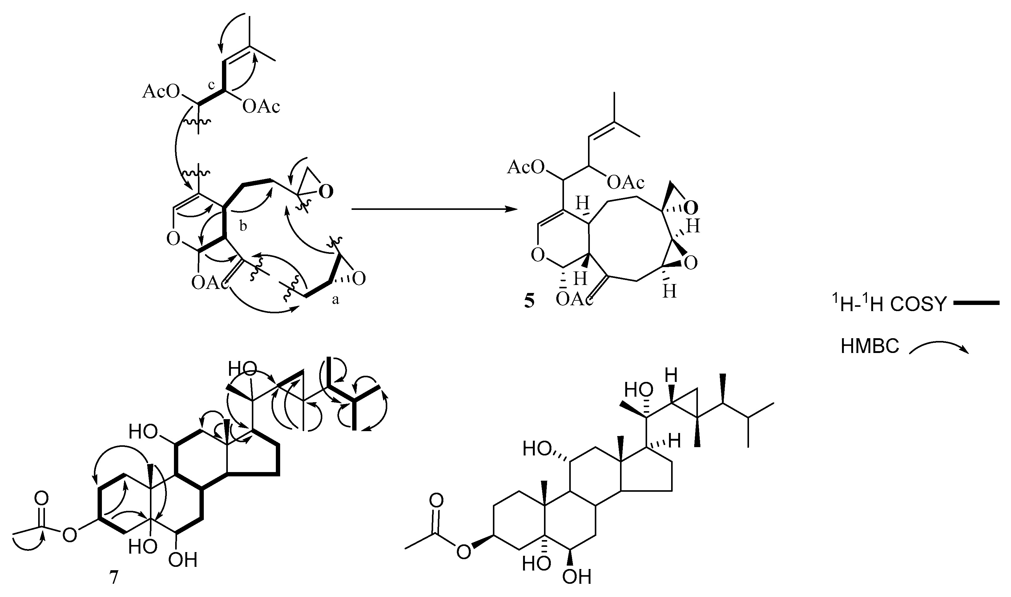
2.1. Chemistry
2.2. Biological Activities
2.2.1. Anti-Proliferative Activity
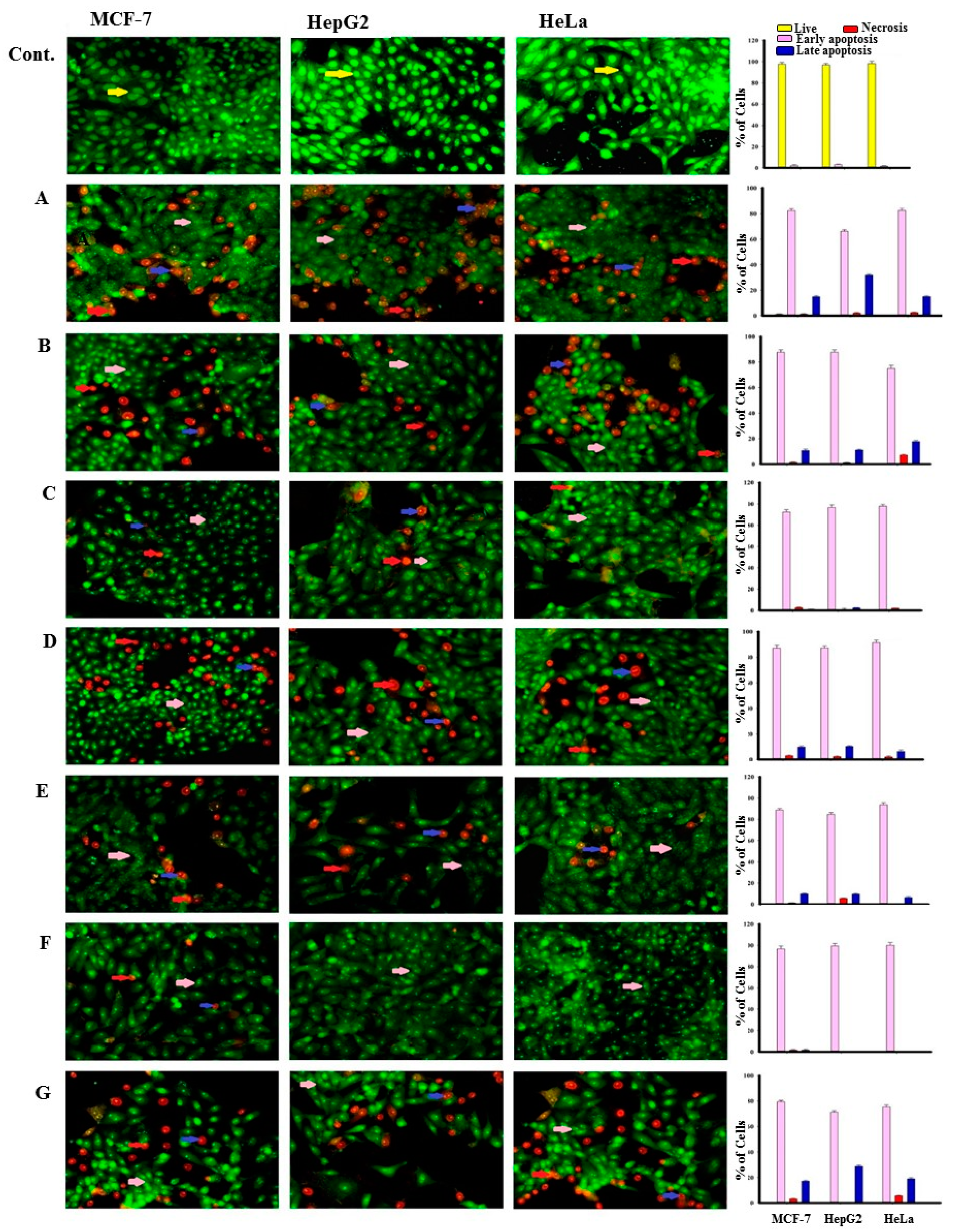
2.2.2. Apoptotic Effect
3. Discussion
3.1. Chemistry
3.2. Biological Activities
4. Material and Methods
4.1. General
4.2. Animal Material
4.3. Extraction and Isolation
4.4. Biological Activities
4.4.1. Antiproliferative Activity
Cell Culture
Sulphorhodamine B Assay (SRB)
Acridine Orange/Ethidium Bromide Staining for Detection of Apoptosis
4.5. Statistical Analysis
5. Conclusions
Supplementary Materials
Author Contributions
Funding
Institutional Review Board Statement
Informed Consent Statement
Data Availability Statement
Acknowledgments
Conflicts of Interest
Sample Availability
Abbreviations
| 1H-NMR | Proton Nuclear Magnetic Resonance |
| 13C-NMR | Carbon-13 Nuclear Magnetic Resonance |
| 1D and 2D NMR COSY | One and two-dimensional Nuclear Magnetic Resonance ¹H-¹H Correlation Spectroscopy |
| DEPT | Heteronuclear Multiple Bond Correlation |
| HMBC | Distortionless Enhancement by polarization transefer |
| HSQC | Heteronuclear Single Quantum Coherence |
| brt | Broad triplet |
| d | Doublet |
| δ | Chemical shift |
| J | Nuclear spin-spin coupling constant |
| dd | Doublet of doublet |
| HeLa | Cervix adenocarcinoma |
| HepG2 | Hepatocellular carcinoma |
| MCF-7 | Human breast cancer |
| IC50 | Half maximal inhibitory concentration |
| SD | Standard Deviation |
| CDCl3 | Deuterated chloroform |
| CH2Cl2 | Dichloromethane |
| MeOH | Methanol |
| TLC | Thin Layer Chromatography |
| PTLC | Preparative Thin Layer Chromatography |
References
- Ferlay, J.; Shin, H.R.; Bray, F.; Forman, D.; Mathers, C.; Parkin, D.M. Estimates of worldwide burden of cancer in 2008: Globocan 2008. Int. J. Cancer 2010, 127, 2893–2917. [Google Scholar] [CrossRef] [PubMed]
- Global Cancer Observatory (GCO). 2020. Available online: https://gco.iarc.fr/today/data/factsheets/populations/682-saudi-arabia-fact-sheets.pdf (accessed on 1 January 2021).
- Mondal, S.; Adhikari, N.; Banerjee, S.; Amin, S.A.; Jha, T. Matrix metalloproteinase-9 (MMP-9) and its inhibitors in cancer: A minireview. Eur. J. Med. Chem. 2020, 194, 112260. [Google Scholar] [CrossRef] [PubMed]
- World Health Organization (WHO). 2018. Available online: https://www.who.int/news-room/fact-sheets/detail/cancer (accessed on 1 January 2021).
- Takaoka, M.; Ando, Y. Chemistry of marine natural products. In Isoprenoids; Schever, P., Ed.; Academic Press: London, UK, 2012; p. 214. [Google Scholar]
- Altmann, K.H. Drugs from the oceans: Marine natural products as leads for drug discovery. Chim. Int. J. Chem. 2017, 71, 646–652. [Google Scholar] [CrossRef] [PubMed]
- Shang, J.; Hu, B.; Wang, J.; Zhu, F.; Kang, Y.; Li, D.; Sun, H.; Kong, D.-X.; Hou, T. Cheminformatic insight into the differences between terrestrial and marine originated natural products. J. Chem. Inf. Model. 2018, 58, 1182–1193. [Google Scholar] [CrossRef]
- Jurasek, M.; Rimpelová, S.; Kmoníčková, E.; Drašar, P.; Ruml, T. Tailor-made fluorescent trilobolide to study its biological relevance. J. Med. Chem. 2014, 57, 7947–7954. [Google Scholar] [CrossRef] [PubMed]
- Peterková, L.; Kmoníčková, E.; Ruml, T.; Rimpelová, S. Sarco/Endoplasmic reticulum calcium ATPase inhibitors: Beyond anticancer perspective. J. Med. Chem. 2020, 63, 1937–1963. [Google Scholar] [CrossRef]
- Jurášek, M.; Džubák, P.; Rimpelová, S.; Sedlák, D.; Konecny, P.; Frydrych, I.; Gurská, S.; Hajdúch, M.; Bogdanová, K.; Kolár, M.; et al. Trilobolide-steroid hybrids: Synthesis, cytotoxic and antimycobacterial activity. Steroids 2017, 117, 97–104. [Google Scholar] [CrossRef]
- Harmatha, J.; Buděšínský, M.; Vokáč, K.; Kostecká, P.; Kmoníčková, E.; Zídek, Z. Trilobolide and related sesquiterpene lactones from laser trilobum possessing immunobiological properties. Fitoterapia 2013, 89, 157–166. [Google Scholar] [CrossRef] [PubMed]
- Li, K.; Chung-Davidson, Y.-W.; Bussy, U.; Li, W. Recent advances and applications of experimental technologies in marine natural product research. Mar. Drugs 2015, 13, 2694–2713. [Google Scholar] [CrossRef]
- Harper, M.K.; Bugni, T.S.; Copp, B.R.; James, R.D.; Lindsay, B.S.; Richardson, A.D.; Schnabel, P.C.; Tasdemir, D.; VanWagoner, R.M.; Verbitski, S.M.; et al. Introduction to the chemical ecology of marine natural products. In Marine Chemical Ecology; McClintock, J.B., Baker, B.J., Eds.; CRC Press: Boca Raton, FL, USA, 2001; p. 453. [Google Scholar]
- Li, G.; Li, P.; Tang, X. Natural Products from Corals. In Symbiotic Microbiomes of Coral Reefs Sponges and Corals; Li, Z., Ed.; Springer: Dordrecht, The Netherlands, 2019; pp. 465–504. [Google Scholar]
- Bergmann, W. Sterols: Their structure and distribution. In Comparative Biochemistry. A Comprehensive Treatise, Vol. III, Constituents of Life, Part A; Florkin, M., Mason, H.S., Eds.; Academic Press: Waltham, MA, USA, 1962; Volume 3, pp. 103–162. [Google Scholar]
- Schmitz, F.J. Uncommon marine steroids. In Marine Natural Products, Chemical and Biological Perspectives; Scheuer, P.J., Ed.; Academic Press: New York, NY, USA, 1978; Volume I, pp. 241–297. [Google Scholar]
- Hale, R.L.; Leclercq, J.; Tursch, B.; Djerassi, C.; Gross Jr, R.; Weinheimer, A.; Gupta, K.C.; Scheuer, P.J. Demonstration of a biogenetically unprecedented side chain in the marine sterol, gorgosterol. J. Am. Chem. Soc. 1970, 92, 2179–2180. [Google Scholar] [CrossRef]
- Ling, N.C.; Hale, R.L.; Djerassi, C. Structure and absolute configuration of the marine sterol gorgosterol. J. Am. Chem. Soc. 1970, 92, 5281–5282. [Google Scholar] [CrossRef]
- Ayyad, S.-E.N.; Alarif, W.M.; Al-Footy, K.O.; Selim, E.A.; Ghandourah, M.A.; Aly, M.M.; Alorfi, H.S. Isolation, antimicrobial and antitumor activities of a new polyhydroxysteroid and a new diterpenoid from the soft coral Xenia umbellata. Z. Naturforsch. C J. Biosci. 2017, 72, 27–34. [Google Scholar] [CrossRef]
- Bawakid, N.O.; Alarif, W.M.; Abdel-Lateff, A. Rare norisodinosterol derivatives from Xenia umbellata: Isolation and anti-proliferative activity. Open Chem. 2021. accepted manuscript. [Google Scholar]
- Vichai, V.; Kirtikara, K. Sulforhodamine B colorimetric assay for cytotoxicity screening. Nat Protoc. 2006, 1, 1112–1116. [Google Scholar] [CrossRef] [PubMed]
- Vanderah, D.J.; Steudler, P.A.; Ciereszko, L.S.; Schmitz, F.J.; Ekstrand, J.D.; van der Helm, D. Marine natural products. Xenicin: A diterpenoid possessing a nice-membered ring from the soft coral, Xenia elongata. J. Am. Chem. Soc. 1977, 99, 5780–5784. [Google Scholar] [CrossRef] [PubMed]
- Kashman, Y.; Groweiss, A. Xeniolide-A and xeniolide-B, two new diterpenoids from the soft-coral Xenia macrospiculata. Tetrahedron Lett. 1978, 19, 4833–4836. [Google Scholar] [CrossRef]
- Kashman, Y.; Groweiss, A. New diterpenoids from the soft corals Xenia macrospiculata and Xenia obscuronata. J. Org. Chem. 1980, 45, 3824–3827. [Google Scholar] [CrossRef]
- Iwagawa, T.; Kawasaki, J.; Hase, T.; Yu, C.M.; Walter, J.A.; Wright, J.L.C. A new tricarbocyclic diterpene structure from the soft coral Xenia florida. J. Chem. Soc. Chem. Commun. 1994, 18, 2073–2074. [Google Scholar] [CrossRef]
- Iwagawa, T.; Amano, Y.; Hase, T.; Shiro, M. New xenia diterpenoids from a soft coral, xenia species 1. Tetrahedron 1995, 51, 11111–11118. [Google Scholar] [CrossRef]
- Iwagawa, T.; Amano, Y.; Nakatani, M.; Hase, T. New xenia diterpenoids from a soft coral, Xenia species containing fatty acyl side chains. Bull. Chem. Soc. Jpn. 1996, 69, 1309–1312. [Google Scholar] [CrossRef]
- Kitagawa, I.; Kobayashi, M.; Cui, Z.; Kiyota, Y.; Ohnishi, M. Marine natural products. XV. Chemical constituents of an Okinawan soft coral of Xenia sp. (Xeniidae). Chem. Pharm. Bull. 1986, 34, 4590–4596. [Google Scholar] [CrossRef]
- Bishara, A.; Rudi, A.; Goldberg, I.; Benayahu, Y.; Kashman, Y. Novaxenicins A–D and xeniolides I–K, seven new diterpenes from the soft coral Xenia novaebrittanniae. Tetrahedron 2006, 62, 12092–12097. [Google Scholar] [CrossRef]
- Musmar, M.; Weinheimer, A.J.; Martin, G.E.; Hurd, R.E. Assignment of the high field resonances of a gorgosterol derivative through the use of autocorrelated two-dimensional proton NMR spectroscopy. J. Org. Chem. 1983, 48, 3580–3581. [Google Scholar] [CrossRef]
- Karplus, M. Vicinal proton coupling in nuclear magnetic resonance. J. Am. Chem. Soc. 1963, 85, 2870–2871. [Google Scholar] [CrossRef]
- Goad, J.; Akihisia, T. Analysis of Sterols, 1st ed.; Blackie Academic & Professional, Chapman Hall: London, UK, 1997; p. 438. [Google Scholar]
- Abu-Izneid, T.; Rauf, A.; Shariati, M.A.; Khalil, A.A.; Imran, M.; Rebezov, M.; Uddin, M.S.; Mahomoodally, M.F.; Rengasamy, K.R.R. Sesquiterpenes and their derivatives-natural anticancer compounds: An update. Pharmacol. Res. 2020, 161, 105165. [Google Scholar] [CrossRef] [PubMed]
- Gupta, A.; Kumar, B.S.; Negi, A.S. Current status on development of steroids as anticancer agents. J. Steroid Biochem. Mol. Biol. 2013, 137, 242–270. [Google Scholar] [CrossRef] [PubMed]
- Alarif, W.M.; Abdel-Lateff, A.; Al-Abd, A.M.; Basaif, S.A.; Badria, F.A.; Shams, M.; Ayyad, S.E. Selective cytotoxic effects on human breast carcinoma of new methoxylated flavonoids from Euryops arabicus grown in Saudi Arabia. Eur. J. Med. Chem. 2013, 66, 204–210. [Google Scholar] [CrossRef] [PubMed]
- Kasibhatla, S.; Amarante-Mendes, G.P.; Finucane, D.; Brunner, T.; Bossy-Wetzel, E.; Green, D.R. Acridine Orange/Ethidium Bromide (AO/EB) Staining to Detect Apoptosis; CSH Protoc: New York, NY, USA, 2006. [Google Scholar]


| Carbon No. | δC b | δH (J in Hz) c |
|---|---|---|
| 1 | 91.2 (CH) | 6.33 (d, 2.5) |
| 3 | 140.2 (CH) | 6.40 (s) |
| 4 | 110.8 (C) | - |
| 4a | 29.6 (CH) | 2.92 (m) |
| 5 | 27.4 (CH2) | 1.92 (m) 1.72 (m) |
| 6 | 26.2 (CH2) | 1.60 (m) 1.45 (m) |
| 7 | 53.5 (C) | - |
| 8 | 56.2 (CH) | 3.22 (d, 4.3) |
| 9 | 57.7 (CH) | 3.05 (ddd, 11.0, 7.7, 4.3) |
| 10 | 33.6 (CH2) | 2.73 (dd, 14.5, 4.3) 2.65 (ddd, 14.5, 11.0, 7.7) |
| 11 | 141.9 (C) | - |
| 11a | 50.7 (CH) | 2.87 (brs) |
| 12 | 74.4 (CH) | 5.33 (d, 6.0) |
| 13 | 74.1 (CH) | 5.77 (dd, 9.5, 6.0) |
| 14 | 119.3 (CH) | 5.11 (dt, 9.5, 6.0) |
| 15 | 140.5 (C) | - |
| 16 | 25.9 (CH3) | 1.71 (brs) |
| 17 | 18.6 (CH3) | 1.73 (brs) |
| 18 | 50.7 (CH2) | 2.85 (d, 6.0) 2.60 (d, 6.0) |
| 19 | 114.0 (CH2) | 5.07 (brs) 5.06 (brs) |
| Ac-1 | 169.5, 21.2 | 2.09 (s) |
| Ac-12 | 170.0, 21.2 | 2.01 (s) |
| Ac-13 | 170.0, 21.2 | 2.01 (s) |
| C No. | δC b | δH (J in Hz) c | C No. | δC b | δH (J in Hz) c | ||
|---|---|---|---|---|---|---|---|
| 1 | 26.9 (CH2) | H2-1a | 1.85 (m) | 16 | 28.2 (CH2) | H2-16a | 2.05 (m) |
| H2-1b | 1.66 (m) | H2-16b | 1.32 (m) | ||||
| 2 | 34.0 (CH2) | H2-2a | 1.83 (m) | 17 | 57.8 (CH) | H1-17 | 1.29 (m) |
| H2-2b | 2.12 (m) | 18 | 13.0 (CH3) | H3-18 | 0.67 (s) | ||
| 3 | 70.8 (CH) | H1-3 | 5.13 (dddd, 11.1, 11.1, 5.1, 5.1) | 19 | 16.9 (CH3) | H3-19 | 1.31 (s) |
| 4 | 37.5 (CH2) | H2-4a | 2.18 (m) | 20 | 76.3 (C) | - | - |
| H2-4b | 1.60 (m) | 21 | 21.1 (CH3) | H3-21 | 1.01 (s) | ||
| 5 | 76.8 (C) | - | 22 | 31.9 (CH) | H1-22 | 0.15 (td, 8.5, 6.0) | |
| 6 | 76.2 (CH) | H1-6 | 3.52 (brt, 2.5) | 23 | 25.8 (C) | - | - |
| 7 | 34.5 (CH2) | H2-7a | 1.77 (m) | 24 | 50.7 (CH) | H1-24 | 0.23 (qd, 13.6, 6.8) |
| H2-7b | 1.56 (m) | 25 | 32.0 (CH) | H1-25 | 1.56 (m) | ||
| 8 | 29.0 (CH) | H1-8 | 1.75 (m) | 26 | 22.17 (CH3) | H3-26 | 0.95 (d, 6.8) |
| 9 | 52.7 (CH) | H1-9 | 1.38 (m) | 27 | 21.5 (CH3) | H3-27 | 0.85 (d, 6.8) |
| 10 | 39.9 (C) | - | 28 | 15.5 (CH3) | H3-28 | 0.93 (d, 6.8) | |
| 11 | 68.6 (CH) | H1-11 | 3.88 (ddd, 10.2, 10.2, 5.1) | 29 | 21.3 (CH2) | H2-29a | 0.46 (dd, 8.5, 4.3) |
| 12 | 51.9 (CH2) | H2-12a | 2.35 (dd, 11.1, 5.1) | H2-29b | −0.13 (dd, 6.0, 4.3) | ||
| H2-12b | 1.19 (m) | 30 | 14.2 (CH3) | H3-30 | 0.89 (s) | ||
| 13 | 43.6 (C) | - | 1′ | 170.8 (C) | - | - | |
| 14 | 54.8 (CH) | H1-14 | 1.19 (m) | 2′ | 21.4 (CH3) | H3-2′ | 2.03 (s) |
| 15 | 24.4 (CH2) | H2-15a | 1.59 (m) | ||||
| H2-15b | 1.05 (m) |
| Compound Number | IC50 (μg/mL) | ||
|---|---|---|---|
| MCF-7 | HepG2 | Hela | |
| 1 | 1.7 ± 0.20 | 2.3 ± 0.14 | 1.1 ± 0.05 |
| 2 | 7.5 ± 0.40 | 21.8 ± 0.20 | 12.8 ± 0.50 |
| 3 | 2.4 ± 0.20 | 3.1 ± 0.10 | 0.9 ± 0.05 |
| 4 | 18.2 ± 1.00 | 30.6 ± 1.10 | 7.8 ± 0.50 |
| 5 | 23.2 ± 1.50 | 21.2 ± 1.70 | 6.7± 1.00 |
| 6 | 1.5 ± 0.10 | 1.8 ± 0.10 | 1.2 ± 0.20 |
| 7 | 19.1 ± 0.50 | 18 ± 0.60 | 11.5 ± 2.20 |
| Doxorubicin | 1.05 ± 0.02 | 0.95 ± 0.01 | 1.65 ± 0.15 |
Publisher’s Note: MDPI stays neutral with regard to jurisdictional claims in published maps and institutional affiliations. |
© 2021 by the authors. Licensee MDPI, Basel, Switzerland. This article is an open access article distributed under the terms and conditions of the Creative Commons Attribution (CC BY) license (http://creativecommons.org/licenses/by/4.0/).
Share and Cite
Althagbi, H.I.; Budiyanto, F.; Abdel-Lateff, A.; Al-Footy, K.O.; Bawakid, N.O.; Ghandourah, M.A.; Alfaifi, M.Y.; Elbehairi, S.E.I.; Alarif, W.M. Antiproliferative Isoprenoid Derivatives from the Red Sea Alcyonacean Xenia umbellata. Molecules 2021, 26, 1311. https://doi.org/10.3390/molecules26051311
Althagbi HI, Budiyanto F, Abdel-Lateff A, Al-Footy KO, Bawakid NO, Ghandourah MA, Alfaifi MY, Elbehairi SEI, Alarif WM. Antiproliferative Isoprenoid Derivatives from the Red Sea Alcyonacean Xenia umbellata. Molecules. 2021; 26(5):1311. https://doi.org/10.3390/molecules26051311
Chicago/Turabian StyleAlthagbi, Hanan I., Fitri Budiyanto, Ahmed Abdel-Lateff, Khalid O. Al-Footy, Nahed O. Bawakid, Mohamed A. Ghandourah, Mohammad Y. Alfaifi, Serag Eldin I. Elbehairi, and Walied M. Alarif. 2021. "Antiproliferative Isoprenoid Derivatives from the Red Sea Alcyonacean Xenia umbellata" Molecules 26, no. 5: 1311. https://doi.org/10.3390/molecules26051311
APA StyleAlthagbi, H. I., Budiyanto, F., Abdel-Lateff, A., Al-Footy, K. O., Bawakid, N. O., Ghandourah, M. A., Alfaifi, M. Y., Elbehairi, S. E. I., & Alarif, W. M. (2021). Antiproliferative Isoprenoid Derivatives from the Red Sea Alcyonacean Xenia umbellata. Molecules, 26(5), 1311. https://doi.org/10.3390/molecules26051311