
Switching-On Fluorescence by Copper (II) and Basic Anions: A Case Study with a Pyrene-Functionalized Squaramide †

 ,
,  , ,
, ,  ,
, 
Abstract
1. Introduction
2. Results
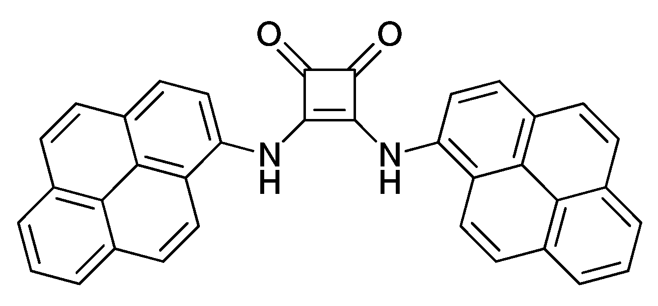
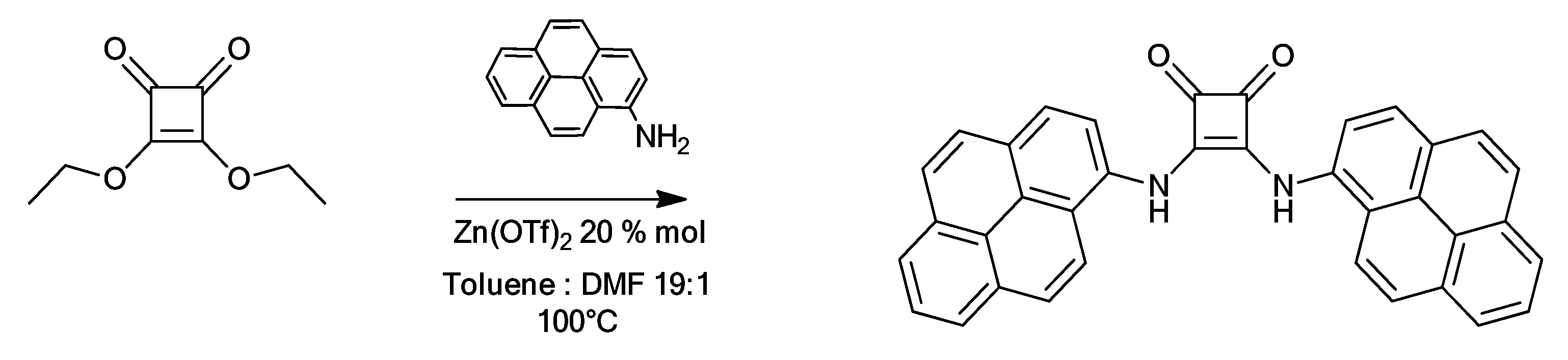
2.1. Synthesis
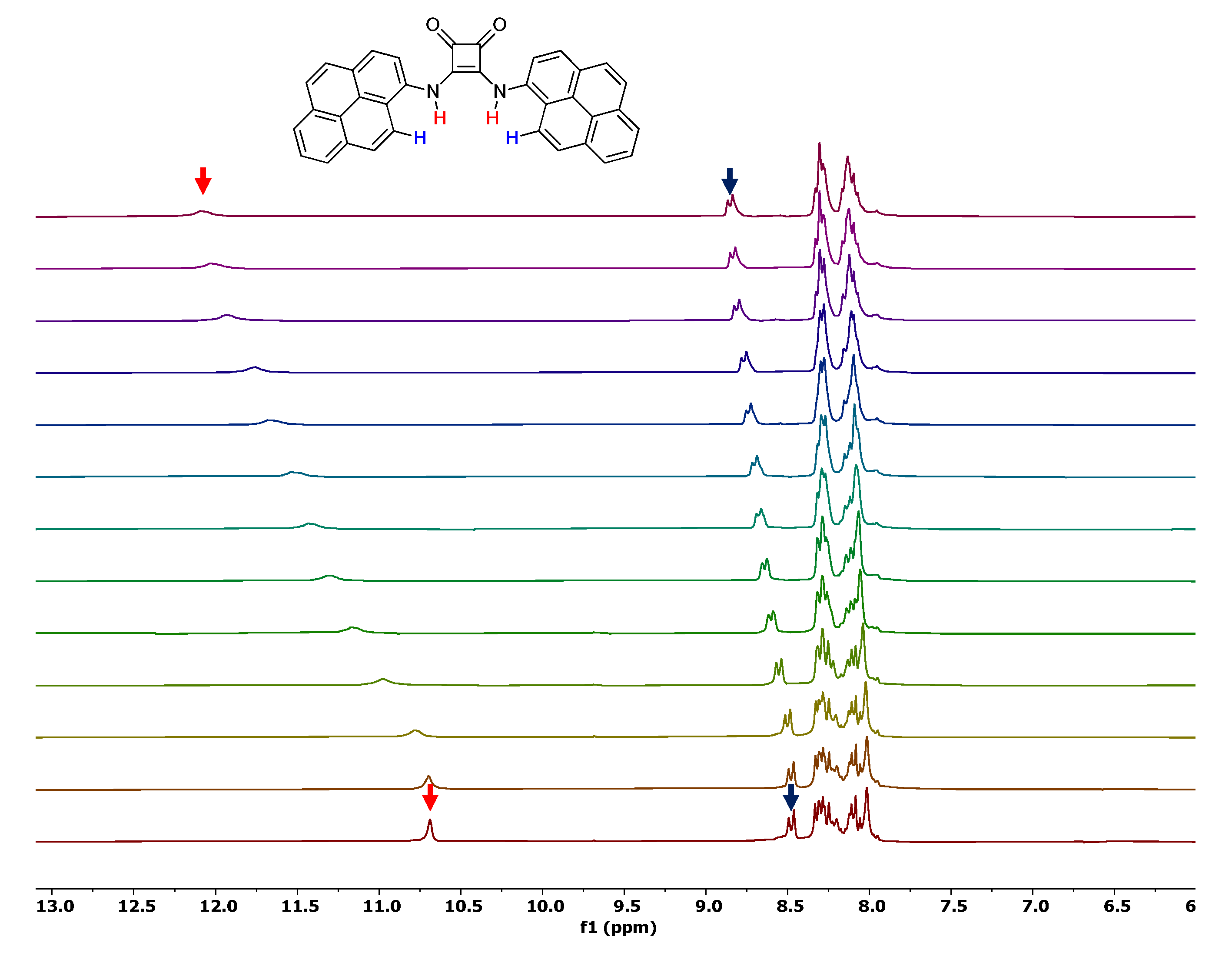
2.2. Anion Binding Studies
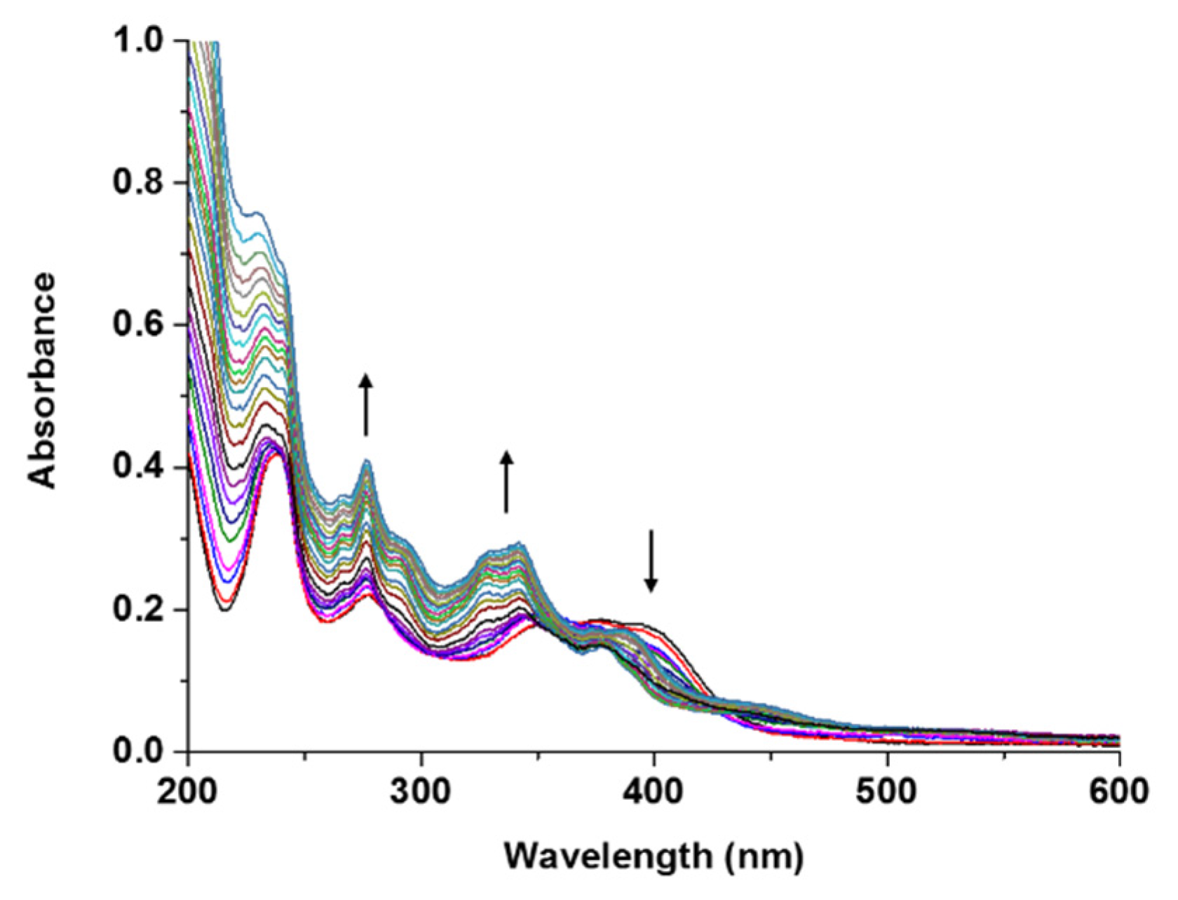
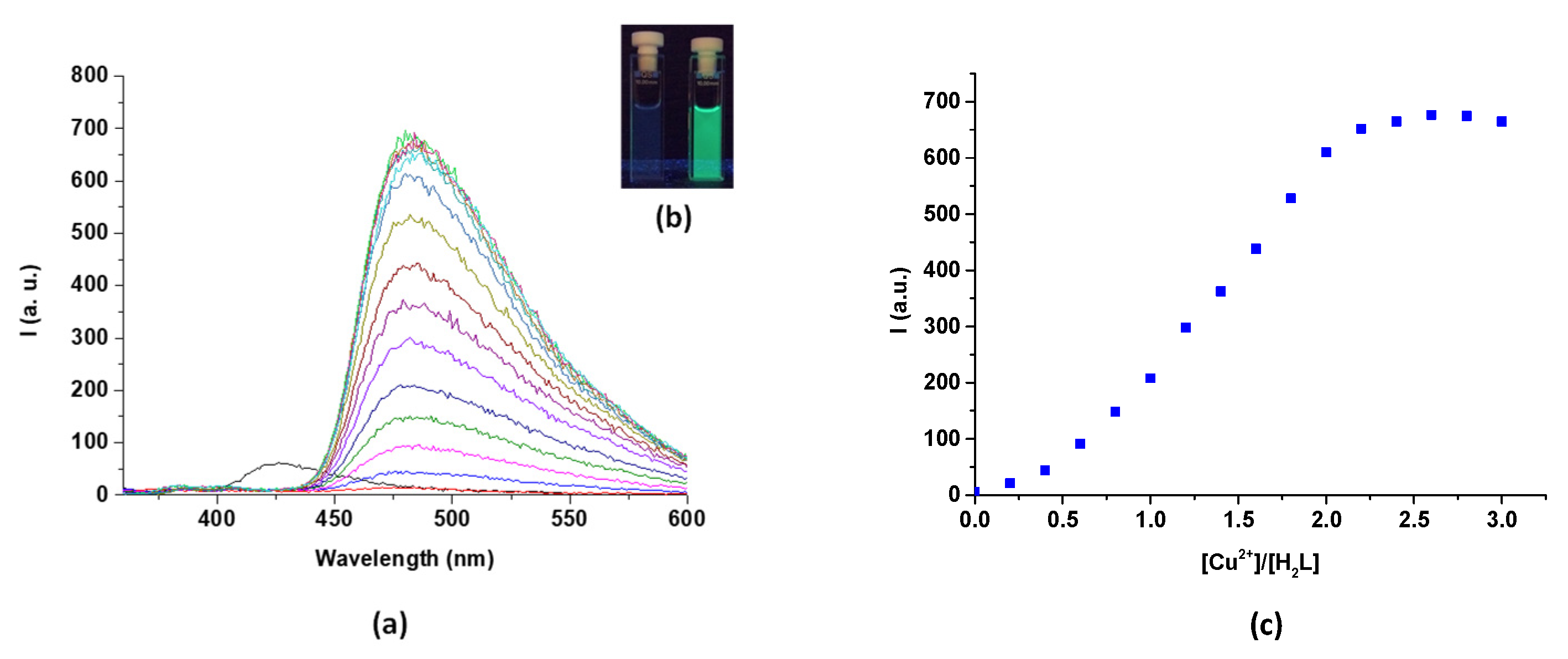
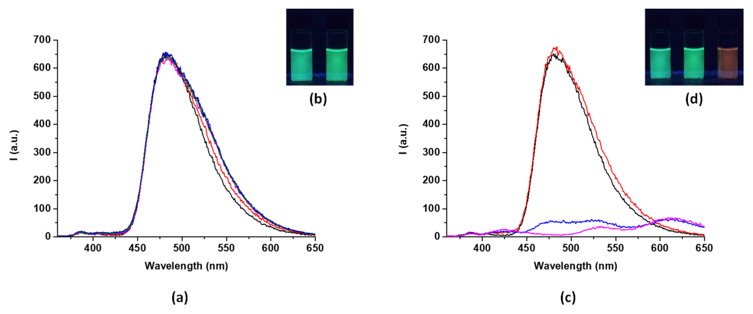
2.3. Metal Ion Sensing by H2L: Spectrophotometric Measurements
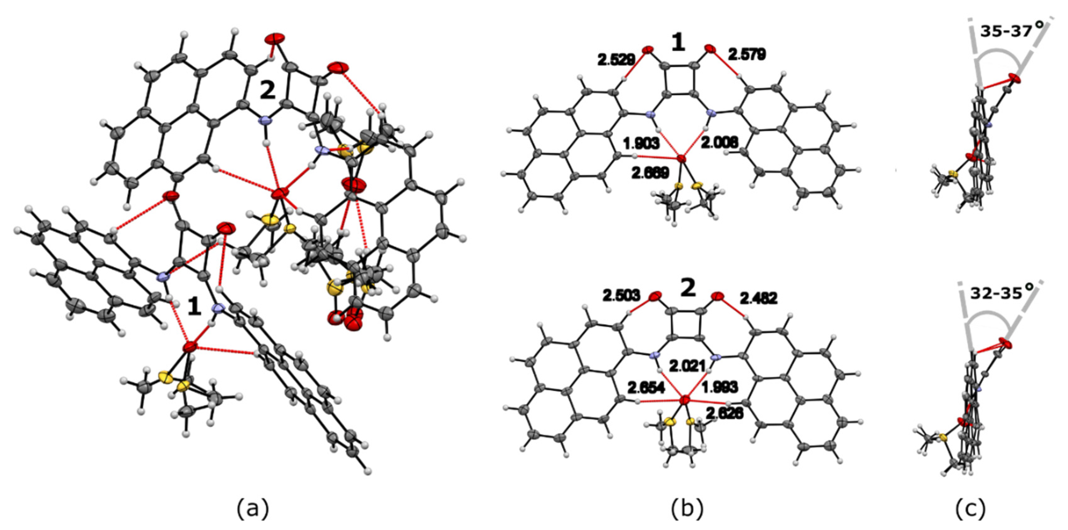
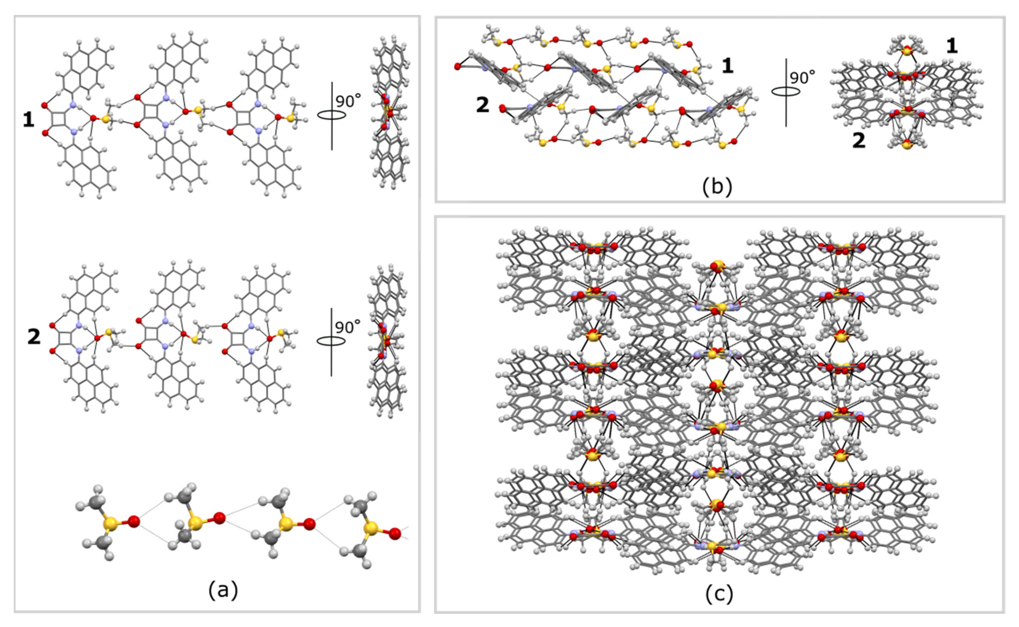
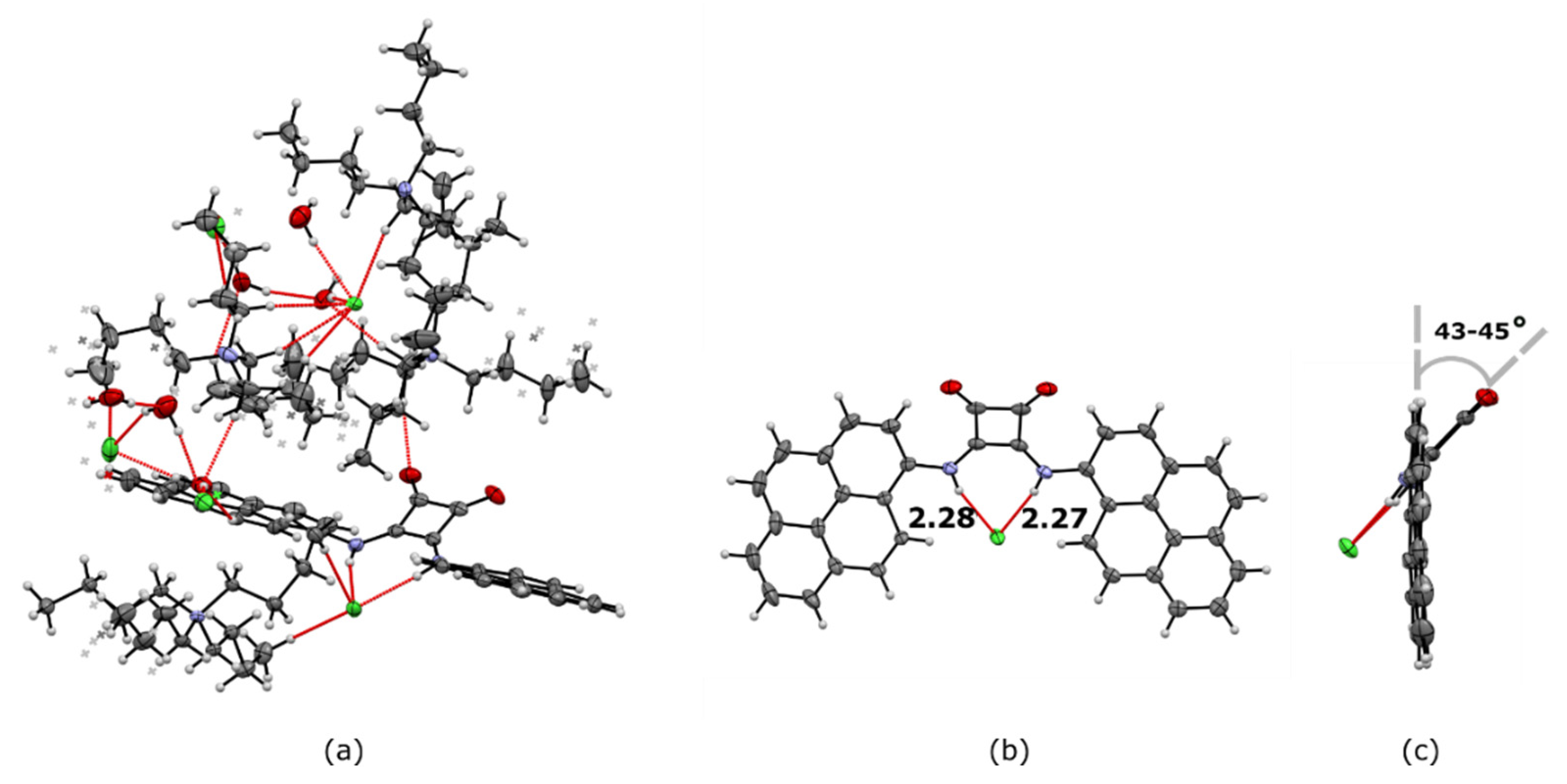
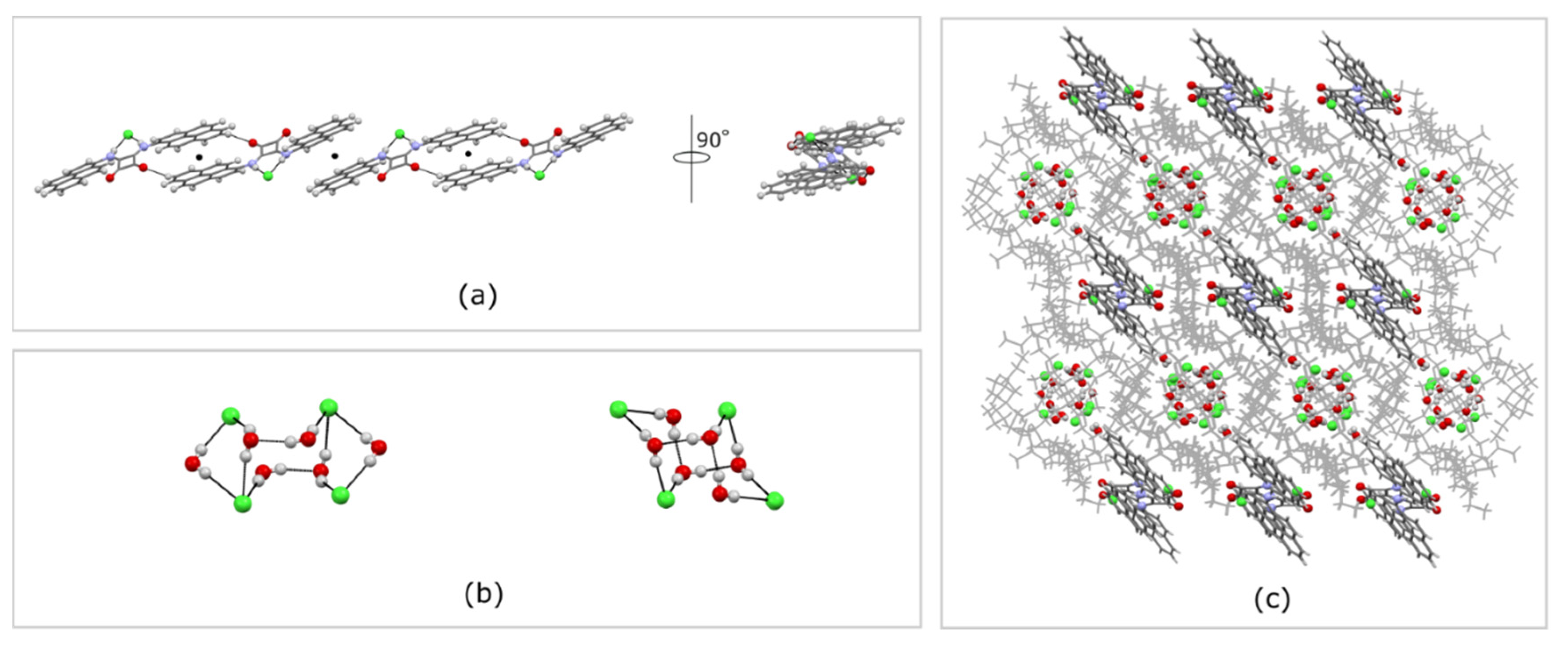
2.4. Solid State Studies
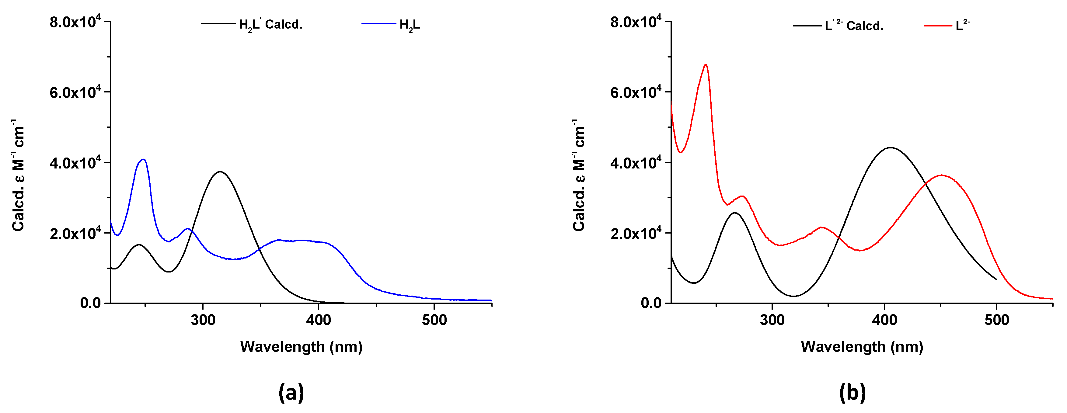
2.5. DFT Studies
3. Materials and Methods
3.1. 1H-NMR Titrations
3.2. Crystallization Methods
X-ray Data Collection
3.3. Theoretical Calculations
3.4. Synthesis of H2L
4. Conclusions
Supplementary Materials
Author Contributions
Funding
Institutional Review Board Statement
Informed Consent Statement
Data Availability Statement
Acknowledgments
Conflicts of Interest
Sample Availability
References
- Busschaert, N.; Park, S.H.; Baek, K.H.; Choi, Y.P.; Park, J.; Howe, E.N.; Hiscock, J.R.; Karagiannidis, L.E.; Marques, I.; Félix, V.; et al. A synthetic ion transporter that disrupts autophagy and induces apoptosis by perturbing cellular chloride concentrations. Nat. Chem. 2017, 9, 667–675. [Google Scholar] [CrossRef] [PubMed]
- Haynes, C.J.E.; Berry, S.N.; Garric, J.; Herniman, J.; Hiscock, J.R.; Kirby, I.L.; Light, M.E.; Perkes, G.; Gale, P.A. Small neutral molecular carriers for selective carboxylate transport. Chem. Commun. 2013, 49, 246–248. [Google Scholar] [CrossRef] [PubMed]
- Marchetti, L.A.; Kumawat, L.K.; Mao, N.; Stephens, J.C.; Elmes, R.B. The Versatility of Squaramides: From Supramolecular Chemistry to Chemical Biology. Chemistry 2019, 5, 1398–1485. [Google Scholar] [CrossRef]
- Picci, G.; Kubicki, M.; Garau, A.; Lippolis, V.; Mocci, R.; Porcheddu, A.; Quesada, R.; Ricci, P.C.; Scorciapino, M.A.; Caltagirone, C. Simple squaramide receptors for highly efficient anion binding in aqueous media and transmembrane transport. Chem. Commun. 2020, 56, 11066–11069. [Google Scholar] [CrossRef] [PubMed]
- Lachowicz, J.I.; Picci, G.; Coni, P.; Lippolis, V.; Mamusa, M.; Murgia, S.; Pichiri, G.; Caltagirone, C. Fluorescent squaramide ligands for cellular imaging and their encapsulation in cubosomes. New J. Chem. 2019, 43, 10336–10342. [Google Scholar] [CrossRef]
- Bao, X.; Wu, X.; Berry, S.N.; Howe, E.N.; Chang, Y.T.; Gale, P.A. Fluorescent squaramides as anion receptors and transmembrane anion transporters. Chem. Commun. 2018, 54, 1363–1366. [Google Scholar] [CrossRef]
- Ramos, J.; Arufe, S.; Martin, H.; Rooney, D.; Elmes, R.B.; Erxleben, A.; Moreira, R.; Velasco-Torrijos, T. Glycosyl squaramides, a new class of supramolecular gelators. Soft Matter 2020, 16, 7916–7926. [Google Scholar] [CrossRef]
- Manesiotis, P.; Riley, A.; Bollen, B. Polymerisable squaramide receptors for anion binding and sensing. J. Mater. Chem. C 2014, 2, 8990–8995. [Google Scholar] [CrossRef]
- Elmes, R.B.; KYYuen, K.; Jolliffe, K.A. Sulfate-Selective Recognition by Using Neutral Dipeptide Anion Receptors in Aqueous Solution. Chem. Eur. J. 2014, 20, 7373–7380. [Google Scholar] [CrossRef]
- Zdanowski, S.; Piątek, P.; Romański, J. An ion pair receptor facilitating the extraction of chloride salt from the aqueous to the organic phase. New J. Chem. 2016, 40, 7190–7196. [Google Scholar] [CrossRef]
- Frontera, A.; Orell, M.; Garau, C.; Quiñonero, D.; Molins, E.; Mata, I.; Morey, J. Preparation, Solid-State Characterization, and Computational Study of a Crown Ether Attached to a Squaramide. Org. Lett. 2005, 7, 1437–1440. [Google Scholar] [CrossRef] [PubMed]
- Mäkelä, T.; Kalenius, E.; Rissanen, K. Cooperatively Enhanced Ion Pair Binding with a Hybrid Receptor. Inorg. Chem. 2015, 54, 9154–9165. [Google Scholar] [CrossRef] [PubMed]
- Ziach, K.; Karbarz, M.; Romański, J. Cooperative binding and extraction of sodium nitrite by a ditopic receptor incorporated into a polymeric resin. Dalton Trans. 2016, 45, 11639–11643. [Google Scholar] [CrossRef]
- Jagleniec, D.; Dobrzycki, Ł.; Karbarz, M.; Romański, J. Ion-pair induced supramolecular assembly formation for selective extraction and sensing of potassium sulfate. Chem. Sci. 2019, 10, 9542–9547. [Google Scholar] [CrossRef] [PubMed]
- Prohens, R.; Martorell, G.; Ballester, P.; Costa, A. A squaramide fluorescent ensemble for monitoring sulfate in water. Chem. Commun. 2001, 1456–1457. [Google Scholar] [CrossRef]
- Danao, A.; Ramalingam, V.; Ramamurthy, V.; Muthyala, R.S. On the origin of chloride-induced emission enhancement in ortho substituted squaramides. J. Photochem. Photobiol. A Chem. 2017, 344, 108–113. [Google Scholar] [CrossRef]
- Porel, M.; Ramalingam, V.; Domaradzki, M.E.; Young, V.G.; Ramamurthy, V.; Muthyala, R.S. Chloride sensing via suppression of excited state intramolecular proton transfer in squaramides. Chem. Commun. 2013, 49, 1633–1635. [Google Scholar] [CrossRef]
- Elmes, R.B.; Turner, P.; Jolliffe, K.A. Colorimetric and Luminescent Sensors for Chloride: Hydrogen Bonding vs. Deprotonation. Org. Lett. 2013, 15, 5638–5641. [Google Scholar] [CrossRef]
- Rostami, A.; Colin, A.; Li, X.Y.; Chudzinski, M.G.; Lough, A.J.; Taylor, M.S. N,N′-Diarylsquaramides: General, High-Yielding Synthesis and Applications in Colorimetric Anion Sensing. J. Org. Chem. 2010, 75, 3983–3992. [Google Scholar] [CrossRef]
- Hibbert, D.B.; Thordarson, P. The death of the Job plot, transparency, open science and online tools, uncertainty estimation methods and other developments in supramolecular chemistry data analysis. Chem. Commun. 2016, 52, 12792–12805. [Google Scholar] [CrossRef]
- Thordarson, P. Determining association constants from titration experiments in supramolecular chemistry. Chem. Soc. Rev. 2011, 40, 1305–1323. [Google Scholar] [CrossRef]
- Duke, R.M.; McCabe, T.; Schmitt, W.; Gunnlaugsson, T. Recognition and Sensing of Biologically Relevant Anions in Alcohol and Mixed Alcohol–Aqueous Solutions Using Charge Neutral Cleft-Like Glycol-Derived Pyridyl–Amidothiourea Receptors. J. Org. Chem. 2012, 77, 3115–3126. [Google Scholar] [CrossRef] [PubMed]
- Rotger, M.C.; Piña, M.N.; Frontera, A.; Martorell, G.; Ballester, P.; Deyà, P.M.; Costa, A. Conformational Preferences and Self-Template Macrocyclization of Squaramide-Based Foldable Modules. J. Org. Chem. 2004, 69, 2302–2308. [Google Scholar] [CrossRef] [PubMed]
- Olivari, M.; Montis, R.; Karagiannidis, L.E.; Horton, P.N.; Mapp, L.K.; Coles, S.J.; Light, M.E.; Gale, P.A.; Caltagirone, C. Anion complexation, transport and structural studies of a series of bis-methylurea compounds. Dalton Trans. 2015, 44, 2138–2149. [Google Scholar] [CrossRef] [PubMed]
- Lin, R.B.; He, Y.; Li, P.; Wang, H.; Zhou, W.; Chen, B. Multifunctional porous hydrogen-bonded organic framework materials. Chem. Soc. Rev. 2019, 48, 1362–1389. [Google Scholar] [CrossRef] [PubMed]
- McGuirk, C.M.; Katz, M.J.; Stern, C.L.; Sarjeant, A.A.; Hupp, J.T.; Farha, O.K.; Mirkin, C.A. Turning On Catalysis: Incorporation of a Hydrogen-Bond-Donating Squaramide Moiety into a Zr Metal–Organic Framework. J. Am. Chem. Soc. 2015, 137, 919–925. [Google Scholar] [CrossRef]
- Busschaert, N.; Kirby, I.L.; Young, S.; Coles, S.J.; Horton, P.N.; Light, M.E.; Gale, P.A. Squaramides as Potent Transmembrane Anion Transporters. Angew. Chem. Int. Ed. 2012, 51, 4426–4430. [Google Scholar] [CrossRef] [PubMed]
- Secci, F.; Arca, M.; Frongia, A.; Piras, P.P. New aminotetrazole derivatives as hydrogen bonding catalysts. A green and selective oxidation of organosulphides with H2O2 in H2O. New J. Chem. 2014, 38, 3622–3629. [Google Scholar] [CrossRef]
- Aragoni, M.C.; Caltagirone, C.; Lippolis, V.; Podda, E.; Slawin, A.M.; Woollins, J.D.; Pintus, A.; Arca, M. Diradical Character of Neutral Heteroleptic Bis(1,2-dithiolene) Metal Complexes: Case Study of [Pd(Me2timdt)(mnt)] (Me2timdt = 1,3-Dimethyl-2,4,5-trithioxoimidazolidine; mnt2– = 1,2-Dicyano-1,2-ethylenedithiolate). Inorg. Chem. 2020, 59, 17385–17401. [Google Scholar] [CrossRef]
- Nascimento, V.; Cordeiro, P.S.; Arca, M.; Marini, F.; Sancineto, L.; Braga, A.L.; Lippolis, V.; Iwaoka, M.; Santi, C. Fast and easy conversion of ortho amidoaryldiselenides into the corresponding ebselen-like derivatives driven by theoretical investigations. New J. Chem. 2020, 44, 9444–9451. [Google Scholar] [CrossRef]
- Cinellu, M.A.; Maiore, L.; Manassero, M.; Casini, A.; Arca, M.; Fiebig, H.H.; Kelter, G.; Michelucci, E.; Pieraccini, G.; Gabbiani, C.; et al. [Au2(phen2Me)2(μ-O)2](PF6)2, a Novel Dinuclear Gold(III) Complex Showing Excellent Antiproliferative Properties. ACS Med. Chem. Lett. 2010, 1, 336–339. [Google Scholar] [CrossRef]
- Alashkar, N.; Arca, M.; Alnasr, H.; Lutter, M.; Lippolis, V.; Jurkschat, K. Water-Soluble Organotin Compounds—Syntheses, Structures and Reactivity towards Fluoride Anions in Water. Eur. J. Inorg. Chem. 2020, 2020, 3925–3936. [Google Scholar] [CrossRef]
- Aragoni, M.C.; Arca, M.; Bencini, A.; Caltagirone, C.; Garau, A.; Isaia, F.; Light, M.E.; Lippolis, V.; Lodeiro, C.; Mameli, M.; et al. Zn2+/Cd2+ optical discrimination by fluorescent chemosensors based on 8-hydroxyquinoline derivatives and sulfur-containing macrocyclic units. Dalton Trans. 2013, 42, 14516–14530. [Google Scholar] [CrossRef] [PubMed]
- Silva, C.E.; Dos Santos, H.F.; Speziali, N.L.; Diniz, R.; de Oliveira, L.F.C. Role of the Substituent Effect over the Squarate Oxocarbonic Ring: Spectroscopy, Crystal Structure, and Density Functional Theory Calculations of 1,2-Dianilinosquairane. J. Phys. Chem. A 2010, 114, 10097–10109. [Google Scholar] [CrossRef] [PubMed]
- Reed, A.E.; Curtiss, L.A.; Weinhold, F. Intermolecular interactions from a natural bond orbital, donor-acceptor viewpoint. Chem. Rev. 1988, 88, 899–926. [Google Scholar] [CrossRef]
- Weinhold, F.; Landis, C.R.; Glendening, E.D. What is NBO analysis and how is it useful? Int. Rev. Phys. Chem. 2016, 35, 399–440. [Google Scholar] [CrossRef]
- Sheldrick, G. A short history of SHELX. Acta Crystallogr. Sect. A 2008, 64, 112–122. [Google Scholar] [CrossRef]
- Dolomanov, O.V.; Bourhis, L.J.; Gildea, R.J.; Howard, J.A.; Puschmann, H. OLEX2: A complete structure solution, refinement and analysis program. J. Appl. Crystallogr. 2009, 42, 339–341. [Google Scholar] [CrossRef]
- Sheldrick, G. Crystal structure refinement with SHELXL. Acta Crystallogr. Sect. C 2015, 71, 3–8. [Google Scholar] [CrossRef]
- Sheldrick, G. SHELXT—Integrated space-group and crystal-structure determination. Acta Crystallogr. Sect. A 2015, 71, 3–8. [Google Scholar] [CrossRef]
- Frisch, M.J.; Trucks, G.W.; Schlegel, H.B.; Scuseria, G.E.; Robb, M.A.; Cheeseman, J.R.; Scalmani, G.; Barone, V.; Petersson, G.A.; Nakatsuji, H.; et al. Gaussian 16 Rev. C.01; Gaussian Inc.: Wallingford, CT, USA, 2016. [Google Scholar]
- Koch, W.; Holthausen, M.C. A Chemist’s Guide to Density Functional Theory, 2nd ed.; Wiley: Hoboken, NJ, USA, 2001. [Google Scholar]
- Adamo, C.; Barone, V. Exchange functionals with improved long-range behavior and adiabatic connection methods without adjustable parameters: The mPW and mPW1PW models. J. Chem. Phys. 1998, 108, 664–675. [Google Scholar] [CrossRef]
- Weigend, F.; Ahlrichs, R. Balanced basis sets of split valence, triple zeta valence and quadruple zeta valence quality for H to Rn: Design and assessment of accuracy. Phys. Chem. Chem. Phys. 2005, 7, 3297–3305. [Google Scholar] [CrossRef] [PubMed]
- Weigend, F. Accurate Coulomb-fitting basis sets for H to Rn. Phys. Chem. Chem. Phys. 2006, 8, 1057–1065. [Google Scholar] [CrossRef] [PubMed]
- Tomasi, J.; Mennucci, B.; Cammi, R. Quantum Mechanical Continuum Solvation Models. Chem. Rev. 2005, 105, 2999–3094. [Google Scholar] [CrossRef]
- Schaftenaar, G.; Noordik, J.H. Molden: A pre- and post-processing program for molecular and electronic structures. J. Comput. Aid. Mol. Des. 2000, 14, 123–134. [Google Scholar] [CrossRef] [PubMed]


| F− | CN− | BzO− | Cl− | Br− | I− | |
|---|---|---|---|---|---|---|
| H2L | deprot | deprot | deprot | 98 ± 6 | <10 | <10 |
Publisher’s Note: MDPI stays neutral with regard to jurisdictional claims in published maps and institutional affiliations. |
© 2021 by the authors. Licensee MDPI, Basel, Switzerland. This article is an open access article distributed under the terms and conditions of the Creative Commons Attribution (CC BY) license (http://creativecommons.org/licenses/by/4.0/).
Share and Cite
Picci, G.; Milia, J.; Aragoni, M.C.; Arca, M.; Coles, S.J.; Garau, A.; Lippolis, V.; Montis, R.; Orton, J.B.; Caltagirone, C. Switching-On Fluorescence by Copper (II) and Basic Anions: A Case Study with a Pyrene-Functionalized Squaramide. Molecules 2021, 26, 1301. https://doi.org/10.3390/molecules26051301
Picci G, Milia J, Aragoni MC, Arca M, Coles SJ, Garau A, Lippolis V, Montis R, Orton JB, Caltagirone C. Switching-On Fluorescence by Copper (II) and Basic Anions: A Case Study with a Pyrene-Functionalized Squaramide. Molecules. 2021; 26(5):1301. https://doi.org/10.3390/molecules26051301
Chicago/Turabian StylePicci, Giacomo, Jessica Milia, Maria Carla Aragoni, Massimiliano Arca, Simon J. Coles, Alessandra Garau, Vito Lippolis, Riccardo Montis, James B. Orton, and Claudia Caltagirone. 2021. "Switching-On Fluorescence by Copper (II) and Basic Anions: A Case Study with a Pyrene-Functionalized Squaramide" Molecules 26, no. 5: 1301. https://doi.org/10.3390/molecules26051301
APA StylePicci, G., Milia, J., Aragoni, M. C., Arca, M., Coles, S. J., Garau, A., Lippolis, V., Montis, R., Orton, J. B., & Caltagirone, C. (2021). Switching-On Fluorescence by Copper (II) and Basic Anions: A Case Study with a Pyrene-Functionalized Squaramide. Molecules, 26(5), 1301. https://doi.org/10.3390/molecules26051301