
Key Factor Study for Generic Long-Acting PLGA Microspheres Based on a Reverse Engineering of Vivitrol®


Abstract
1. Introduction
2. Results
2.1. Drug Loading
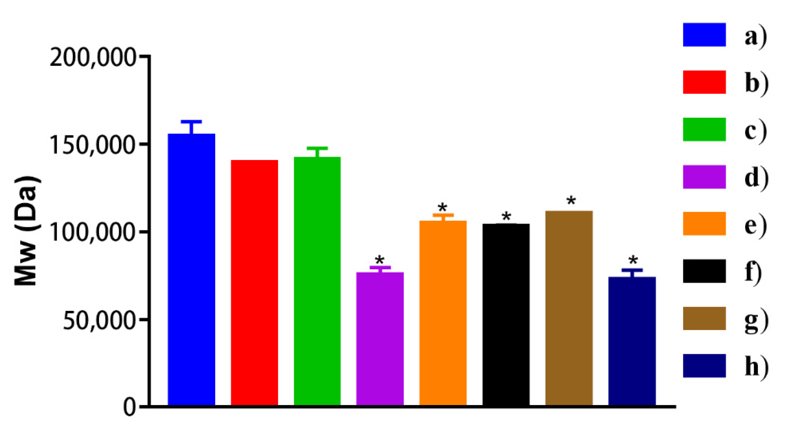
2.2. Molecular Weight of PLGA in Vivitrol®
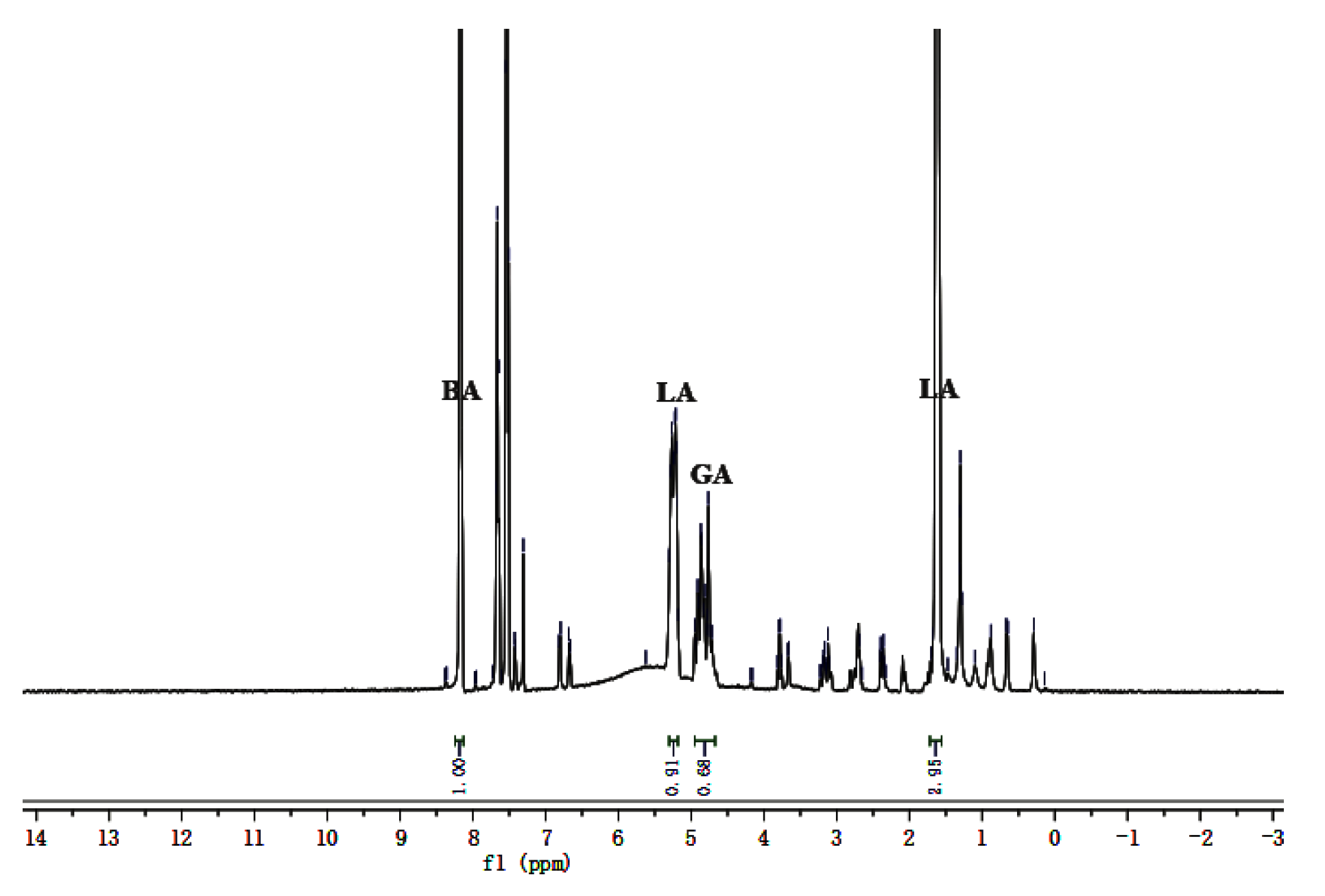
2.3. L/G Ratio and Content of PLGA in Vivitrol®
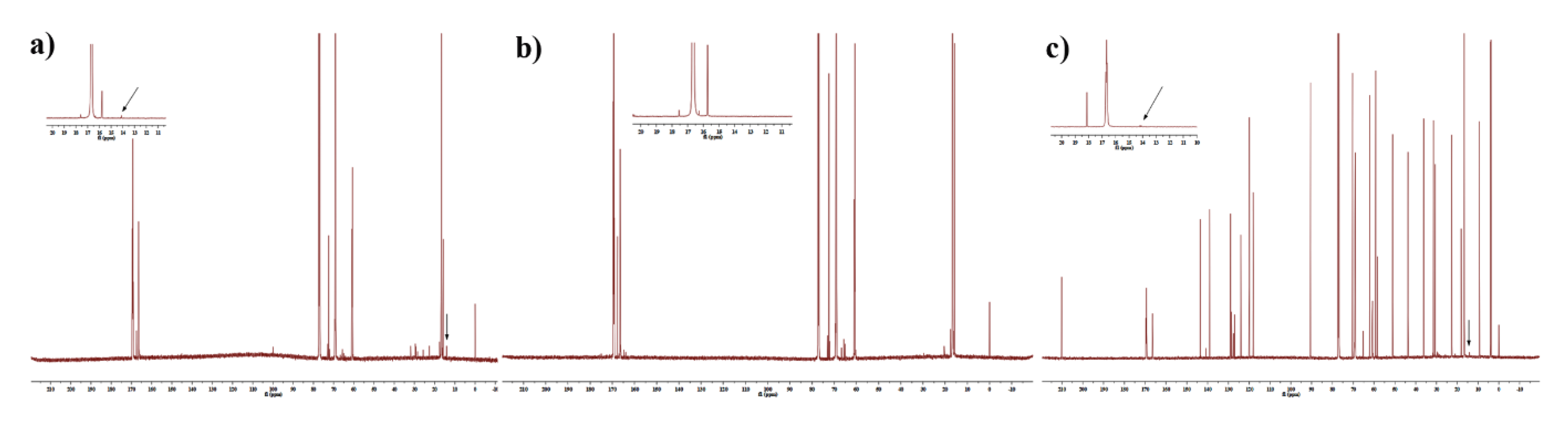
2.4. End-Group of PLGA in Vivitrol®
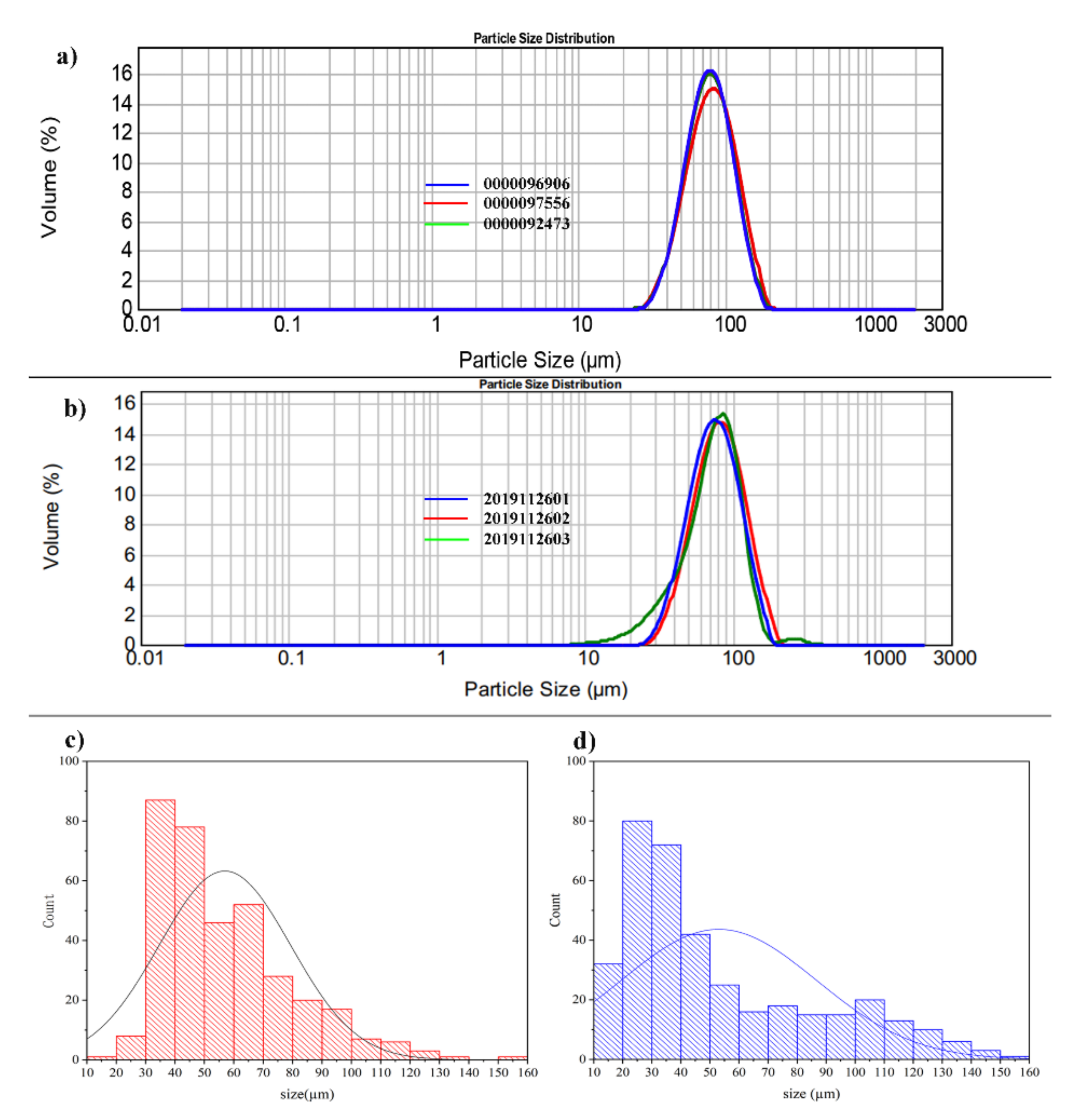
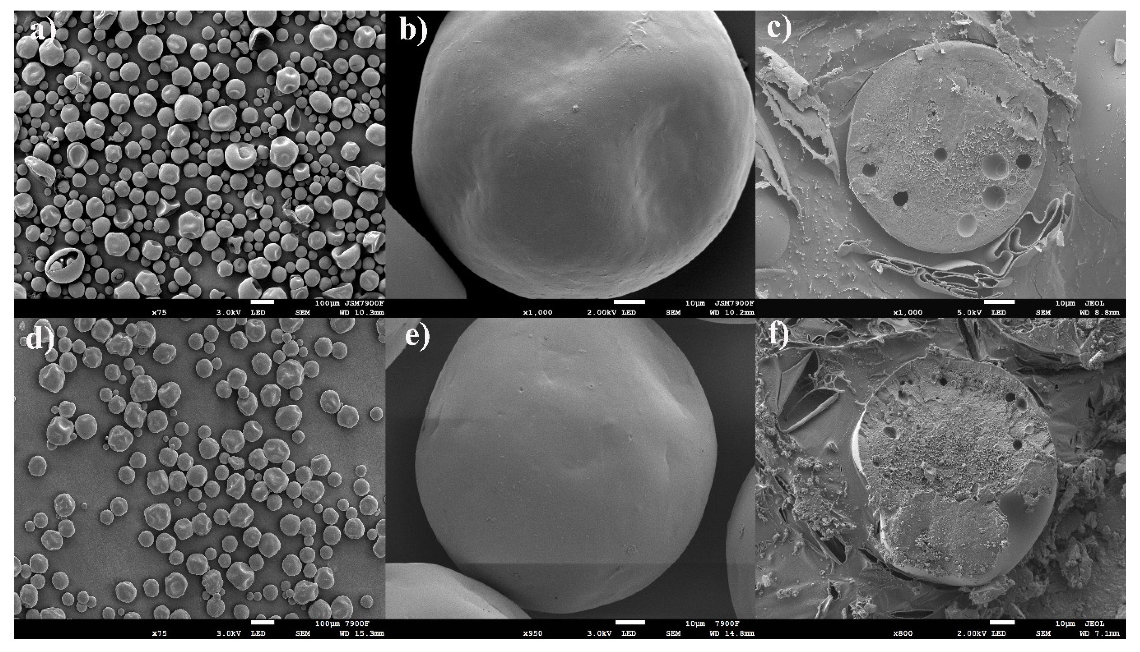
2.5. Particle Size and Distribution
2.6. Molecular Weight of PLGA in Organic Phase With Different Mixing Processes
2.7. Morphology
2.8. Tg and Water Contact Angle
2.9. Residual EA
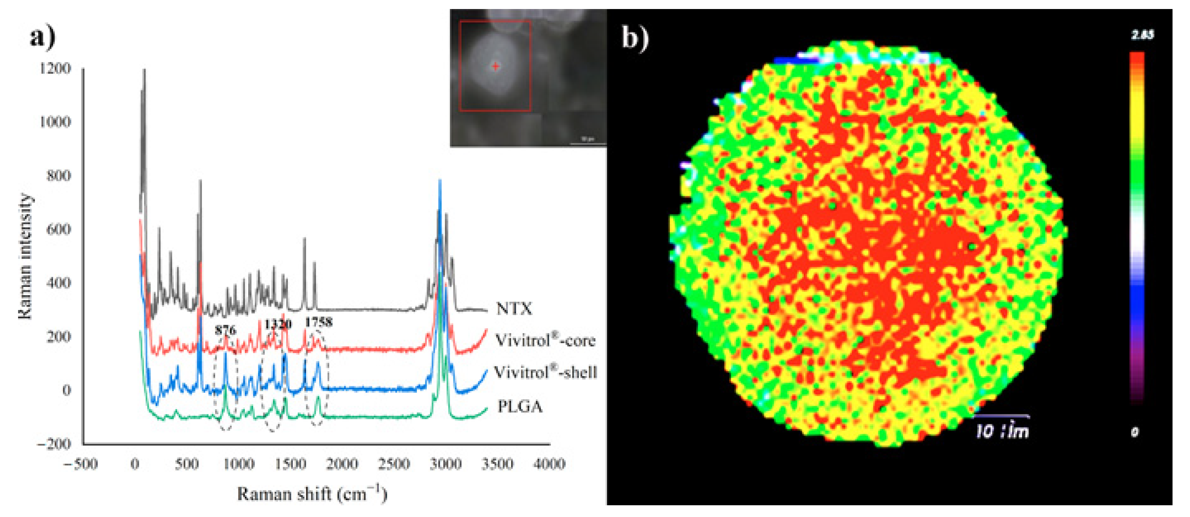
2.10. Raman Spectra
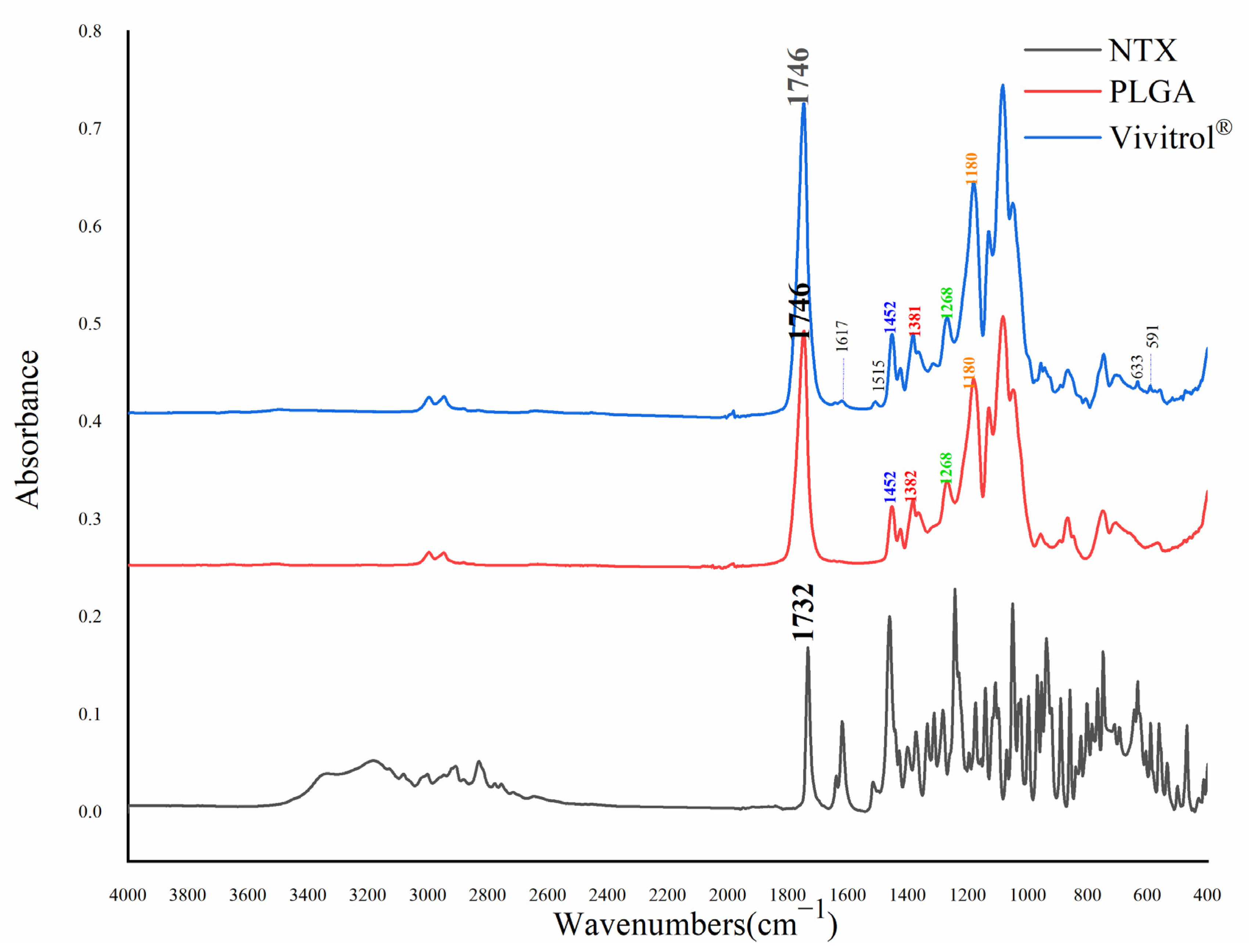
2.11. Fourier Transform Infrared Spectra
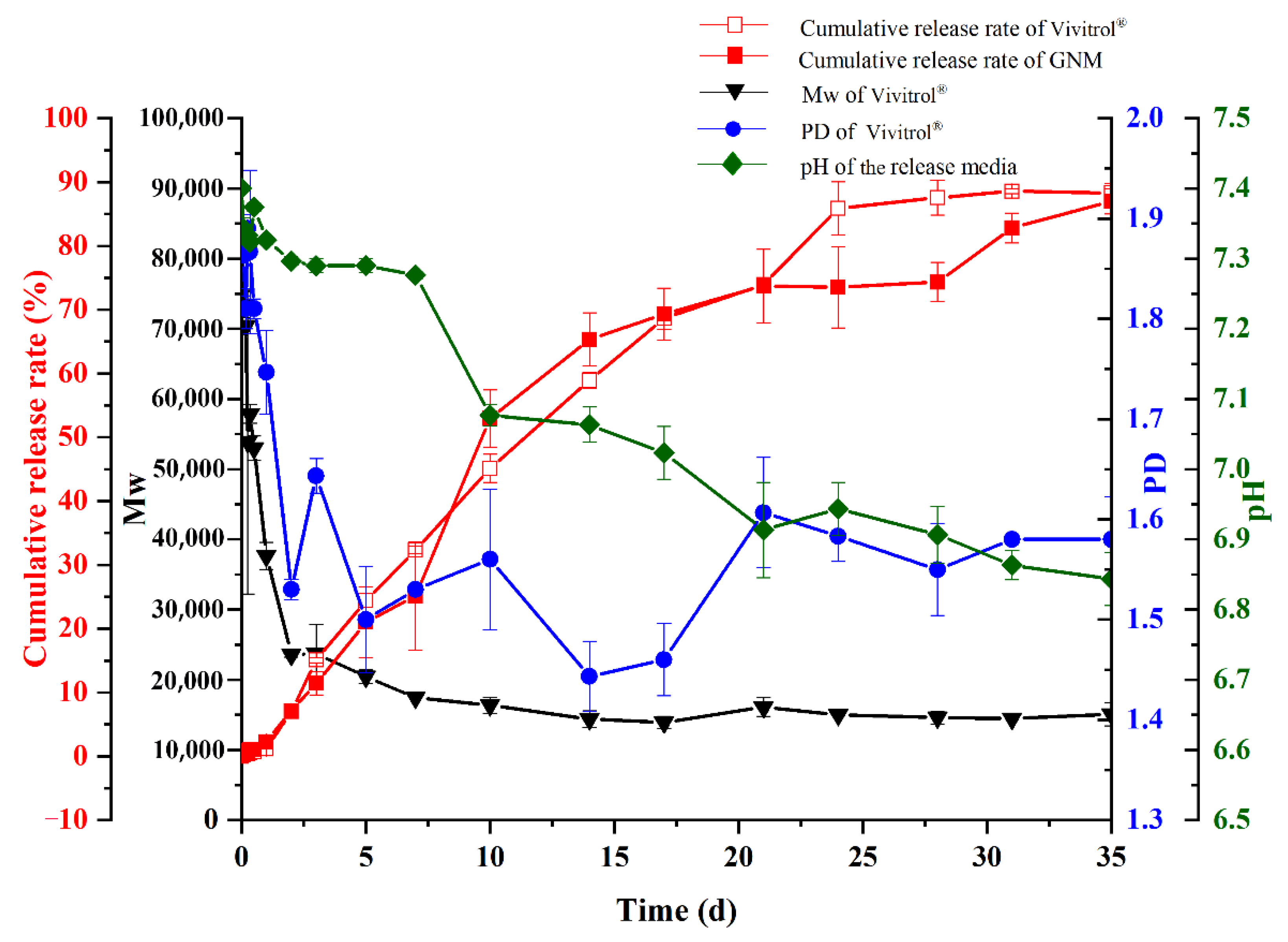
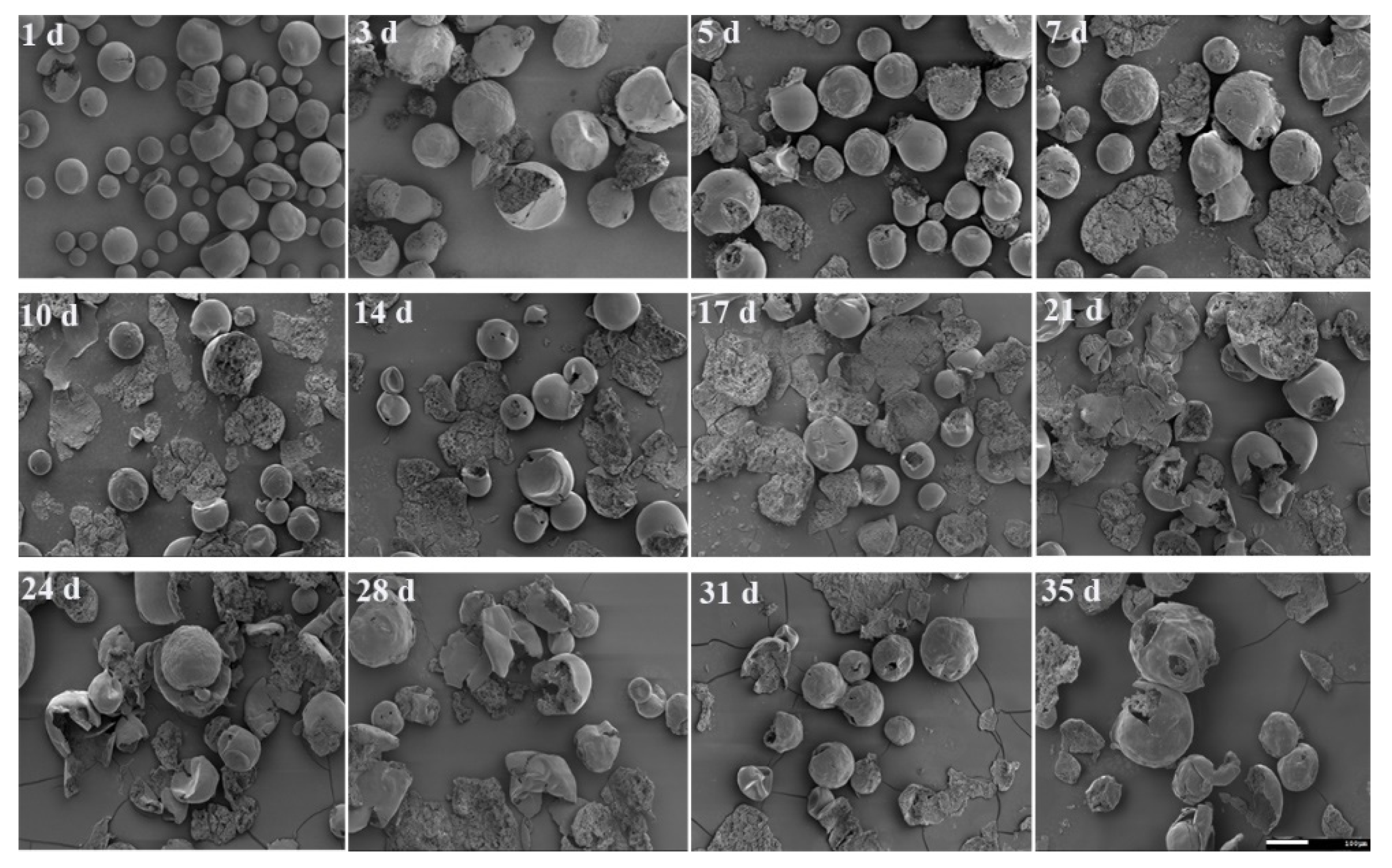
2.12. In Vitro Release and Degradation Studies
3. Discussion
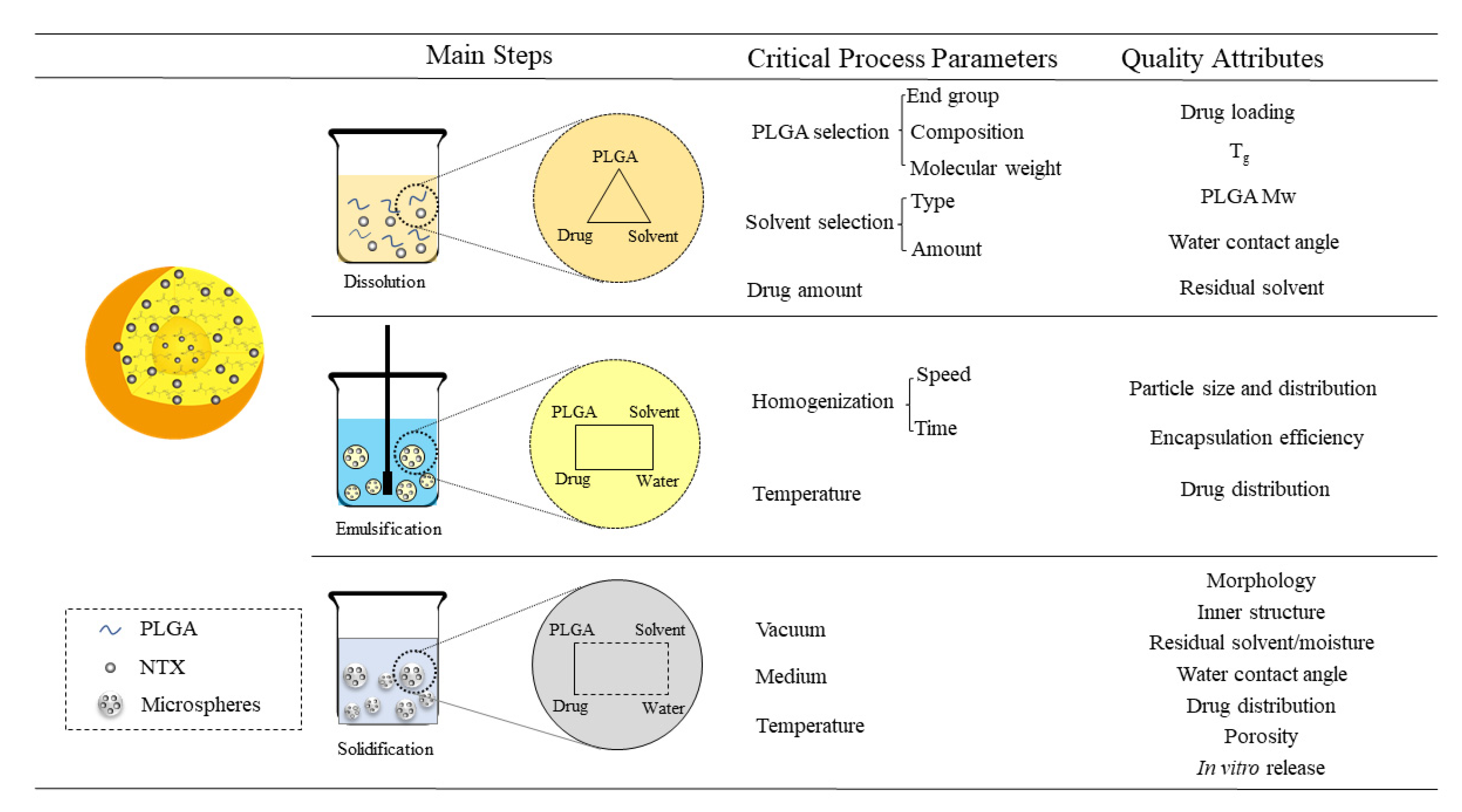
4. Materials and Methods
4.1. Materials
4.2. Characterization of Q1/Q2 PLGA Microspheres
4.2.1. High Performance Liquid Chromatography (HPLC)
4.2.2. Drug Loading
4.2.3. The Molecular Weight of PLGA
4.2.4. Analysis of Content and Lactide/Glycolide (L/G) Ratio of PLGA in Vivitrol®
4.2.5. End-Group Analysis of PLGA in Vivitrol®
4.3. Physicochemical Characterization for Critical Process Parameters
4.3.1. Particle Size and Distribution
4.3.2. Morphology and Internal Structure
4.3.3. Glass Transition Temperature
4.3.4. Residual Solvent
4.3.5. Porosity
4.3.6. Water Contact Angle
4.3.7. Residual Moisture
4.3.8. Raman Spectroscopy
4.3.9. Fourier Transform Infrared Spectra
4.4. Preparation of Naltrexone Loaded PLGA Microspheres
4.5. In Vitro Release and Degradation Studies
4.6. Statistical Analysis
5. Conclusions
Supplementary Materials
Author Contributions
Funding
Institutional Review Board Statement
Informed Consent Statement
Data Availability Statement
Conflicts of Interest
Abbreviations
References
- Park, K.; Skidmore, S.; Hadar, J.; Garner, J.; Park, H.; Otte, A.; Soh, B.K.; Yoon, G.; Yu, D.; Yun, Y.; et al. Injectable, long-acting PLGA formulations: Analyzing PLGA and understanding microparticle formation. J. Controll Release 2019, 304, 125–134. [Google Scholar] [CrossRef]
- Anjali, J.; Konda, R.; Kunduru, A.; Basu, B.; Mizrahi, W.R. Injectable formulations of poly(lactic acid) and its copolymers in clinical use. Adv. Drug Deliver. Rev. 2016, 107, 213–227. [Google Scholar]
- Ramazani, F.; Chen, W.; Van Nostrum, C.F.; Storm, G.; Kiessling, F.; Kok, R.J. Strategies for encapsulation of small hydrophilic and amphiphilic drugs in PLGA microspheres: State-of-the-art and challenges. Int. J. Pharm. 2016, 499, 358–367. [Google Scholar] [CrossRef] [PubMed]
- FDA. Generic Drug Facts. Available online: https://www.fda.gov/drugs/generic-drugs/generic-drug-facts. (accessed on 30 June 2020).
- Zhou, J.; Hirota, K.; Ackermann, R.; Walker, J.; Wang, Y.; Choi, S.; Schwendeman, S.P. Reverse Engineering the 1-Month Lupron Depot®. A.A.P.S. J. 2018, 20, 105. [Google Scholar] [CrossRef] [PubMed]
- Kapoor, D.N.; Bhatia, A.; Kaur, R.; Sharma, R.; Kaur, G.; Dhawan, S.J.T.D. PLGA: A unique polymer for drug delivery. Ther. Deliv. 2015, 6, 41–58. [Google Scholar] [CrossRef] [PubMed]
- Makadia, H.K.; Siegel, S.J. Poly Lactic-co-Glycolic Acid (PLGA) as Biodegradable Controlled Drug Delivery Carrier. Polymers (Basel). 2011, 3, 1377–1397. [Google Scholar] [CrossRef]
- Garnera, J.; Skidmorea, S.; Parka, H.; Parka, K.; Choib, S.; Wang, Y. A protocol for assay of poly(lactide-co-glycolide) in clinical products. Int. J. Pharm. 2015, 495, 87–92. [Google Scholar] [CrossRef] [PubMed]
- Hadar, J.; Skidmore, S.; Garner, J.; Park, H.; Kozak, D.J. Method matters: Development of characterization techniques for branched and glucose-poly(lactide-co-glycolide) polymers. J. Control Release 2020, 320, 484–494. [Google Scholar] [CrossRef]
- Garner, J.; Skidmore, S.; Park, H.; Park, K.; Choi, S.; Wang, Y. Beyond Q1/Q2: The Impact of Manufacturing Conditions and Test Methods on Drug Release From PLGA-Based Microparticle Depot Formulations. J. Pharm. Sci. 2018, 107, 353–361. [Google Scholar] [CrossRef]
- Kumar, R.; Palmieri, M.J., Jr. Points to consider when establishing drug product specifications for parenteral microspheres. AAPS J. 2010, 12, 27–32. [Google Scholar] [CrossRef] [PubMed]
- Andhariya, J.V.; Shen, J.; Wang, Y.; Choi, S.; Burgess, D.J. Effect of minor manufacturing changes on stability of compositionally equivalent PLGA microspheres. Int. J. Pharm. 2019, 566, 532–540. [Google Scholar] [CrossRef]
- Andhariya, J.V.; Shen, J.; Choi, S.; Wang, Y.; Zou, Y.; Burgess, D.J. Development of in vitro-in vivo correlation of parenteral naltrexone loaded polymeric microspheres. J. Control Release 2017, 255, 27–35. [Google Scholar] [CrossRef]
- Sharifi, F.; Otte, A.; Yoon, G.; Park, K.J. Continuous in-line homogenization process for scale-up production of naltrexone-loaded PLGA microparticles. J. Control Release 2020, 325, 347–358. [Google Scholar] [CrossRef]
- Andhariya, J.V.; Choi, S.; Wang, Y.; Zou, Y.; Burgess, D.J.; Shen, J. Accelerated in vitro release testing method for naltrexone loaded PLGA microspheres. Int. J. Pharm. 2017, 520, 79–85. [Google Scholar] [CrossRef]
- Shen, J.; Lee, K.; Choi, S.; Qu, W.; Wang, Y.; Burgess, D.J. A Reproducible Accelerated In Vitro Release Testing Method for PLGA Microspheres. Int. J. Pharm. 2016, 498, 274–282. [Google Scholar] [CrossRef] [PubMed]
- Shen, J.; Choi, S.; Burgess, D.J.; Wang, Y. In vitro-in vivo correlation of parenteral risperidone polymeric microspheres. J. Control Release 2015, 218, 2–12. [Google Scholar] [CrossRef] [PubMed]
- Zhang, Q.; Guo, N.; Sun, Y.; Li, X.; Yang, H.J.; Analysis, B. Absolute quantification of poly(dl-lactide-co-glycolide) in microspheres using quantitative 1H NMR spectroscopy. J. Pharm. Biomed. Anal. 2017, 146, 273–278. [Google Scholar] [CrossRef]
- Kohno, M.; Andhariya, J.V.; Wan, B.; Bao, Q.; Rothstein, S.; Hezel, M.; Wang, Y.; Burgess, D.J. The effect of PLGA molecular weight differences on risperidone release from microspheres. Int. J. Pharm. 2020, 582. [Google Scholar] [CrossRef] [PubMed]
- Andhariya, J.V.; Jog, R.; Shen, J.; Choi, S.; Burgess, D.J. In vitro-in vivo correlation of parenteral PLGA microspheres: Effect of variable burst release. J. Control Release 2019, 314, 25–37. [Google Scholar] [CrossRef]
- Andhariya, J.V.; Jog, R.; Shen, J.; Choi, S.; Burgess, D.J. Development of Level A in vitro-in vivo correlations for peptide loaded PLGA microspheres. J. Control Release 2019, 308, 1–13. [Google Scholar] [CrossRef]
- Wang, M.L.; Lu, X.L.; Yin, X.Z.; Tong, Y.J.; Peng, W.W.; Zhang, J.W. Synchrotron radiation-based Fourier-transform infrared spectromicroscopy for characterization of the protein/peptide distribution in single microspheres. Acta. Pharm. Sin. B 2015, 5, 270–276. [Google Scholar] [CrossRef] [PubMed][Green Version]
- Singh, R.; Lillard, J.W.J.E.; Pathology, M. Nanoparticle-based targeted drug delivery. Exp. Mol. Pathol. 2009, 86, 215–223. [Google Scholar] [CrossRef]
- Akala, E.O.; Wiriyacoonkasem, P.; Pan, G.F. Studies on in vitro availability, degradation, and thermal properties of naltrexone-loaded biodegradable microspheres. Drug Dev. Ind. Pharm. 2011, 37, 673–684. [Google Scholar] [CrossRef] [PubMed]
- Wischke, C.; Schwendeman, S.P. Principles of encapsulating hydrophobic drugs in PLA/PLGA microparticles. Int. J. Pharm. 2008, 364, 298–327. [Google Scholar] [CrossRef] [PubMed]
- Liu, Y.; Ghassemi, A.H.; Hennink, W.E.; Schwendeman, S.P. The microclimate pH in poly(d,l-lactide-co-hydroxymethyl glycolide) microspheres during biodegradation. Biomaterials 2012, 33, 7584–7593. [Google Scholar] [CrossRef]
- Doty, A.C.; Weinstein, D.G.; Hirota, K.; Olsen, K.F.; Schwendeman, S.P. Mechanisms of in vivorelease of triamcinolone acetonide from Plga microspheres. J. Control Release. 2017, 256, 19–25. [Google Scholar] [CrossRef] [PubMed]
- Jeyanthi, R.; Thanoo, B.C.; Metha, R.C.; Deluca, P.P. Effect of solvent removal technique on the matrix characteristics of polylactide/glycolide microspheres for peptide delivery. J. Control Release. 1996, 38, 235–244. [Google Scholar] [CrossRef]
- Busatto, C.; Pesoa, J.; Helbling, I.; Luna, J.; Estenoz, D.J. Effect of particle size, polydispersity and polymer degradation on progesterone release from PLGA microparticles: Experimental and mathematical modeling. Int. J. Pharm. 2018, 536, 360–369. [Google Scholar] [CrossRef]
- Washington, M.A.; Swiner, D.J.; Bell, K.R.; Fedorchak, M.V.; Meyer, T.Y. The impact of monomer sequence and stereochemistry on the swelling and erosion of biodegradable poly(lactic-co-glycolic acid) matrices. Biomaterials. 2017, 117, 66–76. [Google Scholar] [CrossRef] [PubMed]
- Sah, H. Microencapsulation techniques using ethyl acetate as a dispersed solvent: Effects of its extraction rate on the characteristics of PLGA microspheres. J. Control Release 1997, 47, 233–245. [Google Scholar] [CrossRef]
- Chen, W.; Palazzo, A.; Hennink, W.E.; Kok, R.J. Effect of Particle Size on Drug Loading and Release Kinetics of Gefitinib-Loaded PLGA Microspheres. Mol. Pharm. 2017, 14, 459. [Google Scholar] [CrossRef] [PubMed]
- Camilla, O.M.; David, F.; Van, D.E.A.W.; Silko, G.; Rima, J.; Figdor, C.G.; Oya, T.J. A comparative assessment of continuous production techniques to generate sub-micron size PLGA particles. Int. J. Pharm. 2018, 550, 140–148. [Google Scholar]
- Rawat, A.; Stippler, E.; Vinod, P.; Shah, A.J. Pharmaceutics, Validation of USP apparatus 4 method for microsphere in vitro release testing using Risperdal® Consta®. Int. J. Pharm. 2011, 420, 198–205. [Google Scholar] [CrossRef] [PubMed]
- Qi, P.; Bu, R.; Zhang, H.; Yin, J.; Wang, Y.J. Goserelin Acetate Loaded Poloxamer Hydrogel in PLGA Microspheres: Core-Shell Di-Depot Intramuscular Sustained Release Delivery System. Mol. Pharm. 2019, 16. [Google Scholar] [CrossRef]
- Andhariya, J.V.; Burgess, D.J. Recent advances in testing of microsphere drug delivery systems. Expert Opin Drug Deliv. 2016, 13, 593–608. [Google Scholar] [CrossRef] [PubMed]
- Effect of Manufacturing Variables and Raw Materials on the Composition-Equivalent PLGA Microspheres for 1Month Controlled Release of Leuprolide. Mol. Pharm. 2020, 17, 1502–1515. [CrossRef]


| Vivitrol® | GNM | |
|---|---|---|
| Batch No. | 0000097556 0000092473 0000096906 | 2019112601 |
| 2019112602 | ||
| 2019112603 | ||
| Drug loading (%) | 33.50 ± 0.45 | 34.62 ± 1.25 |
| Molecular weight (Da)/ distribution | ||
| Mw | 83118 ± 2698 | 81381 ± 2475 |
| Mn | 47136 ± 1145 | 47376 ± 1480 |
| PD | 1.89 ± 0.12 | 1.93 ± 0.16 |
| Tg (ºC) | 47.32 ± 0.19 | 48.63 ± 3.73 |
| Residual solvent (%) | ||
| EA | 0.13 ± 0.03 | 0.19 ± 0.04 |
| BA | 0.07 ± 0.03 | 0.56 ± 0.06 |
| Porosity | ||
| Average pore diameter(µm) | 0.138 ± 0.006 | 0.194 ± 0.063 |
| Porosity (%) | 51.59 ± 2.56 | 59.109 ± 2.73 |
| Water contact angle (º) | 64.41 ± 0.91 | 55.93 ± 0.38 |
| Residual moisture (%) | 2.32 ± 0.13 | 2.67 ± 0.42 |
Publisher’s Note: MDPI stays neutral with regard to jurisdictional claims in published maps and institutional affiliations. |
© 2021 by the authors. Licensee MDPI, Basel, Switzerland. This article is an open access article distributed under the terms and conditions of the Creative Commons Attribution (CC BY) license (http://creativecommons.org/licenses/by/4.0/).
Share and Cite
Hua, Y.; Wang, Z.; Wang, D.; Lin, X.; Liu, B.; Zhang, H.; Gao, J.; Zheng, A. Key Factor Study for Generic Long-Acting PLGA Microspheres Based on a Reverse Engineering of Vivitrol®. Molecules 2021, 26, 1247. https://doi.org/10.3390/molecules26051247
Hua Y, Wang Z, Wang D, Lin X, Liu B, Zhang H, Gao J, Zheng A. Key Factor Study for Generic Long-Acting PLGA Microspheres Based on a Reverse Engineering of Vivitrol®. Molecules. 2021; 26(5):1247. https://doi.org/10.3390/molecules26051247
Chicago/Turabian StyleHua, Yabing, Zengming Wang, Dan Wang, Xiaoming Lin, Boshi Liu, Hui Zhang, Jing Gao, and Aiping Zheng. 2021. "Key Factor Study for Generic Long-Acting PLGA Microspheres Based on a Reverse Engineering of Vivitrol®" Molecules 26, no. 5: 1247. https://doi.org/10.3390/molecules26051247
APA StyleHua, Y., Wang, Z., Wang, D., Lin, X., Liu, B., Zhang, H., Gao, J., & Zheng, A. (2021). Key Factor Study for Generic Long-Acting PLGA Microspheres Based on a Reverse Engineering of Vivitrol®. Molecules, 26(5), 1247. https://doi.org/10.3390/molecules26051247