Article

Effect of Concentrated Salts Solutions on the Stability of Immobilized Enzymes: Influence of Inactivation Conditions and Immobilization Protocol

by
Sabrina Ait Braham
1,2,†,
El-Hocine Siar
1,3,†,
Sara Arana-Peña
1,
Diego Carballares
1,
Roberto Morellon-Sterling
1,
Hossein Bavandi
1,4,
Diandra de Andrades
1,5,
Jakub F. Kornecki
1,* and
Roberto Fernandez-Lafuente
1,*
1
Departamento de Biocatálisis, ICP-CSIC, Campus UAM-CSIC, 28049 Madrid, Spain
2
Laboratoire de Biotechnologies Végétales et Ethnobotanique, Faculté des Sciences de la Nature et de la Vie, Université de Bejaia, Bejaia 06000, Algeria
3
Transformation and Food Product Elaboration Laboratory, Nutrition and Food Technology Institute (INATAA), University of Brothers Mentouri Constantine 1, Constantine 25017, Algeria
4
Department of Pure Chemistry, Faculty of Chemistry, Shahid Beheshti University, G.C., Tehran 1983969411, Iran
5
Biotechnology, Bioprocess, and Biocatalysis Group, Food Science and Technology Institute, Federal University of Rio Grande do Sul, Porto Alegre 90040-060, RS, Brazil
*
Authors to whom correspondence should be addressed.
These authors contributed equally to this work.
Molecules 2021, 26(4), 968; https://doi.org/10.3390/molecules26040968
Submission received: 26 January 2021 / Revised: 8 February 2021 / Accepted: 9 February 2021 / Published: 12 February 2021
(This article belongs to the Special Issue Enzyme Immobilization Ⅳ)

Abstract

:
This paper aims to investigate the effects of some salts (NaCl, (NH4)2SO4 and Na2SO4) at pH 5.0, 7.0 and 9.0 on the stability of 13 different immobilized enzymes: five lipases, three proteases, two glycosidases, and one laccase, penicillin G acylase and catalase. The enzymes were immobilized to prevent their aggregation. Lipases were immobilized via interfacial activation on octyl agarose or on glutaraldehyde-amino agarose beads, proteases on glyoxyl agarose or glutaraldehyde-amino agarose beads. The use of high concentrations of salts usually has some effects on enzyme stability, but the intensity and nature of these effects depends on the inactivation pH, nature and concentration of the salt, enzyme and immobilization protocol. The same salt can be a stabilizing or a destabilizing agent for a specific enzyme depending on its concentration, inactivation pH and immobilization protocol. Using lipases, (NH4)2SO4 generally permits the highest stabilities (although this is not a universal rule), but using the other enzymes this salt is in many instances a destabilizing agent. At pH 9.0, it is more likely to find a salt destabilizing effect than at pH 7.0. Results confirm the difficulty of foreseeing the effect of high concentrations of salts in a specific immobilized enzyme.

1. Introduction

Enzymes are the most effective, selective and specific catalysts in Nature [1,2,3,4,5]. These features make them the best options for the requirements of green chemistry, as they can catalyze a complex process under the mildest experimental conditions [6]. However, they have evolved to fulfill some physiological requirements (e.g., to give a fast answer under stress situations) and some of their features do not fit those of an industrial biocatalyst: maintain high activity, stability, selectivity and specificity for long periods of time under conditions quite far from the physiological ones and on synthetic substrates. These enzyme limitations may be avoided in some instances by the great advances that have been achieved in the last decades in some disciplines related to biocatalyst design, such as metagenomics [7,8,9,10], enzyme modelling and site-directed mutagenesis [11,12], directed evolution [13,14,15,16], enzyme chemical or physical modification [17,18,19], enzyme immobilization [20,21,22] or reactor design [23,24,25,26]. Some instances show the combination of several of these techniques to get synergetic effects [27,28,29]. For example, in a very nice example of the use of several techniques, an esterase was supplemented with an additional artificial active center (creating the so-called plurizymes) via enzyme modelling and side-directed mutagenesis [30], its activity was later improved by the same tools [31], and an irreversible inhibitor bearing a catalytic organo-metal complex was designed for one of the active centers and coupled to it, enabling the use of just one enzyme molecule to catalyze a cascade process with an enzyme and a metallic active centers in the same protein molecule [31].
Medium design also plays an important role in determining enzyme stability [32,33,34,35,36]. For example, enzymes may be submitted to (or even be used in) media bearing high concentrations of salts. This is a situation that occurs sometimes during the handling of enzymes. For example, when the enzymes are purified via ion exchange, the most strongly adsorbed proteins may require a high concentration of salts to become desorbed from the column [37,38,39]. Another example where the enzymes may be exposed to high salt concentrations is the fractioning of protein extracts by selective precipitation using ammonium sulfate solutions at different saturation percentages [40,41]. Similarly, enzymes may be purified by using salt/polymer aqueous biphasic systems [42,43,44,45], or in some instances these aqueous biphasic systems may become the reaction medium where the enzyme is finally used [46,47,48,49,50,51,52,53,54,55]. In all these examples, the enzymes will be exposed to high ion strength if used in (or extracted to) the salt phase.
While the effects of many additives on enzyme stability have been studied in more or less detail [56], the effect of high ion strength on enzyme stability has not been hitherto studied systematically. One reason for this lack of studies is that changes in the ionic strength may force enzyme aggregations when using soluble enzymes, and that can make understanding of the actual effects of these concentrated salts solutions on enzyme stability complex [57,58]. Some of these studies are just theoretical [59,60] and have reached different conclusions. For example, in one paper the researchers stated that an increased ionic strength may reduce the intensity of the protein ion bridges. In some instances, the ion bridges stabilize the partially inactivated form of the protein with a higher intensity than the native one, and this reduces the enzyme stability. In these specific cases, the weakening of the ion bridges may be positive for the enzyme stability [61]. In another example, using the three-dimensional structure information and some modelling of the proteins, the negative effect of increasing the ionic strength at acid pH on the stability of sperm whale apomyoglobin was related to the decrease of attractive charge-charge interactions which destabilize more the native state of the enzyme than a compact enzyme intermediate formed during its inactivation [62]. In another paper, three proteins obtained from mesophilic, thermophilic and hyperthermophilic bacteria were employed to study the effect of salt concentration on protein stability using continuum electrostatic models [63]. The model shows that the mesophilic protein should be stabilized in the presence of high salt concentration while the thermophile and hyperthermophile enzymes should be destabilized. Other papers analyzed the solubility of different amino acids and some model compounds at increasing ionic strength, trying to correlate this with the effect of the ion strength on enzyme stability [64,65]. In an experimental paper, the dimeric alkaline phosphatase from Vibrio splendidus was found to be extremely unstable at low ionic strengths, and the enzyme stability increased when the concentration of NaCl was increased, although the ionic strength effect on the enzyme stability was pH dependent [66]. That way, the effect of the ionic strength on enzyme stability is quite complex and unclear to date [64], with very few experimental research in the matter.
The complexity of this subject increases considering that in certain cases specific cations/anions are relevant for enzyme stability. Some enzymes stabilities, like the multimeric β-galactosidases from Escherichia coli or Kluyveromyces lactis [67,68,69], depend on some cations that are critical to maintain the assembly of the subunits, and these cations may be released in the presence of high concentrations of other cations. Similarly, phosphate anions are critical for the stability of the multimer stability of the amino acid ester hydrolase from Acetobacter turbidans [70]. Zn2+ was found to be critical for the stability of the multimeric catalase from Aspergillus niger, and this effect was not related to the stability of the multimer, as this effect was found even after the prevention of enzyme subunits dissociation via multisubunit immobilization and further crosslinking [71].
The nature of the salt is also important. For example, lipase from Thermomyces lanuginosus immobilized on octyl agarose is stabilized by high concentrations of NaCl [72] but it is destabilized by sodium phosphate [73]. The fact that, in some instances, the effect of the nature of the buffer on the enzyme stability depends on the way the enzyme is immobilized as well as on the inactivation pH, makes the understanding of this effect more complex. In fact, some cations stabilize lipases immobilized on octyl agarose, but not when they are immobilized on other supports [74,75]. Similarly, the effects of moderate concentrations of phosphate anions on the stability of lipases immobilized on octyl agarose are always negative when inactivated at pH 7.0, but this effect is not general for all lipases when using other immobilization strategies or inactivation pH values [76]. Moreover, the presence of high concentrations of NaCl reduced these negative effects of the phosphate anions [76]. This was explained because while the lipases that have been immobilized on octyl agarose presented the lipase in its stabilized open form [77,78], the covalently immobilized enzyme maintains the open/closed conformational equilibrium [79,80,81].
This new paper tries to compare the effect of the high ionic strength on enzyme stabilities, employing different salts, but using immobilized enzymes to avoid enzyme aggregation that can make understanding the results difficult. The stress inactivations have been performed at different pH values and using different cations and anions to analyze the effect of the nature of the salt (NaCl, (NH4)2SO4 and Na2SO4). In the case of lipases, lipases immobilized on octyl agarose or glutaraldehyde-amino agarose have been employed, as this seems to alter the lipase features response to changes in the medium as stated above [56,57,58,64,65,67,68,69,70,72,73,74,76]. As lipases, this study includes the lipases A and B from Candida antarctica (CALA and CALB) [82,83,84,85,86,87,88], and the lipases from Candida rugosa (CRL) [89] and from Rhizomucor miehei (RML) [90,91]. Moreover, Eversa® Transform 2.0 (EVT), a recombinant commercial enzyme that has been improved from the initial lipase from Thermomyces lanuginosus to improve their performance in biodiesel production [92,93] has been also studied. The proteases ficin from Ficus carica [94,95], chymotrypsin from bovine liver [96,97] and trypsin from bovine liver [98,99] have been immobilized on glyoxyl agarose [100,101] and glutaraldehyde-amino agarose beads [102]. The study also includes other monomeric enzymes and some multimeric enzymes, that have been immobilized on glutaraldehyde-amino agarose [102], such as β-galactosidase from Aspergillus oryzae [103] and laccase from Myceliophthora thermophila [104], a dimeric enzyme such as β-glucosidase from Aspergillus niger [105,106] and the tetrameric catalase from bovine liver [107]. Finally, the penicillin G acylase from Escherichia coli (PGA) [108,109,110] immobilized in glyoxyl agarose [100,101] has been included in this study.

2. Results and Discussion

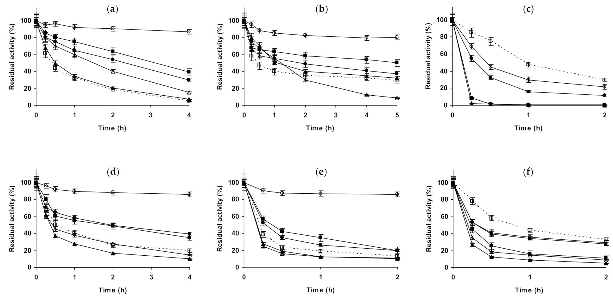
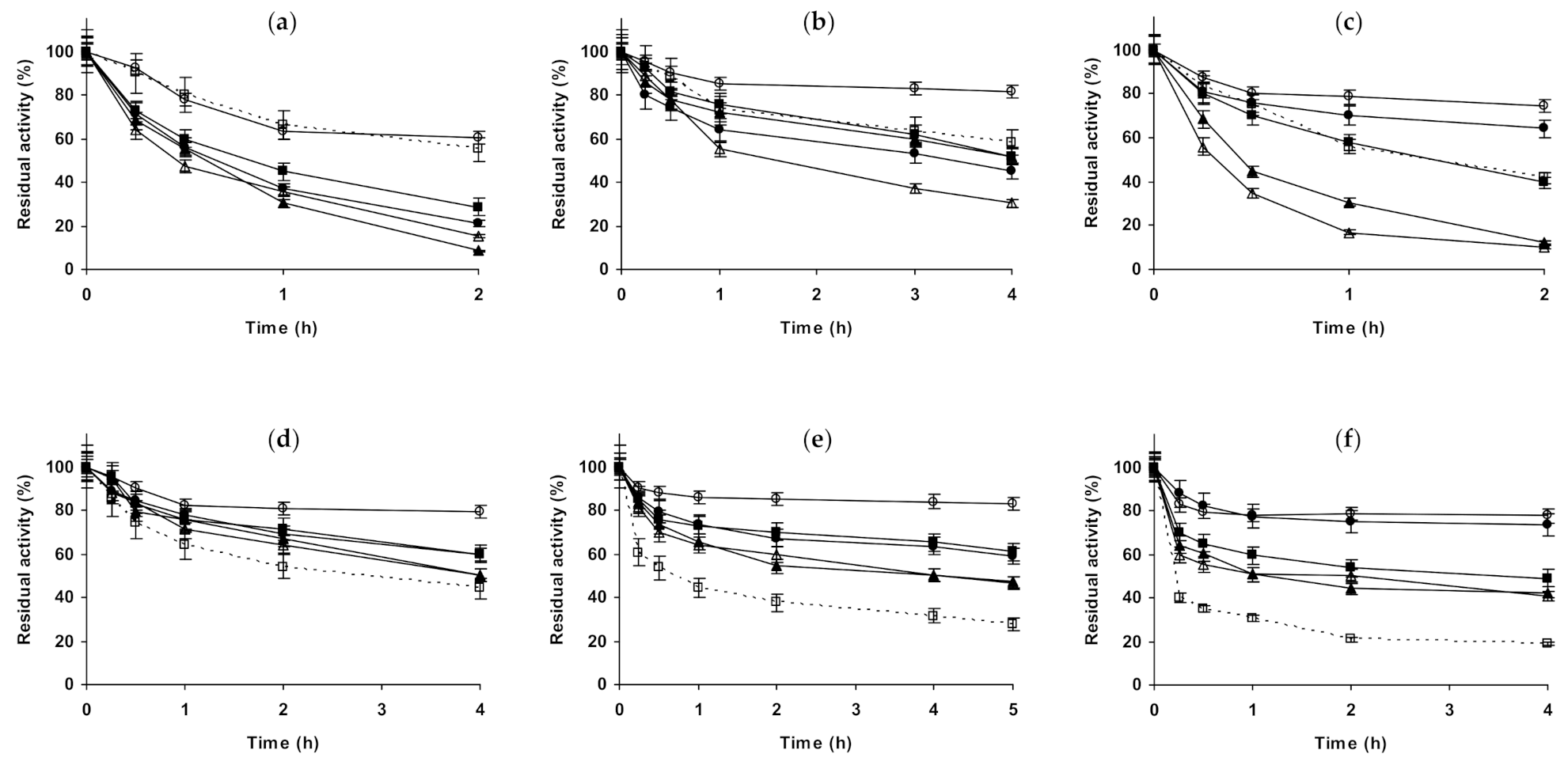
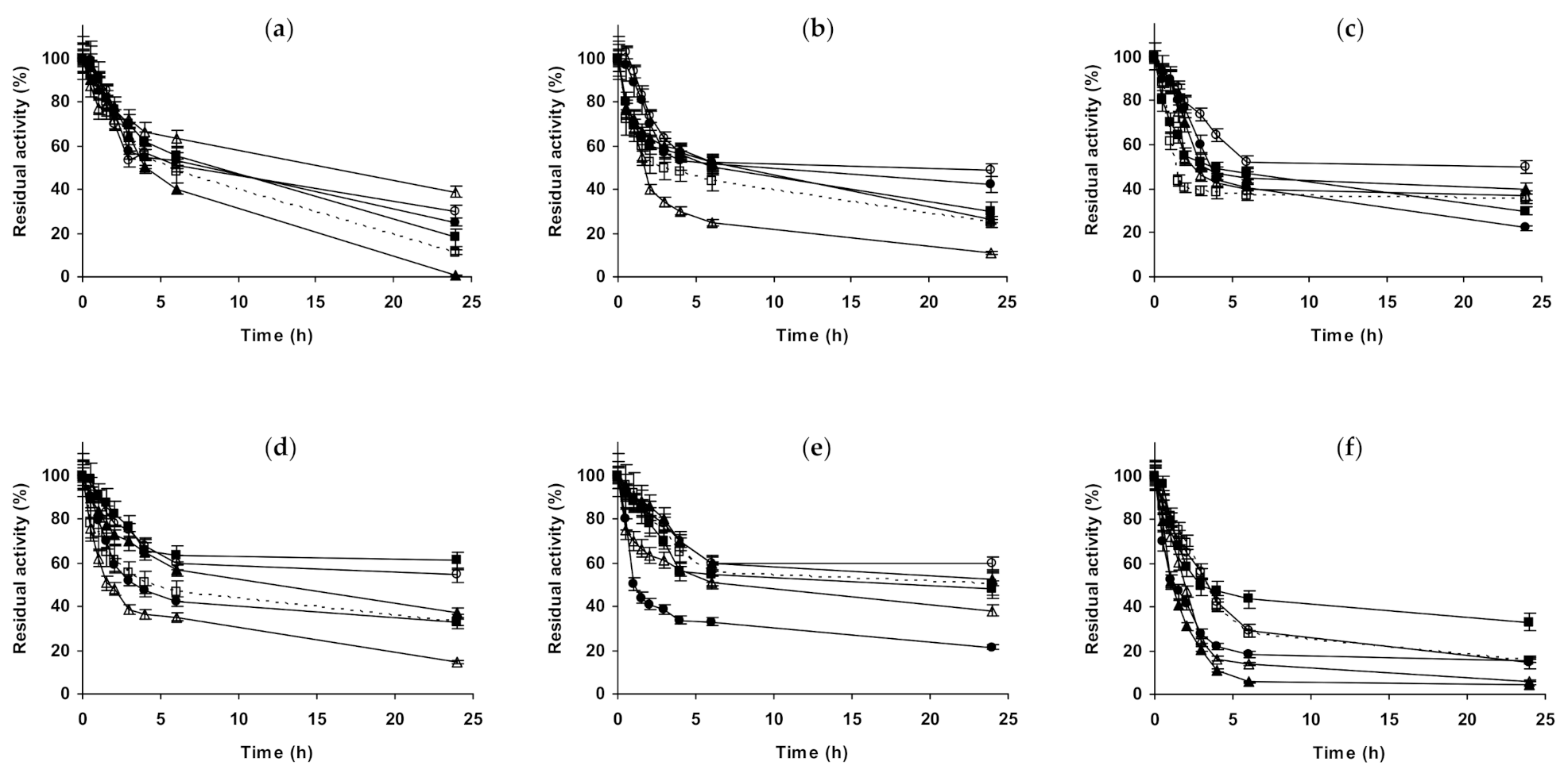
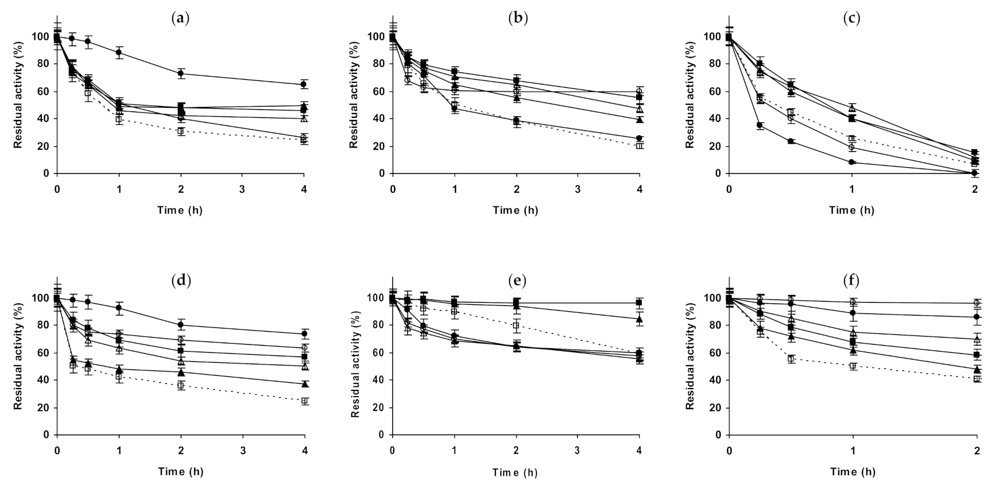
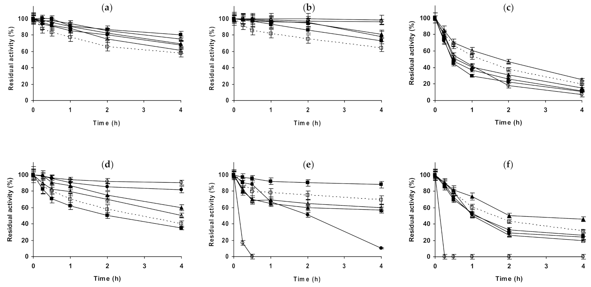
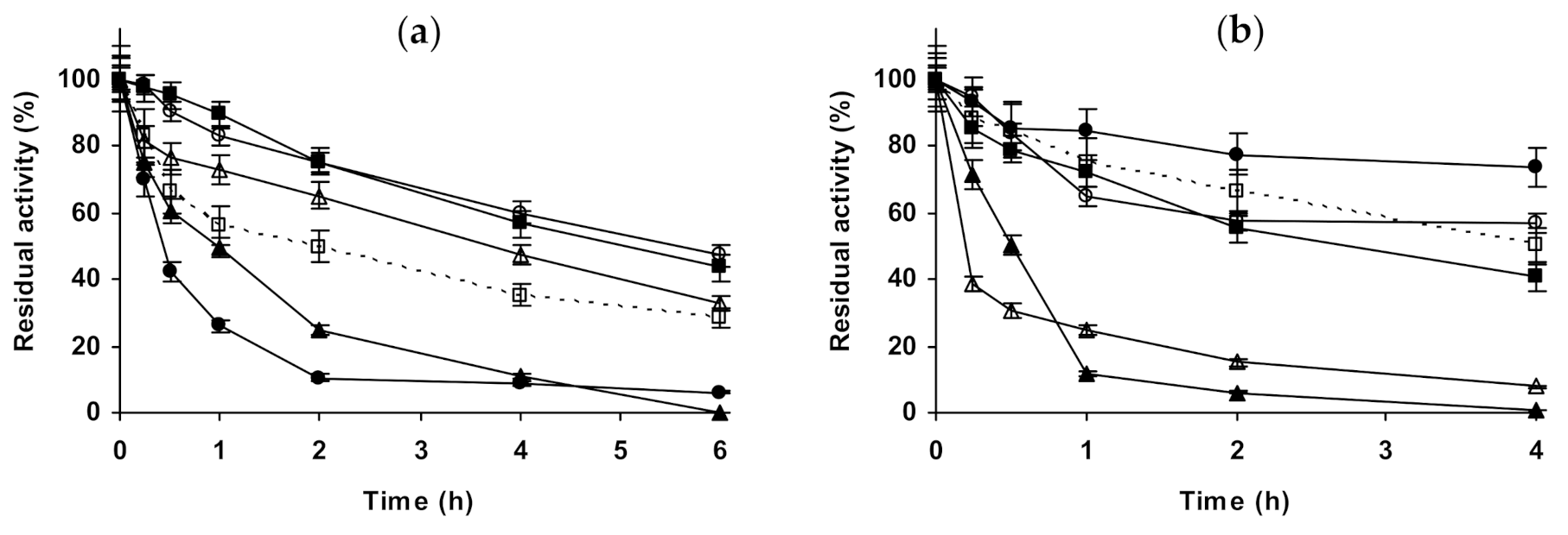
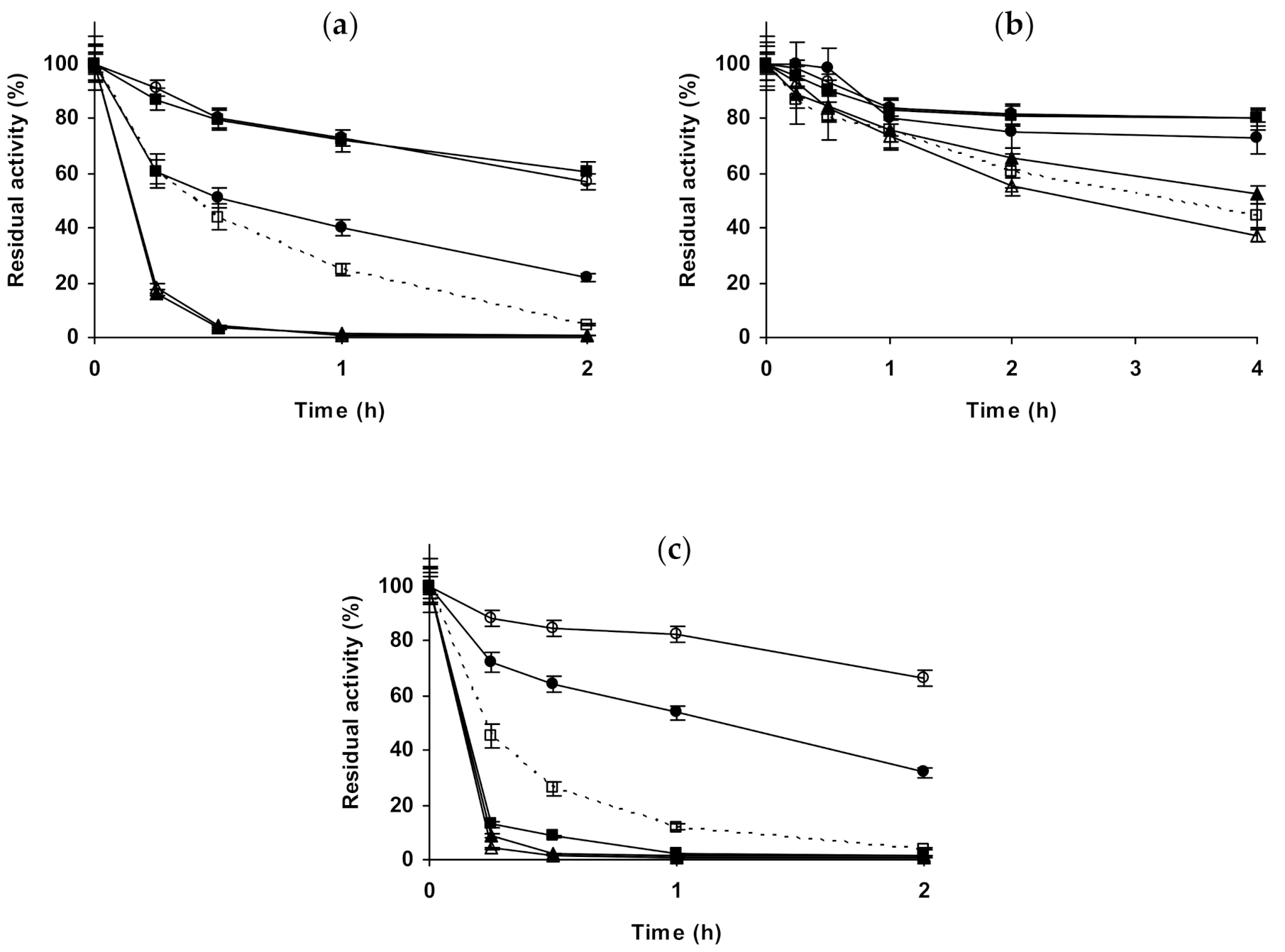
2.1. Effect of Different Salts on the Stability of Immobilized Lipases

As stated in the introduction section, lipases were immobilized on octyl agarose (via interfacial activation [77]) or glutaraldehyde-amino agarose (via multiple factors) [111], to have two immobilized preparations via quite different phenomena.
Starting with CALA (Figure 1), when the enzyme is immobilized on octyl agarose (Figure 1a–c), and with its inactivation at pH 5.0, the presence of 1 M NaCl presented no effect on enzyme stability, while using 3 M of this salt, the stability was slightly improved. (NH4)2SO4 presented a very positive effect on enzyme stability, which reached a maximum using 3 M. In the presence of 1 M Na2SO4, the biocatalyst presented a similar stability to that in the presence of 1 M (NH4)2SO4. At pH 7.0, 1 M NaCl had no significant effect on enzyme stability, while in the presence of 3 M NaCl there was an initial stabilizing effect, but after 5 h the residual activity was under that of the enzyme incubated in absence of additional salts. (NH4)2SO4 remained a stabilizing medium for this immobilized enzyme at pH 7.0, being this effect more significant at 3 M, 1 M Na2SO4 gave similar value that 1 M (NH4)2SO4. At pH 9.0, all concentrated salts strongly reduced the stability of octyl-CALA. The most drastic destabilizing effect was found using the sodium salts (sulfate or chloride), (NH4)2SO4 showed a lower destabilizing effect. Curiously, considering the negative effect of the salt, 3 M concentration gave higher stability than 1 M of the salts (Figure 1c). Using glutaraldehyde-CALA (Figure 1d–f), at pH 5.0, NaCl showed a slight destabilizing effect, more relevant in the presence of 1 M NaCl than using 3 M. The enzyme under these conditions was significantly stabilized by (NH4)2SO4, even more so using 3 M than employing 1 M. Both 1 M sulfate salts permitted the same stabilities for this immobilized enzyme. At pH 7.0, NaCl had a slightly negative effect on enzyme stability at 1 and 3 M. 1 M Na2SO4 showed a slight stabilizing effect, similar to 1 M (NH4)2SO4. However, the inactivation in 3 M (NH4)2SO4 gave the highest stability, even more relevant than using octyl-CALA (Figure 1b). At pH 9.0, as in the case of octyl-CALA, a negative effect on enzyme stability of all concentrated salts was observed, being this effect more significant for both sodium salts than for (NH4)2SO4.
Next, we will present the results using immobilized CALB (Figure 2). In the case of octyl-CALB (Figure 2a–c) at pH 5.0, 3M NaCl produced a positive effect on enzyme stability, while 1 M had no significant effect. Using (NH4)2SO4, some enzyme stabilization could be detected using 1 M, while 3 M of this salt was much more stabilizing, more than 3 M NaCl. 1 M Na2SO4 presented a significant effect on enzyme stability, similar to the value found using 3 M (NH4)2SO4. At pH 7.0, 1 M NaCl had no effects on enzyme stability, while 3 M significantly improved it. (NH4)2SO4 was positive for enzyme stability using 1 M, but when using 3 M the effect was much more significant, the enzyme almost remained fully active after 2 h of incubation. At this pH value, 1 M Na2SO4 had a lower stabilizing effect than 1 M (NH4)2SO4. At pH 9.0, results fully differed from those described using immobilized CALA: the stability increased using all additives. NaCl and (NH4)2SO4 increased the stability of the enzyme when increasing their concentration. The most stabilizing agent was (NH4)2SO4 followed by Na2SO4 and the least stabilizing agent was NaCl (comparing to the effects of the other salts at 1 M). Using glutaraldehyde-CALB (Figure 2d–f), at pH 5.0, 1 M NaCl had no effect on enzyme stability and for 3 M NaCl a slight stabilizing affect was found. 1 M Na2SO4 presented a higher stabilization effect than 3 M NaCl, while 1 M (NH4)2SO4 had scarce stabilizing effect, very similar at that found using 3 M NaCl. The clearest stabilizing effect could be found when inactivating the immobilized enzyme in 3 M (NH4)2SO4. This was quite different from the results found using octyl-CALB (Figure 2a). At pH 7.0, all salts at all concentrations improved enzyme stability. The presence of 1 and 3 M NaCl presented a similar effect on enzyme stability, much smaller than when using the sulfate salts. 1 M (NH4)2SO4 stabilized this biocatalyst to a lower extent the enzyme than 1 M Na2SO4, however the highest stability was observed using 3 M (NH4)2SO4. Again, there are some qualitative differences compared to the effects of the salts in the stability of the octyl-CALB preparation (Figure 2b). At pH 9.0, glutaraldehyde-CALB stability decreased when inactivated in the presence of 1 M of NaCl, becoming the immobilized enzyme stability similar when inactivated in absence of any salt or in the presence of 3 M NaCl. 1 M Na2SO4 presented some stabilizing effects, although smaller than 1 M (NH4)2SO4. This salt gave the same stability when used at 1 or 3 M. Again, this was different to the results using octyl-CALB (Figure 2c).
Next, we studied CRL (Figure 3). In the case of octyl-CRL (Figure 3a–c), at pH 5.0, the stability did not change in the presence of 1 or 3 M NaCl. Sulfate salts presented a positive effect, similar using 1 M of ammonium or sodium salts, more significant using 3 M (NH4)2SO4. At pH 7.0, NaCl presented a slight negative effect on the immobilized octyl-CRL stability. This effect was stronger using 3 M. 1 M Na2SO4 or (NH4)2SO4 were also slightly negative for the enzyme stability. However, 3 M (NH4)2SO4 clearly stabilized the enzyme. At pH 9.0, the negative effect of NaCl remained (similar at both, 1 or 3 M), 1 M Na2SO4 was also negative for enzyme stability. However, (NH4)2SO4 presented positive effects on enzyme stability, and these positive effects increased with the salt concentration. The effect of these salts in the glutaraldehyde-CRL stability was also analyzed (Figure 3d–f). At pH 5.0, NaCl had no significant effect on the stability of the enzyme (similar to the octyl preparation). 1 M Na2SO4 or (NH4)2SO4 increased the enzyme stability in a similar way, but the highest stabilization was found using 3 M (NH4)2SO4. At pH 7.0, this biocatalyst was slightly destabilized by NaCl, and this effect was higher when the salt concentration increased. 1 M Na2SO4 or (NH4)2SO4 presented a marginal stabilizing effect, this stabilizing effect became more evident using 3 M (NH4)2SO4. When the immobilized enzyme was inactivated at pH 9.0, the enzyme destabilizing effect of NaCl was more intense than at the other pH values. 1 M Na2SO4 was slightly destabilizing while the immobilized enzyme in the presence of 1 M (NH4)2SO4 was more stable than in just buffer. This (NH4)2SO4 stabilizing effect was further increased using 3 M of the salt. Results were similar, but not identical, to the ones obtained studying the octyl-CRL preparation (Figure 3a–c), but very different from the results observed using the other two lipases already discussed (Figure 1 and Figure 2).
The next studied lipase was RML (Figure 4). Using octyl-RML (Figure 4a–c), at pH 5.0, the presence of 1 or 3 M NaCl had some stabilizing effect on this enzyme; clearer at 3 M. 1 M Na2SO4 was more stabilizing than 1 M (NH4)2SO4 or 3 M NaCl, the highest enzyme stability was observed using 3 M (NH4)2SO4. At pH 7.0, NaCl presented a negative effect on enzyme stability, being the inactivating results quite similar to the biocatalyst in 1 or 3 M of this salt. 1 M Na2SO4 effect was stabilizing, while 1 M (NH4)2SO4 was slightly destabilizing for this enzyme preparation. However, when the inactivation was performed in 3 M (NH4)2SO4, the enzyme stability reached a maximum (no loss of activity was detected under these conditions for the time of study). At pH 9.0, NaCl produced a stabilization of the enzyme, more significant when the concentration of the salt was increased. 1 M Na2SO4 or (NH4)2SO4 stabilized the enzyme more than 3 M NaCl, and in a very similar fashion. The highest stability was observed in 3 M (NH4)2SO4. Using glutaraldehyde-RML (Figure 4d–f), at pH 5.0, NaCl presented a small stabilizing effect using both concentrations, 1 and 3 M. Using 1 M of both sulfate salts, the stabilizing effect was clearer and almost identical. Again, the highest stabilizing effect was obtained using 3 M (NH4)2SO4. At pH 7.0, the presence of 1 M NaCl had no significant effect on the enzyme stability, while 3 M produced a slight stabilization. The stability of the enzyme further improved in the presence of 1 M Na2SO4, and to a lower extent in the presence of 1 M (NH4)2SO4. The biocatalyst inactivation in 3 M (NH4)2SO4 produced the lower inactivation rate. At pH 9.0, 1 M NaCl produced a negative effect on enzyme stability, while 3 M NaCl produced a positive effect. 1 M Na2SO4 presented no effect on enzyme stability, while 1 M (NH4)2SO4 presented a stabilizing effect, that was augmented using 3 M of this salt. Again, results were diverse depending on the pH and on the enzyme immobilization protocol.
The last lipase that has been studied in this paper was EVT (Figure 5). Using octyl-EVT (Figure 5a–c), at pH 5, 1 and 3 M NaCl presented a similar negative effect on enzyme stability. In the presence of 1 M Na2SO4, the enzyme stability was reduced in a smaller way, results were slightly worse using 1 M (NH4)2SO4, while using 3 M of this salt the enzyme stability was similar to the inactivation performed just in buffer. At pH 7.0, the inactivation in the presence of 1 M NaCl or Na2SO4 presented no differences on enzyme stability, while 3 M NaCl was clearly negative for enzyme stability. 1 M (NH4)2SO4 was slightly negative for enzyme stability, while 3 M of this salt presented a stabilizing effect. At pH 9.0, NaCl presented a negative effect on enzyme stability, 1 M Na2SO4 has no significant effect on enzyme stability, and (NH4)2SO4 presented an enzyme stabilizing effect, higher using 3 M than employing 1 M of this salt. Figure 5d–f show the results using glutaraldehyde-EVT. At pH 5.0, NaCl presented a small stabilizing effect, similar using 1 or 3 M. Using 1 M Na2SO4 or (NH4)2SO4, the stabilization of the enzyme was more evident, and this stabilizing effect increased using 3 M of (NH4)2SO4, although it did not become very relevant (e.g., after 4 h, the reference maintained 44% of the initial activity while in the presence of 3 M (NH4)2SO4 maintained almost 80%). At pH 7.0, the enzyme stabilizing effect of NaCl is more evident, again very similar at 1 or 3 M. 1 M Na2SO4 or (NH4)2SO4 further improved the enzyme stability, and the highest stability was observed in inactivations using 3 M (NH4)2SO4. At pH 9.0, the presence of 1 or 3 M NaCl improved the enzyme stability in a similar fashion. 1 M Na2SO4 permitted stabilities similar to that observed when inactivating the immobilized enzyme in the presence of NaCl, while the inactivation in (NH4)2SO4 gave the highest enzyme stabilities, very similar using 1 or 3 M of these salts.
Thus, the results presented here suggest that the complexity of the effects of the salts in lipase stability is very diverse and a single explanation cannot justify the great differences among enzymes, inactivation pH and immobilization protocol. The different effect of the different salts on the immobilized lipases stability should be due to the different structure of the enzymes, in a similar way that Ca2+ stabilized some lipases and not others, the different cations and anions may promote different effects on the enzyme stability. As a general, but not universal rule (see in Figure 1 the case of CALA at pH 9.0), 3 M (NH4)2SO4 generally stabilize lipases, while NaCl is more risky. In fact, NaCl is even negative for the stability of some of the analyzed immobilized enzymes under certain conditions. The explanation is not always because of the Na+ cations, as in some instances NaCl is destabilizing while Na2SO4 is stabilizing, even more than (NH4)2SO4. The fact that in some instances the negative effect of the salt decreases when increasing its concentration, even becoming positive in certain cases, suggests that a double effect of the salts on the enzyme stability may be occurring in these cases, one negative, perhaps related to the effect of the specific ions, and another positive, perhaps related to an increase in the ionic strength that makes the exposition of partially distorted enzymes, where some internal hydrophobic pockets may be exposed to the medium, less favorable [57,58,59,60,61,62,63,64]. Differences between the different immobilized preparations of the same enzyme may derive from the fact that when immobilized in hydrophobic support, the lipases present the open form of the lipase, that is stabilized versus the hydrophobic support surface, while in the glutaraldehyde-amino, this did not occur [78,102]. Moreover, in the enzymes immobilized via interfacial activation, the enzyme release from the biocatalyst at high temperature should be more difficult at growing ionic strength, and this effect will not occur using covalently immobilized lipases [78].

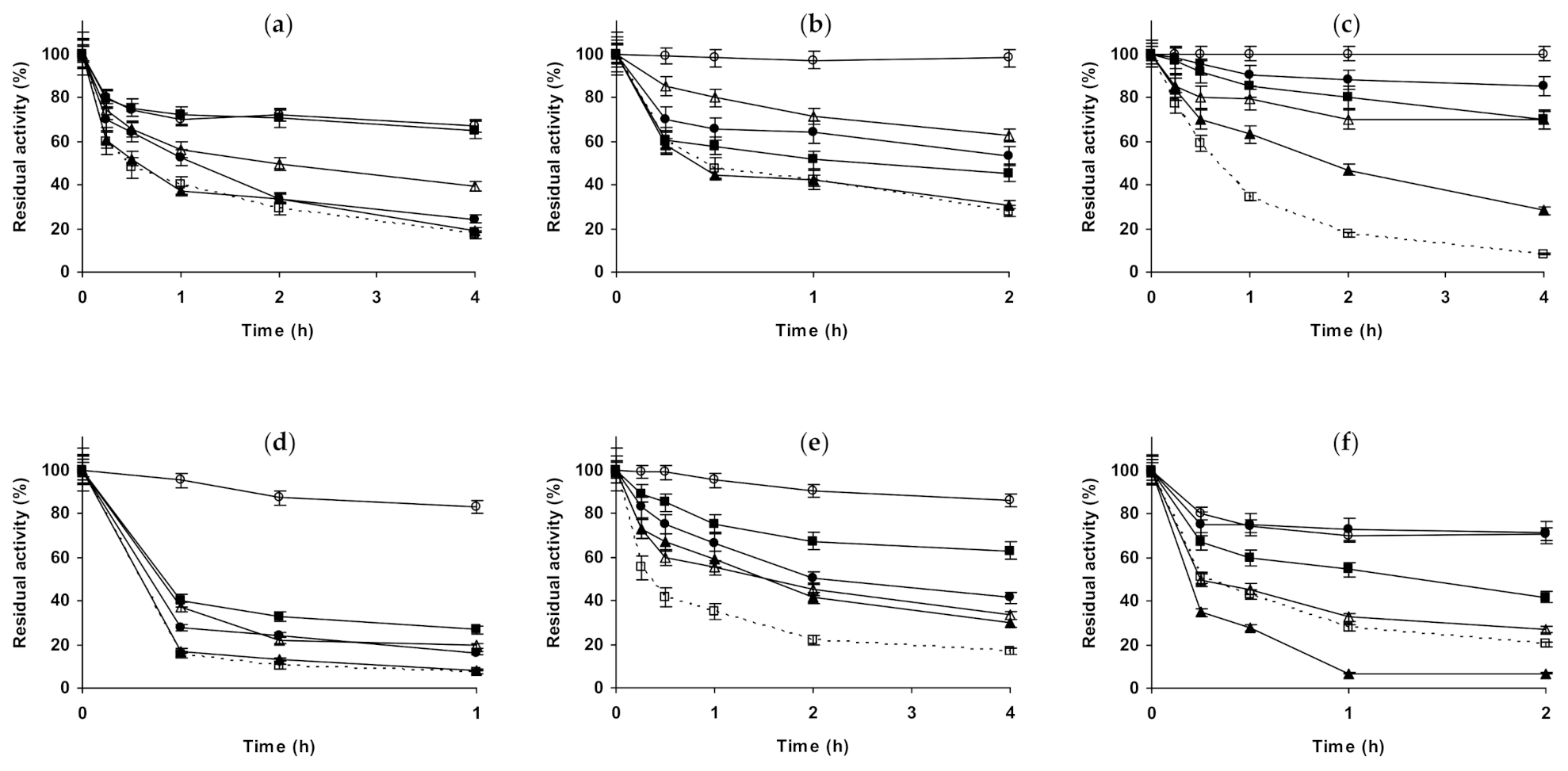
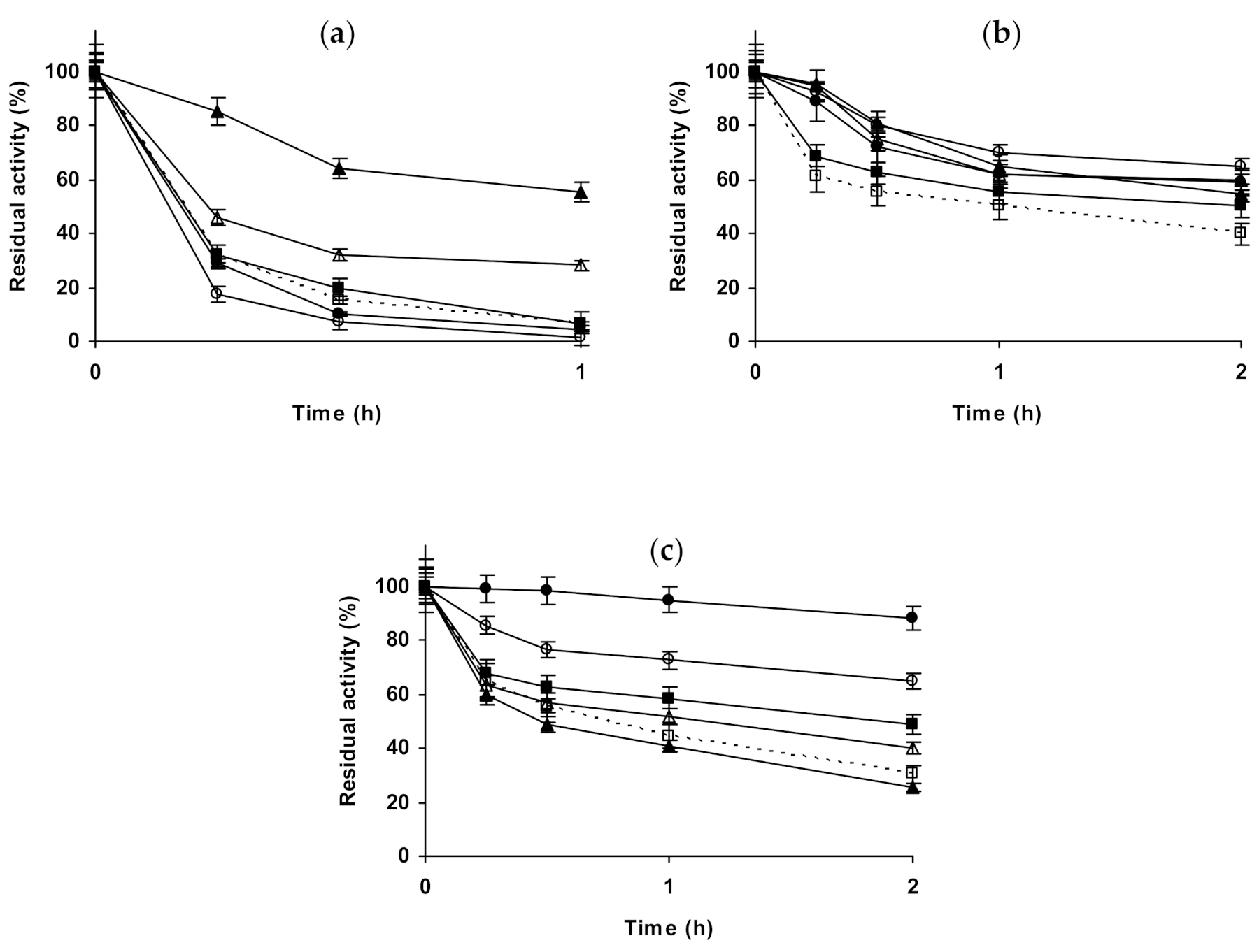
2.2. Effect of Different Salts on the Stability of Immobilized Proteases

To determine if this quite apparently chaotic effect of the presence of salts on enzyme stabilities is general or specific for lipases, we have also analyzed the stability of 3 proteases immobilized on glutaraldehyde-amino [102] and on glyoxyl agarose [101], protocols that should give different enzyme orientations and degrees of enzyme-support multipoint covalent attachment.
Starting with glyoxyl-ficin (Figure 6a–c), the presence of 1 M NaCl at pH 5.0 presented a small enzyme destabilizing effect, while 3 M NaCl was significantly stabilizing for this preparation (the enzyme inactivated under these conditions was the most stable). 1 M Na2SO4 or (NH4)2SO4 presented a similar and small stabilizing effect, similar to that found using 3 M (NH4)2SO4. At pH 7.0, 1 M NaCl, sodium and (NH4)2SO4, or 3 M (NH4)2SO4 presented very small stabilizing effects, while 3 M NaCl was clearly negative for the enzyme stability (in opposition with the results at pH 5). At pH 9.0, the presence of the salts produced a small stabilizing effect, reaching the highest one when inactivating in 3 M (NH4)2SO4. 1 M (NH4)2SO4 and Na2SO4 were slightly destabilizing after 24 h of inactivation. When using glutaraldehyde-ficin (Figure 6d–f), the differences in the stabilities of the biocatalysts in the different solutions were larger than when using glyoxyl-ficin (Figure 6a–c), although not very great. At pH 5.0, 1 M NaCl was slightly stabilizing, while 3 M NaCl was destabilizing for this ficin preparation (this was the contrary using glyoxyl-ficin, Figure 6a). The medium stabilizing effect for this biocatalyst was increased using 1 M Na2SO4, while 1 M (NH4)2SO4 presented no-effect on enzyme stability and 3 M (NH4)2SO4 gave a stabilization similar to that using 1 M Na2SO4. At pH 7.0, the effects of NaCl were similar to those found at pH 5.0, 1 M slightly stabilize the enzyme, while 3 M slightly destabilized the enzyme. 1 M Na2SO4 had no effect on enzyme stability, while 1 M (NH4)2SO4 was clearly negative and 3 M of this salt had a very small positive effect on enzyme stability. At pH 9.0, NaCl was negative for enzyme stability at both, 1 and 3 M. 1 M Na2SO4 was positive for enzyme stability while 1 M (NH4)2SO4 was negative, and using 3 M (NH4)2SO4 the enzyme stability was very similar to that found just in buffer.
Figure 7 shows the studies using immobilized chymotrypsin. Glyoxyl-chymotrypsin (Figure 7a–c), at pH 5.0 and in the presence of 1 M NaCl and 1 M Na2SO, presented a similar increase of enzyme stability. If the inactivation was performed in 3 M NaCl, the stabilizing effect was smaller. 1 M (NH4)2SO4 permitted much higher enzyme stabilization than the other salts, but this effect became similar to the other salts when using 3 M (NH4)2SO4. At pH 7.0, NaCl presented a positive effect that was higher using 3 M NaCl. 1 M Na2SO4 produced a higher stabilization effect, while 1 M (NH4)2SO4 did not produce a significant effect on enzyme stability. Using 3 M (NH4)2SO4, after a first rapid inactivation, the activity of the enzyme did not decrease for a long time, after 4 h the enzyme residual activity became even higher than in 1 M Na2SO4. At pH 9.0, again NaCl produced a positive effect on enzyme stability, slightly higher using 3 M. 1 M Na2SO4 is positive for enzyme stability, while 1 M (NH4)2SO4 reduced the enzyme stability, decreasing this negative effect on enzyme stability when using 3 M of this salt. Using glutaraldehyde-chymotrypsin (Figure 7d–f), the situation is very different. At pH 5.0, NaCl produced enzyme stabilization, higher using 3 M. 1 M Na2SO4 produced an even higher stabilization than when using 3 M NaCl. The highest enzyme stabilization is achieved using 1 M (NH4)2SO4, the use of 3 M of this salt is positive for immobilized enzyme stability, but in a smaller extension. At pH 7.0, 1 M NaCl produced a very positive effect on enzyme stability, while 3 M NaCl produced the enzyme destabilization. 1 M Na2SO4 promoted a strong enzyme stabilization, slightly higher than when using 1 M NaCl (residual activity was 95 versus 85% after 4 h of inactivation). (NH4)2SO4 was negative for enzyme stability, at both concentrations with similar intensity, and similar to the results obtained using 3 M NaCl. At pH 9.0, NaCl produced a positive effect on enzyme stability, higher using 3 M. 1 M Na2SO4 produced an enzyme stabilizing effect slightly smaller than 3 M NaCl, while (NH4)2SO4 promoted the highest enzyme stabilization, higher when using higher salt concentration.
The last studied protease was trypsin (Figure 8). Figure 8a–c show the results using glyoxyl-trypsin. At pH 5.0, all salts additions on the buffer solution produced a stabilization of the enzyme, but this effect was not very significant. 1 M NaCl was slightly more stabilizing than 3 M NaCl, and 1 M Na2SO4 gave a slightly higher stabilization. This stabilization was lower using 1 M (NH4)2SO4 and increased using 3 M (NH4)2SO4 to levels close to 1 M Na2SO4. At pH 7.0, NaCl stabilized the enzyme, an effect which was more pronounced using 3 M than using 1 M. 1 M Na2SO4 stabilized the enzyme less than 1 M NaCl, while 1 M (NH4)2SO4 had a similar effect, which increased using 3 M (NH4)2SO4. At pH 9.0, 1 M NaCl was slightly destabilizing, while 3 M presented a stabilizing effect. 1 M Na2SO4 or (NH4)2SO4 were even more destabilizing than 1 M NaCl, and the enzyme inactivation in 3 M (NH4)2SO4 gave similar results to that in the presence of 1 M NaCl. We can conclude that glyoxyl-trypsin seemed not to be very responsible to the additions of salts in terms of stability. Figure 8d–f shows the results using glutaraldehyde-trypsin. At pH 5.0, NaCl was slightly positive for enzyme stability, more using 1 M than using 3 M. 1 M Na2SO4 was negative for enzyme stability, while 1 M and 3 M (NH4)2SO4 were significantly positive. At pH 7.0, NaCl was negative for enzyme stability at both concentrations, while Na2SO4 was positive (the only condition where stability was improved was using 1 M of this salt). The strongest effect, a destabilizing one, was found using (NH4)2SO4, and the effect was dramatic using 3 M of this salt, as the activity was fully destroyed after just 30 min. At pH 9.0, again everything was different: 1 M NaCl was positive for enzyme stability, while 3 M NaCl was negative. 1 M Na2SO4 and 1 M (NH4)2SO4 were also negative for enzyme stability in a similar way, while 3 M (NH4)2SO4 was again very negative for enzyme stability. That is, trypsin immobilized on glutaraldehyde was more sensible to the presence of high concentrations of salts than the glyoxyl immobilized enzyme.
That way, using proteases, the situation remains very dependent on the inactivation pH, the enzyme and the immobilization protocol. In this instance, the enzyme stability decreased in the presence of some salts more than in the case of lipases, and the stabilizing effects of 3 M (NH4)2SO4 is no longer a general rule, becoming in many instances a strong destabilizing agent. The increase of the salt concentration in some cases revert a negative effect in positive, while in other cases revert a positive effect in a negative one. The only rule that can be extracted from the results is that the effect of the salts on enzyme stability strongly depends on the specific case, suggesting that many different and opposite phenomena are simultaneous determining the final enzyme stability.

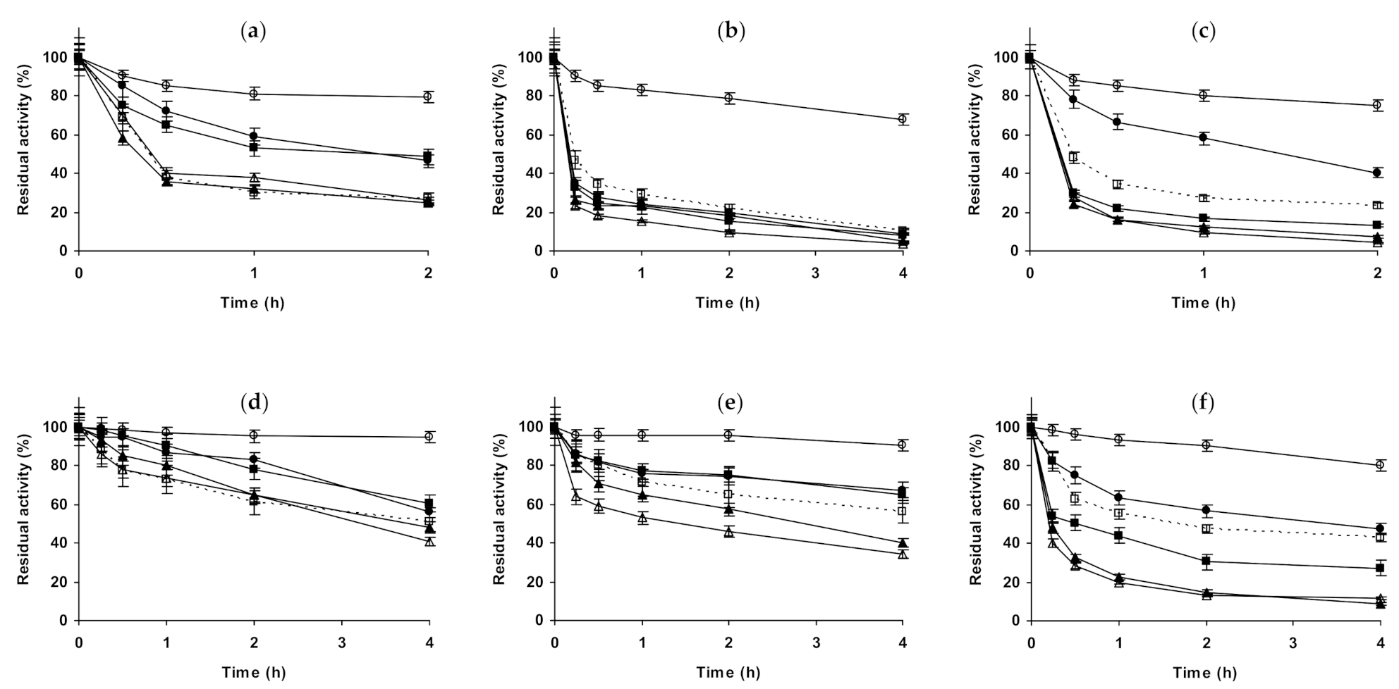
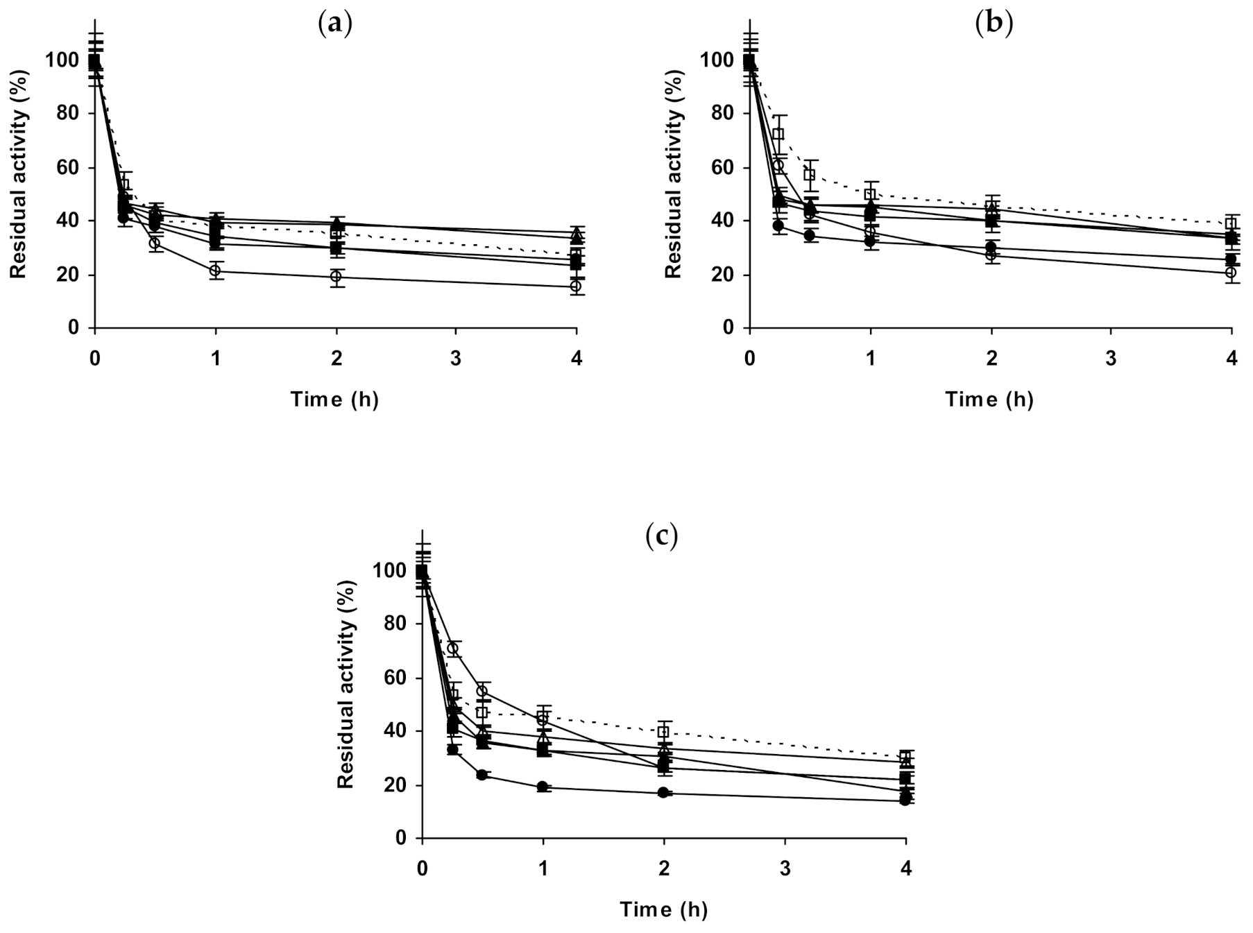
2.3. Effect of Different Salts on the Stability of Some Additional Immobilized Monomeric Enzymes

Here, using immobilized preparations, the effect of the salts on the stability of some additional monomeric enzymes have been analyzed. Starting with glutaraldehyde-β-galactosidase (Figure 9), at pH 5.0, 1 M NaCl produced a clear enzyme destabilization, while 3 M is clearly stabilizing. 1 M Na2SO4 stabilized the immobilized enzyme even more than 3 M NaCl, while the presence of 1 M (NH4)2SO4 was negative for enzyme stability and 3 M of this salt permitted to have a stability similar to that observed using as inactivating medium 1 M Na2SO4. At pH 7.0, the enzyme stability decreased when NaCl were added, the increase in its concentration accelerated the initial steps of the enzyme inactivation but the last activity fraction was more stable. Na2SO4 was not so negative for enzyme stability, as NaCl while 1 M (NH4)2SO4 permitted a higher enzyme stability and 3 M (NH4)2SO4 effect was slightly negative for enzyme stability. At pH 9.0, the study could not be performed due to the very poor stability of the enzyme even at room temperature.
The second studied immobilized monomeric enzyme was glutaraldehyde-laccase (Figure 10). At pH 5.0, 1 M NaCl significantly stabilized the enzyme, while using 3 M the enzyme stabilizing effect was less clear. 1 M Na2SO4 or (NH4)2SO4 did not present an effect on enzyme stability, while 3 M (NH4)2SO4 promoted a destabilization of the enzyme. At pH 7.0, 1 and 3 M NaCl produced similar stabilizing affects, while 1 M Na2SO4 was less stabilizing. 1 M (NH4)2SO4 stabilized the enzyme like NaCl, and using 3 M this effect was slightly improved. At pH 9.0, 1 M NaCl almost did not affect enzyme stability, while 3 M slightly improve it. The stabilizing effect was clearer using 1 M Na2SO4 and further increased using 1 M (NH4)2SO4, although this positive effect on enzyme stability decreased using 3 M of the salt.
The last studied enzyme in this section was glyoxyl-PGA (Figure 11), although it is a heterodimeric enzyme, it is really the result of an auto-processing of pre-enzyme, and it does not establish equilibrium between dissociated and associate subunits [112,113,114]. The effects of the salts in the stability of this enzyme are in general quite small. At pH 5.0, NaCl has a marginal stabilizing effect, similar at 1 and 3 M. 1 M Na2SO4 or (NH4)2SO4 presented not significant effect, while 3 M (NH4)2SO4 destabilized the enzyme. At pH 7.0, all salts have a negative effect on enzyme stability. NaCl was slightly negative at both, 1 and 3 M. 1 M Na2SO4 decreased the enzyme stability in a more significant way, and the stability decreased even more using 1 or 3 M (NH4)2SO4. Results at pH 9.0 were similar, except that using 3 M (NH4)2SO4 the first inactivation was slower than that in just buffer, but later gave less residual activity.
Thus, again a great diversity of results can be found for these enzymes, the pH determines in many instances if one specific salt has a positive or negative result.

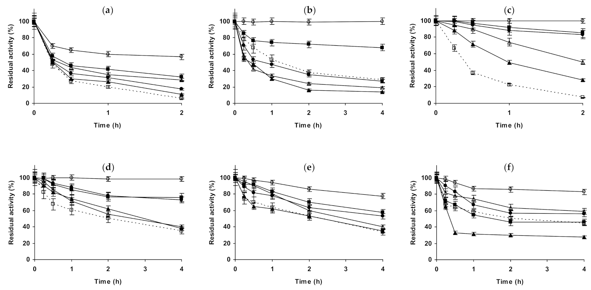
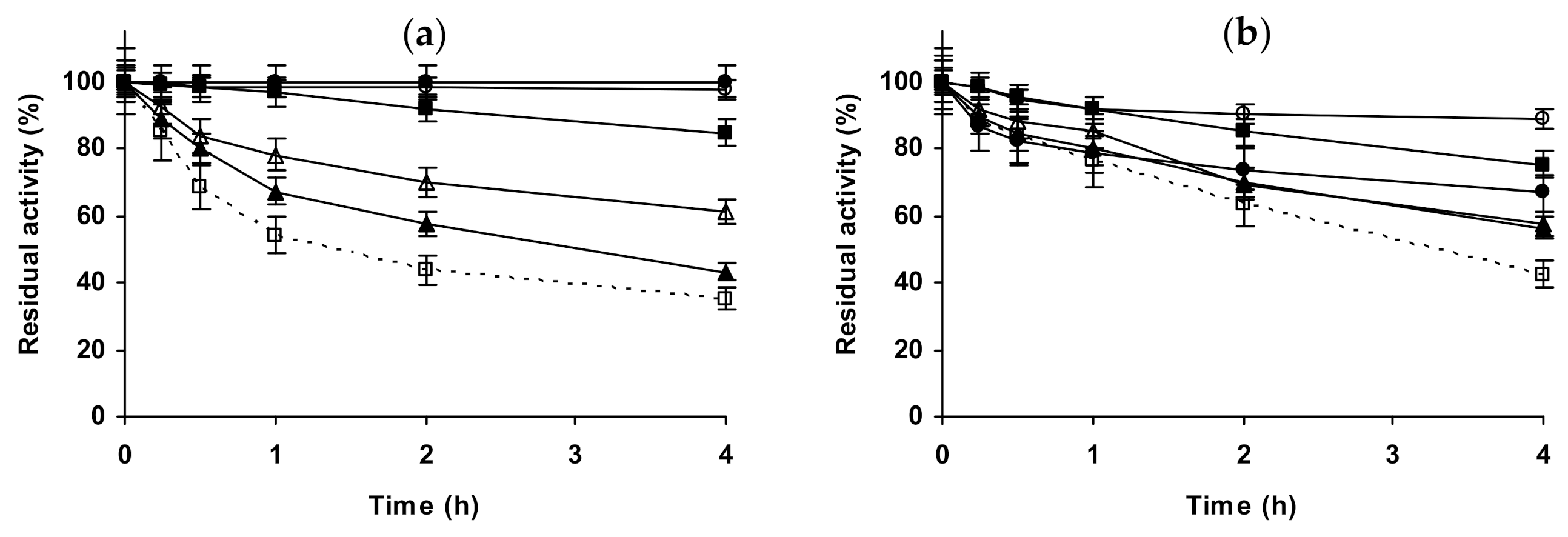
2.4. Effect of Different Salts on the Stability of Some Glutaraldehyde-Amino Agarose Immobilized Multimeric Enzymes

Figure 12 shows the effects of the different salts in the stability of the immobilized dimeric β-glucosidase preparation. At pH 5.0, NaCl produced a stabilizing effect that increased when increased the salt concentration. 1 M Na2SO4 produced a more significant stabilizing effect, while in the presence of 1 M and 3 M (NH4)2SO4 the enzyme retained the full initial activity during the whole inactivation time. At pH 7.0, NaCl has a shorter stabilizing effect than at pH 5.0, and similar using both concentrations. 1 M Na2SO4 produced a higher stabilization than NaCl, while 1 M (NH4)2SO4 gave a value similar to NaCl, however, the maximum stability of this enzyme preparation was found using 3 M (NH4)2SO4. At pH 9.0, the stability of the immobilized enzyme was too low to give reliable data.
The last studied enzyme was an immobilized tetrameric catalase (Figure 13). At pH 5.0, NaCl behaved as a strong destabilizing agent, at both concentrations. However, 1 M or 3 M Na2SO4 produced a similar and significant stabilization, while 1 M (NH4)2SO4 has a short stabilizing effect. At pH 7.0, the effect of NaCl on enzyme stability was low, slightly positive at 1 M and slightly negative at 3 M, while 1 M Na2SO4 remained as a stabilizing condition, 1 M (NH4)2SO4 produced a small stabilization, and 3 M promoted an enzyme stability similar to 1 M Na2SO4. The situation changed again at pH 9.0, NaCl becoming a very destabilizing agent for this enzyme, similar to Na2SO4 (in opposition to the results found at pHs 5.0 and 7.0). However, (NH4)2SO4 promoted the enzyme stabilization, and this effect was higher when the salt concentrations increased. That is, the effect of the salts strongly changed when the inactivation pH changed.
Again, the effects of the salts follow no clear rules, and the same salt may be positive in one condition and negative in other.

3. Materials and Methods

3.1. Materials

CALA (NovoCor® AD L, 7.63 mg of protein/mL), CALB (Lipozyme® CALB L, 5.57 mg of protein/mL), EVT (Eversa® Transform 2.0, 27.5 mg of protein/mL), RML (Palatase® 2000 L, 2.67 mg of protein/mL) and laccase (Novozym® 51033, 39.86 mg of protein/mL) were kindly donated by Novozymes (Madrid, Spain). Ficin (79 mg of protein/mL) was produced as previously described [115]. β-galactosidase (20 units of oNPG/mg of protein), β-glucosidase (350 mg of protein/g of powder), catalase (470 mg of protein/g of powder), CRL (32 mg of protein/g of powder), chymotrypsin and trypsin (lyophilized powder), PGA (68 mg of protein/mL), 6-nitro-3-(phenylacetamido)benzoic acid (NIPAB), Nα-benzoyl-D,L-arginine p-nitroanilide hydrochloride (BAPNA), N-benzoyl-L-tyrosine p-nitroanilide (BTPNA), o-nitrophenyl β-D-galactopyranoside (oNPG), p-nitrophenyl, β-D-glucopyranoside (pNPG), p-nitrophenyl butyrate (pNPB) and sodium borohydride were purchased from Sigma Aldrich (Alcobendas, Spain). 2,2′-azino-bis (3-ethylbenzothiazoline-6-sulfonic acid) diammonium salt (ABTS®) was acquired from Roche (Mannheim, Germany). Horseradish peroxidase (2680 U/mg of powder) was purchased from Orion High Technologies (Parla, Spain). Bradford’s method was used for the protein concentration determination, using bovine serum albumin as standard [116]. Octyl-Sepharose® Cl-4B beads were purchased from GE Healthcare (Madrid, Spain) and 4% BCL Agarose Beads Standard were purchased from ABT (Alcobendas, Spain). Agarose beads were used to produce aminated agarose [117,118], and then modified with glutaraldehyde to produce the glutaraldehyde-amino agarose activated support [119,120,121]. All other reagents were of analytical grade.

3.2. Methods

The experiments were performed in triplicate and the data are given as mean values and standard deviation.

3.2.1. Enzyme Activity Assay

One Unit (U) of activity was defined as the amount of enzyme that hydrolyzes 1 μmol of substrate per minute under the specified conditions. Enzyme activity was determined using a spectrophotometer with magnetic stirring at 200 rpm, at a temperature of 25 °C (except for ficin, where the assay was performed at 55 °C).
Lipases activity was quantified by determining the variance in absorbance at 348 nm produced by the release of p-nitrophenol (isosbestic point, ε under these conditions is 5150 M−1cm−1 [122]) in the hydrolysis of 50 µL of 50 mM pNPB in 25 mM sodium phosphate at pH 7.0. The reaction was started by adding 50 µL of lipase solution or suspension to 2.5 mL of buffer containing pNPB.
Ficin and trypsin activities were determined by measuring the change in absorbance at 405 nm caused by the release of p-nitroaniline (ε under these conditions is 9960 M−1cm−1 [123]), produced by the hydrolysis of BAPNA. For ficin substrate, it was prepared at 1 mM in 100 mM phosphate at pH 7.0, containing 5 mM cysteine and 5 mM EDTA and 200 μL of enzyme solution or suspension were added to 2.5 mL at incubated at 55 °C for 15 min [115]. When it was used as trypsin, BAPNA was prepared at a concentration of 2 mM in 50 mM sodium phosphate at pH 7.0 containing 30% ethanol [124] adding 50–200 μL of enzyme solution or suspension to 2.5 mL of BAPNA solution to start the reaction.
Chymotrypsin activity was determined by measuring the variation in absorbance at 386 nm produced by the release of p-nitroaniline (ε under these conditions is 12,500 M−1 cm−1 [125]), produced by the hydrolysis of BTPNA. A 40 mM BTPNA stock solution was prepared in DMSO. The reaction was started by adding 200 μL of enzyme solution or suspension to 75 μL of 40 mM BTPNA solution in 2.5 mL of 100 mM sodium phosphate at pH 7.0 containing 40% ethanol.
β-Galactosidase activity was measured by the change in absorbance at 380 nm produced by the release of o-nitrophenol (ε under these conditions is 10,493 M−1cm−1 [126]) in the hydrolysis of 10 mM oNPG in 100 mM sodium acetate at pH 4.5. 50–100 μL of enzyme solution or suspension were added to 2.5 mL of the substrate solution to initialize the reaction.
The laccase activity was determined by recording the increase in absorbance at 420 nm produced by the oxidation of the ABTS© (ε under these conditions is 36,000 M−1cm−1 [76]). The reaction was started by adding 25 μL of enzyme solution or suspension to a solution composed by 1 mL of 100 mM sodium acetate at pH 5.0 and 1 mL of 50 mM ABTS© in water.
Activity of PGA was measured using NIPAB as substrate as described by Kutzbach et al. [108]. The assay was performed continuously following the increase of absorbance at 405 nm (ε under these conditions is 8730 M−1cm−1 [108]). The reaction was started by adding 100 μL of enzyme solution or suspension to 2 mL of 0.15 mM NIPAB in 50 mM sodium phosphate at pH 7.5 solution.
β-Glucosidase activity was measured by the increase in absorbance produced at 380 nm by the release of p-nitrophenol (ε under these conditions is 3459.3 M−1cm−1 [120]) caused in the hydrolysis of pNPG. The reaction was started by adding 120 μL of enzyme solution or suspension to a solution composed by 200 μL of 10 mM pNPG prepared in 100 mM sodium phosphate at pH 7.0 and 1.68 mL of 100 mM sodium phosphate at pH 7.0.
The catalase activity was determined by measuring the reduction in the absorbance at 240 nm promoted by the consumption of hydrogen peroxide concentration in the reaction medium (the calculated ε under these conditions is 32.7 M−1cm−1). It consisted of 200 μL of 200 mM H2O2 added to 2.25 mL of 100 mM sodium phosphate, pH 7.0, 50 μL of enzyme solution or suspension were added to start the reaction.

3.2.2. Enzymes Immobilizations

All immobilizations were performed following the activity of supernatant, suspension and a reference of the enzyme under identical conditions, immobilization yield was in all cases very next to 100% [127].

Immobilization of the Lipases on Octyl Agarose Beads

Lipases immobilizations were performed using 1 (CALA, CALB, CRL, RML) or 0.2 (EVT) mg of enzyme/g of wet support to prevent diffusion limitations and protein-protein interactions [128]. The stock enzyme solution was prepared in 5 mM sodium phosphate at pH 7.0 and 25 °C. The support was added in a proportion of 1 g/10 mL of the enzyme solution. The activity was measured using pNPB to determine immobilization yield and expressed activity. After immobilization, the biocatalysts were washed with water, vacuum dried to eliminate inter-particle water and stored at 6–8 °C.

Immobilization of Enzymes on Glutaraldehyde-Amino Agarose Beads

The agarose beads were modified with ethylenediamine to produce aminated agarose as previously described [117,118]. The glyoxyl agarose support was activated using 10% glutaraldehyde prepared in 200 mM sodium phosphate solution at pH 7.0 and left overnight as previously described [119]. Immobilizations on glutaraldehyde-amino agarose were performed using 1 g of support per 10 mL of enzyme solution prepared 5 mM sodium phosphate at pH 7.0 at 25 °C. The immobilization courses were followed during 24 h. Finally, the immobilized enzymes were washed, vacuum dried and stored at 6–8 °C. The enzyme loadings of the immobilized biocatalysts were: CALA and CALB at 2 mg of enzyme/g of support [111], CRL at 1 mg of enzyme/g of support, RML at 4 mg of enzyme/g of support and EVT at 0.4 mg of enzyme/g of support, ficin at 10 mg of enzyme/g of support [129], chymotrypsin at 15 mg of enzyme/g of support and trypsin at 3 mg of enzyme/g of support (3 mM of benzamidine was added to prevent autolysis [130]), β-galactosidase at 1 mg of enzyme/g of support [121], laccase at 20 mg of enzyme/g of support [76], β-glucosidase at 49 mg of enzyme/g of support [120], catalase at 0.5 mg of enzyme/g of support [131].

Immobilization of the Enzyme on Glyoxyl Agarose Beads

Ficin was immobilized using 10 mg of enzyme/g of support, while chymotrypsin and trypsin (in the presence of 3 mM benzamidine [130]) were immobilized using a loading of 3 mg enzyme/g of support, in 100 mM sodium bicarbonate at pH 10.05 and 25 °C, using the protocols previously described [100,115,132,133]. PGA was immobilized using a support load of 5 mg enzyme/g of support. The enzyme was added to a solution composed of 100 mM sodium carbonate containing 100 mM phenyl acetic acid and 20% glycerol to prevent enzyme inactivation at pH 10.05 [134,135]. After enzymes immobilizations, 1 mg/mL of solid sodium borohydride was added and the immobilization suspensions were stirred for 30 min. After, the biocatalysts were washed with water, vacuum dried to eliminate inter-particle water and stored at 6–8 °C.

3.2.3. Stress Inactivation of Different Enzyme Preparations in the Presence of Different Salts on Biocatalyst Stability

The effect of different concentrations of different salts on the stability of all immobilized biocatalysts was studied under different conditions of salts concentration and pHs. Each enzyme was incubated in 100 mM sodium acetate at pH 5.0, 100 mM Tris HCl at pH 7.0 (phosphate was no used by its negative effects on immobilized lipases and galactosidase stabilities [73,76]) or 100 mM sodium carbonate at pH 9.0 and in some instances 1 M or 3 M of NaCl; or 1 M or 3 M of (NH4)2SO4; 1 M of Na2SO4, was added. Inactivation temperatures for each enzyme were selected for each enzyme and inactivation pH value to get inactivation rates of the enzyme (when inactivated in just buffer) that yielded reliable inactivation courses. This way, an easy comparison of the effect of the different salt additions on its stability may be performed.

4. Conclusions

As stated in the Introduction, there are many reports in the literature discussing the likely role of high concentrations of salts on enzyme stability, in an attempt to achieve a mechanistic explanation to these effects. Using free enzymes, the experimental studies are very few because of the possibility of enzyme aggregation, which can make the understanding of the phenomena difficult. That way, most previous studies are mainly theoretical ones. These theoretical papers, involving one or two enzymes, reached different conclusions, some stating that the ionic strength should be negative for enzyme stability, some others stating that it should be positive. In these papers, the conclusions are “clear”, but opposite depending on the enzyme or conditions. These theoretic studies usually conclude that the effect of the high concentrations of salts on enzyme stability should be negative, while we have experimentally found that some salts may greatly improve enzyme stability, although other salts in other conditions may be strong destabilizing agents.
Thanks to the use of a wide variety of enzymes, inactivation conditions and immobilization protocols, the conclusions reached in this new paper on the effects of ion strength on enzyme stability may be considered quite strong. The effect of high ion strength on immobilized enzyme stability may be positive or negative, depending on the enzyme, immobilization protocol and inactivation conditions. That way, the results discussed in this paper show, using 13 different immobilized enzymes, including monomeric and multimeric enzymes, in many instances immobilized following two different protocols, that we are very far from understanding all the phenomena occurring on enzyme inactivation and how that presence of high concentrations of salts may alter this. That is, trying to reduce the effects of the salts on enzyme stability to simple mechanistic phenomenon may be incorrect in the current situation of the enzyme inactivation mechanisms. These simplified models can explain what occurs with a single enzyme, at a single inactivation pH and immobilized following a specific protocol. However, with the available technologies, the researcher cannot, nowadays, be in a condition to predict the effects of a specific salt in a specific immobilized enzyme under some given inactivation condition, which means that this must be empirically studied.
In fact, the results presented in this paper confirm the ones that could be understood from the general reading of previous papers on this matter, the effect of a high concentration of salt on the stability of a specific enzyme may be positive or negative, depending on many factors, which very likely are interacting among them. This effect strongly depends on the properties of each specific enzyme (the ionic bridges that can stabilize active or partially inactivate structures, possible relevance of some specific ions for enzyme stability, the ordering of the water molecules that can reduce enzyme mobility, the solubility of the enzyme groups that go from internal pockets to the outside, etc.).
This is more complex considering that the effect of a specific salt will depend on its concentration; it may be negative at one concentration and positive if using a higher concentration or vice versa. This shows that there are several effects acting in a simultaneous way determining the enzyme stability in these media. The ordering of the water molecules should decrease the freedom of movement of the enzyme moieties, increasing thus enzyme stability. However, in many instances we have detected a negative effect of the presence of salts on enzyme stability that increases with salt concentration, suggesting that other facts are also relevant (see introduction). The effects also strongly depend on the salt nature. However, this effect is not obvious, as sulfate salts may be positive or negative for a specific enzyme, while NaCl could be negative for enzyme stability when sodium sulfate was the most positive studied salt. In this sense, both intensity of the effect on enzyme stability and sense of this effect, depend on the inactivation pH.
A new factor, not previously reported, is how the enzyme immobilization protocol greatly affects the effects of the salts on enzyme stability. However, once again, there is not a clear rule stating that one immobilization protocol will give final biocatalysts more responsive to the presence of these high concentrations of salts. Although with lipases (NH4)2SO4 seems to be the agent that usually produces the highest stabilization of the immobilized enzymes under all conditions, this is not confirmed using other enzymes. In many instances NaCl or Na2SO4 permitted to achieve the highest enzyme stabilities.
Regarding the relation of the inactivation pH with the presence of salt on the effects on enzyme stability, usually (again not a universal rule) the stabilizing effects may be more frequently found at pH 7.0 or 5.0, while at pH 9.0 it is very frequent to observe a very negative effect of the salts. The exact reason for each result would require deep modelling analysis, and even this may be complex considering the effect of the immobilization protocol, that will produce enzymes with different and unknown structures.
However, the current paper shows that it is possible to ensure that the presence of the salts used in this study, in absence of possible enzyme aggregations, has a effect on enzyme stability, very high in some instances, while in other cases it may be weaker, but always significant. This effect may be stabilizing or destabilizing. The exact correlation of this effect with one specific feature of the salt or of the enzyme seems very risky, as a strong correlation of many parameters (and some of them may be fully unknown to date) seem to be relevant for the final result. That way, only experimentally studying the effects of the salts on enzyme stability, the researcher will be sure of the real nature of these effects (but not on the exact causes for these effects).

Author Contributions

S.A.B.: Investigation, Writing-review & editing. E.-H.S.: Investigation, Writing-review & editing. S.A.-P.: Investigation, Writing-review & editing. H.B.: Investigation, Writing-review & editing. D.C.: Investigation, Writing-review & editing. R.M.-S.: Investigation, Writing-review & editing. D.d.A.: Investigation, Writing-review & editing. J.F.K.: Investigation, Writing-review & editing, Writing-original draft. R.F.-L.: Conceptualization, Funding acquisition, Supervision, Writing-review & editing, Writing-original draft. All authors have read and agreed to the published version of the manuscript.

Funding

This research was funded by Ministerio de Ciencia y Tecnologia, grant number CTQ2017-86170-R.

Institutional Review Board Statement

Not applicable.

Informed Consent Statement

Not applicable.

Data Availability Statement

All data are reported in the paper, any specific query may be addressed to [email protected].

Acknowledgments

RMS thank to Ministerio de Educacion-Spanish Government for a FPU fellowship, DC thank to Ministerio de Ciencia e Innovacion-Spanish Government by a FPI, DA thank to CAPES for her fellowship, SAB and EHS thank Algerian Ministry of higher education and scientific research for their fellowships. The help and suggestions from Ángel Berenguer (Departamento de Química Inorgánica, Universidad de Alicante) are gratefully recognized. R. Martinez (Novozymes) is gratefully recognized by the kind donation of lipases.

Conflicts of Interest

The authors declare no conflict of interest.

Sample Availability

Samples of the compounds are available from the authors.

References

  1. Huang, X.; Cao, M.; Zhao, H. Integrating biocatalysis with chemocatalysis for selective transformations. Curr. Opin. Chem. Biol. 2020, 55, 161–170. [Google Scholar]
  2. Fryszkowska, A.; Devine, P.N. Biocatalysis in drug discovery and development. Curr. Opin. Chem. Biol. 2020, 55, 151–160. [Google Scholar]
  3. Woodley, J.M. New frontiers in biocatalysis for sustainable synthesis. Curr. Opin. Green Sustain. Chem. 2020, 21, 22–26. [Google Scholar]
  4. Domínguez de María, P.; de Gonzalo, G.; Alcántara, A.R. Biocatalysis as useful tool in asymmetric synthesis: An assessment of recently granted patents (2014–2019). Catalysts 2019, 9, 802. [Google Scholar]
  5. Woodley, J.M. Accelerating the implementation of biocatalysis in industry. Appl. Microbiol. Biotechnol. 2019, 103, 4733–4739. [Google Scholar]
  6. Sheldon, R.A.; Woodley, J.M. Role of biocatalysis in sustainable chemistry. Chem. Rev. 2018, 118, 801–838. [Google Scholar]
  7. Almeida, J.M.; Alnoch, R.C.; Souza, E.M.; Mitchell, D.A.; Krieger, N. Metagenomics: Is it a powerful tool to obtain lipases for application in biocatalysis? Biochim. Biophys. Acta Proteins Proteom. 2020, 1868, 140320. [Google Scholar]
  8. Ferrer, M.; Beloqui, A.; Timmis, K.; Golyshin, P. Metagenomics for mining new genetic resources of microbial communities. J. Mol. Microbiol. Biotechnol. 2009, 16, 109–123. [Google Scholar]
  9. Fernández-Arrojo, L.; Guazzaroni, M.-E.; López-Cortés, N.; Beloqui, A.; Ferrer, M. Metagenomic era for biocatalyst identification. Curr. Opin. Biotechnol. 2010, 21, 725–733. [Google Scholar]
  10. Ferrer, M.; Martínez-Martínez, M.; Bargiela, R.; Streit, W.R.; Golyshina, O.V.; Golyshin, P.N. Estimating the success of enzyme bioprospecting through metagenomics: Current status and future trends. Microb. Biotechnol. 2016, 9, 22–34. [Google Scholar]
  11. Manning, M.C.; Chou, D.K.; Murphy, B.M.; Payne, R.W.; Katayama, D.S. Stability of protein pharmaceuticals: An update. Pharm. Res. 2010, 27, 544–575. [Google Scholar]
  12. Shortle, D.; Stites, W.E.; Meeker, A.K. Contributions of the large hydrophobic amino acids to the stability of staphylococcal nuclease. Biochemistry 1990, 29, 8033–8041. [Google Scholar]
  13. Cherry, J.R.; Fidantsef, A.L. Directed evolution of industrial enzymes: An update. Curr. Opin. Biotechnol. 2003, 14, 438–443. [Google Scholar]
  14. Eijsink, V.G.H.; GÅseidnes, S.; Borchert, T.V.; Van Den Burg, B. Directed evolution of enzyme stability. Biomol. Eng. 2005, 22, 21–30. [Google Scholar]
  15. Packer, M.S.; Liu, D.R. Methods for the directed evolution of proteins. Nat. Rev. Genet. 2015, 16, 379–394. [Google Scholar]
  16. Renata, H.; Wang, Z.J.; Arnold, F.H. Expanding the enzyme universe: Accessing non-natural reactions by mechanism-guided directed evolution. Angew. Chemie Int. Ed. 2015, 54, 3351–3367. [Google Scholar]
  17. Spicer, C.D.; Davis, B.G. Selective chemical protein modification. Nat. Commun. 2014, 5, 4740. [Google Scholar]
  18. Boutureira, O.; Bernardes, G.J.L. Advances in chemical protein modification. Chem. Rev. 2015, 115, 2174–2195. [Google Scholar]
  19. Chalker, J.M.; Bernardes, G.J.L.; Lin, Y.A.; Davis, B.G. Chemical modification of proteins at cysteine: Opportunities in chemistry and biology. Chem. Asian J. 2009, 4, 630–640. [Google Scholar]
  20. Mateo, C.; Palomo, J.M.; Fernandez-Lorente, G.; Guisan, J.M.; Fernandez-Lafuente, R. Improvement of enzyme activity, stability and selectivity via immobilization techniques. Enzyme Microb. Technol. 2007, 40, 1451–1463. [Google Scholar]
  21. Garcia-Galan, C.; Berenguer-Murcia, Á.; Fernandez-Lafuente, R.; Rodrigues, R.C. Potential of different enzyme immobilization strategies to improve enzyme performance. Adv. Synth. Catal. 2011, 353, 2885–2904. [Google Scholar]
  22. Barbosa, O.; Ortiz, C.; Berenguer-Murcia, Á.; Torres, R.; Rodrigues, R.C.; Fernandez-Lafuente, R. Strategies for the one-step immobilization-purification of enzymes as industrial biocatalysts. Biotechnol. Adv. 2015, 33, 435–456. [Google Scholar]
  23. Tamborini, L.; Fernandes, P.; Paradisi, F.; Molinari, F. Flow bioreactors as complementary tools for biocatalytic process intensification. Trends Biotechnol. 2018, 36, 73–88. [Google Scholar]
  24. Hu, Y.; Wang, X.C.; Ngo, H.H.; Sun, Q.; Yang, Y. Anaerobic dynamic membrane bioreactor (AnDMBR) for wastewater treatment: A review. Bioresour. Technol. 2018, 247, 1107–1118. [Google Scholar]
  25. Asif, M.B.; Hai, F.I.; Kang, J.; van de Merwe, J.P.; Leusch, F.D.L.; Price, W.E.; Nghiem, L.D. Biocatalytic degradation of pharmaceuticals, personal care products, industrial chemicals, steroid hormones and pesticides in a membrane distillation-enzymatic bioreactor. Bioresour. Technol. 2018, 247, 528–536. [Google Scholar]
  26. García-Pérez, T.; López, J.C.; Passos, F.; Lebrero, R.; Revah, S.; Muñoz, R. Simultaneous methane abatement and PHB production by Methylocystis hirsuta in a novel gas-recycling bubble column bioreactor. Chem. Eng. J. 2018, 334, 691–697. [Google Scholar]
  27. Hirata, D.B.; Albuquerque, T.L.; Rueda, N.; Virgen-Ortíz, J.J.; Tacias-Pascacio, V.G.; Fernandez-Lafuente, R. Evaluation of different immobilized lipases in transesterification reactions using tributyrin: Advantages of the heterofunctional octyl agarose beads. J. Mol. Catal. B Enzym. 2016, 133, 117–123. [Google Scholar]
  28. Virgen-Ortíz, J.J.; dos Santos, J.C.S.; Berenguer-Murcia, Á.; Barbosa, O.; Rodrigues, R.C.; Fernandez-Lafuente, R. Polyethylenimine: A very useful ionic polymer in the design of immobilized enzyme biocatalysts. J. Mater. Chem. B 2017, 5, 7461–7490. [Google Scholar]
  29. Rodrigues, R.C.; Barbosa, O.; Ortiz, C.; Berenguer-Murcia, Á.; Torres, R.; Fernandez-Lafuente, R. Amination of enzymes to improve biocatalyst performance: Coupling genetic modification and physicochemical tools. RSC Adv. 2014, 4, 38350–38374. [Google Scholar]
  30. Santiago, G.; Martínez-Martínez, M.; Alonso, S.; Bargiela, R.; Coscolín, C.; Golyshin, P.N.; Guallar, V.; Ferrer, M. Rational engineering of multiple active sites in an ester hydrolase. Biochemistry 2018, 57, 2245–2255. [Google Scholar]
  31. Alonso, S.; Santiago, G.; Cea-Rama, I.; Fernandez-Lopez, L.; Coscolín, C.; Modregger, J.; Ressmann, A.K.; Martínez-Martínez, M.; Marrero, H.; Bargiela, R.; et al. Genetically engineered proteins with two active sites for enhanced biocatalysis and synergistic chemo- and biocatalysis. Nat. Catal. 2020, 3, 319–328. [Google Scholar]
  32. Carpenter, J.F.; Arakawa, T.; Crowe, J.H. Interaction of stabilizing additives with proteins during freeze-thawing and freeze-drying. Dev. Biol. Stand. 1992, 74, 225–238. [Google Scholar]
  33. Han, Y.; Jin, B.-S.; Lee, S.-B.; Sohn, Y.; Joung, J.-W.; Lee, J.-H. Effects of sugar additives on protein stability of recombinant human serum albumin during lyophilization and storage. Arch. Pharm. Res. 2007, 30, 1124. [Google Scholar]
  34. Leibly, D.J.; Nguyen, T.N.; Kao, L.T.; Hewitt, S.N.; Barrett, L.K.; van Voorhis, W.C. Stabilizing additives added during cell lysis aid in the solubilization of recombinant proteins. PLoS ONE 2012, 7, e52482. [Google Scholar]
  35. Brennan, J.D.; Benjamin, D.; DiBattista, E.; Gulcev, M.D. Using sugar and amino acid additives to stabilize enzymes within sol−gel derived silica. Chem. Mater. 2003, 15, 737–745. [Google Scholar]
  36. Gray, C.J. Additives and enzyme stability. Biocatalysis 1988, 1, 187–196. [Google Scholar]
  37. Chang, Y.K.; Chase, H.A. Ion exchange purification of G6PDH from unclarified yeast cell homogenates using expanded bed adsorption. Biotechnol. Bioeng. 1996, 49, 204–216. [Google Scholar]
  38. Rao, C.S. Purification of large proteins using ion-exchange membranes. Process Biochem. 2001, 37, 247–256. [Google Scholar]
  39. Johansson, H.; Jägersten, C.; Shiloach, J. Large scale recovery and purification of periplasmic recombinant protein from E. coli using expanded bed adsorption chromatography followed by new ion exchange media. J. Biotechnol. 1996, 48, 9–14. [Google Scholar]
  40. Perosa, F.; Carbone, R.; Ferrone, S.; Dammacco, F. Purification of human immunoglobulins by sequential precipitation with caprylic acid and ammonium sulphate. J. Immunol. Methods 1990, 128, 9–16. [Google Scholar]
  41. Rothenberg, M.A.; Nachmansohn, D. Studies on cholinesterase. III. Purification of the enzyme by fractional ammonium sulfate precipitation. J. Biol. Chem. 1947, 168, 223–231. [Google Scholar]
  42. Coelho, D.F.; Silveira, E.; Pessoa Junior, A.; Tambourgi, E.B. Bromelain purification through unconventional aqueous two-phase system (PEG/ammonium sulphate). Bioprocess Biosyst. Eng. 2013, 36, 185–192. [Google Scholar]
  43. Pereira, M.M.; Cruz, R.A.P.; Almeida, M.R.; Lima, Á.S.; Coutinho, J.A.P.; Freire, M.G. Single-step purification of ovalbumin from egg white using aqueous biphasic systems. Process Biochem. 2016, 51, 781–791. [Google Scholar]
  44. Wu, W.C.; Ng, H.S.; Sun, I.M.; Lan, J.C.W. Single step purification of bromelain from Ananas comosus pulp using a polymer/salt aqueous biphasic system. J. Taiwan Inst. Chem. Eng. 2017, 79, 158–162. [Google Scholar]
  45. Ho, S.L.; Lan, J.C.W.; Tan, J.S.; Yim, H.S.; Ng, H.S. Aqueous biphasic system for the partial purification of Bacillus subtilis carboxymethyl cellulase. Process Biochem. 2017, 58, 276–281. [Google Scholar]
  46. He, J.Y.; Sun, Z.H.; Ruan, W.Q.; Xu, Y. Biocatalytic synthesis of ethyl (S)-4-chloro-3-hydroxy-butanoate in an aqueous-organic solvent biphasic system using Aureobasidium pullulans CGMCC 1244. Process Biochem. 2006, 41, 244–249. [Google Scholar]
  47. Del-Val, M.I.; Otero, C. Biphasic aqueous media containing polyethylene glycol for the enzymatic synthesis of oligosaccharides from lactose. Enzyme Microb. Technol. 2003, 33, 118–126. [Google Scholar]
  48. Hernandez-Justiz, O.; Fernandez-Lafuente, R.; Terreni, M.; Guisan, J.M. Use of aqueous two-phase systems for in situ extraction of water soluble antibiotics during their synthesis by enzymes immobilized on porous supports. Biotechnol. Bioeng. 1998, 59, 73–79. [Google Scholar]
  49. Andersson, E.; Hahn-Hägerdal, B. Enzyme action in polymer and salt solutions. I. Stability of penicillin acylase in poly(ethylene glycol) and potassium phosphate solutions in relation to water activity. Biochim. Biophys. Acta Protein Struct. Mol. Enzymol. 1987, 912, 317–324. [Google Scholar]
  50. Andersson, E.; Hahn-Hägerdal, B. Enzyme action in polymer and salt solutions. II. Activity of penicillin acylase in poly(ethylene glycol) and potassium phosphate solutions in relation to water activity. Biochim. Biophys. Acta Protein Struct. Mol. Enzymol. 1987, 912, 325–328. [Google Scholar]
  51. Andersson, E.; Hahn-Hägerdal, B. Bioconversions in aqueous two-phase systems. Enzyme Microb. Technol. 1990, 12, 242–254. [Google Scholar]
  52. Andersson, E.; Mattiasson, B.; Hahn-Hägerdal, B. Enzymatic conversion in aqueous two-phase systems: Deacylation of benzylpenicillin to 6-aminopenicillanic acid with penicillin acylase. Enzyme Microb. Technol. 1984, 6, 301–306. [Google Scholar]
  53. Pereira, J.F.B.; Freire, M.G.; Coutinho, J.A.P. Aqueous two-phase systems: Towards novel and more disruptive applications. Fluid Phase Equilib. 2020, 505, 112341. [Google Scholar]
  54. Vobecká, L.; Romanov, A.; Slouka, Z.; Hasal, P.; Přibyl, M. Optimization of aqueous two-phase systems for the production of 6-aminopenicillanic acid in integrated microfluidic reactors-separators. New Biotechnol. 2018, 47, 73–79. [Google Scholar]
  55. Wang, K.; Lu, Y.; Liang, W.Q.; Wang, S.D.; Jiang, Y.; Huang, R.; Liu, Y.H. Enzymatic synthesis of galacto-oligosaccharides in an organic–aqueous biphasic system by a novel β-galactosidase from a metagenomic library. J. Agric. Food Chem. 2012, 60, 3940–3946. [Google Scholar]
  56. Shahid, S.; Ahmad, F.; Hassan, M.I.; Islam, A. Mixture of macromolecular crowding agents has a non-additive effect on the stability of proteins. Appl. Biochem. Biotechnol. 2019, 188, 927–941. [Google Scholar]
  57. Dufrechou, M.; Poncet-Legrand, C.; Sauvage, F.-X.; Vernhet, A. Stability of white wine proteins: Combined effect of pH, ionic strength, and temperature on their aggregation. J. Agric. Food Chem. 2012, 60, 1308–1319. [Google Scholar]
  58. Porter, W.R.; Staack, H.; Brandt, K.; Manning, M.C. Thermal stability of low molecular weight urokinase during heat treatment. I. Effects of protein concentration, pH and ionic strength. Thromb. Res. 1993, 71, 265–279. [Google Scholar]
  59. Stigter, D.; Alonso, D.O.; Dill, K.A. Protein stability: Electrostatics and compact denatured states. Proc. Natl. Acad. Sci. USA 1991, 88, 4176–4180. [Google Scholar]
  60. Ibragimova, G.T.; Wade, R.C. Importance of explicit salt ions for protein stability in molecular dynamics simulation. Biophys. J. 1998, 74, 2906–2911. [Google Scholar]
  61. Nick Pace, C.; Alston, R.W.; Shaw, K.L. Charge–charge interactions influence the denatured state ensemble and contribute to protein stability. Protein Sci. 2000, 9, 1395–1398. [Google Scholar]
  62. Yang, A.-S.; Honig, B. Structural origins of pH and ionic strength effects on protein stability: Acid denaturation of sperm whale apomyoglobin. J. Mol. Biol. 1994, 237, 602–614. [Google Scholar]
  63. Dominy, B.N.; Perl, D.; Schmid, F.X.; Brooks, C.L. The effects of ionic strength on protein stability: The cold shock protein family. J. Mol. Biol. 2002, 319, 541–554. [Google Scholar]
  64. Zhou, R.; Nashine, V.; Palm, T.; Gandhi, R.; Adams, M. Utilization of zwitterion-based solutions to dissect the relative effects of solution pH and ionic strength on the aggregation behavior and conformational stability of a fusion protein. J. Pharm. Sci. 2014, 103, 3065–3074. [Google Scholar]
  65. Baldwin, R.L. How Hofmeister ion interactions affect protein stability. Biophys. J. 1996, 71, 2056–2063. [Google Scholar]
  66. Hjörleifsson, J.G.; Ásgeirsson, B. pH-dependent binding of chloride to a marine alkaline phosphatase affects the catalysis, active site stability, and dimer equilibrium. Biochemistry 2017, 56, 5075–5089. [Google Scholar]
  67. Flores, M.V.; Ertola, R.J.; Voget, C.E. Effect of monovalent cations on the stability and activity of Kluyveromyces lactis β-galactosidase. LWT Food Sci. Technol. 1996, 29, 503–506. [Google Scholar]
  68. Jurado, E.; Camacho, F.; Luzón, G.; Vicaria, J.M. Kinetic models of activity for β-galactosidases: Influence of pH, ionic concentration and temperature. Enzyme Microb. Technol. 2004, 34, 33–40. [Google Scholar]
  69. Ullmann, A.; Monod, J. On the effect of divalent cations and protein concentration upon renaturation of β-galactosidase from E. coli. Biochem. Biophys. Res. Commun. 1969, 35, 35–42. [Google Scholar]
  70. Fernández-Lafuente, R.; Hernández-Jústiz, O.; Mateo, C.; Terreni, M.; Fernández-Lorente, G.; Moreno, M.A.; Alonso, J.; García-López, J.L.; Guisan, J.M. Biotransformations catalyzed by multimeric enzymes: Stabilization of tetrameric ampicillin acylase permits the optimization of ampicillin synthesis under dissociation conditions. Biomacromolecules 2001, 2, 95–104. [Google Scholar]
  71. Kaddour, S.; López-Gallego, F.; Sadoun, T.; Fernandez-Lafuente, R.; Guisan, J.M. Preparation of an immobilized-stabilized catalase derivative from Aspergillus niger having its multimeric structure stabilized: The effect of Zn2+ on enzyme stability. J. Mol. Catal. B Enzym. 2008, 55, 142–145. [Google Scholar]
  72. Zaak, H.; Siar, E.-H.; Kornecki, J.F.; Fernandez-Lopez, L.; Pedrero, S.G.; Virgen-Ortíz, J.J.; Fernandez-Lafuente, R. Effect of immobilization rate and enzyme crowding on enzyme stability under different conditions. The case of lipase from Thermomyces lanuginosus immobilized on octyl agarose beads. Process Biochem. 2017, 56, 117–123. [Google Scholar]
  73. Zaak, H.; Fernandez-Lopez, L.; Velasco-Lozano, S.; Alcaraz-Fructuoso, M.T.; Sassi, M.; Lopez-Gallego, F.; Fernandez-Lafuente, R. Effect of high salt concentrations on the stability of immobilized lipases: Dramatic deleterious effects of phosphate anions. Process Biochem. 2017, 62, 128–134. [Google Scholar]
  74. Fernandez-Lopez, L.; Bartolome-Cabrero, R.; Rodriguez, M.D.; Dos Santos, C.S.; Rueda, N.; Fernandez-Lafuente, R. Stabilizing effects of cations on lipases depend on the immobilization protocol. RSC Adv. 2015, 5, 83868–83875. [Google Scholar]
  75. Cipolatti, E.P.; Pinto, M.C.C.; Robert, J.D.M.; da Silva, T.P.; Beralto, T.D.C.; Santos, J.G.F.; de Castro, R.P.V.; Fernandez-Lafuente, R.; Manoel, E.A.; Pinto, J.C.; et al. Pilot-scale development of core–shell polymer supports for the immobilization of recombinant lipase B from Candida antarctica and their application in the production of ethyl esters from residual fatty acids. J. Appl. Polym. Sci. 2018, 135, 1–13. [Google Scholar]
  76. Kornecki, J.F.; Carballares, D.; Morellon-Sterling, R.; Siar, E.H.; Kashefi, S.; Chafiaa, M.; Arana-Peña, S.; Rios, N.S.; Gonçalves, L.R.B.; Fernandez-Lafuente, R. Influence of phosphate anions on the stability of immobilized enzymes. Effect of enzyme nature, immobilization protocol and inactivation conditions. Process Biochem. 2020, 95, 288–296. [Google Scholar]
  77. Manoel, E.A.; dos Santos, J.C.S.; Freire, D.M.G.; Rueda, N.; Fernandez-Lafuente, R. Immobilization of lipases on hydrophobic supports involves the open form of the enzyme. Enzyme Microb. Technol. 2015, 71, 53–57. [Google Scholar]
  78. Rodrigues, R.C.; Virgen-Ortíz, J.J.; dos Santos, J.C.S.; Berenguer-Murcia, Á.; Alcantara, A.R.; Barbosa, O.; Ortiz, C.; Fernandez-Lafuente, R. Immobilization of lipases on hydrophobic supports: Immobilization mechanism, advantages, problems, and solutions. Biotechnol. Adv. 2019, 37, 746–770. [Google Scholar]
  79. Brzozowski, A.M.; Derewenda, U.; Derewenda, Z.S.; Dodson, G.G.; Lawson, D.M.; Turkenburg, J.P.; Bjorkling, F.; Huge-Jensen, B.; Patkar, S.A.; Thim, L. A model for interfacial activation in lipases from the structure of a fungal lipase-inhibitor complex. Nature 1991, 351, 491–494. [Google Scholar]
  80. Verger, R. ‘Interfacial activation’ of lipases: Facts and artifacts. Trends Biotechnol. 1997, 15, 32–38. [Google Scholar]
  81. van Tilbeurgh, H.; Egloff, M.-P.; Martinez, C.; Rugani, N.; Verger, R.; Cambillau, C. Interfacial activation of the lipase–procolipase complex by mixed micelles revealed by X-ray crystallography. Nature 1993, 362, 814–820. [Google Scholar]
  82. Uppenberg, J.; Hansen, M.T.; Patkar, S.; Jones, T.A. The sequence, crystal structure determination and refinement of two crystal forms of lipase B from Candida antarctica. Structure 1994, 2, 293–308. [Google Scholar]
  83. Anderson, E.M.; Larsson, K.M.; Kirk, O. One biocatalyst—Many applications: The use of Candida antarctica B-lipase in organic synthesis. Biocatal. Biotransform. 1998, 16, 181–204. [Google Scholar]
  84. Gotor-Fernández, V.; Busto, E.; Gotor, V. Candida antarctica lipase B: An ideal biocatalyst for the preparation of nitrogenated organic compounds. Adv. Synth. Catal. 2006, 348, 797–812. [Google Scholar]
  85. Kirk, O.; Christensen, M.W. Lipases from Candida antarctica: Unique biocatalysts from a unique origin. Org. Process Res. Dev. 2002, 6, 446–451. [Google Scholar]
  86. Domínguez De María, P.; Carboni-Oerlemans, C.; Tuin, B.; Bargeman, G.; Van Der Meer, A.; Van Gemert, R. Biotechnological applications of Candida antarctica lipase A: State-of-the-art. J. Mol. Catal. B Enzym. 2005, 37, 36–46. [Google Scholar]
  87. Ericsson, D.J.; Kasrayan, A.; Johansson, P.; Bergfors, T.; Sandström, A.G.; Bäckvall, J.E.; Mowbray, S.L. X-ray structure of Candida antarctica lipase A shows a novel lid structure and a likely mode of interfacial activation. J. Mol. Biol. 2008, 376, 109–119. [Google Scholar]
  88. Monteiro, R.R.C.; Virgen-Ortiz, J.J.; Berenguer-Murcia, Á.; da Rocha, T.N.; dos Santos, J.C.S.; Alcántara, A.R.; Fernandez-Lafuente, R. Biotechnological relevance of the lipase A from Candida antarctica. Catal. Today 2021, 362, 141–154. [Google Scholar]
  89. Grochulski, P.; Li, Y.; Schrag, J.D.; Bouthillier, F.; Smith, P.; Harrison, D.; Rubin, B.; Cygler, M. Insights into interfacial activation from an open structure of Candida rugosa lipase. J. Biol. Chem. 1993, 268, 12843–12847. [Google Scholar]
  90. Rodrigues, R.C.; Fernandez-Lafuente, R. Lipase from Rhizomucor miehei as an industrial biocatalyst in chemical process. J. Mol. Catal. B Enzym. 2010, 64, 1–22. [Google Scholar]
  91. Rodrigues, R.C.; Fernandez-Lafuente, R. Lipase from Rhizomucor miehei as a biocatalyst in fats and oils modification. J. Mol. Catal. B Enzym. 2010, 66, 15–32. [Google Scholar]
  92. Monteiro, R.R.C.; Arana-Peña, S.; da Rocha, T.N.; Miranda, L.P.; Berenguer-Murcia, Á.; Tardioli, P.W.; dos Santos, J.C.S.; Fernandez-Lafuente, R. Liquid lipase preparations designed for industrial production of biodiesel. Is it really an optimal solution? Renew. Energy 2021, 164, 1566–1587. [Google Scholar]
  93. Mibielli, G.M.; Fagundes, A.P.; Bender, J.P.; Vladimir Oliveira, J. Lab and pilot plant FAME production through enzyme-catalyzed reaction of low-cost feedstocks. Bioresour. Technol. Rep. 2019, 5, 150–156. [Google Scholar]
  94. Englund, P.T.; King, T.P.; Craig, L.C.; Walti, A. Studies on ficin. I. Its isolation and characterization. Biochemistry 1968, 7, 163–175. [Google Scholar]
  95. Morellon-Sterling, R.; El-Siar, H.; Tavano, O.L.; Berenguer-Murcia, Á.; Fernández-Lafuente, R. Ficin: A protease extract with relevance in biotechnology and biocatalysis. Int. J. Biol. Macromol. 2020, 162, 394–404. [Google Scholar]
  96. Birktoft, J.J.; Blow, D.M. Structure of crystalline α-chymotrypsin: V. The atomic structure of tosyl-α-chymotrypsin at 2 Å resolution. J. Mol. Biol. 1972, 68, 187–240. [Google Scholar]
  97. Blow, D.M. Structure and mechanism of chymotrypsin. Acc. Chem. Res. 1976, 9, 145–152. [Google Scholar]
  98. Huber, R.; Kukla, D.; Bode, W.; Schwager, P.; Bartels, K.; Deisenhofer, J.; Steigemann, W. Structure of the complex formed by bovine trypsin and bovine pancreatic trypsin inhibitor: II. Crystallographic refinement at 1.9 Å resolution. J. Mol. Biol. 1974, 89, 73–101. [Google Scholar]
  99. Bode, W.; Schwager, P. The refined crystal structure of bovine β-trypsin at 1·8 Å resolution: II. Crystallographic refinement, calcium binding site, benzamidine binding site and active site at pH 7·0. J. Mol. Biol. 1975, 98, 693–717. [Google Scholar]
  100. Mateo, C.; Abian, O.; Bernedo, M.; Cuenca, E.; Fuentes, M.; Fernandez-Lorente, G.; Palomo, J.M.; Grazu, V.; Pessela, B.C.C.; Giacomini, C.; et al. Some special features of glyoxyl supports to immobilize proteins. Enzyme Microb. Technol. 2005, 37, 456–462. [Google Scholar]
  101. Mateo, C.; Palomo, J.M.; Fuentes, M.; Betancor, L.; Grazu, V.; López-Gallego, F.; Pessela, B.C.C.; Hidalgo, A.; Fernández-Lorente, G.; Fernández-Lafuente, R.; et al. Glyoxyl agarose: A fully inert and hydrophilic support for immobilization and high stabilization of proteins. Enzyme Microb. Technol. 2006, 39, 274–280. [Google Scholar]
  102. Barbosa, O.; Ortiz, C.; Berenguer-Murcia, Á.; Torres, R.; Rodrigues, R.C.; Fernandez-Lafuente, R. Glutaraldehyde in bio-catalysts design: A useful crosslinker and a versatile tool in enzyme immobilization. RSC Adv. 2014, 4, 1583–1600. [Google Scholar]
  103. Tanaka, Y.; Kagamiishi, A.; Kiuchi, A.; Horiuchi, T. Purification and properties of β-galactosidase from Aspergillus oryzae. J. Biochem. 1975, 77, 241–247. [Google Scholar]
  104. Berka, R.M.; Schneider, P.; Golightly, E.J.; Brown, S.H.; Madden, M.; Brown, K.M.; Halkier, T.; Mondorf, K.; Xu, F. Characterization of the gene encoding an extracellular laccase of Myceliophthora thermophila and analysis of the recombinant enzyme expressed in Aspergillus oryzae. Appl. Environ. Microbiol. 1997, 63, 3151–3157. [Google Scholar]
  105. Lima, M.A.; Oliveira-Neto, M.; Kadowaki, M.A.S.; Rosseto, F.R.; Prates, E.T.; Squina, F.M.; Leme, A.F.P.; Skaf, M.S.; Polikarpov, I. Aspergillus niger β-glucosidase has a cellulase-like tadpole molecular shape: Insights into glycoside hydrolase family 3 (gh3) β-glucosidase structure and function. J. Biol. Chem. 2013, 288, 32991–33005. [Google Scholar]
  106. Yazaki, T.; Ohnishi, M.; Rokushika, S.; Okada, G. Subsite structure of the β-glucosidase from Aspergillus niger, evaluated by steady-state kinetics with cello-oligosaccharides as substrates. Carbohydr. Res. 1997, 298, 51–57. [Google Scholar]
  107. Longley, W. The crystal structure of bovine liver catalase: A combined study by X-ray diffraction and electron microscopy. J. Mol. Biol. 1967, 30, 323-IN4. [Google Scholar]
  108. Kutzbach, C.; Rauenbusch, E. Preparation and general properties of crystalline penicillin acylase from Escherichia coli ATCC 11 105. Hoppe-Seyler’s Zeitschrift für Physiol. Chemie 1974, 355, 45–53. [Google Scholar]
  109. Schumacher, G.; Sizmann, D.; Haug, H.; Buckel, P.; Böck, A. Penicillin acylase from E. coli: Unique gene-protein relation. Nucleic Acids Res. 1986, 14, 5713–5727. [Google Scholar]
  110. Hewitt, L.; Kasche, V.; Lummer, K.; Lewis, R.J.; Murshudov, G.N.; Verma, C.S.; Dodson, G.G.; Wilson, K.S. Structure of a slow processing precursor penicillin acylase from Escherichia coli reveals the linker peptide blocking the active-site cleft. J. Mol. Biol. 2000, 302, 887–898. [Google Scholar]
  111. Barbosa, O.; Torres, R.; Ortiz, C.; Fernandez-Lafuente, R. Versatility of glutaraldehyde to immobilize lipases: Effect of the immobilization protocol on the properties of lipase B from Candida antarctica. Process Biochem. 2012, 47, 1220–1227. [Google Scholar]
  112. Böck, A.; Wirth, R.; Schmid, G.; Schumacher, G.; Lang, G.; Buckel, P. The two subunits of penicillin acylase are processed from a common precursor. FEMS Microbiol. Lett. 1983, 20, 141–144. [Google Scholar]
  113. Kasche, V.; Lummer, K.; Nurk, A.; Piotraschke, E.; Rieks, A.; Stoeva, S.; Voelter, W. Intramolecular autoproteolysis initiates the maturation of penicillin amidase from Escherichia coli. Biochim. Biophys. Acta Protein Struct. Mol. Enzymol. 1999, 1433, 76–86. [Google Scholar]
  114. Lee, H.; Ku Park, O.; Sam Kang, H. Identification of a new active site for autocatalytic processing of penicillin acylase precursor in Escherichia coli ATCC11105. Biochem. Biophys. Res. Commun. 2000, 272, 199–204. [Google Scholar]
  115. Siar, E.-H.; Zaak, H.; Kornecki, J.F.; Zidoune, M.N.; Barbosa, O.; Fernandez-Lafuente, R. Stabilization of ficin extract by immobilization on glyoxyl agarose. Preliminary characterization of the biocatalyst performance in hydrolysis of proteins. Process Biochem. 2017, 58, 98–104. [Google Scholar]
  116. Bradford, M.M. A rapid and sensitive method for the quantitation of microgram quantities of protein utilizing the principle of protein-dye binding. Anal. Biochem. 1976, 72, 248–254. [Google Scholar]
  117. Fernandez-Lafuente, R.; Rosell, C.M.; Rodriguez, V.; Santana, C.; Soler, G.; Bastida, A.; Guisán, J.M. Preparation of activated supports containing low pK amino groups. A new tool for protein immobilization via the carboxyl coupling method. Enzyme Microb. Technol. 1993, 15, 546–550. [Google Scholar]
  118. Guisán, J. Aldehyde-agarose gels as activated supports for immobilization-stabilization of enzymes. Enzyme Microb. Technol. 1988, 10, 375–382. [Google Scholar]
  119. Betancor, L.; López-Gallego, F.; Hidalgo, A.; Alonso-Morales, N.; Mateo, G.D.-O.C.; Fernández-Lafuente, R.; Guisán, J.M. Different mechanisms of protein immobilization on glutaraldehyde activated supports: Effect of support activation and immobilization conditions. Enzyme Microb. Technol. 2006, 39, 877–882. [Google Scholar]
  120. Vazquez-Ortega, P.G.; Alcaraz-Fructuoso, M.T.; Rojas-Contreras, J.A.; López-Miranda, J.; Fernandez-Lafuente, R. Stabilization of dimeric β-glucosidase from Aspergillus niger via glutaraldehyde immobilization under different conditions. Enzyme Microb. Technol. 2018, 110, 38–45. [Google Scholar]
  121. Zaak, H.; Peirce, S.; de Albuquerque, T.; Sassi, M.; Fernandez-Lafuente, R. Exploiting the versatility of aminated supports activated with glutaraldehyde to immobilize β-galactosidase from Aspergillus oryzae. Catalysts 2017, 7, 250. [Google Scholar]
  122. Wood, A.N.P.; Fernandez-Lafuente, R.; Cowan, D.A. Purification and partial characterization of a novel thermophilic carboxylesterase with high mesophilic specific activity. Enzyme Microb. Technol. 1995, 17, 816–825. [Google Scholar]
  123. Rocha-Martin, J.; Fernández-Lorente, G.; Guisan, J.M. Sequential hydrolysis of commercial casein hydrolysate by immobilized trypsin and thermolysin to produce bioactive phosphopeptides. Biocatal. Biotransform. 2018, 36, 159–171. [Google Scholar]
  124. Pedroche, J.; del Mar Yust, M.; Mateo, C.; Fernández-Lafuente, R.; Girón-Calle, J.; Alaiz, M.; Vioque, J.; Guisán, J.M.; Millán, F. Effect of the support and experimental conditions in the intensity of the multipoint covalent attachment of proteins on glyoxyl-agarose supports: Correlation between enzyme-support linkages and thermal stability. Enzyme Microb. Technol. 2007, 40, 1160–1166. [Google Scholar]
  125. Moyano, F.; Setien, E.; Silber, J.J.; Correa, N.M. Enzymatic Hydrolysis of N-Benzoyl-l-Tyrosine p-Nitroanilide by α-Chymotrypsin in DMSO-Water/AOT/n-Heptane Reverse Micelles. A Unique Interfacial Effect on the Enzymatic Activity. Langmuir 2013, 29, 8245–8254. [Google Scholar]
  126. De Albuquerque, T.L.; Peirce, S.; Rueda, N.; Marzocchella, A.; Gonçalves, L.R.B.; Rocha, M.V.P.; Fernandez-Lafuente, R. Ion exchange of β-galactosidase: The effect of the immobilization pH on enzyme stability. Process Biochem. 2016, 51, 875–880. [Google Scholar]
  127. Boudrant, J.; Woodley, J.M.; Fernandez-Lafuente, R. Parameters necessary to define an immobilized enzyme preparation. Process Biochem. 2020, 90, 66–80. [Google Scholar]
  128. Bastida, A.; Sabuquillo, P.; Armisen, P.; Fernández-Lafuente, R.; Huguet, J.; Guisán, J.M. A single step purification, immobilization, and hyperactivation of lipases via interfacial adsorption on strongly hydrophobic supports. Biotechnol. Bioeng. 1998, 58, 486–493. [Google Scholar]
  129. Siar, E.-H.; Arana-Peña, S.; Barbosa, O.; Zidoune, M.; Fernandez-Lafuente, R. Immobilization/stabilization of ficin extract on glutaraldehyde-activated agarose beads. Variables that control the final stability and activity in protein hydrolyses. Catalysts 2018, 8, 149. [Google Scholar]
  130. Blanco, R.M.; Guisán, J. Protecting effect of competitive inhibitors during very intense insolubilized enzyme-activated support multipoint attachments: Trypsin (amine)-agarose (aldehyde) system. Enzyme Microb. Technol. 1988, 10, 227–232. [Google Scholar]
  131. Fernández-Lafuente, R.; Rodriguez, V.; Guisán, J.M. The coimmobilization of d-amino acid oxidase and catalase enables the quantitative transformation of d-amino acids (d-phenylalanine) into α-keto acids (phenylpyruvic acid). Enzyme Microb. Technol. 1998, 23, 28–33. [Google Scholar]
  132. Guisán, J.M.; Bastida, A.; Cuesta, C.; Fernandez-Lufuente, R.; Rosell, C.M. Immobilization-stabilization of α-chymotrypsin by covalent attachment to aldehyde-agarose gels. Biotechnol. Bioeng. 1991, 38, 1144–1152. [Google Scholar]
  133. Blanco, R.M.; Calvete, J.J.; Guisán, J. Immobilization-stabilization of enzymes; variables that control the intensity of the trypsin (amine)-agarose (aldehyde) multipoint attachment. Enzyme Microb. Technol. 1989, 11, 353–359. [Google Scholar]
  134. Abian, O.; Grazú, V.; Hermoso, J.; González, R.; García, J.L.; Fernández-Lafuente, R.; Guisán, J.M. Stabilization of penicillin G acylase from Escherichia coli: Site-directed mutagenesis of the protein surface to increase multipoint covalent attachment. Appl. Environ. Microbiol. 2004, 70, 1249–1251. [Google Scholar]
  135. Rosell, C.M.; Fernandez-Lafuente, R.; Guisan, J.M. Modification of enzyme properties by the use of inhibitors during their stabilisation by multipoint covalent attachment. Biocatal. Biotransform. 1995, 12, 67–76. [Google Scholar]
Figure 1. Effect of ionic strength and inactivation pH on the stability of different immobilized preparations of CALA. Octyl-CALA: (a) pH 5.0 and 87 °C; (b) pH 7.0 and 82 °C; (c) pH 9.0 and 55 °C. Glutaraldehyde-CALA: (d) pH 5.0 and 83 °C; (e) pH 7.0 and 87 °C; (f) pH 9.0 and 43 °C. Other specifications are described in the Methods section. Dotted line, empty squares: references; full triangles: 1 M NaCl; empty triangles: 3 M NaCl; full circles: 1 M (NH4)2SO4; empty circles: 3 M (NH4)2SO4 and full squares: 1 M Na2SO4.
Molecules 26 00968 g001
Figure 2. Effect of ionic strength and inactivation pH on the stability of different immobilized preparations of CALB. Octyl-CALB: (a) pH 5.0 and 83 °C; (b) pH 7.0 and 80 °C; (c) pH 9.0 and 58 °C. Glutaraldehyde-CALB: (d) pH 5.0 and 78 °C; (e) pH 7.0 and 70 °C; (f) pH 9.0 and 60 °C. Other specifications are described in the Methods section. Dotted line, empty squares: references; full triangles: 1 M NaCl; empty triangles: 3 M NaCl; full circles: 1 M (NH4)2SO4; empty circles: 3 M (NH4)2SO4 and full squares: 1 M Na2SO4.
Molecules 26 00968 g002
Figure 3. Effect of ionic strength and inactivation pH on the stability of different immobilized preparations of CRL. Octyl-CRL: (a) pH 5.0 and 65 °C; (b) pH 7.0 and 64 °C; (c) pH 9.0 and 40 °C. Glutaraldehyde-CRL: (d) pH 5.0 and 68 °C; (e) pH 7.0 and 67 °C; (f) pH 9.0 and 40 °C. Other specifications are described in the Methods section. Dotted line, empty squares: references; full triangles: 1 M NaCl; empty triangles: 3 M NaCl; full circles: 1 M (NH4)2SO4; empty circles: 3 M (NH4)2SO4 and full squares: 1 M Na2SO4.
Molecules 26 00968 g003
Figure 4. Effect of ionic strength and inactivation pH on the stability of different immobilized preparations of RML. Octyl-RML: (a) pH 5.0 and 62 °C; (b) pH 7.0 and 60 °C; (c) pH 9.0 and 45 °C. Glutaraldehyde-RML: (d) pH 5.0 and 64 °C; (e) pH 7.0 and 62 °C; (f) pH 9.0 and 63 °C. Other specifications are described in the Methods section. Dotted line, empty squares: references; full triangles: 1 M NaCl; empty triangles: 3 M NaCl; full circles: 1 M (NH4)2SO4; empty circles: 3 M (NH4)2SO4 and full squares: 1 M Na2SO4.
Molecules 26 00968 g004
Figure 5. Effect of ionic strength and inactivation pH on the stability of different immobilized preparations of EVT. Octyl-EVT: (a) pH 5.0 and 77 °C; (b) pH 7.0 and 75 °C; (c) pH 9.0 and 73 °C. Glutaraldehyde-EVT: (d) pH 5.0 and 75 °C; (e) pH 7.0 and 76 °C; (f) pH 9.0 and 68 °C. Other specifications are described in the Methods section. Dotted line, empty squares: references; full triangles: 1 M NaCl; empty triangles: 3 M NaCl; full circles: 1 M (NH4)2SO4; empty circles: 3 M (NH4)2SO4 and full squares: 1 M Na2SO4.
Molecules 26 00968 g005
Figure 6. Effect of ionic strength and inactivation pH on the stability of different immobilized preparations of ficin. Glyoxyl-ficin: (a) pH 5.0 and 60 °C; (b) pH 7.0 and 60 °C; (c) pH 9.0 and 60 °C. Glutaraldehyde-ficin: (d) pH 5.0 and 60 °C; (e) pH 7.0 and 60 °C; (f) pH 9.0 and 60 °C. Other specifications are described in the Methods section. Dotted line, empty squares: references; full triangles: 1 M NaCl; empty triangles: 3 M NaCl; full circles: 1 M (NH4)2SO4; empty circles: 3 M (NH4)2SO4 and full squares: 1 M Na2SO4.
Molecules 26 00968 g006
Figure 7. Effect of ionic strength and inactivation pH on the stability of different immobilized preparations of chymotrypsin. Glyoxyl-chymotrypsin: (a) pH 5.0 and 83 °C; (b) pH 7.0 and 77 °C; (c) pH 9.0 and 70 °C. Glutaraldehyde-chymotrypsin: (d) pH 5.0 and 56 °C; (e) pH 7.0 and 54 °C; (f) pH 9.0 and 44 °C. Other specifications are described in the Methods section. Dotted line, empty squares: references; full triangles: 1 M NaCl; empty triangles: 3 M NaCl; full circles: 1 M (NH4)2SO4; empty circles: 3 M (NH4)2SO4 and full squares: 1 M Na2SO4.
Molecules 26 00968 g007
Figure 8. Effect of ionic strength and inactivation pH on the stability of different immobilized preparations of trypsin. Glyoxyl-trypsin: (a) pH 5.0 and 82 °C; (b) pH 7.0 and 79 °C; (c) pH 9.0 and 70 °C. Glutaraldehyde-trypsin: (d) pH 5.0 and 52 °C; (e) pH 7.0 and 51 °C; (f) pH 9.0 and 42 °C. Other specifications are described in the Methods section. Dotted line, empty squares: references; full triangles: 1 M NaCl; empty triangles: 3 M NaCl; full circles: 1 M (NH4)2SO4; empty circles: 3 M (NH4)2SO4 and full squares: 1 M Na2SO4.
Molecules 26 00968 g008
Figure 9. Effect of ionic strength and inactivation pH on the stability of glutaraldehyde-β-galactosidase. (a) pH 5.0 and 58 °C; (b) pH 7.0 and 58 °C. Other specifications are described in the Methods section. Dotted line, empty squares: references; full triangles: 1 M NaCl; empty triangles: 3 M NaCl; full circles: 1 M (NH4)2SO4; empty circles: 3 M (NH4)2SO4 and full squares: 1 M Na2SO4.
Molecules 26 00968 g009
Figure 10. Effect of ionic strength and inactivation pH on the stability of glutaraldehyde-laccase. (a) pH 5.0 and 62 °C; (b) pH 7.0 and 59 °C; (c) pH 9.0 and 57 °C. Other specifications are described in the Methods section. Dotted line, empty squares: references; full triangles: 1 M NaCl; empty triangles: 3 M NaCl; full circles: 1 M (NH4)2SO4; empty circles: 3 M (NH4)2SO4 and full squares: 1 M Na2SO4.
Molecules 26 00968 g010
Figure 11. Effect of ionic strength and inactivation pH on the stability of glyoxyl-PGA. (a) pH 5.0 and 65 °C; (b) pH 7.0 and 65 °C; (c) pH 9.0 and 47 °C. Other specifications are described in the Methods section. Dotted line, empty squares: references; full triangles: 1 M NaCl; empty triangles: 3 M NaCl; full circles: 1 M (NH4)2SO4; empty circles: 3 M (NH4)2SO4 and full squares: 1 M Na2SO4.
Molecules 26 00968 g011
Figure 12. Effect of ionic strength and inactivation pH on the stability of glutaraldehyde-β-glucosidase. (a) pH 5.0 and 54 °C; (b) pH 7.0 and 62 °C. Other specifications are described in the Methods section. Dotted line, empty squares: references; full triangles: 1 M NaCl; empty triangles: 3 M NaCl; full circles: 1 M (NH4)2SO4; empty circles: 3 M (NH4)2SO4 and full squares: 1 M Na2SO4.
Molecules 26 00968 g012
Figure 13. Effect of ionic strength and inactivation pH on the stability of glutaraldehyde-catalase. (a) pH 5.0 and 50 °C; (b) pH 7.0 and 50 °C; (c) pH 9.0 and 47 °C. Other specifications are described in the Methods section. Dotted line, empty squares: references; full triangles: 1 M NaCl; empty triangles: 3 M NaCl; full circles: 1 M (NH4)2SO4; empty circles: 3 M (NH4)2SO4 and full squares: 1 M Na2SO4.
Molecules 26 00968 g013
Publisher’s Note: MDPI stays neutral with regard to jurisdictional claims in published maps and institutional affiliations.

