Review

Structural Insights into Ligand—Receptor Interactions Involved in Biased Agonism of G-Protein Coupled Receptors

by
Krzysztof Jóźwiak
* and
Anita Płazińska
Department of Biopharmacy, Medical University of Lublin, ul. Chodźki 4A., 20-093 Lublin, Poland
*
Author to whom correspondence should be addressed.
Molecules 2021, 26(4), 851; https://doi.org/10.3390/molecules26040851
Submission received: 15 January 2021 / Revised: 1 February 2021 / Accepted: 2 February 2021 / Published: 6 February 2021
(This article belongs to the Special Issue G Protein-Coupled Receptors)

Abstract

:
G protein-coupled receptors (GPCRs) are versatile signaling proteins that mediate complex cellular responses to hormones and neurotransmitters. Ligand directed signaling is observed when agonists, upon binding to the same receptor, trigger significantly different configuration of intracellular events. The current work reviews the structurally defined ligand – receptor interactions that can be related to specific molecular mechanisms of ligand directed signaling across different receptors belonging to class A of GPCRs. Recent advances in GPCR structural biology allow for mapping receptors’ binding sites with residues particularly important in recognition of ligands’ structural features that are responsible for biased signaling. Various studies show particular role of specific residues lining the extended ligand binding domains, biased agonists may alternatively affect their interhelical interactions and flexibility what can be translated into intracellular loop rearrangements. Studies on opioid and angiotensin receptors indicate importance of residues located deeper within the binding cavity and direct interactions with receptor residues linking the ortosteric ligand binding site with the intracellular transducer binding domain. Collection of results across different receptors may suggest elements of common molecular mechanisms which are responsible for passing alternative signals from biased agonists.

1. Introduction

G-Protein coupled receptors (GPCRs) comprise a family of membrane proteins which transmits an extracellular signal into the cell interior by coupling to intracellular G proteins what eventually triggers downstream secondary cascades [1,2]. Although GPCRs all share characteristic fold of seven transmembrane helices (TM) and correlated molecular mechanisms of signal transduction, they are enormously versatile; unique structural features of an individual GPCR allow precise recognition of defined extracellular stimulus (chemical or physical) and initiate highly specific biochemical response at the intracellular level [3,4]. Functional investigations in recent years unravel a complex nature of signaling phenomenon, extracellular ligand modulation usually leads its receptor to activate a series of distinct signaling events in the cell [4,5,6]. In addition to canonical pathways regulated by coupling to a G protein, other G-protein independent signaling can be registered usually associated with arrestin recruitment or direct interactions with cellular kinases [7,8]. The current status of GPCR studies envisions the receptor macromolecule as a shapeshifting system which navigates between conformational transitions in response to interactions with either extracellular ligand(s) or intracellular binding partner(s) [9]. Ligand directed signaling is a phenomenon observed when different agonists upon binding to the same receptor trigger significantly altered pattern of interactions with intracellular transducers (G-proteins and/or arrestin), presumably inducing distinct distribution of active conformations of the receptor [10]. Therefore, structurally altered molecules, nominally agonists of a given receptor, can produce qualitatively different cellular response. The phenomenon is also termed biased agonism and its important consequences in current functional, pharmacologic and medicinal chemistry studies of various GPCRs are reviewed elsewhere [11,12].
Class A (rhodopsin-like family) is the largest cohort of GPCRs, which includes receptors sensing light, many hormones, neurotransmitters and other endogenous and exogenous stimuli [13]. Members of the family are important drug targets, thus extensively characterized by medicinal chemists and pharmacologists, also from the ligand directed signaling standpoint. Structural biology studies are resolving molecular mechanisms of activation in many representatives of this class of GPCRs as well as their mechanisms of ligand recognition [14]. The goal of the current work is to review the structurally defined ligand – receptor interactions that can be specifically related to molecular mechanisms of ligand directed signaling across different receptors. While we do not intend to provide a comprehensive review of biased agonism phenomenon and the readers are referred to specialized articles describing ligand bias in specific receptors, the work is focused on interactions between ligands’ structural features and protein residues that can be associated with biasing the signaling in a given receptor. Therefore, specific intermolecular interactions within agonist binding site can be postulated to play a role in aiming the receptor to signal into the agonist specific direction. Class A receptors are known to share similar molecular rearrangements during activation transition that link the ligand binding domain and the intracellular interface binding to a transducer [13]. Inspection of available ligand – receptor interactions involved in biased agonism across various receptors also suggests the elements of common mechanisms in recognition and receptor response to biased ligands that are shared across different receptors. The work describes the following receptor systems which are well characterized by structural biology and their ligand – receptor interactions can be linked with differences in signaling pattern upon activation: β-adrenergic receptors, dopaminergic D2L receptor, muscarinic M2 receptor, serotonin 5HT2B receptor, opioid receptors and angiotensin AT1 receptor.

2. β-Adrenergic Receptors

β-adrenergic receptors (β-ARs) are the most structurally and pharmacologically studied subgroup of GPCRs [15,16]. They are also important drug targets, where both beta- mimetics and beta-blockers are employed in therapies of various conditions [17,18,19]. One of the first structurally evident instances of biased agonism at the β-AR system was characterized for bucindolol, carvedilol and nebivolol. These compounds are known beta blockers and the latter two are approved drugs for treatment of hypertension or congestive heart failure [20]. Their main function is to attenuate the receptor’s mediated Gs signaling, however, in contrast to other known compounds of similar pharmacological function, their binding to the β-ARs was shown to induce additional cellular response regulated by non-G-protein pathways. Warne et al. reported an X-ray crystallography study, where a thermostabilized turkey variant of the β1-AR was complexed with two such biased ligands, bucindolol and carvedilol [21]. The two ligands share the main structural scaffold of other classical beta blockers, both, however, carry a bulky extension of the aminoalkyl tail, (1H-indol-3-yl)-1,1-dimethylethyl- and (2-methoxyphenoxy)ethyl]- for bucindolol and carvedilol, respectively. These structural features are considered to be responsible for their extra activity inducing Gs independent signaling [22]. In the crystal structures of bucindolol–β1-AR and carvedilol–β1-AR complexes ligand molecules assumed typical orientation within the receptor binding. As other beta blockers, both the bucindolol and carvedilol bound complexes did not exhibit the contraction of the binding pocket characteristic for the structures with full and partial agonist bound, an indication that these two ligands did not induce the initial conformational changes in the receptor needed to activate G proteins [21]. Their bulky aromatic moieties at the aminoalkyl end occupied the so-called extended ligand binding domain (ELBD), the space between the upper part of TM6 and TM7 capped by the extracellular loop 2 (ECL2). The work postulated that additional contact between that part of the ligand and ELBD domain could lead to increased probability of subtle conformational changes that might be transmitted to the receptor’s C-terminus where phosphorylation by GPCR-specific kinases promotes binding of arrestin [21]. In a consecutive study from the same group, ligand interactions with the ELBD were further characterized with thermostabilized β1-AR receptor co-crystalized to a conformation specific nanobody [23]. Therefore, the previously obtained structures representing an inactive state of β1-AR could be compared with a nanobody stabilized active conformations. At the ligand binding domain, a significant decrease in the volume of the cavity was characterized as a main effect of transition from inactive into active conformation. That was mainly due to inward movements of the extracellular ends of TM5, TM6 and TM7, however, the authors identified reorientation of certain residues forming the ELBD as a critical change during activation. Specifically, the pincer-like movement of the two residues, F3257.35 (in the whole text, the superscript numbers the TM residues according to the Ballesteros-Weinstein scheme [24]) and F201ECL2 and towards the ligand had the largest effect on reduction of the volume of the binding pocket [23].
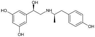
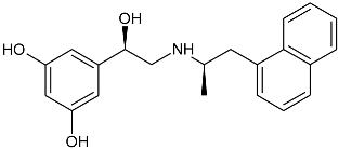
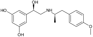
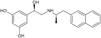
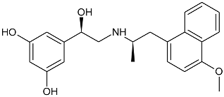
While the canonical part of the binding pocket in β-adrenergic receptors remains highly evolutionary conserved, the residues forming ELBDs may differ depending on the receptor subtype and this fact is widely exploited by subtype specific ligands. In particular, the β2-AR selective ligands contain elaborated substructures at their aminoalkyl tails aimed to explore the ELBD of the β2-AR. Remarkably, a very peculiar example of biased agonism at the β2-AR subtype was reported and related to the specific structural features introduced to β2-AR selective agonists. Studies by Xiao et al., indicated that β2-AR upon agonist activation couples to Gs and, to a lesser extent, Gi proteins [25]. The two events have the opposite effects on studied cell system (rat cardiomyocytes) affecting positively (Gs-coupling) and negatively (Gi-coupling) contractility of the cells [26]. The Gi coupling can be blocked by pertussis toxin (PTx) and adding PTx to the experiment resulted in significantly increased potency of many β2-AR agonists e.g., salbutamol, procaterol, zinterol, etc. [25]. Fenoterol, a selective β2-AR agonist was characterized as an exemption, activation pattern induced by that ligand in cardiomyocytes appeared to be PTx independent. The observation was interpreted as biased agonism, fenoterol and later some of its derivatives activated the receptor to a form that coupled Gs protein only, while other typical agonists induce dual, [Gs + Gi] coupling. [27]. Our group performed a medicinal chemistry project by chiral switching and aminoalkyl tail optimization of fenoterol scaffold aimed at development of highly selective β2-AR agonists [28,29,30]. The project generated a number of structures with diverse biological and pharmacological activities [31,32,33,34]. This congeneric cohort of compounds proved to be very effective chemical biology tool in structural characterization of β2-AR activation patterns leading to either Gs selective coupling (PTx insensitive) or dual, [Gs + Gi] coupling (PTx sensitive). Table 1 shows a selection of structures generated in the project along with their potency to activate the receptor to either Gs exclusive or dual signaling pattern [27]. While many tested molecules induced [Gs + Gi] profile, three of the derivatives (R,R)-01, (R,R)-02 and (R,R)-03 activated β2-AR at cardiomyocytes in PTx insensitive manner. SAR analysis allowed assigning key structural condition at the aminoalkyl tail of those three compounds, a hydrogen bond acceptor atom attached at the 4′ position of benzene ring system. It led to the hypothesis that the presence of –OH, –OCH3 or –NH2 substituent at that position was essential to form a specific interaction with the receptor, which might be a key event to induce specific activation of the β2-AR leading to Gs selective coupling. Subsequent docking simulations postulated that such an interaction may be realized by hydrogen bond (HB) created between a 4′-substituent of a ligand and hydroxyl group of Y3087.35 within the ELBD. Molecular modeling observation was further confirmed by mutagenesis studies, Y3087.35A mutation introduced to a receptor resulted in significant drop of the affinity of the three Gs selective ligands in comparison of the affinity on the wild type. When other derivatives, inducing dual, [Gs+Gi] signaling pattern were tested (e.g., (R,R)-05, (R,R)-06 and (R,R)-07, Table 1), their affinities to the Y3087.35A mutant remained at the level measured previously at the wild type receptor. Another set of experiments on additional mutant, Y3087.35F evidenced that such mutated receptor regained the PTx sensitive pattern of activity when activated by (R,R)-01, (R,R)-02 and (R,R)-03 [27]. Taking together, all above results strongly suggested that unusual behavior of (R,R)-01, (R,R)-02 or (R,R)-03 eliciting exclusively Gs cellular response could result from capability of these ligands to form a HB with Y3087.35 residue located at ELBD. Figure 1A shows postulated mechanism of molecular interactions between (R,R)-02, a Gs signaling ligand forming HB with Y3087.35 residue. The complex is compared with results of docking simulations of (R,R)-05, a ligand lacking hydrogen-bond acceptor at 4′-position and inducing dual [Gs + Gi] signaling pattern, Figure 1B, where Y3087.35 residue forms alternative interhelical HB with nearby N2936.55 residue.

3. Dopaminergic Receptors

Growing body of evidence indicates that ELBD is an important structural hotspot to elicit ligand directed signaling patterns in other GPCRs as well. Tschammer et al. in their work on 1,4-disubstituted aromatic piperidines/piperazines acting on dopaminergic D2L receptor identified that H3936.55 played a crucial role in imposing a ligand directed signaling in the system [35]. The operational model of agonism was used to quantify the ligand bias between the ability of the compounds to inhibit cAMP accumulation or stimulate intracellular ERK1/2 phosphorylation and substantial ligand biased signaling was observed for the wild type receptor. When the H3936.55A variant of the receptor was tested, the overall increase of agonism was observed, however, the system lost its ability to produce biased signaling imposed by the agonists [35]. In another work, Fowler et al., studied D2L signaling in various cellular responses; adenylate cyclase, MAPK, arachidonic acid release, and guanosine 5′-O-(3-thio)triphosphate binding produced by three rigid agonists, dihydrexidine, dinapsoline and dinoxyline [36]. Alanine substitution approach was employed to explain the role of three serine residues located on TM5, S1935.42, S1945.43 and S1975.46 and the functional studies of the mutants showed that S1935.42A and S1975.46A mutations abolished the activity of dopamine and three other ligands although dihydrexidine retained intrinsic activity at MAPK function only with S1935.42A. Remarkably, S1945.43A mutation did not affect the intrinsic activity for adenyl cyclase and MAPK for any of the ligands, but eliminated arachidonic acid release activity for dinapsoline and dihydrexidine but not dinoxyline [36]. That was interesting observation particularly from the structural point of view, TM5 located S1945.43 residue faced the direction of TM6 and was capable to form a HB interaction with H3936.55 residue. Parallel molecular modeling studies by the same group postulated a possibility of two alternative ELBD conformations which could be responsible for different recognition of agonists and subsequently biased signaling of downstream receptor effects. The central role was played by H3936.55 residue swinging between two positions; in the first state the residue was shifted towards TM5 and formed interhelical HB with S1945.43 residue, whereas in the second state the H3936.55 residue was placed more towards TM7 and created alternative HB bridge with Y4297.35 residue [36]. Interestingly, the two-conformation mechanism proposed to control alternative ligand directed signaling patterns in D2L receptor highly resembles the mechanism proposed by Woo et al., for explanation of Gs vs. [Gs+Gi] alternative signaling observed in β2-AR system. Both mechanisms are realized by the residues being positional equivalents in the respective ELBDs. In addition, in both cases two alternative interhelical HBs has been postulated, the one linking residues at positions 7.35 and 6.55, the other connecting residue 6.55 with residue 5.43, see Figure 1.
Other elements of structural requirements for G protein-biased agonist activity in the D2L system were described in studies which elucidated four structural features that were crucial for agonist efficacy and signaling bias for MLS1547 and its derivatives [37,38,39]. One of the most important determinants for G protein-biased signaling was the interaction of quinoline ring of the ligand molecule with a hydrophobic pocket comprised of I184ECL2, F1895.38, and V1905.41 residues. The two HBs created between: (i) nitrogen atom in the ortho-position of the pyridine moiety of MLS1547 and T4126.54 of D2L and (ii) hydroxyl group at the quinoline ring of MLS1547 and D1143.32 were found crucial for the G biased signaling. Additionally, an important HB involving a conserved aspartate residue D1143.32 can be formed by the positively charged nitrogen atom at the piperazine moiety of MLS1547 [37]. Moreover, the chlorine atom at the quinoline ring of MLS1547 was hypothesized to interact with hydrophobic pocket and prevent the tilting of TM5 during receptor activation. The structural analogues of MLS1547 that dispossessed the chlorine atom at the quinoline ring presented a modest decrease in agonist potency and loss of signaling bias [38]. Additionally, F1895.38 was identified as a micro-switch that regulates the active state of D2L for recruiting β-arrestin [39]. Interestingly, such a switch exists not only in dopamine receptor but also in several other related GPCRs, including the β2-AR [40] and 5-HT2B serotonin receptors [41].

4. Muscarinic Receptors

Muscarinic acetylcholine receptors are another member of class A GPCRs, where ELBD is extensively exploited by medicinal chemists, particularly in development of allosteric molecules exploring this site [42]. Studies showed that allosteric interaction within ELBD altered the receptor signaling pattern and could be used as a fine tuner of downstream signaling. For example, Bock et al. developed a cohort of hybrid compounds targeting M2 receptor in a dualsteric manner: the iperoxo building block while targeting the ortosteric site with super high affinity was linked to phtalimide or naphtalimide buinding blocks designated to allosterically interact with ELBD; hexamethylene or octamethylene spacer was used to link the two blocks [43]. The authors characterized synthesized structures by the bias plot comparing M2 receptor signaling via Gi ([35S]guanosine -5′-O-(3-thio)triphosphate binding to membranes of CHO-hM2 cells) and Gs (cAMP accumulation in CHO-hM2 cells pretreated with PTx) routes. The study showed that, while acetylcholine and iperoxo itself induced dual Gi and Gs signaling upon M2 binding, the hybrid molecules induced signaling where Gi component was much more dominant and Gs component significantly diminished in comparison to the control compounds. Therefore, a significant structure dependent Gi bias could be assigned to those dual steric probes. Molecular modeling simulations suggested that observed difference in binding modes was due to interactions of an allosteric building block with two key residues located at the ELBD, W4227.35 and Y177ECL2. The authors hypothesized that dual steric ligands induced reduction in flexibility of that domain, what can be translated into restricted intracellular loop rearrangement and subsequently to impaired downstream signaling events. The hypothesis was further explored in mutational study where W4227.35A alteration was introduced to the receptor. For all studied structures binding affinities were significantly reduced in the mutant when compared to the wild type receptor. But most importantly, W4227.35A mutant gained in efficacy towards Gs pathway upon binding to the studied dual-steric ligand, in comparison to the wild type [43].
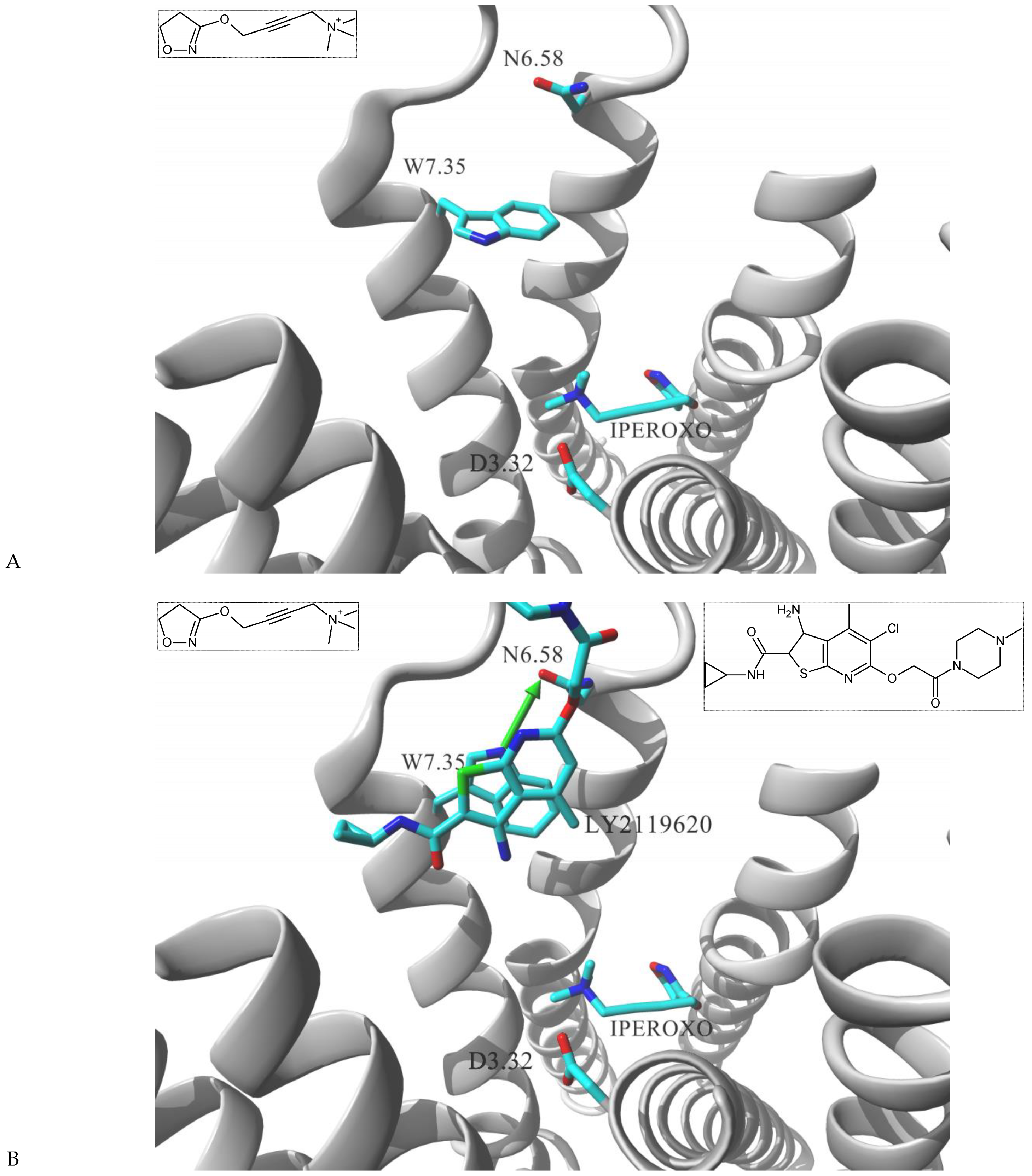
The importance of W7.35 residue in ligand directing signaling of muscarinic receptors was further confirmed in the structural biology study of agonist bound, active state of the human M2 receptor stabilized by a G-protein mimetic camelid antibody fragment [44]. In that work Kruse et al. resolved two ligand – receptor complexes, the first contained iperoxo molecule present in the ortosteric site; the second enclosed two ligand molecules, the orthosteric iperoxo and positive modulator molecule, LY2119620 occupying the allosteric vestibule. Figure 2 shows the comparison of these two complexes focusing on differences within the ELBD. Receptor arrangements are almost identical at the ortosteric domain recognizing iperoxo molecule. Comparison at the level of allosteric site also shows high similarities, but W4227.35 is a noteworthy exemption; its sidechain assumed two alternative conformations depending on whether LY2119620 molecule was present at ELBD or not. In the iperoxo–M2 complex, the indole ring of the residue adopts a fairly perpendicular orientation in relation to TM7 axis (nearly parallel in relation to the membrane surface). Such conformation allows W4227.35 residue for flexible exploration of the cavity devoid of allosteric ligand (Figure 2A). However, such bulky residue switched that conformation while in the complex containing both iperoxo and LY2119620. As shown in Figure 2B, the indole ring of W4227.35 is sterically required to vacate the space for the allosteric ligand and reorients parallel to the LY2119620 ring system, parallel to TM7 axis and perpendicular to membrane surface. Such conformation has a significant consequence; the W4227.35 conformer while in (iperoxo + LY2119620)–M2 complex is able to create interhelical HB with N4106.58.

5. Serotonin Receptors

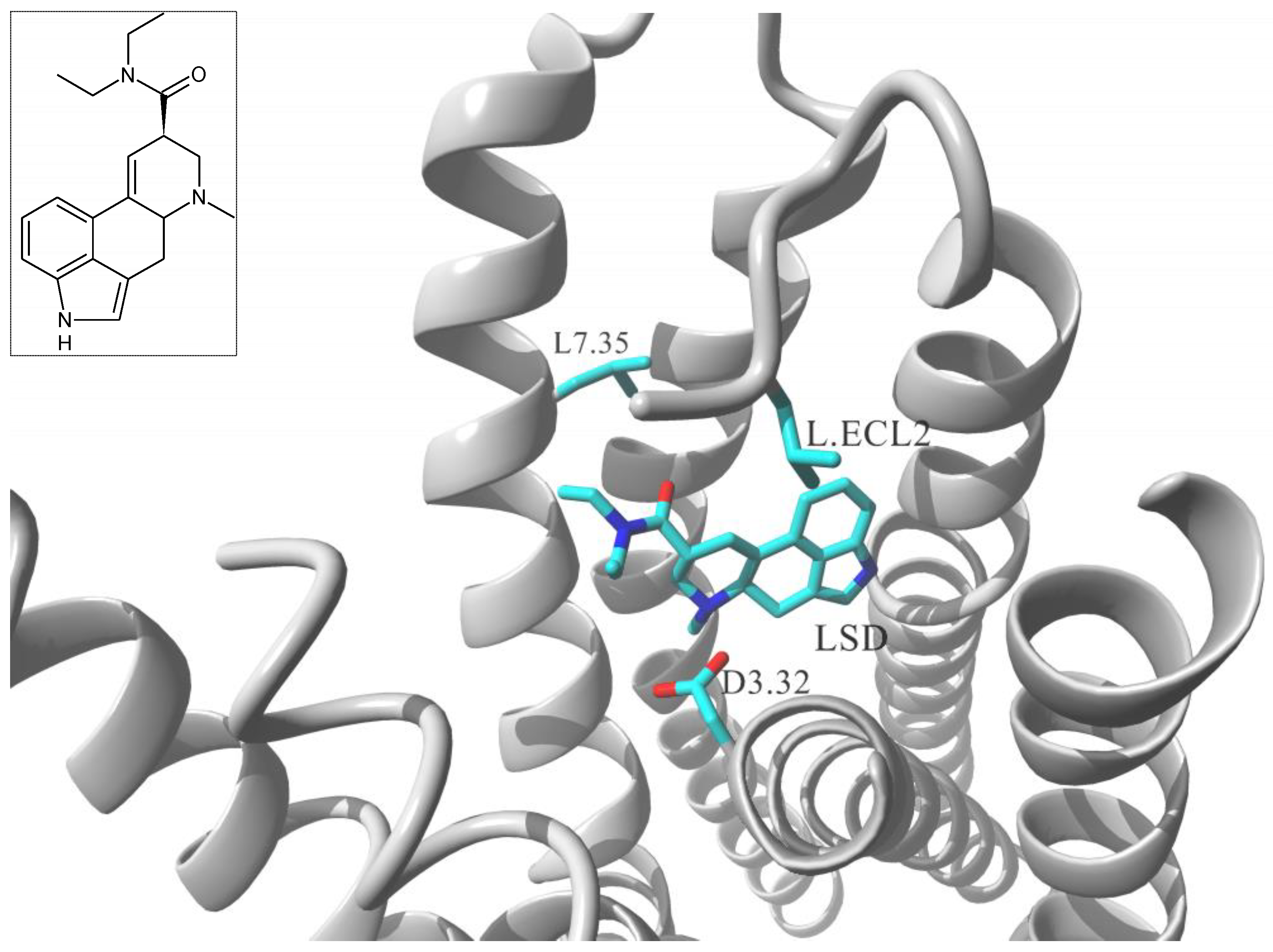
Lisergic acid diethylamide (LSD) is the prototypical hallucinogen that primarily acts via serotonin (5-HT) receptors. Wacker et al. resolved crystal structure of human 5-HT2B receptor in complex with LSD [45] and the study revealed a conformational reorganization of the receptor ECL2 in order to accommodate that particular ligand. Subsequent molecular dynamics (MD) simulations postulated that rearrangement to be responsible for exceptionally slow dissociation rate in LSD–5-HT2B and LSD–5-HT2A complexes [45]. Particular role in that lid – like function of ECL2 section was assigned to L209ECL2 residue of the 5-HT2B receptor; the side chain of that residue was directed towards LSD and acted as a bulky steric blocker fixing the ligand molecule within contracted binding domain. The authors validated their postulate by 5-HT2B mutational study; the receptor where L209ECL2 was substituted with less bulky alanine side chain showed markedly faster off-rate of LSD when compared to the wild type readouts [38]. The study provided additional insight into structural features of the LSD–5-HT2B complex that might be related to biased agonism properties. Single-point mutation of L209ECL2A changed not only the ligand dissociation kinetics; it additionally changed the signaling pattern; LSD upon binding to wild type 5-HT2B induced signaling both via Gq protein coupling and via β-arrestin recruitment. In L209ECL2A mutant Gq coupling remained at relatively the same level in comparison to the wild type but the time dependent augmentation of β-arrestin recruitment was selectively attenuated [45].
5-HT receptors are known to be very sensitive in functional recognition of ligands within the binding site; even a very minute structural change of LSD molecule may translate its agonistic profile into an antagonist upon binding to the receptor [46]. In the structural study by McCorvy et al. the 5-HT2B binding modes of several chemically congeneric agonists and antagonists were compared [47]. The authors attempted to identify key residues responsible for the receptor activation and to postulate differential mechanisms of recognition of receptor agonists as opposed to antagonists; additional structure – guided mutagenesis experiments revealed residues that were essential for agonist mediated biased signaling [47]. The study characterized a series of residues essential for ligand recognition at the binding site but that was L3627.35 residue which was associated to quantitative differential in alternative signaling patterns. L3627.35F mutation at 5-HT2B receptor was shown to restore the agonistic properties of a nominal antagonist molecule, lisuride. However, the mutation reestablished only the Gq coupling signaling, while the ligand did not elicit β-arrestin-2 related agonism. Similarly, the L3627.35F mutation did not affect LSD’s Gq agonism, but it abolished β-arrestin recruitment observed for LSD on the wild type receptor [40]. Further structural studies of receptor cocrystalized with non-ergoline, purportedly 5-HT2B selective antagonist, LY266097 revealed that extracellular tip of TM7 acted as a trigger for biased agonism. While the tetrahydro-β-carboline core of the ligand molecule was placed within the ortosteric site and interacted with residues Asp1353.32, Phe3406.51 and 3416.52, its 2-chloro-3,4-dimethoxybenzyl substituent was oriented much closer to TM7 than lisuride molecule. Functional studies determined that LY266097 was a modest Gq partial agonists without detectable β-arrestin-2 activity. The authors hypothesized that the extend of Gq agonism was determined by a ligand contact with L3627.35 residue. The hypothesis was elegantly verified by functional studies at the L3627.35F mutant, where LY266097 elicited diminished Gq activity suggesting that bulkier phenylalanine at that position sterically clashed with 2-chloro-3,4-dimethoxy- benzyl moiety and abolished the agonism [47].
Taken together, two residues located at the ELBD of 5-HT2B receptor, Leu209ECL2 and Leu3627.35 can be directly linked with biased signaling induced by different ligands (see Figure 3). Interestingly, each of these residues can be precisely associated with two alternative signaling patterns; Leu209ECL2 residue has been found essential for massive β-arrestin-2 bias of LSD molecule [45], while Leu3627.35 emerged as involved in biasing the signaling towards Gq related pathways upon binding to LSD and some related molecules [47]. Notably, the two residues 5-HT2B receptor are the positional equivalents of the residues involved in pincer like movement responsible for recognition of biased ligands described earlier in β1-AR receptor system, F201ECL2 and F3257.35.

6. Opioid Receptors

Three principal members of opioid receptor (OR), μ, δ and κ are important targets for pain management. Multiple lines of evidence show that the analgesic effect of μ-OR is related to Gi intracellular signaling induced upon receptor activation, while respiratory depression, impaired intestinal motility, opioid tolerance and addiction can be linked to the pathways regulated by β-arrestin receptor interactions. [48]. It underlines the importance of medicinal chemistry efforts to develop Gi biased ligands of μ-OR with minimal to no β-arrestin-2 signaling as future analgesics (for review, c.f. [49]). Numerous studies were aimed at explanation of ligand – receptor interaction that might be linked to a biased signaling of the receptor. Hothersall et al. identified two μ-OR mutations, W3207.35A and Y3287.43F that changed pathway bias with different patterns between peptide agonists. Subtype selective ligand DAMGO exhibited increase in β-arrestin activity at the W3207.35A mutant, while Y3287.43F substitution completely abrogated β-arrestin signaling. Endomorphin-1 gained the efficacies at both pathways with Y3287.43F mutation but lost them at W3207.35A. For endomorphin-2, the W3207.35A mutation resulted in a shift from Gi preferred bias at the wildtype towards β-arrestin2 bias at the mutant [50]. Cheng et al. performed molecular simulations of the Gi protein biased activation and inactivation mechanisms of μ-OR system [51]. MD of the receptor alone or in complex with Gi biased agonists, TRV130 or BU72 or antagonists, β-FNA and NTx suggested two residues, W2956.48 and Y3287.43 that acted as a paired activation switch. The authors postulated the switch to be critical for receptor activation and the ligand interactions that positioned the two residues influenced the increased or decreased signaling via β-arrestin. In more recent molecular modeling study de Waal et al. employed adaptively biased MD simulations to determine the molecular mechanisms of interactions between μ-OR system and fentanyl and two its derivatives, highly potent β-arrestin biased agonists [52]. The authors proposed an activation mechanism where agonist molecules mediated β-arrestin signaling of μ-OR through a novel M1533.36 “microswitch” at the ortosteric site. The residue was identified to directly interact with ligands’ N–aniline ring and its conformational change affected the rotameric state of W2956.48 residue. The findings were further validated by design and synthesis of novel fentanyl based derivatives with confirmed complete, clinically desirable, Gi protein biased coupling [52].
Che at al. presented the nanobody stabilized active state structure of another subtype, the κ-OR [53]. A combination of structural biology analyses, binding and functional assays allowed identification of key residues important as molecular determinants of κ-OR ligand binding and agonist efficacy. Among them, κ-OR residues involved in conferring different patterns of biased signaling were postulated. The authors showed for example that replacing Y3127.35 in the κ-OR binding pocket with tryptophan residue found in the corresponding position of μ-OR, transformed IBNtxA, a balanced κ-OR agonist into a G-protein-biased ligand (thus mimicking its activity at the μ-OR subtype). That was an elegant illustration that certain residues located at corresponding positions of closely related subtypes may be responsible for eliciting differential signaling patterns between the two receptors [53].
A structural study of another subtype of opioid receptor, human δ-OR, revealed a key role of complexed sodium ion in mediating allosteric control of receptor functional selectivity towards a specific transducer [54]. Functional studies on the δ-OR variants, where key residues forming the allosteric locus for Na+ ion were mutated, identified that N1313.35A/V augmented constitutive β-arrestin-mediated signaling, while D952.50A, N3107.45A and N3147.49A mutations transformed classical δ-opioid antagonists into a β-arrestin-biased agonists. Overall, the data revealed that sodium-coordinating residues acted as “efficacy switches” at a prototypic G-protein-coupled receptor. [54]. It revealed that the sodium ion regulated the activation of a GPCR through cooperation with the ligand at the orthosteric site. Subsequently, Sun at al. presented the results of computational simulation of δ-OR system that postulated W2746.48 residue located adjacent to the allosteric Na+ binding site to establish a bridge between the sodium allosteric site and the orthosteric site. During MD simulations allosteric sodium ion exploited a distinct conformation of that key residue to propagate the modulation to TM5 and TM6, which was further transmitted along the helixes and regulated their positions on the intracellular side. The models hypothesized the contrast between the allosteric effects towards the two transducing partners of the receptor, Na+ allosteric modulation significantly altered the β-arrestin recruitment, while it affected the G protein to much lesser extent [55].

7. Angiotensin Receptor

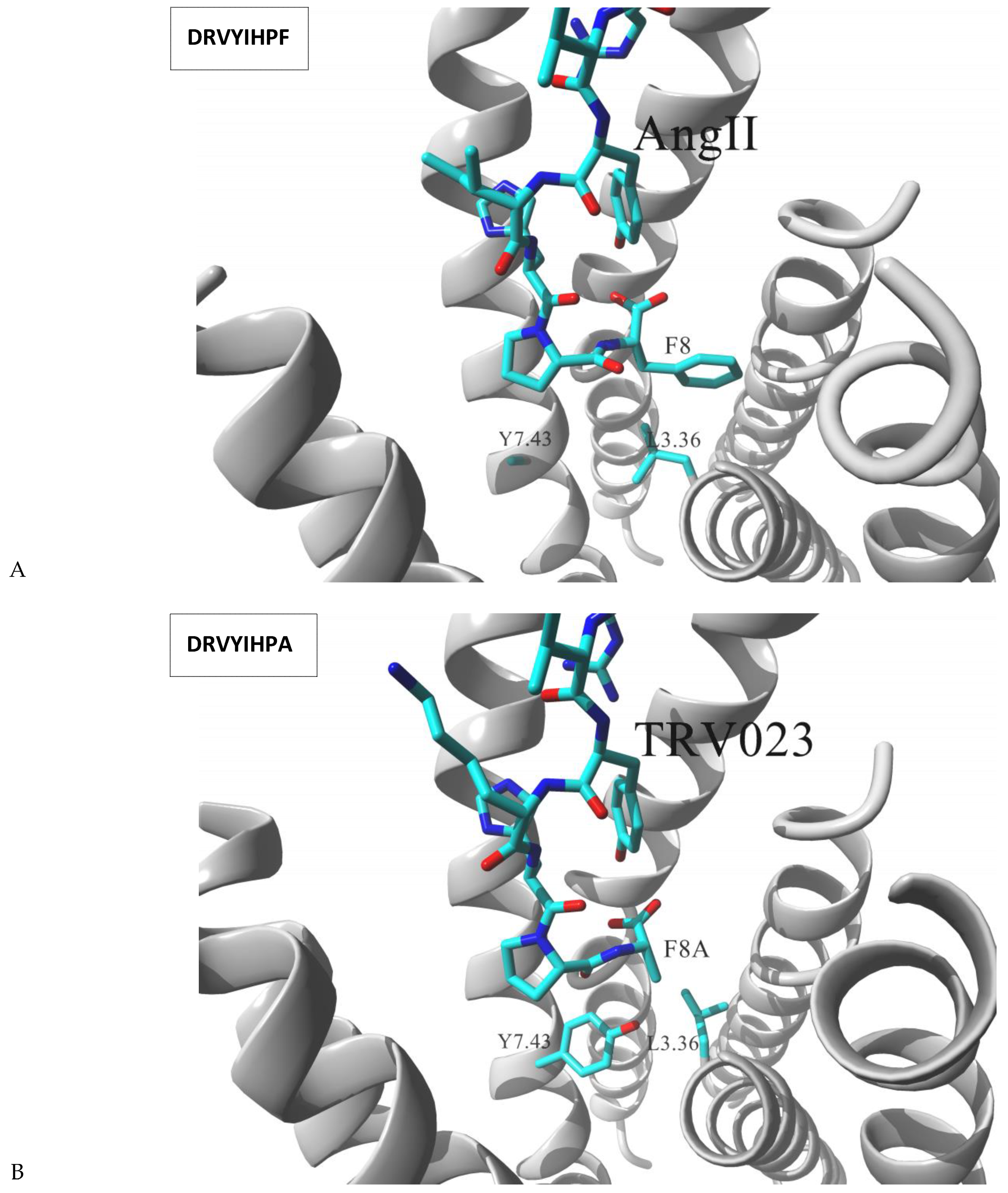
Angiotensin receptor 1 (AT1) is very well characterized for its ligand directed signaling properties. Binding of receptor’s endogenous ligand, the octapeptide angiotensin II (AngII) stimulates both Gq protein mediated and arrestin mediated signaling pathways. Functional studies of AngII showed that its single point mutations could result in either β-arrestin – biased or Gq – biased response in comparison to the wild type hormone [56]. For example, F8A mutation significantly altered the AT1 signaling pattern and such octapeptide did not activate the receptor to elicit Gq dependent routes while still maintained high level of β-arrestin signaling. The receptor is a major drug target, and there is a significant interest in development of arrestin – biased AT1 ligands for treatment of heart failure, such ligands have clinically relevant antihypertensive effects but also improve cardiac function through β-arrestin – mediated pathways [57]. Recent structural biology report [58] positioned the AngII molecule within the AT1 structure locating the hormone N-terminus at the extracellular entrance and the C-terminus at the base of the binding pocket. Closer inspection of the complex indicated that AngII F8 residue pointed towards the vestibule formed by the central portions of TM3, TM4 and TM5 of AT1, however, that C-terminal residue of the hormone was poorly resolved with markedly high B-factors. Additionally, weak or none electron density was observed for the side chains of the two AT1 residues located nearby F8 of AngII, L1123.36 and Y2927.43 suggesting that region of the AngII – AT1 complex to be conformationally heterogeneous [58]. In the same study, two alternative versions of AngII molecule, F8A mutant (TRV023) and F8del (TRV026) were cocrystalized with AT1, and the results evidenced that both L1123.36 and Y2927.43 residues were resolved better with lower B-factors while complexed with those β-arrestin biased molecules. Additionally, clear rearrangement of the two residues could be observed, TM3 was rotated on-axis moving L1123.36 away from position occupied by Y2927.43 in comparison to the wild type AngII – AT1 complex (Figure 4). Consistent with crystallographic observations, L1123.36A mutation of AT1, a residue expected to better accommodate the flexibility of AngII F8, increased receptor affinity to the wild type AngII but decreased affinity for TRV026 or TRV023. Similarly, Y2927.43A substitution (which was designed by the authors to mimic disordered state induced by the hormone — see Figure 4A) increased AT1 affinity for AngII, but had relatively minor effects on TRV026 or TRV023. The authors also noted that both mutations, despite enhancing affinities to AngII, did not induce increased efficacy or potency in activating AngII-dependent Gq signaling [58].
Above structural results led to the postulate that L1123.36 and Y2927.43 residues of AT1 played a decisive role in inducing the receptor coupling to Gq protein upon binding of AngII. The hypothesis was additionally validated by extensive molecular dynamics simulations of the AngII–AT1 complex [59]. The simulations suggested that the agonist bound receptor was oscillating between two conformations referred to as canonical active conformation or alternative active conformation. During that transition TM7 twisted above its proline kink leading the intracellular portion of TM7 to shift away from TM2 and TM3. In details, the movement induced N461.50 to switch hydrogen bond interaction from C2967.47 to N2957.46 and further side chains rearrangement of adjacent residues. Importantly, the authors additionally noted that those intracellular transitions of TM7 region were allosterically linked to the ligand binding region, affecting in particular Y2927.43 residue position. In the alternative active conformation, the residue pointed towards TM3 while in the canonical active conformation the Tyr2927.43 was shifted toward TM2. The change affected side chain conformations of adjacent residues in AT1, L1123.36, N1113.35 and F772.52 as well as F8 residue of AngII. Further replica exchange MD simulations showed that β-arrestin biased ligands favored the alternative active conformation while ligands with Gq-protein bias properties favored binding to the canonical active state [59].

8. Conclusions

A growing number of structural biology studies support the idea that class A GPCRs, despite having high variance in amino acid sequences, share similar three dimensional folding as well as analogous patterns of conformational transitions in response to interactions with their binding partners, intracellular transduces and extracellular ligands [10]. One of the most canonical hallmarks of GPCRs activation is the outward movement of the intracellular portions of TM5 and TM6 allowing the receptor to open the interface for interaction with a G protein [60]. Evolutionary developed variations in residues shaping this interface play a decisive role in recognition of specific type(s) of protein G, in a similar manner differences in residues lining orthosteric binding domain are critical for specific recognition of endogenous hormone or neurotransmitter. Hence, different receptors belonging to class A (having the ligand binding domain located at the bundle of transmembrane helices) maintain relatively uniform molecular mechanisms of conformational transitions upon activation, while differences in their sequences allows for enormous versatility in both recognition of agonists and signal transmission via interactions with transducing protein(s). Ligand directed signaling is a phenomenon that has been puzzling researches for years as it opens novel possibilities of using specialized ligand molecules for precise tuning of induced signaling pathway(s) via one pharmacological target. Combination of medicinal chemistry, molecular pharmacology, structural biology and mutagenesis studies sheds the light on the structural mechanisms responsible for biased agonism functionality in some representatives of class A receptors. Very interestingly, these mechanisms show significant similarities when compared across characterized receptor systems.
First group of GPCRs discussed above are receptors for neurotransmitters; small molecules like epinephrine (β-ARs) acetylcholine (M2), dopamine (D2L) and serotonin (5-HT2B). All these receptors can be characterized that their ortosteric sites accommodating an endogenous neurotransmitter molecule are accompanied by its extension, the ELBD. The latter domain does not seem to interact significantly with a neurotransmitter, however, many exogenous agonists including drugs introduced into clinical practice explore both the ortosteric and ELBD while binding to the receptor. Examples presented above illustrate that, while the core drug – receptor interactions occurs at the ortosteric site, additional interactions with residues lining ELBDs can play important roles in subtle tuning of observed ligand directed signaling. Noteworthy, the residues that were postulated to form essential interactions biasing the ligand functions shared analogous positioning within ELBDs of different receptors. The studies identified those hot spots residues to be typically located at upper tips of TM7 (7.35 position at β1-AR, β2-AR, M2, D2L and 5-HT2B systems); TM6 (6.55 position in β2-AR and M2 receptor and 6.58 position at D2L receptor) and TM5 (5.43 at β2-AR and M2). Typically, residues at those positions were involved in interhelical HB interactions which were switched on and off while interacting with agonists of alternative signaling profiles (observed in β2-AR, M2 and D2L systems). Additional important position was found at the ECL2 (in β1-AR and 5HT2B receptors), the loop acts of specific lid of many ELBDs, and the residues involved in interactions with biased agonists pointed downward the ECL2. It was suggested that ligands of alternative signaling patterns stabilized certain interactions bridging TM helices and shaped the position of ECL2, thus, affecting the flexibility of ELBD what could translate into restricted intracellular loop rearrangement and subsequently to alter downstream signaling events [36].
In contrast to neurotransmitter receptors, ORs and AT1 are GPCRs sensing endogenous peptides. These bulkier molecules natively bind to a large receptor vestibule covering completely a space previously referred to as ortosteric and ELBD domains. Studies on μ-OR and κ-OR indicated again important role of 7.35 residue played in recognition of agonists eliciting alternative signaling patterns. δ-OR investigations on the other hand underlined the role of region located at the bottom of the ligand binding cavity, W6.48 toggle switch residue was hypothesized to establish a bridge transmitting biased signals from the ortosteric site to the allosteric cavity accommodating Na+ ion deep in the TM helical bundle. The importance of residues lining the bottom of ortosteric site was explicitly showed in the AT1 system where studies of AngII molecule cocrystalized with the receptor identified conformationally heterogeneous region in the complex: F8 residue of AngII and L3.36 and Y7.43 of AT1. Simultaneous structural studies of β-arrestin biased AngII mutants, F8A and F8del evidenced that both residues at 3.36 and 7.43 positions showed much better resolution. Additional rearrangements of positions of these residues were observed when compared to native, wild type AngII cocrystalized structure. The observation strongly suggests that these two residues located at the center of the orthosteric site play a crucial role in inducing the receptor coupling to Gq protein upon binding of AngII.
While growing number of structural biology studies characterize molecular nature of specific interactions between a G-protein heterodimer and various GPCRs [6,60,61], potential mechanisms coupling the receptors to arrestins remain much more elusive. Recently, however, structures of β-arrestin-1 complexes with neurotensin-1 receptor [62] and turkey thermostable β1-AR [63] were reported. In the latter study, Lee et al. described cryo-electron microscopy structure of formoterol–β1-AR complex coupled to β-arrestin-1 in lipid nanodiscs. The authors compared their β-arrestin-1 bound β1-AR complex with previously reported Gs heterotrimer bound formoterol–β2-AR complex [64] and the difference in conformational arrangement of receptors was found; the finger loop of β-arrestin-1 occupied a narrower cleft on the intracellular surface and it was closer to TM7 of the receptor when compared with the C-terminal α5 helix of Gs [63]. The authors suggested that, upon G protein dissociation, coupling of β-arrestin-1 to the receptor was accompanied with slight inward movement of the cytoplasmic end of TM5, resulting in an outward movement of its extracellular tip; the move of the helix away from the ligand binding domain could weaken the HB interactions between formoterol and TM5 [63]. Based on that model, one can hypothesize that the β-arrestin-1 induced change in TM5 rearranges the ELBD and might affect a pattern of interactions between TM5 and adjacent helices and/or ECL2 lid. Therefore, specific parts of various agonists by fine interactions within these regions may affect the conformational state(s) that are preferential for interactions with one or another transducers eventually leading to a ligand directed signaling.
Emerging structural information on ligand – receptor interactions involved in alternative recognition of biased agonists is of the frontline importance in current and future GPCR pharmacology. Related receptor subtypes sensing the same neurotransmitter have their orthosteric sites formed by specialized and highly evolutionary conserved residues. It is not necessarily true in case of their ELBDs and the fact is widely explored by medicinal chemistry in development of ligands selective towards a specific receptor. Many such molecules effectively exercise their selectivity via “address” residues located at ELBDs that differ between subtypes. We must be aware that plenty of subtype selective ligands that have been developed so far, may express distinct, sometimes unexpected or unwelcome, signaling patterns and the fact might have clinical consequences. On the other hand, future development of subtype selective ligands opens the possibility for fine tuning of induced signaling pathways by a control of agonist – ELBD interactions. One example has been described above, fenoterol-based agonists of β2-AR activating the receptor to couple either Gs only or dually [Gs + Gi] are highly selective towards that particular receptor subtype. Their cellular (and possibly physiological) effects, however, can be fine-tuned by a presence of a hydrogen bond acceptor substituent at the agonist aminoalkyl tail that controls possible ligand interaction with 7.35 receptor residue [27]. AT1, on the other hand, is a hallmark for its ligand directed signaling properties and currently approved antihypertensive drugs targeting the receptor may be considered suboptimal as they attenuate both Gq related signaling and β-arrestin related signaling in response to AngII [58]. The former is a desired therapeutic effect in hypertension treatment, the latter is undesired as β-arrestin signaling via AT1 is considered cardioprotective [57] and heart failure treatment may significantly benefit from development of β-arrestin biased AT1 agonists, such ligands can increase cardiac contractility without undesired hypertensive effects [65]. Also, novel agonists of μ-OR biased towards Gi signaling with minimal effect on β-arrestin signaling is a promising strategy to obtain a new class of antinociceptives devoid of adverse and drug tolerance effects. TRV130 is the first such a candidate introduced into the phase III clinical trial for treatment of acute severe pain [66]. Hence, more detailed understanding of structural aspects of interactions between pharmacologically relevant receptor and their biased ligands gives novel therapeutic perspective.

Funding

This research was partially founded by National Science Centre, Poland (Grant no. 2017/25/B/NZ7/02654) (to A.P.). The APC was founded by 2017/25/B/NZ7/02654 and the Medical University of Lublin statutory founds.

Conflicts of Interest

The authors declare no conflict of interest.

References

  1. Lefkowitz, R.J. A brief history of G-protein coupled receptors (Nobel Lecture). Angew. Chem. Int. Ed. Engl. 2013, 52, 6366–6378. [Google Scholar] [CrossRef]
  2. Kobilka, B. The structural basis of G-protein-coupled receptor signaling (Nobel Lecture). Angew. Chem. Int. Ed. Engl. 2013, 52, 6380–6388. [Google Scholar] [CrossRef] [Green Version]
  3. Latorraca, N.R.; Venkatakrishnan, A.J.; Dror, R.O. GPCR Dynamics: Structures in Motion. Chem. Rev. 2017, 117, 139–155. [Google Scholar] [CrossRef]
  4. Manglik, A.; Kruse, A.C. Structural Basis for G Protein-Coupled Receptor Activation. Biochemistry 2017, 56, 5628–5634. [Google Scholar] [CrossRef] [PubMed]
  5. Kenakin, T. Biased Receptor Signaling in Drug Discovery. Pharmacol. Rev. 2019, 71, 267–315. [Google Scholar] [CrossRef] [Green Version]
  6. Wingler, L.M.; Lefkowitz, R.J. Conformational Basis of G Protein-Coupled Receptor Signaling Versatility. Trends Cell Biol. 2020, 30, 736–747. [Google Scholar] [CrossRef] [PubMed]
  7. Gurevich, V.V.; Gurevich, E.V. GPCR Signaling Regulation: The Role of GRKs and Arrestins. Front. Pharmacol. 2019, 10, 125. [Google Scholar] [CrossRef] [PubMed] [Green Version]
  8. Zhou, X.E.; Meicher, K.; Xu, H.E. Understanding the GPCR biased signaling through G protein and arrestin complex structures. Curr. Opin. Struct. Biol. 2017, 45, 150–159. [Google Scholar] [CrossRef]
  9. Rankovic, Z.; Brust, T.F.; Bohn, L.M. Biased agonism: An emerging paradigm in GPCR drug discovery. Bioorg. Med. Chem. Lett. 2016, 26, 241–250. [Google Scholar] [CrossRef] [Green Version]
  10. Kenakin, T.; Miller, L.J. Seven transmembrane receptors as shapeshifting proteins: The impact of allosteric modulation and functional selectivity on new drug discovery. Pharmacol. Rev. 2010, 62, 265–304. [Google Scholar] [CrossRef] [Green Version]
  11. Shonberg, J.; Lopez, L.; Scammells, P.J.; Christopoulos, A.; Capuano, B.; Lane, J.R. Biased agonism at G protein-coupled receptors: The promise and the challenges--a medicinal chemistry perspective. Med. Res. Rev. 2014, 34, 1286–1330. [Google Scholar] [CrossRef]
  12. Wootten, D.; Christopoulos, A.; Marti-Solano, M.; Babu, M.M.; Sexton, P.M. Mechanisms of signalling and biased agonism in G protein-coupled receptors. Nat. Rev. Mol. Cell Biol. 2018, 19, 638–653. [Google Scholar] [CrossRef] [PubMed]
  13. Lee, S.-M.; Booe, J.M.; Pioszak, A.A. Structural insights into ligand recognition and selectivity for classes A, B, and C GPCRs. Eur. J. Pharmacol. 2015, 763 Pt B, 196–205. [Google Scholar] [CrossRef] [Green Version]
  14. Hilger, D.; Masureel, M.; Kobilka, B.K. Structure and dynamics of GPCR signaling complexes. Nat. Struct. Mol. Biol. 2018, 25, 4–12. [Google Scholar] [CrossRef] [PubMed]
  15. Shonberg, J.; Kling, R.C.; Gmeiner, P.; Löber, S. GPCR crystal structures: Medicinal chemistry in the pocket. Bioorg. Med. Chem. 2015, 23, 3880–3906. [Google Scholar] [CrossRef]
  16. Kolinski, M.; Plazinska, A.; Jozwiak, K. Recent progress in understanding of structure, ligand interactions and the mechanism of activation of the β₂-adrenergic receptor. Curr. Med. Chem. 2012, 19, 1155–1163. [Google Scholar] [CrossRef]
  17. Wendell, S.G.; Fan, H.; Zhang, C. G Protein-Coupled Receptors in Asthma Therapy: Pharmacology and Drug Action. Pharmacol. Rev. 2020, 72, 1–49. [Google Scholar] [CrossRef] [Green Version]
  18. Fumagalli, C.; Maurizi, N.; Marchionni, N.; Fornasari, D. beta-blockers: Their new life from hypertension to cancer and migraine. Pharmacol. Res. 2020, 151, 104587. [Google Scholar] [CrossRef] [PubMed]
  19. Wnorowski, A. To block it, or not to block it? J. Cancer Res. Clin. Oncol. 2017, 143, 2631–2633. [Google Scholar] [CrossRef] [Green Version]
  20. Sinagra, G.; Corrà, U.; Contini, M.; Magrì, D.; Paolillo, S.; Perrone Filardi, P.; Sciomer, S.; Badagliacca, R.; Agostoni, P. Choosing among β-blockers in heart failure patients according to β-receptors’ location and functions in the cardiopulmonary system. Pharmacol. Res. 2020, 156, 104785. [Google Scholar] [CrossRef]
  21. Warne, T.; Edwards, P.C.; Leslie, A.G.; Tate, C.G. Crystal structures of a stabilized β1-adrenoceptor bound to the biased agonists bucindolol and carvedilol. Structure 2012, 20, 841–849. [Google Scholar] [CrossRef] [Green Version]
  22. Wisler, J.W.; DeWire, S.M.; Whalen, E.J.; Violin, J.D.; Drake, M.T.; Ahn, S.; Shenoy, S.K.; Lefkowitz, R.J. A unique mechanism of beta-blocker action: Carvedilol stimulates beta-arrestin signaling. Proc. Natl. Acad. Sci. USA 2007, 104, 16657–16662. [Google Scholar] [CrossRef] [PubMed] [Green Version]
  23. Warne, T.; Edwards, P.C.; Doré, A.S.; Leslie, A.G.W.; Tate, C.G. Molecular basis for high-affinity agonist binding in GPCRs. Science 2019, 364, 775–778. [Google Scholar] [CrossRef] [PubMed]
  24. Ballesteros, J.A.; Weinstein, H. Integrated methods for the construction of three-dimensional models and computational probing of structure-function relations in G protein-coupled receptors. In Methods in Neurosciences; Sealfon, S.C., Ed.; Academic Press: Cambridge, MA, USA, 1995; Volume 25, pp. 366–428. [Google Scholar]
  25. Xiao, R.P.; Ji, X.; Lakatta, E.G. Functional coupling of the beta 2-adrenoceptor to a pertussis toxin-sensitive G protein in cardiac myocytes. Mol. Pharmacol. 1995, 47, 322–329. [Google Scholar] [PubMed]
  26. Xiao, R.P.; Avdonin, P.; Zhou, Y.Y.; Cheng, H.; Akhter, S.A.; Eschenhagen, T.; Lefkowitz, R.J.; Koch, W.J.; Lakatta, E.G. Coupling of beta2-adrenoceptor to Gi proteins and its physiological relevance in murine cardiac myocytes. Circ. Res. 1999, 84, 43–52. [Google Scholar] [CrossRef] [PubMed] [Green Version]
  27. Woo, A.Y.; Jozwiak, K.; Toll, L.; Tanga, M.J.; Kozocas, J.A.; Jimenez, L.; Huang, Y.; Song, Y.; Plazinska, A.; Pajak, K.; et al. Tyrosine 308 is necessary for ligand-directed Gs protein-biased signaling of β2-adrenoceptor. J. Biol. Chem. 2014, 289, 19351–19363. [Google Scholar] [CrossRef] [Green Version]
  28. Jozwiak, K.; Khalid, C.; Tanga, M.J.; Berzetei-Gurske, I.; Jimenez, L.; Kozocas, J.A.; Woo, A.; Zhu, W.; Xiao, R.P.; Abernethy, D.R.; et al. Comparative molecular field analysis of the binding of the stereoisomers of fenoterol and fenoterol derivatives to the beta2 adrenergic receptor. J. Med. Chem. 2007, 50, 2903–2915. [Google Scholar] [CrossRef]
  29. Jozwiak, K.; Woo, A.Y.; Tanga, M.J.; Toll, L.; Jimenez, L.; Kozocas, J.A.; Plazinska, A.; Xiao, R.P.; Wainer, I.W. Comparative molecular field analysis of fenoterol derivatives: A platform towards highly selective and effective beta(2)-adrenergic receptor agonists. Bioorg. Med. Chem. 2010, 18, 728–736. [Google Scholar] [CrossRef] [Green Version]
  30. Plazinska, A.; Pajak, K.; Rutkowska, E.; Jimenez, L.; Kozocas, J.; Koolpe, G.; Tanga, M.; Toll, L.; Wainer, I.W.; Jozwiak, K. Comparative molecular field analysis of fenoterol derivatives interacting with an agonist-stabilized form of the β₂-adrenergic receptor. Bioorg. Med. Chem. 2014, 22, 234–246. [Google Scholar] [CrossRef] [Green Version]
  31. Jozwiak, K.; Toll, L.; Jimenez, L.; Woo, A.Y.; Xiao, R.P.; Wainer, I.W. The effect of stereochemistry on the thermodynamic characteristics of the binding of fenoterol stereoisomers to the beta(2)-adrenoceptor. Biochem. Pharmacol. 2010, 79, 1610–1615. [Google Scholar] [CrossRef] [Green Version]
  32. Toll, L.; Jimenez, L.; Waleh, N.; Jozwiak, K.; Woo, A.Y.; Xiao, R.P.; Bernier, M.; Wainer, I.W. β2-adrenergic receptor agonists inhibit the proliferation of 1321N1 astrocytoma cells. J. Pharmacol. Exp. Ther. 2011, 336, 524–532. [Google Scholar] [CrossRef] [PubMed] [Green Version]
  33. Wnorowski, A.; Sadowska, M.; Paul, R.K.; Singh, N.S.; Boguszewska-Czubara, A.; Jimenez, L.; Abdelmohsen, K.; Toll, L.; Jozwiak, K.; Bernier, M.; et al. Activation of β2-adrenergic receptor by (R,R’)-4’-methoxy-1-naphthylfenoterol inhibits proliferation and motility of melanoma cells. Cell Signal. 2015, 27, 997–1007. [Google Scholar] [CrossRef] [Green Version]
  34. Wnorowski, A.; Such, J.; Paul, R.K.; Wersto, R.P.; Indig, F.E.; Jozwiak, K.; Bernier, M.; Wainer, I.W. Concurrent activation of β2-adrenergic receptor and blockage of GPR55 disrupts pro-oncogenic signaling in glioma cells. Cell Signal. 2017, 36, 176–188. [Google Scholar] [CrossRef] [PubMed]
  35. Tschammer, N.; Bollinger, S.; Kenakin, T.; Gmeiner, P. Histidine 6.55 is a major determinant of ligand-biased signaling in dopamine D2L receptor. Mol. Pharmacol. 2011, 79, 575–585. [Google Scholar] [CrossRef] [Green Version]
  36. Fowler, J.C.; Bhattacharya, S.; Urban, J.D.; Vaidehi, N.; Mailman, R.B. Receptor conformations involved in dopamine D(2L) receptor functional selectivity induced by selected transmembrane-5 serine mutations. Mol. Pharmacol. 2012, 81, 820–831. [Google Scholar] [CrossRef] [PubMed] [Green Version]
  37. Michino, M.; Free, R.B.; Doyle, T.B.; Sibley, D.R.; Shi, L. Structural basis for Na(+)-sensitivity in dopamine D2 and D3 receptors. Chem. Commun. (Camb) 2015, 1, 8618–8621. [Google Scholar] [CrossRef] [Green Version]
  38. Free, R.B.; Chun, L.S.; Moritz, A.E.; Miller, B.N.; Doyle, T.B.; Conroy, J.L.; Padron, A.; Meade, J.A.; Xiao, J.; Hu, X.; et al. Discovery and characterization of a G protein-biased agonist that inhibits β-arrestin recruitment to the D2 dopamine receptor. Mol. Pharmacol. 2014, 86, 96–105. [Google Scholar] [CrossRef] [Green Version]
  39. Sanchez-Soto, M.; Verma, R.K.; Willette, B.K.A.; Gonye, E.C.; Moore, A.M.; Moritz, A.E.; Boateng, C.A.; Yano, H.; Free, R.B.; Shi, L.; et al. A structural basis for how ligand binding site changes can allosterically regulate GPCR signaling and engender functional selectivity. Sci. Signal. 2020, 13, eaaw5885. [Google Scholar] [CrossRef]
  40. Ring, A.M.; Manglik, A.; Kruse, A.C.; Enos, M.D.; Weis, W.I.; Garcia, K.C.; Kobilka, B.K. Adrenaline-activated structure of beta2-adrenoceptor stabilized by an engineered nanobody. Nature 2013, 502, 575–579. [Google Scholar] [CrossRef] [Green Version]
  41. Wacker, D.; Wang, C.; Katritch, V.; Han, G.W.; Huang, X.P.; Vardy, E.; McCorvy, J.D.; Jiang, Y.; Chu, M.; Siu, F.Y.; et al. Structural features for functional selectivity at serotonin receptors. Science 2013, 340, 615–619. [Google Scholar] [CrossRef] [Green Version]
  42. Christopoulos, A.; Kenakin, T. G protein-coupled receptor allosterism and complexing. Pharmacol. Rev. 2002, 54, 323–374. [Google Scholar] [CrossRef]
  43. Bock, A.; Merten, N.; Schrage, R.; Dallanoce, C.; Bätz, J.; Klöckner, J.; Schmitz, J.; Matera, C.; Simon, K.; Kebig, A.; et al. The allosteric vestibule of a seven transmembrane helical receptor controls G-protein coupling. Nat. Commun. 2012, 3, 1044. [Google Scholar] [CrossRef]
  44. Kruse, A.C.; Ring, A.M.; Manglik, A.; Hu, J.; Hu, K.; Eitel, K.; Hübner, H.; Pardon, E.; Valant, C.; Sexton, P.M.; et al. Activation and allosteric modulation of a muscarinic acetylcholine receptor. Nature 2013, 504, 101–106. [Google Scholar] [CrossRef] [PubMed] [Green Version]
  45. Wacker, D.; Wang, S.; McCorvy, J.D.; Betz, R.M.; Venkatakrishnan, A.J.; Levit, A.; Lansu, K.; Schools, Z.L.; Che, T.; Nichols, D.E.; et al. Crystal Structure of an LSD-Bound Human Serotonin Receptor. Cell 2017, 168, 377–389.e12. [Google Scholar] [CrossRef] [PubMed] [Green Version]
  46. Nichols, D.E.; Frescas, S.; Marona-Lewicka, D.; Kurrasch-Orbaugh, D.M. Lysergamides of isomeric 2,4-dimethylazetidines map the binding orientation of the diethylamide moiety in the potent hallucinogenic agent N,N-diethyllysergamide (LSD). J. Med. Chem. 2002, 45, 4344–4349. [Google Scholar] [CrossRef]
  47. McCorvy, J.D.; Wacker, D.; Wang, S.; Agegnehu, B.; Liu, J.; Lansu, K.; Tribo, A.R.; Olsen, R.H.J.; Che, T.; Jin, J.; et al. Structural determinants of 5-HT2B receptor activation and biased agonism. Nat. Struct. Mol. Biol. 2018, 25, 787–796. [Google Scholar] [CrossRef] [PubMed]
  48. Raehal, K.M.; Walker, K.L.; Bohn, L.M. Morphine side effects in beta-arrestin 2 knockout mice. J. Pharmacol. Exp. Ther. 2005, 314, 1195–1201. [Google Scholar] [CrossRef] [Green Version]
  49. Conibear, A.E.; Kelly, E. A Biased View of μ-Opioid Receptors? Mol. Pharmacol. 2019, 96, 542–549. [Google Scholar] [CrossRef]
  50. Hothersall, J.D.; Torella, R.; Humphreys, S.; Hooley, M.; Brown, A.; McMurray, G.; Nickolls, S.A. Residues W320 and Y328 within the binding site of the mu-opioid receptor influence opiate ligand bias. Neuropharmacology 2017, 118, 46–58. [Google Scholar] [CrossRef] [PubMed]
  51. Cheng, J.X.; Cheng, T.; Li, W.H.; Liu, G.X.; Zhu, W.L.; Tang, Y. Computational insights into the G-protein-biased activation and inactivation mechanisms of the μ opioid receptor. Acta Pharmacol. Sin. 2018, 39, 154–164. [Google Scholar] [CrossRef] [Green Version]
  52. de Waal, P.W.; Shi, J.; You, E.; Wang, X.; Melcher, K.; Jiang, Y.; Xu, H.E.; Dickson, B.M. Molecular mechanisms of fentanyl mediated β-arrestin biased signaling. PLoS Comput. Biol. 2020, 16, e1007394. [Google Scholar] [CrossRef] [PubMed] [Green Version]
  53. Che, T.; Majumdar, S.; Zaidi, S.A.; Ondachi, P.; McCorvy, J.D.; Wang, S.; Mosier, P.D.; Uprety, R.; Vardy, E.; Krumm, B.E.; et al. Structure of the Nanobody-Stabilized Active State of the Kappa Opioid Receptor. Cell 2018, 172, 55–67.e15. [Google Scholar] [CrossRef] [PubMed] [Green Version]
  54. Fenalti, G.; Giguere, P.M.; Katritch, V.; Huang, X.P.; Thompson, A.A.; Cherezov, V.; Roth, B.L.; Stevens, R.C. Molecular control of delta-opioid receptor signalling. Nature 2014, 506, 191–196. [Google Scholar] [CrossRef] [PubMed] [Green Version]
  55. Sun, X.; Laroche, G.; Wang, X.; Ågren, H.; Bowman, G.R.; Giguère, P.M.; Tu, Y. Propagation of the Allosteric Modulation Induced by Sodium in the δ-Opioid Receptor. Chemistry 2017, 23, 4615–4624. [Google Scholar] [CrossRef]
  56. Holloway, A.C.; Qian, H.; Pipolo, L.; Ziogas, J.; Miura, S.; Karnik, S.; Southwell, B.R.; Lew, M.J.; Thomas, W.G. Side-chain substitutions within angiotensin II reveal different requirements for signaling, internalization, and phosphorylation of type 1A angiotensin receptors. Mol. Pharmacol. 2002, 61, 768–777. [Google Scholar] [CrossRef] [Green Version]
  57. Ryba, D.M.; Li, J.; Cowan, C.L.; Russell, B.; Wolska, B.M.; Solaro, R.J. Long-Term Biased β-Arrestin Signaling Improves Cardiac Structure and Function in Dilated Cardiomyopathy. Circulation 2017, 135, 1056–1070. [Google Scholar] [CrossRef] [Green Version]
  58. Wingler, L.M.; Skiba, M.A.; McMahon, C.; Staus, D.P.; Kleinhenz, A.L.W.; Suomivuori, C.M.; Latorraca, N.R.; Dror, R.O.; Lefkowitz, R.J.; Kruse, A.C. Angiotensin and biased analogs induce structurally distinct active conformations within a GPCR. Science 2020, 367, 888–892. [Google Scholar] [CrossRef]
  59. Suomivuori, C.M.; Latorraca, N.R.; Wingler, L.M.; Eismann, S.; King, M.C.; Kleinhenz, A.L.W.; Skiba, M.A.; Staus, D.P.; Kruse, A.C.; Lefkowitz, R.J.; et al. Molecular mechanism of biased signaling in a prototypical G protein-coupled receptor. Science 2020, 367, 881–887. [Google Scholar] [CrossRef]
  60. Manglik, A.; Kobilka, B. The role of protein dynamics in GPCR function: Insights from the β2AR and rhodopsin. Curr. Opin. Cell Biol. 2014, 27, 136–143. [Google Scholar] [CrossRef] [Green Version]
  61. Sutkeviciute, I.; Vilardaga, J.P. Structural insights into emergent signaling modes of G protein-coupled receptors. J. Biol. Chem. 2020, 295, 11626–11642. [Google Scholar] [CrossRef]
  62. Huang, W.; Masureel, M.; Qu, Q.; Janetzko, J.; Inoue, A.; Kato, H.E.; Robertson, M.J.; Nguyen, K.C.; Glenn, J.S.; Skiniotis, G.; et al. Structure of the neurotensin receptor 1 in complex with β-arrestin 1. Nature 2020, 579, 303–308. [Google Scholar] [CrossRef]
  63. Lee, Y.; Warne, T.; Nehmé, R.; Pandey, S.; Dwivedi-Agnihotri, H.; Chaturvedi, M.; Edwards, P.C.; García-Nafría, J.; Leslie, A.G.W.; Shukla, A.K.; et al. Molecular basis of β-arrestin coupling to formoterol-bound β1-adrenoceptor. Nature 2020, 583, 862–866. [Google Scholar] [CrossRef] [PubMed]
  64. Rasmussen, S.G.; DeVree, B.T.; Zou, Y.; Kruse, A.C.; Chung, K.Y.; Kobilka, T.S.; Thian, F.S.; Chae, P.S.; Pardon, E.; Calinski, D.; et al. Crystal structure of the β2 adrenergic receptor-Gs protein complex. Nature 2011, 477, 549–555. [Google Scholar] [CrossRef] [PubMed] [Green Version]
  65. Greenberg, B. Novel Therapies for Heart Failure—Where Do They Stand? Circ. J. 2016, 80, 1882–1891. [Google Scholar] [CrossRef] [Green Version]
  66. Viscusi, E.R.; Webster, L.; Kuss, M.; Daniels, S.; Bolognese, J.A.; Zuckerman, S.; Soergel, D.G.; Subach, R.A.; Cook, E.; Skobieranda, F. A randomized, phase 2 study investigating TRV130, a biased ligand of the μ-opioid receptor, for the intravenous treatment of acute pain. Pain 2016, 157, 264–272. [Google Scholar] [CrossRef] [PubMed] [Green Version]
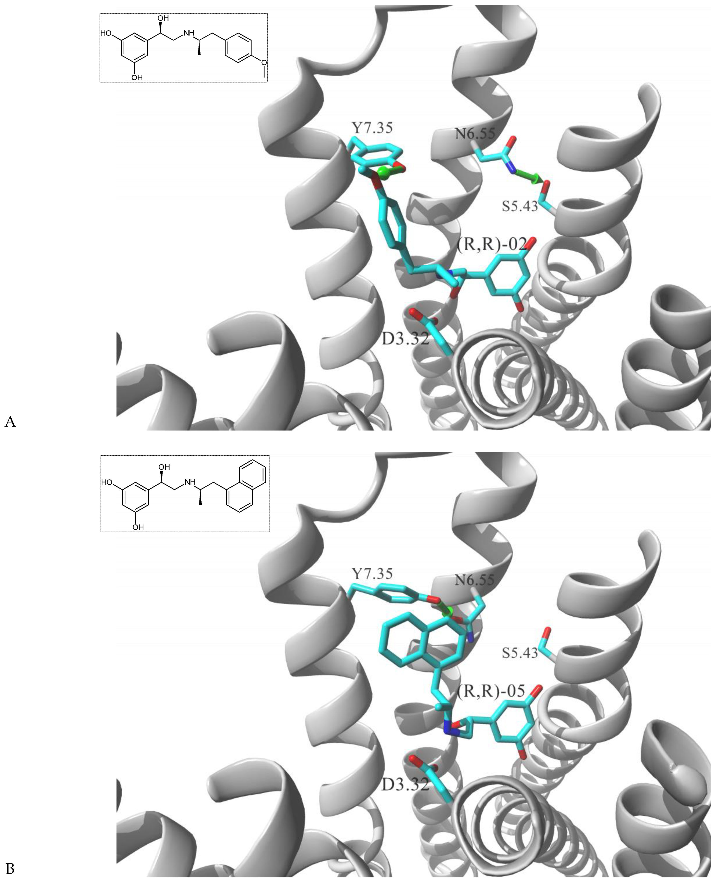
Figure 1. Postulated mechanism of differential interactions between β2-AR molecule and (A) (R,R)-02, (a ligand inducing Gs signaling pattern), or (B) (R,R)-05, a ligand inducing [Gs+Gi] signaling pattern. The receptor molecule is shown in secondary structure mode (gray ribbon), parts are hidden for clarity. Ligands molecules and Y3087.35, N2936.55, S2045.43 and D1133.32 residues are rendered in stick mode, key HBs discussed in the text are shown as green arrows. Inset: chemical formula of a ligand. Adopted from [27].
Molecules 26 00851 g001
Figure 2. Structural model of M2 receptor cocrystalized with (A) iperoxo alone (4mqs.pdb) and (B) iperoxo + LY2119620 ligands (4mqt.pdb) [44]. Iperoxo resides at the ortosteric site, LY2119620 occupies the allosteric ELBD. The receptor molecule is shown in secondary structure mode (gray ribbon), parts are hidden for clarity. Ligand molecules and W4227.35, N4106.58 and D1033.32 residues are rendered in stick mode, interhelical HB in (B) is shown as a green arrow. Inset: chemical formula of ligands.
Molecules 26 00851 g002
Figure 3. Structural model of 5-HT2B receptor cocrystalized with LSD (5tvn.pdb) [45] and location of the two ELBD residues, L3627.35, L209ECL2, postulated to be involved in biased signaling. The receptor molecule is shown in secondary structure mode (gray ribbon), parts are hidden for clarity. Ligand molecules and L3627.35, L209ECL2 and D1353.32 residues are rendered in stick mode. Inset: chemical formula of a ligand.
Molecules 26 00851 g003
Figure 4. AT1 receptor cocrystalized with (A) wild type AngII peptide (6os0.pdb) (Gq + β-arrestin signaling) and (B) F8A AngII mutant (TRV023; 6os1.pdb) (β-arrestin biased signaling). Due to high B-factor, conformation of the Y2927.43 cannot be fully visualized in (A) [58]. The receptor is shown in secondary structure mode (gray ribbon), parts are hidden for clarity. Ligand molecules, L1123.36 and Y2927.43 residues are rendered in stick mode. Inset: sequence of cocrystalized peptide.
Molecules 26 00851 g004
Table 1. Chemical structures of fenoterol derivatives inducing ligand directed signaling of β2-AR at rat cardiomyocytes, controlled by PTx [27].
Gs Signaling Ligands (PTx Insensitive)[Gs+Gi] Signaling Ligands (PTx Sensitive)
Molecules 26 00851 i001 (R,R)-01 Molecules 26 00851 i002 (R,R)-05
Molecules 26 00851 i003 (R,R)-02 Molecules 26 00851 i004 (R,R)-06
Molecules 26 00851 i005 (R,R)-03 Molecules 26 00851 i006 (R,R)-07
Publisher’s Note: MDPI stays neutral with regard to jurisdictional claims in published maps and institutional affiliations.