Article

Synthesis of Tetravalent Thio- and Selenogalactoside-Presenting Galactoclusters and Their Interactions with Bacterial Lectin PA-IL from Pseudomonas aeruginosa

1
Department of Organic Chemistry, University of Debrecen, Egyetem tér 1, H-4032 Debrecen, Hungary
2
Central European Institute of Technology, Masaryk University, Kamenice 5, 625 00 Brno, Czech Republic
3
National Centre for Biomolecular Research, Faculty of Science, Masaryk University, Kotlářská 2, 611 37 Brno, Czech Republic
4
Department of Pharmaceutical Chemistry, University of Debrecen, Egyetem tér 1, H-4032 Debrecen, Hungary
5
Department of Inorganic and Analytical Chemistry, University of Debrecen, Egyetem tér 1, H-4032 Debrecen, Hungary
6
Department of Biochemistry, Faculty of Science, Masaryk University, Kotlářská 2, 611 37 Brno, Czech Republic
7
Research Group for Molecular Recognition and Interaction, Hungarian Academy of Sciences, University of Debrecen, Egyetem tér 1, H-4032 Debrecen, Hungary
*
Author to whom correspondence should be addressed.
Molecules 2021, 26(3), 542; https://doi.org/10.3390/molecules26030542
Submission received: 15 December 2020 / Revised: 18 January 2021 / Accepted: 18 January 2021 / Published: 21 January 2021
(This article belongs to the Special Issue Targeting Carbohydrate–Protein Interactions)

Abstract

:
Synthesis of tetravalent thio- and selenogalactopyranoside-containing glycoclusters using azide-alkyne click strategy is presented. Prepared compounds are potential ligands of Pseudomonas aeruginosa lectin PA-IL. P. aeruginosa is an opportunistic human pathogen associated with cystic fibrosis, and PA-IL is one of its virulence factors. The interactions of PA-IL and tetravalent glycoconjugates were investigated using hemagglutination inhibition assay and compared with mono- and divalent galactosides (propargyl 1-thio- and 1-seleno-β-d-galactopyranoside, digalactosyl diselenide and digalactosyl disulfide). The lectin-carbohydrate interactions were also studied by saturation transfer difference NMR technique. Both thio- and seleno-tetravalent glycoconjugates were able to inhibit PA-IL significantly better than simple d-galactose or their intermediate compounds from the synthesis.

1. Introduction

Lectins from pathogenic organisms could be important virulence factors. These specific carbohydrate-binding proteins could be involved in the recognition and adhesion processes in host-pathogens interactions [1]. Consequently, carbohydrate-based inhibitors of lectins are promising potential therapeutics [2]. Lectins are usually multivalent oligomeric proteins, frequently displaying an avidity effect and increased affinity to complex glycosylated surfaces. Therefore, the multivalent inhibitors containing several carbohydrate residues are suitable for disrupting lectins’ binding to host cells or tissues [3]. The opportunistic pathogen Pseudomonas aeruginosa is a Gram-negative bacterium, causing chronic and potentially lethal lungs infections in immunocompromised humans, mainly patients suffering from cystic fibrosis (CF). It is the most widespread pulmonary pathogen associated with CF and significantly influences morbidity and mortality [4,5]. P. aeruginosa produces a tetrameric d-galactose-specific lectin named PA-IL (LecA) which is considered to be involved in adhesion, biofilm formation, cellular invasion and cytotoxicity [6,7,8,9,10].
Our previous works presented several potential inhibitors of various lectins of bacterial and fungal origin [11]. Recently, a tetravalent lead compound I for anti-adhesion therapy of Pseudomonas aeruginosa infections was developed (Figure 1) [12].
Our current aim is to synthesize the thio- and selenoglycoside analogues of the tetravalent lead-structure as well as their intermediate compounds to investigate their binding properties towards lectin PA-IL and the effect of sulfur and selenium on lectin binding. Several selenium-containing carbohydrates are known from the literature. They were synthesized for various purposes [13], exploiting the inherent potential of the selenium nucleus [14]. Selenoglycosides were used as glycosyl donors [15], for protein glycoconjugation in site-selective glycosylation by Se-S-mediated ligation [16], as enzyme inhibitors (O-GlcNAcase [17] and as novel glycosidase inhibitors [18]). Mono- and divalent selenogalactosides and diselenide digalactosides proved to be potential ligands to biomedically relevant galactophilic lectins [19]; non-glycosidically linked Se-containing pseudodisaccharides were also synthesized as BanLec and ConA lectin ligands [20]. Selenium-linked neoglycoconjugates, pseudodisaccharides [21], selenenylsulfide-linked glycopeptides and glycoproteins [22] were also prepared.  Moreover, the presence of a selenium nucleus provides an excellent opportunity for structural analysis of biomolecules by NMR and X-ray spectroscopy [14] and to study the lectin-carbohydrate interactions using sophisticated 77Se-NMR methods [23]. Selenium could be also potentially used as a trace for the selective detection of compounds in the biofluids [24]. A further advantage of the Se-interglycosidic linkage is its higher stability towards hydrolases [17,18].

2. Results and Discussion

2.1. Synthesis

PA-IL is known to recognize and interact with d-galactose-containing ligands specifically. In order to develop potential enzymatically stable ligands for this lectin, we have synthesized thio- and selenogalactoside-containing glycoconjugates based on the lead structure (Figure 1).
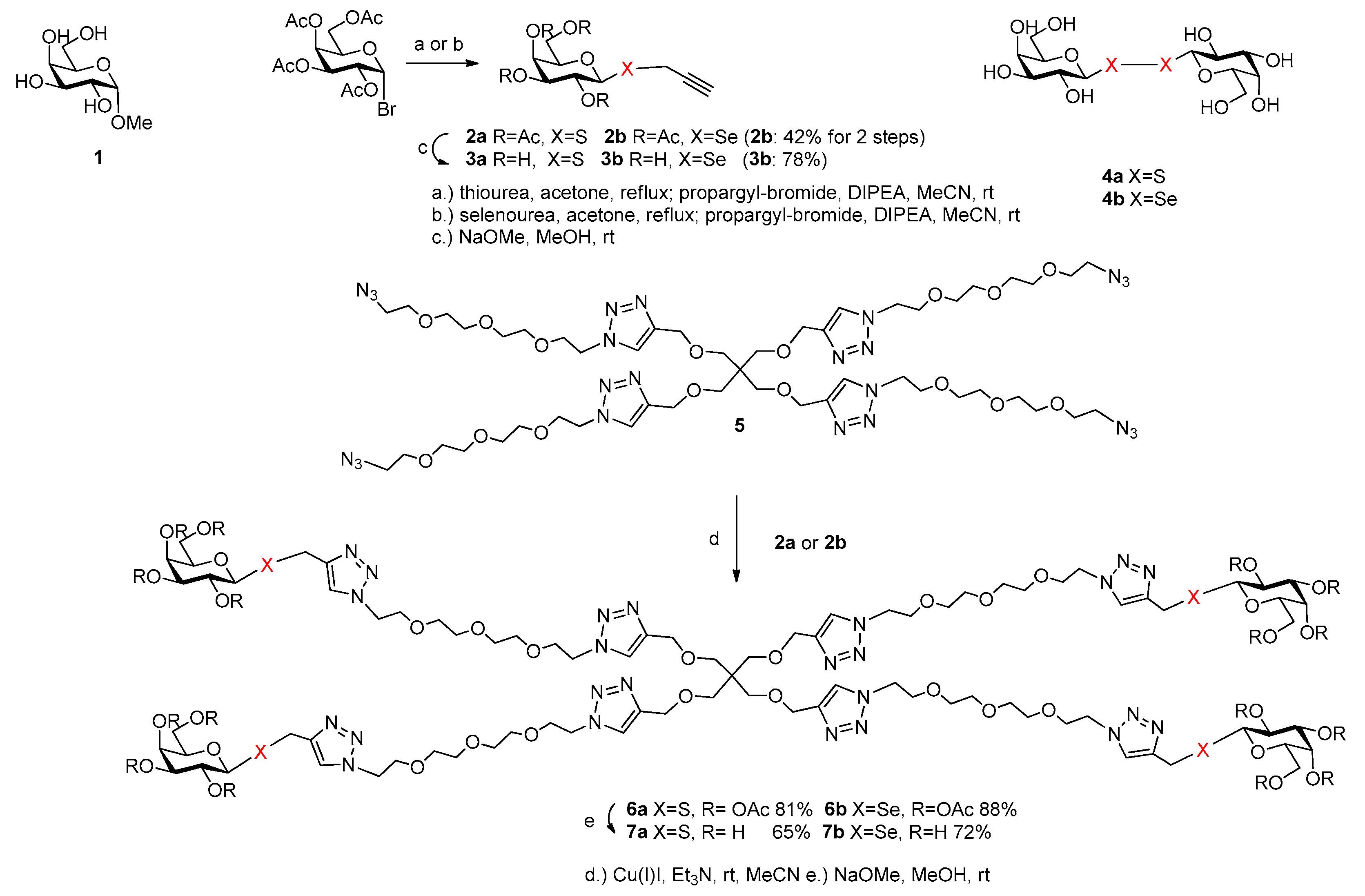
Methyl α-d-galactopyranoside 1 was used as standard, as a natural ligand of the lectin. For the synthesis of an oligovalent ligand by click-strategy [25], propargyl 2,3,4,6-tetra-O-acetyl-1-thio-β-d-galactopyranoside [26] (2a) and propargyl 2,3,4,6-tetra-O-acetyl-1-seleno-β-d-galactopyranoside (2b) were synthesized starting from peracetylated galactopyranoside bromide via thio- or selenouronium salt and propargylation (Figure 2). The deacetylated propargyl S-galactoside 3a [27] and propargyl Se-galactoside 3b were also suitable for investigations of their potential binding properties to the lectin PA-IL. Digalactosyl diselenide 4a and digalactosyl disulfide 4b [19] are known from the literature and were also suitable and available for binding studies as divalent galactoside ligands. The tetravalent glycoconjugates were built up by copper (I)-mediated azide-alkyne click reaction of alkyne 2a or 2b with azido-scaffold 5 [12]. The acetylated thiogalactocluster 6a and selenogalactocluster 6b were isolated with a yield of 81% and 88%, respectively. The tetravalent galactoclusters 6a and 6b were deprotected by Zemplén-deacetylation method. Altogether, easy and efficient syntheses of the tetravalent thiogalactocluster 7a and selenogalactocluster 7b were achieved; moreover, monovalent thiogalactoside (3a) and selenogalactoside (3b), divalent selenogalactoside (4a) and thiogalactoside (4b) were also available for binding studies as potential ligands of galactose-specific PA-IL.

2.2. Inhibition of PA-IL with Thio- and Selenogalactosides

The inhibitory potential of tetravalent thio- and selenogalactosides as well as their intermediate compounds was investigated by hemagglutination inhibition assay with microscope detection [28]. The minimal inhibitory concentrations (MIC) of compounds were determined and their inhibitory potencies were calculated and semi-quantitatively evaluated by the comparison with simple monosaccharide d-galactose (standard). All tested compounds were able to inhibit hemagglutination caused by the lectin PA-IL (see Table 1 and Figure S3). The monovalent intermediates 3a and 3b (propargyl 1-thio- and 1-seleno-ß-d-galactopyranoside) showed eight times better inhibitory potency than d-galactose, possibly due to the additional interactions via their sidechains. Compounds 4a and 4b (digalactosyl disulfide and digalactosyl diselenide) displayed inhibitory potency 16 and 8, respectively. Although these compounds are theoretically divalent, they have no spacer and are not supposed to bind to the two binding sites simultaneously; therefore, the potencies comparable with monovalent compounds were expected. Both tetravalent compounds 7a and 7b were 256 times better inhibitors than d-galactose; taking into account the effect of several galactose units in the single compound (parameter β), they were 64 times better than d-galactose. These results are the same as the potency obtained for the lead structure I. The substitution of oxygen in the glycosidic linkage did not affect the inhibitory effect on lectin PA-IL.

2.3. STD-NMR Studies: Binding of Galactoside-Containing Ligands to PA-IL Lectin Characterized by 1H STD and Competition NMR Experiments

Ligand-based STD NMR experiments [29,30,31] were performed to support and further characterize the binding of β-d-galactoside-containing ligands to PA-IL. This technique is able to disclose the structural regions of the ligands that are involved in the binding. Moreover, further information on the interaction—such as binding site and relative affinity—can be obtained in competition experiments when suitable reference (natural) ligand is available.
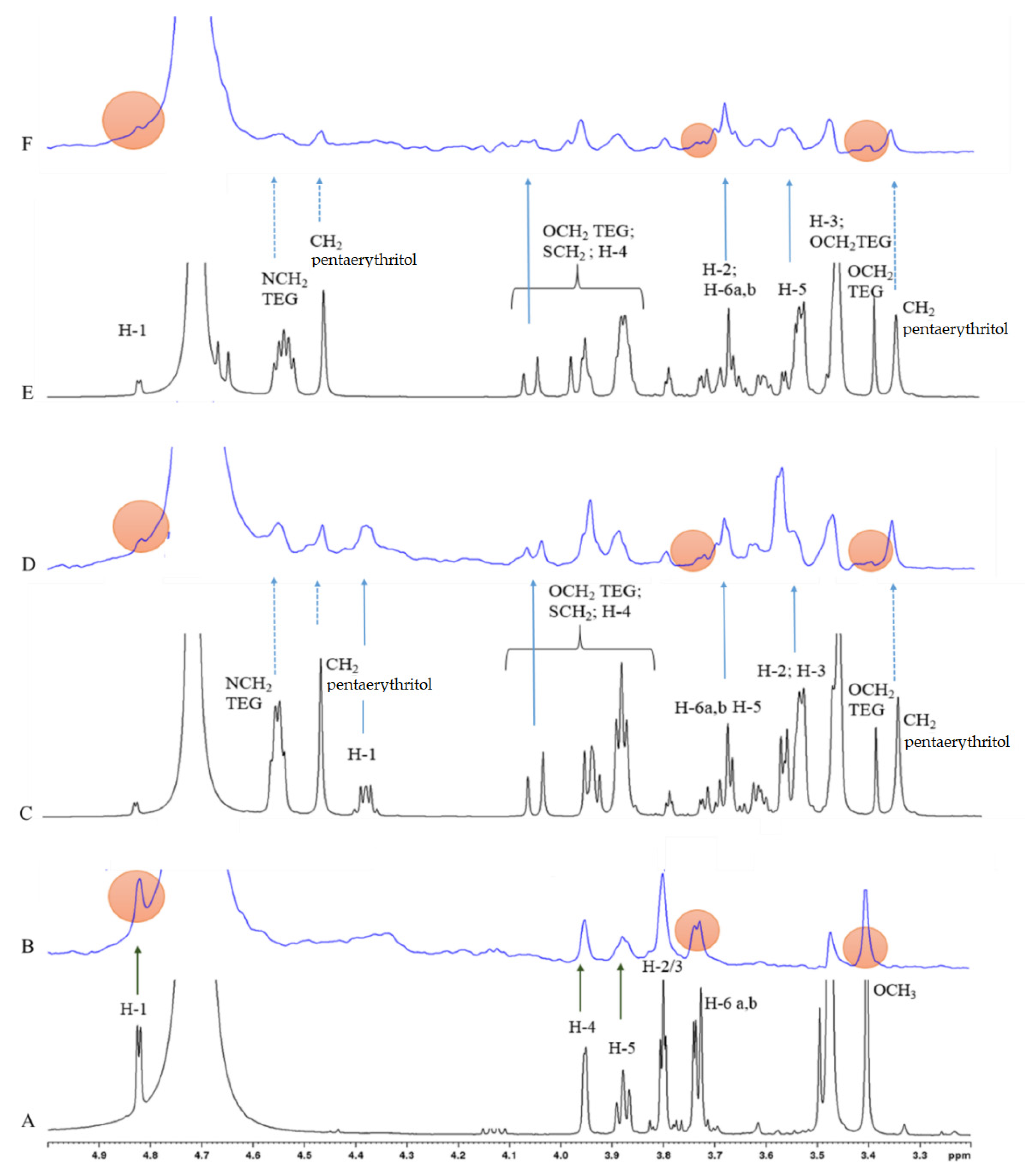
In the present study, Me α-d-Gal (1) served as a reference ligand and its binding to PA-IL was unambiguously confirmed with the STD NMR spectra shown in Figure 3B. All galactosyl ring protons and also the methyl protons of OCH3 group show similar STD effects (for resonance assignment see the 1H NMR spectrum in Figure 3A), suggesting that Me α-d-Gal being a small ligand may fit completely in the binding pocket of PA-IL.
In the succeeding competition experiments STD NMR spectra were recorded on samples containing both the natural (1) and one of the mono- or tetravalent ligands in a one-to-one molar ratio. The STD signals belonging to the well-resolved resonances (i.e., the ones separated from the resonances of 1 of tested compounds—marked by blue arrows in the STD spectra of Figure 3D,F, and also in Figure S2) confirm that all investigated mono- and tetravalent ligands bind to PA-IL. Moreover, the STD effects observed on the CH2 protons—marked by dotted blue arrows in Figure 3D,F—suggest that binding of compounds 7a and 7b to PA-IL involves certain hydrophobic contacts of the spacers in the formation of the complex.
Moreover, the STD signal attenuation of the reference ligand 1—monitored on well-separated (non-overlapping) resonances of 1 and marked by filled red circles in Figure 3B,D,F—confirms that the particular ligands compete for the same (or partially overlapped) binding site of PA-IL. Considering the 1:1 ratio of the competing ligands in the sample, the substantial drop observed in the STD signal intensities suggest that the tetravalent ligands 7a and 7b show significantly higher affinity towards PA-IL than the natural ligand 1 used in the competition assay.
The STD NMR spectra of the monovalent ligands (3a and 3b) show similar signal patterns (see Figure S2), confirming their binding to PA-IL. The attenuation of STD signals of 1 observed in the competition experiments, however, was significantly weaker, indicating lower affinity of the monovalent ligands towards PA-IL. It should be noted that the relative affinity order of the ligands assessed (qualitatively) in the competition NMR experiments is in accordance with the inhibition data given in Table 1.
In summary, tetravalent S- and Se-galactoclusters synthesized by click-chemistry were found to be suitable ligands of the lectin PA-IL in vitro, with significant, about 64 times better inhibitory activity than simple d-galactose. We can also conclude that enzymatically stable S- [32] and Se-interglycosidic linkages [17,18] do not influence the potency of ligands compared with the appropriate O-glycosides [12]. We could prove using STD-NMR techniques that the multivalent ligands compete with the natural ligand for the binding sites of the protein. In the future, multivalent selenoglycosides will provide a great opportunity to investigate the lectin-carbohydrate interactions in biologically relevant environments by highly sensitive and selective advanced 77Se-NMR methods. As d-galactose and l-fucose were applied for treatment of CF in an open clinical trial [33], novel S- and Se-galactoconjugates can be possible candidates in anti-adhesion therapy of cystic fibrosis as inhalational drugs.

3. Materials and Methods

3.1. General Methods

Optical rotations were measured at room temperature with a Perkin-Elmer 241 automatic polarimeter. TLC analysis was performed on Kieselgel 60 F254 (Merck) silica gel plates with visualization by immersing in a sulfuric-acid solution (5% in EtOH) followed by heating. Column chromatography was performed on silica gel 60 (Merck 0.063–0.200 mm) and flash column chromatography was performed on silica gel 60 (Merck 0.040–0.063 mm). Organic solutions were dried over MgSO4 and concentrated under vacuum. The 1H (500 MHz) and 13C NMR (125.76 MHz) spectra were recorded with Bruker Avance II 500 spectrometer (Bruker, Billerica, MA, USA). Chemical shifts are referenced to Me4Si or DSS (0.00 ppm for 1H) and to solvent signals (CDCl3: 77.00 ppm, CD3OD: 49.15 ppm for 13C). ESI-QTOF MS measurements were carried out on a maXis II UHR ESI-QTOF MS instrument (Bruker, Billerica, MA, USA), in positive ionization mode. The following parameters were applied for the electrospray ion source: capillary voltage: 3.6 kV; end plate offset: 500 V; nebulizer pressure: 0.5 bar; dry gas temperature: 200 °C and dry gas flow rate: 4.0 L/min. Constant background correction was applied for each spectrum, the background was recorded before each sample by injecting the blank sample matrix (solvent). Na-formate calibrant was injected after each sample, which enabled internal calibration during data evaluation. Mass spectra were recorded by otofControl version 4.1 (build: 3.5, Bruker, Billerica, MA, USA) and processed by Compass DataAnalysis version 4.4 (build: 200.55.2969) (Bruker, Billerica, MA, USA).

3.2. Synthesis

3.2.1. Compound 2b

Peracetylated galactopyranosyl bromide (1 g, 2.43 mmol) was dissolved in dry acetone (10 mL) and selenourea (300 mg, 2.43 mmol) was added then heated and stirred at reflux temperature for 1 h. When the TLC showed complete conversion of the starting material, it was evaporated, and the residue was dissolved in dry acetonitrile (10 mL). Propargyl bromide (80% solution in toluene, 1.2 mL, 2.8 mmol, 1.1 equiv.) and N,N-diisopropylethylamine (0.50 mL, 2.8 mmol) were added and stirred overnight at room temperature. The reaction mixture was evaporated, dissolved in ethyl acetate (50 mL), washed with distilled water (2 × 15 mL), dried over Na2SO4, filtered and evaporated. The crude product was purified by flash column chromatography (Merck, Darmstadt, Germany) (8:2 n-hexane:EtOAc) to give compound 2b (482 mg, 42%) as a white powder. [α]24D—53.5 (c 0.32, CHCl3); Rf 0.26 (7:3 n-hexane:EtOAc).
1H NMR (500 MHz, CDCl3) δ = 5.43 (d, J = 3.2 Hz, 1H, H-4), 5.28 (t, 1H, H-2), 5.05 (dd, J = 3.7 Hz, J = 9.9 Hz, 1H, H-3), 4.97 (d, J = 9.9 Hz, H, H-1), 4.17-4.04 (m, 2H, H-6a,b), 3.93 (m, 1H, H-5), 3.47 (dd, 1H, J = 15.4 Hz, J = 2.6 Hz, SeCH2(A) propargyl), 3.26 (dd, 1H, J = 15.4 Hz, J = 2.6 Hz, SeCH2(B) propargyl), 2.26 (t, J = 2.6 Hz, 1H, CH propargyl); 2.13, 2.04, 2.02, 1.96 (4 × s, 12H, 4 × CH3 acetyl), ppm; 13C-NMR (125 MHz, CDCl3): δ = 170.3, 170.1, 169.9, 169.7 (4C, 4 × CO acetyl), 79.8 (Cq propargyl), 77.7 (C-1), 75.6 (C-5), 71.7 (C-3), 71.5 (CH propargyl), 67.9 (C-2), 67.3 (C-4), 61.3 (C-6), 20.7, 20.6, 20.5 (4C, 4 × CH3 acetyl), 7.2 (SeCH2 propargyl) ppm.
ESI-HRMS: m/z calcd for C17H22NaO9Se [M+Na]+ 473.0327, found 473.0322.

3.2.2. Compound 3b

A catalytic amount of NaOMe (pH~9) was added to a stirred solution of ester 2b (150 mg, 0.33 mmol) in dry MeOH (5 mL) and stirred overnight at room temperature. The reaction mixture was neutralized with Amberlite IR-120 H+ ion-exchange resin, filtered and evaporated; then, the crude product was purified by flash column chromatography (7:3 CH2Cl2:MeOH) to give compound 3b (94 mg, 78%) as a colorless syrup. [α]24D—69.8 (c 0.21, MeOH); Rf 0.34 (9:1 CH2Cl2:MeOH).
1H NMR (500 MHz, D2O) δ = 4.94 (d, J = 9.9 Hz, 1H, H-1), 3.98 (d, J = 2.1 Hz, 1H, H-4); 3.80–3.68 (overlapping signals, 4H, H-6a,b, H-2, H-5); 3.65 (dd, J = 2.1 Hz, J = 9.3 Hz, 1H, H-3); 3.59 (d, 1H, J 16.5 Hz, SeCH2(A) propargyl), 3.49 (d, 1H, J 16.5 Hz, SeCH2(B) propargyl) ppm; 13C-NMR (125 MHz, D2O): δ = 81.0 (Cpropargyl), 80.8 (C-1), 79.9 (C-5), 73.5 (C-3), 71.6 (CH propargyl), 69.8 (C-2), 68.5 (C-4), 60.7 (C-6), 6.2 (SeCH2 propargyl) ppm.
ESI-HRMS: m/z calcd for C9H14NaO5Se [M+Na]+ 304.9904, found 304.9900.

3.2.3. Compound 6a

Et3N (42 μL, 0.3 mmol, 4 equiv.) and Cu(I)I (5.7 mg, 0.03 mmol, 0.4 equiv.) were added to a stirred solution of propargyl 1-thiogalactoside peracetate 2a (181 mg, 0.45 mmol, 6.0 equiv.) and azide scaffold 5 (95 mg, 0.075 mmol) in CH3CN (5 mL) under an argon atmosphere and stirred overnight at room temperature. The reaction mixture was evaporated, and the crude product was purified by flash column chromatography (95:5 CH2Cl2:MeOH) to give compound 6a (176 mg, 81%) as a colorless syrup. [α]24D—28.8 (c 0.13, MeOH); Rf 0.48 (95:5 CH2Cl2: MeOH).
1H NMR (500 MHz, CDCl3) δ = 7.75, 7.72 (2 × s, 8H, 8 × CH triazole), 5.41 (d, J = 2.8 Hz, 4H, 4 × H-4), 5.23 (dd, J = 8.0 Hz, J = 10.4 Hz, 4H, 4 × H-2), 5.03 (dd, J = 3.4 Hz, J = 10.4 Hz, 4H, 4 × H-3), 4.62 (d, J = 8.0 Hz, 4H, 4 × H-1), 4.50 (s, 8H, 4 × CH2 pentaerythritol), 4.40 (16H, 8 × NCH2 TEG), 4.15 (d, J 16.5, 4H, 4 × SCH2(A)), 4,10 (m, 8H, 4 × H-6a,b), 3.99-3.96 (m, 4H, 4 × H-5, 4H, 4× SCH2(B)), 3.70-3.65 (m, 20H, 10 × OCH2 TEG), 3.62–3.58 (m, 32H, 16 × OCH2 TEG), 3.48 (s, 8H, 4 × CH2 pentaerythritol), 2.15, 2.06, 1.98, 1.97 (4 × s, 48H, 16 × CH3 acetyl) ppm; 13C NMR (125 MHz, CDCl3) δ = 170.3, 170.2, 169.9, 169.6 (16C, 16 × CO acetyl), 145.0, 144.5 (8C, Cq triazole), 124.0, 123.5 (8C, CH triazole), 83.4 (4C, 4 × C-1), 74.3 (4C, 4 × C-5), 71.7 (4C, 4 × C-3), 70.4, 69.3 (28C, 24 × OCH2 TEG, 4 × CH2 pentaerythritol), 67.3 (8C, 4 × C-2, 4 × C-4), 64.6 (4C, 4 × CH2 pentaerythritol), 61.2 (4C, 4 × C-6), 50.5, 50.4 (8C, 8 × NCH2 TEG), 45.3 (1C, Cq pentaerythritol), 24.7 (4C, 4 × SCH2), 20.8, 20.7, 20.5 (16C, 16 × CH3 acetyl) ppm.
ESI-HRMS: m/z calcd for C117H172N24NaO52S4 [M+Na]+ 2896.0333, found: 2896.0350.

3.2.4. Compound 6b

Et3N (42 μL, 0.3 mmol, 4 equiv.) and Cu(I)I (5.7 mg, 0.03 mmol, 0.4 equiv.) were added to a stirred solution of propargyl 1-selenogalactoside peracetate 2b (169 mg, 0.375 mmol, 5.0 equiv.) and azide scaffold 5 (95 mg, 0.075 mmol) in CH3CN (5 mL) under an argon atmosphere and stirred overnight at room temperature. The reaction mixture was evaporated, and the crude product was purified by flash column chromatography (95:5 CH2Cl2:MeOH) to give compound 6b (121 mg, 88%) as a colorless syrup. [α]24D + 23.1 (c 0.11, CHCl3); Rf 0.38 (95:5 CH2Cl2: MeOH).
1H NMR (500 MHz, CDCl3) δ = 7.67, 7.60 (2 × s, 8H, 8 × CH triazole), 5.40 (d, J = 2.7 Hz, 4H, 4 × H-4), 5.25 (t, J = 10.1 Hz, 4H, 4 × H-2), 5.00 (dd, J = 3.2 Hz, J = 10.0 Hz, 4H, 4 × H-3), 4.84 (d, J = 10.0 Hz, 4H, 4 × H-1), 4.54–4.41 (m, 24 H, 4 × CH2 pentaerythritol, 8 × NCH2 TEG), 4.07 (m, 12H, 4H, 4 × SeCH2(A), 4 × H-6a,b), 3.98–3.87 (m, 8H, 4 × H-5, 4H, 4 × SeCH2(B)), 3.82 (m, 20H, 10 × OCH2 TEG), 3.60–3.46 (m, 32H, 16 × OCH2 TEG), 3.41 (s, 8H, 4 × CH2 pentaerythritol), 2.15, 2.06, 1.98, 1.97 (4 × s, 48H, 16 × CH3 acetyl) ppm; 13C NMR (125 MHz, CDCl3) δ = 170.2, 170.0, 169.8, 169.6 (16C, 16 × CO acetyl), 145.0, 144.9 (8C, Cq triazole), 123.5, 122.8 (8C, CH triazole), 78.0 (4C, 4 × C-1), 75.4 (4C, 4 × C-5), 71.5 (4C, 4 × C-3), 70.3, 69.2 (28C, 24 × OCH2 TEG, 4 × CH2 pentaerythritol), 67.9 (4C, 4 × C-2), 67.2 (4C, 4 × C-4), 64.8 (4C, 4 × CH2 pentaerythritol), 61.1 (4C, 4 × C-6), 50.1, 49.9 (8C, 8 × NCH2 TEG), 45.2 (1C, Cq pentaerythritol), 20.7, 20.5, 20.4 (16C, 16 × CH3 acetyl), 15.6 (4C, 4 × SeCH2) ppm.
ESI-HRMS: m/z calcd for C117H172N24Na2O52Se4 [M+2Na]2+ 1555.4005, found 1555.3999 [M+2Na]2+.

3.2.5. Compound 7a

A catalytic amount of NaOMe (pH ~ 9) was added to a stirred solution of ester 6a (115 mg, 0.4 mmol) in dry MeOH (5 mL) and stirred overnight at room temperature. The reaction mixture was neutralized with Amberlite IR-120 H+ ion-exchange resin, filtered and evaporated; then, the crude product was purified by flash column chromatography (7:3 CH3CN:H2O) to give compound 7a (58 mg, 65%) as a colorless syrup. [α]24D +21.5 (c 0.23, CHCl3); Rf 0.23 (7:3 CH3CN:H2O).
1H NMR (500 MHz, D2O) δ = 7.95, 7.93 (2 × s, 8H, 8 × CH triazole), 4.60–4.51 (m, 16H, 8 × NCH2 TEG), 4.46 (d, 4H, 4 × CH2pentaerythritol), 4.38 (d, 1H, 4 × H-1, J = 9.8 Hz), 4.05 (d, J 16.5, 4H, 4 × SCH2(A)), 3.98–3.82 (m, 24H, 8 × OCH2 TEG, 4 × SCH2(B), 4 × H-4), 3.75–3.67 (m, 8H, 4 × H-6a,b), 3.62 (m, 4H, 4 × H-5), 3.56–3.53 (m, 8H, 4 × H-3, 4 × H-2), 3.54 (m, 16H, 8 × OCH2 TEG), 3.48 (m, 16H, 8 × OCH2 TEG), 3.36 (m, 8H, 4 × CH2 pentaerythritol) ppm; 13C NMR (125 MHz, D2O) δ = 144.8, 144.1 (8C, Cq triazole), 125.1, 124.4 (8C, CH triazole), 85.0 (4C, 4 × C-1), 78.8 (4C, 4 × C-5), 73.8 (4C, 4 × C-3), 69.6 (12C, 12 × OCH2 TEG), 69.3 (12C, 12 × OCH2 TEG), 68.6 (8C, 4 × C-4, 4 × C-2), 68.2 (4C, 4 × CH2 pentaerythritol), 63.5 (4C, 4 × CH2 pentaerythritol), 60.9 (4C, 4 × C-6), 49.9 (16C, 16 × NCH2 TEG), 44.5 (1C, Cq pentaerythritol), 23.4 (4C, 4 × SCH2) ppm.
ESI-HRMS: m/z calcd for C85H140N24NaO36S4 [M+Na]+ 2223.8643, found 2223.8637.

3.2.6. Compound 7b

A catalytic amount of NaOMe (pH ~ 9) was added to a stirred solution of ester 6b (100 mg, 0.26 mmol) in dry MeOH (5 mL) and stirred overnight at room temperature. The reaction mixture was neutralized with Amberlite IR-120 H+ ion-exchange resin, filtered and evaporated; then, the crude product was purified by flash column chromatography (7:3 CH3CN:H2O) to give compound 7b (56 mg, 72%) as a colorless syrup. [α]24D—19.40 (c 0.53, H2O); Rf 0.26 (7:3 CH3CN:H2O).
1H NMR (500 MHz, D2O + CD3OD) δ = 7.90, 7.89 (2 × s, 8H, 8 × CH triazole), 4.61 (d, J = 9.8 Hz 4H, H-1), 4.53–4.44 (m, 16H, 8 × NCH2 TEG), 4.41 (8H, 4 × CH2 pentaerythritol), 4.02 (d, J 16.5 Hz, 4H, 4 × SCH2(A)), 3.95–3.88 (m, 24H, 8 × OCH2 TEG, 4 × SeCH2(B), 4 × H-4), 3.75–3.67 (m, 12H, 4 × H-2, 4 × H-6a,b), 3.57 (m, 4H, 4 × H-5), 3.54–3.46 (m, 20H, 4 × H-3, 8 × OCH2 TEG), 3.45–3.37 (m, 16H, 8 × OCH2 TEG), 3.31 (m, 8H, 4 × CH2 pentaerythritol) ppm; 13C NMR (125 MHz, D2O + CD3OD) δ = 147.0, 145.3 (8C, Cq triazole), 126.3, 125.3 (8C, CH triazole), 82.1 (4C, 4 × C-1), 81.4 (4C, 4 × C-5), 75.1 (4C, 4 × C-3), 71.4 (4C, 4 × C-2), 70.8, 70.6 (16C, 16 × OCH2 TEG), 70.0 (4C, 4 × C-4), 69.8 (8C, 8 × OCH2 TEG), 69.4 (4C, 4 × CH2 pentaerythritol), 64.8 (4C, 4 × CH2 pentaerythritol) 62.2 (4C, 4 × C-6), 51.1 (8C, 8 × NCH2 TEG), 45.9 (1C, Cq pentaerythritol), 15.4 (4C, 4 × SeCH2) ppm.
ESI-HRMS: m/z calcd for C85H140N24Na2O36Se4 [M+2Na]2+ 1219.3159, found 1219.3154 [M+2Na]2+.

3.3. Hemagglutination Inhibition Assay (HIA)

PA-IL was produced and purified as previously described [7]. The lectin was dissolved in the suitable buffer (20 mM Tris/HCl, 150 mM NaCl, 5 mM CaCl2, pH 7.5) to a concentration of 0.25 mg·mL−1. The lectin was mixed with synthetized galactosides and serially diluted in the buffer in a 5 µL:5 µL ratio. The final (working) concentration of the lectin was therefore 0.125 mg·mL−1. Then, a total volume of 10 µL of 20% papain-treated, azid-stabilized red blood cells B in the buffer was added, after which the mixture was thoroughly mixed and incubated for 5 min at room temperature. After incubation, the mixture was again mixed, transferred to a microscope slide and examined. The examination was conducted using the Levenhuk D2L NG Digital Microscope (Levenhuk, Tampa, FL, USA). Images were obtained with a Levenhuk D2L digital camera (Levenhuk, Tampa, FL, USA) using the software ToupView for Windows (Levenhuk, Tampa, FL, USA). The positive (experiment without an inhibitor) and negative control (experiment without the lectin) were prepared and processed in the same way using the appropriate volume of dissolving buffer instead of the omitted components. The minimal inhibitory concentration (MIC) of the inhibitor able to inhibit hemagglutination was determined and compared with the standard (d-galactose), and the potency of the inhibitor was calculated (MIC of the standard/MIC of the inhibitor).

3.4. 1H STD NMR Experiment

All NMR measurements were performed on a Bruker Avance II 500 spectrometer (Bruker, Billerica, MA, USA) operating at 500.13 MHz for 1H and equipped with 5-mm triple-resonance (txi) probe-head with z-axis gradients.
1H STD NMR spectra were recorded on samples dissolved in D2O (1M Tris-d11, 0.5 mM CaCl2, pH 7.5, T = 303 K) with the molar ratio of the ligand to PA-IL of about 100:1. The concentration of PA-IL tetramer was kept as low as ca. 10 μM to avoid aggregation upon addition of the ligand(s). For selective saturation of protein resonances, a train of band-selective E-BURP-1 (90°) shaped pulses of 50 ms each with a maximum B1 field strength of 75 Hz was employed yielding a total irradiation time of 3 s. For irradiation at aliphatic (CH3) region of PA-IL the E-BURP-1 pulses were applied at −0.3 ppm, while for recording the reference (off-resonance) spectrum, the irradiation frequency was set at −27 ppm. Off- and on-resonance data were recorded at alternate scans and the corresponding FIDs were collected in separate memories for subsequent processing and for the generation of STD spectra. Competition STD NMR experiments were performed following the experimental protocol as given above on samples containing two ligands (natural ligand and one of the mono- or multivalent ligands) and PA-IL lectin in 100:100:1 molar ratio. STD spectra were typically recorded with 2000–2400 transients to obtain a suitable signal-to-noise ratio for the analysis.

Supplementary Materials

The following are available. Figure S1: NMR spectra of the new compounds. Figure S2: The STD NMR spectra of the monovalent ligand 1, 3a and 3b in the presence of PA-IL. Figure S3: Influence of d-galactose, compounds 3a, 3b, 4a, 4b, 7a and 7b on hemagglutination caused by lectin PA-IL.

Author Contributions

Conceptualization K.E.K., M.W. and M.C.; investigation, T.Z.I., L.M., E.R., B.T., B.F. and M.K.; writing—original draft preparation, L.M., M.C. and K.E.K.; writing—review and editing, L.M., K.E.K. and M.C.; supervision, K.E.K., M.W. and M.C.; funding acquisition, M.C., K.E.K. and M.W. All authors have read and agreed to the published version of the manuscript.

Funding

The synthetic work was supported by the National Research and Development and Innovation Office of Hungary (K119509, M.C., K128368, K.E.K.) and co-financed by the European Regional Development Fund under the project GINOP-2.3.2-15-2016-00008. The project was supported by the János Bolyai Fellowship (M.C.) of the Hungarian Academy of Sciences. The project was further supported by the Czech Science Foundation (18-18964S). We acknowledge the Biomolecular Interactions and Crystallization Core Facility of CEITEC MU supported by the CIISB research infrastructure (LM2018127 funded by MEYS CR) for their support with obtaining the scientific data presented in this paper.

Institutional Review Board Statement

Not applicable.

Informed Consent Statement

Not applicable.

Data Availability Statement

The data presented in this study are available in Supplementary Material.

Conflicts of Interest

The authors declare no conflict of interest.

Sample Availability

Samples of the compounds 2a, 2b, 3a, 3b, 4a, 4b, 7a and 7b are available from the authors.

References

  1. Sharon, N.; Lis, H. Lectins: Carbohydrate-specific proteins that mediate cellular recognition. Chem. Rev. 1998, 98, 637–674. [Google Scholar] [CrossRef]
  2. Sharon, N. Carbohydrates as future anti-adhesion drugs for infectious diseases. Biochim. Biophys. Acta 2006, 1760, 527–537. [Google Scholar] [CrossRef] [PubMed]
  3. Cecioni, S.; Imberty, A.; Vidal, S. Glycomimetics versus Multivalent Glycoconjugates for the Design of High Affinity Lectin Ligands. Chem. Rev. 2015, 115, 525–561. [Google Scholar] [CrossRef] [PubMed]
  4. Folkesson, A.; Jelsbak, L.; Yang, L.; Johansen, H.K.; Ciofu, O.; Høiby, N.; Molin, S. Adaptation of Pseudomonas aeruginosa to the cystic fibrosis airway: An evolutionary perspective. Nat. Rev. Microbiol. 2012, 10, 841–851. [Google Scholar] [CrossRef]
  5. Bhagirath, A.Y.; Li, Y.; Somayajula, D.; Dadashi, M.; Badr, S.; Duan, K. Cystic fibrosis lung environment and Pseudomonas aeruginosa infection. BMC Pulm. Med. 2016, 16, 174–196. [Google Scholar] [CrossRef] [Green Version]
  6. Cioci, G.; Mitchell, E.P.; Gautier, C.; Wimmerová, M.; Sudakevitz, D.; Pérez, S.; Gilboa-Garber, N.; Imberty, A. Structural basis of calcium and galactose recognition by the lectin PA-IL of Pseudomonas aeruginosa. FEBS Lett. 2003, 555, 297–301. [Google Scholar] [CrossRef] [Green Version]
  7. Chemani, C.; Imberty, A.; de Bentzmann, S.; Pierre, M.; Wimmerová, M.; Guery, B.P.; Faure, K. Role of LecA and LecB lectins in Pseudomonas aeruginosa-induced lung injury and effect of carbohydrate ligands. Infect. Immun. 2009, 77, 2065–2075. [Google Scholar] [CrossRef] [Green Version]
  8. Diggle, S.P.; Stacey, R.E.; Dodd, C.; Cámara, M.; Williams, P.; Winzer, K. The galactophilic lectin, LecA, contributes to biofilm development in Pseudomonas aeruginosa. Environ. Microbiol. 2006, 8, 1095–1104. [Google Scholar] [CrossRef]
  9. Novoa, A.; Eierhoff, T.; Topin, J.; Varrot, A.; Barluenga, S.; Imberty, A.; Römer, W.; Winssinger, N. A LecA Ligand Identified from a Galactoside-Conjugate Array Inhibits Host Cell Invasion by Pseudomonas aeruginosa. Angew. Chem. Int. Ed. 2014, 53, 8885–8889. [Google Scholar] [CrossRef]
  10. Bajolet-Laudinat, O.; Bentzmann, S.G.-D.; Tournier, J.M.; Madoulet, C.; Plotkowski, M.C.; Chippaux, C.; Puchelle, E. Cytotoxicity of Pseudomonas aeruginosa internal lectin PA-I to respiratory epithelial cells in primary culture. Infect. Immun. 1994, 62, 4481–4487. [Google Scholar] [CrossRef] [Green Version]
  11. Thai, L.S.; Malinovská, L.; Vašková, M.; Mező, E.; Kelemen, V.; Borbás, A.; Hodek, P.; Wimmerová, M.; Csávás, M. Investigation of the Binding Affinity of a Broad Array of L-Fucosides with Six Fucose-Specific Lectins of Bacterial and Fungal Origin. Molecules 2019, 24, 2262. [Google Scholar] [CrossRef] [PubMed] [Green Version]
  12. Malinovská, L.; Thai, L.S.; Herczeg, M.; Vašková, M.; Houser, J.; Fujdiarová, E.; Komárek, J.; Hodek, P.; Borbás, A.; Wimmerová, M.; et al. Synthesis of β-D-galactopyranoside-presenting glycoclusters, investigation of their interactions with Pseudomonas aeruginosa lectin A (PA-IL) and evaluation of their anti-adhesion potential. Biomolecules 2019, 9, 686. [Google Scholar] [CrossRef] [Green Version]
  13. Mangiavacchi, F.; Dias, I.F.C.; Di Lorenzo, I.; Grzes, P.; Palomba, M.; Rosati, O.; Bagnoli, L.; Marini, F.; Santi, C.; Lenardao, E.J.; et al. Sweet Selenium: Synthesis and Properties of Selenium-Containing Sugars and Derivatives. Pharmaceuticals 2020, 13, 211. [Google Scholar] [CrossRef] [PubMed]
  14. Suzuki, T.; Makyio, H.; Ando, H.; Komura, N.; Menjo, M.; Yamada, Y.; Imamura, A.; Ishida, H.; Wakatsuki, S.; Kato, R.; et al. Expanded potential of seleno-carbohydrates as a molecular tool for X-ray structural determination of a carbohydrate–protein complex with single/multi-wavelength anomalous dispersion phasing. Bioorg. Med. Chem. 2014, 22, 2090–2101. [Google Scholar] [CrossRef] [PubMed]
  15. Valerio, S.; Iadonisi, A.; Adinolfi, M.; Ravidá, A. Novel Approaches for the Synthesis and Activation of Thio- and Selenoglycoside Donors. J. Org. Chem. 2007, 72, 6097–6106. [Google Scholar] [CrossRef] [PubMed]
  16. Gamblin, D.P.; Garnier, P.; Van Kasteren, S.; Oldham, N.J.; Fairbanks, A.J.; Davis, B.G. Glyco-SeS: Selenenylsulfide-Mediated Protein Glycoconjugation—A New Strategy in Post-Translational Modification. Angew. Chem. Int. Ed. 2004, 43, 828–833. [Google Scholar] [CrossRef]
  17. Kim, E.J.; Love, N.C.; Darout, E.; Abdo, M.; Rempel, B.; Withers, S.G.; Rablen, P.R.; Hanover, J.A.; Knapp, S. OGA inhibition by GlcNAc-selenazoline. Bioorg. Med. Chem. 2010, 18, 7058–7064. [Google Scholar] [CrossRef] [Green Version]
  18. Mehta, S.; Andrews, J.S.; Svensson, B.; Pinto, B.M. Synthesis and Enzymic Activity of Novel Glycosidase Inhibitors Containing Sulfur and Selenium. J. Am. Chem. Soc. 1995, 117, 9783–9790. [Google Scholar] [CrossRef]
  19. André, S.; Kövér, K.E.; Gabius, H.-J.; Szilágyi, L. Thio- and selenoglycosides as ligands for biomedically relevant lectins: Valency–activity correlations for benzene-based dithiogalactoside clusters and first assessment for (di)selenodigalactosides. Bioorg. Med. Chem. Lett. 2015, 25, 931–935. [Google Scholar] [CrossRef] [Green Version]
  20. Cumpstey, I.; Ramstadius, C.; Akhtar, T.; Goldstein, I.J.; Winter, H.C. Non-Glycosidically Linked Pseudodisaccharides: Thioethers, Sulfoxides, Sulfones, Ethers, Selenoethers, and Their Binding to Lectins. Eur. J. Org. Chem. 2010, 10, 1951–1970. [Google Scholar] [CrossRef]
  21. Affeldt, R.F.; Braga, H.C.; Baldassari, L.L.; Luedtke, D.S. Synthesis of selenium-linked neoglycoconjugates and pseudodisaccharides. Tetrahedron 2012, 68, 10470–10475. [Google Scholar] [CrossRef]
  22. Boutureira, O.; Bernardes, G.J.L.; Fernández-González, M.; Anthony, D.C.; Davis, B.G. Selenenylsulfide-Linked Homogeneous Glycopeptides and Glycoproteins: Synthesis of Human “Hepatic Se Metabolite A”. Angew. Chem. Int. Ed. 2011, 51, 1432–1436. [Google Scholar] [CrossRef] [PubMed]
  23. Raics, M.; Timári, I.; Diercks, T.; Szilágyi, L.; Gabius, H.-J.; Kövér, K.E. Selenoglycosides as Lectin Ligands: 77Se-Edited CPMG-HSQMBC NMR Spectroscopy To Monitor Biomedically Relevant Interactions. ChemBioChem 2019, 20, 1688–1692. [Google Scholar] [CrossRef] [PubMed] [Green Version]
  24. Llamas, I.; Boutureira, O.; Claridge, T.D.W.; Davis, B.G. Glycosyldiselenides as lectin ligands detectable by NMR in biofluids. Chem. Commun. 2015, 51, 12208–12211. [Google Scholar] [CrossRef] [Green Version]
  25. Dondoni, A. Triazole: The Keystone in Glycosylated Molecular Architectures Constructed by a Click Reaction. Chem. Asian J. 2007, 2, 700–708. [Google Scholar] [CrossRef]
  26. Ziegler, T.; Pietrzik, N.; Schips, C. Efficient Synthesis of Glycosylated Asparaginic Acid Building Blocks via Click Chemistry. Synthesis 2008, 4, 519–526. [Google Scholar] [CrossRef]
  27. Giguere, D.; Bonin, M.-A.; Cloutier, P.; Patnam, R.; St-Pierre, C.; Sato, S.; Roy, R. Synthesis of stable and selective inhibitors of human galectins-1 and -3. Bioorg. Med. Chem. 2008, 16, 7811–7823. [Google Scholar] [CrossRef]
  28. Adamová, L.; Malinovská, L.; Wimmerová, M. New sensitive detection method for lectin hemagglutination using microscopy. Microsc. Res. Tech. 2014, 77, 841–849. [Google Scholar] [CrossRef]
  29. Mayer, M.; Meyer, B. Characterization of Ligand Binding by Saturation Transfer Difference NMR Spectroscopy. Angew. Chem. Int. Ed. 1999, 38, 1784–1788. [Google Scholar] [CrossRef]
  30. Mayer, M.; Meyer, B. Group Epitope Mapping by Saturation Transfer Difference NMR To Identify Segments of a Ligand in Direct Contact with a Protein Receptor. J. Am. Chem. Soc. 2001, 123, 6108–6117. [Google Scholar] [CrossRef]
  31. Groves, P.; Kövér, K.E.; André, S.; Bandorowicz-Pikuła, J.; Batta, G.; Bruix, M.; Buchet, R.; Canales, Á.; Cañada, F.J.; Gabius, H.-J.; et al. Temperature dependence of ligand-protein complex formation as reflected by saturation transfer difference NMR experiments. J. Magn. Reson. Chem. 2007, 45, 745–748. [Google Scholar] [CrossRef] [PubMed] [Green Version]
  32. Driguez, H. Thiooligosaccharides in glycobiology. In Glycoscience Synthesis of Substrate Analogs and Mimetics; Springer: Berlin/Heidelberg, Germany, 1997; pp. 85–116. [Google Scholar] [CrossRef]
  33. Hauber, H.-P.; Schulz, M.; Pforte, A.; Mack, D.; Zabel, P.; Schumacher, U. Inhalation with Fucose and Galactose for Treatment of Pseudomonas Aeruginosa in Cystic Fibrosis Patients. Int. J. Med. Sci. 2008, 5, 371–376. [Google Scholar] [CrossRef] [PubMed] [Green Version]
Figure 1. Lead compound (I) for anti-adhesion therapy of Pseudomonas aeruginosa infection.
Molecules 26 00542 g001
Figure 2. Synthesis of tetravalent thiogalactoside 7a and selenogalactoside 7b.
Molecules 26 00542 g002
Figure 3. 500 MHz 1H and STD NMR spectra of 1, 7a and 7b in the presence of 10 μM PA-IL tetramer. (A,B) 1H and STD NMR spectra of 1. (C,D) 1H and STD spectra of the 1:1 mixture of 1 and 7a, (E,F) 1H and STD spectra of the 1:1 mixture of 1 and 7b, respectively.
Molecules 26 00542 g003
Table 1. The MIC (minimal inhibitory concentration) values and potencies of tested inhibitors obtained for the inhibition of hemagglutination caused by PA-IL lectin from Pseudomonas aeruginosa.
InhibitorMIC Potency 2Valencyβ 3
d-galactose 16.25 mM111
Me α-d-Gal1.562 mM414
Compound 3a0.781 mM818
Compound 3b0.781 mM818
Compound 4a0.391 mM1628
Compound 4b0.781 mM824
Compound I 424.41 µM256464
Compound 7a24.41 µM256464
Compound 7b24.41 µM256464
1 Standard, 2 MIC of standard/MIC of inhibitor, 3 Potency/Valency, 4 From ref. 12.
Publisher’s Note: MDPI stays neutral with regard to jurisdictional claims in published maps and institutional affiliations.