
Ultrasonication Improves Solid Phase Synthesis of Peptides Specific for Fibroblast Growth Factor Receptor and for the Protein-Protein Interface RANK-TRAF6

 , , ,
, , ,  ,
,  and
and 
Abstract
:
1. Introduction
2. Results and Discussion
3. Conclusions
4. Materials and Methods
4.1. Materials
4.2. Reactor Vessels for “Classical” and US-Assisted SPPS
4.3. Synthesis of the Peptides
4.4. Kaiser Test Colorimetric Assay Procedure
4.5. RP-HPLC Analysis
4.6. Applied Binary HPLC Gradients
Supplementary Materials
Author Contributions
Funding
Institutional Review Board Statement
Informed Consent Statement
Data Availability Statement
Acknowledgments
Conflicts of Interest
Sample Availability
References
- Hahn, E.M.; Estrada-Ortiz, N.; Han, J.; Ferreira, V.F.C.; Kapp, T.G.; Correia, J.D.G.; Casini, A.; Kuehn, F.E. Functionalization of Ruthenium(II) Terpyridine Complexes with Cyclic RGD Peptides to Target Integrin Receptors in Cancer Cells. Eur. J. Inorg. Chem. 2017, 12, 1667–1672. [Google Scholar] [CrossRef]
- Machado, J.F.; Machuqueiro, M.; Marques, F.; Robalo, M.P.; Piedade, M.F.M.; Garcia, M.H.; Correia, J.D.G.; Morais, T.S. Novel “ruthenium cyclopentadienyl”—Peptide conjugate complexes against human FGFR(+) breast cancer. Dalton Trans. 2020, 49, 5974–5987. [Google Scholar] [CrossRef] [PubMed]
- Woods, B.; Silva, R.D.M.; Schmidt, C.; Wragg, D.; Cavaco, M.; Neves, V.; Ferreira, V.F.C.; Gano, L.; Morais, T.S.; Mendes, F.; et al. Bioconjugate Supramolecular Pd2+ Metallacages Penetrate the Blood Brain Barrier In Vitro and In Vivo. Bioconjugate Chem. 2021, 32, 1399–1408. [Google Scholar] [CrossRef] [PubMed]
- Vultos, F.; Fernandes, C.; Correia, J.D.G.; Santos, I.; Gano, L. In-111 labeled peptides targeting the estrogen receptor for theranostic of cancer. Nucl. Med. Biol. 2014, 41, 645. [Google Scholar] [CrossRef]
- Morais, M.; Oliveira, B.L.; Correia, J.D.G.; Oliveira, M.C.; Jimenez, M.A.; Santos, I.; Raposinho, P.D. Influence of the Bifunctional Chelator on the Pharmacokinetic Properties of Tc-99m(CO)(3)-Labeled Cyclic alpha-Melanocyte Stimulating Hormone Analog. J. Med. Chem. 2013, 56, 1961–1973. [Google Scholar] [CrossRef]
- Correia, J.D.G.; Paulo, A.; Raposinho, P.D.; Santos, I. Radiometallated peptides for molecular imaging and targeted therapy. Dalton Trans. 2011, 40, 6144–6167. [Google Scholar] [CrossRef] [PubMed]
- Ornitz, D.M.; Itoh, N. The Fibroblast Growth Factor signaling pathway. Wiley Interdiscip. Rev. Dev. Biol. 2015, 4, 215–266. [Google Scholar] [CrossRef] [Green Version]
- Katoh, M. Fibroblast growth factor receptors as treatment targets in clinical oncology. Nat. Rev. Clin. Oncol. 2019, 16, 105–122. [Google Scholar] [CrossRef]
- Jin, M.; Yu, Y.; Qi, H.B.; Xie, Y.L.; Su, N.; Wang, X.F.; Tan, Q.Y.; Luo, F.T.; Zhu, Y.; Wang, Q.; et al. A novel FGFR3-binding peptide inhibits FGFR3 signaling and reverses the lethal phenotype of mice mimicking human thanatophoric dysplasia. Hum. Mol. Genet. 2012, 21, 5443–5455. [Google Scholar] [CrossRef] [Green Version]
- Perrault, D.P.; Lee, G.K.; Park, S.Y.; Lee, S.; Choi, D.; Jung, E.; Seong, Y.J.; Park, E.K.; Sung, C.; Yu, R.; et al. Small Peptide Modulation of Fibroblast Growth Factor Receptor 3-Dependent Postnatal Lymphangiogenesis. Lymphat. Res. Biol. 2019, 17, 19–29. [Google Scholar] [CrossRef]
- Choi, I.; Lee, S.; Chung, H.K.; Lee, Y.S.; Kim, K.E.; Choi, D.; Park, E.K.; Yang, D.; Ecoiffier, T.; Monahan, J.; et al. 9-Cis Retinoic Acid Promotes Lymphangiogenesis and Enhances Lymphatic Vessel Regeneration Therapeutic Implications of 9-Cis Retinoic Acid for Secondary Lymphedema. Circulation 2012, 125, 872–882. [Google Scholar] [CrossRef] [PubMed]
- Moreno, V.; Font-Bardia, M.; Calvet, T.; Lorenzo, J.; Aviles, F.X.; Garcia, M.H.; Morais, T.S.; Valente, A.; Robalo, M.P. DNA interaction and cytotoxicity studies of new ruthenium(II) cyclopentadienyl derivative complexes containing heteroaromatic ligands. J. Inorg. Biochem. 2011, 105, 241–249. [Google Scholar] [CrossRef] [PubMed]
- Tomaz, A.I.; Jakusch, T.; Morais, T.S.; Marques, F.; de Almeida, R.F.M.; Mendes, F.; Enyedy, E.A.; Santos, I.; Pessoa, J.C.; Kiss, T.; et al. Ru-II(eta(5)-C5H5)(bipy)(PPh3) (+), a promising large spectrum antitumor agent: Cytotoxic activity and interaction with human serum albumin. J. Inorg. Biochem. 2012, 117, 261–269. [Google Scholar] [CrossRef]
- Corte-Real, L.; Matos, A.P.; Alho, I.; Morais, T.S.; Tomaz, A.I.; Garcia, M.H.; Santos, I.; Bicho, M.P.; Marques, F. Cellular Uptake Mechanisms of an Antitumor Ruthenium Compound: The Endosomal/Lysosomal System as a Target for Anticancer Metal-Based Drugs. Microsc. Microanal. 2013, 19, 1122–1130. [Google Scholar] [CrossRef]
- Corte-Real, L.; Mendes, F.; Coimbra, J.; Morais, T.S.; Tomaz, A.I.; Valente, A.; Garcia, M.H.; Santos, I.; Bicho, M.; Marques, F. Anticancer activity of structurally related ruthenium(II) cyclopentadienyl complexes. J. Biol. Inorg. Chem. 2014, 19, 853–867. [Google Scholar] [CrossRef]
- Perez-Garcia, J.; Munoz-Couselo, E.; Soberino, J.; Racca, F.; Cortes, J. Targeting FGFR pathway in breast cancer. Breast 2018, 37, 126–133. [Google Scholar] [CrossRef] [PubMed] [Green Version]
- Paradis-Bas, M.; Tulla-Puche, J.; Albericio, F. The road to the synthesis of “difficult peptides”. Chem. Soc. Rev. 2016, 45, 631–654. [Google Scholar] [CrossRef] [PubMed]
- Takahash, S.; Shimonis, Y. Solid phase peptide synthesis using ultrasonic waves. Chem. Lett. 1974, 3, 51–56. [Google Scholar] [CrossRef]
- Merlino, F.; Tomassi, S.; Yousif, A.M.; Messere, A.; Marinelli, L.; Grieco, P.; Novellino, E.; Cosconati, S.; Di Maro, S. Boosting Fmoc Solid-Phase Peptide Synthesis by Ultrasonication. Org. Lett. 2019, 21, 6378–6382. [Google Scholar] [CrossRef]
- Wolczanski, G.; Plociennik, H.; Lisowski, M.; Stefanowicz, P. A faster solid phase peptide synthesis method using ultrasonic agitation. Tetrahedron Lett. 2019, 60, 1814–1818. [Google Scholar] [CrossRef]
- Raheem, S.J.; Schmidt, B.W.; Solomon, V.R.; Salih, A.K.; Price, E.W. Ultrasonic-Assisted Solid-Phase Peptide Synthesis of DOTA-TATE and DOTA-linker-TATE Derivatives as a Simple and Low-Cost Method for the Facile Synthesis of Chelator-Peptide Conjugates. Bioconjugate Chem. 2020, 32, 1204–1213. [Google Scholar] [CrossRef] [PubMed]
- Ye, H.; Arron, J.R.; Lamothe, B.; Cirilli, M.; Kobayashi, T.; Shevde, N.K.; Segal, D.; Dzivenu, O.K.; Vologodskaia, M.; Yim, M.; et al. Distinct molecular mechanism for initiating TRAF6 signalling. Nature 2002, 418, 443–447. [Google Scholar] [CrossRef]
- Poblenz, A.T.; Jacoby, J.J.; Singh, S.; Darnay, B.G. Inhibition of RANKL-mediated osteoclast differentiation by selective TRAF6 decoy peptides. Biochem. Biophys. Res. Commun. 2007, 359, 510–515. [Google Scholar] [CrossRef]
- Chen, H.M.; Li, M.J.; Sanchez, E.; Wang, C.S.; Lee, T.; Soof, C.M.; Casas, C.E.; Cao, J.; Xie, C.L.; Udd, K.A.; et al. Combined TRAF6 Targeting and Proteasome Blockade has Anti-myeloma and Anti-Bone Resorptive Effects. Mol. Cancer Res. 2017, 15, 598–609. [Google Scholar] [CrossRef] [PubMed] [Green Version]
- Kaiser, E.; Colescot, R.L.; Bossinge, C.D.; Cook, P.I. Color test for detection of free terminal amino groups in solid phase synthesis of peptides. Anal. Biochem. 1970, 34, 595–598. [Google Scholar] [CrossRef]


| Peptide/Sequence | Calc. Exact Mass (Da) | Found [ion] | tR (Min)/Purity | Total Synthesis (Min) |
|---|---|---|---|---|
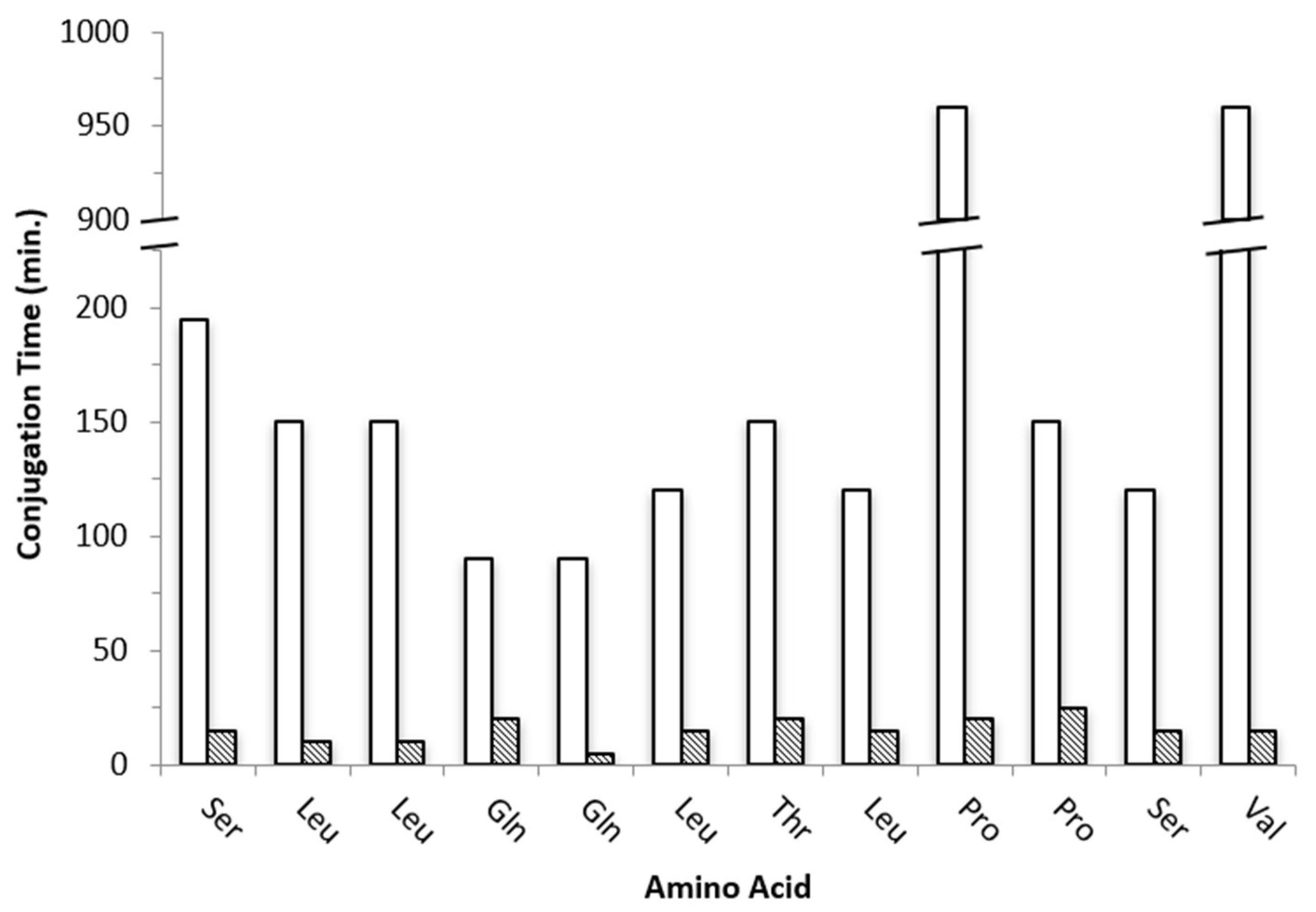
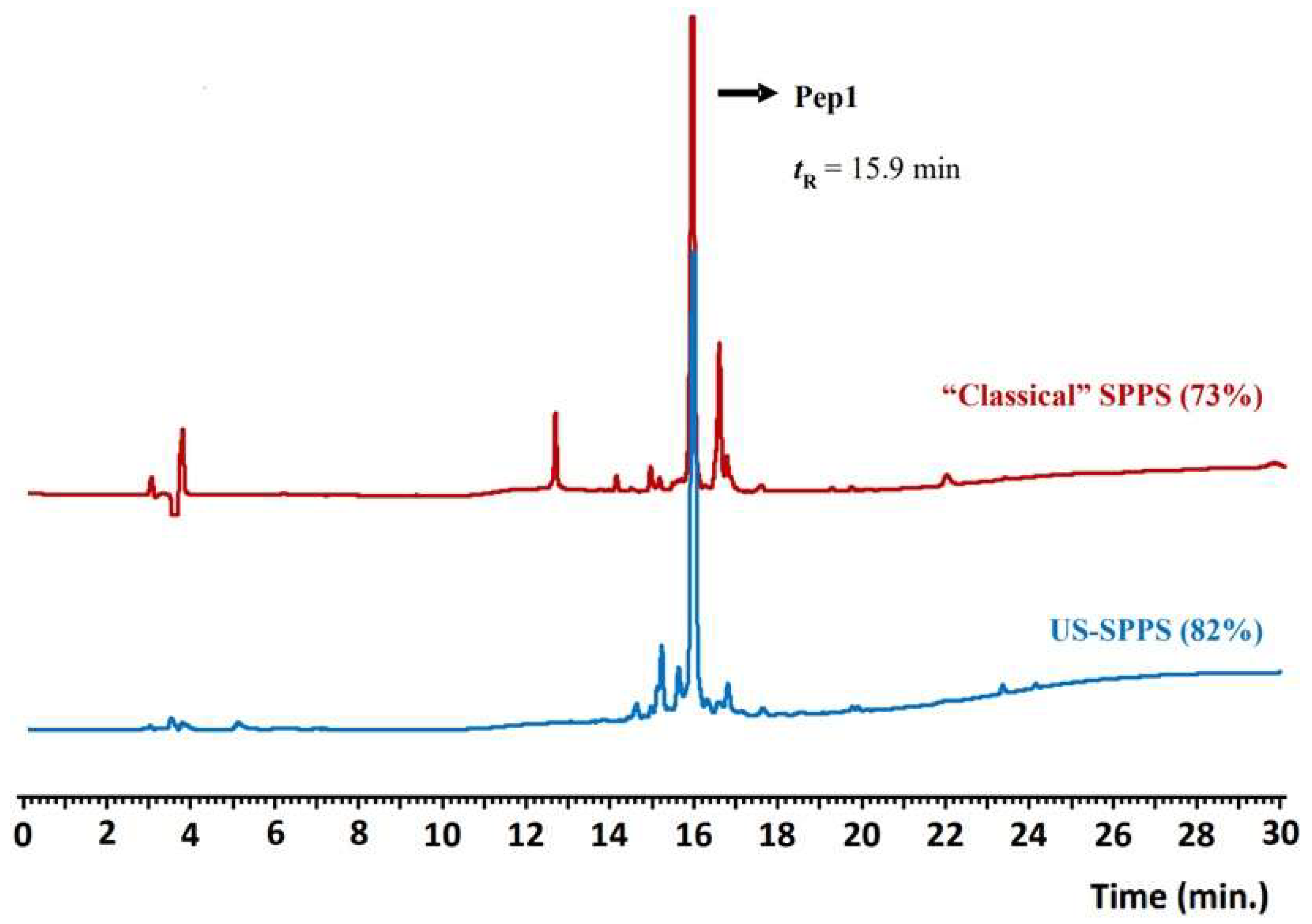
| Pep1 VSPPLTLGQLLS-NH2 | C56H98N14O16 1222.7 | 1223.8 [M+H]+ 612.5 [M+2H]2+ | 15.9 a Cl. 73% US 82% | Cl. 3515 US 250 |
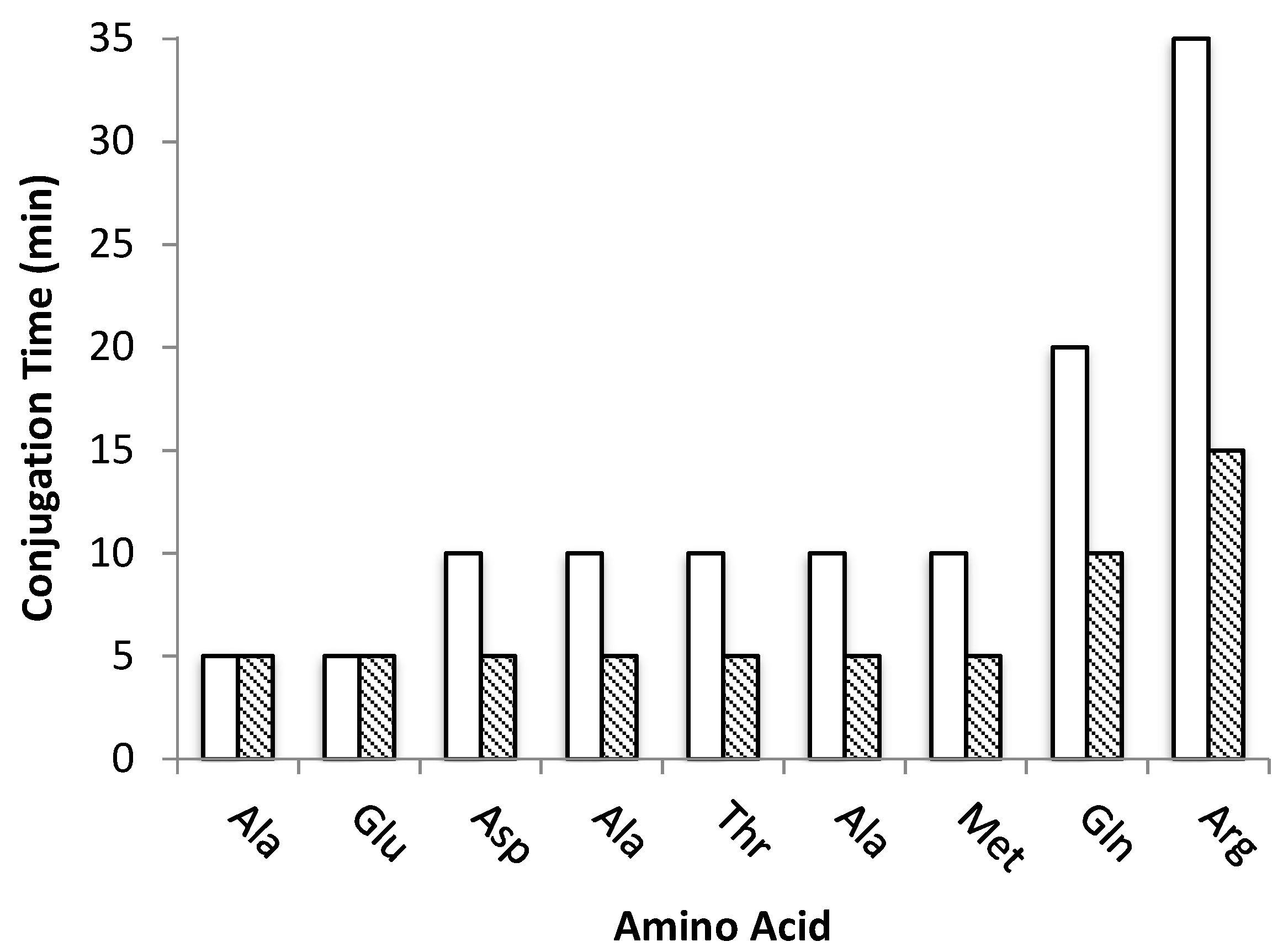
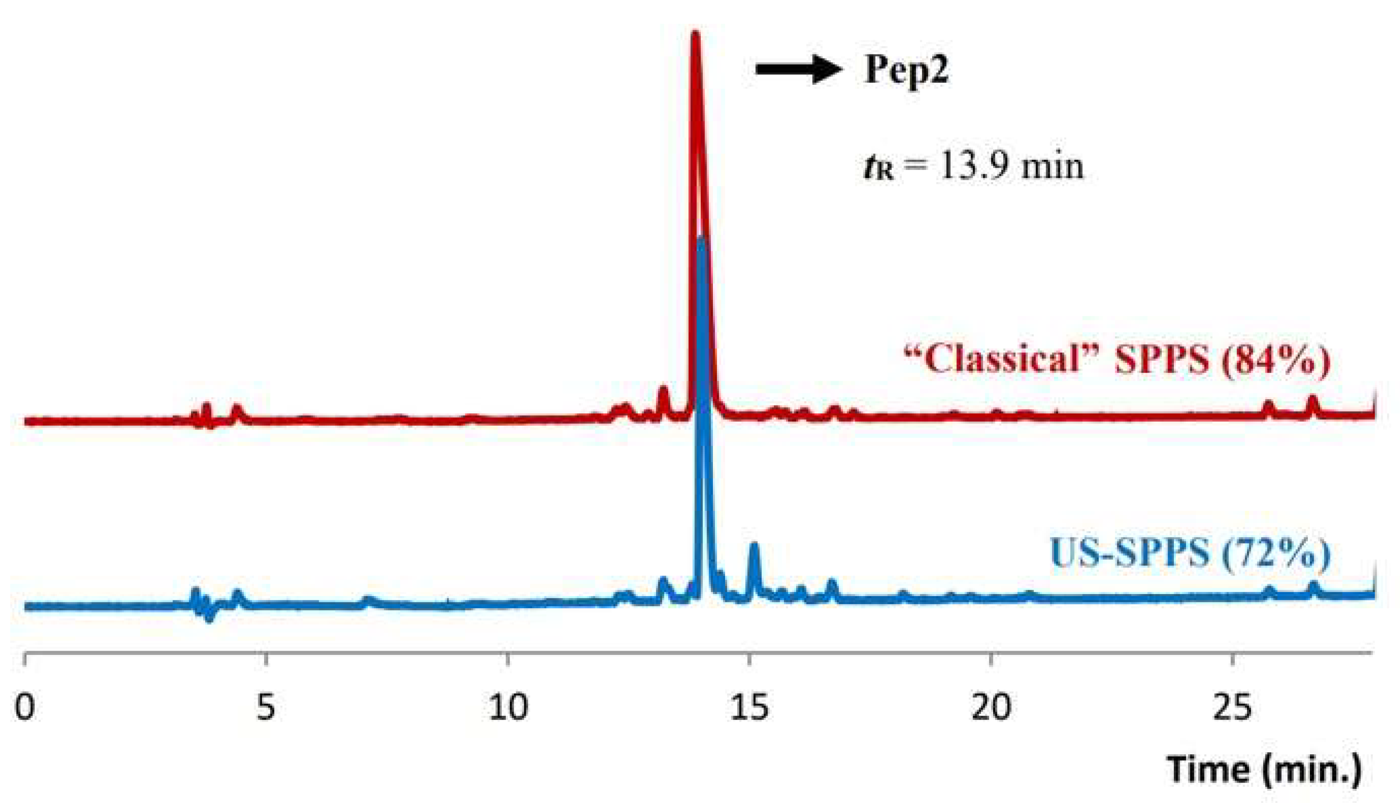
| Pep2 RQMATADEA-NH2 | C38H64N14O15S 991.09 | 991.6 [M+H]+ 496.3 [M+2H]2+ | 13.9 b Cl. 84% US 72% | Cl. 365 US 85 |
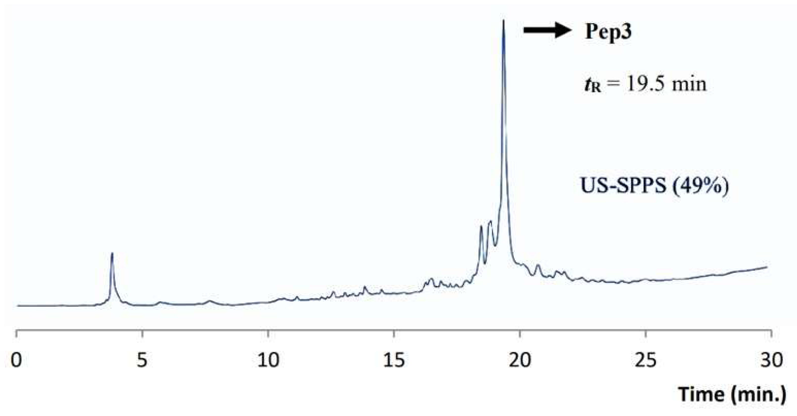
| Pep3 AAVALLPAVLLALLAPRQMATADEA-NH2 | C112H192N30O31S 2489.02 | 1245.5 [M+2H]2+ 830.9 [M+3H]3+ | 19.5 c US 50% | US 347 |
| Peptide | Classical SPPS | US-SPPS |
|---|---|---|
| Pep1 | 42 | 54 |
| Pep2 | 32 | 49 |
| Pep3 | - | 19 |
Publisher’s Note: MDPI stays neutral with regard to jurisdictional claims in published maps and institutional affiliations. |
© 2021 by the authors. Licensee MDPI, Basel, Switzerland. This article is an open access article distributed under the terms and conditions of the Creative Commons Attribution (CC BY) license (https://creativecommons.org/licenses/by/4.0/).
Share and Cite
Silva, R.D.M.; Franco Machado, J.; Gonçalves, K.; Lucas, F.M.; Batista, S.; Melo, R.; Morais, T.S.; Correia, J.D.G. Ultrasonication Improves Solid Phase Synthesis of Peptides Specific for Fibroblast Growth Factor Receptor and for the Protein-Protein Interface RANK-TRAF6. Molecules 2021, 26, 7349. https://doi.org/10.3390/molecules26237349
Silva RDM, Franco Machado J, Gonçalves K, Lucas FM, Batista S, Melo R, Morais TS, Correia JDG. Ultrasonication Improves Solid Phase Synthesis of Peptides Specific for Fibroblast Growth Factor Receptor and for the Protein-Protein Interface RANK-TRAF6. Molecules. 2021; 26(23):7349. https://doi.org/10.3390/molecules26237349
Chicago/Turabian StyleSilva, Rúben D. M., João Franco Machado, Kyle Gonçalves, Francisco M. Lucas, Salete Batista, Rita Melo, Tânia S. Morais, and João D. G. Correia. 2021. "Ultrasonication Improves Solid Phase Synthesis of Peptides Specific for Fibroblast Growth Factor Receptor and for the Protein-Protein Interface RANK-TRAF6" Molecules 26, no. 23: 7349. https://doi.org/10.3390/molecules26237349
APA StyleSilva, R. D. M., Franco Machado, J., Gonçalves, K., Lucas, F. M., Batista, S., Melo, R., Morais, T. S., & Correia, J. D. G. (2021). Ultrasonication Improves Solid Phase Synthesis of Peptides Specific for Fibroblast Growth Factor Receptor and for the Protein-Protein Interface RANK-TRAF6. Molecules, 26(23), 7349. https://doi.org/10.3390/molecules26237349