Article

Structure and Cytotoxicity of Novel Lignans and Lignan Glycosides from the Aerial Parts of Larrea tridentata

School of Pharmacy, Tokyo University of Pharmacy and Life Sciences, 1432-1, Horinouchi, Hachioji, Tokyo 192-0392, Japan
*
Author to whom correspondence should be addressed.
Molecules 2021, 26(20), 6186; https://doi.org/10.3390/molecules26206186
Submission received: 24 September 2021 / Revised: 9 October 2021 / Accepted: 11 October 2021 / Published: 14 October 2021
(This article belongs to the Section Natural Products Chemistry)

Abstract

:
Previously, the authors conducted phytochemical investigations of the aerial parts of Larrea tridentata and reported triterpene glycosides and lignan derivatives. In continuation of the preceding studies, 17 lignans and lignan glycosides (117) were isolated, including seven new compounds (17). Herein, the structure of the new compounds was determined based on spectroscopic analysis and enzymatic hydrolysis. The cytotoxicity of 117 against HL-60 human promyelocytic leukemia cells was examined. Compounds 411 and 1416 were cytotoxic to HL-60 cells, with IC50 values in the range of 2.7–17 μM. Compound 6, which was the most cytotoxic among the unprecedented compounds, was shown to induce apoptotic cell death in HL-60 cells.

1. Introduction

Larrea tridentata is an evergreen small shrub belonging to the Zygophyllaceae family. It grows in the desert areas of the southwestern United States and Mexico, and is commonly called ‘Creosote bush’. It has been used as a medicinal plant for the treatment of a variety of illnesses—including infertility, rheumatism, arthritis, diabetes, gallbladder and kidney stones, pain, and inflammation [1]. The phytochemical constituents of L. tridentata have been extensively investigated with the aim of discovering new biologically active secondary metabolites. In this quest, bioactive compounds with antibacterial [2,3], antimicrobial [3], cytotoxic [2,4], anti-inflammatory [5], anti-tuberculosis [6], anti-fungal [7], and anti-protozoal [8] potential have been isolated and identified. Previously, we reported 25 triterpene glycosides and three lignan derivatives, including larrealignans A and B, from the aerial parts of L. tridentata, and their cytotoxicity against HL-60 human promyelocytic leukemia cells [9,10]. Some plant lignans are promising seed compounds for new anticancer agents, and etoposide, a clinically applied anticancer medicine, is a chemically modified plant lignan. Acute promyelocytic leukemia is a cancer of the white blood cells, and there will be approximately 14,287 new cases and 8809 deaths in 2018 in Japan [11,12]. It is relatively sensitive to chemotherapy agents, often successfully leading to remission. However, when leukemia recurs, the present anticancer agents are not satisfactorily effective against leukemia. Thus, the development of new antileukemia agents is still expected to overcome leukemia. Our phytochemical investigation of this plant with the focus on lignan constituents resulted in the isolation of 17 lignan derivatives (117), seven (17) of which were previously undescribed. This study deals with the structural characterization of novel lignan derivatives based on spectroscopic analysis and hydrolysis. The cytotoxicity of the isolated compounds against HL-60 cells is evaluated.

2. Results and Discussion

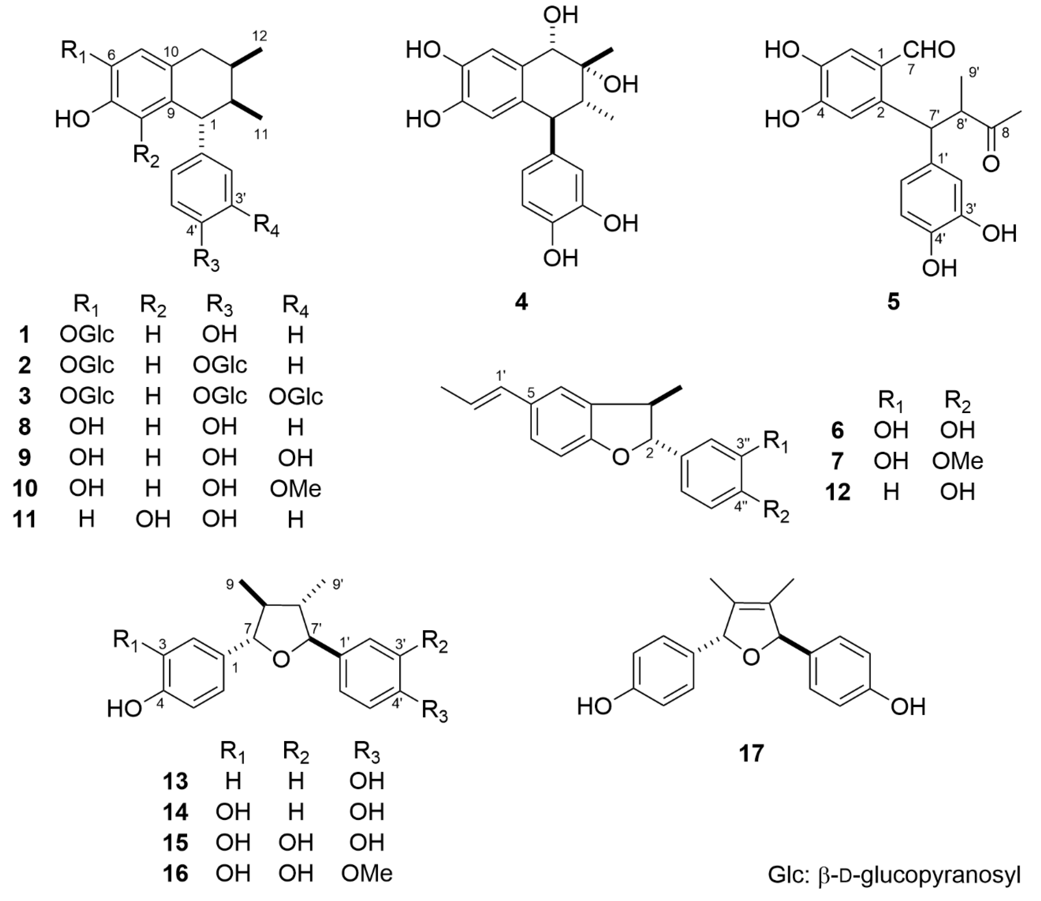
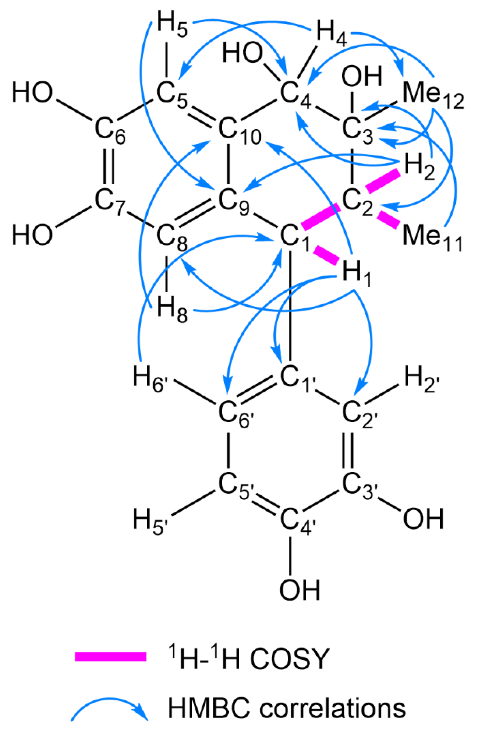
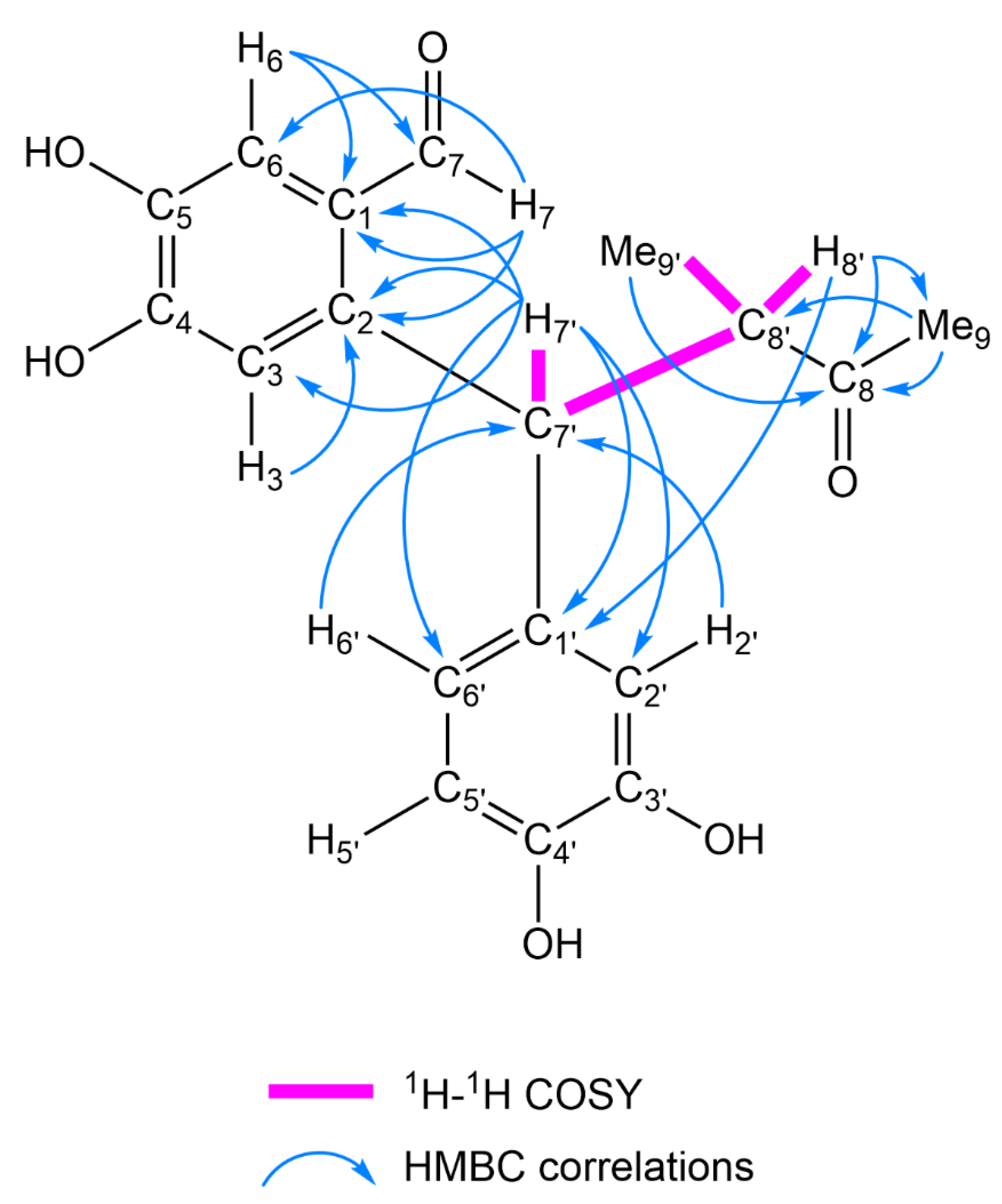
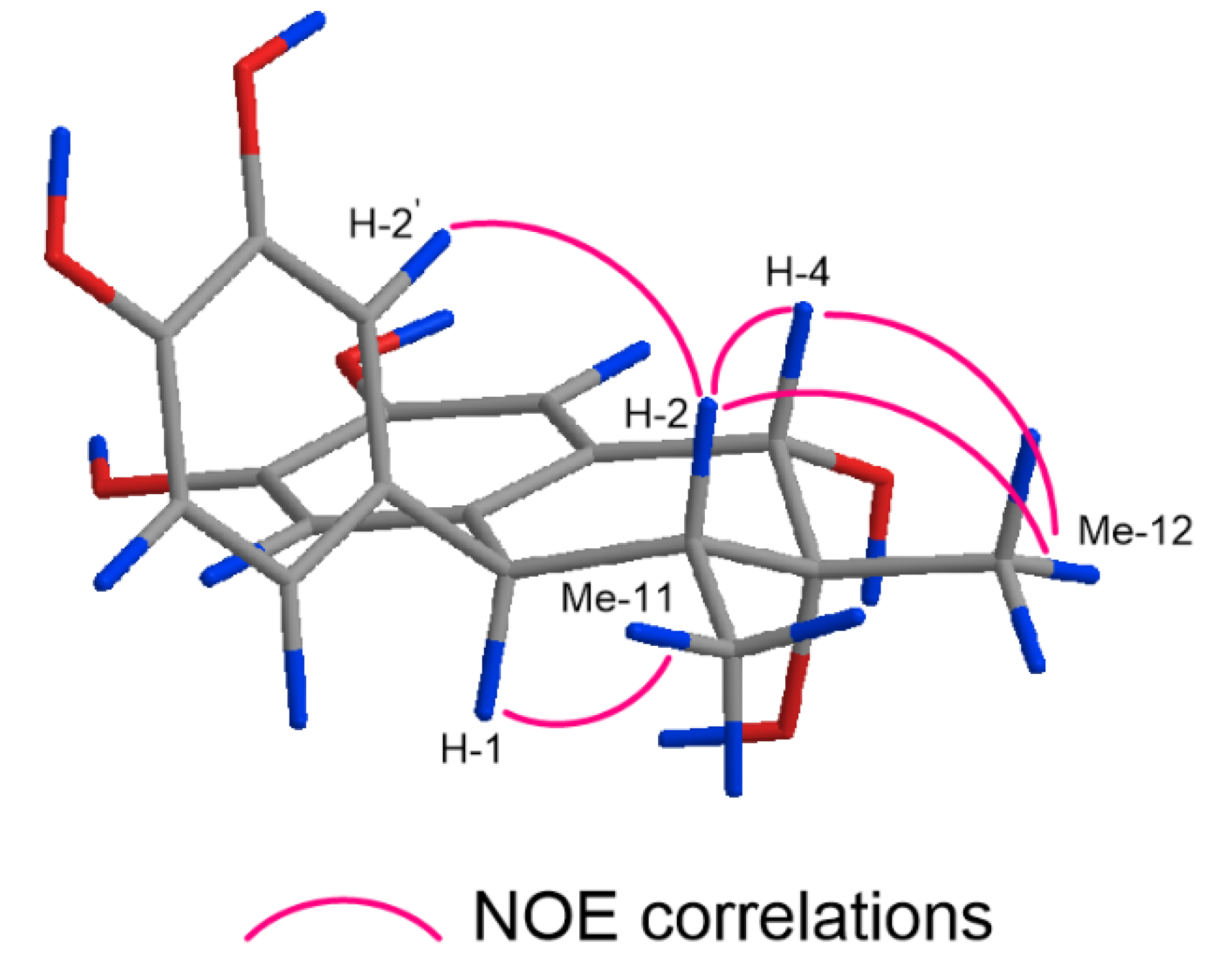
2.1. Structure Elucidation of 1–17

The MeOH extract (940 g) of the aerial parts of L. tridentata (dry weight, 3.0 kg) was separated by silica gel and octadecylsilanized (ODS) silica gel column chromatography (CC) to collect 17 compounds (117) (Figure 1). Compounds 817 were identified as 3′-demethoxy-6-demethylisoguaiacin (8) [13], 6,3′-didemethylisoguaiacin (9) [14], 6-demethylisoguaiacin (10) [15], 6,3′-didemethoxy-8-hydroxyisoguaiacin (11) [16], (−)-conocarpan (12) [17], (−)-8′-epi-larreatricin (13) [18], (7S,7′S,8S,8′S)-3,4,4′-trihydroxy-7,7′-epoxylignan (14) [14], (7S,7′S,8S,8′S)-3,3′,4,4′-tetrahydroxy-7,7′-epoxylignan (15) [14], (7S,7′S,8S,8′S)-3,3′,4-trihydroxy-4′-methoxy-7,7′-epoxylignan (16) [19], and (7R,7′R)-4,4′-dihydroxy-8,8′-didehydro-7,7′-epoxylignan (17) [14].
Compound 1 was obtained as an amorphous powder. The molecular formula was determined as C24H30O8 based on high-resolution electrospray ionization time-of-flight mass spectroscopy (HR-ESI-TOF-MS; m/z: 469.1843 [M + Na]+, calculated for C24H30NaO8: 469.1838) and 13C-NMR. The ultraviolet (UV) spectrum of 1 exhibited an absorption maximum indicative of aromatic rings (281 nm). The 1H- and 13C-NMR spectra of 1 indicated the presence of a 1,2,4,5-tetrasubstituted aromatic ring [δH 6.97 (1H, s, H-5), 6.31 (1H, s, H-8); δC 116.9, 143.3, 144.2, 116.7, 133.2, 127.1 (C-5–C-10)], a 1,4-disubstituted aromatic ring [δH 6.85 (2H, d, J = 8.5 Hz, H-2′, H-6′), 6.71 (2H, d, J = 8.5 Hz, H-3′, H-5′); δC 137.4, 129.1, 113.9, 154.5, 113.9, 129.1 (C-1′–C-6′)], two methyl groups [δH 0.93 (3H, d, J = 7.0 Hz, Me-12), 0.91 (3H, d, J = 6.8 Hz, Me-11); δC 14.3 (C-11), 14.1 (C-12)], a methylene group [δH 2.90 (1H, dd, J = 16.0, 5.5 Hz, H-4a) and 2.49 (1H, dd, J = 16.0, 7.0 Hz, H-4b); δC 34.2 (C-4)], and three methine groups [δH 3.60 (1H, d, J = 6.6 Hz, H-1), 2.04 (1H, m, H-3), 1.94 (1H, ddd, J = 6.8, 6.6, 2.5 Hz, H-2); δC 49.4 (C-1), 40.2 (C-2), 28.7 (C-3)]. These spectral features are analogous to those of 8. The glycosidic nature of 1 was corroborated by the strong absorption band of the hydroxy groups (3365 cm−1) in the infrared (IR) spectrum, and the signals of an anomeric proton and carbon [δH 4.78 (1H, d, J = 8.0 Hz, H-1″); δC 102.7 (C-1″)] in the 1H- and 13C-NMR spectra. Enzymatic hydrolysis of 1 with β-D-glucosidase in AcOH/AcONa buffer (pH 5.0) yielded 8 and D-glucose. D-Glucose was identified by HPLC analysis using a combination of refractive index and optical rotation detectors. In the heteronuclear multiple bond correlation (HMBC) spectrum of 1, a long-range correlation was observed between H-1″ of β-D-glucopyranosyl (Glc) and C-6 of the aglycone moiety (δC 143.3). The β-anomeric configuration was ascertained based on the large coupling constant of the anomeric proton (J = 8.0 Hz). Therefore, 1 was characterized as 3′-demethoxy-6-demethylisoguaiacin 6-O-β-D-glucopyranoside.
The 1H- and 13C-NMR spectra of 2 (C30H40O13) were closely related to those of 1; however, signals of an additional anomeric proton and carbon were observed. Furthermore, the molecular formula of 2 had an additional C6H10O5 compared to that of 1. Thus, 2 was suggested to have one more hexopyranosyl group than 1. Compound 2 was enzymatically hydrolyzed under the same conditions as those used for 1 to afford 8 and D-glucose. Analysis of the 1H-1H correlation spectroscopy (COSY), heteronuclear single quantum coherence (HSQC), 1-dimensional selective total correlation spectroscopy (1D-TOCSY), and HSQC-TOCSY data for the sugar moieties indicated the presence of two terminal β-D-glucopyranosyl units [Glc (I): δH 4.78 (1H, d, J = 7.9 Hz, H-1″ of Glc (I)); δC 102.7, 73.1, 76.1, 69.5, 76.2, 60.6 (C-1″–C-6″ of Glc (I))] and [Glc (II): δH 4.92 (1H, d, J = 7.9 Hz, H-1′″ of Glc (II)); δC 100.5, 73.1, 75.8, 69.5, 76.4, 60.6 (C-1′″–C-6′″ of Glc (II))]. The HMBC spectrum of 2 exhibited long-range correlations from H-1″ of Glc (I) to C-6 of the aglycone moiety (δC 143.4), and from H-1′″ of Glc (II) to C-4′ of the aglycone moiety (δC 155.4). Accordingly, 2 was identified as 3′-demethoxy-6-demethylisoguaiacin 6,4′-di-O-β-D-glucopyranoside.
Compound 3 (C36H50O19) was obtained as an amorphous powder. The 1H- and 13C-NMR spectra of 3 were similar to those of 2, except for the signals assignable to the aromatic ring attached to C-1 of the aliphatic moiety. Signals arising from a 1,2,4-trisubstituted aromatic ring [δH 7.54 (1H, d, J = 8.3 Hz, H-5′), 7.51 (1H, br s, H-2′), 6.68 (1H, d, J = 8.3 Hz, H-6′); δC 143.0, 120.8, 148.3, 147.2, 118.9, 124.1 (C-1′–C-6′)] were observed in the 1H- and 13C-NMR spectra of 3. Enzymatic hydrolysis of 3 with β-D-glucosidase yielded 9 and D-glucose. 1H-1H COSY, HSQC, 1D-TOCSY, and HSQC-TOCSY analyses of the sugar moieties revealed the following three terminal β-D-glucopyranosyl units: Glc (I) [δH 5.53 (1H, d, J = 8.0 Hz, H-1″ of Glc (I)); δC 105.1, 75.2, 78.3, 71.2, 79.0, 62.3 (C-1″–C-6″ of Glc (I))], Glc (II) [δH 5.62 (1H, d, J = 7.5 Hz, H-1′″ of Glc (II)); δC 103.8, 75.0, 78.3, 71.2, 78.5, 62.2 (C-1′″–C-6′″ of Glc (II))], and Glc (III) [δH 5.56 (1H, d, J = 7.4 Hz, H-1″″ of Glc (III)); δC 104.1, 75.1, 78.1, 71.2, 78.8, 62.3 (C-1″″–C-6″″ of Glc (III))]. The HMBC spectrum of 3 showed correlations between H-1″ of Glc (I) and C-6 of the aglycone moiety (δC 145.4), H-1′″ of Glc (II) and C-3′ of the aglycone moiety (δC 148.3), and between H-1″″ of Glc (III) and C-4′ of the aglycone moiety (δC 147.2). Thus, 3 was determined to be 6,3′-didemethylisoguaiacin 6,3′,4′-tri-O-β-D-glucopyranoside.
Compound 4 (C18H20O6) was obtained as an amorphous powder. The 1H- and 13C-NMR spectra of 4 were suggestive of a 1-aryl tetralin-type lignan closely related to 9, and 4 and 9 shared the same 1,2,4-trisubstituted aromatic ring and 1,2,4,5-tetrasubstituted aromatic ring. Compound 4 differed from 9 in the aliphatic ring moiety; that of 4 was composed of two methyl groups [δH 1.41 (3H, s, Me-12), 0.92 (3H, d, J = 6.7 Hz, Me-11); 21.9 (C-12), 11.0 (C-11)], two methine groups [δH 3.56 (1H, d, J = 11.0 Hz, H-1), 1.88 (1H, dd, J = 11.0, 6.7 Hz, H-2); δC 48.3 (C-1), 43.6 (C-2)], an oxygenated methine group [δH 4.48 (1H, s, H-4); δC 73.5 (C-4)], and an oxygenated quaternary carbon [δC 72.3 (C-3)]. The 1H-1H COSY spectrum of 4 indicated spin-coupling correlations of the H-2 methine proton with the H-1 methine proton and Me-11 methyl protons. The HMBC spectrum of 4 indicated long-range correlations from H-2 to C-3 and C-4, Me-12 to C-2, C-3, and C-4, and from H-4 to C-12. These correlations indicated the introduction of hydroxy groups at C-3 and C-4 (Figure 2). Thus, the planar structure of 4 was determined to be 1,2,4-trihydro-1-(3,4-dihydroxyphenyl)-2,3-dimethyl-3,4,6,7-naphthalenetetrol. In the nuclear Overhauser effect spectroscopy (NOESY) spectrum of 4, the NOE correlations between H-1 and Me-11, H-2 and H-4/Me-12/H-2′, and between H-4 and Me-12 are indicative of the 1β, 2α, 3α-hydroxy, and 4α relative configurations (Figure 3). The circular dichroism (CD) spectrum of 4 exhibited a positive Cotton effect at 244 and 275 nm, confirming the 1R absolute configuration [20]. The configurations at C-2, C-3, and C-4 were determined as 2R, 3R, and 4S, respectively. Thus, 4 was identified as (1R,2R,3R,4S)-1,2,4-trihydro-1-(3,4-dihydroxyphenyl)-2,3-dimethyl-3,4,6,7-naphthalenetetrol.
Compound 5 (C18H18O6) was collected as an amorphous powder. The IR spectrum of 5 shows absorption bands of the hydroxy groups (3271 cm−1) and carbonyl groups (1715 and 1695 cm−1). The 1H- and 13C-NMR spectra of 5 showed signals of a 1,2,4,5-tetrasubstituted aromatic ring [δH 7.22 (1H, s, H-6), 7.03 (1H, s, H-3); δC 125.2, 140.3, 113.4, 151.1, 143.1, 116.4 (C-1–C-6)], a 1,2,4-trisubstituted aromatic ring [δH 6.78 (1H, d, J = 1.7 Hz, H-2′), 6.73 (1H, d, J = 8.0 Hz, H-5′), 6.70 (1H, dd, J = 8.0, 1.7 Hz, H-6′); δC 133.3, 114.6, 144.5, 143.1, 114.4, 118.9 (C-1′–C-6′)], two methyl groups [δH 2.09 (3H, s, Me-9), 1.08 (3H, d, J = 6.9 Hz, Me-9′); δC 27.1 (C-9), 15.1 (C-9′)], two methine groups [δH 5.05 (1H, d, J = 11.4 Hz, H-7′), 3.50 (1H, dq, J = 11.4, 6.9 Hz, H-8′); δC 51.1 (C-8′), 44.7 (C-7′)], an aldehyde group [δH 10.2 (1H, s, H-7); δC 190.4 (C-7)], and a carbonyl carbon [δC 213.2 (C-8)]. The spin-coupling correlation H-7′/H-8′/Me-9′ in the 1H-1H COSY spectrum of 5 indicates the existence of a structural fragment of -C(7′)H-C(8′)H(Me(9′))- (Figure 4). The HMBC spectrum displayed long-range correlations from H-8′, Me-9′, and Me-9 to the C-8 carbonyl carbon. These correlations allowed the structural fragment to be extended to -C(7′)H-C(8′)H(Me(9′))-C(8)(=O)-Me(9), which was shown to be linked to C-1′ of the 1,2,4-trisubstituted aromatic ring and C-2 of the 1,2,4,5-tetrasubstituted aromatic ring based on the observed HMBC correlations between H-7′ and C-1′/C-2′/C-6′/C-1/C-2/C-3. Furthermore, the aldehyde group was confirmed to be attached to C-1 of the 1,2,4,5-tetrasubstituted aromatic ring based on the long-range correlation from the H-7 aldehyde proton to C-1/C-2/C-6 of the aromatic carbons (Figure 4). The configurations of C-7′ and C-8′ have yet to be determined. Accordingly, 5 was deduced to be 2-[1-(3,4-dihydroxyphenyl)-2-methyl-3-oxobutyl]-4,5-dihydroxybenzaldehyde.
Compound 6 (C18H18O3) was obtained as an amorphous powder and is suggested to be a benzofuran-type neolignan, the 1H- and 13C-NMR spectra of which resembled those of 12, except for the signals arising from the aromatic ring bonded to C-2 of the benzofuran moiety. In the 1H-NMR spectrum of 6, the aromatic proton showed a typical 1,2,4-trisubstituted spin-coupling system at δH 6.87 (1H, d, J = 2.1 Hz, H-2″), 6.80 (1H, d, J = 8.2 Hz, H-5″), and 6.76 (1H, dd, J = 8.2, 2.1 Hz, H-6″). Furthermore, the molecular formula of 6 was larger than that of 12 by an oxygen atom. The above data revealed that 6 is the C-3″ hydroxy derivative of 12. Because the proton spin-coupling constant between H-2 and H-3 was 8.6 Hz, and the CD spectrum of 6 was in good agreement with that of 12 [17], the absolute configurations of C-2 and C-3 were determined to be R and R, respectively. Thus, 6 was identified as (2R,3R)-2,3-dihydro-2-(3,4-dihydroxyphenyl)-3-methyl-5-(E)-propenylbenzofuran.
The 1H- and 13C-NMR spectra of 7 (C19H20O3) were similar to those of 6. However, the molecular formula of 7 was larger than that of 6 by CH2, and the presence of a methoxy group [δH 3.88 (3H, s); δC 54.6] was verified based on 1H- and 13C-NMR spectral analysis. Comparison of the 13C-NMR spectrum of 7 with that of 6 showed that the signal assignable to C-4″ was moved downfield by 2.5 ppm, whereas that attributable to C-5″ was shifted upfield by 3.5 ppm. Additionally, an HMBC correlation from δH 3.88 (OMe) to δC 147.2 (C-4″) was detected. The above data revealed that 7 is a C-4″-O-methyl derivative of 6. Accordingly, 7 was deduced to be (2R,3R)-2,3-dihydro-2-(3-hydroxy-4-methoxyphenyl)-3-methyl-5-(E)-propenylbenzofuran.

2.2. Cytotoxicity of 1–17

The cytotoxicity of 117 to HL-60 cells was evaluated using a modified 3-(4,5-dimethylthiazol-2-yl)-2,5-diphenyl-2H-tetrazolium bromide (MTT) assay (Table 1). Compounds 411 and 1416 exhibited cytotoxicity to HL-60 cells, with IC50 values in the range of 2.7–17 μM. Comparison of the cytotoxicity of 8 with that of 1 and 2, and the cytotoxicity of 9 with that of 3 suggests that glucosylation(s) of the aromatic group(s) diminished the cytotoxicity. In benzofuran-type neolignans (6, 7, and 12), the catechol group (C-3″, C-4″-hydroxy) was necessary to induce cytotoxicity (6), and C-4″-O-methylation reduced the cytotoxicity (7). For the tetrahydrofuran-type lignans (1317), the presence of the catechol group was also essential for exerting cytotoxicity (14, 15, and 16).

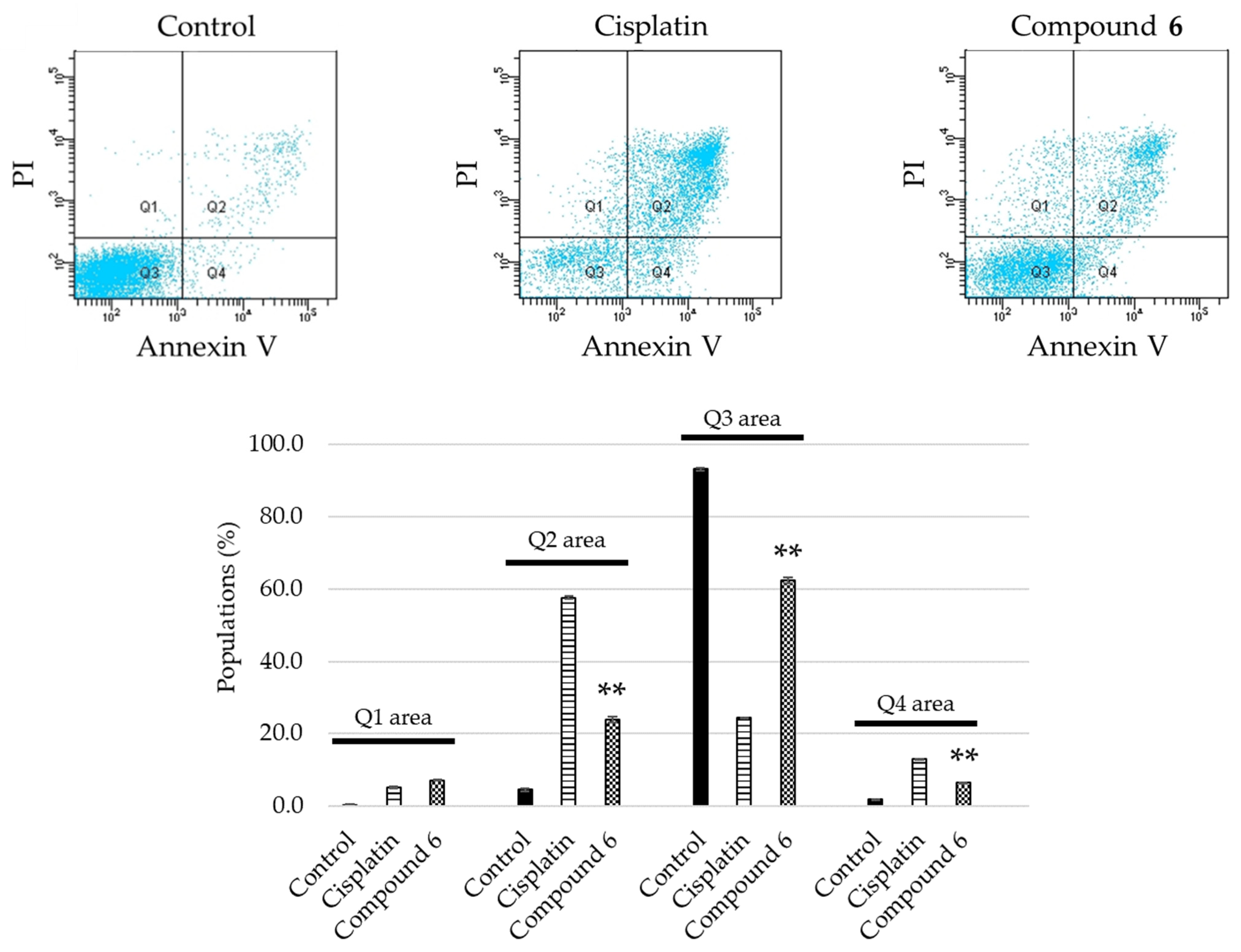
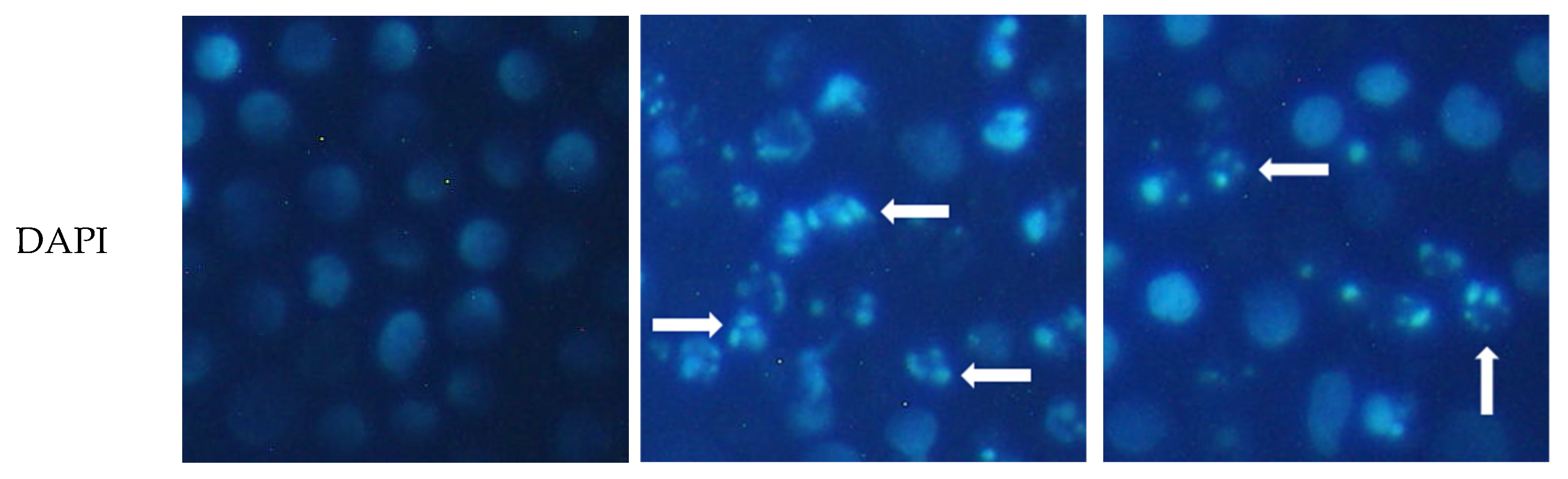
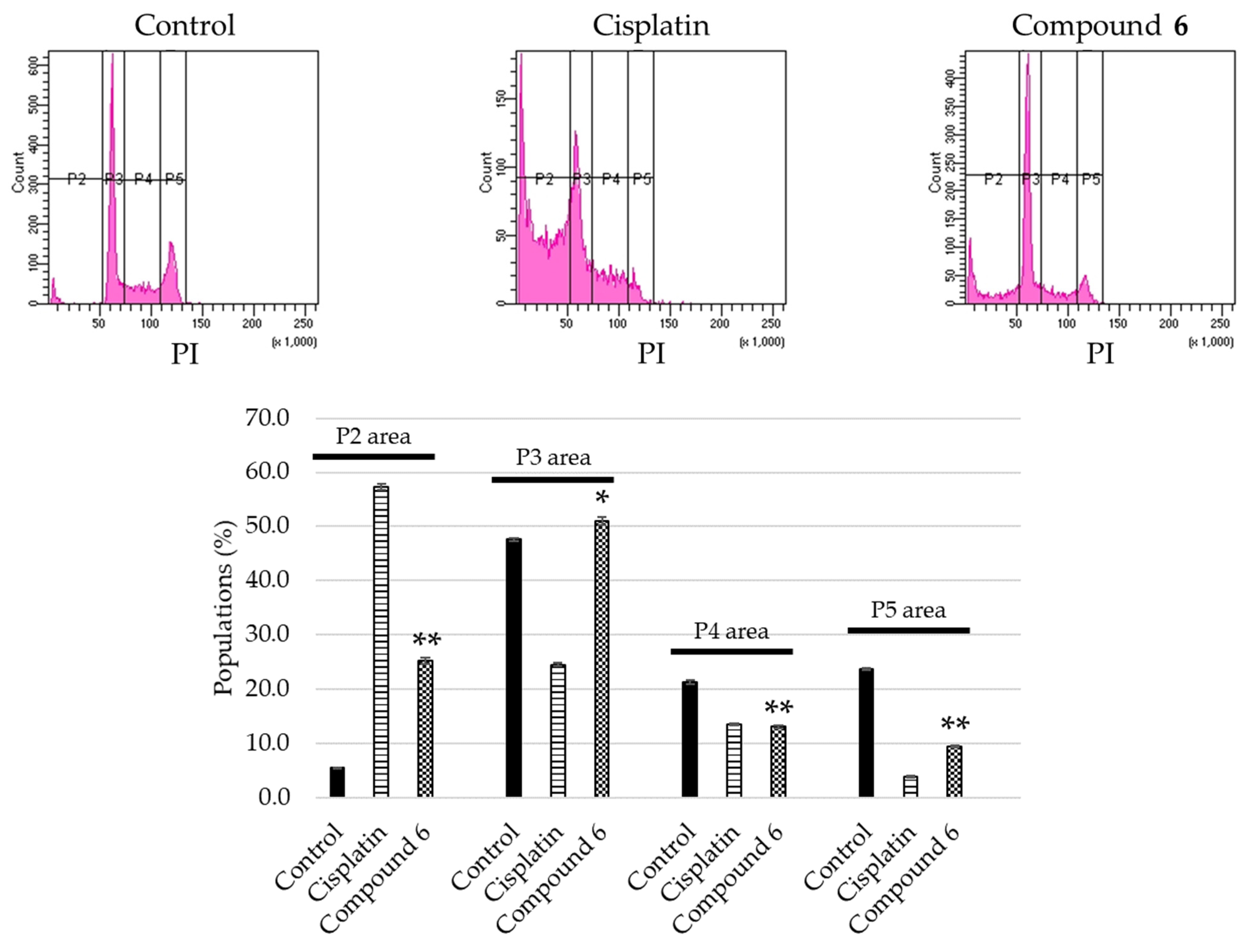
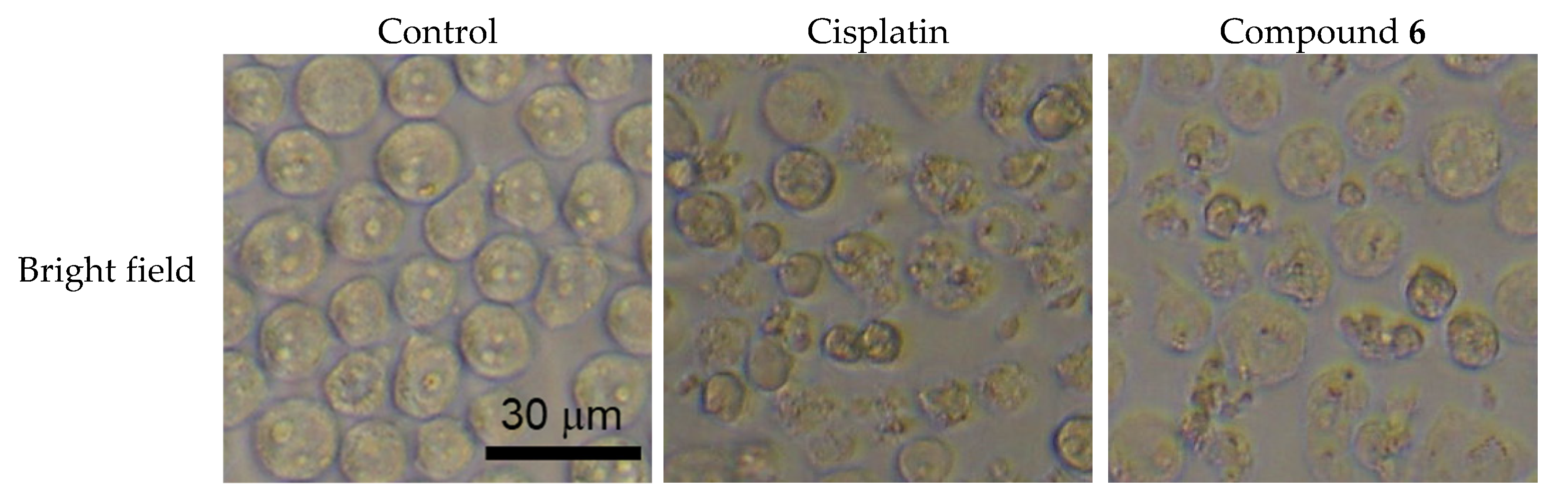
2.3. Apoptosis Inducing Activity of 6

The apoptosis-inducing activity of 6 was evaluated. Among the unprecedented compounds, 6 exerted the highest cytotoxicity in HL-60 cells. HL-60 cells were treated with 40 μM of 6 for 24 h, stained with Annexin V and propidium iodide (PI), and analyzed by flow cytometry. The cell populations of early (Q4 area) and late (Q2 area) apoptotic cells, for which the vehicle control was 1.9 ± 0.033% and 4.5 ± 0.38%, increased to 6.5 ± 0.033% and 24 ± 0.87%, respectively (Figure 5). HL-60 cells treated with 40 μM of 6 were stained with 4′,6-diamidino-2-phenylindole (DAPI), and their morphology was observed under a fluorescence microscope. As shown in Figure 6, the HL-60 cells exhibited chromatin condensation and nuclear disassembly, which are representative phenomena in apoptosis. In addition, the cell cycle distribution of HL-60 cells was analyzed using a flow cytometer. When the cells were treated with 40 μM of 6 for 24 h, the sub-G1 population increased to 25 ± 0.58%, compared to 5.5 ± 0.088% in the vehicle control populations (Figure 7). Notably, the results showed that, for HL-60 cells treated with 6, the population of cells in the G0/G1 phase (P3 area) increased (control: 48 ± 0.35%; 6: 51 ± 0.71%), whereas those in the S phase declined (P4 area) (control: 21 ± 0.38%; 6: 13 ± 0.29%) compared with the vehicle control.

3. Material and Methods

3.1. General Experimental Procedures

Optical rotations were measured on a JASCO P-1030 automatic digital polarimeter (Tokyo, Japan). IR, UV, and CD spectral data were collected using a FT-IR 620 spectrometer (JASCO), V-630 spectrometer (JASCO), and J-720 spectrometer (JASCO), respectively. NMR spectral data were obtained by a Bruker DPX-400 (1H-NMR: 400 MHz; 13C-NMR: 100 MHz), Bruker DRX-500 (1H-NMR: 500 MHz; 13C-NMR: 125 MHz), Bruker AV-500 (1H-NMR: 500 MHz; 13C-NMR: 125 MHz), or Bruker AV-600 (1H-NMR: 600 MHz; 13C-NMR: 150 MHz) spectrometer using a standard Bruker pulse programs at 300 K (Karlsruhe, Germany). Chemical shifts are rendered as δ values with reference to tetramethylsilane (TMS) as an internal standard. HR-ESI-TOF mass were measured using a Waters Micromass LCT mass spectrometer (Milford, MA, USA). Diaion HP-20 porous polymer polystyrene resin (Mitsubishi-Chemical, Tokyo, Japan), silica gel Chromatorex BW-300 (Fuji-Silysia Chemical, Aichi, Japan), and ODS silica gel COSMOSIL 75C18-OPN (Nacalai Tesque, Kyoto, Japan) were employed for CC. Thin layer chromatography (TLC) analysis carried out by precoated silica gel 60F254 or RP18 F254S plates (0.25 mm thick; Merch, Darmstadt, Germany), and the sample spots were detected by spraying the TLC plates with H2SO4/H2O (1:9), followed by heating. The HPLC system was constituted with the following instrument; a Tosoh CCPM pump (Tokyo, Japan), a Shodex OR-2 detector (Showa-Denko, Tokyo, Japan), a RheodyneTM injection port (Thermo Fisher Scientific, Waltham, MA, USA), and a Capcell Pak NH2 UG80 (4.6 mm i.d. × 250 mm, 5 μm; Shiseido, Tokyo, Japan). Enzymatic hydrolysis was conducted using β-D-glucosidase (Sigma, St. Louis, MO, USA). The following materials and biochemical-grade reagents were used for the cell culture assays: a 96-well flat-bottom plate (Iwaki Glass, Chiba, Japan); RPMI-1640 medium, cisplatin, and MTT (Sigma); penicillin G sodium salt and streptomycin sulfate (Gibco, Gland Island, NY, USA); paraformaldehyde and phosphate-buffered saline (PBS) (FUJIFILM Wako Pure Chemical Corporation, Osaka, Japan); fetal bovine serum (FBS) (Bio-Whittaker, Walkersville, MD, USA); a Spectra Classic microplate reader (Tecan, Salzburg, Austria).

3.2. Plant Material

The aerial parts of L. tridentata were purchased from Richters Herbs (Ontario, Canada) in 2007. A voucher specimen was kept at the herbarium of the Tokyo University of Pharmacy and Life Sciences.

3.3. Extraction and Isolation Procedures

The aerial parts of L. tridentata (dry weight, 3.0 kg) were extracted with hot MeOH (15 L × 4 times), and the extract was evaporated under reduced pressure. The MeOH extract (940 g) was loaded on a Diaion HP-20 column and successively eluted with MeOH/H2O (3:7), MeOH/H2O (1:1), MeOH, EtOH, and EtOAc. The MeOH eluted portion [Fraction (Fr) C; 477 g] was subjected to silica gel CC and eluted with CHCl3/MeOH/H2O (50:1:0; 19:1:0; 9:1:0; 40:10:1) to obtain 7 fractions (Frs. C-1–C-7). Fr. C-3 was separated by silica gel CC eluted with CHCl3/MeOH (19:1; 9:1) and ODS silica gel CC eluted with MeOH/H2O (1:3; 3:2; 2:1; 4:1) to obtain 8 (79 mg) and 13 (5.2 mg). Fr. C-4 was applied to silica gel CC eluted with CHCl3/MeOH/H2O (9:1:0; 40:10:1), and ODS silica gel CC eluted with MeOH/H2O (2:5; 1:2; 2:3; 1:1; 3:2; 2:1) and MeCN/H2O (2:3) to collect 4 (4.6 mg) and 5 (5.0 mg). Fr. C-5 was divided by silica gel CC eluted with CHCl3/MeOH/H2O (9:1:0; 50:10:1; 40:10:1), and ODS silica gel CC eluted with MeOH/H2O (4:3; 8:3) and MeCN/H2O (2:5; 1:1) to get 1 (11 mg). Fr. C-6 was subjected to silica gel CC eluted with CHCl3/MeOH/H2O (7:4:1; 27:18:5; 2:4:1; 60:10:1; 30:10:1), and ODS silica gel CC eluted with MeOH/H2O (1:2; 2:3; 9:11; 11:9; 2:1; 4:1) and MeCN/H2O (1:4; 1:3) to afford 2 (12 mg) and 3 (7.0 mg). The EtOH eluted portion (Fr. D; 41 g) was chromatographed on silica gel CC eluted with CHCl3/MeOH (19:1; 9:1) to get 8 fractions (Frs. D-1–D-8). Fr. D-2 was purified using silica gel CC eluted with hexane/EtOAc (5:1) and ODS silica gel CC eluted with MeOH/H2O (7:3; 4:1) to yield 7 (5.3 mg). Fr. D-5 was separated by silica gel CC eluted with CHCl3/MeOH (19;1) and hexane/EtOAc (3:1; 5:1) and ODS silica gel CC eluted with MeCN/H2O (1:1) to obtained 6 (12 mg) and 12 (4.7 mg). Fr. D-7 was loaded on silica gel CC eluted with hexane/EtOAc (2:1), hexane/CHCl3/MeOH (1:19:1; 4:38:1), and CHCl3/MeOH (19:1), and ODS silica gel CC eluted with MeOH/H2O (3:2) and MeCN/H2O (1:2) to afford 10 (7.8 mg), 11 (2.6 mg), 16 (10 mg), and 17 (12 mg). Fr. D-8 was divided by silica gel CC eluted with CHCl3/MeOH (19:1; 9:1) to furnish 9 (17 mg), 14 (12 mg), and 15 (21 mg).

3.4. Structural Elucidation

Compound 1: Amorphous powder; [α]D25 −71.6 (c = 0.09, MeOH); IR (film) νmax: 3365 (OH), 1613, 1513, 1453 (aromatic ring) cm−1; HR-ESI-TOF-MS m/z: 469.1843 [M + Na]+ (calculated for C24H30NaO8: 469.1838); UV λmax (MeOH): 281 (log ε = 3.74) nm; CD λmax (MeOH) (Δε): 232 (−9.41), 273 (−1.32), 289 (+1.97). 1H-NMR spectral data for the aglycone moiety (500 MHz, CD3OD): δH 6.97 (1H, s, H-5), 6.85 (2H, d, J = 8.5 Hz, H-2′ and H-6′), 6.71 (2H, d, J = 8.5 Hz, H-3′ and H-5′), 6.31 (1H, s, H-8), 3.60 (1H, d, J = 6.6 Hz, H-1), 2.90 (1H, dd, J = 16.0, 5.5 Hz, H-4a), 2.49 (1H, dd, J = 16.0, 7.0 Hz, H-4b), 2.04 (1H, m, H-3), 1.94 (1H, ddd, J = 6.8, 6.6, 2.5 Hz, H-2), 0.93 (3H, d, J = 7.0 Hz, Me-12), 0.91 (3H, d, J = 6.8 Hz, Me-11). For 1H-NMR spectral data of the sugar moiety, see Table 2. For 13C-NMR spectral data, see Table 3. For NMR data, see Supplementary Materials.
Compound 2: Amorphous powder; [α]D25 −99.8 (c = 0.10, MeOH); IR (film) νmax: 3363 (OH), 1643, 1507, 1453 (aromatic ring) cm−1; HR-ESI-TOF-MS m/z: 631.2376 [M + Na]+ (calculated for C30H40NaO13: 631.2367); UV λmax (MeOH): 282 (log ε = 3.78) nm; CD λmax (MeOH) (Δε): 237 (−10.2), 270 (−2.66), 286 (+1.92). 1H-NMR spectral data for the aglycone moiety (500 MHz, CD3OD): δH 7.03 (2H, d, J = 8.7 Hz, H-3′ and H-5′), 6.98 (2H, d, J = 8.7 Hz, H-2′ and H-6′), 6.96 (1H, s, H-5), 6.28 (1H, s, H-8), 3.67 (1H, d, J = 6.6 Hz, H-1), 2.91 (1H, dd, J = 16.4, 5.3 Hz, H-4a), 2.51 (1H, dd, J = 16.4, 7.1 Hz, H-4b), 2.04 (1H, m, H-3), 1.95 (1H, ddd, J = 6.9, 6.6, 2.9 Hz, H-2), 0.93 (3H, d, J = 6.9 Hz, Me-12), 0.92 (3H, d, J = 6.9 Hz, Me-11). For 1H-NMR spectral data of the sugar moieties, see Table 2. For 13C-NMR spectral data, see Table 3. For NMR data, see Supplementary Materials.
Compound 3: Amorphous powder; [α]D25 −104.5 (c = 0.10, MeOH); IR (film) νmax: 3287 (OH), 1639, 1506, 1453 (aromatic ring) cm−1; HR-ESI-TOF-MS m/z: 809.2808 [M + Na]+ (calculated for C36H50NaO19: 809.2844); UV λmax (MeOH): 281 (log ε = 3.73) nm; CD λmax (MeOH) (Δε): 233 (−11.2), 270 (−3.23), 286 (+1.68). 1H-NMR spectral data for the aglycone moiety (600 MHz, C5D5N): δH 7.54 (1H, d, J = 8.3 Hz, H-5′), 7.51 (1H, br s, H-2′), 7.38 (1H, s, H-5), 6.85 (1H, s, H-8), 6.68 (1H, d, J = 8.3 Hz, H-6′), 3.76 (1H, d, J = 5.2 Hz, H-1), 2.70 (1H, dd, J = 16.4, 4.8 Hz, H-4a), 2.31 (1H, dd, J = 16.4, 7.9 Hz, H-4b), 1.92 (1H, m, H-3), 1.91 (1H, m, H-2), 0.79 (3H, d, J = 6.7 Hz, Me-11), 0.69 (3H, d, J = 6.7 Hz, Me-12). For 1H-NMR spectral data of the sugar moieties, see Table 2. For 13C-NMR spectral data, see Table 3. For NMR data, see Supplementary Materials.
Compound 4: Amorphous powder; [α]D25 36.8 (c = 0.10, MeOH); IR (film) νmax: 3348 (OH), 1607, 1521, 1448 (aromatic ring) cm−1; HR-ESI-TOF-MS m/z: 355.1118 [M + Na]+ (calculated for C18H20NaO6: 355.1158); UV λmax (MeOH): 285 (log ε = 3.75) nm; CD λmax (MeOH) (Δε): 244 (+1.13), 275 (+4.64), 294 (−5.53). 1H-NMR spectral data (400 MHz, CD3OD): δH 7.04 (1H, s, H-5), 6.73 (1H, d, J = 8.0 Hz, H-5′), 6.54 (1H, d, J = 2.0 Hz, H-2′), 6.51 (1H, dd, J = 8.0, 2.0 Hz, H-6′), 6.15 (1H, s, H-8), 4.48 (1H, s, H-4), 3.56 (1H, d, J = 11.0 Hz, H-1), 1.88 (1H, dd, J = 11.0, 6.7 Hz, H-2), 1.41 (3H, s, Me-12), 0.92 (3H, d, J = 6.7 Hz, Me-11). For 13C-NMR spectral data, see Table 3. For NMR data, see Supplementary Materials.
Compound 5: Amorphous powder; [α]D25 −87.0 (c = 0.10, MeOH); IR (film) νmax: 3271 (OH), 1715 and 1695 (C=O), 1667, 1519, 1453 (aromatic ring) cm−1; HR-ESI-TOF-MS m/z: 353.1021 [M + Na]+ (calculated for C18H18NaO6: 353.1001); UV λmax (MeOH): 282 (log ε = 3.97) nm; CD λmax (MeOH) (Δε): 238 (+11.6), 319 (−3.72). 1H-NMR spectral data (400 MHz, CD3OD): δH 10.2 (1H, s, H-7), 7.22 (1H, s, H-6), 7.03 (1H, s, H-3), 6.78 (1H, d, J = 1.7 Hz, H-2′), 6.73 (1H, d, J = 8.0 Hz, H-5′), 6.70 (1H, dd, J = 8.0, 1.7 Hz, H-6′), 5.05 (1H, d, J = 11.4 Hz, H-7′), 3.50 (1H, dq, J = 11.4, 6.9 Hz, H-8′), 2.09 (3H, s, Me-9), 1.08 (3H, d, J = 6.9 Hz, Me-9′). For 13C-NMR spectral data, see Table 3. For NMR data, see Supplementary Materials.
Compound 6: Amorphous powder; [α]D25 95.6 (c = 0.10, MeOH); IR (film) νmax: 3232 (OH), 1609, 1524, 1486 (aromatic ring) cm−1; HR-ESI-TOF-MS m/z: 283.1386 [M + H]+ (calculated for C18H19O3: 283.1334); UV λmax (MeOH): 264 (log ε = 4.37) nm; CD λmax (MeOH) (Δε): 230 (−3.44), 262 (+12.0), 269 (+10.2). 1H-NMR spectral data (500 MHz, CD3OD): δH 7.20 (1H, br s, H-4), 7.13 (1H, dd, J = 8.2, 1.6 Hz, H-6), 6.87 (1H, d, J = 2.1 Hz, H-2″), 6.80 (1H, d, J = 8.2 Hz, H-5″), 6.76 (1H, dd, J = 8.2, 2.1 Hz, H-6″), 6.71 (1H, d, J = 8.2 Hz, H-7), 6.39 (1H, dd, J = 15.8, 1.7 Hz, H-1′), 6.13 (1H, dq, J = 15.8, 6.6 Hz, H-2′), 5.01 (1H, d, J = 8.6 Hz, H-2), 3.35 (1H, m, H-3), 1.88 (3H, dd, J = 6.6, 1.7 Hz, Me-3′), 1.40 (3H, d, J = 6.8 Hz, Me-10). For 13C-NMR spectral data, see Table 3. For NMR data, see Supplementary Materials.
Compound 7: Amorphous powder; [α]D25 113.1 (c = 0.10, MeOH); IR (film) νmax: 3366 (OH), 1650, 1512, 1484 (aromatic ring) cm−1; HR-ESI-TOF-MS m/z: 297.1489 [M + H]+ (calculated for C19H21O3: 297.1491); UV λmax (MeOH): 265 (log ε = 4.62) nm; CD λmax (MeOH) (Δε): 221 (−14.5), 261 (+25.0), 271 (+21.4). 1H-NMR spectral data (500 MHz, CD3OD): δH 7.20 (1H, br s, H-4), 7.14 (1H, br d, J = 8.2 Hz, H-6), 6.94 (1H, d, J = 8.0 Hz, H-5″), 6.89 (1H, d, J = 2.0 Hz, H-2″), 6.87 (1H, dd, J = 8.0, 2.0 Hz, H-6″), 6.72 (1H, d, J = 8.2 Hz, H-7), 6.39 (1H, dd, J = 15.7, 1.6 Hz, H-1′), 6.13 (1H, dq, J = 15.7, 6.6 Hz, H-2′), 5.04 (1H, d, J = 8.5 Hz, H-2), 3.88 (3H, s, OMe), 3.35 (1H, m, H-3), 1.87 (3H, dd, J = 6.6, 1.6 Hz, H-3′), 1.40 (3H, d, J = 6.8 Hz, Me-10). For 13C-NMR spectral data, see Table 3. For NMR data, see Supplementary Materials.
Enzymatic hydrolysis of 13: Compounds 1 (4.0 mg), 2 (5.1 mg), and 3 (2.7 mg) were independently dealt with β-D-glucosidase (each 30 mg) in AcOH/AcONa (pH 5.0, each 5.0 mL) at 28 °C for 42 h, 16 h, and 28 h, respectively. Each reactant was divided by silica gel CC eluted with CHCl3/MeOH/H2O (7:4:1) to afford 8 (1.4 mg) from 1, 8 (1.8 mg) from 2, 9 (0.1 mg) from 3, and sugar fractions (1.1 mg from 1, 1.3 mg from 2, and 1.3 mg from 3), respectively. HPLC analysis of D-glucose were carried out following conditions: solvent, MeCN/H2O (17:3); flow rate, 1.0 mL/min. D-Glucose was specified by comparing the retention time and optical rotation with those of an authentic sample: D-glucose (16.45, positive optical rotation).

3.5. Assay for Cytotoxic Activity

HL-60 cells (JCRB 0085; Human Science Research Resources Bank, Osaka, Japan) were maintained in RPMI-1640 medium, which contained heat-inactivated 10% FBS supplemented with L-glutamine, penicillin G sodium salt, and 100 μg/mL streptomycin sulfate. The cells kept in a humidified incubator at 37 °C with 5% CO2. HL-60 cells (4 × 10⁴ cells/mL) seeded in a 96-well plate. After 24 h, 4 μL of an EtOH/H2O (1:1) solution containing the test samples was added and 4 μL of EtOH/H2O (1:1) was added into control wells. HL-60 cells treated with each compound for 72 h. Then, cell viability was measured with a modified MTT reduction assay method [21]. Briefly, 10 μL of MTT solution (5 mg/mL in PBS) was added into each well, and then incubated at 37 °C. After 4 h, dissolved the formazan in dimethyl sulfoxide (DMSO) and measured the absorbance at a wavelength of 405 nm. A dose–response curve was plotted for 411 and 1416, all of which resulted in less than 50% cell growth at a concentration of 20 μM, and the exact concentration at which 50% inhibition (IC50) of cell growth occurred was calculated.

3.6. Detection of Apoptosis

HL-60 cells (1 × 10⁶ cells/well) were pre-incubated for 4 h in a 6 well plate, and then treated with either EtOH/H2O (1:1) (control), 20 μM of cisplatin, or 40 μM of 6. After 24 h treatment, HL-60 cells were collected and washed with PBS, and incubated for 15 min at 28 °C in 1 × Annexin V binding buffer containing Annexin V-FITC and propidium iodide (PI) as provided by the manufacturer (15342, Nacalai Tesque). Apoptotic cells were analyzed by a BD FACSCelestaTM flow cytometer (BD Biosciences, Franklin Lakes, NJ, USA).

3.7. DAPI Staining

HL-60 cells (5 × 10⁵ cells/mL) were pre-incubated in a 96 well plate. After 24 h, HL-60 cells were delt with either EtOH/H2O (1:1), 20 μM of cisplatin, or 40 μM of 6 for 24 h. After treatment, the cells fixed with 1% glutaraldehyde for 30 min at 28 °C. Then stained with DAPI (0.5 μg/mL in PBS) for 10 min at 28 °C. Finally, HL-60 cells were observed by a CKX41 fluorescence microscopy (Olympus, Tokyo, Japan) [22].

3.8. Cell Cycle Distribution Analysis

HL-60 cells (1 × 10⁶ cells/well) were seeded for 4 h in a 6 well plate, and then treated with either EtOH/H2O (1:1), 20 μM of cisplatin, or 40 μM of 6. HL-60 cells were collected using PBS and fixed EtOH/H2O (7:3) overnight at −20 °C. Following procedures were carried out same as previously described [23]. Analysis of cell cycle distribution was performed by a BD FACSCelestaTM flow cytometer (BD Biosicences).

3.9. Statistical Anlysis

Statistical analysis was carried out one-way analysis of variance (ANOVA) followed by Dunnett′s test. A probability (p) value of less than 0.001 or 0.05 was considered to represent a statistically significant difference.

4. Conclusions

A phytochemical investigation of the aerial parts of L. tridentata was performed to obtain 17 lignans and lignan glycosides (117), including seven novel compounds (17). Compound 3 has a unique structure with the D-glucosyl moiety on three different aromatic hydroxy groups. Compounds 411 and 1416 are cytotoxic to HL-60 cells, with IC50 values in the range of 2.7–17 μM. Additionally, 6 induced apoptotic cell death in HL-60 cells. The apoptosis-inducing mechanism of 6 is currently under investigation.

Supplementary Materials

The following are available online. Figures S1–S36 exhibited NMR spectral data of 17.

Author Contributions

A.Y., T.I., and Y.M. conceived and designed the experiments; A.Y., T.I. and M.J. performed the experiments; A.Y., T.I., and Y.M. wrote the paper. All authors have read and agreed to the published version of the manuscript.

Funding

This research received no external funding.

Conflicts of Interest

The authors declare no conflict of interest.

Sample Availability

Isolated compounds are not available from the authors.

References

  1. Arteaga, S.; Andrade-Cetto, A.; Cárdenas, R. Larrea tridentata (Creosote bush), an abundant plant of Mexico and US-American deserts and its metabolite nordihydroguaiaretic acid. J. Ethnopharmacol. 2005, 98, 231–239. [Google Scholar] [CrossRef]
  2. Núñez-Mojica, G.; Vázquez-Ramírez, A.L.; García, A.; Rivas-Galindo, V.M.; Garza-González, E.; González-Bravo, G.E.C.; Toscano, R.A.; Moo-Puc, R.E.; Villanueva-Toledo, J.R.; Marchand, P.; et al. New cyclolignans of Larrea tridentata and their antibacterial and cytotoxic activities. Phytochem. Lett. 2021, 43, 212–218. [Google Scholar] [CrossRef]
  3. Favela-Hernández, J.M.J.; García, A.; Garza-González, E.; Rivas-Galindo, V.M.; Camacho-Corona, M.R. Antibacterial and antimycrobacterial lignans and flavonoids from Larrea tridentata. Phytother. Res. 2012, 26, 1957–1960. [Google Scholar] [CrossRef]
  4. Lambert, J.D.; Sang, S.; Dougherty, A.; Galdwell, C.G.; Meyers, R.O.; Dorr, R.T.; Timmermann, B.N. Cytotoxic lignans from Larrea tridentata. Phytochemistry 2005, 66, 811–815. [Google Scholar] [CrossRef]
  5. Rahman, S.; Ansari, R.A.; Rehman, H.; Parvez, S.; Raisuddin, S. Nordihydroguaiaretic acid from Creosote Bush (Larrea tridentata) mitigates 12-O-tetradecanoylphorbol-13-acetate-induced inflammatory and oxidative stress response of tumor promotion cascade in mouse skin. Evid.-Based Complement. Altern. Med. 2009, 2011, 734785. [Google Scholar] [CrossRef] [Green Version]
  6. Camacho-Corona, M.d.R.; Ramírez-Cabrera, M.A.; González-Santiago, O.; Garza-González, E.; Palacios, I.d.P.; Luna-Herrera, J. Activity against drug resistant-tuberculosis strains of plants used in Mexican traditional medicine to treat tuberculosis and other respiratory diseases. Phytother. Res. 2008, 22, 82–85. [Google Scholar] [CrossRef]
  7. Vargas-Arispuro, I.; Contreras-Valenzuela, A.; Martínez-Téllez, M.A. Lignans from Larrea tridentata (creosote bush) as fungal β-1,3-glucanase inhibitors. Pest. Biochem. Physiol. 2009, 94, 60–63. [Google Scholar] [CrossRef]
  8. Bashyal, B.; Li, L.; Bains, T.; Debnath, A.; LaBarbera, D.V. Larrea tridentata: A novel source for antiparasitic agents active against Entamoeba histolytica, Giardia lamblia and Naegleria fowleri. PLoS Neglect. Trop. Dis. 2017, 11, e0005832. [Google Scholar] [CrossRef] [PubMed] [Green Version]
  9. Jitsuno, M.; Mimaki, Y. Triterpene glycosides from the aerial parts of Larrea tridentata. Phytochemistry 2010, 71, 2157–2167. [Google Scholar] [CrossRef] [PubMed]
  10. Yokosuka, A.; Matsuo, Y.; Jitsuno, M.; Adachi, K.; Mimaki, Y. Larrealignans A and B, novel lignan glycosides from the aerial parts of Larrea tridentata. Chem. Pharm. Bull. 2011, 59, 1467–1470. [Google Scholar] [CrossRef] [Green Version]
  11. Cancer Statistics. Cancer Information Service, National Cancer Center, Japan (National Cancer Registry, Ministry of Health, Labour and Welfare). Available online: https://ganjoho.jp/reg_stat/statistics/data/dl/index.html (accessed on 12 October 2021).
  12. Cancer Statistics. Cancer Information Service, National Cancer Center, Japan (Vital Statistics of Japan, Ministry of Health, Labour and Welfare). Available online: https://ganjoho.jp/reg_stat/statistics/data/dl/index.html (accessed on 12 October 2021).
  13. Konno, C.; Xue, H.Z.; Lu, Z.Z.; Ma, B.X.; Erdelmeier, C.A.J.; Che, C.T.; Cordell, G.A.; Soejarto, D.D.; Waller, D.P.; Fong, H.H.S. 1-aryl tetralin lignans from Larrea tridentata. J. Nat. Prod. 1989, 52, 1113–1117. [Google Scholar] [CrossRef]
  14. Schmidt, T.J.; Rzeppa, S.; Kaiser, M.; Brun, R. Larrea tridentata-Absolute configuration of its epoxylignans and investigations on its antiprotozoal activity. Phytochem. Lett. 2012, 5, 632–638. [Google Scholar] [CrossRef]
  15. Liu, J.S.; Huang, M.F.; Gao, Y.L. The structure of chicanine, a new lignan from Schisandra sp. Can. J. Chem. 1981, 59, 1680–1684. [Google Scholar] [CrossRef]
  16. Ganbre, J.; Huang, R.C.C.; Bates, R.B.; Burns, J.J.; Caldera, S.; Malcomson, M.E.; McClure, K.J. Characterization of anti-HIV lignans from Larrea tridentata. Tetrahedron 1995, 51, 12203–12210. [Google Scholar]
  17. Achenbach, H.; Grob, J.; Dominguez, X.A.; Cano, G.; Star, J.V.; Brussolo, L.D.C.; Muñoz, G.; Salgado, F.; López, L. Lignans, neolignans and norneolignans from Krameria cystisoides. Phytochemistry 1987, 26, 1159–1166. [Google Scholar] [CrossRef]
  18. Konno, C.; Lu, Z.Z.; Xue, H.Z.; Erdelmeier, C.A.J.; Meksuriyen, D.; Che, C.T.; Cordell, G.A.; Soejarto, D.D.; Waller, D.P.; Fong, H.H.S. Furanoid lignans from Larrea tridentata. J. Nat. Prod. 1990, 53, 396–406. [Google Scholar] [CrossRef]
  19. Abou-Gazar, H.; Bedir, E.; Takamatsu, S.; Ferreira, D.; Khan, I.A. Antioxidant lignans from Larrea tridentata. Phytochemistry 2004, 65, 2499–2505. [Google Scholar] [CrossRef]
  20. Hulbert, P.B.; Klyne, W.; Scopes, P.M. Chiroptical studies. Part 100: Lignans. J. Chem. Res. (S). 1981, 27. [Google Scholar]
  21. Iguchi, T.; Kuroda, M.; Naito, R.; Watanabe, T.; Matsuo, Y.; Yokosuka, A.; Mimaki, Y. Cholestane glycosides from Ornithogalum sundersiae bulbs and the induction of apoptosis in HL-60 cells by OSW-1 through a mitochondrial-independent signaling pathway. J. Nat. Med. 2019, 73, 131–145. [Google Scholar] [CrossRef]
  22. Iguchi, T.; Kuroda, M.; Naito, R.; Watanabe, T.; Matsuo, Y.; Yokosuka, A.; Mimaki, Y. Structural characterization of cholestane rhamnosides from Ornithogalum saundersiae bulbs and their cytotoxic activity against cultured tumor celds. Molecules 2017, 22, e1243. [Google Scholar] [CrossRef] [PubMed] [Green Version]
  23. Yokosuka, A.; Iguchi, T.; Kawahata, R.; Mimaki, Y. Cytotoxic bufadienolides from the whole plants of Helleborus foetidus. Phytochem. Lett. 2018, 23, 94–99. [Google Scholar] [CrossRef]
Figure 1. Structures of 117.
Molecules 26 06186 g001
Figure 2. 1H-1H COSY and key HMBC correlations of 4.
Molecules 26 06186 g002
Figure 3. Key NOE correlations of 4.
Molecules 26 06186 g003
Figure 4. 1H-1H COSY and key HMBC correlations of 5.
Molecules 26 06186 g004
Figure 5. Detection of apoptosis in HL-60 cells treated with cisplatin or 6. Top: HL-60 cells were treated with either 20 μM of cisplatin or 40 μM of 6 for 24 h, and then stained with Annexin V and propidium iodide (PI), followed by flow cytometry analysis. Bottom: the percentage of dead cells (Q1 area), late apoptotic cells (Q2 area), live cells (Q3 area), and early apoptotic cells (Q4 area) in the cell population are shown as mean ± S.E.M. of three experiments (** p < 0.001 vs. control group).
Molecules 26 06186 g005
Figure 6. Morphology of HL-60 cells treated with cisplatin or 6. HL-60 cells were stained with DAPI after treatment with either 20 μM of cisplatin or 40 μM of 6 for 24 h, and observed by a fluorescence microscopy (magnification: 200×).
Molecules 26 06186 g006aMolecules 26 06186 g006b
Figure 7. Cell cycle progression for HL-60 cells treated with cisplatin or 6. Top: HL-60 cells were treated with either 20 μM of cisplatin or 40 μM of 6 for 24 h, and the cell cycle distribution was analyzed by flow cytometry. Bottom: percentage of cells in the sub-G1 (P2 area), G0/G1 (P3 area), S (P4 area), and G2/M (P5 area) phase are exhibited as mean ± S.E.M. of three experiments (** p < 0.001, * p < 0.05 vs. control group).
Molecules 26 06186 g007
Table 1. Cytotoxic activity of 117 and cisplatin for HL-60 cells (1).
CompoundsIC50 (μM)CompoundsIC50 (μM)
1>20104.2±0.25
2>20112.7±0.23
3>2012>20
416±0.9513>20
517±0.83145.1±0.14
64.1±0.521516±1.5
715±1.9169.3±0.45
89.2±0.4417>20
911±0.17Cisplatin1.1±0.19
(1) Data represent the mean value ± S.E.M. from three experiments performed.
Table 2. 1H-NMR spectral data for the sugar moieties of 13.
123
PositionsδHJ (Hz)PositionsδHJ (Hz)PositionsδHJ (Hz)
Glc 1″ 4.78d8.0Glc 1″ 4.78d7.9Glc 1″ 5.53d8.0
2″ 3.52dd9.0, 8.02″ 3.52m 2″ 4.34m
3″ 3.51dd9.0, 8.53″ 3.50m 3″ 4.28m
4″ 3.45dd9.5, 8.54″ 3.42m 4″ 4.36m
5″ 3.46m 5″ 3.47m 5″ 4.10m
6″a3.96dd12.0, 2.06″a3.95dd12.1, 1.86″a4.57br d11.0
b3.76dd12.0, 5.0 b3.76dd12.1, 5.4 b4.42br d11.0
Glc 1″′ 4.92d7.9Glc 1″′ 5.62d7.5
2″′ 3.50m 2″′ 4.28m
3″′ 3.48m 3″′ 4.30m
4″′ 3.42m 4″′ 4.32m
5″′ 3.45m 5″′ 3.93m
6″′a3.92br d12.26″′a4.44br d11.3
b3.72dd12.2, 4.6 b4.38br d11.3
Glc 1″″ 5.56d7.4
2″″ 4.32m
3″″ 4.32m
4″″ 4.32m
5″″ 4.01m
6″″a4.50br d10.7
b4.39br d10.7
The 1H-NMR spectra of 1 and 2 were recorded at 500 MHz in CD3OD and 3 was recorded at 600 MHz in C5D5N.
Table 3. 13C-NMR spectral data for 17.
Positions1234567
149.449.550.948.3125.2--
240.240.240.843.6140.392.492.1
328.728.728.872.3113.444.644.7
434.234.234.673.5151.1120.0120.0
5116.9117.0119.6112.5143.1130.8130.8
6143.3143.4145.4142.7116.4125.4125.4
7144.2144.3147.4143.2190.4107.9108.0
8116.7116.7118.9115.0213.2157.8157.7
9133.2132.8133.6131.527.1131.9131.8
10127.1127.0127.5128.7 16.616.6
1114.314.215.411.0
1214.114.116.421.9
1′137.4140.5143.0137.6133.3130.3130.3
2′129.1129.1120.8115.5114.6121.5121.5
3′113.9115.5148.3144.4144.516.516.6
4′154.5155.4147.2142.7143.1
5′113.9115.5118.9114.1114.4
6′129.1129.1124.1120.3118.9
7′ 44.7
8′ 51.1
9′ 15.1
1″102.7102.7105.1 131.8133.3
2″73.173.175.2 112.3112.1
3″75.876.178.3 144.7145.9
4″69.569.571.2 144.7147.2
5″76.476.279.0 114.3110.8
6″60.660.662.3 117.1116.8
OMe 54.6
1″′ 100.5103.8
2″′ 73.175.0
3″′ 75.878.3
4″′ 69.571.2
5″′ 76.478.5
6″′ 60.662.2
1″″ 104.1
2″″ 75.1
3″″ 78.1
4″″ 71.2
5″″ 78.8
6″″ 62.3
The 13C-NMR of 4 and 5 were recorded at 100 MHz in CD3OD, 1, 2, 6, and 7 at 125 MHz in CD3OD, and 3 at 150 MHz in C5D5N.
Publisher’s Note: MDPI stays neutral with regard to jurisdictional claims in published maps and institutional affiliations.

