Article

Novel Boronate Probe Based on 3-Benzothiazol-2-yl-7-hydroxy-chromen-2-one for the Detection of Peroxynitrite and Hypochlorite

by
Julia Modrzejewska
1,
Marcin Szala
1,
Aleksandra Grzelakowska
1,
Małgorzata Zakłos-Szyda
2,
Jacek Zielonka
3,* and
Radosław Podsiadły
1,*
1
Institute of Polymer and Dye Technology, Faculty of Chemistry, Lodz University of Technology, Stefanowskiego 12/16, 90-924 Lodz, Poland
2
Institute of Molecular and Industrial Biotechnology, Faculty of Biotechnology and Food Sciences, Lodz University of Technology, Stefanowskiego 2/22, 90-537 Lodz, Poland
3
Department of Biophysics, Medical College of Wisconsin, 8701 Watertown Plank Road, Milwaukee, WI 53226, USA
*
Authors to whom correspondence should be addressed.
Molecules 2021, 26(19), 5940; https://doi.org/10.3390/molecules26195940
Submission received: 30 August 2021 / Revised: 22 September 2021 / Accepted: 24 September 2021 / Published: 30 September 2021
(This article belongs to the Special Issue Fluorescent Probes as Powerful Tools in Medicinal Chemistry)

Abstract

:
Derivatives of coumarin, containing oxidant-sensitive boronate group, were recently developed for fluorescent detection of inflammatory oxidants. Here, we report the synthesis and the characterization of 3-(2-benzothiazolyl)-7-coumarin boronic acid pinacol ester (BC-BE) as a fluorescent probe for the detection of peroxynitrite (ONOO), with high stability and a fast response time. The BC-BE probe hydrolyzes in phosphate buffer to 3-(2-benzothiazolyl)-7-coumarin boronic acid (BC-BA) which is stable in the solution even after a prolonged incubation time (24 h). BC-BA is slowly oxidized by H2O2 to form the phenolic product, 3-benzothiazol-2-yl-7-hydroxy-chromen-2-one (BC-OH). On the other hand, the BC-BA probe reacts rapidly with ONOO. The ability of the BC-BA probe to detect ONOO was measured using both authentic ONOO and the system co-generating steady-state fluxes of O2 and NO. BC-BA is oxidized by ONOO to BC-OH. However, in this reaction 3-benzothiazol-2-yl-chromen-2-one (BC-H) is formed in the minor pathway, as a peroxynitrite-specific product. BC-OH is also formed in the reaction of BC-BA with HOCl, and subsequent reaction of BC-OH with HOCl leads to the formation of a chlorinated phenolic product, which could be used as a specific product for HOCl. We conclude that BC-BA shows potential as an improved fluorescent probe for the detection of peroxynitrite and hypochlorite in biological settings. Complementation of the fluorescence measurements by HPLC-based identification of oxidant-specific products will help to identify the oxidants detected.

1. Introduction

Coumarin skeleton is frequently used to construct a range of fluorescent dyes due to high fluorescent quantum yields and tunable emission wavelengths. The emission of coumarin-based fluorophores can be finely tuned by appropriate substitution in 2H-chromen-2-one skeleton. Fluorescence can be red shifted by the placement of electron-donating groups in the six- or seven-position or electron-accepting groups in the three- or four-position of the skeleton [1].
A widely used example of coumarin dyes is C.I. Disperse Yellow 82 [2] which, in the 7-position of the coumarin ring, contains the N,N-diethylamino group and, in the 3-position, a benzimidazole residue. Derivatives of coumarin generally show good photostability which is rather unusual among fluorescent dyes [3].
7-Hydroxy-2H-chromen-2-one (also known as 7-hydroxycoumarin, COH, or umbelliferone) is widely used as a fluorophore in sensors and probes due to the relatively simple methods of masking its hydroxyl group and a high water solubility (621 mg L−1 [4]). Umbelliferone was chosen as a scaffold to construct a wide variety of probes, by listing only several example, e.g., for the detection of anions [1,5,6], formaldehyde [7], hydrogen sulfide [8,9,10], biothiols [11,12], enzymes [13,14], Ser/Thr protein phosphatases [15], sulfane sulfurs [16], peroxynitrite [17], and hydrogen peroxide [18,19].
However, COH has several disadvantages, including a short wavelength UV-absorption at 320 nm, a relatively low extinction coefficient (ε = 1.3 × 10⁴ M−1 cm−1) [20], and a pH dependent fluorescence efficiency [17,20,21]. Therefore, especially for biomedical applications, alternative coumarin analogs with red-shifted absorption are sought [22].
The detection of reactive oxygen and nitrogen species is becoming more important these days. An excess of reactive oxygen species (ROS) and reactive nitrogen species (RNS) are implicated in pathologies such as cancer, cardiovascular, and neurodegenerative diseases [23]. To understand how these oxidative stressors participate in cellular function it is important to detect when, where, and what kind of specific products are produced. Significant information about reactive oxidants can be obtained using high performance liquid chromatography, mass spectrometry or other analytical procedure, but only connecting these methods with fluorescence and chemiluminescence approaches provide real-time monitoring.
There are two main fluorescence and chemiluminescence strategies for ROS/RNS detection. The first one is based on aromatic compounds that undergo oxidation to a fluorescent product (redox probes) and, the second, in which compound contains masked fluorophore. These are often called “non-redox” probes, as the fluorescence of the probe is uncovered through nucleophilic attack of the reactive species on the blocking group. Boronates are most often investigated as leaving groups [24,25,26,27,28,29,30,31].
Continuing our research on the synthesis of low molecular weight boronate probes for the detection of peroxynitrite [27,28,29], we focused our efforts on the synthesis of a coumarin probe with the excitation band located above 400 nm. With the exception of coumarin 7-boronic acid, CBA, so far boronate probes based on the coumarin skeleton have been obtained by the boronobenzylation process [30]. However, the oxidative conversion of boronobenzyloxycarbonyl- and boronobenzylcoumarin derivatives not only produces a fluorophore but also releases quinone methide (QM) moiety. The self-immolation of such a moiety results in the delayed formation of a fluorescent product, while fluorophores with direct derivatization by the boronate group produce the fluorescent product instantly upon oxidation [28]. Another potential disadvantage of boronobenzylated probes arises from the fact that QM as an electrophile may influence the redox state of the cell and thus influence the redox environment studied. [31]
Here, we report the synthesis and the characterization of a novel 3-(2-benzothiazolyl)-7-coumarin boronic acid pinacol ester, BC-BE, an analogue of the CBA probe with a benzothiazole residue in the 3-position of the coumarin ring (Scheme 1). In aqueous solutions containing a phosphate buffer (pH 7.4), the BC-BE probe undergoes fast hydrolysis to its boronic acid (BC-BA). Oxidation of BC-BE results in instantaneous formation of 3-benzothiazol-2-yl-7-hydroxy-chromen-2-one BC-OH, a fluorescent dye with improved photophysical properties as compared to COH.

2. Results

2.1. Synthesis

The BC-BE boronate probe was obtained in a two-step protocol starting from the fluorophore BC-OH, prepared from 3-cyano-7-hydroxycoumarin (3) and ortho-aminothiophenol (4) according to the published procedure [32] (Scheme 2). In the first step, the phenolic hydroxyl group was converted into the appropriate triflate BC-OTf with a 99% yield. In the second step, BC-OTf was transformed to BC-BE in a Pd(dppf)Cl2-assisted reaction with bis(pinacolato)diboron. Since the probe was designed to detect peroxynitrite, the anticipated minor product, BC-H coumarin derivative was also prepared via a classical one-step condensation between appropriate ortho-hydroxybenzaldehyde (1b) and benzothiazole-2-carbonitrile (2).

2.2. Spectroscopic Response of BC-BE

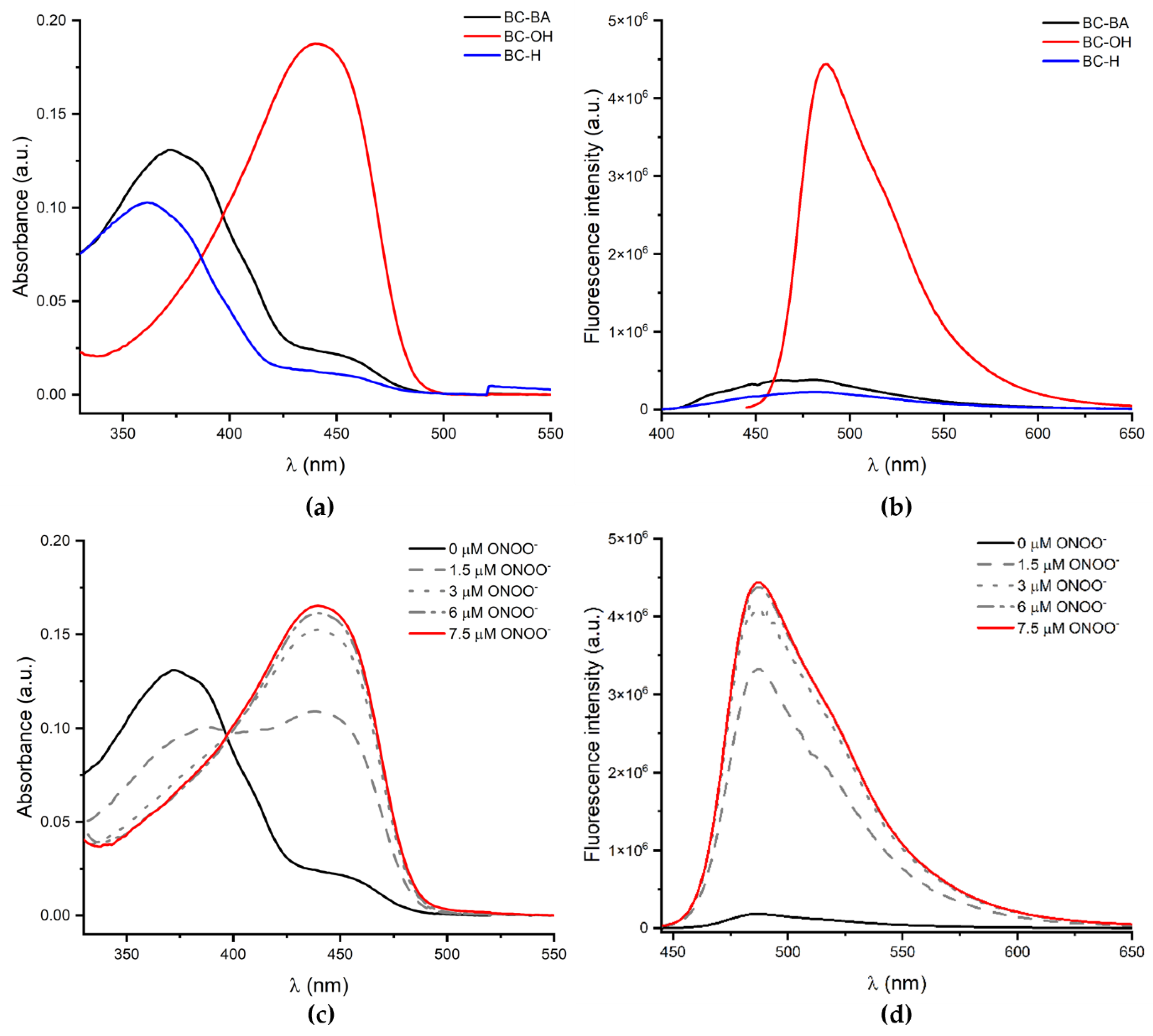
Introduction of the benzothiazolyl group into the coumarin skeleton has only a minor effect on the acid-base properties of the phenolic hydroxyl group. In fact, the reported pKa values of COH and BC-OH are 6.89 [33] and 7.0 [34], respectively. Photophysical properties of the novel boronate probe and the parent hydroxycoumarin BC-OH, as well as COH are compared in Table 1. Figure 1 shows absorption and emission spectra of BC-BA, BC-H and BC-OH. Presented data demonstrate that boronate group significantly reduces the emission of the BC-OH coumarin. BC-BA exhibited a maximal UV-absorption at 371 nm (ε = 25,500 M−1 cm−1) and a maximal emission at 473 nm (Φ = 0.17). BC-OH has absorption and emission bands red shifted in comparison with BC-BA. More importantly, this dye has a higher quantum yield of emission than BC-BA and a higher extinction coefficient (resulting in higher brightness) than COH. Bolus addition of peroxynitrite to the solution of the BC-BA probe red shifts the absorption band (Figure 1c) and turns on fluorescence (Figure 1d).

2.3. Reactivity towards Biological Oxidants

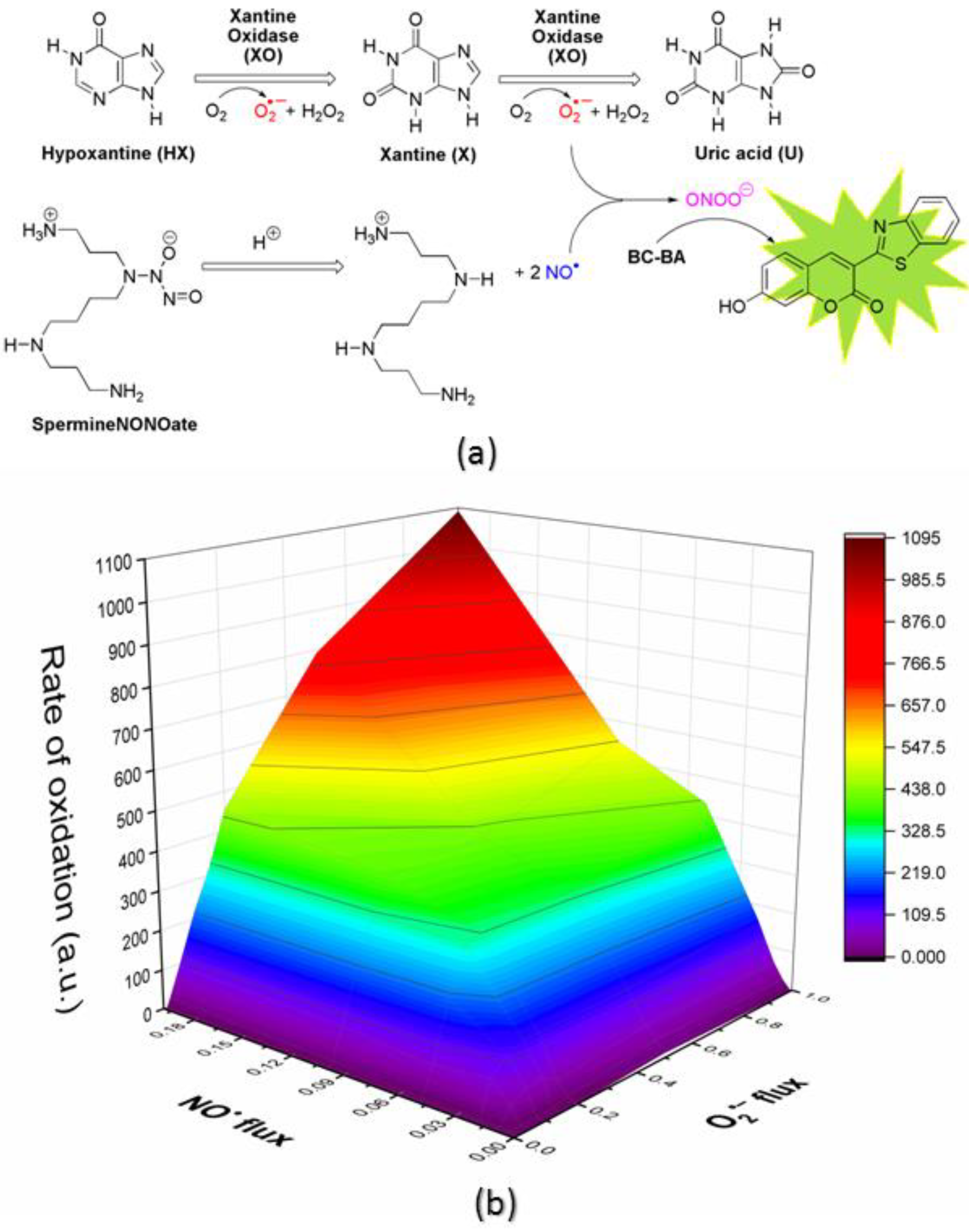
The effectiveness of the BC-BA probe for the detection of peroxynitrite was also measured using the system co-generating steady fluxes of O2•– (from hypoxanthine (HX) and xanthine oxidase XO) and NO (from spermine-NONOate). The profile of the BC-BA probe oxidation as a measure of ONOO formation in a matrix of various fluxes of O2•– and NO is shown in Figure 2a.
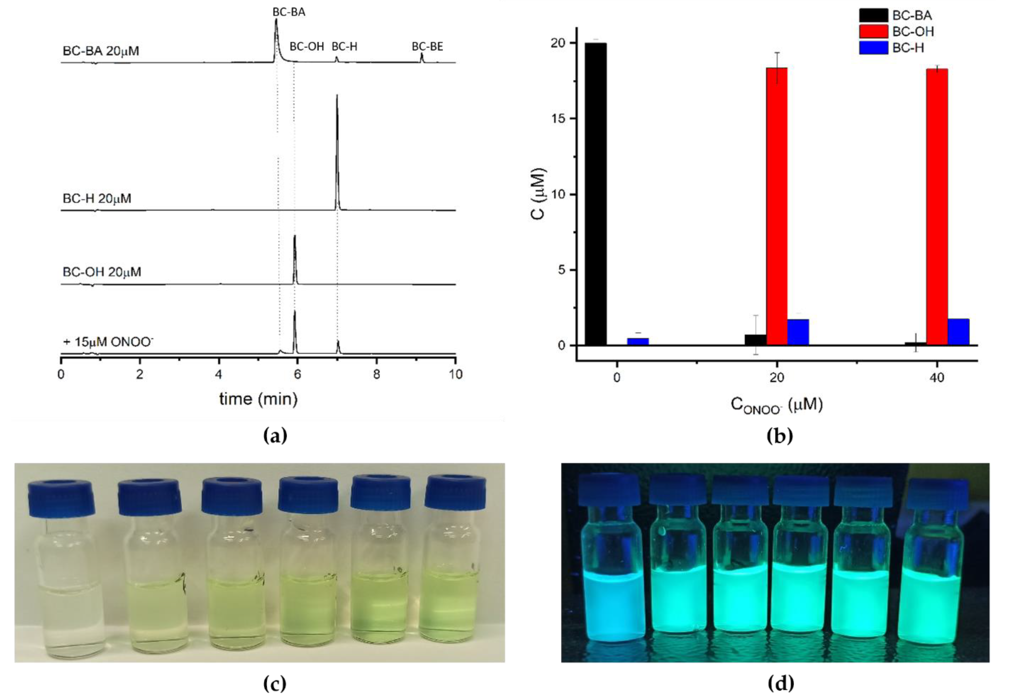
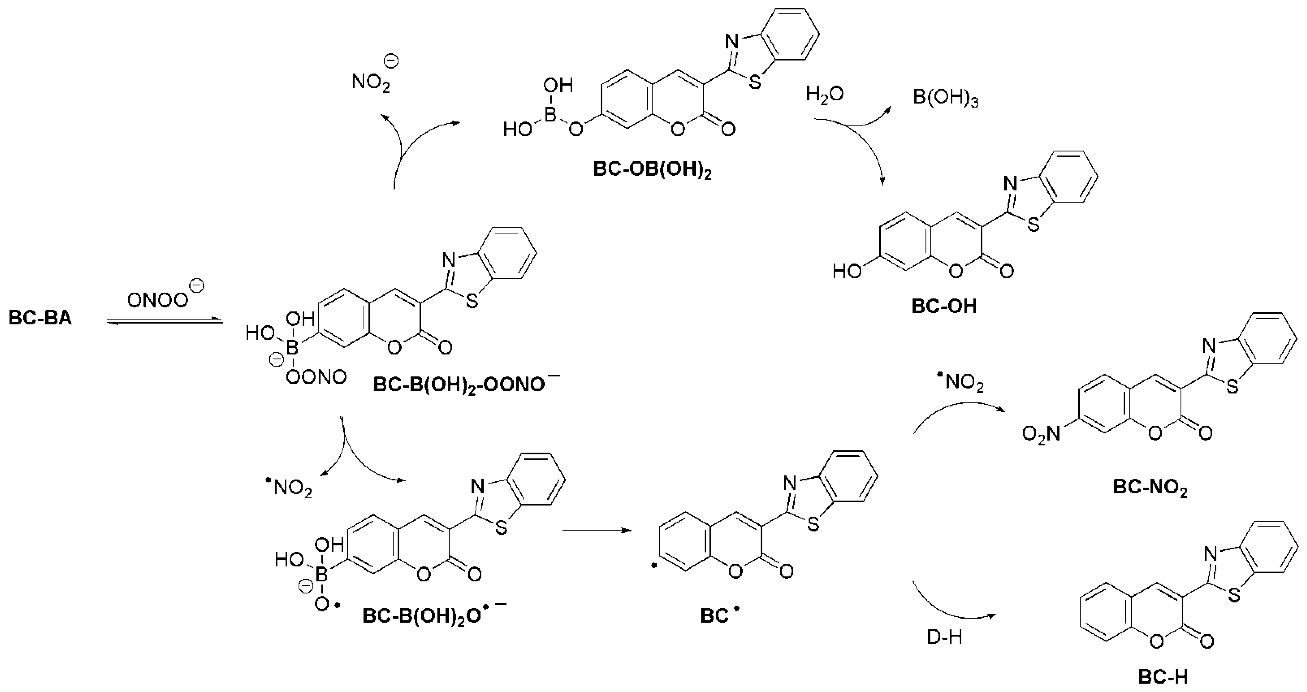
Next, we determined the stoichiometry of the reaction between BC-BA and peroxynitrite. Using HPLC, we analyzed products formed after the bolus addition of ONOO to the phosphate buffer containing the BC-BA probe. Figure 3a shows that, after 5 min of incubation, we observed the formation of the BC-OH coumarin as the main product. The formation of BC-OH can be observed with the naked eye, as shown in Figure 3c. The hue of fluorescence also changes (Figure 3d). Figure 3b showed that BC-BA reacts with ONOO forming the BC-OH dye with c.a. 90% yield. These results are consistent with a previously reported reactivity of arylboronate-derived probes toward peroxynitrite [30]. Peroxynitrite-induced oxidation of boronic acid Ar-B(OH)2 or ester Ar-B(pin) proceeds via two pathways, and typically leads to the formation of minor but ONOO-specific products (ArNO2, ArH) in addition to the major phenolic product (ArOH). Therefore, we anticipated that the BC-BA reaction with ONOO would produce the corresponding minor products (BC-NO2 and BC-H), as shown in Scheme 3. However, under the experimental conditions used we only detected BC-H (c.a. 9% yield), probably due to the usage of ethanol as an organic co-solvent, which efficiently reduces the phenyl-type radical BC to BC-H (Scheme 2). Previously, it has shown that in the presence of phenyl radical scavengers (2-propanol), the phenyl radical formed in the radical pathway is almost quantitatively converted into the product in which the boronate moiety is replaced by a hydrogen atom [36,37].
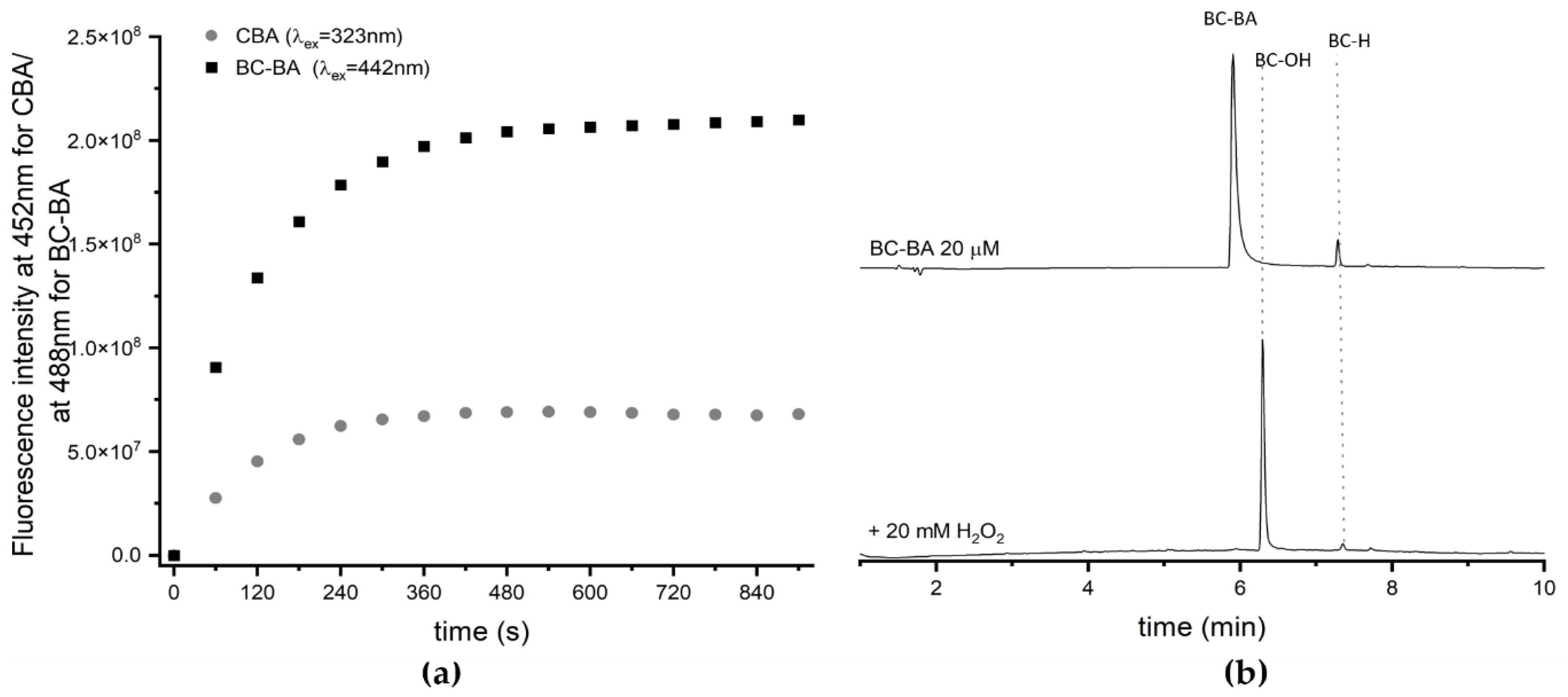
Boronate probe is also oxidized by other inflammatory oxidants such as hydrogen peroxide and hypochlorous acid [27,28,29,30]. Therefore, we also tested the reactivity of BC-BA toward H2O2 and HOCl. We also compared the kinetic profile of BC-OH formation during the oxidation of BC-BA to the profile of the COH formation from a simple boronate probe, coumarin-7-boronic acid (CBA). Figure 4a shows the buildup of emission at 442 nm during the reaction between those boronic probes and H2O2 (5 mM). It is evident that both probes release products (BC-OH or COH) with comparable reaction rates. However, the signal intensity of BC-OH is 3-fold higher than the signal emission of COH, demonstrating significantly higher brightness of the product, consistent with the fluorescence parameters listed in Table 1. It is also worth emphasizing that in the reaction of the BC-BA probe with H2O2, the BC-OH coumarin is formed as the sole product (Figure 4b). [27,28,29,30].
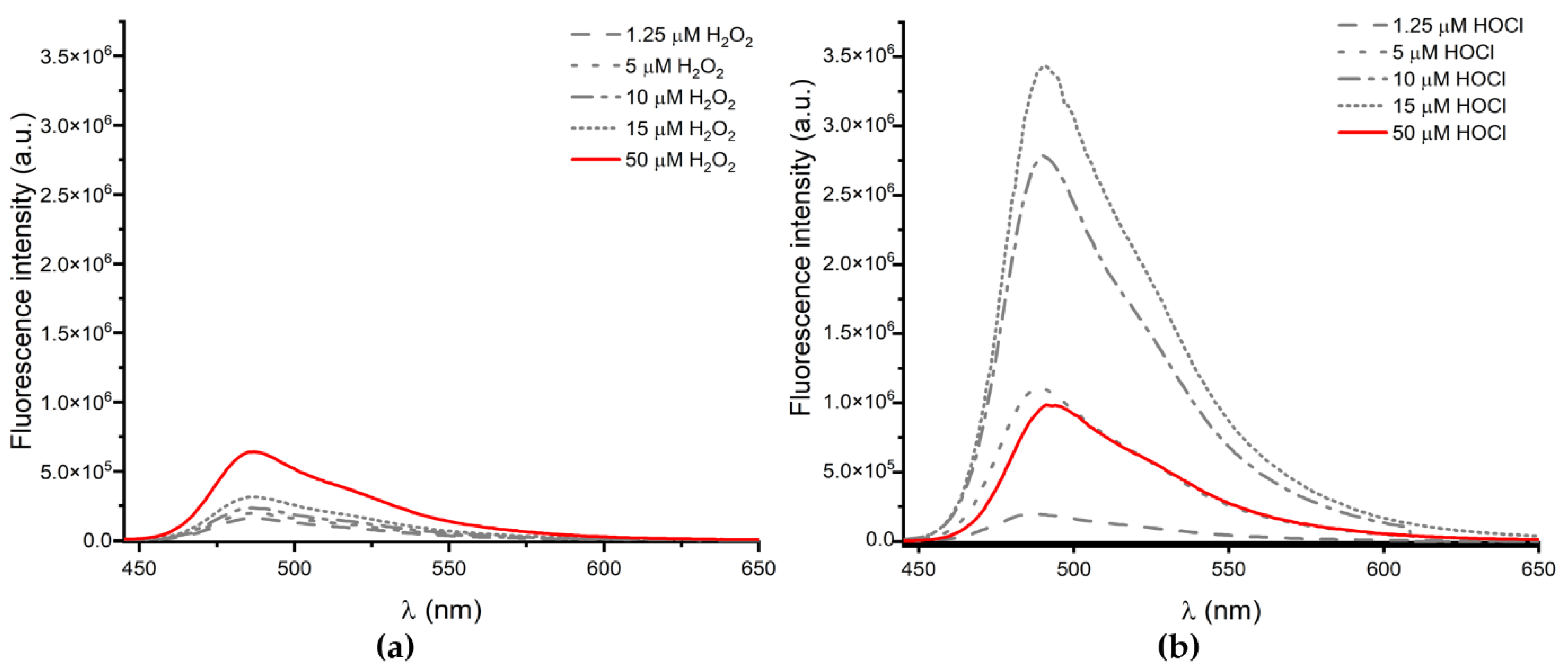
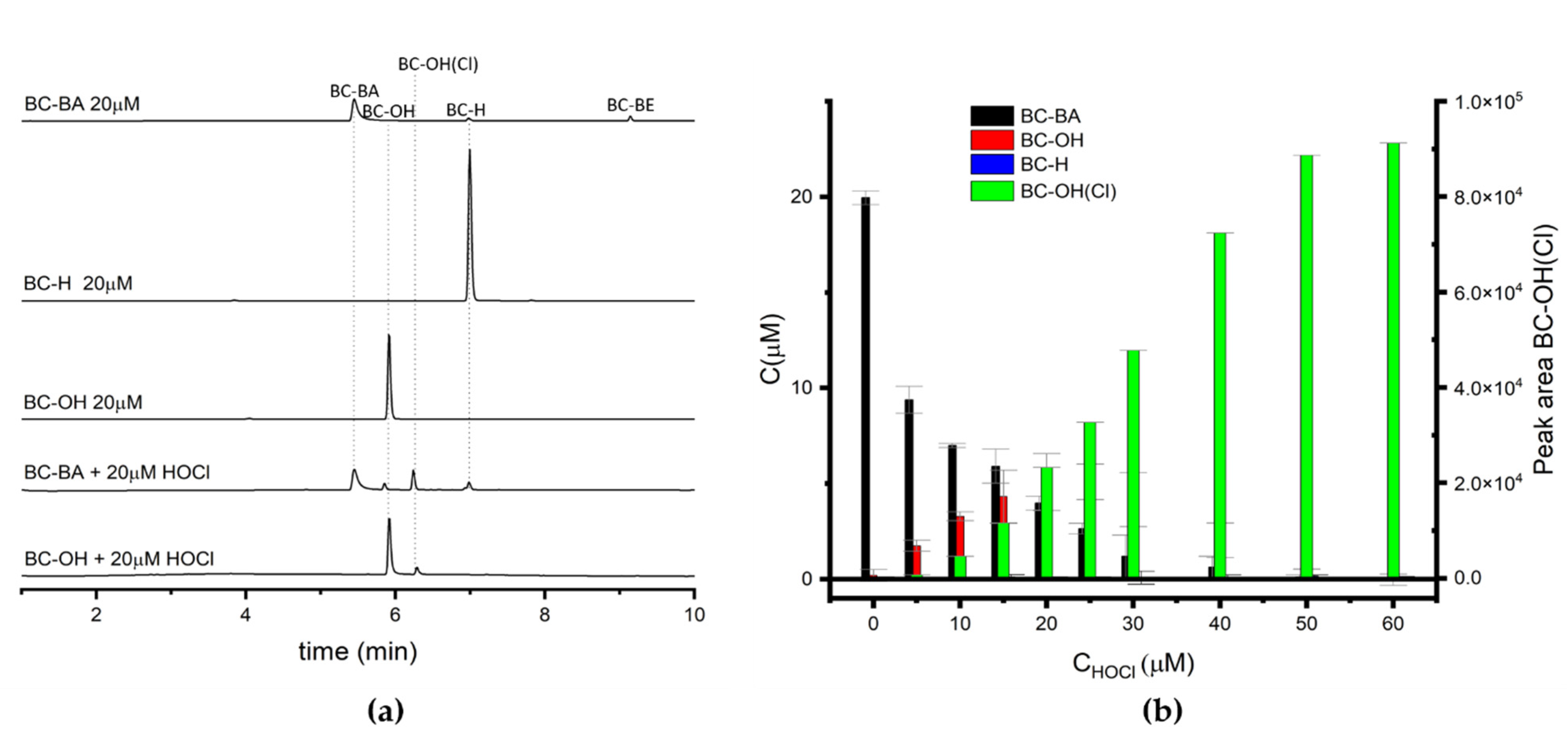
Emission spectra recorded after the addition of a micromolar concentration of H2O2 into the phosphate buffer solution containing BC-BA are shown in Figure 5a. The intensity of recorded spectra are lower in comparison with the intensity of fluorescence measured after the reaction of BC-BA with ONOO (Figure 1d). For comparison, we also studied the reaction of BC-BA with hypochlorous acid. Emission spectra (Figure 5b) reveal that the BC-BA probe is converted to the BC-OH dye. In contrast to the reaction of the probe with hydrogen peroxide, however, the signal intensity is significantly higher. HPLC analysis of the reaction mixture (Figure 6a) shows that besides BC-OH, another product is also formed. We attributed this product to the chlorinated derivative of the coumarin BC-OH(Cl) since the product with the same retention time (6.1 min) was also detected after the addition of HOCl to the BC-OH solution. By analyzing the disappearance of the BC-BA probe and the concentration of products formed with an increase in the amount of added HOCl, it can be seen that the maximum yield of BC-OH reaches only ca. 25%. Moreover, an excess of HOCl causes the disappearance of BC-OH. This is consistent with our previous investigations in which we demonstrated that the phenolic product (luciferin or 6-(2-benzothiazolyl)-2-naphthalenol) released from the boronate probes (LBA, PCL-1 or NAB-BE) undergoes a further reaction with HOCl, leading to the formation of 7′-chloroluciferin [27,28] or 6-(1,3-benzothiazol-2-yl)-1-chloronaphthalen-2-ol [29], a product specific for HOCl.

2.4. The Effect of Compounds on Cell Metabolic Activity

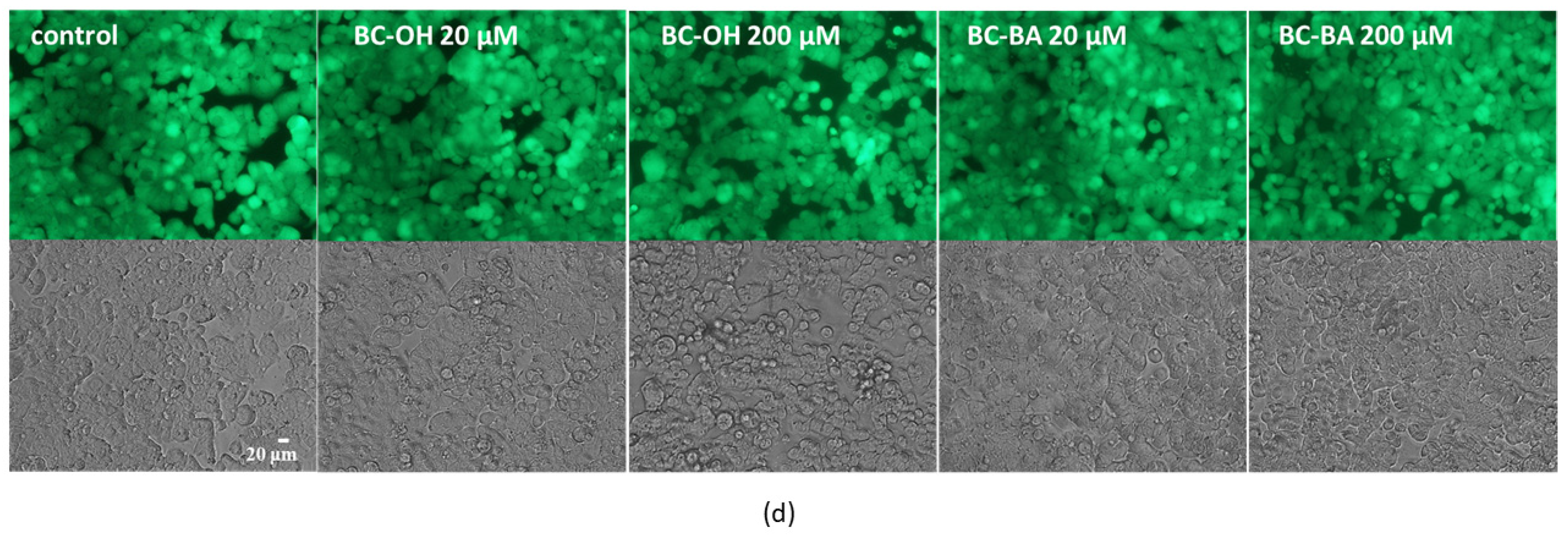
To determine the potential usage of synthesized compounds in cell-based assays, next the impact of probes on the metabolic activity of human colon cancer HT29 cells was studied. Compounds were dissolved in ethanol/dimethylsulfoxide mixture (9:1) and added to the cells in a volume not exceeding 2% of the medium, which had no cytotoxic effect on cells. To determine the cytotoxic potential of studied compounds, cells were incubated for 8 h in their presence at the range of 0–200 µM concentration. As it is presented in Figure 7a–c, compounds BC-OH and BC-BA at 20 µM concentration had no impact on HT29 cells’ metabolic activity; therefore, both of them can be potentially used in studies requiring cell incubation for at least 20 min. The direct comparison of cytotoxic potential within the studied range of concentration revealed that compound BC-OH was more cytotoxic than BC-BA. The highest cytotoxicity of BC-OH was detected for 200 µM with decreased metabolic activity by almost 20% compared to the control cells. Microscopic observations performed with calcein AM ester confirmed the lack of cytotoxic effects of both compounds at 20 µM on HT29 cells, as shown in Figure 7d. In healthy cells with active esterases, there is a visible strong cytosolic green fluorescence of calcein. Cells incubated with 200 µM of BC-OH had lower cytoplasmic esterase activity, thus a decreased green fluorescence of calcein was observed, as well as the presence of some less attached and more rounded cells.

3. Materials and Methods

3.1. General

The reagents used for the synthesis were commercially available. 3-benzothiazol-2-yl-7-hydroxy-chromen-2-one (BC-OH) and 3-benzothiazol-2-yl-chromen-2-one (BC-H) were synthesized according to published protocols [32,38].
The purity of the final compounds was tested by HPLC (Shimadzu) equipped with a photodiode array detector and analytical column—Phenomenex Kinetex Core-Shell C18 (100 mm × 4.6 mm; 2.6 μm). The mobile phase was a gradient prepared from acetonitrile with 0.1% of TFA (component A) and water with 0.1% of TFA (component B). The analytes were eluted by an increase of A concentration from 10–100% over 10 min at the flow rate of 1.5 mL/min. The column temperature was set at 30 °C.
1H NMR spectra were recorded with a Bruker Avance DPX 250 spectrometer at 250 MHz, respectively (see Supplementary materials). Compounds were dissolved in DMSO-d6 and TMS was added as internal reference. Mass spectra [TOF MS (ESI+)] were recorded on a Synapt G2-Si spectrometer (Waters, Milford, MA, USA).
Absorption spectra were recorded on UV-Vis-NIR spectrophotometer Jasco-V670. Steady-state and time-resolved fluorescence spectra were recorded on Edinburgh Analytical Instruments FL900.
BC-OH:1H NMR (250 MHz, DMSO-d6): δ 11.06 (bs, 1H), 9.16 (s, 1H), 8.16 (d, J = 7.7 Hz, 1H), 8.05 (d, J = 8.1 Hz, 1H), 7.92 (d, J = 8.6 Hz, 1H), 7.57 (t, J = 7.6 Hz, 1H), 7.46 (t, J = 7.5 Hz, 1H), 6.93 (dd, J = 8.6, 2.2 Hz, 1H), 6.86 (d, J = 2.1 Hz, 1H). TOF MS ES+ calcd for C16H10NO3S 296.0381, found 296.0390. m.p. 303–305 °C, lit. 302–304 °C [39].
BC-H: 1H NMR (250 MHz, DMSO-d6): δ 9.06 (s, 1H), 8.07 (d, J = 8.1, 1H), 7.96 (d, J = 8.0, 1H), 7.71 (dd, J = 7.7, 1.6 Hz, 1H), 7.6–7.59 (m, 1H), 7.55–7.48 (m, 1H), 7.44–7.33 (m, 3H). TOF MS ES+ calcd for C16H10NO2S 280.0432, found 280.0439. m.p. 213–215 °C, lit. 213–215 °C [40].

3.2. Synthesis of 3-(Benzo[d]thiazol-2-yl)-2-oxo-2H-chromen-7-yl Trifluoromethanesulfonate (BC-OTf)

3-benzothiazol-2-yl-7-hydroxycoumarin BC-OH (148 mg, 0.5 mmol) and N-phenyl-bis(trifluoromethanesulfonimide) (214 mg, 0.6 mmol) were dissolved in 10 mL of anhydrous chloroform. Triethylamine (0.36 mL, 2.6 mmol) was added to the mixture and the resulting solution was stirred under reflux for 4 h. After cooling to room temperature, it was diluted with chloroform and washed three times with citric acid (20%, 10 mL), three times with water (10 mL), and three times with saturated solution NaHCO3 (10 mL). The organic layer was dried over MgSO4 and the solvent was removed by rotary evaporation to give a yellowish solid (211 mg, 99%).
BC-OTf: 1H NMR (250 MHz, DMSO-d6): δ 9.26 (s, 1H), 8.26 (d, J = 8.8 Hz, 1H), 8.18 (d, J = 7.7 Hz, 1H), 8.08 (d, J = 7.8 Hz, 1H), 7.90 (d, J = 2.4 Hz, 1H), 7.64–7.54 (m, 2H), 7.47 (t, 1H). TOF MS ES+ calcd for C17H9NO5S2F3 427.9874, found 427.9876. m.p. 167–170 °C.

3.3. Synthesis of 3-(2-Benzothiazolyl)-7-coumarin Boronic Acid Pinacol Ester (BC-BE)

3-benzothiazol-2-yl-7-trifluoromethanesulfonate coumarin BC-OTf (43 mg, 0.1 mmol), bis(pinacolato)diboron (28 mg, 0.11 mmol), Pd(dppf)Cl2 (2.2 mg, 0.003 mmol), dppf (1.7 mg, 0.003 mmol), and potassium acetate (29 mg, 0.3 mmol) were dissolved in 5 mL of anhydrous 1,4-dioxane. The reaction was heated in a microwave at 100 °C for 45 min under argon. After cooling to room temperature, the mixture was diluted with toluene and washed three times with brine. The organic layer was dried over MgSO4 and the solvent was removed by rotary evaporation to give a brown residue. The residue was purified with column chromatography (eluent DCM:MeOH; 19:1; v/v) to give a beige solid (41 mg, 73%).
BC-BE: 1H NMR (250 MHz, DMSO-d6): δ 9.24 (s, 1H), 8.18 (d, J = 6.7 Hz, 1H), 8.07 (t, J = 7.7 Hz, 2H), 7.93 (s, 1H), 7.72–7.53 (m, 1H), 7.47 (t, J = 7.5 Hz, 1H), 7.17 (d, J = 8.2Hz, 1H), 1.31 (s, 12H). TOF MS ES+ calcd for C22H21NO4SB 406.1284, found 406.1288. m.p. 229–233 °C.

3.4. Real-Time Monitoring of Oxidation BC-BE Boronic Probe in Cell-Free System Generating NO and O2●–

All solutions were prepared using deionized water (Millipore Milli-Q system). Due to poor solubility of probes in water, all experiments were performed with addition of an up 1% (v/v) of DMSO. The profiles of ONOO formation in a solution with hypoxanthine (HX) and xanthine oxidase (XO) as a source of steady flux of superoxide O2●– and spermine-NONOate as a source of NO, were monitored using BC-BA as fluorogenic probe. The total fluorescence intensity of formed BC-OH dye was measured using a Beckman Coulter DTX880 plate reader. The instrument was kept at 37 °C during the measurements. The changes in fluorescence intensity were monitored over a 2 h period. NO fluxes were determined from the rate of decomposition of spermine-NONOate measured by following the decrease of its characteristic absorbance at 252 nm (ε = 8.5 × 10³ M−1 cm−1) [41]. The flux of O2●– was determined by monitoring the cytochrome c(Fe3+) reduction and the increase in absorbance at 550 nm (using a difference in the values of the extinction coefficients between reduced and oxidized cytochrome of 2.1 × 10⁴ M−1 cm−1). In our experiments peroxynitrite was co-generated from fluxes of O2●− and NO in the phosphate buffer (pH 7.4, 50 mM) with DTPA (100 µM), HX/XO (1 mM/70 µU/mL), spermine-NONOate (75 µM) and CAT (100 U/mL). Such systems produced up to 0.2 µM/min flux of peroxynitrite.

3.5. Cell Culture and Exposure Conditions

Human colon carcinoma cell line HT29 was obtained from ATCC, (Manassas, VA, USA). Cells were grown in DMEM with a 10% fetal bovine serum (FBS) medium supplemented with 100 U/mL penicillin, 100 µg/mL streptomycin, and 25 µg/mL amphotericin B. All cell culture experiments were performed in a humidified 5% CO2 and 95% atmosphere at 37 °C. All cell culture reagents were obtained from Life Technologies (Carlsbad, CA, USA).

3.6. Cell Metabolic Activity

Metabolic activity was evaluated with MTT assay. Briefly, cells were seeded into 96-well plate at 1 × 10⁴ cells/well density in 100 µL complete medium and grown overnight, and then incubated in the presence of studied compounds for another 8 h. After this, 20 µL of MTT reagent (5 mg/mL) was added for 120 min. After that time, MTT was removed, and formazan precipitates were solubilized by adding 100 µL of DMSO. Absorbance was measured at 570 nm using the Synergy 2 BioTek Microplate Reader (BioTek, Winooski, VT, USA).

4. Conclusions

We used 3-benzothiazol-2-yl-7-hydroxy-chromen-2-one fluorophore to develop a novel boronate probe for the selected biological oxidants. The BC-BA probe showed similar reactivity toward selected inflammatory oxidants to the previously reported CBA probe [17], producing fluorescent BC-OH phenolic product. In comparison to CBA, BC-BA is significantly more lipophilic, which may improve its cellular uptake. Furthermore, the fluorescence of the oxidation product is red shifted and its brightness is ca. 3-fold higher. This may help in the successful application of the probe for the imaging of biological oxidants in cultured cells. Detection of the minor products characteristic for a specific oxidant (ONOO or HOCl) will allow the unambiguous identification of the oxidants involved in probe oxidation.
Further structure modifications may be introduced to the developed scaffold to modulate the water solubility of the probe, fluorescence properties of the probe and the product and to target the probe to specific subcellular or extracellular compartments.

Supplementary Materials

The following are available online. Figure S1: HPLC chromatograms of the BC-BA in aqueous solution containing phosphate buffer (0.1 M, pH 7.4), dtpa (10 µM) and EtOH (10%): freshly made solution (above), after 10 min (below). The traces were collected using the absorption detector set at 330 nm, Figure S2: 1H NMR spectrum of BC-OH in d6-DMSO, Figure S3: 1H NMR spectrum of BC-H in CDCl3, Figure S4: 1H NMR spectrum of BC-OTf in d6-DMSO, Figure S5: 1H NMR spectrum of BC-BE in d6-DMSO, Figure S6: Mass spectrum of BC-OH, Figure S7: Mass spectrum of BC-H, Figure S8: Mass spectrum of BC-OTf, Figure S9: Mass spectrum of BC-BE.

Author Contributions

Conceptualization, J.Z. and R.P.; methodology, J.M.; validation, J.M., M.S., M.Z.-S. and A.G.; formal analysis, J.M.; investigation, J.M. and M.Z.-S.; resources, J.M.; data curation, J.M.; writing—original draft preparation, J.M.; writing—review and editing, R.P. and J.Z.; visualization, J.M.; supervision, R.P. and J.Z.; project administration, R.P. and J.Z.; funding acquisition, J.Z. and R.P. All authors have read and agreed to the published version of the manuscript.

Funding

This research was funded by a grant from the Polish National Science Centre within the SONATA BIS 6 program (Grant no. 2016/22/E/ST4/00549) to R.P. J.Z. was supported in part by the Institutional Research Grant IRG #16-183-31 from the American Cancer Society and the MCW Cancer Center.

Institutional Review Board Statement

Not applicable.

Informed Consent Statement

Not applicable.

Data Availability Statement

The data presented in this study are available in article and supplementary material.

Conflicts of Interest

The authors declare no conflict of interest.

Sample Availability

Samples of the compounds BC-BA, BC-OH, and BC-H are available from the authors.

References

  1. Cao, D.; Liu, Z.; Verwilst, P.; Koo, S.; Jangjili, P.; Kim, J.S.; Lin, W. Coumarin-based small-molecule fluorescent chemosensors. Chem. Rev. 2019, 119, 10403–10519. [Google Scholar] [CrossRef]
  2. The Colour Index™. Available online: https://colour-index.com/ (accessed on 10 April 2021).
  3. Sokołowska, J.; Czajkowski, W.; Podsiadły, R. The photostability of some fluorescent disperse dyes derivatives of coumarin. Dye. Pigment. 2001, 49, 187–191. [Google Scholar] [CrossRef]
  4. Telange, D.R.; Nirgulkar, S.B.; Umekar, M.J.; Patil, A.T.; Pethe, A.M.; Bali, N.R. Enhanced transdermal permeation and anti-inflammatory potential of phospholipids complex-loaded matrix film of umbelliferone: Formulation development, physico-chemical and functional characterization. Eur. J. Pharm. Sci. 2019, 131, 23–38. [Google Scholar] [CrossRef]
  5. Mahoney, K.M.; Goswami, P.P.; Winter, A.H. Self-immolative aryl phthalate esters. J. Org. Chem. 2013, 78, 702–705. [Google Scholar] [CrossRef] [Green Version]
  6. Li, L.; Zhang, M.; Chang, K.; Kang, Y.; Ren, G.; Hou, X.; Liu, W.; Wang, H.; Wang, B.; Diao, H. A novel fluorescent off–on probe for the sensitive and selective detection of fluoride ions. RSC Adv. 2019, 9, 32308–32312. [Google Scholar] [CrossRef] [Green Version]
  7. Salahuddin, S.; Renaudet, O.; Reymond, J.-L. Aldehyde detection by chromogenic/fluorogenic oxime bond fragmentation. Org. Biomol. Chem. 2004, 2, 1471–1475. [Google Scholar] [CrossRef]
  8. Gu, X.; Zhu, H.; Yang, S.; Zhu, Y.-C.; Zhu, Y.-Z. Development of a highly selective H2S fluorescent probe and its application to evaluate CSE inhibitors. RSC Adv. 2014, 4, 50097–50101. [Google Scholar] [CrossRef]
  9. Bae, J.; Choi, J.; Park, T.J.; Chang, S.-K. Reaction-based colorimetric and fluorogenic signaling of hydrogen sulfide using a 7-nitro-2,1,3-benzoxadiazole–coumarin conjugate. Tetrahedron Lett. 2014, 55, 1171–1174. [Google Scholar] [CrossRef]
  10. Wang, L.; Li, W.; Zhi, W.; Ye, D.; Wang, Y.; Ni, L.; Bao, X. A rapid-responsive fluorescent probe based on coumarin for selective sensing of sulfite in aqueous solution and its bioimaging by turn-on fluorescence signal. Dye Pigments 2017, 147, 357–363. [Google Scholar] [CrossRef]
  11. Zhang, D.; Chen, W.; Kang, J.; Ye, Y.; Zhao, Y.; Xian, M. Highly selective fluorescence off–on probes for biothiols and imaging in live cells. Org. Biomol. Chem. 2014, 12, 6837–6841. [Google Scholar] [CrossRef] [PubMed] [Green Version]
  12. Sun, W.; Li, J.; Li, W.H.; Su, L.J.; Du, L.P.; Li, M.Y. Design of OFF/ON fluorescent thiol probes based on coumarin fluorophore. Sci. China Chem. 2012, 55, 1776–1780. [Google Scholar] [CrossRef]
  13. Badalassi, F.; Wahler, D.; Klein, G.; Crotti, P.; Reymond, L.-J. A versatile periodate-coupled fluorogenic assay for hydrolytic enzymes. Angew. Chem. Int. Ed. 2000, 39, 4067–4070. [Google Scholar] [CrossRef]
  14. Nyfeler, E.; Grognux, J.; Wahler, D.; Reymond, J.-L. A sensitive and selective high-throughput screening fluorescence assay for lipases and esterases. Helv. Chim Acta 2003, 86, 2919–2927. [Google Scholar] [CrossRef]
  15. Xue, F.; Seto, C.T. Fluorogenic peptide substrates for serine and threonine phosphatases. Org. Lett. 2010, 12, 1936–1939. [Google Scholar] [CrossRef]
  16. Chen, W.; Liu, C.; Peng, B.; Zhao, Y.; Pacheco, A.; Xian, M. New fluorescent probes for sulfane sulfurs and the application in bioimaging. Chem. Sci. 2013, 4, 2892–2896. [Google Scholar] [CrossRef] [Green Version]
  17. Zielonka, J.; Sikora, A.; Joseph, J.; Kalyanaraman, B. Peroxynitrite is the major species formed from different flux ratios of co-generated nitric oxide and superoxide. J. Biol. Chem. 2010, 285, 14210–14216. [Google Scholar] [CrossRef] [Green Version]
  18. Zielonka, J.; Zielonka, M.; Verplank, L.; Cheng, G.; Hardy, M.; Ouari, O.; Ayhan, M.M.; Podsiadły, R.; Sikora, A.; Lambeth, D.J.; et al. Mitigation of NADPH oxidase 2 activity as a strategy to inhibit peroxynitrite formation. J. Biol. Chem. 2016, 291, 7029–7044. [Google Scholar] [CrossRef] [Green Version]
  19. Wang, H.; Li, W.-G.; Zeng, K.; Wu, Y.-J.; Zhang, Y.; Xu, T.-L.; Chen, Y. Photocatalysis Enables Visible-Light Uncaging of Bioactive Molecules in Live Cells. Angew. Chem. Int. Ed. 2019, 58, 561–565. [Google Scholar] [CrossRef] [PubMed]
  20. Shaobing, Q.; Dongdong, D.; Chunlei, G.; Zhenglong, S.; Hui, L.; Xuhong, Q.; Youjun, Y. Amino-substituted C-coumarins: Synthesis, spectral characterizations and their applications in cell imaging. Dye Pigments 2019, 163, 55–61. [Google Scholar] [CrossRef]
  21. Sączewski, J.; Hinc, K.; Obuchowski, M.; Gdaniec, M. The tandem Mannich–electrophilic amination reaction: A versatile platform for fluorescent probing and labeling. Chem. Eur. J. 2013, 19, 11531–11535. [Google Scholar] [CrossRef] [PubMed]
  22. Tasior, M.; Kim, D.; Singha, S.; Krzeszewski, M.; Ahn, K.H.; Gryko, D.T. П-Expanded coumarins: Synthesis, optical properties and applications. J. Mater. Chem. C 2015, 3, 1421–1446. [Google Scholar] [CrossRef]
  23. Wang, X.; Fang, H.; Huang, Z.; Shang, W.; Hou, T.; Cheng, A.; Cheng, H. Imaging ROS signaling in cells and animals. J. Mol. Med. 2013, 91, 917–927. [Google Scholar] [CrossRef] [PubMed] [Green Version]
  24. Winterbourn, C.C. The challenges of using fluorescent probes to detect and quantify specific reactive oxygen species in living cells. Biochim. Biophys. Acta 2014, 1840, 730–738. [Google Scholar] [CrossRef] [PubMed]
  25. Prolo, C.; Rios, N.; Piacenza, L.; Álvarez, M.N.; Radi, R. Fluorescence and chemiluminescence approaches for peroxynitrite detection. Free Radic. Biol. Med. 2018, 128, 59–68. [Google Scholar] [CrossRef] [PubMed]
  26. Wardman, P. Fluorescent and luminescent probes for measurement of oxidative and nitrosative species in cells and tissues: Progress, pitfalls, and prospects. Free Radic. Biol. Med. 2007, 43, 995–1022. [Google Scholar] [CrossRef] [PubMed]
  27. Zielonka, J.; Podsiadły, R.; Zielonka, M.; Hardy, M.; Kalyanaraman, B. On the use of peroxy-caged luciferin (PCL-1) probe for bioluminescent detection of inflammatory oxidants in vitro and in vivo—dentification of reaction intermediates and oxidant-specific minor products. Free Radic. Biol. Med. 2016, 99, 32–42. [Google Scholar] [CrossRef] [PubMed] [Green Version]
  28. Szala, M.; Grzelakowska, A.; Modrzejewska, J.; Siarkiewicz, P.; Słowiński, D.; Świerczyńska, M.; Zielonka, J.; Podsiadły, R. Characterization of the reactivity of luciferin boronate—A probe for inflammatory oxidants with improved stability. Dye Pigments 2020, 183, 108693. [Google Scholar] [CrossRef]
  29. Grzelakowska, A.; Zielonka, M.; Dębowska, K.; Modrzejewska, J.; Szala, M.; Sikora, A.; Zielonka, J.; Podsiadły, R. Two-photon fluorescent probe for cellular peroxynitrite: Fluorescence detection, imaging, and identification of peroxynitrite-specific products. Free Radic. Biol. Med. 2021, 169, 24–35. [Google Scholar] [CrossRef]
  30. Sikora, A.; Zielonka, J.; Dębowska, K.; Michalski, R.; Smulik-Izydorczyk, R.; Pięta, J.; Podsiadły, R.; Artelska, A.; Pierzchała, K.; Kalyanaraman, B. Boronate-based probes for inflammatory oxidants—A novel class of molecular tools for redox biology. Front. Chem. 2020, 8, 843–875. [Google Scholar] [CrossRef]
  31. Parvez, S.; Long, M.J.C.; Poganik, J.R.; Aye, Y. Redox signaling by reactive electrophiles and oxidants. Chem. Rev. 2018, 118, 8789–8888. [Google Scholar] [CrossRef] [PubMed]
  32. Khoobia, M.; Ramazania, A.; Foroumadib, A.R.; Hamadic, H.; Hojjatia, Z.; Shafieeb, A. Efficient microwave-assisted synthesis of 3-benzothiazolo and 3-benzothiazolino coumarin derivatives catalyzed by heteropoly acids. J. Iran. Chem. Soc. 2011, 8, 1036–1042. [Google Scholar] [CrossRef]
  33. Garcia-Molina, M.; Munoz-Munoz, J.L.; Garcia-Molina, F.; Rodriguez-Lopez, J.N.; Garcia-Canovas, F. Study of umbelliferone hydroxylation to esculetin catalyzed by polyphenol oxidase. Biol. Pharm. Bull. 2013, 36, 1140–1145. [Google Scholar] [CrossRef]
  34. Roubinet, B.; Chevalier, A.; Renard, P.-Y.; Romieu, A. A synthetic route to 3-(heteroaryl)-7-hydroxycoumarins designed for biosensing applications. Eur. J. Org. Chem. 2015, 1, 166–182. [Google Scholar] [CrossRef]
  35. Tan, B.; Liu, L.; Zheng, H.; Cheng, T.; Zhu, D.; Yang, X.; Luan, X. Two-in-one strategy for fluorene-based spirocycles via Pd(0)-catalyzed spiroannulation of o-iodobiaryls with bromonaphthols. Chem. Sci. 2020, 11, 10198–10203. [Google Scholar] [CrossRef] [PubMed]
  36. Sikora, A.; Zielonka, J.; Lopez, M.; Dybala-Defratyka, A.; Joseph, J.; Marcinek, A.; Kalyanaraman, B. Reaction between peroxynitrite and boronates: EPR spin-trapping, HPLC Analyses, and quantum mechanical study of the free radical pathway. Chem. Res. Toxicol. 2011, 24, 687–697. [Google Scholar] [CrossRef] [Green Version]
  37. Sikora, A.; Zielonka, J.; Adamus, J.; Debski, D.; Dybala-Defratyka, A.; Michalowski, B.; Joseph, J.; Hartley, R.C.; Murphy, M.P.; Kalyanaraman, B. Reaction between peroxynitrite and triphenylphosphonium-substituted arylboronic acid isomers: Identification of diagnostic marker products and biological implications. Chem. Res. Toxicol. 2013, 26, 856–867. [Google Scholar] [CrossRef] [Green Version]
  38. Yang, J.; Yu, Y.; Wang, B.; Jiang, Y. A sensitive fluorescent probe based on coumarin for detection of cysteine in living cells. J. Photochem. Photobiol. A Chem. 2017, 338, 178–182. [Google Scholar] [CrossRef]
  39. Wang, K.; Lai, G.; Li, Z.; Liu, M.; Shen, Y.; Wang, C. A novel colorimetric and fluorescent probe for the highly selective and sensitive detection of palladium based on Pd(0) mediated reaction. Tetrahedron 2015, 71, 7874–7878. [Google Scholar] [CrossRef]
  40. Yu, T.C.; Zhang, Y.; Zhao, H.; Chai, W.; Li, A. one-pot reaction to synthesize two types of fluorescent materials containing benzothiazolyl moiety. Spectrochim. Acta A 2013, 108, 274–279. [Google Scholar] [CrossRef] [PubMed]
  41. Maragos, C.M.; Morley, D.; Wink, D.A.; Dunams, T.M.; Saavedra, J.E.; Hoffman, A.; Bove, A.A.; Isaac, L.; Hrabie, J.A.; Keefer, L.K. Complexes of NO with nucleophiles as agents for the controlled biological release of nitric oxide. Vasorelaxant effects. J. Med. Chem. 1991, 34, 3242–3247. [Google Scholar] [CrossRef]
Scheme 1. Pro-coumarin boronate based probes.
Molecules 26 05940 sch001
Scheme 2. Synthesis of novel boronates based on 3-benzothiazol-2-yl-7-hydroxy-chromen-2-one. Reagents and conditions: (i) 1. EtOH, piperidine, rt, 3 h; 2. 10% HCl, reflux, 3 h; (ii) acetic acid, 150 °C, MW, 40 min; (iii) PhN(SO2CF3)2, DCM, TEA, reflux, 4 h; (iv) Pd(dppf)Cl2, dppf, AcOK, 1,4-dioxane, 100 °C, MW, 45 min; (v) phosphate buffer, pH 7.4, 30 min.
Molecules 26 05940 sch002
Figure 1. Absorption and emission spectra of studied coumarins recorded in mixture of phosphate buffer (0.1 M, pH 7.4) and EtOH (10%). (a) Absorption spectra of BC-OH, BC-BA, and BC-H (5 µM each); (b) fluorescence spectra of BC-OH, BC-BA, and BC-H (5 µM each, λex = λmax); (c) absorption spectra of BC-BA probe (5 µM) recorded before and after bolus addition of different amount of peroxynitrite (0, 1.5, 4.5, 6, 7.5 µM); (d) emission spectra of the BC-BA probe (5 µM, λex = 442 nm) recorded before and after bolus addition of different amount of peroxynitrite (0, 1.5, 4.5, 6, 7.5 µM).
Molecules 26 05940 g001
Figure 2. Measurement of peroxynitrite formation in situ by monitoring oxidation of BC-BA (20 µM) using a 96-well fluorescence plate reader. (a) The formation of O2•– from incubations containing hypoxanthine HX (100 µM) and xanthine oxidase XO (0–0.4 milliunits/mL) in a phosphate buffer (pH 7.4; 50 mM) containing DTPA (100 µM), and NO generation from a thermal decomposition of spermine-NONOate (0–50 µM). Peroxynitrite, formed from co-generated fluxes of O2•– and NO, oxidizes BC-BA to fluorescent BC-OH; (b) the rate of increase fluorescence intensity during the oxidation of the BC-BA probe in the presence of varying fluxes of O2•– and NO in the phosphate buffer (pH 7.4, 50 mM) with DTPA (100 µM), HX/XO, spermine-NONOate, and CAT (100 U/mL). The fluorescence intensity (λex at 405 nm, λem at 535 nm) of the reaction mixtures consisting of BC-BA (20 µM) and HX/XO (O2•– flux: 0–1 µM/min) and spermine-NONOate (NO flux: 0–0.2 µM/min) was measured over a period of 2 h.
Molecules 26 05940 g002
Figure 3. Peroxynitrite-induced oxidation of the BC-BA probe. (a) HPLC traces of the mixture of BC-BA, BC-OH, and BC-H (20 µM each) and the reaction mixtures of BC-BA (20 µM) with peroxynitrite (15 µM) after 5 min of incubation. Tests were conducted in aqueous solutions containing EtOH (10%), phosphate buffer (0.1 M, pH 7.4) and dtpa (10 µM). The traces were collected using an absorption detector set at 330 nm; (b) HPLC based titration of BC-BA probe (20 µM) with ONOO (0–40 µM). Pictures of BC-BA probe (20 µM) with different amount of peroxynitrite (0, 6, 12, 18, 24, 30 µM): (c) in day light; (d) under UV.
Molecules 26 05940 g003
Scheme 3. Peroxynitrite induced oxidative conversion of BC-BA leading to benzothiazolyl-coumarin derived dyes BC-OH, BC-H, and BC-NO2.
Molecules 26 05940 sch003
Figure 4. (a) Kinetic profiles of product formation during the oxidation of probes BC-BA and CBA (5 µM each) by H2O2 (5 mM). Experiments were repeated three times independently. Representative result displayed; (b) HPLC traces of the BC-BA probe (20 µM) before and after addition of H2O2 (20 mM).
Molecules 26 05940 g004
Figure 5. Emission spectra of the BC-BE probe (5 µM, λex = 442 nm) recorded before and after bolus addition of different amount (0, 1.5, 4.5, 6, 7.5 µM) of: (a) H2O2 (after 24 h of incubation); (b) HOCl (after 30 min of incubation). Tests were conducted in aqueous solutions containing EtOH (10%), phosphate buffer (0.1 M, pH 7.4) and dtpa (10 µM).
Molecules 26 05940 g005
Figure 6. HOCl-induced oxidation of the BC-BA probe. (a) HPLC traces of the mixture of BC-BA, BC-OH and BC-H (20 µM each) and the reaction mixtures of BC-BA (20 µM) and BC-OH (20 µM) with HOCl (20 µM) after 15 min of incubation. Tests were conducted in aqueous solutions containing EtOH (10%), phosphate buffer (0.1 M, pH 7.4) and dtpa (10 µM). The traces were collected using an absorption detector set at 330 nm; (b) HPLC based titration of the BC-BA probe (20 µM) with HOCl (0–60 µM). Pictures of the BC-BE probe (20 µM) with different amount of HOCl (0–60 µM): (c) in day light; (d) under UV.
Molecules 26 05940 g006aMolecules 26 05940 g006b
Figure 7. The influence of BC-OH (a) and BC-BA (b) on HT29 cell metabolic activity, determined by MTT assay after 8 h exposure; control cells were not exposed to any compound but the vehicle (EtOH/DMSO (9:1) (c); values are means ± SEM, n ≥ 7; statistical significance was calculated versus control cells (untreated), * p ≤ 0.05, ** p ≤ 0.01. Morphology of HT29 cells stained with 2 μM calcein AM observed after incubation with 20 and 200 μM of BC-OH and BC-BA (d); randomly chosen fields were photographed at × 200 with fluorescent microscope Nikon TS Eclipse 100.
Molecules 26 05940 g007aMolecules 26 05940 g007b
Table 1. Photophysical properties of studied compounds.
Solventλmax
[nm]
ε
[104 M−1 cm−1]
λex
[nm]
λem
[nm]
Stokes Shift
[nm]
Φemτ [ns] 5
COHEtOH3261.22326391650.10
H2O 13231.273234521290.80 3 (0.76) 4
BC-OHEtOH3963.30396467710.70 33.07
EtOH:PB 24423.81442488460.85 33.32
BC-HEtOH3642.363644641000.14 31.00
EtOH:PB 23652.723654701050.09 31.63
BC-BAEtOH3692.22369464950.21 32.60
EtOH:PB 23712.553714731020.17 31.33
1 In H2O (pH 7.2) [20], 2 Aqueous phosphate buffer (100 mM, pH 7.4):EtOH (4:1), 3 According to quinine sulfate (Φref 0.54 in water) [21], 4 NaHCO3–NaOH buffer (10 mM, pH 10.0, containing 1% EtOH) [35], 5 Fluorescence lifetime was determined by TCSPC single photon counting using FL900 spectrofluorometer(Edinburgh Instruments, Livingston, UK).
Publisher’s Note: MDPI stays neutral with regard to jurisdictional claims in published maps and institutional affiliations.