Article

Epithelial-to-Mesenchymal Transition Is Not a Major Modulating Factor in the Cytotoxic Response to Natural Products in Cancer Cell Lines

by
Baris Kucukkaraduman
1,
Ekin Gokce Cicek
1,
Muhammad Waqas Akbar
1,
Secil Demirkol Canli
2,
Burcak Vural
3 and
Ali Osmay Gure
4,*
1
Department of Molecular Biology and Genetics, Bilkent University, Ankara 06800, Turkey
2
Molecular Pathology Application and Research Center, Hacettepe University, Ankara 06100, Turkey
3
Department of Genetics, Aziz Sancar Institute of Experimental Medicine, Istanbul University, Istanbul 34093, Turkey
4
Department of Medical Biology, Acibadem University, Istanbul 34684, Turkey
*
Author to whom correspondence should be addressed.
Molecules 2021, 26(19), 5858; https://doi.org/10.3390/molecules26195858
Submission received: 19 July 2021 / Revised: 20 September 2021 / Accepted: 22 September 2021 / Published: 27 September 2021

Abstract

:
Numerous natural products exhibit antiproliferative activity against cancer cells by modulating various biological pathways. In this study, we investigated the potential use of eight natural compounds (apigenin, curcumin, epigallocatechin gallate, fisetin, forskolin, procyanidin B2, resveratrol, urolithin A) and two repurposed agents (fulvestrant and metformin) as chemotherapy enhancers and mesenchymal-to-epithelial (MET) inducers of cancer cells. Screening of these compounds in various colon, breast, and pancreatic cancer cell lines revealed anti-cancer activity for all compounds, with curcumin being the most effective among these in all cell lines. Although some of the natural products were able to induce MET in some cancer cell lines, the MET induction was not related to increased synergy with either 5-FU, irinotecan, gemcitabine, or gefitinib. When synergy was observed, for example with curcumin and irinotecan, this was unrelated to MET induction, as assessed by changes in E-cadherin and vimentin expression. Our results show that MET induction is compound and cell line specific, and that MET is not necessarily related to enhanced chemosensitivity.

1. Introduction

Cancer is the second leading cause of death worldwide and about one in six deaths are due to cancer [1]. Chemotherapy is a main treatment option for cancer and has been used as a monotherapy, or in combination with surgery or radiation therapy, for decades. However, drug resistance, innate or acquired, is a major determinant of treatment response and disease progression. Many studies have shown that epithelial-to-mesenchymal transition (EMT) plays an important role in chemotherapy resistance [2,3,4,5]. Therefore, targeting EMT to improve the response to chemotherapy has recently become a priority in cancer research [6].
Nutrition and diet are effective in reducing cancer risk. Numerous dietary natural products have shown potential in prevention and/or treatment of cancers [7,8,9]. Accumulating evidence shows that naturally occurring bioactive compounds interfere with carcinogenesis by inhibiting tumor cell proliferation and targeting multiple abnormally activated signaling pathways [10,11]. Furthermore, many natural products have been shown to reverse EMT [12,13], and to overcome drug resistance in cancer [14,15].
In line with these observations, modulators of EMT, combined with chemotherapeutics and/or targeted drugs, may possibly improve clinical outcomes in cancer. Most studies aiming to address this issue, however, were conducted either in single cell lines, or only in one cancer type [12]. Hence, a comprehensive study, which evaluates the effectiveness of naturally occurring bioactive molecules in the modulation of EMT and their role as chemotherapy enhancers, is needed. To fulfill this need, we investigated the effects of eight natural compounds: apigenin, curcumin, epigallocatechin gallate (EGCG), fisetin, forskolin, procyanidin B2, resveratrol, urolithin A, and two repurposed agents, fulvestrant and metformin, on cancer cell viability, modulation of EMT phenotype, and sensitization to chemotherapeutics in vitro in three different cancer types. Curcumin is one of the best studied natural products, present in turmeric. Many studies have showed that curcumin sensitizes cancer cells to chemotherapeutics by modulating EMT [16,17,18,19]. Resveratrol (phytoalexin) is a polyphenol, found in grapes, chocolate, and peanuts [20]. The regulatory role of resveratrol in EMT, leading to chemosensitization, has also been extensively studied [15,21,22,23]. Recently, apigenin, a natural flavonoid, was shown to act as an inhibitor of Snail, which is a key regulator of EMT [24,25]. Apigenin inhibits IL-6 linked downstream pathways, resulting in epithelial marker E-cadherin upregulation, and downregulation of the mesenchymal marker N-cadherin [26]. Another natural product, EGCG (epigallocatechin gallate), the most prevalent tea polyphenol, was also shown to reverse EMT, and synergistic effects of EGCG were shown in pancreatic cancer cells when combined with gemcitabine through the modulation of EMT markers [27]. Fisetin, a natural flavanol found in various fruits and vegetables, was shown to reverse EMT in vivo in a xenograft model of the MDA-MB-231 breast cancer cell line [28]. Forskolin, a natural product derived from the root of the plant Cleus forskohlii, was shown to induce tumor-initiating cells to undergo mesenchymal-to-epithelial transition, and to cause them to lose their tumor-initiating ability [29]. Procyanidin B2, an abundant polyphenol in red wine, was shown to reverse high-glucose-induced-EMT in a human renal epithelial cell line [30]. Additionally, urolithin A, a metabolite of ellagitannins, was shown to inhibit metastasis in a SW620 colon cancer cell line, and to increase sensitivity against 5-FU in a Caco-2 colon cancer cell line [31,32]. It was also shown to upregulate the epithelial marker E-cadherin and to downregulate the mesenchymal markers, vimentin and N-cadherin, in lung cancer cells [33]. Fulvestrant and metformin are FDA-approved compounds that have been shown to be capable of reducing the mesenchymal features of cancer cells. Fulvestrant is an estrogen antagonist which was shown to reduce the mesenchymal features of lung carcinoma cells, resulting in tumor sensitization to chemotherapy. The anti-diabetic drug metformin was also shown to have capability in the modulation of EMT [34,35,36,37]. These compounds have been reported for their low-toxicity against normal cells in many studies [38,39,40,41,42,43,44,45,46,47,48,49,50,51,52,53]. Here, we report on the activity of eight natural products, and two repurposed agents, in sixteen cell lines of colon, breast, and pancreatic cancer origin.

2. Results

2.1. Screening of Natural Products in Various Breast, Colon, and Pancreatic Cancer Cell Lines Reveals Notable Anti-Cancer Activity

We first sought to characterize the inhibitory effect of natural products and repurposed agents on various breast, colon, and pancreatic cancer cell lines. We selected eight naturally occurring molecules, an estrogen antagonist, and an anti-diabetic drug that were previously shown to inhibit and/or reverse EMT. The cells were designated resistant if an IC50 value was not observed within the concentration ranges tested. For the breast cancer cell lines, curcumin exhibited a remarkable inhibition of cell viability, with IC50 values ranging from 12 to 40 μM among the cell lines tested (Table 1). Additionally, apigenin and resveratrol showed variance in the inhibitory activity on cell viability, whereas fisetin and forskolin exhibited a more restricted cytotoxicity on cell lines. MDA-MB-453 was the only breast cancer cell line sensitive to fulvestrant, which may be a result of HER2-overexpression in this cell line, while other cell lines were triple-negative. On the other hand, EGCG, procyanidin B2, urolithin A, and metformin, in micromolar concentrations, did not have any effect on cell viability in breast cancer cell lines at the concentrations tested.
Since diet is an important factor in colon cancer risk, studying the effects of natural products on colon cancer may provide new insights into cancer prevention and treatment [54]. In a similar way to our findings in breast cancer, curcumin was the most effective compound for inhibition of viability in colon cancer cell lines, with IC50 values ranging from 6 to 15 μM (Table 2). We observed moderate inhibition of cell viability with apigenin, EGCG, fisetin, resveratrol, and urolithin A, at micromolar levels. In addition, metformin in the millimolar range was cytotoxic for colon cancer cell lines (Table 2). The fact that KM12 is the only cancer cell line sensitive to forskolin among our colon cancer cell line panel, is also evident in the NCI-60 drug sensitivity data (Figure S1) [55]. On the other hand, procyanidin B2 and fulvestrant did not exhibit any inhibitory effects on colon cancer cell viability.
Pancreatic ductal adenocarcinoma (PDAC) is among the most lethal cancer types, and a number of naturally occurring molecules in preclinical studies exhibit promising pharmacological features [56,57]. We examined the effects of natural compounds on the viability of four pancreatic cancer cell lines (Table 3). Similar to what we observed in breast and colon cancer cells, curcumin was the most effective natural compound in terms of pancreatic cancer cell viability. We observed moderate cytotoxicity with apigenin, EGCG, fisetin, and resveratrol. In addition, forskolin and urolithin A exhibited selective and mild effects on the pancreatic cancer cell lines. Millimolar levels of metformin also had growth inhibitory effects in these cells. In a similar way to the colon cancer cell lines, procyanidin B2 and fulvestrant had no effect on cell viability.
In total, 12, 13, and 15 cancer cell lines (out of 16) were resistant (IC50 could not be determined within the concentration ranges tested) to forskolin, procyanidin B2, and fulvestrant, respectively, suggesting that these compounds were not effective inhibitors of cancer cell viability. Apigenin, curcumin, and resveratrol were the only three compounds for which IC50 values could be generated for all of the cell line types tested. Among these compounds, the lowest mean IC50 was obtained for curcumin for all three cancer types, with values of 23.8 μM, 10 μM, and 15 μM for breast, colon, and pancreatic cancer cells, respectively.

2.2. Natural Products Induce Mesenchymal-to-Epithelial Transition Selectively in Cancer Cell Lines

We treated six cell lines from three cancer types with natural compounds and evaluated changes in the epithelial/mesenchymal phenotypes (Figure 1 and Figure S2). We used four colon cancer cell lines (Caco-2, KM12, LoVo, and WiDr), among which Caco-2 is known for its high plasticity, with the flexibility to shift through the spectrum between epithelial and mesenchymal phenotypes [58]. Three other cell lines showed variance in expression of EMT marker genes in which KM12 was the most mesenchymal and WiDr was the most epithelial (Figure S3). We performed a 48-h treatment on these cells with the compounds at multiple doses and quantified the change, via qRT-PCR, in the expression of E-cadherin (CDH1) and vimentin (VIM), as markers of epithelial and mesenchymal phenotypes, respectively. An increase in CDH1, and a decrease in VIM expression upon treatment, compared to the untreated control cells, were considered as indicators of MET induction, and vice versa for EMT. In Caco-2 cells, none of the compounds induced MET or EMT under our test conditions. In contrast, some products did induce MET in three other colon cancer cell lines (Figure 1). Among tested compounds, higher doses of fisetin and forskolin exhibited MET induction only in the KM12 cell line. Neither MET nor EMT could be observed in LoVo and WiDr cell lines with these compounds. On the other hand, treatment with 5 μM doses of apigenin and fulvestrant resulted in MET induction in LoVo but not in KM12 and WiDr (Figure S2). Therefore, even within the same cancer type, the compounds that can induce MET vary. We then evaluated EMT or MET in the breast cancer cell line MDA-MB-157, for which we previously showed phenotypic plasticity in 3D culture [59]. With the exception of resveratrol treatment (20 μM), most treatments resulted in the downregulation of CDH1, contrary to our expectations. Lastly, we treated a mesenchymal pancreatic cancer cell line, MIA PaCa-2, with various doses of natural compounds and repurposed agents. Almost all the treatments resulted in a two- to three-fold upregulation of CDH1 expression, possibly suggesting MET induction, however the VIM expression levels did not change. The increase in CDH1 expression may be considered as a mild phenotypic switch in the MIA PaCa-2 cells since CDH1 levels were already very low in this cell line when compared to breast and colon cell lines (Figures S3 and S4).
Overall, we noted transcriptional changes suggesting a shift towards a more epithelial phenotype in four out of the six cell lines tested upon treatment with at least one compound. The MET induction effects summarized in Table S1 show that none of the natural compounds or targeted agents could trigger MET in all cancer cells screened. In addition, compounds with clear or slight MET induction only caused these effects in a specific cell line, or only in a specific cancer type.

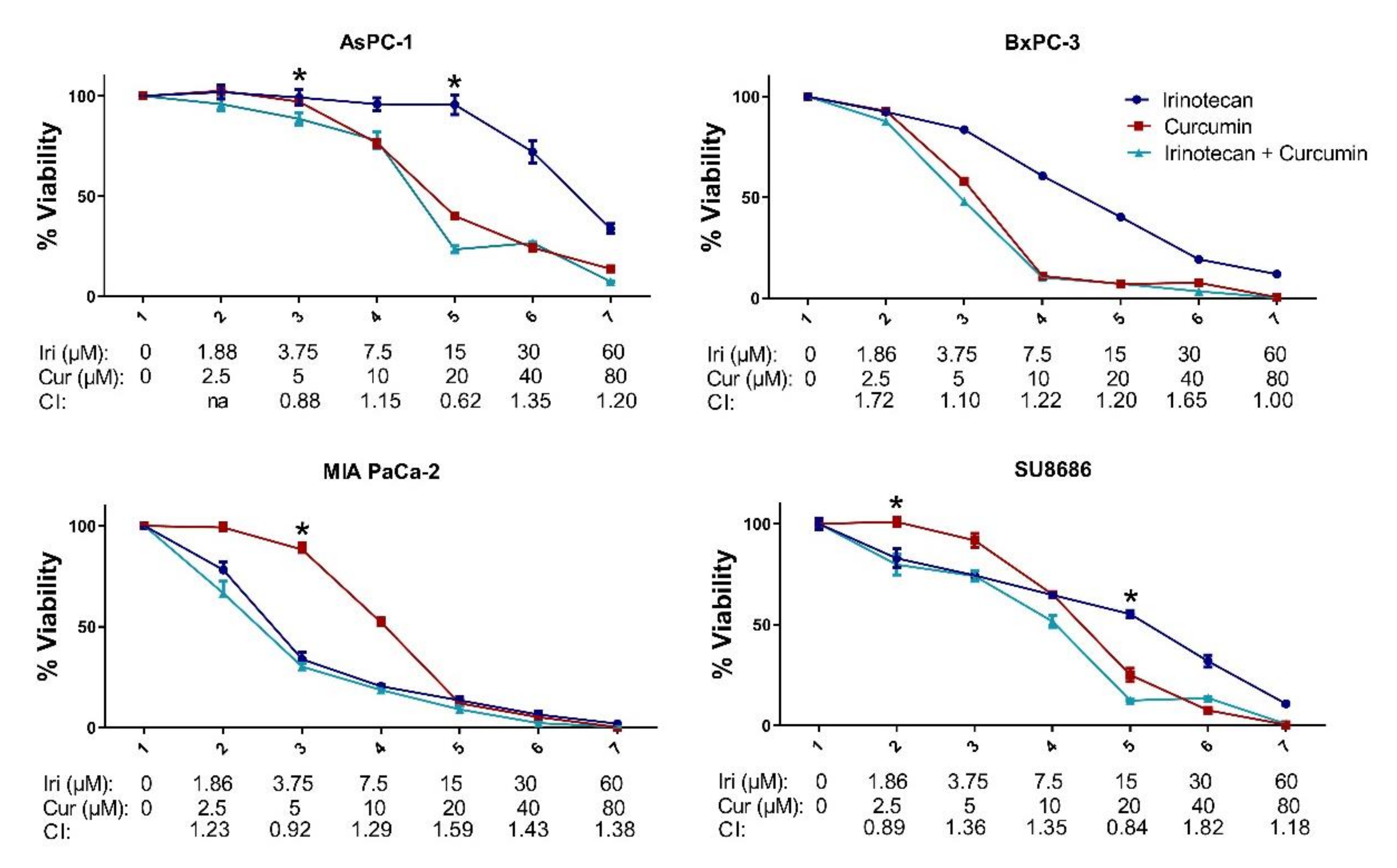
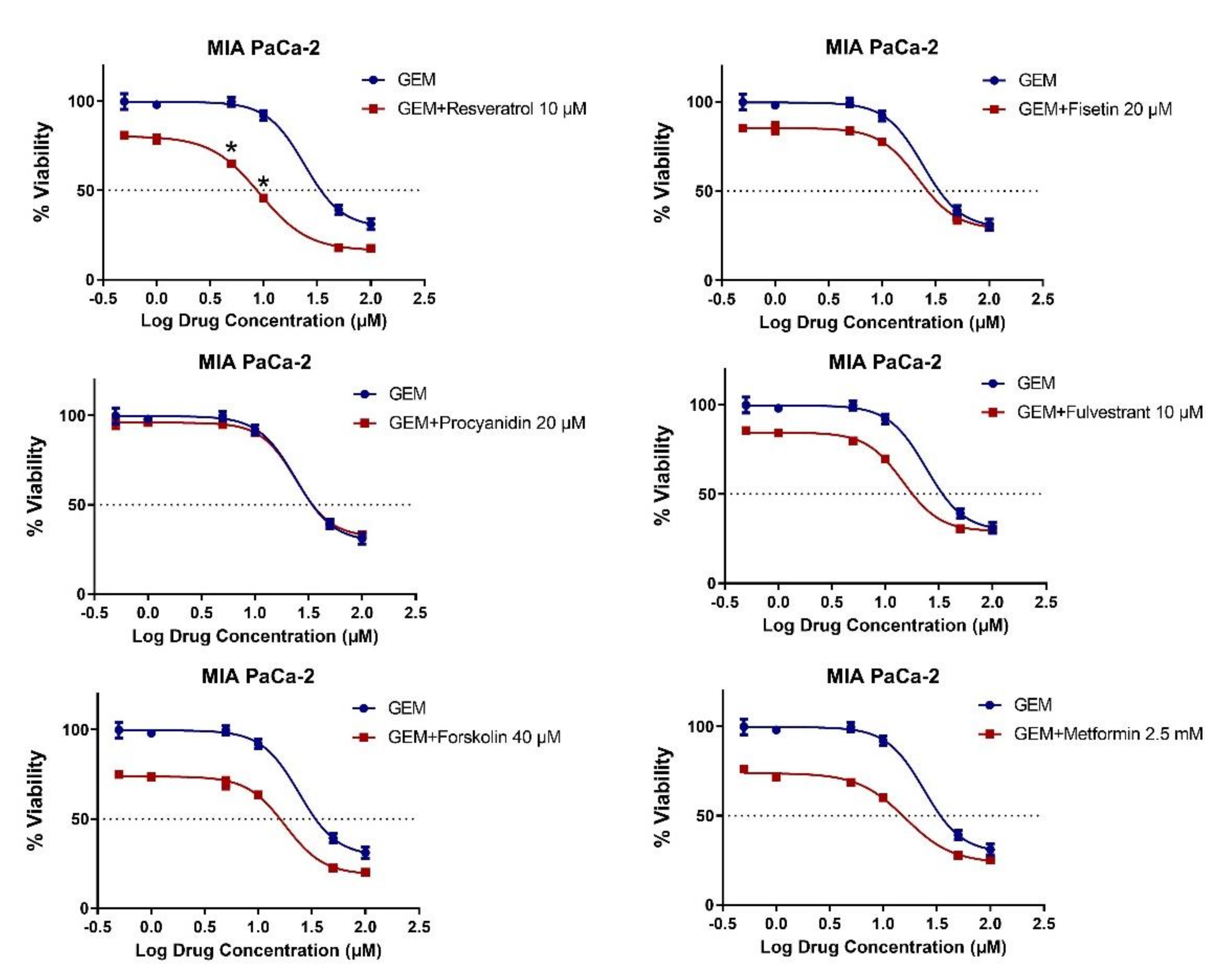
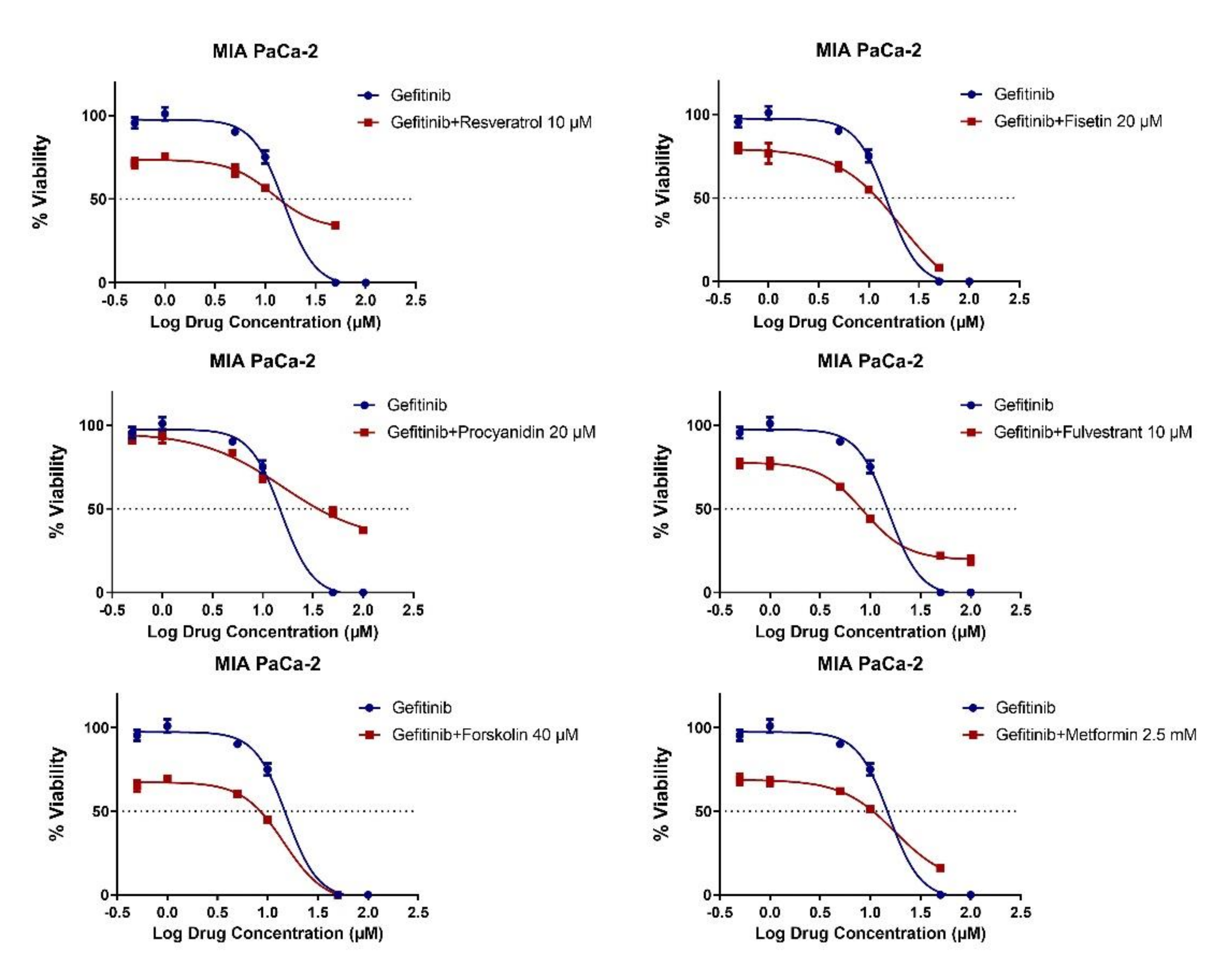
2.3. MET Induction by Natural Compounds Is Not Uniformly Related to Increased Sensitivity to Chemotherapeutics but Can Result in Occasional Synergistic or Additive Effects

We next asked whether the treatment with natural compounds would result in chemosensitization. For this purpose, the KM12 and WiDr colon cancer cell lines were treated with various doses of natural products, alone or in combination with two doses (1 μM and 10 μM) of 5-FU, a commonly used chemotherapeutic in colon cancer, after twenty-four hours of cell seeding. Cell viability assays were then performed 72 h after the treatments. In KM12, we observed MET induction with 20 uM of fisetin and two doses (10 μM and 30 μM) of forskolin. However, only the 10 uM 5-FU and forskolin combination treatments resulted in synergy, with the combination index (CI) score ranging from 0.91 to 0.37 (Table S2). Additionally, 10 uM 5-FU and urolithin A combinations showed slight synergy, with the CI ranging from 0.95 to 0.77, despite no significant MET induction with urolithin A in this cell line. Interestingly, 5 μM fulvestrant and 5-FU combinations also resulted in synergy, with the CI ranging from 0.80 to 0.24. All the other combinations tested in KM12 showed additive effects or a slight antagonism. In the WiDr cell line, where we observed a mild induction of MET with forskolin only, the forskolin and 5-FU combinations showed slight antagonisms. Synergisms were observed with a combination of 1 μM 5-FU with various doses of apigenin, EGCG, and fisetin. In a similar way to the KM12 line, in the WiDr cell line, the fulvestrant and 5-FU combination resulted in a slight synergism. Additionally, we used the rectal cancer cell line SW837 in our analysis to examine the ability of natural products and repurposed agents to act as chemotherapy enhancers by combining them with irinotecan, which is a component of FOLFIRI routinely used in colorectal cancer, mainly as a second-line treatment [60,61]. Procyanidin B2, which did not have an effect on either the inhibition of cellular viability, or the induction of MET in the colon cancer cell lines, exhibited synergism in the SW837 line, with the CI ranging from 0.89 to 0.35. Additionally, specific doses of urolithin A, fulvestrant, and metformin, combined with irinotecan, also resulted in a slight synergism. In contrast, all other combinations showed either additive effects or even antagonism for SW837 (Tables S2 and S3).
We next investigated the combinatory effects of irinotecan, used in pancreatic cancer, and some widely studied natural compounds, curcumin and resveratrol, in pancreatic cancer cell lines, AsPC-1, BxPC-3, MIA PaCa-2, and SU8686 (Figure 2 and Figure 3). Most of the curcumin/irinotecan combinations resulted in antagonistic effects, while a 15 μM irinotecan and 20 μM curcumin combination showed synergy in AsPC-1 and SU8686 cell lines. Only the highest dose combinations of irinotecan and resveratrol resulted in a slight synergy in BxPC-3, MIA PaCa-2, and SU8686. The mild induction of MET by natural products in the MIA PaCa-2 cell line did not enhance irinotecan sensitivity.
Gemcitabine is a standard agent for the treatment of pancreatic cancer, either alone or in combination [62,63]. We therefore evaluated the combinatory effects of natural products with gemcitabine (Figure 4), as well as with the targeted agent gefitinib, an EGFR inhibitor, since EGFR inhibition has shown promising activity in pancreatic cancer, in the MIA PaCa-2 cell line (Figure 5) [64]. Six different combinations of gemcitabine and gefitinib were combined with a single dose of the natural products. Synergy was observed for only 10 μM of resveratrol, and 5 μM and 10 μM of gemcitabine combinations, with CI scores 0.71 and 0.47 (Figure 4), whereas no synergistic effect was noted for gefitinib (Table S3). On the other hand, combinations of low dose gemcitabine and gefitinib, showed additive effects in MIA PaCa-2, however, the effect evolved from an additive to an antagonistic one at higher doses. The fact that synergy was observed with only gemcitabine among the three conventional drugs (irinotecan, gemcitabine, gefitinib) combined with resveratrol shows that the synergistic effects of a specific natural compound cannot be generalized even for one cell line; instead, the specific molecular cascades targeted by natural compound–drug pairs are most likely critical factors. Overall, these data indicate that there is no perfect overlap between MET-inducing and chemosensitizing compounds, which is likely to stem from the existence of more complex mechanisms underlying synergy patterns.

3. Discussion

Innate or acquired resistance of cancer cells to chemotherapeutics is responsible for tumor growth and metastasis [65,66]. Several studies have suggested that tumors exhibit a high degree of molecular and genetic heterogeneity, resulting in their adaptation to the conventional cytotoxic therapeutics. Alterations in the drug metabolic pathway, tumor evolution resulting in new mutations and thus the inhibition of cell death, epigenetic changes, mutations in the drug targets, and many other undiscovered mechanisms, cause resistance to cancer treatment. These mechanisms can act independently or in combination, and through various signaling pathways [67]. EMT is a well-known phenomenon whereby tumor cells lose their epithelial characteristics and acquire mesenchymal characteristics. Emerging evidence suggests a molecular and phenotypic association between EMT and chemotherapy resistance in several cancer types [68]. Therefore, targeting EMT has been considered a novel strategy to overcome therapy resistance in cancer.
A wide range of natural products have been reported as being cytotoxic for cancer cells and therefore useful in cancer therapy. Natural products can act as therapeutic, preventive, or chemosensitizer agents. Enhancement of chemotherapy sensitivity in tumor cells using naturally occurring biomolecules is a new and promising strategy aiming to promote the efficacy of conventional chemotherapeutics and targeted drugs [69]. Several studies suggest that inhibition or reversal of EMT by natural products results in resensitizing drug resistant cancer cells [15,21,22,27,36].
In the present study, we used eight natural compounds and two repurposed agents, that were previously shown to modulate EMT in different types of cancer cell lines, to test their capability to sensitize cancer cells to chemotherapy in a comprehensive manner. Although there are studies supporting the modulatory effects of all these compounds on EMT, only a few of them were able to induce MET in specific cancer cell lines in our experiments, suggesting a highly selective cell-line specific action. Interestingly, none of the compounds were able to induce MET in the Caco-2 cell line, which can easily be induced to undergo EMT and MET under various other conditions [58]. In fact, some treatments resulted in upregulation of VIM, suggesting the induction of EMT. Moreover, the MET induction with fisetin and forskolin seen in KM12 was not observed in other colon cancer cell lines. This suggests that MET induction with these natural compounds may vary according to cellular and molecular characteristics of cell lines even within the same cancer type. Similarly, the breast cancer cell line MDA-MB-157 exhibited MET only with resveratrol. Since various cellular mechanisms are effected by resveratrol, including mammalian target of rapamycin (mTOR) signaling, miRNA modulation, AMP-activated protein kinase (AMPK) activation, NF-B activation, nitric oxide production, histone deacetylase (HDAC) inhibition, and cytochrome P450 inhibition [70], it might be that MET induced in this cell line is managed through one or more of these mechanisms which may not have been targeted or effected sufficiently by the other compounds in our panel.
Different transcriptomic signatures have been evaluated for determining the EMT status of cancer cells [71,72,73]. Evaluation of the expression of both CHD1 and VIM genes was shown to be a highly reliable and consistent predictor of the EMT phenotype [72,74,75,76]. Our experience also shows that CDH1/VIM-based evaluation of EMT is very reliable [59]. Throughout this study, induction of MET has been assessed based on a reduction in expression of VIM, and an increase in the expression of CDH1 upon treatment. Exceptionally, MIA PaCa-2, which had a very low basal expression of CDH1, had a very dramatic increase in CDH1 expression induced by multiple natural compounds. Almost no change was recorded for VIM expression, therefore we did not consider these changes as markers of MET in this cell line. Evaluation of changes in the expression of multiple mesenchymal markers may be needed to better identify if these compounds can induce MET in this cell line.
Among the compounds we examined in this study, almost none were found to both trigger MET and induce chemosensitization. In KM12, although fisetin and forskolin induced MET, the fisetin and 5-FU combination did not show a synergistic effect. On the other hand, urolithin A synergized with 5-FU in KM12 in the absence of MET induction. Furthermore, slight MET induction in MIA PaCa-2 with natural compounds did not lead to improved sensitivity to irinotecan, gemcitabine, and gefitinib in almost all combinations.
These findings show that synergy between natural compounds and chemotherapeutics/targeted agents can be observed only for some cell lines and that the underlying mechanism is likely not to be the simple induction of MET, but possibly a combination of several other mechanisms. The fact that synergistic effects were also quite variable across cancer types as well as in cell lines within a given cancer type suggests that the mechanisms underlying these observations are highly cell line specific.

4. Materials and Methods

4.1. Cell Lines and Culture Conditions

Breast cancer cell lines, CAL51, MDA-MB-157, MDA-MB-231, MDA-MB-436, MDA-MB-453, and MDA-MB-468 were cultured in DMEM medium (Biowest, MO, USA). Colon cancer cell lines, Caco-2, HCT-8, KM-12, LS513, LoVo, SW837, and WiDr were cultured in RPMI-1640 medium (Biowest, MO, USA). Pancreatic cancer cell lines, AsPC-1, BxPC-3, and SU8686 were cultured in RPMI-1640 medium, while MIA PaCa-2 cells were cultured in DMEM medium. The respective media were supplemented with 10% fetal bovine serum (FBS) (Biowest, MO, USA), 1% penicillin-streptomycin (Lonza, Switzerland), and 1% 200 mM L-Glutamine (Lonza, Switzerland) at 37 °C with 5% CO2.

4.2. Chemicals

Chemotherapeutics, 5-FU (S1224), gemcitabine (S1149) and irinotecan (S2217), were purchased from Selleckchem (Houston, TX, USA). Targeted cancer therapy drugs, gefitinib (S1025) and selumetinib (S1008) were purchased from Selleckchem, while lapatinib (11493) was purchased from Cayman Chemical (Ann Harbor, MI, USA). Fulvestrant (S1191, Selleckchem) and metformin (13118, Cayman Chemical) were used as repurposed agents. Natural products, apigenin (S2262), curcumin (S1848), fisetin (S2298), and forskolin (S2449) were purchased from Selleckchem, while (-)- epigallocatechin gallate (70935), procyanidin B2 (19865), resveratrol (70675) and urolithin A (22607) were purchased from Cayman Chemical. All chemicals had more than 98% purity. Stock solutions were prepared according to the manufacturers’ instructions.

4.3. Cell Viability Assay

Cancer cell lines were seeded at 2000 cells per well in 96-well plates (Corning, NY, USA). After incubation for 24 h, cells were treated with chemotherapeutics, targeted drugs, and natural compounds using 6 different concentrations: apigenin, curcumin, EGCG, fisetin, forskolin, procyanidin B2, resveratrol, urolithin A, fulvestrant: 0.1–100 μM; metformin: 0.4–60 mM (in colon and pancreatic cancers); metformin: 0.1–100 μM (in breast cancer). Cell viability was analyzed in quadruplicates using the CellTiter-Glo Luminescent Cell Viability Assay (Promega, WI, USA), according to the manufacturer’s protocol, after 72 h of treatment. IC50 values were obtained by a method previously described [59]. Different methods were used to assess combination treatments. The first approach was the combination of a single dose of a natural compound (resveratrol: 10 μM; procyanidin B2: 20 μM; forskolin: 40 μM; fisetin: 20 μM; fulvestrant: 10 μM; metformin: 2.5 mM) with six different doses of a chemotherapeutic (gemcitabine: 1–100 μM) or targeted agent (gefitinib: 0.1–100 μM) in the MIA PaCa-2 cell line. In this approach, a total of 2000 cells were seeded in quadruplicates. In the second approach, four different doses of natural compounds were tested with two different doses of chemotherapeutics (5-FU: 1 μM and 10 μM; irinotecan: 2 μM and 40 μM). In this approach, a total of 2000 cells were seeded in duplicates for KM12 and WiDr cell lines and in quadruplicates for the SW837 cell line. In the third approach, six different doses of natural compounds (curcumin and resveratrol: 2.5–80 μM) were combined with six different doses of irinotecan: 0.186–60 μM in pancreatic cancer cell lines, AsPC-1, BxPC-3, MIA PaCa-2, and SU8686. In this approach, a total of 2000 cells were seeded in triplicates. All combination treatments were administered to cancer cell lines in 96-well plates after the cell lines were seeded and incubated for 24 h. After 72 h treatment, cell viability was quantified with the CellTiter-Glo Luminescent Cell Viability Assay.

4.4. Combination Index Analysis

To quantitate synergy combination treatments of natural compounds, chemotherapeutics and targeted drugs were analyzed with the Chou-Talalay methods using CompuSyn Software (Paramus, NJ, USA) which generates a combination index (CI) which was calculated with the cell viability values from combination treatments. CI < 1 indicates synergy, CI = 1 indicates additive effect, while CI > 1 indicates antagonism.

4.5. Quantitative Real Time PCR Analysis

Total RNA was isolated using Purezol (Biorad, CA, USA) according to the manufacturer’s instructions. Two micrograms of total RNA were reverse-transcribed into cDNA using the Revert-aid first strand cDNA synthesis kit (Thermo Fisher Scientific, MA, USA) according to the supplier’s protocol using a random hexamer. Quantitative profiling of EMT marker genes CDH1 and VIM by qRT-PCR was performed in triplicates using a LightCycler 480 II real-time PCR system (Roche, Switzerland) and iTaq Universal SYBR Green Supermix (Bio-rad, CA, USA). Real-time PCR reactions were run under cycling conditions according to the manufacturer’s instructions. GAPDH was used as an endogenous control in qPCR reactions in triplicates and gene expression signals were normalized to GAPDH. Relative expression values of CDH1 and VIM were calculated by the ddCt method according to samples with least expression as previously described [77]. qPCR primer pairs used in this study were as follows: GAPDH_F: GCTCATTTCCTGGTATGACAACG, GAPDH_R: CTCTTGTGCTCTTGCTGGGG, CDH1_F: TCCAGGAACCTCTGTGATGGA, CDH1_R: CGTAGGGAAACTCTCTCGGTC, VIM_F: CGGGAGAAATTGCAGGAGGA, and VIM_R: AAGGTCAAGACGTGCCAGAG.

4.6. Statistical Analysis

Statistical analysis was performed using R Statistical Software (R Foundation, Vienna, Austria) and different treatment groups were compared using the Mann–Whitney U test. p-values less than 0.05 were considered as statistically significant.

Supplementary Materials

Figure S1. Growth inhibition screening assay data in NCI-60 for forskolin in melanoma, ovarian, lung, leukemia, colon, central nervous system, and renal cancer cell lines; Figure S2. Effect of natural compounds and repurposed agents on the expression of EMT marker genes CDH1 and VIM IN LoVo, KM12, and WiDr colon cancer cell lines; Figure S3. Basal expression levels of CDH1 and VIM genes in Caco-2, KM12, LoVo, WiDr, MDA-MB-157, and MIA PaCa-2 cancer cell lines by qRT-PCR; Figure S4. CCLE RNA-seq expression levels of CDH1 and VIM genes in LoVo, KM12, SW837, MDA-MB-157, AsPC-1, BxPC-3, MIA PaCa-2, and SU8686 cell lines; Table S1. Overall analysis of selective MET induction by natural products in colon, breast and pancreatic cancer cell lines; Table S2. Effects of natural product treatments alone or in combination with chemotherapeutics on cell viability and analysis of combination index in KM12, WiDr and SW837 colon cancer cell lines; Table S3. Overall analysis of combinations and combination index values in this study.

Author Contributions

Conceptualization, B.K., S.D.C. and A.O.G.; methodology, B.K. and A.O.G.; validation, B.K., E.G.C., M.W.A. and A.O.G.; formal analysis, B.K. and A.O.G.; data curation, B.K., E.G.C. and M.W.A.; writing—original draft preparation, B.K.; writing—review and editing, B.K., E.G.C., M.W.A., S.D.C., B.V. and A.O.G.; funding acquisition, B.V. and A.O.G. All authors have read and agreed to the published version of the manuscript.

Funding

This research was funded by the Research Fund of Istanbul University (ONAP 46784). B.K. was supported by the Scientific and Technological Research Council of Turkey (TUBITAK-BIDEB 2211-E).

Conflicts of Interest

The authors declare no conflict of interest.

Sample Availability

Samples of the compounds are available from the authors.

References

  1. Bray, F.; Ferlay, J.; Soerjomataram, I.; Siegel, R.L.; Torre, L.A.; Jemal, A. Global cancer statistics 2018: GLOBOCAN estimates of incidence and mortality worldwide for 36 cancers in 185 countries. CA Cancer J. Clin. 2018, 68, 394–424. [Google Scholar] [CrossRef] [Green Version]
  2. Fischer, K.R.; Durrans, A.; Lee, S.; Sheng, J.; Li, F.; Wong, S.T.; Choi, H.; El Rayes, T.; Ryu, S.; Troeger, J.; et al. Epithelial-to-mesenchymal transition is not required for lung metastasis but contributes to chemoresistance. Nature 2015, 527, 472–476. [Google Scholar] [CrossRef]
  3. Gasiule, S.; Dreize, N.; Kaupinis, A.; Razanskas, R.; Ciupas, L.; Stankevicius, V.; Kapustina, Z.; Laurinavicius, A.; Valius, M.; Vilkaitis, G. Molecular Insights into miRNA-Driven Resistance to 5-Fluorouracil and Oxaliplatin Chemotherapy: miR-23b Modulates the Epithelial-Mesenchymal Transition of Colorectal Cancer Cells. J. Clin. Med. 2019, 8, 2115. [Google Scholar] [CrossRef] [Green Version]
  4. Song, K.A.; Niederst, M.J.; Lochmann, T.L.; Hata, A.N.; Kitai, H.; Ham, J.; Floros, K.V.; Hicks, M.A.; Hu, H.; Mulvey, H.E.; et al. Epithelial-to-Mesenchymal Transition Antagonizes Response to Targeted Therapies in Lung Cancer by Suppressing BIM. Clin. Cancer Res. Off. J. Am. Assoc. Cancer Res. 2018, 24, 197–208. [Google Scholar] [CrossRef] [PubMed] [Green Version]
  5. Zheng, X.; Carstens, J.L.; Kim, J.; Scheible, M.; Kaye, J.; Sugimoto, H.; Wu, C.C.; LeBleu, V.S.; Kalluri, R. Epithelial-to-mesenchymal transition is dispensable for metastasis but induces chemoresistance in pancreatic cancer. Nature 2015, 527, 525–530. [Google Scholar] [CrossRef] [Green Version]
  6. Jonckheere, S.; Adams, J.; De Groote, D.; Campbell, K.; Berx, G.; Goossens, S. Epithelial-Mesenchymal Transition (EMT) as a Therapeutic Target. Cells Tissues Organs 2021, 1–26. [Google Scholar] [CrossRef] [PubMed]
  7. Li, Y.; Li, S.; Meng, X.; Gan, R.Y.; Zhang, J.J.; Li, H.B. Dietary Natural Products for Prevention and Treatment of Breast Cancer. Nutrients 2017, 9, 728. [Google Scholar] [CrossRef] [PubMed] [Green Version]
  8. Kim, C.; Kim, B. Anti-Cancer Natural Products and Their Bioactive Compounds Inducing ER Stress-Mediated Apoptosis: A Review. Nutrients 2018, 10, 1021. [Google Scholar] [CrossRef] [PubMed] [Green Version]
  9. Fabiani, R. Antitumoral Properties of Natural Products. Molecules 2020, 25, 650. [Google Scholar] [CrossRef] [PubMed] [Green Version]
  10. Guilford, J.M.; Pezzuto, J.M. Natural products as inhibitors of carcinogenesis. Expert Opin. Investig. Drugs 2008, 17, 1341–1352. [Google Scholar] [CrossRef]
  11. Subramaniam, S.; Selvaduray, K.R.; Radhakrishnan, A.K. Bioactive Compounds: Natural Defense Against Cancer? Biomolecules 2019, 9, 758. [Google Scholar] [CrossRef] [Green Version]
  12. Zhang, L.; Wang, X.; Lai, M. Modulation of epithelial-to-mesenchymal cancerous transition by natural products. Fitoterapia 2015, 106, 247–255. [Google Scholar] [CrossRef]
  13. Avila-Carrasco, L.; Majano, P.; Sanchez-Tomero, J.A.; Selgas, R.; Lopez-Cabrera, M.; Aguilera, A.; Gonzalez Mateo, G. Natural Plants Compounds as Modulators of Epithelial-to-Mesenchymal Transition. Front. Pharmacol. 2019, 10, 715. [Google Scholar] [CrossRef] [Green Version]
  14. Zhang, C.; Xu, Y.; Wang, H.; Li, G.; Yan, H.; Fei, Z.; Xu, Y.; Li, W. Curcumin reverses irinotecan resistance in colon cancer cell by regulation of epithelial-mesenchymal transition. Anti-Cancer Drugs 2018, 29, 334–340. [Google Scholar] [CrossRef]
  15. Jin, X.; Wei, Y.; Liu, Y.; Lu, X.; Ding, F.; Wang, J.; Yang, S. Resveratrol promotes sensitization to Doxorubicin by inhibiting epithelial-mesenchymal transition and modulating SIRT1/beta-catenin signaling pathway in breast cancer. Cancer Med. 2019, 8, 1246–1257. [Google Scholar] [CrossRef] [Green Version]
  16. Bahrami, A.; Majeed, M.; Sahebkar, A. Curcumin: A potent agent to reverse epithelial-to-mesenchymal transition. Cell. Oncol. 2019, 42, 405–421. [Google Scholar] [CrossRef] [PubMed]
  17. Toden, S.; Okugawa, Y.; Jascur, T.; Wodarz, D.; Komarova, N.L.; Buhrmann, C.; Shakibaei, M.; Boland, C.R.; Goel, A. Curcumin mediates chemosensitization to 5-fluorouracil through miRNA-induced suppression of epithelial-to-mesenchymal transition in chemoresistant colorectal cancer. Carcinogenesis 2015, 36, 355–367. [Google Scholar] [CrossRef] [PubMed] [Green Version]
  18. Meng, X.; Cai, J.; Liu, J.; Han, B.; Gao, F.; Gao, W.; Zhang, Y.; Zhang, J.; Zhao, Z.; Jiang, C. Curcumin increases efficiency of gamma-irradiation in gliomas by inhibiting Hedgehog signaling pathway. Cell Cycle 2017, 16, 1181–1192. [Google Scholar] [CrossRef] [PubMed] [Green Version]
  19. Lu, Y.; Zhang, R.; Zhang, X.; Zhang, B.; Yao, Q. Curcumin may reverse 5-fluorouracil resistance on colonic cancer cells by regulating TET1-NKD-Wnt signal pathway to inhibit the EMT progress. Biomed. Pharmacother. 2020, 129, 110381. [Google Scholar] [CrossRef] [PubMed]
  20. Burns, J.; Yokota, T.; Ashihara, H.; Lean, M.E.; Crozier, A. Plant foods and herbal sources of resveratrol. J. Agric. Food Chem. 2002, 50, 3337–3340. [Google Scholar] [CrossRef] [PubMed]
  21. Yar Saglam, A.S.; Kayhan, H.; Alp, E.; Onen, H.I. Resveratrol enhances the sensitivity of FL118 in triple-negative breast cancer cell lines via suppressing epithelial to mesenchymal transition. Mol. Biol. Rep. 2021, 48, 475–489. [Google Scholar] [CrossRef]
  22. Xu, J.; Liu, D.; Niu, H.; Zhu, G.; Xu, Y.; Ye, D.; Li, J.; Zhang, Q. Resveratrol reverses Doxorubicin resistance by inhibiting epithelial-mesenchymal transition (EMT) through modulating PTEN/Akt signaling pathway in gastric cancer. J. Exp. Clin. Cancer Res. CR 2017, 36, 19. [Google Scholar] [CrossRef] [PubMed] [Green Version]
  23. Baribeau, S.; Chaudhry, P.; Parent, S.; Asselin, E. Resveratrol inhibits cisplatin-induced epithelial-to-mesenchymal transition in ovarian cancer cell lines. PLoS ONE 2014, 9, e86987. [Google Scholar] [CrossRef] [Green Version]
  24. Tong, J.; Shen, Y.; Zhang, Z.; Hu, Y.; Zhang, X.; Han, L. Apigenin inhibits epithelial-mesenchymal transition of human colon cancer cells through NF-kappaB/Snail signaling pathway. Biosci. Rep. 2019, 39, BSR20190452. [Google Scholar] [CrossRef] [Green Version]
  25. Qin, Y.; Zhao, D.; Zhou, H.G.; Wang, X.H.; Zhong, W.L.; Chen, S.; Gu, W.G.; Wang, W.; Zhang, C.H.; Liu, Y.R.; et al. Apigenin inhibits NF-kappaB and snail signaling, EMT and metastasis in human hepatocellular carcinoma. Oncotarget 2016, 7, 41421–41431. [Google Scholar] [CrossRef] [Green Version]
  26. Lee, H.H.; Jung, J.; Moon, A.; Kang, H.; Cho, H. Antitumor and Anti-Invasive Effect of Apigenin on Human Breast Carcinoma through Suppression of IL-6 Expression. Int. J. Mol. Sci. 2019, 20, 3143. [Google Scholar] [CrossRef] [PubMed] [Green Version]
  27. Tang, S.N.; Fu, J.; Shankar, S.; Srivastava, R.K. EGCG enhances the therapeutic potential of gemcitabine and CP690550 by inhibiting STAT3 signaling pathway in human pancreatic cancer. PLoS ONE 2012, 7, e31067. [Google Scholar] [CrossRef] [PubMed]
  28. Li, J.; Gong, X.; Jiang, R.; Lin, D.; Zhou, T.; Zhang, A.; Li, H.; Zhang, X.; Wan, J.; Kuang, G.; et al. Fisetin Inhibited Growth and Metastasis of Triple-Negative Breast Cancer by Reversing Epithelial-to-Mesenchymal Transition via PTEN/Akt/GSK3beta Signal Pathway. Front. Pharmacol. 2018, 9, 772. [Google Scholar] [CrossRef] [PubMed]
  29. Pattabiraman, D.R.; Bierie, B.; Kober, K.I.; Thiru, P.; Krall, J.A.; Zill, C.; Reinhardt, F.; Tam, W.L.; Weinberg, R.A. Activation of PKA leads to mesenchymal-to-epithelial transition and loss of tumor-initiating ability. Science 2016, 351, aad3680. [Google Scholar] [CrossRef] [Green Version]
  30. Li, D.; Zhao, T.; Meng, J.; Jing, Y.; Jia, F.; He, P. Procyanidin B2 inhibits high glucoseinduced epithelialmesenchymal transition in HK2 human renal proximal tubular epithelial cells. Mol. Med. Rep. 2015, 12, 8148–8154. [Google Scholar] [CrossRef] [Green Version]
  31. Zhao, W.; Shi, F.; Guo, Z.; Zhao, J.; Song, X.; Yang, H. Metabolite of ellagitannins, urolithin A induces autophagy and inhibits metastasis in human sw620 colorectal cancer cells. Mol. Carcinog. 2018, 57, 193–200. [Google Scholar] [CrossRef] [PubMed] [Green Version]
  32. Gonzalez-Sarrias, A.; Tome-Carneiro, J.; Bellesia, A.; Tomas-Barberan, F.A.; Espin, J.C. The ellagic acid-derived gut microbiota metabolite, urolithin A, potentiates the anticancer effects of 5-fluorouracil chemotherapy on human colon cancer cells. Food Funct. 2015, 6, 1460–1469. [Google Scholar] [CrossRef]
  33. Cheng, F.; Dou, J.; Zhang, Y.; Wang, X.; Wei, H.; Zhang, Z.; Cao, Y.; Wu, Z. Urolithin A Inhibits Epithelial-Mesenchymal Transition in Lung Cancer Cells via P53-Mdm2-Snail Pathway. Onco Targets Ther. 2021, 14, 3199–3208. [Google Scholar] [CrossRef]
  34. Qu, C.; Zhang, W.; Zheng, G.; Zhang, Z.; Yin, J.; He, Z. Metformin reverses multidrug resistance and epithelial-mesenchymal transition (EMT) via activating AMP-activated protein kinase (AMPK) in human breast cancer cells. Mol. Cell. Biochem. 2014, 386, 63–71. [Google Scholar] [CrossRef]
  35. Wang, Y.; Wu, Z.; Hu, L. The regulatory effects of metformin on the [SNAIL/miR-34]:[ZEB/miR-200] system in the epithelial-mesenchymal transition (EMT) for colorectal cancer (CRC). Eur. J. Pharmacol. 2018, 834, 45–53. [Google Scholar] [CrossRef]
  36. de Anda-Jauregui, G.; Hernandez-Lemus, E. Computational Oncology in the Multi-Omics Era: State of the Art. Front. Oncol. 2020, 10, 423. [Google Scholar] [CrossRef]
  37. Di Matteo, S.; Nevi, L.; Overi, D.; Landolina, N.; Faccioli, J.; Giulitti, F.; Napoletano, C.; Oddi, A.; Marziani, A.M.; Costantini, D.; et al. Metformin exerts anti-cancerogenic effects and reverses epithelial-to-mesenchymal transition trait in primary human intrahepatic cholangiocarcinoma cells. Sci. Rep. 2021, 11, 2557. [Google Scholar] [CrossRef]
  38. Adachi, S.I.; Sasaki, K.; Kondo, S.; Komatsu, W.; Yoshizawa, F.; Isoda, H.; Yagasaki, K. Antihyperuricemic Effect of Urolithin A in Cultured Hepatocytes and Model Mice. Molecules 2020, 25, 5136. [Google Scholar] [CrossRef]
  39. El-Wetidy, M.S.; Ahmad, R.; Rady, I.; Helal, H.; Rady, M.I.; Vaali-Mohammed, M.A.; Al-Khayal, K.; Traiki, T.B.; Abdulla, M.H. Urolithin A induces cell cycle arrest and apoptosis by inhibiting Bcl-2, increasing p53-p21 proteins and reactive oxygen species production in colorectal cancer cells. Cell Stress Chaperones 2021, 26, 473–493. [Google Scholar] [CrossRef] [PubMed]
  40. Follin-Arbelet, V.; Misund, K.; Naderi, E.H.; Ugland, H.; Sundan, A.; Blomhoff, H.K. The natural compound forskolin synergizes with dexamethasone to induce cell death in myeloma cells via BIM. Sci. Rep. 2015, 5, 13001. [Google Scholar] [CrossRef] [PubMed]
  41. Li, D.; Wang, G.; Jin, G.; Yao, K.; Zhao, Z.; Bie, L.; Guo, Y.; Li, N.; Deng, W.; Chen, X.; et al. Resveratrol suppresses colon cancer growth by targeting the AKT/STAT3 signaling pathway. Int. J. Mol. Med. 2019, 43, 630–640. [Google Scholar] [CrossRef] [PubMed] [Green Version]
  42. Liu, H.; Scholz, C.; Zang, C.; Schefe, J.H.; Habbel, P.; Regierer, A.C.; Schulz, C.O.; Possinger, K.; Eucker, J. Metformin and the mTOR inhibitor everolimus (RAD001) sensitize breast cancer cells to the cytotoxic effect of chemotherapeutic drugs in vitro. Anticancer Res. 2012, 32, 1627–1637. [Google Scholar]
  43. Liu, Y.S.; Chang, Y.C.; Kuo, W.W.; Chen, M.C.; Hsu, H.H.; Tu, C.C.; Yeh, Y.L.; Viswanadha, V.P.; Liao, P.H.; Huang, C.Y. Inhibition of protein phosphatase 1 stimulates noncanonical ER stress eIF2alpha activation to enhance fisetin-induced chemosensitivity in HDAC inhibitor-resistant hepatocellular carcinoma cells. Cancers 2019, 11, 918. [Google Scholar] [CrossRef] [PubMed] [Green Version]
  44. Ni, J.; Guo, X.; Wang, H.; Zhou, T.; Wang, X. Differences in the Effects of EGCG on Chromosomal Stability and Cell Growth between Normal and Colon Cancer Cells. Molecules 2018, 23, 788. [Google Scholar] [CrossRef] [Green Version]
  45. Park, J.B. The effects of fulvestrant, an estrogen receptor antagonist, on the proliferation, differentiation and mineralization of osteoprecursor cells. Mol. Med. Rep. 2013, 7, 555–558. [Google Scholar] [CrossRef]
  46. Safari, Z.; Safaralizadeh, R.; Seyedzadeh, M.H.; Valinezad Orang, A.; Zare, A.; Hosseinpour Feizi, M.A.; Kardar, G.A. The Induction of Metformin Inhibitory Effects on Tumor Cell Growth in Hypoxic Condition. Iran. J. Allergy Asthma Immunol. 2015, 14, 605–614. [Google Scholar]
  47. Shilpi, A.; Parbin, S.; Sengupta, D.; Kar, S.; Deb, M.; Rath, S.K.; Pradhan, N.; Rakshit, M.; Patra, S.K. Mechanisms of DNA methyltransferase-inhibitor interactions: Procyanidin B2 shows new promise for therapeutic intervention of cancer. Chem. Biol. Interact. 2015, 233, 122–138. [Google Scholar] [CrossRef]
  48. Smith, M.L.; Murphy, K.; Doucette, C.D.; Greenshields, A.L.; Hoskin, D.W. The Dietary Flavonoid Fisetin Causes Cell Cycle Arrest, Caspase-Dependent Apoptosis, and Enhanced Cytotoxicity of Chemotherapeutic Drugs in Triple-Negative Breast Cancer Cells. J. Cell Biochem. 2016, 117, 1913–1925. [Google Scholar] [CrossRef]
  49. Sutcliffe, T.C.; Winter, A.N.; Punessen, N.C.; Linseman, D.A. Procyanidin B2 Protects Neurons from Oxidative, Nitrosative, and Excitotoxic Stress. Antioxidants 2017, 6, 77. [Google Scholar] [CrossRef]
  50. Syng-Ai, C.; Kumari, A.L.; Khar, A. Effect of curcumin on normal and tumor cells: Role of glutathione and bcl-2. Mol. Cancer Ther. 2004, 3, 1101–1108. [Google Scholar] [PubMed]
  51. van Ginkel, P.R.; Sareen, D.; Subramanian, L.; Walker, Q.; Darjatmoko, S.R.; Lindstrom, M.J.; Kulkarni, A.; Albert, D.M.; Polans, A.S. Resveratrol inhibits tumor growth of human neuroblastoma and mediates apoptosis by directly targeting mitochondria. Clin. Cancer Res. 2007, 13, 5162–5169. [Google Scholar] [CrossRef] [PubMed] [Green Version]
  52. Yan, X.; Qi, M.; Li, P.; Zhan, Y.; Shao, H. Apigenin in cancer therapy: Anti-cancer effects and mechanisms of action. Cell Biosci. 2017, 7, 50. [Google Scholar] [CrossRef] [PubMed] [Green Version]
  53. Yu, W.; Sun, H.; Zha, W.; Cui, W.; Xu, L.; Min, Q.; Wu, J. Apigenin Attenuates Adriamycin-Induced Cardiomyocyte Apoptosis via the PI3K/AKT/mTOR Pathway. Evid.-Based Complement. Alternat. Med. 2017, 2017, 2590676. [Google Scholar] [CrossRef] [PubMed] [Green Version]
  54. Costea, T.; Hudita, A.; Ciolac, O.A.; Galateanu, B.; Ginghina, O.; Costache, M.; Ganea, C.; Mocanu, M.M. Chemoprevention of Colorectal Cancer by Dietary Compounds. Int. J. Mol. Sci. 2018, 19, 3787. [Google Scholar] [CrossRef] [PubMed] [Green Version]
  55. Shoemaker, R.H. The NCI60 human tumour cell line anticancer drug screen. Nat. Rev. Cancer 2006, 6, 813–823. [Google Scholar] [CrossRef]
  56. Lohse, I.; Wildermuth, E.; Brothers, S.P. Naturally occurring compounds as pancreatic cancer therapeutics. Oncotarget 2018, 9, 35448–35457. [Google Scholar] [CrossRef] [Green Version]
  57. Yue, Q.; Gao, G.; Zou, G.; Yu, H.; Zheng, X. Natural Products as Adjunctive Treatment for Pancreatic Cancer: Recent Trends and Advancements. BioMed Res. Int. 2017, 2017, 8412508. [Google Scholar] [CrossRef] [Green Version]
  58. Yilmaz-Ozcan, S.; Sade, A.; Kucukkaraduman, B.; Kaygusuz, Y.; Senses, K.M.; Banerjee, S.; Gure, A.O. Epigenetic mechanisms underlying the dynamic expression of cancer-testis genes, PAGE2-2B and SPANX-B, during mesenchymal-to-epithelial transition. PLoS ONE 2014, 9, e107905. [Google Scholar] [CrossRef] [Green Version]
  59. Akbar, M.W.; Isbilen, M.; Belder, N.; Canli, S.D.; Kucukkaraduman, B.; Turk, C.; Sahin, O.; Gure, A.O. A Stemness and EMT Based Gene Expression Signature Identifies Phenotypic Plasticity and is A Predictive but Not Prognostic Biomarker for Breast Cancer. J. Cancer 2020, 11, 949–961. [Google Scholar] [CrossRef] [Green Version]
  60. Goodwin, R.A.; Asmis, T.R. Overview of systemic therapy for colorectal cancer. Clin. Colon Rectal Surg. 2009, 22, 251–256. [Google Scholar] [CrossRef] [Green Version]
  61. Guglielmi, A.P.; Sobrero, A.F. Second-line therapy for advanced colorectal cancer. Gastrointest. Cancer Res. GCR 2007, 1, 57–63. [Google Scholar]
  62. Mohammed, S.; Van Buren, G., 2nd; Fisher, W.E. Pancreatic cancer: Advances in treatment. World J. Gastroenterol. 2014, 20, 9354–9360. [Google Scholar] [CrossRef]
  63. Binenbaum, Y.; Na’ara, S.; Gil, Z. Gemcitabine resistance in pancreatic ductal adenocarcinoma. Drug Resist. Updates Rev. Comment. Antimicrob. Anticancer. Chemother. 2015, 23, 55–68. [Google Scholar] [CrossRef] [PubMed]
  64. Mohammed, A.; Janakiram, N.B.; Li, Q.; Madka, V.; Ely, M.; Lightfoot, S.; Crawford, H.; Steele, V.E.; Rao, C.V. The epidermal growth factor receptor inhibitor gefitinib prevents the progression of pancreatic lesions to carcinoma in a conditional LSL-KrasG12D/+ transgenic mouse model. Cancer Prev. Res. 2010, 3, 1417–1426. [Google Scholar] [CrossRef] [PubMed] [Green Version]
  65. Ramos, P.; Bentires-Alj, M. Mechanism-based cancer therapy: Resistance to therapy, therapy for resistance. Oncogene 2015, 34, 3617–3626. [Google Scholar] [CrossRef] [PubMed]
  66. Boumahdi, S.; de Sauvage, F.J. The great escape: Tumour cell plasticity in resistance to targeted therapy. Nat. Rev. Drug Discov. 2020, 19, 39–56. [Google Scholar] [CrossRef]
  67. Bukowski, K.; Kciuk, M.; Kontek, R. Mechanisms of Multidrug Resistance in Cancer Chemotherapy. Int. J. Mol. Sci. 2020, 21, 3233. [Google Scholar] [CrossRef]
  68. Smith, B.N.; Bhowmick, N.A. Role of EMT in Metastasis and Therapy Resistance. J. Clin. Med. 2016, 5, 17. [Google Scholar] [CrossRef] [Green Version]
  69. de Oliveira Junior, R.G.; Christiane Adrielly, A.F.; da Silva Almeida, J.R.G.; Grougnet, R.; Thiery, V.; Picot, L. Sensitization of tumor cells to chemotherapy by natural products: A systematic review of preclinical data and molecular mechanisms. Fitoterapia 2018, 129, 383–400. [Google Scholar] [CrossRef]
  70. Britton, R.G.; Kovoor, C.; Brown, K. Direct molecular targets of resveratrol: Identifying key interactions to unlock complex mechanisms. Ann. N. Y. Acad. Sci. 2015, 1348, 124–133. [Google Scholar] [CrossRef]
  71. Byers, L.A.; Diao, L.; Wang, J.; Saintigny, P.; Girard, L.; Peyton, M.; Shen, L.; Fan, Y.; Giri, U.; Tumula, P.K.; et al. An epithelial-mesenchymal transition gene signature predicts resistance to EGFR and PI3K inhibitors and identifies Axl as a therapeutic target for overcoming EGFR inhibitor resistance. Clin. Cancer Res. 2013, 19, 279–290. [Google Scholar] [CrossRef] [Green Version]
  72. George, J.T.; Jolly, M.K.; Xu, S.; Somarelli, J.A.; Levine, H. Survival Outcomes in Cancer Patients Predicted by a Partial EMT Gene Expression Scoring Metric. Cancer Res. 2017, 77, 6415–6428. [Google Scholar] [CrossRef] [PubMed] [Green Version]
  73. Tan, T.Z.; Miow, Q.H.; Miki, Y.; Noda, T.; Mori, S.; Huang, R.Y.; Thiery, J.P. Epithelial-mesenchymal transition spectrum quantification and its efficacy in deciphering survival and drug responses of cancer patients. EMBO Mol. Med. 2014, 6, 1279–1293. [Google Scholar] [CrossRef]
  74. Chakraborty, P.; George, J.T.; Tripathi, S.; Levine, H.; Jolly, M.K. Comparative Study of Transcriptomics-Based Scoring Metrics for the Epithelial-Hybrid-Mesenchymal Spectrum. Front. Bioeng. Biotechnol. 2020, 8, 220. [Google Scholar] [CrossRef] [PubMed] [Green Version]
  75. Park, S.M.; Gaur, A.B.; Lengyel, E.; Peter, M.E. The miR-200 family determines the epithelial phenotype of cancer cells by targeting the E-cadherin repressors ZEB1 and ZEB2. Genes Dev. 2008, 22, 894–907. [Google Scholar] [CrossRef] [PubMed] [Green Version]
  76. Qiu, Y.; Lyu, J.; Dunlap, M.; Harvey, S.E.; Cheng, C. A combinatorially regulated RNA splicing signature predicts breast cancer EMT states and patient survival. RNA 2020, 26, 1257–1267. [Google Scholar] [CrossRef] [PubMed]
  77. Livak, K.J.; Schmittgen, T.D. Analysis of relative gene expression data using real-time quantitative PCR and the 2−ΔΔCT Method. Methods 2001, 25, 402–408. [Google Scholar] [CrossRef] [PubMed]
Figure 1. Effect of natural compounds and repurposed agents on the expression of EMT marker genes CDH1 and VIM. Cells were treated with indicated concentrations for 48 h. Gene expression levels were then evaluated by qRT-PCR and normalized to GAPDH levels. Data are presented as the mean with standard deviation for three replicates. Asterisks (*) before compound names indicate significant upregulation in CDH1 or downregulation in VIM expressions according to the Mann–Whitney U test; * indicates p-value < 0.05.
Molecules 26 05858 g001
Figure 2. Combination treatment of curcumin and irinotecan in pancreatic cancer cell lines. The cells were treated with indicated concentrations for 72 h. Data are presented as the mean with standard deviation of 4 replicates. CI = combination index value; * shows combinations with CI < 1 indicating synergy. Other combinations resulted in additive or antagonistic effects.
Molecules 26 05858 g002
Figure 3. Combination treatment of resveratrol and irinotecan in pancreatic cancer cell lines. The cells were treated with indicated concentrations for 72 h. Data are presented as the mean with standard deviation of 4 replicates. CI = combination index value; * shows combinations with CI < 1 indicating synergy. Other combinations resulted in additive or antagonistic effects.
Molecules 26 05858 g003
Figure 4. Combination treatment of natural compounds and gemcitabine in the MIA PaCa-2 pancreatic cancer cell line. The cells were treated with indicated concentrations for 72 h. Data were presented as the mean with standard deviation of 4 replicates. * shows combinations with CI < 1 indicating synergy. Other combinations resulted in additive (CI = 1) or antagonistic effects (CI > 1).
Molecules 26 05858 g004
Figure 5. Combination treatment of natural compounds and gefitinib in the MIA PaCa-2 pancreatic cancer cell line. The cells were treated with indicated concentrations for 72 h. Data are presented as the mean with standard deviation of 4 replicates. All combinations resulted in additive (CI = 1) or antagonistic effects (CI > 1).
Molecules 26 05858 g005
Table 1. In vitro cytotoxicity analyses of natural compounds and repurposed agents in breast cancer cell lines.
CAL-51MDA-MB-157MDA-MB-231MDA-MB-436MDA-MB-453MDA-MB-468
Apigenin34 ± 2.9 *78 ± 4.6>10052 ± 4.236 ± 5.843 ± 13
Curcumin24 ± 3.423 ± 1.240 ± 2.712 ± 2.214 ± 3.230 ± 3.8
EGCG80 ± 3.2>10079 ± 3.6>10085 ± 3.883 ± 3.1
Fisetin41 ± 7.639 ± 2.1>10086 ± 9.372 ± 5.1>100
Forskolin48 ± 3.3>100>100>10038 ± 5.3>100
Procyanidin B2>100>100>100>100>100>100
Resveratrol59 ± 4.465 ± 5.633 ± 2.654 ± 5.764 ± 7.830 ± 5.7
Urolithin A>100>100>100>10057 ± 3.3>100
Fulvestrant >100>100>100>10029 ± 7.5>100
Metformin(μM)>100>100>100>100>100>100
* Values correspond to IC50 ± standard error of mean (μM).
Table 2. In vitro cytotoxicity analyses of natural compounds and repurposed agents in colon cancer cell lines.
HCT8KM12LS513LoVoSW837WiDr
Apigenin19 ± 2.9 *26 ± 2.312 ± 3.928 ± 6.8 50 ± 3.640 ± 8
Curcumin12 ± 1.110 ± 1.96 ± 0.69 ± 0.6 15 ± 1.68 ± 0.5
EGCG14 ± 2.617 ± 1.717 ± 1.923 ± 2.915 ± 2.120 ± 2.9
Fisetin35 ± 6.916 ± 3.126 ± 3.324 ± 3.138 ± 4.366 ± 7.9
Forskolin>1004 ± 0.3>100>100>100>100
Procyanidin B2>100>100>100>100>100>100
Resveratrol35 ± 5.147 ± 4.624 ± 1.927 ± 3.325 ± 1.839 ± 2.7
Urolithin A40 ± 3.3>10030 ± 3.128 ± 2.740 ± 2.252 ± 3
Fulvestrant >100>100>100>100>100>100
Metformin(mM)2 ± 1.213 ± 4.12 ± 2.110 ± 2.523 ± 2.710 ± 3.3
* Values correspond to IC50 ± standard error of mean (μM).
Table 3. In vitro cytotoxicity analyses of natural compounds and repurposed agents in pancreatic cancer cell lines.
IC50 (μM)AsPC-1BxPC-3MIA PaCa-2SU8686
Apigenin34 ± 1.3 *18 ± 2.340 ± 5.421 ± 0.9
Curcumin18 ± 2.27 ± 0.411 ± 0.817 ± 1.7
EGCG35 ± 7.626 ± 3.787 ± 10.222 ± 2.4
Fisetin90 ± 7.725 ± 1.138 ± 4.745 ± 11.9
Forskolin>10045 ± 3.1>100>100
Procyanidin B2>100>100>100>100
Resveratrol65 ± 4.432 ± 1.723 ± 1.840 ± 4.1
Urolithin A>10092 ± 4.896 ± 1.8>100
Fulvestrant >100>100>100>100
Metformin(mM)40 ± 2.17 ± 1.227 ± 3.74 ± 5.5
* Values correspond to IC50 ± standard error of mean (μM).
Publisher’s Note: MDPI stays neutral with regard to jurisdictional claims in published maps and institutional affiliations.

