Article

Chemical Composition and Antioxidant, Anti-Inflammatory, and Enzyme Inhibitory Activities of an Endemic Species from Southern Algeria: Warionia saharae

1
Faculty of Sciences of Nature and Life, Mohamed Cherif Messaadia University, Souk-Ahras 41000, Algeria
2
Department of Biology of Organisms, Faculty of Sciences of Nature and Life, University of Batna 2, Mostefa Ben Boulaid, Batna 05078, Algeria
3
LAQV-REQUIMTE & Department of Chemistry, University of Aveiro, 3810-193 Aveiro, Portugal
4
Unité de Valorisation des Ressources Naturelles, Molécules Bioactives et Analyse Physicochimiques et Biologiques (VARENBIOMOL), Université des Frères Mentouri, Constantine 25000, Algeria
5
Department of Biology, Faculty of Sciences of Nature and Life, University of Oued Souf, Oued Souf 39000, Algeria
6
Laboratory of Applied Animal Biology, Badji Mokhtar University, Annaba 23000, Algeria
7
Department of Technology, Faculty of Technology, 20 August 1955 Skikda University, Skikda 21000, Algeria
8
Centre de Recherche en Biotechnologie, Ali Mendjli Nouvelle Ville UV 03, Constantine 25000, Algeria
*
Authors to whom correspondence should be addressed.
Molecules 2021, 26(17), 5257; https://doi.org/10.3390/molecules26175257
Submission received: 11 July 2021 / Revised: 19 August 2021 / Accepted: 26 August 2021 / Published: 30 August 2021
(This article belongs to the Section Medicinal Chemistry)

Abstract

:
Warionia saharae Benth. & Coss. (Asteraceae) is an endemic species of North Africa naturally grown in the southwest of the Algerian Sahara. In the present study, this species’ hydromethanolic leaf extract was investigated for its phenolic profile characterized by ultra-high-performance liquid chromatography coupled with a diode array detector and an electrospray mass spectrometer (UHPLC-DAD-ESI/MS). Additionally, the chemical composition of W. saharae was analyzed by gas chromatography–mass spectrometry, and its antioxidant potential was assessed through five in vitro tests: DPPH scavenging activity, ABTS●+ scavenging assay, galvinoxyl scavenging activity, ferric reducing power (FRP), and cupric reducing antioxidant capacity. The UHPLC-DAD-ESI/MS analysis allowed the detection and quantification of 22 compounds, with taxifolin as the dominant compound. The GC–MS analysis allowed the identification of 37 compounds, and the antioxidant activity data indicate that W. saharae extract has a very high capacity to capture radicals due to its richness in compounds with antioxidant capacity. The extract also showed potent α-glucosidase inhibition as well as a good anti-inflammatory activity. However, weak anti-α-amylase and anticholinesterase activities were recorded. Moreover, an in silico docking study was performed to highlight possible interactions between three significant compounds identified in W. saharae extract and α-glucosidase enzyme.

1. Introduction

It is recognized that medicinal plants have been used since the dawn of civilization to treat a wide range of ailments [1,2]. Their therapeutic potential has been attributed to a large quantity and variety of substances called secondary metabolites belonging to several classes such as phenolic compounds (e.g., flavonoids, chlorogenic acids) [3], terpenes [4], and alkaloids [5]. In parallel with increasing knowledge about their biological properties, plant secondary metabolites are increasingly used as drugs [6] and pharmaceutical food additives for aromatic and culinary purposes [7]. Various biotic and abiotic (light, temperature, soil water, soil fertility, and salinity) elicitors can influence their biosynthesis and accumulation [8,9,10].
Asteraceae is one of the most prominent plant families. It covers 23,000 species distributed across more than 1,620 genera. Among them, Warionia saharae Benth. & Coss. is an endemic species grown in Beni Ounif Zousfana and Bechar (south-west of Algerian Sahara). It was also found in various regions in the south of Morocco [11,12]. The leaves of W. saharae are used in folk medicine to treat many health problems, among them inflammatory diseases, gastrointestinal disorders, and epileptic crises [13,14].
Warionia saharae, until now, did not arouse researchers’ interest, and few works have been published about this species. These works were focused on different types of W. saharae extracts: aqueous extract [15], essential oil and ethyl acetate extract [16,17], and flavonoid-enriched extract [14]. Their antimicrobial [16,18], antioxidant [16,19], antidiabetic, and antihypertensive [14,20] activities were revealed. Moreover, only ten known compounds were isolated from W. saharae chloroform and ethyl acetate extracts and were identified using UV, IR, and NMR methods [19]. As far as we know, the UHPLC-ESI-MS and GC–MS compositions of W. saharae leaves have not been studied.
Furthermore, this species’ extracts and secondary metabolites remain less explored regarding their anti-inflammatory, cytotoxic, and enzyme inhibition activities. Therefore, our study will be the pioneer in this field. Simultaneously, evaluating the extracts’ antioxidant and anti-inflammatory potentials and their inhibition ability toward key enzymes involved in diabetes and neurodegenerative diseases (α-glucosidase, α-amylase, and acetylcholinesterase enzymes) was also an objective. Molecular docking experiments investigate the most energetically favorable interactions between bioactive compounds and enzymes [21]. In this context, molecular docking studies against α-glucosidase were conducted to unveil any possible interactions with the major phenolic compounds of W. saharae extract.
This research is expected to provide a better understanding of the chemical composition and the health benefits of this plant, which could contribute to new perspectives for developing new and potent pharmacological agents.

2. Results and Discussion

2.1. UHPLC-DAD-ESI-MS/MSn Characterization of W. saharae Extract

The UHPLC-DAD-ESI-MS/MSn analysis of W. saharae hydromethanolic (80/20, v/v) extract allowed the identification, for the first time, of 24 compounds. Figure 1 shows the chromatographic profile of this extract, and in Table 1, with respect to their elution order, the peak characteristics, the assigned identification, and the quantification are detailed. The compounds’ assigned identification was enabled by comparing retention times and MS data from reference standards or comparing with previously reported MS and UV–Vis data.
Among the 24 metabolites, hydroxycinnamic acid derivatives and flavonoids were identified, which are secondary metabolites usually associated with anti-inflammatory properties. Actually, the quantification shows around 76 µg/mg extract of hydroxycinnamic acid derivatives and around 80 µg/mg extract of flavonoids (Table 1).
Considering the hydroxycinnamic acid derivatives, they are mostly chlorogenic acid derivatives, from which 1,3-O-dicaffeoylquinic acid (1,3-O-diCQA) can be highlighted because of its quantity (peak 18 at 13.03 min; Figure 1 and Table 1).
The other dicaffeoylquinic acids, isomers 3,5-O-diCQA, 3,4-O-diCQA, and 4,5-O-diCQA, were also identified, respectively, with peaks 11, 17, and 19. An extra dicaffeoylquinic acid was detected (peak 9, Table 1), but we could not establish its full assignment. These isomeric dicaffeoylquinic acids present a pseudomolecular ion [M-H] at m/z 515, and the characteristic fragments at m/z 353, 191, and 179, corresponding to monocaffeoyl quinic, quinic acid, and caffeic acid moieties, respectively [22,23,24]. Monocaffeoylquinic acids, compounds with a pseudomolecular ion [M-H] at m/z 353 and the typical MS2 fragment at m/z 191 (due to quinic acid), were also observed. Again, their identification was confirmed by literature data, and peaks 4, 5, and 6 were assigned to 5-O-CQA, 3-O-CQA, and 4-O-CQA, respectively [22].
The flavonoid family is represented in the hydromethanolic extract of W. saharae by 10 derivatives, 7 aglycones, and 3 glycosides. These results are consistent with those previously reported because we also found luteolin, esculetin, taxifolin, and quercetin derivatives, although Mezhoud et al. [19] mainly reported the flavonoids in their aglycone form.
Among the identified flavonoids, taxifolin derivatives can be highlighted (peaks 14 and 15, Table 1) with a pseudomolecular ion [M-H] at m/z 303, MS2 fragment ion at m/z 285, and UV spectrum data also in agreement with a taxifolin derivative [25,26,27]. Other flavonoid derivatives, such as luteolin derivatives (peaks 20, 21and 22, Table 1) and quercetin derivatives (peaks 10 and 13, Table 1), were identified using a pure standard [23,28] and based on MS2 fragmentation [29]. Isorhamnetin (peak 23) was identified using a standard and literature data, from which the MS2 fragment at m/z 300 obtained by losing the methyl group can be highlighted [30,31]. Finally, peak 16, with a pseudomolecular ion [M-H] at m/z 477 and MS2 fragment at m/z 315, which corresponds to the loss of a glycosyl group, was assigned to isorhamnetin-O-hexoside (Table 1).
Other compounds were identified, for example, peak 2 with a pseudomolecular ion [M-H] at m/z 137, characteristic of hydroxybenzoic acid [32], peak 3 with a pseudomolecular ion [M-H] at m/z 339 and a fragment ion at m/z 177 characteristic of esculetin-6-O-glucoside [33], and peak 24 with a pseudomolecular ion [M-H] at m/z 299 and predominant fragment ions at m/z 271 and 255 characteristic of naringenin derivatives (Table 1).

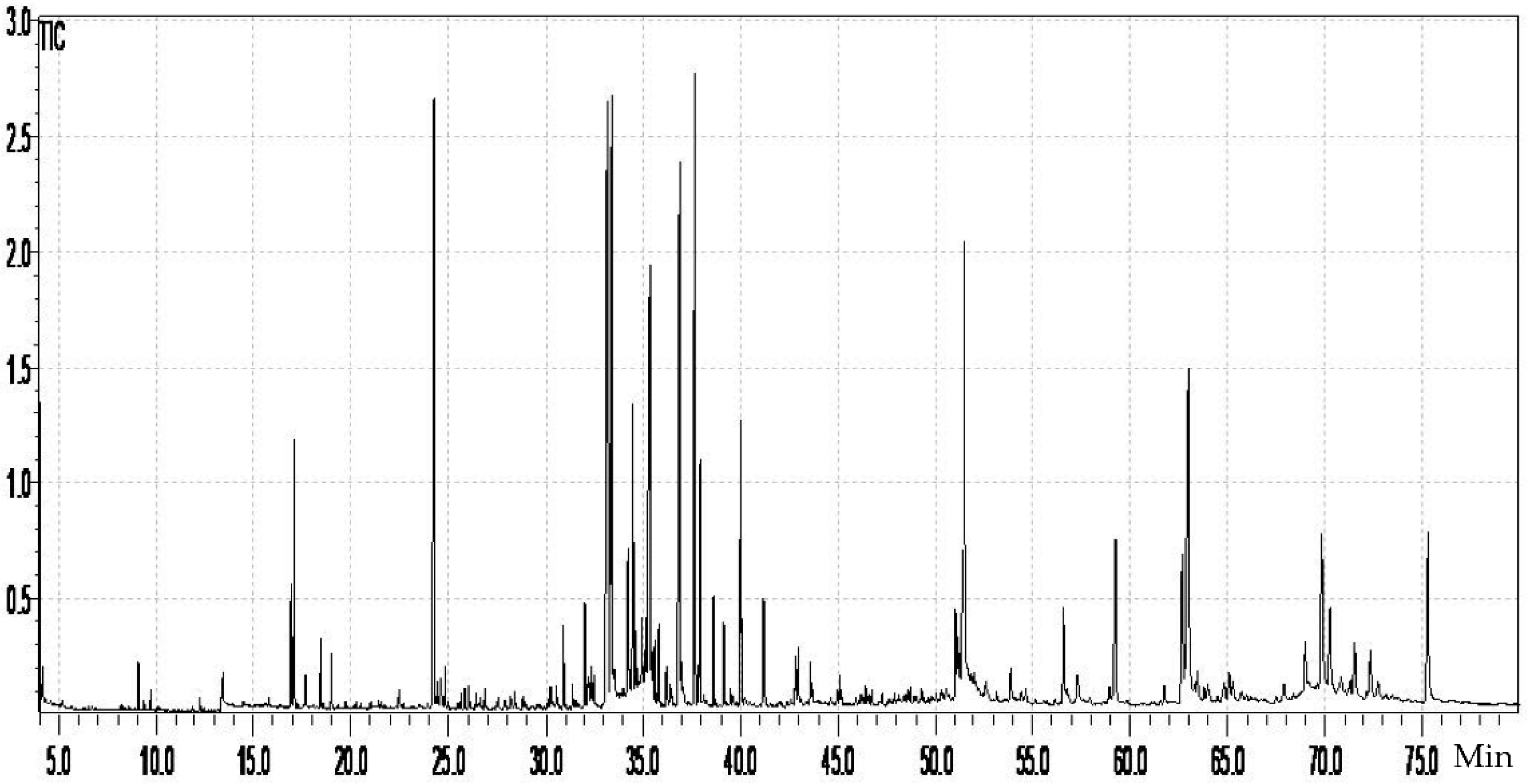
2.2. GC–MS Characterization of W. saharae Extract

GC–MS analysis is the most suitable technique to assess plant extracts′ lipophilic profile [34,35]. It requires, in some cases, a derivatization procedure to convert many secondary metabolites to volatile derivatives [36], and silylation is the most recent derivatization procedure [37,38]. The GC–MS analysis of the hydromethanolic extract of W. saharae allowed the identification of four families of compounds: sugars (214.735 µg/mg), alcohols (128.520 µg/mg), carboxylic acids and esters (124.6 µg/mg), and fatty acids (11.364 µg/mg). The peak characteristics and the identification of the compounds are presented in Table 2, respecting their elution order. The quantification data were expressed as µg compound per mg of extract. The chromatogram is shown in Figure 2.
As far as we know, no studies have been carried out on the lipophilic composition of W. saharae, so this research provides new knowledge about this plant′s fatty acids and important alcohols (Table 2). Since we analyzed the hydromethanolic extract and, before its analysis by GC–MS, the extract was silylated, important chemical compounds such as sugars were revealed by the GC–MS analysis since they became lipophilic upon the silylation procedure. The sugars profile is, however, important to establish the plant nutritional value, and its richness in small carboxylic acids and alcohols also emphasizes the plant value. Moreover, they can be responsible for the observed activities discussed herein. Naturally, fatty acids represented the less significant group identified in the hydromethanolic extract of W. saharae because they are more lipophilic. Nevertheless, these compounds are known to benefit human health by decreasing cancer risk, cardiovascular and neurological disorders and through their antioxidant potential and anti-inflammatory activity [39,40].

2.3. Total Bioactive Content

Total phenolic and flavonoid contents in the hydromethanolic extract of W. saharae were found to be 119.8 ± 8.3 mg GAE/g of extract and 24.25 ± 0.070 mg QE/g extract, respectively. The most important properties of these compounds are their capacity to act as potent antioxidant [41], anti-inflammatory [42], anti-atherogenic [43], and anti-carcinogenic agents [44,45].

2.4. Antioxidant Activities

The antioxidant activity of W. saharae extract was evaluated through five in vitro tests: 1,1-diphenyl-2-picrylhydrazyl (DPPH) scavenging activity, 2,2′-azino-bis(3-ethylbenzothiazoline-6-sulfonic acid (ABTS●+) scavenging assay, galvinoxyl scavenging assay, ferric reducing power (FRP), and cupric reducing antioxidant capacity (CUPRAC). The results obtained by the different tests, expressed in IC50 and A0.50, respectively, are shown in Table 3.
The antioxidant reaction with free radicals involves different mechanisms: the hydrogen atom transfer (HAT) method, in which the free radical removes one hydrogen atom from the antioxidant, and single electron transfer (SET), in which the antioxidant provides an electron to reduce compounds such as metals, carbonyls, and radicals. An antioxidant reaction can also involve both methods (HAT and SET) [46].
The use of a single assay could not assess the total antioxidant potential because of the implication of different mechanisms in the neutralization of free radicals [47]. Hence, to better assess the overall antioxidant effect, the W. saharae extract was assessed through five different trials (see Table 3).
According to ANOVA (F4,12 = 438.85, p < 0.001), data of DPPH scavenging activity show that the W. saharae hydromethanolic extract exhibited high antioxidant potential (IC50 = 7.12 ± 0.09 µg/mL) close to positive controls Trolox, BHT, and ascorbic acid, 5.12 ± 0.21, 6.14 ± 0.41, and 4.39 ± 0.01 µg/mL, respectively, but more potent than BHA (12.99 ± 0.41 µg/mL) with qTukey = 5.99.
According to the HSD Tukey test following ANOVA (F4,12 = 88.01, p < 0.001), in the ABTS●+ scavenging assay, there is not a significant difference between Trolox and ascorbic acid and between BHT and BHA (qTukey = 1.04 and 4.23, respectively). The studied extract exhibits high antioxidant activity (IC50 = 4.19 ± 0.35 µg/mL), very close to those of the standards.
This high antioxidant potential was also noted when analyzing the data of the galvinoxyl assay. Thus, W. saharae extract (IC50 = 3.55 ± 0.08 µg/mL) exhibits very high activity, statistically similar to both standards BHT and Trolox (F4,12 = 266.31, p < 0.001, qTukey = 1.38 and 4.65, respectively).
On the other hand, the reduction potential of the hydromethanolic extract of W. saharae was determined using FRP. In this assay, the antioxidant activity causes the reduction of the Fe3+/ferricyanid complex to the ferrous form. The FRP of extracts is linked with reductants, which exert antioxidant action by donating electrons and reacting with free radicals to convert them to more stable products [48,49]. A significant difference in the FRP assay was noted with ANOVA comparing all tests (F4,12 = 1446.84, p < 0.001, qTukey > 4.65). Data show that W. saharae extract activity (A0.5 = 31.21 ± 0.14 µg/mL) was weak compared to the used standards (5.25 ± 0.20, 7.99 ± 0.87, and 3.62 ± 0.29 µg/mL for Trolox, BHA, and ascorbic acid, respectively). Reducing capacity detected by cupric acid activity was significantly higher compared to that by the FRP assay.
The CUPRAC assay enables total antioxidant capacity (TAC) measurements of a plant extract using the copper (II)–neocuproine reagent in ammonium acetate buffer and based on an electron transfer mechanism [50]. According to the HSD Tukey test, all standards show statistically similar cupric acid activities (p > 0.05). A significant difference was noted with ANOVA and HSD Tukey tests (F4, 12 = 4.38, p < 0.026, qTukey < 4.65), which unveil the best activity of the W. saharae extract (A0.5 = 3.84 ± 0.16 μg/mL) as compared to all tested standards: Trolox, BHA, BHT, and ascorbic acid (8.69 ± 0.14, 6.62 ± 0.05, 8.97 ± 3.94, and 8.31 ± 0.15 µg/mL, respectively).
Overall, present data indicate that W. saharae extract showed a greater capacity to capture radicals. The results of the DPPH activity are in agreement with those reported in previous works on the ethyl acetate extract of the Algerian W. saharae (IC50 = 6.98 µg/mL) [19], but are significantly higher than those reported in another study conducted on the same species grown in Morocco (IC50 = 182 µg/mL) [16]. The nature of the extraction solvent, the plant growth area, and the collection period point out the discrepancy in antioxidant activity results between the referred studies. It should be mentioned that no previous studies were conducted on the antioxidant activity of W. saharae using the ABTS●+ scavenging assay, galvinoxyl assay, FRP, and cupric reducing antioxidant capacity.
The DPPH, ABTS●+, and galvinoxyl scavenging data suggest that the components present in W. saharae extract act as excellent scavenging free radicals through a mechanism of electron or hydrogen donation to stabilize the free radicals, and should be able to protect biological matrices from free radical-mediated oxidative degradation [51,52].
According to the CUPRAC and FRP data, W. saharae was as effective as the positive standards in reducing Cu2+ to Cu+ but had a moderate effect in reducing the Fe3+ to Fe2+. The good reducing behavior of W. saharae extract suggests that its constituents, mainly chlorogenic acids, flavonoids, and sugars, are capable of terminating a chain reaction by eliminating free radical intermediates and hence are considered antioxidants [53]. A significant positive correlation between total phenolic and reducing sugar contents with the potent antioxidant capacity was established for other species [54]. Therefore, it is important to emphasize the important role of reducing sugars in antioxidant activity, since the extract of W. saharae contains a significant amount (around 215 μg/mg).
Furthermore, phenolic acids’ and flavonoids’ antioxidant potentials are closely related to their chemical structure [55] with double bonds and hydroxyl groups [56,57], making them ideal for scavenging free radicals and chelating metal ions. These properties also conferred effective antioxidant potential in vivo [58]. In the present study, the chemical composition analysis of the hydromethanolic extract of W. saharae reveals that phenolic acids are the most dominant group identified in this species. In addition, many compounds detected in the W. saharae hydromethanolic extract are known to possess great antioxidant capacity. For example, simple hydroxycinnamic acids have been reported to possess potent antioxidant activity against DPPH and ABTS●+ [59]. Chlorogenic acids are considered responsible for both green and medium roasted coffee’s antioxidant capacity, with 5-O-caffeoylquinic acid being the major contributor [60].
Li et al. [61] reported that di-O-caffeoylquinic acids exhibit high antioxidant activity using conventional antioxidant assays. The same authors proposed that their antioxidant mechanisms might include electron transfer, H+ transfer, and Fe2+ chelation.
Taxifolin (peak 14) is the major compound present in the hydromethanolic extract of W. saharae and may contribute to its strong antioxidant activity. A previous study reported that in antioxidant assays, taxifolin scavenges HO, DPPH, and ABTS●+ efficiently and enhances the relative Cu2+ and Fe3+ reducing levels [62]. Zu et al. [63] also mentioned that taxifolin is a free radical scavenger, and its antioxidant capacity is superior to other flavonoids. The results obtained by Topal et al. [64] from an in vitro study showed that taxifolin possesses marked antioxidant, reducing ability, radical scavenging, and metal-chelating activities.
Although it is present in lower amounts, luteolin has received increasing attention due to its various biological activities and pharmacological effects, including antioxidant activity [65], and has been widely applied in medicine and functional foods [66,67].
The high antioxidant potential recorded in this study may be linked to the presence of these compounds that could act individually or combined to provide the hydromethanolic extract of W. saharae with its strong antioxidant power.

2.5. Anti-Inflammatory Activity

The anti-inflammatory effect of W. saharae was evaluated by the NO scavenging method. NO is one of the main chemical mediators involved in inflammatory events. Regarding its ability to scavenge NO, the relevant anti-inflammatory activity of W. saharae extract (IC50 = 76.66 ± 4.16 µg/mL) is better than that of ascorbic acid (IC50 = 144.380 ± 7.035 µg/mL) but lower than that of curcumin (IC50 = 7.49 ± 0.58 µg/mL). To the best of our knowledge, no anti-inflammatory effect had been previously described for W. saharae extract. Thus, this is the first report describing the anti-inflammatory potential of this species.
The phenolic compounds highly present in W. saharae extract are probably responsible for the anti-inflammatory property observed in this study, and flavonoids can be highlighted [42,68]. Taxifolin, the main phenolic compound present in W. saharae extract, was previously found to possess significant anti-inflammatory activity against carrageenan-induced edema involving the exudative phase of inflammation and also against formaldehyde-induced arthritis [69]. These results corroborate the traditional use of this species in folk medicine.

2.6. Enzyme Inhibitory Activities

The enzyme inhibitory activities of the W. saharae extract and the standard inhibitors tested toward α-glucosidase, α-amylase, and acetylcholinesterase expressed in IC50 or the percentage of inhibition are shown in Table 4.
The α-glucosidase and α-amylase inhibitors are used to treat diabetes mellitus as they slow down the digestion of carbohydrates in the small intestine and thus reduce the rapid rise in blood sugar after eating [70,71]. The inhibitory effect of the W. saharae species toward these two enzymes was evaluated in this study.
According to an ANOVA test (F1,5 = 349.79, p < 0.001), W. saharae extract expressed significant strong activity against the α-glucosidase enzyme with an IC50 = 23.52 ± 6.33 μg/mL lower than that of acarbose (IC50 = 405.77 ± 34.83 mg/mL). However, low inhibitory activity against α-amylase was recorded (38.41 ± 5.51% inhibition at the concentration of 4.2 mg/mL).
Several earlier studies have demonstrated the role of medicinal plants’ phenolic compounds as inhibitors of α-glucosidase. Among those flavonoids such as taxifolin and luteolin, and hydroxycinnamic acids, major compounds of W. saharae extract exhibited strong inhibitory activity against α-glucosidase in in vitro studies [72,73,74]. In a recent report, it was found that taxifolin′s pre-administration can significantly improve postprandial hyperglycemia in rats [74].
Furthermore, the obtained results with the α-amylase inhibition assay agree with those of Nyambe-Silavwe and Williamson [75] and Proença et al. [76], suggesting that chlorogenic and other phenolic acids, major compounds in W. saharae extract, weakly inhibit human α-amylase activity despite publications that claim otherwise [74].
The UHPLC-DAD-ESI/MS analysis shows that W. saharae extract contains these phenolic compounds abundantly. Consequently, it can be said that the synergistic effect could explain the high inhibitory effect recorded in the W. saharae extract among these compounds.
Apostolidis et al. [77] reported that medicinal plants that selectively inhibit α-glucosidase are better for controlling glucose absorption. Indeed, both enzymes’ inhibition leads to abnormal bacterial fermentation in the colon resulting from the undigested carbohydrates. Thus, the inhibition of α-glucosidase by the W. saharae extract could be considered a promising way to control diabetes mellitus.
Acetylcholinesterase inhibitors prevent the breakdown of acetylcholine in the body by blocking the action of the enzyme acetylcholinesterase responsible for its separation, which leads to an increase in its level in the space between nerve endings and thus gives it more opportunity to cross into another cell and carry out its functions. This mechanism is important for people with Alzheimer′s disease, who already suffer from a decrease in acetylcholine level since these inhibitors temporarily improve the symptoms of the disease and its stability [78].
We also evaluated the inhibitory effect of the acetylcholinesterase enzyme here. At the highest dose of the extract (1 mg/mL in this assay), the enzyme′s inhibition was observed (28.578 ± 2.979%). The inhibitory action on AChE in the in vitro assay proved to be lower than the donepezil used as a positive control (IC50 = 13.32 ± 1.34 µM). Thus, the weak inhibitory activity of W. saharae extract toward AChE activity could be related to the chemical composition of this species since we considered that the major substance in the plant extract is usually responsible for its biological activity.
Taxifolin, highly present in the W. saharae extract, has been shown to have relatively weak AChE inhibitory properties (IC50 = 133.1 μg/mL) [79] despite studies that claim otherwise [80]. Although chlorogenic acids are mainly responsible for inhibiting the AChE enzyme in green coffee extracts [81], their presence in W. saharae extract could not be sufficient to inhibit this enzyme strongly. Some of them, like 4,5-di-O-caffeoylquinic, one of the major compounds in W. saharae extract, showed AChE inhibition under 50% [81].
In addition, some phenolic compounds may act as activators of AChE. Earlier studies demonstrated that caffeic acid was shown to increase AChE activity in in vitro studies [82]. Some authors have hypothesized that caffeic acid (or its derivatives) could trigger the AChE catalytic site′s activation in a reaction at a second site [83]. Hence, the presence of caffeic acid in W. saharae extract could, in part, explain these results.

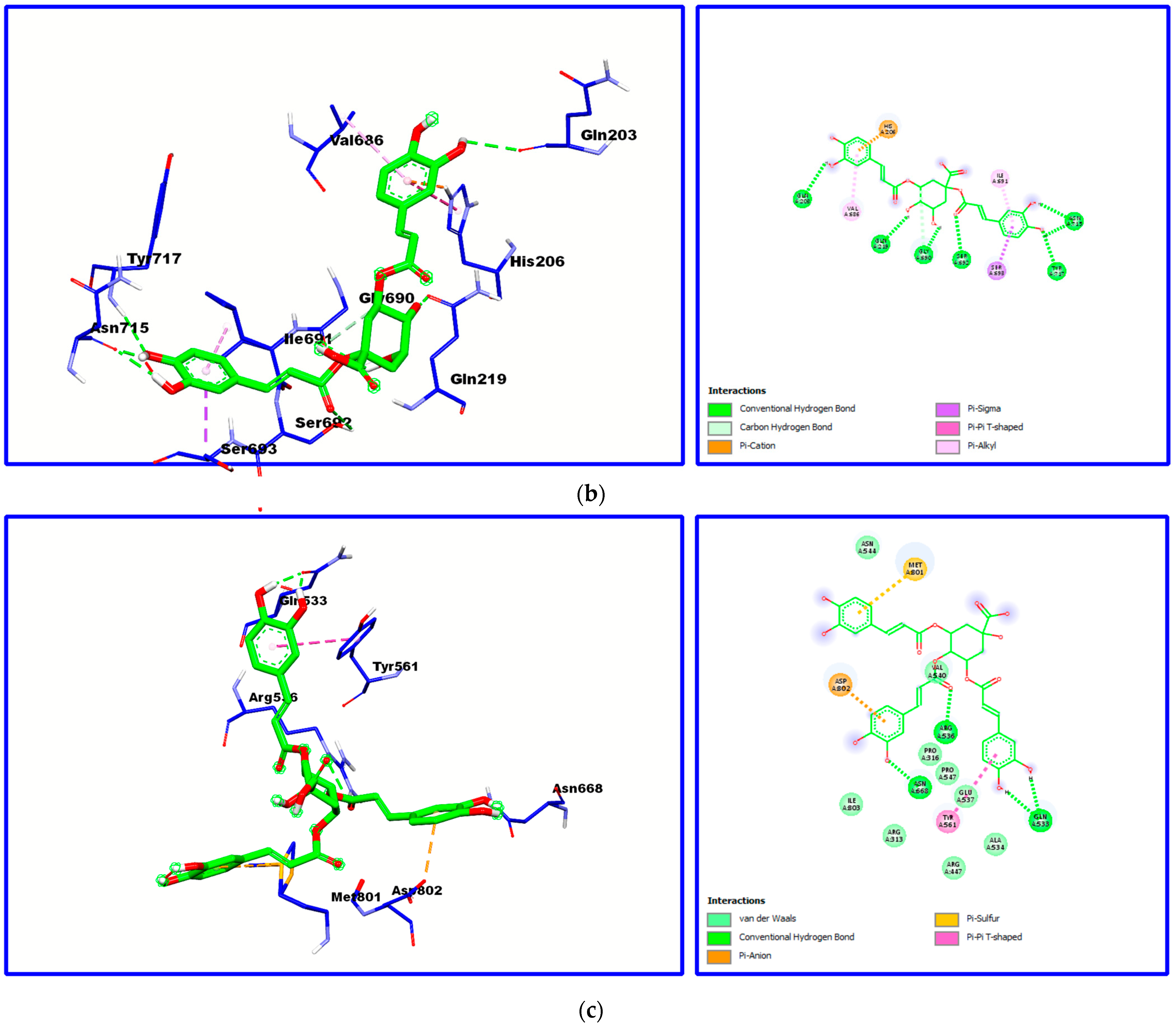
2.7. Molecular Docking

An in silico study was performed to further screen the mechanisms of interaction between α-glucosidase and the three major compounds in W. saharae extract. Figure 3 shows the 2D and 3D structures of the docked compounds at the active site of the α-glucosidase enzyme and the types of interactions involved.
Based on the docking test’s interaction data, the major constituents of W. saharae extract interact with α-glucosidase, engaging different amino acids (Table 5). Taxifolin showed a higher binding affinity, as indicated by its lower binding energy of –5.89 (kcal/mol), followed by 4,5-O-dicaffeoylquinic acid and 1,3-O-dicaffeoylquinic acid with a binding energy of –4.17 and –4.02 (kcal/mol), respectively.
Herein, it can be observed that the best docking pose obtained was for taxifolin docked to α-glucosidase. Different interactions, such as three hydrogen bonds with Asn715, Ala687, and Gly690, π–cation, and π–alkyl with Ile691 to the aromatic cycle and many other Van Der Waals interactions can be observed. Taxifolin, which has shown the best binding affinity within the major compounds, was previously reported as a competitive inhibitor of the α-glucosidase enzyme with an IC50 of 0.038 mg/mL [74].
Several types of interactions were found between both compounds, 4,5-O-dicaffeoylquinic acid and 1,3-O-dicaffeoylquinic acid, and the α-glucosidase active site (Figure 3), most of which are hydrogen bonds, π interactions, van der Waals, and carbon-hydrogen bonds.
These findings further support our in vitro assay results. However, in vivo studies are still necessary to better confirm these results.

3. Materials and Methods

3.1. Chemicals

The following chemicals: butylated hydroxyanisole (BHA), hydroxytoluene (BHT) and α-tocopherol, 1,1-diphenyl-2-picrylhydrazyl (DPPH), 2,2′-azinobis(3-ethylbenzothiazoline-6-sulfonic acid) diammonium salt (ABTS●+), trichloroacetic acid (TCA), potassium ferricyanide, and neocuproine, were used for the determination of the antioxidant activities and were purchased from Sigma Chemical Co. (Sigma-Aldrich GmbH, Stern-heim, Germany). α-Glucosidase from Saccharomyces cerevisiae, 4-nitrophenyl α-D-glucopyranoside (pNPG), α-amylase from porcine pancreas, β-nicotinamide adenine dinucleotide (β-NADH), phenazine methosulphate (PMS), nitrotetrazolium blue chloride (NBT), and ascorbic acid were obtained from Sigma (St. Louis, MO, USA). Acarbose was purchased from Fluka (Bucharest, Romania) and potato starch from Fisher (Pittsburgh, PA, USA). Sodium nitroprusside, sulfanilamide, and 3,5-dinitrosalicylic acid (DNS) were obtained from Acros Organics (Hampton, NH, USA). Folin–Ciocalteu reagent, Na2CO3, and gallic acid were purchased from Panreac (Barcelona, Spain). Acetylcholinesterase (AChE) from Electrophorus electricus, 5,5-dithiobis(2-nitrobenzoic acid), acetylthiocholine iodine, and donepezil were purchased from Sigma-Aldrich (St. Louis, MO, USA).
Compounds used as standards to elucidate the identification of the phenolic constituents and to elaborate the calibration curves were obtained from EXTRASYNTHESE (Genay CEDEX, France).
Formic acid and acetonitrile HPLC-grade solvents were purchased from Panreac (Barcelona, Spain). Ultrapure water was obtained by a Direct-Q® water purification system (Merck Life Science, Darmstadt, Germany). All other chemicals were of analytical grade.
Solvents used for GC–MS analysis were purchased from Panreac and Acros Organics and were of analytical grade. Other chemicals such as palmitic acid (99%), N,O-bis(trimethylsilyl)trifluoroacetamide (BSTFA) (99%), trimethylsilyl chloride (TMSCl) (99%), eicosane (99%), hexatriacontane (99%), citric acid (99.5%), cinnamic acid (99%), palmitic acid (99%), mannose (99%), D-mannitol (98%), 5-cholesten-3β-ol (99%), tetradecanol (98%), and 1-palmitoylglycerol (99%) were purchased from Sigma-Aldrich.

3.2. Extract Preparation

Aerial parts of W. saharae were collected from Djebel Antar (Bechar, Southwest Algeria; 31°56′34′′ N, 1°55′52′′ W). A voucher specimen was deposited in the Herbarium of the VAREN laboratory of Mentouri University of Constantine, Algeria, under the reference number VAREN/CWS04/05. The leaves were shade air-dried at room temperature (25 °C) before they were ground to a fine powder. The powder was then macerated at room temperature with a 80:20 mixture of methanol:water (v/v) for 72 h three times. The filtrates were concentrated under reduced pressure using a rotary evaporator at 30 °C. The residual water was freeze-dried in a freeze dryer and then stored at 4 °C until future use.

3.3. Total Bioactive Content

3.3.1. Total Phenolic Content

Total phenolic content was determined using the Folin–Ciocalteu method [84]. An aliquot of 15 μL of plant extract (1 mg/mL) was mixed with 15 μL of Folin’s reagent and 60 μL of water. After 5 min of incubation, 150 μL of Na2CO3 solution (20% w/v) was added. The mixture was then incubated in the darkness for 60 min, and absorbance was read at 760 nm. The standard curve of gallic acid was obtained under the same conditions as above, and the total phenolic content was measured as μg equivalents of gallic acid per mg of extract (μg GAE mg of extract).

3.3.2. Total Flavonoid Content

The total flavonoid content of the plant’s crude extract was determined according to the method described by Türkoğlu et al. [85] with a slight modification. A total of 100 µL of plant extract was mixed with 100 mL of AlCl3 (2%) solution. The mixture was incubated at room temperature for 10 min, and then the absorbance was measured at 415 nm. The standard curve of quercetin was obtained under the same conditions as above, and the total phenolic content was measured as μg equivalents of quercetin per mg of extract (μg GAE/mg of extract).

3.4. UHPLC-DAD-ESI-MS/MSn Characterization of W. saharae Extract

Phenolic composition of hydromethanolic extract of W. saharae was determined using an Ultimate 3000 (Dionex Co., San Jose, CA, USA) apparatus equipped with a binary pump, an automatic sampler, and a diode array detector (Dionex Co., San Jose, CA, USA). The MS analysis was performed using a Thermo LTQ XL mass spectrometer (Thermo Scientific, San Jose, CA, USA), which was equipped with an electrospray ionization interface (ESI). The separation was carried out with a Hypersil Gold (Thermo Scientific, USA) C18 column (100 mm length; 2.1 mm i.d.; 1.9 μm particle diameter, end-capped) at room temperature 25 °C. The extract was prepared at a 1 mg/mL concentration and the injection volume was 10 µL. The solvents used were formic acid in water (A) and acetonitrile (B). The flow rate was set at 2 mL/min. Spectra were recorded in negative mode. MS and MS/MS data were processed using the Thermo Xcalibur Qual Browser data system (Thermo Scientific, USA).

3.5. GC–MS Characterization of W. saharae Extract

The lipophilic composition of the hydromethanolic extract of W. saharae was analyzed by GC–MS using a Shimadzu Gas Chromatograph QP2010 Ultra. Three aliquots of the W. saharae extract (20 mg) were submitted to silylation procedure according to the method described by Freire et al. [86]. Silylation is the substitution of the active hydrogen atoms by a trimethylsilyl group (TMS), so that all the compounds with hydroxy groups will be transformed into the correspondent TMS derivatives. The sample was dissolved in 250 µL of dichloromethane. A total of 200 µL of hexatriacontane as internal standard (IS), 250 µL of pyridine, 250 µL of BSTFA, and 50 µL of TMSCl were then added to the sample. The mixtures were incubated for 30 min at 70 °C and then injected in triplicate into the GC–MS apparatus. Compound separation was carried out in J&W DB-5 capillary column (30 m × 0.25 mm inner diameter, 0.25 μm film thickness) using helium as the carrier gas (35 cm s−1). The following standards: eicontane, palmitic acid, tetradecanol, D-(+)-mannose, mannitol, and cholestanol, were used to elaborate calibration curves. Compounds were identified by comparing their mass spectra with mass spectra in the Wiley and NIST libraries and those available in the literature.

3.6. Antioxidant Activities

3.6.1. Determination of 1,1-Diphenyl-2-Picrylhydrazyl Radical Scavenging Activity

The 1,1-diphenyl-2-picrylhydrazyl (DPPH) free radical-scavenging activity was evaluated by a slightly modified method [87,88]. Briefly, the solution of 0.1 mM of DPPH was prepared in ethanol, and 160 µL of this solution was added to 40 µL of sample solutions diluted in methanol at different concentrations. Twenty-five minutes later, the absorbance was measured at 517 nm. The scavenging capability of DPPH was calculated using the following equation. The results were given as IC50 (mg/mL) corresponding to the concentration of 50% inhibition.
DPPH scavenging effect (%) = (AC − AS)/AC × 100
where Ac refers to the absorbance of control; AS refers to the absorbance of sample.
The sample concentration providing 50% free radical scavenging activity (IC50) was calculated from the graph of DPPH scavenging effect percentage against sample concentration. BHA, BHT, Trolox, and ascorbic acid were used as antioxidant standards for comparison of the activity.

3.6.2. Determination of 2,2′-Azinobis(3-Ethylbenzothiazoline-6-Sulfonic Acid) Scavenging Activity

The 2,2′-azino-bis(3-ethylbenzothiazoline-6-sulfonic acid) free cation-radical (ABTS●+) scavenging activity was obtained by spectrophotometric analysis according to the slightly modified method proposed by Re et al. [89]. The ABTS●+ was regenerated by adding 2.45 mM potassium persulfate to an aqueous solution of 7 mM ABTS●+, which was stored in the dark at room temperature for 12 h. Before use, the ABTS●+ solution was diluted until an absorbance of 0.700 ± 0.025 at 734 nm with ethanol was obtained. Then, to each well, in a 96-well plate, 160 µL of this solution and 40 µL of sample solutions diluted in methanol at different concentrations were added. After 10 min, the percentage inhibition at 734 nm was calculated for each concentration relative to a blank absorbance (methanol). The scavenging capability of ABTS●+ was calculated using the following equation:
ABTS●+ Scavenging effect (%) = (AC − AS)/AC × 100
where Ac refers to the absorbance of control; As refers to the absorbance of sample.
The sample concentration providing 50% cation radical scavenging activity (IC50) was calculated from the graph of ABTS●+ scavenging effect percentage against sample concentration. BHA, BHT, Trolox, and ascorbic acid were used as antioxidant standards for comparison of the activity.

3.6.3. Galvinoxyl Radical Scavenging Activity

The trapping activity of the galvinoxyl radical was determined according to the method described by Shi et al. [90] in 96-well microplates. A total of 40 µL of the extract to be tested at different concentrations was placed in the wells of the microplate. A total of 160 µL of methanolic galvinoxyl solution (4 mg of galvinoxyl in 100 mL of methanol) was added to each well. The plate was then incubated in the dark for 2 h. Absorbance was measured at the wavelength of 428 nm.
A blank was prepared by mixing 160 µL of methanol with 40 µL of each extract. All tests were performed in triplicate. The percentage of inhibition of the galvinoxyl is expressed by the following formula:
Galvinoxyl Scavenging effect (%) = (Ac − As)/Ac × 100
where Ac refers to the absorbance of control; As refers to the absorbance of sample.
The concentration of the sample providing 50% inhibition (IC50) was calculated by relating the inhibition percentages to the concentrations of the extracts. The antioxidant capacity of the extract was compared with ascorbic acid, BHT, BHA, and Trolox used as antioxidant standards and prepared under the same conditions.

3.6.4. Ferric Reducing Power Assay

The FRP assay was carried out according to the method of Oyaizu [91] with slight modification. Briefly, 40 μL of phosphate buffer (pH 6.6) and 50 μL of potassium ferricyanide (1%) (1 g of K3Fe(CN)6 in 100 mL H2O) were mixed with 10 μL plant extract at different concentrations and then incubated 20 min at 50 °C. A mixture of 50 mL trichloroacetic acid (TCA) (10%), 40 mL H2O, and 10 μL FeCl3 (0.1%) was then added. The absorbance of the reaction mixture was then recorded at 700 nm. Increased absorbance of the reaction mixture indicated increased FRP. The results were given as A0.50, which corresponds to the concentration producing 0.500 absorbance.

3.6.5. Cupric Ion Reducing Antioxidant Capacity Assay

The cupric ion reducing antioxidant capacity (CUPRAC) assay was determined by the method described by Apak et al. [92]. A total of 40 µL of W. saharae extract at different concentrations was added to a solution composed of 50 µL Cu Cl2, 2H2O (10 mM), 50 µL neocuproine (7.5 mM), and 60 µL ammonium acetate buffer solution (1 M, pH 7.0). The reaction mixtures were incubated in the dark for 1 h. The absorbance was then measured at 450 nm.
The results were calculated as A0.50 (µg/mL) corresponding to the concentration indicating 0.500 of absorbance. The reduction capacity of the extracts was compared with that of BHA, BHT, Trolox, and ascorbic acid prepared under the same conditions.

3.7. Anti-Inflammatory Activity

The in vitro anti-inflammatory activity was determined by the ability of the extract to scavenge NO following the method described by Afonso et al. [93]. A total of 100 µL of each extract concentration was mixed with 100 µL of sodium nitroprusside (3.33 mM) solution prepared in PBS 100 mM (pH 7.4). After incubation for 10 min at room temperature under light irradiation, 100 µL of Griess reagent (equal volume of 1% of sulfanilamide and 0.1% of naphthyl ethylenediamine dihydrochloride in 2.5% of phosphoric acid) was then added to the mixture. The absorbance was measured at 562 nm in spectrophotometer. Curcumin was used as the reference compound.
The scavenging ability of NO was calculated using the following equation:
NO Scavenging effect (%) = (Ac − As)/Ac × 100
where Ac refers to the absorbance of control; As refers to the absorbance of sample.
The sample concentration providing 50% NO scavenging activity (IC50) was calculated from the graph of NO scavenging effect percentage against sample concentration.

3.8. Inhibition of Enzymatic Activities

3.8.1. Inhibition of α-Glucosidase Activity

α-Glucosidase inhibition activity was measured by the method described by Neto et al. [94] with brief modifications. A total of 50 µL of each extract concentration was mixed with 50 µL of 4-nitrophenyl α-d-glucopyranoside (PNPG) solution used as substrate. A total of 100 µL of the α-glucosidase enzyme solution was then added to the mixture. The absorbance was measured at 405 nm at 37 °C, for 20 min every minute. Acarbose was used as reference.
The α-glucosidase inhibitory activity was expressed as percentage inhibition and was determined according to the following formula:
Inhibition % = (Ac − As)/Ac × 100
where Ac refers to the absorbance of control (enzyme and buffer); As refers to the absorbance of sample (enzyme and inhibitor).
IC values (concentration of inhibitor required to inhibit 50% of enzyme activity) were determined.

3.8.2. Inhibition of α-Amylase Activity

α-Amylase inhibition activity was determined according to the method previously described by Wickramaratne et al. [95] with slight modifications. Briefly, 200 µL of each extract concentration was added to 400 µL of a 0.8% (w/v) starch solution dissolved in 20 mM phosphate buffer (pH 6.9, containing 6 mM of NaCl). After incubation at 37 °C for 5 min, the reaction was started with the addition of 200 µL of α-amylase solution. A total of 200 µL of the mixture was then mixed with 600 µL of 3,5-dinitrosalicylic acid (DNS) reagent.
A second aliquot of 200 µL of the first mixture was also collected 15 min later and mixed with DNS reagent. The mixtures were boiled for 10 min. After cooling, 250 µL was transferred to each well in a 96-well microplate and the absorbance was read at 450 nm.
The α-amylase inhibitory effect was calculated by the following equation:
Inhibition % = (Ac − As)/Ac × 100
where Ac refers to the absorbance of control (enzyme and buffer); As refers to the absorbance of sample (enzyme and inhibitor).
IC values (concentration of inhibitor required to inhibit 50% of enzyme activity) were determined. Acarbose was used as reference compound.

3.8.3. Inhibition of Acetylcholinesterase Activity

Inhibition of acetylcholinesterase (AChE) activity was determined as described by Ellman et al. [96] with slight modifications. A total of 150 µL of 0.025 U/mL solution of AChE was added to 50 µL of each extract at different concentrations. After incubating at 37 °C for 5 min, 50 µL of DTNB (0.5 mM) and 50 µL of acetylthiocholine iodide (2.5 mM) were added to the mixture. The absorbance was read at 415 nm each 150 s for 7.5 min. Donepezil was used as a reference. The percentage inhibition of AChE enzyme was determined according to the following formula:
Percentage of inhibition = (E − S)/E × 100.
where E is the activity of the enzyme without extract; S is the activity of the enzyme with the extract.
IC50 values (concentration of inhibitor required to inhibit 50% of enzyme activity) were determined.

3.9. Molecular Docking Study

To support our findings from in vitro inhibition of α-glucosidase activity by the W. saharae extract, three compounds were docked to the α-glucosidase enzymatic activity. The selected compounds were taxifolin, 1,3-O-dicaffeoylquinic acid, and 4,5-O-dicaffeoylquinic acid, which are the most abundant in the W. saharae extract. The 3D structures of the considered molecules were downloaded from PubChem database [97]. All three structures were performed by AM1 [H2] semi empirical method by using Orca-4.2.1 program package [98]. The crystal structure of the α-glucosidase (PDB code 3W37) was retrieved from the Protein Data Bank [99]. Water molecules were removed from the protein structure. The docking software AutoGrid and AutoDock (version 4.2.6) implemented in AutoDock Tools (ADT 1.5.6) program were used for the docking experiments. The most stable receptor–ligand complexes were generated using Chimera [100] and BIOVIA Discovery studio visualizer (version 1.10.2) [101].

3.10. Statistical Analysis

All of the data were analyzed with MINITAB software (version 16, State College, PA, USA) using one-way ANOVA followed by a post hoc honestly significant difference (HSD) Tukey’s test at p ≤ 0.05. Data were recorded as the mean ± standard deviation (m ± SD) from three independent assays.

4. Conclusions

Our results provided new information and allowed the characterization of the chemical profile of the hydromethanolic extract of W. saharae. The extract showed potent antioxidant and α-glucosidase inhibition properties, highlighting that this species provides a promising source for obtaining natural compounds that could be used to develop food additives or pharmaceutical products. Anti-inflammatory properties were also observed with the hydromethanolic extract of W. saharae. Finally, further toxicological and pharmacological studies must be performed for pharmaceutical purposes.

Author Contributions

H.R. contributed to conceptualization, investigation, data curation, and writing—original draft preparation. K.H. contributed to conceptualization, supervision, and writing—reviewing the original draft. A.H. contributed to conceptualization, investigation, and data curation. H.A. contributed to investigation (docking study). T.B. contributed to investigation (plant collection, sample preparation). D.C.G.A.P. contributed to conceptualization, supervision, and writing—reviewing the original draft. S.M.C. contributed to conceptualization, data curation, and supervision. A.M.S.S. contributed to conceptualization, supervision, and writing—reviewing the original draft. C.B. contributed to investigation and supervision. N.S. contributed to conceptualization and review. All authors have read and agreed to the published version of the manuscript.

Funding

This research was funded by FCT (Fundação para a Ciência e a Tecnologia), the European Union, QREN, FEDER, and COMPETE for funding the LAQV-REQUIMTE (UIDB/50006/2020) and PRFU Project to Dr. K. Hamaidia (No: D01N01UN410120210001).

Institutional Review Board Statement

Not applicable.

Informed Consent Statement

Not applicable.

Data Availability Statement

Data is contained within the article.

Acknowledgments

Thanks are due to the University of Aveiro and FCT (Fundação para a Ciência e a Tecnologia), the European Union, QREN, FEDER, and COMPETE for funding the LAQV-REQUIMTE (UIDB/50006/2020) and PRFU Project to K. Hamaidia (No: D01N01UN410120210001). Thanks are also due to the Algerian Higher Ministry and Scientific Research via PNE (Programme National Exceptionnel) for financial support, namely the H.R. and A.H. displacements.

Conflicts of Interest

The authors declare no conflict of interest.

Sample Availability

Samples of the compounds are available from the authors.

References

  1. Das, A.; Chaudhuri, D.; Sarkar, R.; Ghate, N.B.; Panja, S.; Mandal, N. Plants of Indian traditional medicine with antioxidant activity. In Nutritional Antioxidant Therapies: Treatments and Perspectives; Al-Gubory, K.H., Laher, I., Eds.; Springer: Cham, Switzerland, 2018; pp. 27–64. [Google Scholar]
  2. Joshee, N.; Dhekney, S.A.; Parajuli, P. Medicinal Plants: From Farm to Pharmacy; Springer International Publishing: New York, NY, USA, 2019; p. 439. [Google Scholar] [CrossRef]
  3. Panche, A.N.; Diwan, A.D.; Chandra, S.R. Flavonoids: An overview. J. Nutr. Sci. 2016, 5, e47. [Google Scholar] [CrossRef] [Green Version]
  4. Brahmkshatriya, P.P.; Brahmkshatriya, P.S. Terpenes: Chemistry, biological role, and therapeutic applications. In Natural Products; Ramawat, K., Mérillon, J.M., Eds.; Springer: Berlin, Germany, 2013; pp. 2665–2691. [Google Scholar] [CrossRef]
  5. Liu, C.M.; Kao, C.L.; Wu, H.M.; Li, W.J.; Huang, C.T.; Li, H.T.; Chen, C.Y. Antioxidant and anticancer aporphine alkaloids from the leaves of Nelumbo nucifera Gaertn. cv. Rosa-plena. Molecules 2014, 19, 17829–17838. [Google Scholar] [CrossRef]
  6. Cardoso, J.C.; Oliveira, M.E.B.S.; Cardoso, F.C.I. Advances and challenges on the in vitro production of secondary metabolites from medicinal plants. Hortic. Bras. 2019, 37, 124–132. [Google Scholar] [CrossRef] [Green Version]
  7. Jain, C.; Khatana, S.; Vijayvergia, R. Bioactivity of secondary metabolites of various plants: A review. Int. J. Pharm. Sci. Res. 2019, 10, 494–504. [Google Scholar] [CrossRef]
  8. Yang, L.; Wen, K.S.; Ruan, X.; Zhao, Y.X.; Wei, F.; Wang, Q. Response of Plant Secondary Metabolites to Environmental Factors. Molecules 2018, 23, 762. [Google Scholar] [CrossRef] [Green Version]
  9. Kabtni, S.; Sdouga, D.; Bettaib Rebey, I.; Mattew Save, N.T.F.; Fauconnier, M.L.; Marghali, S. Influence of climate variation on phenolic composition and antioxidant capacity of Medicago minima populations. Sci. Rep. 2020, 10, 8293. [Google Scholar] [CrossRef]
  10. Li, Y.; Kong, D.; Fu, Y.; Sussman, M.R.; Wu, H. The effect of developmental and environmental factors on secondary metabolites in medicinal plants. Plant Physiol. Biochem. 2020, 148, 80–89. [Google Scholar] [CrossRef]
  11. Ozenda, P. Flore du Sahara Septentrional et Central; CNRS: Paris, France, 1958; p. 444. [Google Scholar]
  12. Katinas, L.; Pruski, J.; Gisela Sancho, G.; Tellería, M.C. The subfamily Mutisioideae (Asteraceae). Bot. Rev. 2008, 74, 469–716. [Google Scholar] [CrossRef]
  13. Bellakhdar, J.; Baayaoui, A.; Kazdari, A.; Marechal, J. Herboristes et medecine traditionnelle à Tissint, oasis presaharien du sud Marocain (province de Tata). Al Biruniya 1986, 3, 7–50. [Google Scholar]
  14. Ajebli, M.; Eddouks, M. Flavonoid-enriched extract from desert plant Warionia saharae improves glucose and cholesterol levels in diabetic rats. Cardiovasc. Hematol. Agents Med. Chem. 2019, 17, 28–39. [Google Scholar] [CrossRef]
  15. Beghalia, M.; Ghalem, S.; Allali, H. Comparison of the inhibitory capacity of two groups of pure natural extract on the crystallization of two types of material compound urinary stones in vitro study. IOP Conf. Ser. Mater. Sci. Eng. 2015, 92, 012025. [Google Scholar] [CrossRef]
  16. Amezouar, F.; Badri, W.; Hsaine, M.; Bourhim, N.; Fougrach, H. Chemical composition, antioxidant and antibacterial activities of leaves essential oil and ethanolic extract of Moroccan Warionia saharae Benth. & Coss. J. Appl. Pharm. Sci. 2012, 2, 212–217. [Google Scholar] [CrossRef]
  17. Sellam, K.; Ramchoun, M.; Alem, C.; Khallouki, F.; El Moualij, B.; El Rhaffari, L. Chemical composition, antioxidant and antimicrobial activities of essential oil of Warionia saharae from Oases of Marocco. In Gas Chromatography–Biochemicals, Narcotics and Essential Oils; Salih, B., Ed.; IntechOpen: Rijeka, Croatia, 2012; Chapter 10; pp. 213–220. [Google Scholar] [CrossRef] [Green Version]
  18. Znini, M.; Cristofari, G.; Majidi, L.; Paolini, J.; Desjobert, J.M.; Costa, J. Essential oil composition and antifungal activity of Pulicaria mauritanica Coss., against postharvest phytopathogenic fungi in apples. LWT-Food Sci. Technol. 2013, 54, 564–569. [Google Scholar] [CrossRef]
  19. Mezhoud, S.; Derbré, S.; Ameddah, S.; Mekkiou, R.; Boumaza, O.; Seghiri, R.; Benayache, S.; Richomme, P.; Benayache, F. Antioxidant activity and chemical constituents of Warionia saharae Benth. & Coss. (Compositae) from Algeria. Int. J. Med. Arom. Plants. 2012, 2, 509–513. [Google Scholar]
  20. El-Ouady, F.; Eddouks, M. Warionia saharae induces antihypertensive and vasorelaxant activities through nitric oxide and KATP channels pathways in rats. J. Complement. Integr. Med. 2019, 26, 17. [Google Scholar] [CrossRef]
  21. Boittier, E.D.; Tang, Y.Y.; Buckley, M.E.; Schuurs, Z.P.; Richard, D.J.; Gandhi, N.S. Assessing molecular docking tools to guide targeted drug discovery of CD38 inhibitors. Int. J. Mol. Sci. 2020, 21, 5183. [Google Scholar] [CrossRef]
  22. Clifford, M.N.; Johnston, K.L.; Knight, S.; Kuhnert, N. Hierarchical Scheme for LC-MSn Identification of chlorogenic acids. J. Agric. Food Chem. 2003, 51, 2900–2911. [Google Scholar] [CrossRef]
  23. Luo, C.; Wang, X.; Gao, G.; Wang, L.; Li, Y.; Sun, C. Identification and quantification of free, conjugate and total phenolic compounds in leaves of 20 sweet potato cultivars by HPLC–DAD and HPLC–ESI–MS/MS. Food Chem. 2013, 141, 2697–2706. [Google Scholar] [CrossRef]
  24. Rahmouni, N.; Pinto, D.C.G.A.; Beghidja, N.; Benayache, S.; Silva, A.M.S. Scabiosa stellata L. phenolic content clarifies its antioxidant activity. Molecules 2018, 23, 1285. [Google Scholar] [CrossRef] [PubMed] [Green Version]
  25. Fu, S.; Arraez-Roman, D.; Segura-Carretero, A.; Menendez, J.A.; Menendez-Gutierrez, M.P.; Micol, V.; Fernandez-Gutierrez, A. Qualitative screening of phenolic compounds in olive leaf extracts by hyphenated liquid chromatography and preliminary evaluation of cytotoxic activity against human breast cancer cells. Anal. Bioanal. Chem. 2010, 397, 643–654. [Google Scholar] [CrossRef]
  26. Kang, J.; Price, W.E.; Ashton, J.; Tapsell, L.C.; Johnson, S. Identification and characterization of phenolic compounds in hydromethanolic extracts of Sorghum wholegrains by LC-ESI-MSn. Food Chem. 2016, 211, 215–226. [Google Scholar] [CrossRef] [Green Version]
  27. Rodriguez-Perez, C.; Quirantes-Pin, R.; Amessis-Ouchemoukh, N.; Khodir, M.; Segura-Carretero, A.; Fernandez-Gutierrez, A. A metabolite-profiling approach allows the identification of new compounds from Pistacia lentiscus leaves. J. Pharm. Biomed. Anal. 2013, 77, 167–174. [Google Scholar] [CrossRef]
  28. Fabre, N.; Rustan, I.; Hoffmann, E.; Quetin-Leclercq, J. Determination of flavone, flavonol, and flavanone aglycones by negative ion liquid chromatography electrospray ion trap mass spectrometry. J. Am. Soc. Mass. 2001, 12, 707–715. [Google Scholar] [CrossRef] [Green Version]
  29. Boukhalkhal, S.; Gourine, N.; Pinto, D.C.G.A.; Silva, A.M.S.; Yousfi, M. UHPLC-DAD-ESI-MSn profiling variability of the phenolic constituents of Artemisia campestris L. populations growing in Algeria. Biocatal. Agric. Biotechnol. 2020, 23, 101483. [Google Scholar] [CrossRef]
  30. Lin, L.Z.; Harnly, J.M. Identification of the phenolic components of Chrysanthemum flower (Chrysanthemum morifolium ramat). Food Chem. 2010, 120, 319–326. [Google Scholar] [CrossRef]
  31. Santos, S.A.O.; Freire, C.S.A.; Domingues, M.R.M.; Silvestre, A.J.D.; Pascoal Neto, C. Characterization of phenolic components in polar extracts of Eucalyptus globules Labill. Bark by high-performance liquid chromatography mass spectrometry. J. Agric. Food Chem. 2011, 59, 9386–9393. [Google Scholar] [CrossRef] [PubMed]
  32. Fang, N.; Yu, S.; Prior, R.L. LC/MS/MS characterization of phenolic constituents in dried plums. J. Agric. Food Chem. 2002, 50, 3579–3585. [Google Scholar] [CrossRef]
  33. Vuković, N.L.; Vukić, M.D.; Đelić, G.T.; Kacaniova, M.M.; Cvijović, M. The investigation of bioactive secondary metabolites of the methanol extract of Eryngium amethystinum. Kragujevac J. Sci. 2018, 40, 113–129. [Google Scholar] [CrossRef]
  34. Guedes, J.A.C.; Alves Filho, E.G.; Silva, M.F.S.; Rodrigues, T.H.S.; Ramires, C.M.C.; Lima, M.A.C.; Silva, G.S.; Pessoa, C.Ó.; Canuto, K.M.; Brito, E.S.; et al. GC-MS-Based metabolomic profiles combined with chemometric tools and cytotoxic activities of non-polar leaf extracts of Spondias mombin L. and Spondias tuberosa Arr. Cam. J. Braz. Chem. Soc. 2020, 31, 331–340. [Google Scholar] [CrossRef]
  35. Jahan, I.; Tona, M.R.; Sharmin, S.; Sayeed, M.A.; Tania, F.Z.; Paul, A.; Chy, M.N.U.; Rakib, A.; Bin Emran, T.; Simal-Gandara, J. GC-MS Phytochemical profiling, pharmacological properties, and in silico studies of Chukrasia velutina leaves: A novel source for bioactive agents. Molecules 2020, 25, 3536. [Google Scholar] [CrossRef]
  36. Tankiewicz, M.; Olkowska, E.; Berg, A.; Wolska, L. Advancement in determination of phthalate metabolites by gas chromatography eliminating derivatization step. Front. Chem. 2020, 7, 928. [Google Scholar] [CrossRef] [Green Version]
  37. Engel, B.; Suralik, P.; Marchetti-Deschmann, M. Critical considerations for trimethylsilyl derivatives of 24 primary metabolites measured by gas chromatography–tandem mass spectrometry. Sep. Sci. Plus. 2020, 3, 407–418. [Google Scholar] [CrossRef]
  38. Pautova, A.; Khesina, Z.; Getsina, M.; Sobolev, P.; Revelsky, A.; Beloborodova, N. Determination of tryptophan metabolites in serum and cerebrospinal fluid samples using microextraction by packed sorbent, silylation and GC-MS detection. Molecules 2020, 25, 3258. [Google Scholar] [CrossRef]
  39. D’Angelo, S.; Motti, M.L.; Meccariello, R. ω-3 and ω-6 Polyunsaturated Fatty Acids, Obesity and Cancer. Nutrients 2020, 12, 2751. [Google Scholar] [CrossRef]
  40. Lei, X.; Cheng, S.; Peng, H.; He, Q.; Zhu, H.; Xu, M.; Wang, Q.; Liu, L.; Zhang, C.; Zhou, Q.; et al. Anti-inflammatory effect of Zanthoxylum bungeanum-cake-separated moxibustion on rheumatoid arthritis rats. Afr. J. Tradit. Complement. Altern. Med. 2016, 13, 45–52. [Google Scholar] [CrossRef] [Green Version]
  41. Wang, X.; Ding, G.; Liu, B.; Wang, Q. Flavonoids and antioxidant activity of rare and endangered fern: Isoetes sinensis. PLoS ONE 2020, 15, e0232185. [Google Scholar] [CrossRef]
  42. Choy, K.W.; Murugan, D.; Leong, X.F.; Abas, R.; Alias, A.; Mustafa, M.R. Flavonoids as natural anti-inflammatory agents targeting nuclear Factor-Kappa B (NFκB) signaling in cardiovascular diseases: A mini review. Front. Pharmacol. 2019, 10, 1295. [Google Scholar] [CrossRef] [Green Version]
  43. Fardoun, M.M.; Maaliki, D.; Halabi, N.; Iratni, R.; Bitto, A.; Baydoun, E.; Eid, A.H. Flavonoids in adipose tissue inflammation and atherosclerosis: One arrow, two targets. Clin. Sci. 2020, 134, 1403–1432. [Google Scholar] [CrossRef]
  44. Kopustinskiene, D.M.; Jakstas, V.; Savickas, A.; Bernatoniene, J. Flavonoids as anticancer agents. Nutrients 2020, 12, 457. [Google Scholar] [CrossRef] [PubMed] [Green Version]
  45. Chen, S.; Lin, R.; Lu, H.; Wang, Q.; Yang, J.; Liu, J.; Yan, C. Effects of phenolic acids on free radical scavenging and heavy metal bioavailability in Kandelia obovata under cadmium and zinc stress. Chemosphere 2020, 249, 126341. [Google Scholar] [CrossRef]
  46. Sadeer, N.B.; Montesano, D.; Albrizio, S.; Zengin, G.; Mahomoodally, M.F. The versatility of antioxidant assays in food science and safety-chemistry, applications, strengths, and limitations. Antioxidants 2020, 9, 709. [Google Scholar] [CrossRef]
  47. Zeghad, N.; Ahmed, E.; Belkhiri, A.; Heyden, Y.V.; Demeyer, K. Antioxidant activity of Vitis vinifera, Punica granatum, Citrus aurantium and Opuntia ficus indica fruits cultivated in Algeria. Heliyon 2019, 5, e01575. [Google Scholar] [CrossRef] [PubMed] [Green Version]
  48. Loganayaki, N.; Siddhuraju, P.; Manian, S. Antioxidant activity and free radical scavenging capacity of phenolic extracts from Helicteres isora L. and Ceiba pentandra L. J. Food Sci. Technol. 2016, 50, 687–695. [Google Scholar] [CrossRef] [Green Version]
  49. Kobus-Cisowska, J.; Szczepaniak, O.; Szymanowska-Powałowska, D.; Piechocka, J.; Szulc, P.; Dziedziński, M. Antioxidant potential of various solvent extract from Morus alba fruits and its major polyphenols composition. Ciência Rural. 2020, 50, 20190371. [Google Scholar] [CrossRef] [Green Version]
  50. Pérez-Gálvez, A.; Viera, I.; Roca, M. Carotenoids and chlorophylls as antioxidants. Antioxidants 2020, 9, 505. [Google Scholar] [CrossRef]
  51. Mordi, R.C.; Ademosun, O.T.; Ajanaku, C.O.; Olanrewaju, I.O.; Walton, J.C. Free radical mediated oxidative degradation of carotenes and xanthophylls. Molecules 2020, 25, 1038. [Google Scholar] [CrossRef] [Green Version]
  52. Salehi, B.; Azzini, E.; Zucca, P.; Varoni, E.M.; Kumar, N.V.A.; Dini, L.; Panzarini, E.; Rajkovic, J.; Fokou, P.V.T.; Peluso, I.; et al. Plant-derived bioactives and oxidative stress-related disorders: A key trend towards healthy aging and longevity promotion. Appl. Sci. 2020, 10, 947. [Google Scholar] [CrossRef] [Green Version]
  53. Dastmalchi, K.; Dorman, H.J.D.; Laakso, I.; Hiltunen, R. Chemical composition and antioxidative activity of Moldavian balm (Dracocephalum moldavica L.) extracts. LWT-Food Sci. Technol. 2007, 40, 1655–1663. [Google Scholar] [CrossRef]
  54. Khatri, D.; Chhetri, S.B.B. Reducing Sugar, Total Phenolic Content, and Antioxidant Potential of Nepalese Plants. BioMed Res. Int. 2020, 2020, 7296859. [Google Scholar] [CrossRef]
  55. Rzepecka-Stojko, A.; Stojko, J.; Kurek-Górecka, A.; Górecki, M.; Kabała-Dzik, A.; Kubina, R.; Moździerz, A.; Buszman, E. Polyphenols from bee pollen: Structure, absorption, metabolism and biological activity. Molecules 2015, 20, 21732–21749. [Google Scholar] [CrossRef] [Green Version]
  56. Rice-Evans, C.A.; Miller, N.J.; Paganga, G. Structure-antioxidant activity relationships of flavonoids and phenolic acids. Free Radic. Biol. Med. 1996, 20, 933–956. [Google Scholar] [CrossRef]
  57. Gómez-Caravaca, A.M.; Gómez-Romero, M.; Arráez-Román, D.; Segura-Carretero, A.; Fernández-Gutiérrez, A. Advances in the analysis of phenolic compounds in products derived from bees. J. Pharm. Biomed. Anal. 2006, 41, 1220–1234. [Google Scholar] [CrossRef] [PubMed]
  58. Benslama, A.; Harrar, A. Free radicals scavenging activity and reducing power of two Algerian Sahara medicinal plants extracts. Int. J. Herb. Med. 2016, 4, 158–161. [Google Scholar] [CrossRef] [Green Version]
  59. Gonçalves, A.C.; Bento, C.; Silva, B.M.; Silva, L.R. Sweet cherries from Fundão possess antidiabetic potential and protect human erythrocytes against oxidative damage. Food Res. Int. 2017, 95, 91–100. [Google Scholar] [CrossRef] [Green Version]
  60. Stalmach, A.; Mullen, W.; Nagai, C.; Crozier, A. On-line HPLC analysis of the antioxidant activity of phenolic compounds in brewed, paper-filtered coffee. Braz. J. Plant. Physiol. 2006, 18, 253–262. [Google Scholar] [CrossRef] [Green Version]
  61. Li, X.; Li, K.; Xie, H.; Xie, Y.; Li, Y.; Zhao, X.; Jiang, X.; Chen, D. Antioxidant and cytoprotective effects of the di-O-caffeoylquinic acid family: The mechanism, structure-activity relationship, and conformational effect. Molecules 2018, 23, 222. [Google Scholar] [CrossRef] [Green Version]
  62. Li, X.; Xie, H.; Jiang, Q.; Wei, G.; Lin, L.; Li, C.; Ou, X.; Yang, L.; Xie, Y.; Fu, Z.; et al. The mechanism of (+) taxifolin’s protective antioxidant effect for •OH-treated bone marrow-derived mesenchymal stem cells. Cell. Mol. Biol. Lett. 2017, 22, 31. [Google Scholar] [CrossRef] [Green Version]
  63. Zu, Y.; Wu, W.; Zhao, X.; Li, Y.; Wang, W.; Zhong, C.; Zhang, Y.; Zhao, X. Enhancement of solubility, antioxidant ability and bioavailability of taxifolin nanoparticles by liquid antisolvent precipitation technique. Int. J. Pharmaceut. 2014, 471, 366–376. [Google Scholar] [CrossRef]
  64. Topal, F.; Nar, M.; Gocer, H.; Kalin, P.; Kocyigit, U.M.; Gülçin, İ.; Alwasel, S.H. Antioxidant activity of taxifolin: An activity-structure relationship. J. Enzym. Inhib. Med. Chem. 2016, 31, 674–683. [Google Scholar] [CrossRef]
  65. Li, G.; Zhou, J.; Sun, M.; Cen, J.; Xu, J. Role of luteolin extracted from Clerodendrum cyrtophyllum Turcz leaves in protecting HepG2 cells from TBHP-induced oxidative stress and its cytotoxicity, genotoxicity. J. Funct. Foods 2020, 74, 104196. [Google Scholar] [CrossRef]
  66. Orhan, I.E.; Senol, F.S.; Ozturk, N.; Celik, S.A.; Pulur, A.; Kan, Y. Phytochemical contents and enzyme inhibitory and antioxidant properties of Anethum graveolens L. (dill) samples cultivated under organic and conventional agricultural conditions. Food Chem. Toxicol. 2013, 59, 96–103. [Google Scholar] [CrossRef]
  67. Dong, X.; Zhang, J.; Yang, F.; Wu, J.; Cai, R.; Wang, T.; Zhang, J. Effect of luteolin on the methylation status of the OPCML gene and cell growth in breast cancer cells. Exp. Ther. Med. 2018, 16, 3186–3194. [Google Scholar] [CrossRef] [Green Version]
  68. Serafini, M.; Peluso, I.; Raguzzini, A. Flavonoids as anti-inflammatory agents. Proc. Nutr. Soc. 2010, 69, 273–278. [Google Scholar] [CrossRef] [Green Version]
  69. Muramatsu, D.; Uchiyama, H.; Kida, H.; Iwai, A. In vitro anti-inflammatory and anti-lipid accumulation properties of taxifolin-rich extract from the Japanese larch, Larix kaempferi. Heliyon 2020, 6, e05505. [Google Scholar] [CrossRef]
  70. Lankatillake, C.; Huynh, T.; Dias, D.A. Understanding glycaemic control and current approaches for screening antidiabetic natural products from evidence-based medicinal plants. Plant. Methods 2019, 15, 105. [Google Scholar] [CrossRef] [Green Version]
  71. Patil, S.P.; Goswami, A.; Kalia, K.; Kate, A.S. Plant-derived bioactive peptides: A treatment to cure diabetes. Int. J. Pept. Res. Ther. 2020, 26, 955–968. [Google Scholar] [CrossRef]
  72. Oboh, G.; Agunloye, O.M.; Adefegha, S.A.; Akinyemi, A.J.; Ademiluyi, A.O. Caffeic and chlorogenic acids inhibit key enzymes linked to type 2 diabetes (in vitro): A comparative study. J. Basic Clin. Physiol. Pharmacol. 2014, 26, 165–170. [Google Scholar] [CrossRef] [PubMed]
  73. Proenca, C.; Freitasa, M.; Ribeiroa, D.; Oliveira, E.F.T.; Sousa, J.L.C.; Tome, S.M.; Ramos, M.J.; Silva, A.M.S.; Fernandes, P.A.; Fernandes, E. α-Glucosidase inhibition by flavonoids: An in vitro and in silico structure–activity relationship study. J. Enzym. Inhib. Med. Chem. 2017, 32, 1216–1228. [Google Scholar] [CrossRef] [PubMed] [Green Version]
  74. Su, H.; Ruan, Y.T.; Li, Y.; Chen, J.G.; Yin, Z.P.; Zhang, Q.F. In vitro and in vivo inhibitory activity of taxifolin on three digestive enzymes. Int. J. Biol. Macromol. 2020, 150, 31–37. [Google Scholar] [CrossRef]
  75. Nyambe-Silavwe, H.; Williamson, G. Chlorogenic and phenolic acids are only very weak inhibitors of human salivary α-amylase and rat intestinal maltase activities. Food Res. Int. 2018, 113, 452–455. [Google Scholar] [CrossRef]
  76. Proença, C.; Freitas, M.; Ribeiro, D.; Tomé, S.M.; Oliveir, E.F.T.; Viegas, M.F.; Araújo, A.N.; Ramo, M.J.; Silva, A.M.S.; Fernandes, P.A.; et al. Evaluation of a flavonoids library for inhibition of pancreatic α-amylase towards a structure–activity relationship. J. Enzym. Inhib. Med. Chem. 2019, 34, 577–588. [Google Scholar] [CrossRef] [PubMed] [Green Version]
  77. Apostolidis, E.; Kwon, Y.I.; Shetty, K. Potential of cranberry-based herbal synergies for diabetes and hypertension management. Asia Pac. J. Clin. Nutr. 2006, 15, 433–441. [Google Scholar]
  78. Sharma, K. Cholinesterase inhibitors as Alzheimer’s therapeutics (Review). Mol. Med. Rep. 2019, 20, 1479–1487. [Google Scholar] [CrossRef] [Green Version]
  79. Ding, X.; Ouyang, M.A.; Liu, X.; Wang, R.Z. Acetylcholinesterase inhibitory activities of flavonoids from the leaves of Ginkgo biloba against brown plant hopper. J. Chem. 2013, 2013, 645086. [Google Scholar] [CrossRef] [Green Version]
  80. Gocer, H.; Topal, F.; Topal, M.; Küçük, M.; Teke, D.; Gülçin, İ.; Alwasel, S.; Supuran, C.T. Acetylcholinesterase and carbonic anhydrase isoenzymes I and II inhibition profiles of taxifolin. J. Enzym. Inhib. Med. Chem. 2016, 31, 441–447. [Google Scholar] [CrossRef]
  81. Grzelczyk, J.; Budryn, G. Analysis of the activity of hydroxycinnamic acids from green and roasted coffee extracts as acetylcholinesterase inhibitors using an isothermal method of titration calorimetry. Folia Pomer. Univ. Technol. Stetin. Agric. Aliment. Pisc. Zootech. 2019, 50, 15–24. [Google Scholar] [CrossRef]
  82. Anwar, J.; Spanevello, M.; Thomé, G.; Stefanello, N.; Schmatz, R.; Gutierres, J.; Vieira, J.; Baldissarelli, J.; Carvalho, F.B.; da Rosa, M.M.; et al. Effects of caffeic acid on behavioral parameters and on the activity of acetylcholinesterase in different tissues from adult rats. Pharmacol. Biochem. Behav. 2012, 103, 386–394. [Google Scholar] [CrossRef] [Green Version]
  83. Rezg, R.; Mornagui, B.; El-Fazaa, S.; Gharbi, N. Caffeic acid attenuates malathion induced metabolic disruption in rat liver, involvement of acetylcholinesterase activity. Toxicology 2008, 250, 27–31. [Google Scholar] [CrossRef]
  84. Singleton, V.L.; Orthofer, R.; Lamuela-Raventos, R.M. Analysis of total phenols and other oxidation substrates and antioxidants by means of Folin Ciocalteu reagent. Met. Enzym. 1999, 299, 152–178. [Google Scholar] [CrossRef]
  85. Türkoglu, A.; Duru, M.E.; Mercan, N.; Kivrak, I.; Gezer, K. Antioxidant and antimicrobial activities of Laetiporus sulphureus (Bull.) Murrill. Food Chem. 2007, 101, 267–273. [Google Scholar] [CrossRef]
  86. Freire, C.S.R.; Silvestre, A.J.D.; Neto, C.P.; Cavaleiro, J.A.S. Lipophilic extractives of the inner and outer barks of Eucalyptus globulus. Holzforschung 2002, 56, 372–379. [Google Scholar] [CrossRef]
  87. Blois, M.S. Antioxidant determination by the use of a stable free radical. Nature 1958, 26, 1199–1200. [Google Scholar] [CrossRef]
  88. Bensouici, C.; Kabouche, A.; Karioti, A.; Ozturk, M.; Duru, M.E.; Bilia, A.R.; Kabouche, Z. Compounds from Sedum caeruleum with antioxidant, anticholinesterase and antibacterial activities. Pharm. Biol. 2016, 54, 174–179. [Google Scholar] [CrossRef] [PubMed]
  89. Re, R.; Pellegrini, N.; Proteggente, A.; Pannala, A.; Yang, M.; Rice-Evans, C. Antioxidant activity applying an improved ABTS radical cation decolorization assay. Free Radic. Biol. Med. 1999, 26, 1231–1237. [Google Scholar] [CrossRef]
  90. Shi, H.; Noguchi, N.; Niki, E. Galvinoxyl method for standardizing electron and proton donation activity, methods in enzymology. Methods Enzymol. 2001, 335, 157–166. [Google Scholar] [CrossRef]
  91. Oyaizu, M. Studies on products of browning reactions: Antioxidative activities of browning reaction prepared from glucosamine. Jpn. J. Nutr. Diet. 1986, 44, 307–315. [Google Scholar] [CrossRef] [Green Version]
  92. Apak, R.; Guclu, K.; Ozyurek, M.; Karademir, S.E. Novel total antioxidant capacity index for dietary polyphenols and vitamins C and E, Using their cupric ion reducing capability in the presence of neocuproine: CUPRAC method. J. Agric. Food Chem. 2004, 52, 7970–7981. [Google Scholar] [CrossRef]
  93. Afonso, A.F.; Pereira, O.R.; Válega, M.; Silva, A.M.S.; Cardoso, S.M. Metabolites and biological activities of Thymus zygis, Thymus pulegioides, and Thymus fragrantissimus grown under organic cultivation. Molecules 2018, 23, 1514. [Google Scholar] [CrossRef] [Green Version]
  94. Neto, R.T.; Marçal, C.; Queirós, A.S.; Abreu, H.; Silva, A.M.S.; Cardoso, S.M. Screening of Ulva rigida, Gracilaria sp., Fucus vesiculosus and Saccharina latissima as functional ingredients. Int. J. Mol. Sci. 2018, 19, 2987. [Google Scholar] [CrossRef] [Green Version]
  95. Wickramaratne, M.N.; Punchihewa, J.C.; Wickramaratne, D.B.M. In-vitro alpha amylase inhibitory activity of the leaf extracts of Adenanthera pavonina. BMC Complement. Altern. Med. 2016, 16, 466. [Google Scholar] [CrossRef] [Green Version]
  96. Ellman, G.L.; Courtney, K.D.; Andres, V.; Featherston, R.M. A new and rapid colorimetric determination of acetylcholinesterase activity. Biochem. Pharmacol. 1961, 7, 88–90. [Google Scholar] [CrossRef]
  97. Kim, S.; Thiessen, P.A.; Bolton, E.E.; Chen, J.; Fu, G.; Gindulyte, A.; Han, L.; He, J.; He, S.; Shoemaker, B.A.; et al. PubChem substance and compound databases. Nucleic Acids Res. 2016, 44, 1202–1213. [Google Scholar] [CrossRef] [PubMed]
  98. Neese, F. Software update: The ORCA program system, version 4.0. WIREs Comput. Mol. Sci. 2018, 8, e1327. [Google Scholar] [CrossRef]
  99. Berman, H.; Henrick, K.; Nakamura, H. Announcing the worldwide protein data bank. Nat. Struct. Biol. 2003, 10, 980. [Google Scholar] [CrossRef] [PubMed]
  100. Pettersen, E.F.; Goddard, T.D.; Huang, C.C.; Couch, G.S.; Greenblatt, D.M.; Meng, E.C.; Ferrin, T.E. UCSF Chimera? A visualization system for exploratory research and analysis. J. Comput. Chem. 2004, 25, 1605–1612. [Google Scholar] [CrossRef] [PubMed] [Green Version]
  101. Biovia, D.S. Discovery Studio Modeling Environment; Release 4.5; Dassault Systèmes: San Diego, CA, USA, 2017. [Google Scholar]
Figure 1. UHPLC chromatogram of Warionia saharae hydromethanolic extract recorded at 280 nm.
Molecules 26 05257 g001
Figure 2. Total ion chromatogram (TIC) of Warionia saharae extract.
Molecules 26 05257 g002
Figure 3. Two-dimensional and three-dimensional structures of the docked compounds taxifolin (a), 1,3-O-dicaffeoylquinic acid (b), and 4,5-O-dicaffeoylquinic acid (c) at the active site of α-glucosidase enzyme.
Molecules 26 05257 g003aMolecules 26 05257 g003b
Table 1. Chemical composition of W. saharae extract by UHPLC-DAD-ESI/MS (retention time (RT), not quantified (NQ), traces (Tr), caffeoylquinic acid (CQA)).
RTΛmax[M − H]MS2 (m/z)Identified CompoundQuantification
(µg/mg Extract)
11.53256215179,160Caffeic acid adduct with chlorideNQ
21.75194137110,82Hydroxybenzoic acid0.855 ± 0.037
36.58204,224,250,289,340339177Esculetin-6-O-glucosideNQ
48.05325,238,218353191,1795-O-CQA10.333 ± 2.360
58.29203,225,337353191,1793-O-CQA1.537 ± 0.239
68.46202,225,298,342353191,179,173,1354-O-CQA 3.039 ± 0.420
78.84299,235,322179135Caffeic acid1.380 ± 0.790
89.06260536-UnknownNQ
910.54198,229,288515353O-diCQA isomer1.631 ± 0.430
1010.96234,288,330477301Quercetin glucuronide0.560 ± 0.110
1111.23232,290515353,335,1793,5-O-diCQA1.280 ± 0.190
1211.76261609459,315,299UnknownNQ
1312.03204,256,353463301,300Quercetin-O-hexoside2.460 ± 0.790
1412.15227,289303285,175,125Taxifolin62.800 ± 16.690
1512.47231,290303285,177Taxifolin isomer1.540 ± 0.590
1612.60236,256,268,341477315,300Isorhamnetin-O-hexoside0.640 ± 0.130
1712.78220,241,324515353,335,172,1913,4-O-diCQA4.185 ± 1.340
1813.03220,241,324515353,335,1911,3-O-diCQA43.120 ± 17.390
1913.67219,242,326515353,299,255,2034,5-O-diCQA9.660 ± 2.970
2015.97207,254,351285241,217,199,175Luteolin7.410 ± 0.530
2117.91245,274,3322992843′-O-Methylluteolin (Chrysoeriol)0.740 ± 0.520
2218.07246,342299284Luteolin 4′-methyl ether (Diosmetin)Tr
2318.43296,234315300Isorhamnetin3.435 ± 1.390
2418.76200,230,290299271,255,243Naringenin derivativeNQ
Table 2. Identified compounds on the hydromethanolic extract of W. saharae aerial parts by GC–MS (µg compound/mg extract). (SD: standard deviation, Rt: retention time, NQ: not quantified, Tr: traces).
FamiliesPeakRt (min)Identified Compound *Quantification
Carboxylic acids and esters
Total=124.6 µg/mg
424.259Malic acid50.79 ± 5.67
1133.568Citric acid4.01 ± 1.98
1334.517Quinic acid18.53 ± 2.19
1434.9602-n-Propyl valerate4.63 ± 0.47
2337.945D-Gluconic acid17.11 ± 1.37
2841.191Caffeic acid6.02 ± 0.72
3762.690Chlorogenic acid 119.89 ± 2.89
3965.073Chlorogenic acid 23.62 ± 0.77
Fatty acids
Total=11.364 µg/mg
318.452Butanedioic acid3.58 ± 0.56
2539.147Palmitic acid5.73 ± 0.12
2942.814Linoelaidic acid1.056 ± 0.80
3042.961Oleic acid0.98 ± 0.11
Alcohols
Total=128.520 µg/mg
217.099Glycerol12.03 ± 2.07
530.892D-arabitol2.17 ± 0.66
1635.335Dulcitol31.99 ± 1.93
2136.916Myo-inositol60.91 ± 6.91
2438.630Scyllo-Inositol4.24 ± 0.90
2640.015Myo-Inositol17.18 ± 3.16
Sugars
Total=214.735 µg/mg
632.041Ribonic acidTr
732.222D-TaloseTr
832.379D-TagatoseTr
933.230D-Fructose (isomer 1) a51.21 ± 14.36
1033.442D-Fructose (isomer 2) a32.19 ± 19.31
1234.249D-Galactose (isomer 1) b2.18 ± 0.14
1735.393D-Galactose (isomer 2) b34.57 ± 0.46
1835.615D-Galactose (isomer 3) bTr
2237.677D-Galactose (isomer 4) b19.81 ± 4.66
3151.024D-Fructose (isomer 3) aTr
3251.153D-TuranoseTr
3351.515Sucrose derivative36.27 ± 12.46
3453.916D-TrehaloseTr
3556.602β-Lyxose0.075 ± 0.01
3862.999D-Glucose (isomer 1) c26.06 ± 4.58
4068.978D-GuloseTr
4169.824Sucrose12.37 ± 0.03
4371.578D-Glucose (isomer 2) cTr
4472.381D-Glucose (isómer 3) cTr
Other compounds116.930Phosphoric acidNQ
1535.172GluconolactoneNQ
3659.258CatechinNQ
2740.082EsculetinNQ
1935.829AcrylsaeureNQ
4270.2265-MethyluridineNQ
* Compounds were identified by comparison with the GC–MS spectral libraries NIST14.lib and WILEY229.LIB; a Different forms of D-fructose that can be obtained during the silylation; b Different forms of D-galactose that can be obtained during the silylation; c Different forms of D-glucose that can be obtained during the silylation.
Table 3. Antioxidant activity of the hydromethanolic extract of W. saharae by DPPH, ABTS●+, galvinoxyl, FRP, and CUPRAC assays.
SampleRadical Scavenging Activity IC50 (μg/mL)Radical Scavenging Activity A0.5 (μg/mL)
DPPHABTS●+GalvinoxylFRPCUPRAC
W. saharae7.12 ± 0.094.19 ± 0.353.55 ± 0.0831.21 ± 0.143.84 ± 0.16
Trolox5.12 ± 0.213.21 ± 0.064.31 ± 0.055.25 ± 0.208.69 ± 0.14
BHA6.14 ± 0.411.81 ± 0.105.38 ± 0.067.99 ± 0.876.62 ± 0.05
BHT12.99 ± 0.411.29 ± 0.303.32 ± 0.18>2008.97 ± 3.94
Ascorbic acid4.39 ± 0.013.04 ± 0.055.02 ± 0.013.62 ± 0.298.31 ± 0.15
IC50 and A0.50 values corresponded to the concentration of 50% inhibition percentages and the concentration at 0.500 absorbance, respectively. For the same test, different small letters in each column are statistically different at p ≤ 0.05 (HSD Tukey test). DPPH: 1,1-diphenyl-2-picrylhydrazyl, ABTS●+: 2,2′-azino-bis(3-ethylbenzothiazoline-6-sulfonic acid, FRP: ferric reducing power, CUPRAC: cupric ion reducing antioxidant capacity, Trolox: 6-hydroxy-2,5,7,8-tetramethylchroman-2-carboxylic acid, BHA: butylated hydroxyanisole, BHT: hydroxytoluene (BHT).
Table 4. Enzyme inhibition activities of W. saharae extract and standards (IC50 value corresponded to the concentration of 50% inhibition.
SampleEnzyme Inhibitory Activity
α-Glucosidase
IC50 (μg/mL)
α-Amylase
(% inhibition)
Acetylcholinesterase
(% inhibition)
W. saharae extract23.52 ± 6.33 A38.41 ± 5.51 a28.578 ± 2.979 b
Acarbose (μg/mL)405.77 ± 34.83 BNDND
For the same enzyme, different capital letters in each column are statistically different at p ≤ 0.05 (HSD Tukey test).a Determined at the concentration of 4.2 mg/mL of the extract. b Determined at the concentration of 1 mg/mL of the extract.
Table 5. Binding energy and amino acids involved in the interactions between α-glucosidase enzyme and the docked compounds.
CompoundBEAmino Acid Involved in Interaction
Taxifolin−5.89Asn715, Ile691, Ala687, Gly690.
4,5-O-Dicaffeoylquinic acid−4.02His206, Ile691, Asn415, Tyr717, Ser693, Ser692, Gly690, Gln219, Val686, Gln208.
1,3-O-Dicaffeoylquinic acid−4.17Met801, Gln533, Tyr561, Arg536, Asn668, Asp802.
BE: Binding energy (kcal/mol).
Publisher’s Note: MDPI stays neutral with regard to jurisdictional claims in published maps and institutional affiliations.

