Article

Junipers of Various Origins as Potential Sources of the Anticancer Drug Precursor Podophyllotoxin

by
Diana I. Ivanova
1,*,†,
Paraskev T. Nedialkov
2,†,
Alexander N. Tashev
3,
Marta Olech
4,
Renata Nowak
4,*,
Yana E. Ilieva
2,5,
Zlatina K. Kokanova-Nedialkova
2,
Teodora N. Atanasova
2,
George Angelov
1 and
Hristo M. Najdenski
5
1
Institute of Chemical Engineering, Bulgarian Academy of Sciences, 1113 Sofia, Bulgaria
2
Faculty of Pharmacy, Medical University, 1000 Sofia, Bulgaria
3
Department of Dendrology, University of Forestry, 1756 Sofia, Bulgaria
4
Department of Pharmaceutical Botany, Faculty of Pharmacy, Medical University of Lublin, 20-059 Lublin, Poland
5
The Stephan Angeloff Institute of Microbiology, Bulgarian Academy of Sciences, 1113 Sofia, Bulgaria
*
Authors to whom correspondence should be addressed.
Both are considered first authors.
Molecules 2021, 26(17), 5179; https://doi.org/10.3390/molecules26175179
Submission received: 29 July 2021 / Revised: 16 August 2021 / Accepted: 17 August 2021 / Published: 26 August 2021
(This article belongs to the Special Issue Phytochemical and Pharmacological Evaluation of Natural Products)

Abstract

:
Juniper representatives are natural sources of plenty of bioactive metabolites and have been used since ancient times as folk remedies against tapeworms, warts, cancer, etc. The antiproliferative activities of junipers are attributed to podophyllotoxin (PPT), which is a precursor for the synthesis of efficient anticancer drugs. However, the natural sources of PPT, Sinopodophyllum hexandrum (Royle) T. S. Ying and Podophyllum peltatum L., are already endangered species because of their intensive industrial exploitation. Therefore, identification of other sources of PPT is necessary. This study is a broad comparative investigation of junipers, for which original sources have been accessed from different continents of the world. The present research is aimed at the identification of species, producing PPT and other lignans at concentrations that are sufficient for the high antiproliferative activity of the corresponding extracts. Cytotoxic juniper leaf extracts demonstrated a broad spectrum of activity on a panel of cancer cell lines. The antiproliferative properties of junipers were attributed to the combined activity of great diversity of lignans (podophyllotoxin, deoxypodophyllotoxin, β-peltatin, yatein, matairesinol, anhydropodorhizol, etc.), detected by UHPLC-HRMS and LC-ESI-MS/MS in the corresponding extracts. Several species of the genus Juniperus L. were outlined as perspective sources of drug precursors with potential pharmaceutical applications.

1. Introduction

The genus Juniperus L. (Cupressaceae) comprises about 50–75 species, depending on the taxonomic classification [1], as well as more than 220 cultivars [2]. Junipers belong to the Pinophyta division (Coniferae) of plants, producing plenty of bioactive metabolites, contributing to their various biological activities [3]. Since ancient times junipers have been known as folk remedies against various diseases. The first information about the healing properties of junipers has been found in the Egyptian Papyrus Ebers (c. 1500 B.C.), which informed about the application of junipers against tapeworms and roundworms [4]. The prominent review of Hartwell (1967–1971) on medicinal plants reported the application of Juniperus virginiana L. (red cedar) leaves for the treatment of venereal warts [5]. The usage of the red cedar against warts and tapeworms was also confirmed by other authors [6]. The work of Hartwell was extended by Graham et al. [7], who informed about the prevention and treatment of cancer by Juniperus recurva Buch.-Ham. ex D. Don and Juniperus squamata Buch.-Ham. ex D. Don in the traditional medicine of Nepal [8]. Various antibacterial, antifungal, and antiviral activities of J. communis have been also reported [9,10]. Juniper essential oils (J. communis, J. virginiana, J. excelsa, J. oxycedrus, etc.) have been used to treat wounds, acne, skin infections [11], etc. Abortifacient effects of junipers (J. communis, J. virginiana, etc.) have also been reported [12,13].
Podophyllotoxin (PPT, Figure 1) is a secondary metabolite that mediates the antiviral, anthelmintic, anticancer, and other activities of junipers. PPT was isolated in the 1880s from the American mayapple (Podophyllum peltatum L.) and its structure was elucidated in the 1930s [14]. Both Indian physicians and Western physicians in New Orleans have used mayapple for the treatment of genital warts [15]. At present, PPT is used in the topical agent Podofilox (containing 0.5% PPT) against Condyloma acuminatum (genital warts), caused by the human papillomavirus.
Due to its various biological activities, PPT attracted scientific attention as a lead compound for the design of different drugs. Structural modifications of the original PPT molecule (glycosylation, acetalization, etc.) were aimed at increasing the water solubility and decreasing of the toxicity of the corresponding drugs. After screenings of hundreds of substances over a period of 20 years, the PPT derivatives Etoposide, Etopophos, and Teniposide were discovered. Today, these drugs belong to the golden standards in the therapy of lung, breast, ovarian, testicular, stomach, bladder, pancreatic, brain, blood cancers [16,17,18] etc. The combination of PPT with cisplatin has been useful in the treatment of neuroblastoma and the combination of PPT derivatives with interferon has shown efficacy against genital infections [19]. PPT analogs have been applied to treat other infections, such as malaria, psoriasis, etc. [20]. Moreover, recent clinical trials develop Etoposide as drug to treat cytokine activation in COVID-19 patients, caused by SARS-CoV-2 (“Etoposide in patients with COVID-19 infection”, clinicaltrials.gov, NTC 04356690).
At present, natural sources of PPT are Sinopodophyllum hexandrum (Royle) T. S. Ying (Himalayan mayapple) and several species of the genus Podophyllum L. (Berberidaceae), e.g., Podophyllum peltatum L. (American mayapple). The resin, obtained from the roots and rhizomes of P. peltatum, was found to contain about 10% PPT, whereas the resin from the Indian S. hexandrum was reported to contain more (about 40%) PPT [21]. However, the PPT content in the Himalayan species, estimated by the weight of dry roots and rhizomes, is highly variable (0.01–5.5%), which reveals a difficult reproducibility of the PPT delivery from the roots and rhizomes of these plants [22,23]. That is why the leaves of Podophyllum species were studied as alternative sources of PPT. The American mayapple leaves were found to contain 0.6–3% PPT [24], but later studies revealed that some representatives of the species contained up to 4.5% PPT in their leaves [25]. The PPT concentration in the Indian mayapple leaves was reported also to be widely distributed in the range of 1–6%, based on the dry raw material weight [26]. The PPT biosynthesis in these species is highly variable, depending on the plant age, growing conditions, such as altitude and shade/sun exposure [22,24]. In addition, the American and Himalayan mayapples have become an endangered species because of their intensive industrial exploitation.
Therefore, other natural sources of PPT are necessary for the pharmaceutical industry. Plenty of plants were screened for various cytotoxic activities, based on their content of PPT and other lignans. Plant species belonging to more than 60 families, including genera other than the genus Podophyllum, were found to produce PPT and derivatives: Jeffersonia, Diphylleia and Dysosma (Berberidaceae); Catharanthus (Apocynaceae); Polygala (Polygalaceae); Anthriscus (Apiaceae); Linum (Linaceae); Hyptis, Teucrium, Nepeta and Thymus (Lamiaceae); Thuja, Juniperus, Callitris and Thujopsis (Cupressaceae); Cassia (Fabaceae), Haplophyllum (Rutaceae); Commiphora (Burseraceae); Hernandia (Hernandiaceae), etc. [27]. However, difficulties in cultivation hampered the extraction of PPT from plants. In addition, only some of the species, including junipers, were found to contain PPT or its derivatives at concentrations, ensuring efficient activity of the corresponding extracts.
Junipers are natural factories for the biosynthesis of nearly 580 compounds, including cytotoxic lignans (PPT and other derivatives), sesquiterpenes, diterpenes, flavonoids, etc., responsible for their diverse biological activities [28]. Recently, the content of various polyphenols and antioxidant activity of about 30 junipers of different origins was reported [29]. Junipers are evergreen plants that have the advantage to produce huge amounts of plant mass all throughout the year. In addition, studies of a juniper representative (J. virginiana) found no significant interindividual and seasonal changes in its biosynthesis of PPT, being independent from the collection site [30]. Therefore, the genus Juniperus attracted an increased scientific attention for finding of plant species as potential sources of podophyllotoxin and other drug precursors.
PPT, deoxy-PPT, and/or their derivatives, considered as potential anticancer agents [31], were found in various juniper species: J. virginiana L. [32,33,34], J. × media Pfitzeriana [35], J. bermudiana L. [30], J. horizontalis Moench [36], J. communis L. [37], J. sabina L. [38,39,40,41], juniper cultivars [42], etc. Identification of other lignans was reported only for a few juniper species: yatein in J. chinensis L. [43]; matairesinol in J. martinezii Pérez de la Rosa; yatein and matairesinol in J. squamata Buch.-Ham. ex D. Don and J. virginiana L. [44]; anhydropodorhizol and β-peltatin derivatives in J. sabina [38]; β-peltatin-A-methyl ether in J. phoenicea L. [45]. The presence of other lignans in addition to PPT derivatives and/or deoxy-PPT set the pattern for efficient antiproliferative activity of the corresponding juniper extracts. Indeed, β-peltatin-A-methyl ether [46], yatein [47], and anhydropodorhizol [48] were reported to inhibit efficiently various cancer cells proliferation. A synergistic action of matairesinol with the leading anticancer drug doxorubicin has been observed, suggesting a potential use of this compound in adjuvant anticancer therapy [49,50].
To our knowledge, the present research is the first broad comparative investigation of the Juniperus species, with original sources being accessed from different continents around the world. The study is aimed at the identification of PPT and other lignans in junipers and the evaluation of the antiproliferative activity of the corresponding juniper extracts. Ultra-high performance liquid chromatography, coupled to high-resolution mass spectrometry (UHPLC-HRMS), and LC-ESI-MS/MS-MRM were used for the analysis of the cytotoxic metabolites in the studied junipers. As a result, in the cytotoxic juniper extracts, a great diversity of lignans was identified. Their combined activity set the pattern for efficient cytotoxic properties of the corresponding leaf extracts. Several lignans, such as β-peltatin derivatives, yatein, matairesinol, and anhydropodorhizol were identified in addition to PPT and/or deoxy-PPT for the first time in many cytotoxic juniper extracts. Several species of the genus Juniperus of various origins were outlined as potential natural sources of drug precursors with expected pharmacological applications.

2. Results

2.1. Quantitative Determination of the Podophyllotoxin Content in the Juniper Extracts

The identification of plant species as sources of efficient cytotoxic agents is a permanent challenge in the anticancer research. In this connection, junipers, originating from various continents of the world, were assayed for the quantitative determination of the PPT content and antiproliferative activity evaluation of their extracts. The accession summaries of the original juniper sources are presented in Table 1.
Podophyllotoxin was identified using LC-ESI-MS/MS by comparing its retention time and MRM (Multiple Reaction Monitoring) transitions with the values and transitions from a standard compound tested under the same conditions. The calibration curve for the quantification of PPT was obtained in MRM mode for the transition 415 m/z → 397 m/z (Table 2). Quantification was based on the corresponding peak area. The limits of detection (LOD) and quantification (LOQ) for PPT (Table 3) were determined at a signal-to-noise ratio of 3:1 and 10:1, respectively, by injecting a series of dilute solutions with known concentrations.

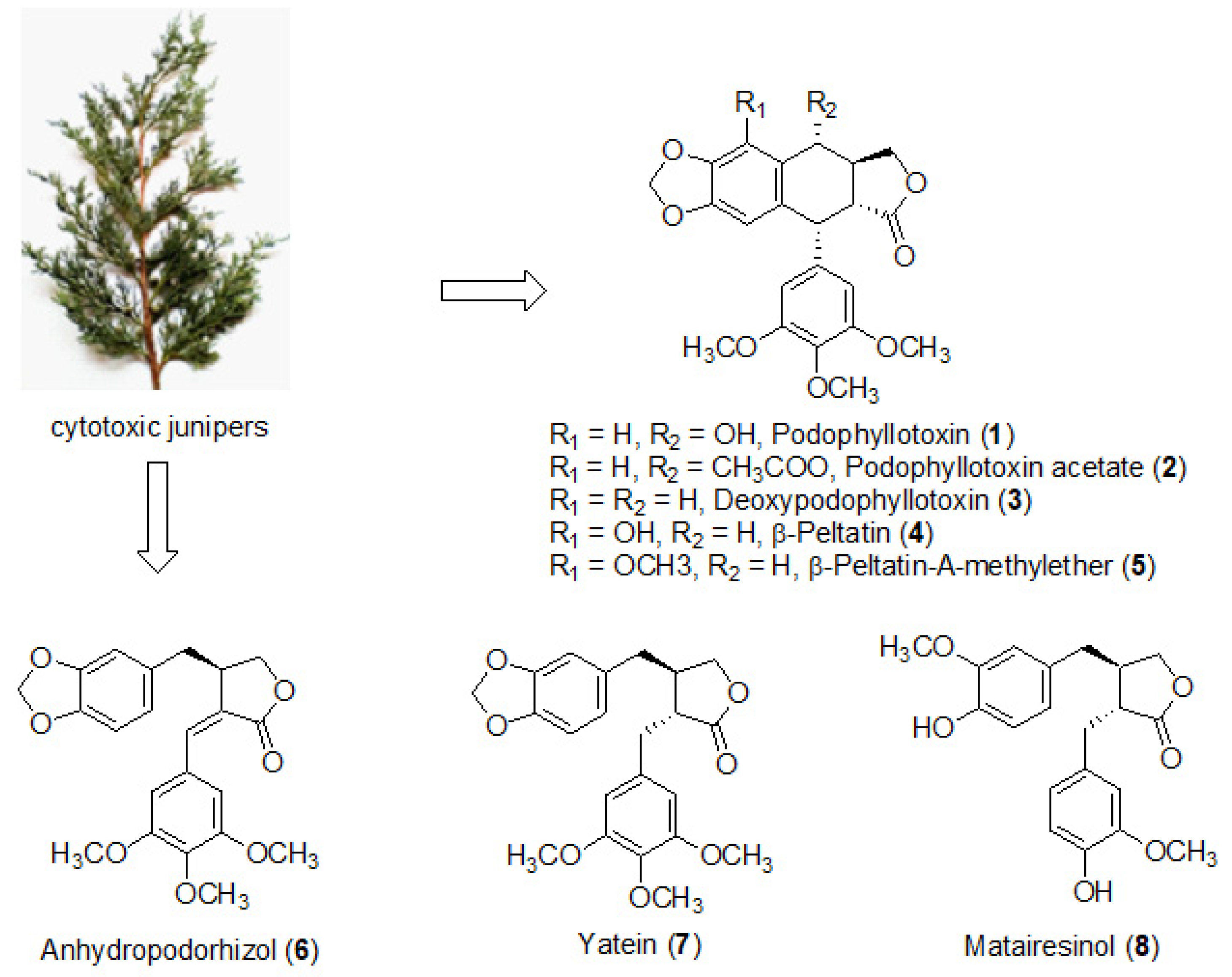
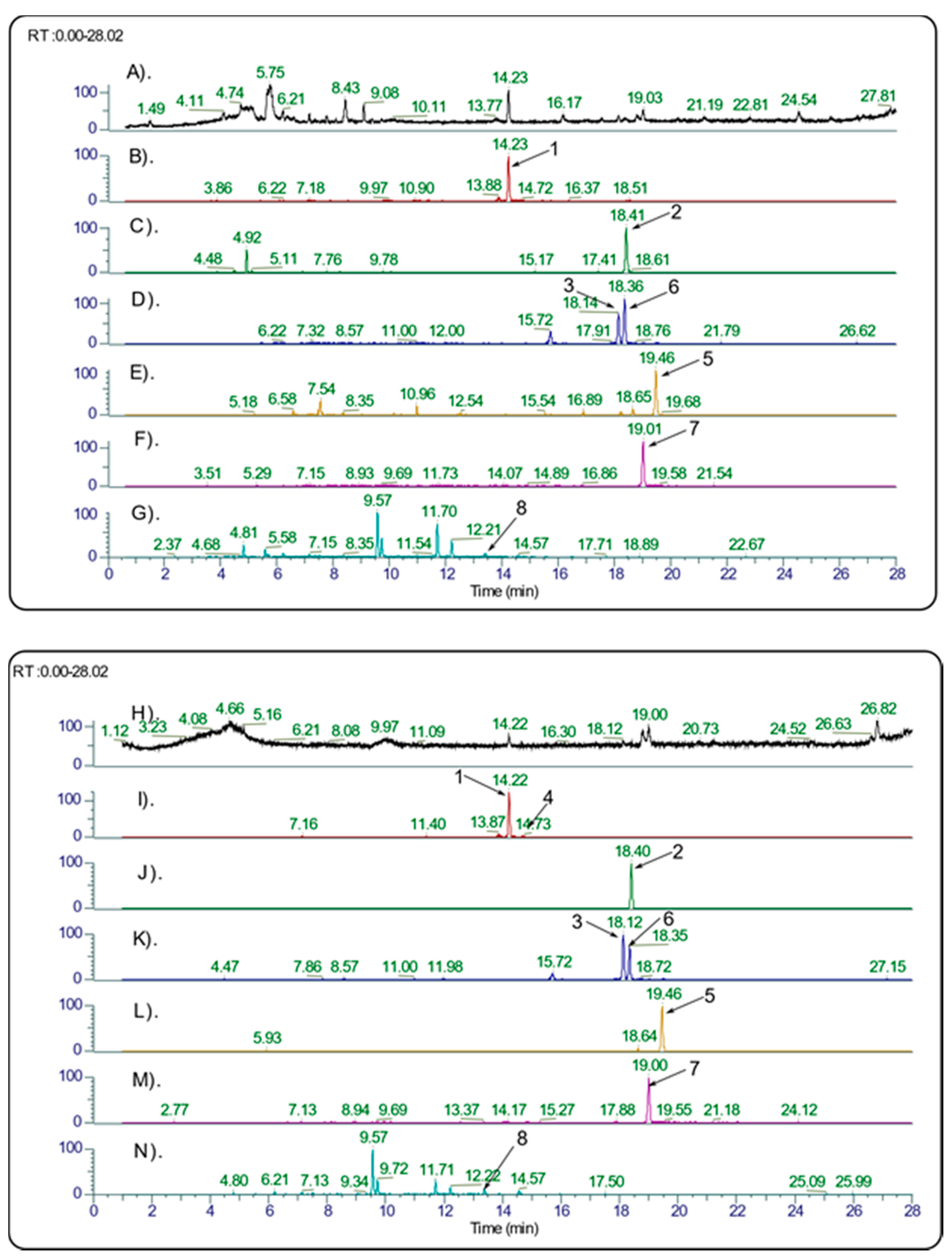
2.2. Identification of PPT Derivatives and Other Lignans in the Cytotoxic Juniper Extracts

The identification of various lignans in the juniper extracts was carried out by UHPLC coupled to HRMS (Figure 2). Generally, the leaf extracts contained more PPT than the galbuli extracts (Table 1). Thus, the leaf extracts were subjected to further detailed investigation. High-resolution MS/MS fragmentation analysis of the juniper leaf extracts identified a great diversity of lignans, for which structures were assigned in correspondence with the literature data (Figure 1, Table 4).

2.3. Identification of Juniperus Species with Efficient Antiproliferative Activity of Their Extracts

In this research, NB-4 acute promyelocytic leukemia (APL) cells were used as a model cancer cell line to study the antiproliferative activity of various juniper extracts. This cell line is characterized by a balanced reciprocal chromosomal translocation t(15;17), involving fusion of the retinoic acid receptor alfa (RARα) gene to the promyelocytic leukemia (PML) gene, leading to formation of an oncogene PML-RARα with abnormal properties [53]. This oncogene prevents the normal maturation of the promyelocytes to granulocytes, inducing accumulation of immature white blood cells in APL patients. All-trans-retinoic acid (ATRA) and anthracycline antibiotics induce remission in up to 80–90% of the PML-RARα positive patients. However, about one-quarter of the patients become resistant or suffer from life-threatening retinoic acid syndrome (characterized by fever, dyspnea, vomiting, etc.). Thus, new cytotoxic agents are required for the therapy of APL and other types of cancer. In continuation of previous studies [40,42], where juniper representatives were found to possess superior antiproliferative activity, the genus Juniperus attracted our attention for the identification of species with efficient biosynthesis of cytotoxic PPT and other lignans with perspectives to be used as anticancer drug precursors. Plenty of junipers, for which original sources were accessed from different continents of the world, were studied in this research for their antiproliferative activity.
The antiproliferative activity of the studied plant extracts was evaluated by their half-maximum growth-inhibitory concentrations (IC50), calculated from the corresponding dose–response curves of MTT-assay of treated NB-4 cells. Lower IC50 values indicated higher activity of the extracts.
Representatives of several juniper species with high antiproliferative activity of their leaf extracts in NB-4 cells were also assayed on a panel of other cancer cell lines (Table 5). These extracts demonstrated highly efficient activity in K-562 human chronic myeloid leukemia (CML) and BV-173 human B cell precursor leukemia (Philadelphia chromosome-positive cell lines, bearing t(9;22) BCR-ABL1 fusion gene); T-24 human urinary bladder carcinoma and HT-29 human colon adenocarcinoma cell lines.

3. Discussion

3.1. Quantification of PPT and Identification of Other Lignans in the Juniper Leaf Extracts

The concentration of podophyllotoxin in the studied extracts was determined by comparison with a standard compound (Table 1). In addition, the UHPLC, coupled to high-resolution MS/MS fragmentation analysis, referred to the literature data, revealed that the cytotoxic juniper species produce a diversity of other lignans in addition to PPT (Table 4) [54,55,56]. Matairesinol and yatein were supposed to be biosynthetic precursors of PPT [16], β-peltatin, and deoxypodophyllotoxin [57]. Anhydropodorhizol was also identified as a secondary metabolite. Thus, plenty of other lignans in addition to previously detected PPT, were found in the studied juniper leaf extracts of J. virginiana, J. sabina, J. scopulorum, J. horizontalis, J. × media, and J. chinensis representatives (Table 4), of which the combined activity contributed to high antiproliferative properties of the corresponding extracts.
The extracts of J. pinchotii, J. ashei, J. deltoides, and J. excelsa representatives with the lowest cytotoxic activity in NB-4 cells (Table 1) contained negligible amounts of PPT and other lignans, or these compounds were not found.

3.2. Selection of Juniper Species with High Antiproliferative Activity, Based on the PPT Content of Their Extracts

The antiproliferative properties of the studied junipers are a result of the combined action of a diversity of lignans in addition to PPT in the cytotoxic leaf extracts. The highest PPT content and antiproliferative activity, respectively, were obtained for the leaf extracts of J. virginiana L. (incl. its cultivars ‘Glauca’, ‘Cinerascens’, ‘Grey Owl’), J. sabina L., (incl. J. sabina var. balkanensis), J. scopulorum ‘Moon light’, J. chinensis ‘Pfitzer Mattews Blue’, J. chinensis ‘Plumosa Aurea’, J. horizontalis Moench, and various J. × media hybrids. The PPT concentrations of these leaf extracts varied in the range of 0.2–1.3% PPT and their IC50 values were found in the range of 0.2–0.7 μg/mL (Table 1).
The biosynthesis of cytotoxic metabolites is determined by the plant genome, as only distinct species of the genus Juniperus possess cytotoxic properties. For example, J. × media hybrids, studied here, must have inherited efficient biosynthesis of cytotoxic metabolites from their parent species J. sabina and J. chinensis [58]. ‘Grey Owl’ juniper is considered to be a hybrid of J. virginiana and J. × pfitzeriana (synonym J. × media). Thus, the genome of three species was supposed to be involved in the activity of the ‘Grey Owl’ cultivar, because J. × pfitzeriana is a hybrid of J. chinensis and J. sabina.
A recently identified juniper representative, namely Juniperus sabina var. balkanensis R. P. Adams and A. N. Tashev, also demonstrated superior antiproliferative properties in the group of studied junipers. J. sabina var. balkanensis grows naturally in the Balkan Peninsula (Albania, Bosnia-Herzegovina, Bulgaria, Croatia, Macedonia, Greece, Turkey, etc.) and Italian regions [59,60]. This juniper variety has inherited the cytotoxic properties of its predecessors by hybridization of Juniperus sabina and Juniperus thurifera, when the natural habitats of these species overlapped in ancient times [61].
Considering the potential use of junipers as industrial crops for the delivery of drug precursors, the shrub-like representatives were considered as more perspective plants for cultivation and industrial applications than the magnificent trees. J. virginiana and its cultivars ‘Glauca’, ‘Cinerascens’, as well as J. scopulorum ‘Moon light’ grow as magnificent trees. However, junipers that grow as shrubs are easier for cultivation. Hence, shrub-like representatives J. sabina, J. horizontalis, J. virginiana ‘Grey Owl’, various J. × media hybrids, J. chinensis ‘Plumosa Aurea’, and J. chinensis ‘Pfitzer Mattews Blue’, which produce PPT in concentrations, sufficient for high antiproliferative activities of their leaf extracts, were considered as preferable sources of PPT for industrial cultivation.
Other junipers, which leaf extracts also exhibited efficient antiproliferative properties, such as J. communis L., J. chinensis L., J. sibirica Burgsd, J. pigmaea K. Koch, J. formosana Hayata, etc., were found to contain predominantly deoxypodophyllotoxin and other lignans instead of PPT (Table 4), as was detected by UHPLC/HRMS and MS/MS-fragmentation analysis. The IC50 values of these leaf extracts showed also excellent antiproliferative activity on NB-4 cells and varied in the range of 0.5–5 μg/mL (Table 1). Deoxypodophyllotoxin (deoxy-PPT) is a strong cytotoxic agent; however, the absence of a hydroxyl group in its molecule restricts the possibilities for the reduction of its toxicity, because the formation of glycoside derivatives is prevented. This structural specificity of deoxy-PPT limits its perspectives for pharmaceutical applications.
The less efficient activity was determined for the leaf extracts of J. pinchotii Sudw., J. deltoides R. P. Adams, J. ashei J. Buchholz, J. excelsa M. Bieb. Their IC50 values after treatment of NB-4 cells were determined in the range of 17–137 μg/mL, consistent with the observation that all analyzed lignans persisted in these extracts in negligible amounts or were not detected.
The broad spectrum of the antiproliferative activity of juniper species with a high cytotoxic activity in NB-4 cells was confirmed also by their excellent IC50 values after treatment of other cancer cell lines: K-562 human chronic myeloid leukemia and BV-173 human B cell precursor leukemia (Philadelphia chromosome positive cell lines); T-24 human urinary bladder carcinoma and HT-29 human colon adenocarcinoma (Table 5). This result revealed excellent perspectives of the genus Juniperus as a potential source of anticancer agents for the treatment of various forms of cancer.
To our knowledge, the present research is the first comparative analysis of the PPT content in the extracts of plenty of species of the genus Juniperus, with original sources being accessed from various continents around the world. For the first time, matairesinol, yatein, and anhydropodorhizol were identified in many of the studied juniper extracts in addition to previously detected in some junipers PPT, deoxy-PPT, and β-peltatin derivatives. Their combined activity contributed to the efficient antiproliferative properties of the corresponding cytotoxic leaf extracts.
The juniper representatives with efficient biosynthesis of antiproliferative agents, identified in this research, revealed the genus Juniperus around the world as a perspective natural source of drug precursors for the pharmaceutical industry.

4. Materials and Methods

4.1. Chemicals and Reagents

Podophyllotoxin (standard compound, ≥98%), MTT [3-(4,5-dimethylthiazol-2-yl)-2,5-diphenyltetrazolium bromide], LC grade acetonitrile, and RPMI 1640 medium were purchased from Sigma-Aldrich Fine Chemicals (Saint Louis, MO, USA), Fetal calf serum (FCS) for cell culture was purchased from Biochrom GmbH (Berlin, Germany), and DMSO was purchased from Fluka Chemie AG (Buchs, Switzerland). LC grade water was prepared using a Millipore Direct-Q3 purification system (Bedford, MA, USA).

4.2. Plant Material

Junipers from the Arnold Arboretum (AA), Harvard University, Boston, USA, were collected in June 2017 (species № 7–10, 24–26, 32, 35) and in October 2018 (species № 1–6, 12–21, 31). The accession summaries and the original sources of the specimen were reported (Table 1). Junipers from the Balkan Peninsula region were collected as follows: J. communis L. was from the village Ognyanovo, Blagoevgrad Province, Rhodope Mountains (41°37′47.3″ N; 23°47′14.5″ E, 700 m a.s.l., April 2017); J. sibirica Burgsd. was from the Vitosha mountain, on the outskirts of Sofia (42°34′59.6″ N; 23°17′28.6″ E, 1803 m a.s.l., April 2017); J. pigmaea C. Koch was from the Smolyan Province, Mursalitsa region of the Rhodope Mountains (41°38′40.8″ N; 24°29′58.5″ E, 1898 m a.s.l., May 2017); J. deltoides R. P. Adams was from the village Ognyanovo, Blagoevgrad Province, Rhodope Mountains (41°37′46.6″ N; 23°47′15.4″ E, 695 m a.s.l., April 2017); J. excelsa M. Bieb. was from the reserve Tisata, on the riverside of Struma (41°44′01.6″ N; 23°09′22.5″ E, 199 m a.s.l., April 2017). Juniperus sabina var. balkanensis R. P. Adams and A. N. Tashev were collected from the eastern Rhodopes (peak Veikata), Bulgaria (November 2017). J. virginiana L. (sample № 4) was collected from the Arboretum of the University of Forestry, Sofia, Bulgaria (April 2017). Their voucher specimens were deposited in the Herbarium (SOM) of the Institute of Biodiversity and Ecosystem Research, Bulgarian Academy of Sciences (IBER-BAS). Plant species from the Balkan Peninsula region were authenticated by A. N. Tashev (University of Forestry, Sofia, Bulgaria) and according to R. P. Adams (Adams, 2014).

4.3. Extraction Procedure

The plant material was dried for a week at room temperature and then kept in the freezer (−20 °C) in vacuum bags until extraction. For the preparation of the extracts, the plant material (5 g) was ground and mixed with methanol (50 mL, 80% v/v) in an Erlenmeyer flask with a stopper. The suspension was stirred for 1.5 h in a shaker water bath at 20 °C. The mixture was filtered and the extract was collected. The remaining solid material was subjected to a second extraction for 1.5 h with a new portion of 80% methanol (50 mL). After filtration, the solid mass was stirred again for 1.5 h in 80% methanol (25 mL). The combined extracts were concentrated by a vacuum evaporator. During the vacuum evaporation, a chlorophyll-containing dark green oil appeared in traces amounts (2–4% yield), and was removed by decantation from the main extract. All extracts were freeze-dried (24 h, −50 °C, 0.1 mbar) and kept in the freezer (−20 °C) until analyses.

4.4. Cell Culture and MTT-Assays

Cancer cell lines NB-4 acute promyelocytic leukemia, K-562 human chronic myeloid leukemia, BV-173 human B cell precursor leukemia, T-24 human urinary bladder carcinoma, and HT-29 human colon adenocarcinoma were purchased from DSMZ, Germany. The cells were cultured in a humidified incubator (37 °C, 5% CO2) in RPMI-1640 with 10% FCS, L-glutamine (2 mM), and HEPES buffer (25 mM). Stock solutions of extracts (20 mg/mL in DMSO) were diluted with the cell culture medium to obtain the desired concentrations. The solvent in the medium was less than 0.5% (v/v). Cells (3 × 10⁵ cells/mL) were seeded into 96-well plates (100 µL/well) and after 24 h of incubation, the cells were exposed for 72 h to various extract concentrations. MTT assays were performed as described previously [62,63].

4.5. Data Processing and Statistics

MTT-assays were carried out in at least four experiments. MTT data were fitted to dose–response curves and IC50 values were calculated using non-linear regression analysis by GraphPad Prism (version 6.01 for Windows, GraphPad Software, San Diego, CA USA, www.graphpad.com, accessed on 1 July 2021). Statistical analysis exploited Student’s t-test with p ≤ 0.05 set as the lowest level of statistical significance.

4.6. LC-ESI-MS/MS Analyses

Reversed-phase high-performance liquid chromatography coupled to electrospray ionization mass spectrometry (LC-ESI-MS/MS) was performed on an Agilent 1200 Series HPLC system (Agilent Technologies, Wood Dale, IL, USA) equipped with a binary gradient solvent pump, a degasser, an autosampler, and a column oven connected to 3200 QTRAP Mass spectrometer (AB Sciex, Foster City, CA, USA). Chromatographic separations were carried out on an Eclipse XDB-C18 column (4.6 × 50 mm, 1.8-µm particle size; Agilent Technologies, USA) maintained at 2 5 °C. The gradient program consisted of mobile phase A (0.1% formic acid in water) and mobile phase B (acetonitrile containing 0.1% formic acid) at 0–0.5 min hold at 35% B, a linear ramp to 53% B at 1 min, a hold at 53% B until 4 min, a linear ramp to 90% B at 5 min, and a hold at 90% B until 6 min. The total run time was 9 min, including a 2 min equilibration. The injection volume was 5 μL with a mobile phase flow rate of 450 μL/min.
The QTRAP-MS system was equipped with an electrospray ionization source (ESI) operated in the positive-ion mode, with a capillary temperature of 500 °C and source voltage of 4500 V. Nitrogen was used as a curtain and collision gas. The optimum conditions of the Multiple Reaction Mode (MRM) were determined in the infusion mode. The data were acquired and processed using Analyst 1.5 software (AB Sciex, USA). Triplicate injections were made for each standard solution and sample. Before injection, the samples were subjected to solid-phase purification with Sep-Pak C18 Cartridges (Waters, Dublin, Ireland).

4.7. UHPLC-HRMS Analyses

Ultra-high-performance liquid chromatography (UHPLC) coupled to high-resolution mass spectrometry (HRMS) was performed on a Thermo Scientific Dionex Ultimate 3000 RSLC system, connected to a Thermo Scientific Q Exactive Plus mass spectrometer (Bremen, Germany), equipped with Thermo Scientific Xcalibur 3.0 software. AkzoNobel Kromasil Externity XT-1.8-C18 (Bohus, Sweden) narrow-bore column (2.1 × 100 mm, 1.8 µm) with Phenomenex Security Guard ULTRA UHPLC EVO C18 (Torrance, CA, USA) was used and maintained at 40 °C. The mobile phase consisted of systems A (0.1% formic acid in water) and B (0.1% formic acid in acetonitrile). The following gradient was used: the mobile phase was held at 5% B for 0.5 min, gradually turned to 60% B for 22.5 min, kept at 60% B for 2 min, followed by a gradual increase to 85% B for 2.5 min, kept at 85% B for 2 min, and the system was turned to the initial condition of 5% B for 0.5 min. The system was conditioned at 5% B for 4.5 min before injection. The flow rate and injection volumes were 300 µL/min and 2 µL, respectively. Prior to injection, samples were subjected to solid-phase purification by Sep-Pak C18 Cartridges (Waters, Ireland), prepared for sample loading using 80% (v/v) methanol.

5. Conclusions

Junipers are evergreen plants, producing plenty of metabolites all the year. The present study is a comparative investigation of the podophyllotoxin concentration, other lignans identification and antiproliferative activity of plant extracts, obtained from a great diversity of junipers, with original sources being accessed from different continents around the world. The chromatography analysis coupled to mass spectrometry showed that many of the studied junipers produce cytotoxic podophyllotoxin at concentrations that are sufficient for the high antiproliferative activities of the corresponding leaf extracts (Table 1).
In general, the juniper leaf extracts contained more PPT than the corresponding galbuli extracts.
The highest concentrations of PPT and best cytotoxic activity of the corresponding leaf extracts, respectively, were determined for J. virginiana (including its cultivars ‘Glauca’, ‘Cinerascens’, ‘Grey Owl’), J. scopulorum ‘Moon light’, J. chinensis ‘Pfitzer Mattews Blue’, J. chinensis ‘Plumosa Aurea’, J. horizontalis, various J. × media hybrids, and J. sabina, including J. sabina var. balkanensis. These extracts contained 0.2–1.3% PPT and their efficient IC50 values were found in the range of 0.2–0.7 μg/mL.
Junipers that grow as shrubs are considered to be species with more potential for cultivation and industrial exploitation than representatives that grow as large trees. In this respect, shrub-like representatives J. sabina, J. horizontalis, various J. × media hybrids, J. virginiana ‘Grey Owl’, etc. were outlined in this research as preferable sources of PPT for industrial cultivation.
The broad spectrum of activity of highly efficient cytotoxic juniper extracts, detected initially on NB-4 APL cells, was also demonstrated on a panel of other cancer cell lines, including K-562 human chronic myeloid leukemia and BV-173 human B cell precursor leukemia (Philadelphia chromosome positive cell lines), T-24 human urinary bladder carcinoma, and HT-29 human colon adenocarcinoma.
In addition to previously detected PPT and β-peltatin derivatives in some junipers, other lignans, such as matairesinol, yatein, and anhydropodorhizol, were also found for the first time in a number of juniper species (Table 4). The combined activity of the identified lignans set the pattern for the efficient antiproliferative properties of the corresponding juniper leaf extracts.
In conclusion, a number of juniper species of different origins around the world were revealed to be natural factories for the efficient biosynthesis of cytotoxic podophyllotoxin and other lignans that represent the genus Juniperus as a source of drug precursors, with potential pharmacological perspectives in the treatment of cancer and other diseases.

Author Contributions

Conceptualization, D.I.I., P.T.N., A.N.T., M.O., R.N. and G.A.; methodology, D.I.I., P.T.N., M.O., R.N., Z.K.K.-N. and G.A.; software, P.T.N., M.O. and Y.E.I., validation, P.T.N., M.O. and Y.E.I.; formal analysis, P.T.N., M.O. and Y.E.I.; investigation, D.I.I., P.T.N., A.N.T., M.O. and R.N.; resources, A.N.T., P.T.N. and R.N.; data curation, P.T.N., Z.K.K.-N., Y.E.I., T.N.A. and D.I.I.; writing—original draft preparation, review and editing, D.I.I., P.T.N., A.N.T., M.O., R.N., Y.E.I., G.A. and H.M.N.; visualization, P.T.N. and Y.E.I.; supervision, A.N.T., P.T.N., R.N. and G.A.; project administration, D.I.I.; funding acquisition, A.N.T., P.T.N., R.N. and G.A. All authors have read and agreed to the published version of the manuscript.

Funding

The support of this research within the following contracts is greatly acknowledged: contract № 37-2016 of the Arnold Arboretum, Harvard University, Boston, MA, USA; contract № DN 07/25 (2016) of the Bulgarian National Science Fund; an Erasmus plus agreement between the Bulgarian Academy of Sciences and Medical University of Lublin; and project “Development of eco-technologies and identification of natural bioactive substances” of the Institute of Chemical Engineering, Bulgarian Academy of Sciences.

Institutional Review Board Statement

Not applicable.

Informed Consent Statement

Not applicable.

Acknowledgments

The authors are very grateful to Michael Dosmann and Kathryn Richardson for the collection and supply of the plant material according to the contract № 37-2016 with Arnold Arboretum, Harvard University, Boston, MA, USA. We thank Galina I. Kalotova (IChE-BAS) for technical assistance at an early stage of this work.

Conflicts of Interest

The authors declare no conflict of interest.

Sample Availability

Samples of the compounds are not available from the authors.

References

  1. Adams, R.P. Junipers of the World: The Genus Juniperus, 4th ed.; Trafford Publishing Co.: Bloomington, IN, USA, 2014; Available online: https://www.amazon.com/Junipers-World-Genus-Juniperus-4th/dp/1490723250 (accessed on 1 July 2021)ISBN 978-1-4907-2325-9.
  2. Krüssmann, G. Manual of Cultivated Conifers; Timber Press: Portland, OR, USA, 1991; Available online: https://www.amazon.com/Manual-Cultivated-Conifers-Gerd-Krüssmann/dp/1604691115 (accessed on 1 July 2021)ISBN 978-1604691115.
  3. Bhardwaj, K.; Silva, A.; Atanassova, M.; Sharma, R.; Nepovimova, E.; Musilek, K.; Sharma, R.; Alghuthaymi, M.; Dhanjal, D.; Nicoletti, M.; et al. Conifers Phytochemicals: A Valuable Forest with Therapeutic Potential. Molecules 2021, 26, 3005. [Google Scholar] [CrossRef]
  4. Dawson, W.R.; Ebbell, B. The Papyrus Ebers; The Greatest Egyptian Medical Document. J. Egypt. Archaeol. 1938, 24, 250. [Google Scholar] [CrossRef]
  5. Hartwell, J.L. Plants used against cancer. A survey. Lloydia 1971, 34, 386–425. Available online: https://www.ncbi.nlm.nih.gov/pubmed/5173435 (accessed on 1 July 2021). [PubMed]
  6. Lewis, W.H.; Elvin-Lewis, M.P.F. Medical Botany: Plants Affecting Human Health; Wiley: Hoboken, NJ, USA, 2003. [Google Scholar]
  7. Graham, J.; Quinn, M.; Fabricant, D.; Farnsworth, N. Plants used against cancer—An extension of the work of Jonathan Hartwell. J. Ethnopharmacol. 2000, 73, 347–377. [Google Scholar] [CrossRef]
  8. Lechner-Knecht, S. Sacred healing plants in Nepal. Dtsch. Apoth. Ztg. 1982, 122, 2122–2129. [Google Scholar]
  9. Gordien, A.Y.; Gray, A.I.; Franzblau, S.G.; Seidel, V. Antimycobacterial terpenoids from Juniperus communis L. (Cuppressaceae). J. Ethnopharmacol. 2009, 126, 500–505. [Google Scholar] [CrossRef] [PubMed]
  10. Carpenter, C.D.; O’Neill, T.; Picot, N.; Johnson, J.A.; Robichaud, G.A.; Webster, D.; Gray, C.A. Anti-mycobacterial natural products from the Canadian medicinal plant Juniperus communis. J. Ethnopharmacol. 2012, 143, 695–700. [Google Scholar] [CrossRef]
  11. Orchard, A.; Van Vuuren, S. Commercial Essential Oils as Potential Antimicrobials to Treat Skin Diseases. Evidence-Based Complement. Altern. Med. 2017, 2017, 4517971. [Google Scholar] [CrossRef] [Green Version]
  12. Gardner, D.R.; Panter, K.E.; James, L.F.; Stegelmeier, B.L. Abortifacient effects of lodgepole pine (Pinus contorta) and common juniper (Juniperus communis) on cattle. Vet. Hum. Toxicol. 1998, 40, 260–263. [Google Scholar]
  13. Johnson, W. Final Report on the Safety Assessment of Juniperus communis Extract, Juniperus oxycedrus Extract, Juniperus oxycedrus Tar, Juniperus phoenicea Extract, and Juniperus virginiana Extract. Int. J. Toxicol. 2001, 20, 41–56. [Google Scholar] [CrossRef]
  14. Borsche, W.; Niemann, J. Über Podophyllin. Eur. J. Org. Chem. 1932, 494, 126–142. [Google Scholar] [CrossRef]
  15. Lipke, M.M. An Armamentarium of Wart Treatments. Clin. Med. Res. 2006, 4, 273–293. [Google Scholar] [CrossRef] [PubMed] [Green Version]
  16. Canel, C.; Moraes, R.M.; Dayan, F.; Ferreira, D. Podophyllotoxin. Phytochemistry 2000, 54, 115–120. [Google Scholar] [CrossRef]
  17. Guerram, M.; Jiang, Z.-Z.; Zhang, L.-Y. Podophyllotoxin, a medicinal agent of plant origin: Past, present and future. Chin. J. Nat. Med. 2012, 10, 161–169. [Google Scholar] [CrossRef]
  18. Stähelin, H.F.; von Wartburg, A. The chemical and biological route from podophyllotoxin glucoside to etoposide: Memorial award lecture. Cancer Res. 1991, 51, 5–15. Available online: https://www.ncbi.nlm.nih.gov/pubmed/1988106 (accessed on 1 July 2021).
  19. Shah, Z.; Gohar, U.; Jamshed, I.; Mushtaq, A.; Mukhtar, H.; Zia-Ui-Haq, M.; Toma, S.; Manea, R.; Moga, M.; Popovici, B. Podophyllotoxin: History, Recent Advances and Future Prospects. Biomolecules 2021, 11, 603. [Google Scholar] [CrossRef]
  20. Leander, K.; Rosen, B. Medicinal Uses for Podophyllotoxin. U.S. Patent 4,788,216, 29 November 1988. Available online: https://patents.google.com/patent/US4788216A/en (accessed on 1 July 2021).
  21. Fay, D.A.; Ziegler, H.W. Botanical Source Differentiation of Podophyllum Resin by HPLC. J. Liq. Chromatogr. 1985, 8, 1501–1505. [Google Scholar] [CrossRef]
  22. Pandey, H.; Nandi, S.K.; Kumar, A.; Palni, U.T.; Palni, L.M.S. Podophyllotoxin content in Podophyllum hexandrum Royle plants of known age of seed origin and grown at a lower altitude. Acta Physiol. Plant. 2007, 29, 121–126. [Google Scholar] [CrossRef]
  23. Kumar, P.; Pal, T.; Sharma, N.; Kumar, V.; Sood, H.; Chauhan, R.S. Expression analysis of biosynthetic pathway genes vis-à-vis podophyllotoxin content in Podophyllum hexandrum Royle. Protoplasma 2015, 252, 1253–1262. [Google Scholar] [CrossRef]
  24. Cushman, K.E.; Maqbool, M.; Lata, H.; Bedir, E.; Khan, I.A.; Moraes, R.M. Podophyllotoxin Content and Yield of American Mayapple Leaves in Sun and Shade. HortScience 2005, 40, 60–63. [Google Scholar] [CrossRef] [Green Version]
  25. Zheljazkov, V.D.; Jones, A.M.; Avula, B.; Maddox, V.; Rowe, D.E. Lignan and Nutrient Concentrations in American Mayapple (Podophyllum peltatum L.) in the Eastern United States. HortScience 2009, 44, 349–353. [Google Scholar] [CrossRef]
  26. Sultan, P.; Shawl, A.; Ramteke, P.; Kour, A.; Qazi, P. Assessment of diversity in Podophyllum hexandrum by genetic and phytochemical markers. Sci. Hortic. 2008, 115, 398–408. [Google Scholar] [CrossRef]
  27. Ardalani, H.; Avan, A.; Ghayour-Mobarhan, M. Podophyllotoxin: A novel potential natural anticancer agent. Avicenna J. Phytomed. 2017, 7, 285–294. [Google Scholar]
  28. Seca, A.M.L.; Silva, A.M.S. Recent Progress in Medicinal Plants, Vol. 16. In The Chemical Composition of the Juniperus Genus (1970–2004); Govil, J.N., Singh, V.K., Eds.; Studium Press LLC: Houston, TX, USA, 2006; pp. 401–522. Available online: https://www.researchgate.net/publication/283432291_The_chemical_composition_of_the_Juniperus_Genus_1970-2004 (accessed on 1 July 2021).
  29. Olech, M.; Nowak, R.; Ivanova, D.; Tashev, A.; Boyadzhieva, S.; Kalotova, G.; Angelov, G.; Gawlik-Dziki, U. LC-ESI-MS/MS-MRM Profiling of Polyphenols and Antioxidant Activity Evaluation of Junipers of Different Origin. Appl. Sci. 2020, 10, 8921. [Google Scholar] [CrossRef]
  30. Renouard, S.; Lopez, T.; Hendrawati, O.; Dupré, P.; Doussot, J.; Falguieres, A.; Ferroud, C.; Hagège, D.; Lamblin, F.; Lainé, E.; et al. Podophyllotoxin and Deoxypodophyllotoxin in Juniperus bermudiana and 12 Other Juniperus Species: Optimization of Extraction, Method Validation, and Quantification. J. Agric. Food Chem. 2011, 59, 8101–8107. [Google Scholar] [CrossRef]
  31. Bedir, E.; Khan, I.; Moraes, R.M. Bioprospecting for podophyllotoxin. In Trends in New Crops and New Uses; Janick, J., Whipkey, A., Eds.; ASHS Press: Alexandria, VA, USA, 2002; pp. 545–549. Available online: https://www.hort.purdue.edu/newcrop/ncnu02/pdf/moraes-545.pdf (accessed on 1 July 2021)ISBN 0-970756-5-5.
  32. Cushman, K.E.; Maqbool, M.; Gerard, P.D.; Bedir, E.; Lata, H.; Moraes, R.M. Variation of Podophyllotoxin in Leaves of Eastern Red Cedar (Juniperus virginiana). Planta Med. 2003, 69, 477–478. [Google Scholar] [CrossRef] [PubMed]
  33. Gawde, A.; Cantrell, C.L.; Zheljazkov, V.D. Dual extraction of essential oil and podophyllotoxin from Juniperus virginiana. Ind. Crop. Prod. 2009, 30, 276–280. [Google Scholar] [CrossRef]
  34. Gawde, A.; Zheljazkov, V.D.; Maddox, V.; Cantrell, C.L. Bioprospection of Eastern red cedar from nine physiographic regions in Mississippi. Ind. Crop. Prod. 2009, 30, 59–64. [Google Scholar] [CrossRef]
  35. Kusari, S.; Zühlke, S.; Spiteller, M. Chemometric Evaluation of the Anti-cancer Pro-drug Podophyllotoxin and Potential Therapeutic Analogues in Juniperus and Podophyllum Species. Phytochem. Anal. 2010, 22, 128–143. [Google Scholar] [CrossRef] [PubMed]
  36. Zheljazkov, V.D.; Cantrell, C.L.; Donega, M.A.; Astatkie, T.; Heidel, B. Podophyllotoxin Concentration in Junipers in the Big Horn Mountains in Wyoming. HortScience 2012, 47, 1696–1697. [Google Scholar] [CrossRef]
  37. Benzina, S.; Harquail, J.; Jean, S.; Beauregard, A.-P.; Colquhoun, C.D.; Carroll, M.; Bos, A.; Gray, C.A.; Robichaud, G.A. Deoxypodophyllotoxin Isolated from Juniperus communis Induces Apoptosis in Breast Cancer Cells. Anti-Cancer Agents Med. Chem. 2014, 15, 79–88. [Google Scholar] [CrossRef]
  38. Feliciano, A.S.; del Corral, J.M.M.; Gordaliza, M.; Castro, A. Lignans from Juniperus sabina. Phytochemistry 1990, 29, 1335–1338. [Google Scholar] [CrossRef]
  39. Hartwell, J.L.; Johnson, J.M.; Fitzgerald, D.B.; Belkin, M. Podophyllotoxin from Juniperus Species; Savinin. J. Am. Chem. Soc. 1953, 75, 235–236. [Google Scholar] [CrossRef]
  40. Ivanova, D.I.; Tashev, A.N.; Nedialkov, P.T.; Ilieva, Y.E.; Atanassova, T.N.; Olech, M.; Nowak, R.; Angelov, G.; Tsvetanova, F.V.; Iliev, I.A.; et al. Antioxidant and antiproliferative activity of Juniperus L. species of Bulgarian and foreign origin and their anticancer metabolite identification. Bulg. Chem. Commun. 2018, 50C, 144–150. Available online: http://www.bcc.bas.bg/BCC_Volumes/Volume_50_Special_C_2018/pdf/BCC-50-C-2018-144-150-Ivanova-28.pdf (accessed on 1 July 2021).
  41. Zheljazkov, V.; Cantrell, C.; Semerdjieva, I.; Radoukova, T.; Stoyanova, A.; Maneva, V.; Kačániová, M.; Astatkie, T.; Borisova, D.; Dincheva, I.; et al. Essential Oil Composition and Bioactivity of Two Juniper Species from Bulgaria and Slovakia. Molecules 2021, 26, 3659. [Google Scholar] [CrossRef] [PubMed]
  42. Och, M.; Och, A.; Ciesla, L.; Kubrak, T.; Pecio, Ł.; Stochmal, A.; Kocki, J.; Bogucka-Kocka, A. Study of cytotoxic activity, podophyllotoxin, and deoxypodophyllotoxin content in selected Juniperus species cultivated in Poland. Pharm. Biol. 2014, 53, 831–837. [Google Scholar] [CrossRef] [PubMed]
  43. Hwang, G.S.; Phuong, N.T.; Park, K.R.; Kim, Y.H.; Kim, K.H.; Kang, J.S. Distribution of (−)-yatein in Cupressaceae family analysed by high performance liquid chromatography. Arch. Pharmacal Res. 2004, 27, 35–39. [Google Scholar] [CrossRef] [PubMed]
  44. Doussot, J.; Mathieu, V.; Colas, C.; Molinié, R.; Corbin, C.; Montguillon, J.; Banuls, L.M.Y.; Renouard, S.; Lamblin, F.; Dupré, P.; et al. Investigation of the Lignan Content in Extracts from Linum, Callitris and Juniperus Species in Relation to Their In Vitro Antiproliferative Activities. Planta Medica 2016, 83, 574–581. [Google Scholar] [CrossRef] [Green Version]
  45. Cairnes, D.A.; Ekundayo, O.; Kingston, D.G.I. Plant Anticancer Agents. X. Lignans From Juniperus phoenicea. J. Nat. Prod. 1980, 43, 495–497. [Google Scholar] [CrossRef]
  46. Rojas-Sepúlveda, A.M.; Mendieta-Serrano, M.; Mojica, M.Y.A.; Salas-Vidal, E.; Marquina, S.; Villarreal, M.L.; Puebla, A.M.; Delgado, J.I.; Álvarez, L. Cytotoxic Podophyllotoxin Type-Lignans from the Steam Bark of Bursera fagaroides var. fagaroides. Molecules 2012, 17, 9506–9519. [Google Scholar] [CrossRef] [Green Version]
  47. Donoso-Fierro, C.; Tiezzi, A.; Ovidi, E.; Ceccarelli, D.; Triggiani, D.; Mastrogiovanni, F.; Taddei, A.R.; Pérez, C.; Becerra, J.; Silva, M.; et al. Antiproliferative activity of yatein isolated from Austrocedrus chilensis against murine myeloma cells: Cytological studies and chemical investigations. Pharm. Biol. 2014, 53, 378–385. [Google Scholar] [CrossRef]
  48. Chen, Y.-L.; Lin, S.-Z.; Chang, J.-Y.; Cheng, Y.-L.; Tsai, N.-M.; Chen, S.-P.; Chang, W.-L.; Harn, H.-J. In vitro and in vivo studies of a novel potential anticancer agent of isochaihulactone on human lung cancer A549 cells. Biochem. Pharmacol. 2006, 72, 308–319. [Google Scholar] [CrossRef] [PubMed]
  49. Su, S.; Cheng, X.; Wink, M. Natural lignans from Arctium lappa modulate P-glycoprotein efflux function in multidrug resistant cancer cells. Phytomedicine 2015, 22, 301–307. [Google Scholar] [CrossRef] [PubMed]
  50. Su, S.; Cheng, X.; Wink, M. Cytotoxicity of arctigenin and matairesinol against the T-cell lymphoma cell line CCRF-CEM. J. Pharm. Pharmacol. 2015, 67, 1316–1323. [Google Scholar] [CrossRef]
  51. Schmidt, T.J.; Hemmati, S.; Fuss, E.; Alfermann, A.W. A combined HPLC-UV and HPLC-MS method for the identification of lignans and its application to the lignans of Linum usitatissimum L. and L. bienne Mill. Phytochem. Anal. 2006, 17, 299–311. [Google Scholar] [CrossRef] [PubMed]
  52. Hendrawati, O.; Woerdenbag, H.; Michiels, P.J.; Aantjes, H.G.; Van Dam, A.; Kayser, O. Identification of lignans and related compounds in Anthriscus sylvestris by LC–ESI-MS/MS and LC-SPE–NMR. Phytochemistry 2011, 72, 2172–2179. [Google Scholar] [CrossRef]
  53. Lanotte, M.; Martin-Thouvenin, V.; Najman, S.; Balerini, P.; Valensi, F.; Berger, R. NB4, a maturation inducible cell line with t(15;17) marker isolated from a human acute promyelocytic leukemia (M3). Blood 1991, 77, 1080–1086. [Google Scholar] [CrossRef] [Green Version]
  54. Wong, S.-K.; Tsui, S.-K.; Kwan, S.-Y.; Su, X.-L.; Lin, R.-C. Identification and characterization of Podophyllum emodi by API-LC/MS/MS. J. Mass Spectrom. 2000, 35, 1246–1251. [Google Scholar] [CrossRef]
  55. Kusari, S.; Lamshöft, M.; Spiteller, M. Aspergillus fumigatus Fresenius, an endophytic fungus from Juniperus communis L. Horstmann as a novel source of the anticancer pro-drug deoxypodophyllotoxin. J. Appl. Microbiol. 2009, 107, 1019–1030. [Google Scholar] [CrossRef]
  56. Koulman, A.; Bos, R.; Medarde, M.; Pras, N.; Quax, W.J. A Fast and Simple GC MS Method for Lignan Profiling in Anthriscus sylvestris and Biosynthetically Related Plant Species. Planta Medica 2001, 67, 858–862. [Google Scholar] [CrossRef]
  57. Khaled, M.; Jiang, Z.-Z.; Zhang, L.-Y. Deoxypodophyllotoxin: A promising therapeutic agent from herbal medicine. J. Ethnopharmacol. 2013, 149, 24–34. [Google Scholar] [CrossRef]
  58. Adams, R.P.; Johnson, S.T.; Anderson, J.; Rushforth, K.; Farhat, P.; Valentin, N.; Siljak-Yakovlev, S. The origin of Juniperus x pfitzeriana, an allotetraploid hybrid of J. chinensis x J. sabina. Phytologia 2019, 101, 164–174. Available online: https://www.juniperus.org/uploads/2/2/6/3/22639912/416._-_phyto101_2_164-174adamsetal_xfitzeriana_w_2_seagreen_6-11-19.pdf (accessed on 1 July 2021).
  59. Adams, R.P.; Boratynski, A.; Mataraci, T.; Tashev, A.N.; Schwarzbach, A.E. Discovery of Juniperus sabina var. balkanensis R. P. Adams and A. N. Tashev in western Turkey (Anatolia). Phytologia 2017, 99, 22–31. Available online: https://www.juniperus.org/uploads/2/2/6/3/22639912/375-_2017_phyto_99_1_22-31adams_boratynski_taskev_dist_v_balkanensis.pdf (accessed on 1 July 2021).
  60. Adams, R.P.; Farhat, P.; Shuka, L.; Siljak-Yakovlev, S. Discovery of Juniperus sabina var. balkanensis R. P. Adams and A. N. Tashev in Albania and relictual polymorphisms found in nrDNA. Phytologia 2018, 100, 187–194. Available online: https://www.juniperus.org/uploads/2/2/6/3/22639912/402._-_2019_phyto100_3_187-194adams_et_al_discovery_balkanensisalkbaniatypo4-27-19.pdf (accessed on 1 July 2021).
  61. Adams, R.P.; Schwarzbach, A.E.; Tashev, A.N. Chloroplast capture by a new variety, Juniperus sabina var. balkanensis R. P. Adams and A. N. Tashev, from the Balkan peninsula: A putative stabilized relictual hybrid between J. sabina and ancestral J. thurifera. Phytologia 2016, 98, 100–111. Available online: https://www.juniperus.org/uploads/2/2/6/3/22639912/361_-_2016_phyto_98_2_100-111adams_tashev_schwarzback_balcanensis_new_sp__rev_2-18-16.pdf (accessed on 1 July 2021).
  62. Mosmann, T. Rapid colorimetric assay for cellular growth and survival: Application to proliferation and cytotoxicity assays. J. Immunol. Methods 1983, 65, 55–63. [Google Scholar] [CrossRef]
  63. Konstantinov, S.M.; Eibl, H.; Berger, M.R. BCR-ABL influences the antileukaemic efficacy of alkylphosphocholines. Br. J. Haematol. 1999, 107, 365–374. [Google Scholar] [CrossRef] [PubMed]
Figure 1. Structures of the lignans 18, identified in the cytotoxic juniper leaf extracts.
Molecules 26 05179 g001
Figure 2. Example UHPLC chromatograms of cytotoxic juniper leaf extracts. The lignan numbers, shown on the chromatograms with arrows, correspond to Figure 1. (A) Total Ion Chromatogram (TIC) of Grey Owl juniper extract (extract № 7, Table 1); (BG) Extracted Ion Chromatograms (XIC) of Grey Owl juniper extract at m/z 415.1387, 457.1496, 399.1438, 429.1544, 401.1595, and 359.1489, respectively, with a 5 ppm isolation window; (H). TIC chromatogram of J. virginiana L. extract (extract №1, Table 1); (IN) XIC chromatograms of J. virginiana L. extract at m/z 415.1387, 457.1496, 399.1438, 429.1544, 401.1595, and 359.1489, respectively, with a 5 ppm isolation window.
Molecules 26 05179 g002
Table 1. Original source accession summary of junipers of different origins and comparison of the podophyllotoxin content, antiproliferative activity in NB-4 cells, and yields of the juniper extracts.
Extract №Specimen NumberJuniper SpeciesF/MOrgan UsedOriginal Source
Accession Summary
%PPT ± SD in the Dry ExtractNB-4 IC50 ± SD
[μg/mL]
Q%
1AA 1746–81/AJ. virginiana L.Fleaveswild, USA, Bald Head Cliff, Maine, Ogunquit, 19810.91 ± 0.010.21 ± 0.0112
2AA 1746–81/AJ. virginiana L.Fgalbuliwild, USA, Bald Head Cliff, Maine, Ogunquit, 19810.23 ± 0.010.54 ± 0.036
3AA 4714/AJ. virginiana ‘Cinerascens’FleavesBerlin, Späth Arb., 19011.03 ± 0.030.37 ± 0.0312
4SOM 174406J. virginiana L.MEleavescultivar, UFA, Sofia, BG0.37 ± 0.010.48 ± 0.1015
5AA 14882/AJ. virginiana ‘Glauca’FleavesRochester Park, New York, 19030.74 ± 0.010.29 ± 0.028
6AA 14882/AJ. virginiana ‘Glauca’FgalbuliRochester Park, New York, 19030.04 ± 0.010.26 ± 0.019
7AA 1136–61/AJ. virginiana ‘Grey Owl’FleavesDominion Arb., Ottawa, Canada, 19610.39 ± 0.010.72 ± 0.1011
8AA 1136–61/AJ. virginiana ‘Grey Owl’FgalbuliDominion Arb., Ottawa, Canada, 19610.09 ± 0.010.49 ± 0.207
9AA 211–57/AJ. scopulorum ‘Moon light’MleavesUSA, D. Hill Nurs., Dundee, Illinois, 19570.85 ± 0.020.20 ± 0.0418
10AA 14868/EJ. sabina L.Fleaveswild, 1904, Uzbekistan0.50 ± 0.010.34 ± 0.0113
11SOM 177009J. sabina var. balkanensis Adams & TashevMleaveswild, from eastern Rhodopes, BG0.20 ± 0.010.29 ± 0.0316
12AA 1164–56/AJ. horizontalis MoenchFleavesD. Hill Nurs., USA, Dundee, Illinois, 19560.38 ± 0.010.23 ± 0.0312
13AA 1164–56/AJ. horizontalis MoenchFgalbuliD. Hill Nurs., USA, Dundee, Illinois, 19560.03 ± 0.012.43 ± 0.227
14AA 74–42/CJ. × media ‘Pfitzeriana Argentea’MleavesMorris Arb, Pennsylvania, USA, 19420.42 ± 0.010.51 ± 0.0711
15AA 183–62/AJ. × media ‘Óld Gold’MleavesGrootendorst Nurs., Holland, 19620.47 ± 0.010.42 ± 0.0410
16AA 639–48/AJ. × media ‘Richeson’MleavesArmstrong Nurs., Ontario, CA, 19480.22 ± 0.010.43 ± 0.0313
17AA 1–51/AJ. chinensis ‘Pfitzer Mattews Blue’MleavesInterstate Nursery, Iowa, USA, 19511.30 ± 0.050.24 ± 0.0113
18AA 219–61/AJ. chinensis ‘Plumosa Aurea’MleavesPennsylvania, USA, 19610.22 ± 0.020.45 ± 0.0210
19AA 265–33/AJ. chinensis L.MleavesCalifornia, USA, 1933BQL0.5 ± 0.314
20AA 14809/AJ. chinensis L.FleavesRoyal Botanic Gardens, UK, Kew, 1908BQL1.0 ± 0.115
21AA 14809/AJ. chinensis L.FgalbuliRoyal Botanic Gardens, UK, Kew, 1908BQL0.8 ± 0.17
22SOM 174400J. communis L.Fleaveswild from Rhodopes, BGBQL1.0 ± 0.415
23SOM 174400J. communis L.Fgalbuliwild from Rhodopes, BGBQL3.6 ± 1.713
24AA 49–66/AJ. communis ‘Laxa’MleavesU. S. Natl. Arb., 1966, WashingtonBQL0.8 ± 0.216
25AA 4176–1/AJ. communis ‘Oblonga
Pendula’
MleavesUSA, Biltmore Estate, North Carolina, 1907BQL4.7 ± 0.617
26AA 280–98/AJ. formosana HayataFleaveswild, from Taiwan, 1998BQL1.7 ± 0.317
27SOM 174401J. sibirica Burgsd.Fleaveswild, from Vitosha, BGND3 ± 115
28SOM 174401J. sibirica Burgsd.Fgalbuliwild, from Vitosha, BGBQL15 ± 39
29SOM 174402J. pigmaea K. KochFleaveswild from Rhodopes, BGBQL5 ± 115
30SOM 174402J. pigmaea K. KochFgalbuliwild from Rhodopes, BGBQL29 ± 711
31AA 20–89/AJ. squamata ‘Meyeri’FleavesHicks Nurs., Westbury, NY, 1989BQL6.3 ± 0.510
32AA 642–88/BJ. pinchotii Sudw.Mleaveswild, USA, Oklahoma, Kiowa reserv., 1988BQL17 ± 219
33SOM 174403J. deltoides R. P. AdamsFleaveswild from Rhodopes, BGBQL66 ± 816
34SOM 174403J. deltoides R. P. AdamsFgalbuliwild from Rhodopes, BGND70 ± 517
35AA 276–86/AJ. ashei J. BuchholzMleaveswild, USA, Oklahoma, Murray, 1986BQL130 ± 1828
36SOM 174404J. excelsa M. Bieb.Fleaveswild, Struma riverside, BGND137 ± 1219
37SOM 174404J. excelsa M. Bieb.Fgalbuliwild, Struma riverside, BGND188 ± 5516
-controlPodophyllotoxin -standard compound-0.005 ± 0.001-
Legend: PPT—podophyllotoxin; NB-4—acute promyelocytic leukemia cell line; AA—specimen of the Arnold Arboretum, Harvard University, Boston, USA, a capital letter qualifier is applied to each individual plant and the year of the original source accession is given; SOM—specimen of the Herbarium of the Institute of Biodiversity and Ecosystem Research, Bulgarian Academy of Sciences; UFA—University of Forestry Arboretum, Sofia, Bulgaria; Arb.—Arboretum; Nurs.—Nursery; F/M—female or male representative; ME—monoecious; NB-4 IC50: half-maximum growth-inhibitory concentrations of the extracts in NB-4 cells, given in micrograms of the dry extract per milliliter of cell culture medium. Lower IC50 values denote higher activity; Q—yield [%] of the extract related to the weight of the starting plant material; BQL—below the quantification limit (peak detected, concentration > LOD, but < LOQ); LOD/LOQ—limit of detection/quantification; ND—not detected.
Table 2. LC-ESI-MS/MS parameters for identification of podophyllotoxin (PPT).
CompoundRT
[min]
Molecular Weight[M + H]+
[m/z]
Fragment Ions
[m/z]
Collision Energy
[eV]
DP
[V]
EP
[V]
CEP
[V]
CXP
[V]
PPT3.6041441539718301023.62
24720301023.62
22920301023.62
Abbreviations: RT—retention time, given in minutes; [M + H]+ protonated molecular ion; m/z—mass-to-charge ratio; declustering potential (DP); entrance potential (EP); collision cell entrance potential (CEP); collision cell exit potential (CXP).
Table 3. Analytical parameters of the LC-MS/MS quantitative method and data for calibration curves for the determination of the podophyllotoxin (PPT) concentration.
CompoundLOD
[ng mL−1]
LOQ
[ng mL−1]
r2EquationLinearity Range
[ng mL−1]
PPT512.50.9992y = 87.3x − 14412.5 to 500
Legend: LOD—limit of detection; LOQ—limit of quantification; r—regression coefficient.
Table 4. HRMS/MS analysis of lignans, identified in cytotoxic juniper leaf extracts.
CompoundRT minMolecular Formula[M + H]+HRMS/MS Fragments **Extract №Ref
Calcd. *FoundΔppm *
Podophyllotoxin14.23C22H22O8415.1387415.1385−0.61397.1277 (100), 313.1066 (10), 247.0598 (85), 229.0493 (20), 185.0596 (5)1, 3–5, 7, 9–12, 14–18 ***[51,52]
Podophyllotoxin acetate18.41C24H24O9457.1496457.14930.57397.1287 (80), 355.1169 (70), 313.1070 (100), 229.0492 (50), 185.0596 (25)7, 9, 10, 14–16, 18[46]
Deoxypodophyllotoxin18.14C22H22O7399.1438399.1437−0.23231.0649 (100), 187.0752 (10)1, 3–5, 7, 9–12, 14–20, 22, 24–27, 29[51,52]
β-Peltatin14.72C22H22O8415.1387415.13900.54247.0599 (100), 203.0701 (5), 189.0545 (2)1, 4, 5, 14–20,[51,52]
β-Peltatin-A-methyl-ether19.47C23H24O8429.1544429.15460.57261.0755 (100), 217.0860 (10)1, 3, 4, 7, 9, 10, 12, 14–19, 22, 24, 25[51,52]
Anhydropodorhizol18.35C22H22O7399.1438399.14400.38381.1327 (30), 363.1227 (10), 231.0649 (90), 203.0701 (40), 187.0752 (20), 181.0857 (10), 135.0441 (15)1, 3–5, 7, 9–12, 14–19[52]
Yatein19.02C22H24O7401.1595401.15980.74383.1484 (100), 365.1381 (5), 223.0963 (5), 181.0858 (30), 161.0596 (10), 135.0441 (5)1, 3, 5, 7, 9–12, 14–20, 26[51,52]
Matairesinol13.40C20H22O6359.1489359.14900.15341.1379 (60), 323.1274 (25), 223.0964 (10), 163.0752 (10), 137.0596 (100)1, 3–5, 7, 9, 10, 20, 24, 25, 29[51]
Legend: RT—retention time in minutes by UHPLC analysis. * The molecular mass of the protonated molecules and Δppm values were determined by Thermo Scientific Xcalibur 4.0 software (Waltham, MA, USA). ** Fragmentations are with normalized collision energy (NCE) 10; the ion abundance in brackets is given in %; *** Traces of PPT were found also in the leaf extracts № 19, 20, 22, 24–26, 29, 31–33, and 35. The extract numbers refer to Table 1.
Table 5. Antiproliferative activity presented as IC50 values [μg/mL] of the corresponding cytotoxic juniper leaf extracts after treatment of a panel of cancer cell lines.
Extract №Juniperus RepresentativesK-562BV-173T-24HT-29
4J. virginiana0.4 ± 0.10.14 ± 0.061.3 ± 0.55 ± 1
7J. virginiana ‘Grey Owl’0.4 ± 0.30.11 ± 0.054.0 ± 3.58 ± 5
9J. scopulorum ‘Moon light’0.3 ± 0.10.22 ± 0.030.8 ± 0.22 ± 1
10J. sabina0.4 ± 0.10.24 ± 0.030.8 ± 0.32 ± 1
11J. sabina var. balkanensis0.2 ± 0.10.16 ± 0.030.7 ± 0.32 ± 1
22J. communis1.6 ± 1.21.6 ± 0.219 ± 1217 ± 7
controlPodophyllotoxin0.006 ± 0.0030.007 ± 0.0020.007 ± 0.0020.007 ± 0.002
Legend: Cancer cell lines: K-562 human chronic myeloid leukemia (CML), BV-173 human B cell precursor leukemia (Philadelphia chromosome-positive cell lines, bearing t(9;22) BCR-ABL1 fusion gene); T-24 human urinary bladder carcinoma; HT-29 human colon adenocarcinoma; IC50—half-maximum growth-inhibitory concentration of the extracts in the corresponding cancer cells, given in micrograms of the dry extract per milliliter of the cell culture medium. Lower IC50 values denote higher activities.
Publisher’s Note: MDPI stays neutral with regard to jurisdictional claims in published maps and institutional affiliations.

