Article

Inhibitory Effects of Cucurbitane-Type Triterpenoids from Momordica charantia Fruit on Lipopolysaccharide-Stimulated Pro-Inflammatory Cytokine Production in Bone Marrow-Derived Dendritic Cells

1
Drug Research and Development Center, College of Pharmacy, Daegu Catholic University, Gyeongsan-si 38430, Korea
2
Institute of Pharmaceutical Research and Development, College of Pharmacy, Wonkwang University, Iksan 54538, Korea
3
Institute of Marine Biochemistry (IMBC), Vietnam Academy of Science and Technology (VAST), Hanoi 100000, Vietnam
4
Department of Herbal Crop Research, National Institute of Horticultural and Herbal Science, RDA, Eumseon 27709, Korea
5
College of Pharmacy, Chungnam National University, Daejeon 34134, Korea
6
Center for Research and Technology Transfer, VAST, Hanoi 100000, Vietnam
7
Department of Medicine, School of Medicine, Jeju National University, 102 Jejudaehakno, Jeju 63243, Korea
8
Department of Pharmaceutical Engineering, Sangji University, Wonju 26339, Korea
*
Author to whom correspondence should be addressed.
These authors contributed equally to this work.
Molecules 2021, 26(15), 4444; https://doi.org/10.3390/molecules26154444
Submission received: 8 June 2021 / Revised: 18 July 2021 / Accepted: 20 July 2021 / Published: 23 July 2021
(This article belongs to the Special Issue Natural Product-Inspired Molecules: From Weed to Remedy)

Abstract

:
The bitter melon, Momordica charantia L., was once an important food and medicinal herb. Various studies have focused on the potential treatment of stomach disease with M. charantia and on its anti-diabetic properties. However, very little is known about the specific compounds responsible for its anti-inflammatory activities. In addition, the in vitro inhibitory effect of M. charantia on pro-inflammatory cytokine production by lipopolysaccharide (LPS)-stimulated bone marrow-derived dendritic cells (BMDCs) has not been reported. Phytochemical investigation of M. charantia fruit led to the isolation of 15 compounds (115). Their chemical structures were elucidated spectroscopically (one- and two-dimensional nuclear magnetic resonance) and with electrospray ionization mass spectrometry. The anti-inflammatory effects of the isolated compounds were evaluated by measuring the production of the pro-inflammatory cytokines interleukin IL-6, IL-12 p40, and tumor necrosis factor α (TNF-α) in LPS-stimulated BMDCs. The cucurbitanes were potent inhibitors of the cytokines TNF-α, IL-6, and IL-12 p40, indicating promising anti-inflammatory effects. Based on these studies and in silico simulations, we determined that the ligand likely docked in the receptors. These results suggest that cucurbitanes from M. charantia are potential candidates for treating inflammatory diseases.

1. Introduction

Inflammation is a beneficial host response to an external challenge or cellular injury that leads to the release of inflammatory mediators, finalizing the restoration of tissue function and structure [1]. Once activated, inflammatory cells secrete increased amounts of nitric oxide (NO), prostaglandin E2, and cytokines (e.g., interleukin (IL)-1β, IL-6, IL-12, and tumor necrosis factor-α (TNF-α)) to initiate the inflammatory process. These pro-inflammatory mediators can also serve as targets to disrupt this mechanism therapeutically [2]. Inflammation is subclassified into acute and chronic inflammation. Acute inflammation is a rapid response in the early stage of inflammation that typically lasts from minutes to a few days. It is usually beneficial for the host to initiate healing and promote tissue recovery. Once acute inflammation fails to resolve the damage, inflammation will progress to the chronic stage—a low-grade inflammation that persists for weeks, months, or even years [3]. The World Health Organization (WHO) ranks chronic diseases as the greatest threat to human health. Chronic inflammation is related to many human diseases, including diabetes, rheumatoid arthritis, atherosclerosis, asthma, neurodegenerative disorders, and cancer [3,4].
Natural products are rich sources for drug discovery, and the development of active components from medicinal herbs continues to be an important source of therapeutic agents for chronic inflammation [5]. The bitter gourd (Momordica charantia L.) belongs to the family Cucurbitaceae and has long been used in foods and medicines [6]. Bitter gourd possess anti-diabetic [6], anti-inflammation [7], anti-oxidant [8], anti-viral [8], anti-cancer [9], and anti-hyperlipidemic [10] effects. Phytochemical investigations revealed that cucurbitane-type triterpenes are the major subclass of compounds in M. charantia [7,9], and more than 270 cucurbitane-type triterpenoids have been isolated from plant organs with various pharmaceutical effects [11,12,13,14]. For instance, xuedanencins G and H isolated from the tubers of Hemsleya penxianensis were cytotoxic with IC50 values of 1.82 and 2.45 μM, respectively [14]. Cucurbitacin B isolated from Hemsleya endecaphylla had potent anti-HIV-1 effects in C8166 cells (EC = 0.09 μg/mL), with a selectivity index of 16.7 [15]. Cucurbitane-type triterpenoids from M. charantia reduced NO production with IC50 values of 11.3–29.1 μM [16].
Our continued efforts to study biologically active compounds isolated from medicinal herbs led to the isolation of 15 cucurbitane-type terpenoids (115) from M. charantia fruit. Here, we report the isolation, structure elucidation, and in vitro and in silico anti-inflammatory activities of the isolated metabolites in lipopolysaccharide (LPS)-stimulated bone marrow-derived dendritic cells (BMDCs).

2. Results and Discussion

To characterize the bioactive metabolites responsible for the anti-inflammatory effects, efficient chromatographic separation techniques enabled the isolation of 15 compounds (115) from M. charantia. (Figure 1), which were identified as 3,7,25-trimethoxycucurbita-5,23(E)-dien,19-al (1) [17], goyaglycoside-d (2) [18], momordicoside G (3) [19], karaviloside II (4) [20], goyaglycoside-b (5) [18], momordicoside F2 (6) [19], 3-[(5-formyl-7β-hydroxy-25-methoxycucurbita-5,23-dien-3-yl)oxy]-3-oxopropanoic acid (7) [21], 3-[(25-O-methylkaravilagenin d-3-yl)oxy]-2-oxoacetic acid (8) [21], 7β,25-dimethoxycucurbita-5(6),23(E)-dien-19-al 3-O-β-D-allopyranoside (9) [22], 3-[(5-formyl-7β-methoxy-7,23S-dimethoxycucurbita-5,23-dien-3-yl)oxy]-3-oxopropanoic acid (10) [21], (19R,23E)-5β,19-epoxy-19-methoxycucurbita-6,23-diene-3β,25-diol (11) [9], (23E)-3β-hydroxy-7β,25-dimethoxycucurbita-5,23-dien-19-al (12) [17], 3β,7β,25-trihydroxycucurbita-5,23(E)-dien-19-al (13) [17], 3-[(5-formyl-7β,25-dihydroxymethoxycucurbita-5,23-dien-3-yl)oxy]-3-oxopropanoic acid (14) [21], and 19S, 25-dimethoxy-5,23-dien-5β,19-epoxycucurbitane-3-O-β-allopyranoside (15) [18], by comparison of their spectral data with values reported in the literature.
Compound 1 was obtained as a white amorphous powder, with [α]D20 = −77.8 (c = 0.2, MeOH). The infrared (IR) spectrum of 1 showed strong absorption by hydroxyl (3398 cm−1) and ketone (1730 cm−1) groups, and a distinctive absorption band for an olefinic group (1066 cm−1). Using high-resolution electrospray ionization mass spectrometry (HR-ESI-MS), the molecular formula was determined to be C33H54O4 from the positive-ion [M + Na]+ at m/z 537.3918. To our knowledge, this is the first report of the nuclear magnetic resonance (NMR) assignment of this compound. The NMR spectra of 1 indicated a triterpenoid, one of the major components of M. charantia. The 1H-NMR spectrum of 1 showed seven tertiary methyl groups at δH 1.07, 1.15, 1.25, 1.43, and 1.50 (s, 3H) and 1.56 (s, 6H); three proton resonances at δH 5.71 (br m, H-23 and H-24) and 6.26 (d, J = 4.8 Hz, H-6); and three methoxy groups at δH 3.58 (s, 3H, H-3) and 3.45 (s, 6H, H-7 and H-25). The 13C-NMR spectrum together with the HMQC data showed 33 carbon signals, four olefinic carbons (δC 146.1, C-5; 124.5, C-6; 128.5 C-23; and 137.1, C-24), and one ketone (δC 208.0, C-19), including three methoxy at δC 56.1, 51.2, and 50.3. The NMR data of 1 were very similar to those of 3β,7β,25-trihydroxycucurbita-5,23(E)-dien-19-al, except for the presence of three methoxy groups at C-3, C-7, and C-25 [17]. Detailed analyses of the 1H-1H COSY, HMQC, and HMBC data confirmed the planar structure of 1, which was identified as 3,7,25-trimethoxycucurbita-5,23(E)-dien,19-al. Previously, compound 1 has been produced by the methanolysis reaction [17].

2.1. Inhibition of IL-6 Expression

IL-6 is a pleiotropic, pro-inflammatory cytokine produced by various cell types, including lymphocytes, monocytes, and fibroblasts. In joints, the major source of IL-6 is from synovial fibroblasts, with some released by activated macrophages and lymphocytes [23]. Excess IL-6 production contributes to the anemia of chronic disease, which is common in active rheumatoid arthritis, by increasing hepcidin production and inducing thrombocytosis via increased megakaryocyte differentiation [24]. Furthermore, IL-6 may prompt synovial fibroblast differentiation and osteoclast activation, contributing to pannus formation and cartilage and bone destruction [25]. Dysregulation of IL-6 may explain some of the common clinical manifestations associated with active rheumatoid arthritis, including fever, weight loss, fatigue, and poor appetite [23].
To examine the inhibitory effects of M. charantia on IL-6 expression, LPS-stimulated BMDCs were treated with isolated metabolites 115. As Table 1 shows, all of the isolated cucurbitane-type triterpenoids inhibited IL-6 production with IC50 values of 0.028–1.962 μM, while the positive control SB203580 had an IC50 of 5.000 μM. Compounds 3, 4, 6, 11, and 12 had dramatic effects, with IC50 values of 0.245, 0.363, 0.381, 0.157, and 0.028 μM, respectively.
We demonstrated the docking of the active metabolites and SB203580 into 2L3Y protein (Figure 2) using the PyRx 0.9.4 virtual screening software. Table 2 shows the docking score energy and root-mean-square deviation (RMSD) between the active compounds and protein with various interactions, including hydrogen bond interactions and the interaction distances between amino acids and the active sites. From the docking results, compounds 3, 4, 6, 11, and 12 had binding energies of −7.18, −7.48, −7.26, −6.99, and −6.41 kcal·mol−1, respectively. The corresponding ligand interactions of 3 and 4 with 2L3Y protein were hydrogen bond interactions between the enzyme residues and the hydroxyl groups in the sugar rings of the two compounds (Figure 3). As illustrated in Figure 3C, the ligand interactions of 6 with the IL-6 protein involved four hydrogen bonds with Lys156, Thr166, Arg165, and Asn109 with bond distances of 2.3, 2.0, 2.6, and 2.3 Å, respectively. These interactions corresponded to the hydrogen-bonding interactions between the enzyme residues and hydroxyl groups in both sugar rings and position C-25. Interestingly, the activities of compounds 11 and 12 were interpreted as hydrogen bonds with residues Lys137 and Asn133, respectively, with bond distances of 2.3 Å (Table 2). The corresponding ligand interactions of 11 and 12 with the protein involved hydrogen bonds between the enzyme residues and hydroxyl groups at C-25 and C-3, respectively (Figure 3). These findings suggest the important roles of the hydroxyl groups at positions C-3 and C-25 as well as the sugar rings in inhibition of IL-6 expression.

2.2. Inhibition of IL-12 Expression

IL-12 is a heterodimeric pro-inflammatory cytokine that was first recovered from EBV-transformed B cell lines [26]. It comprises a 35-kDa light chain (p35) and a 40-kDa heavy chain (p40). p35 is homologous with other single-chain cytokines, whereas p40 is homologous with the extracellular domain of members of the hematopoietic cytokine receptor family. The unusual structure of IL-12 might have evolved from a primordial cytokine in the IL-6 family and one of its receptors [27]. Recent evidence points to a critical role for IL-12 in the pathogenesis of rodent models of Th1-mediated autoimmune diseases, such as type-1 diabetes, rheumatoid arthritis, multiple sclerosis, and inflammatory bowel disease [28].
This study evaluated the inhibitory effects 15 isolated cucurbitane-type triterpenoids (115) on IL-12 p40 expression. Surprisingly, all 15 isolated compounds markedly inhibited IL-12 production with IC50 values of 0.012–1.360 μM, whereas the positive control SB203580 had an IC50 of 3.500 μM (Table 1). Compounds 46, 8, 9, 11, and 12 had the greatest activity with IC50 values of 0.031, 0.063, 0.012, 0.085, 0.052, 0.073, and 0.045 μM, respectively. Subsequently, the most active compounds and positive control were docked with the 1F42 protein (Figure 2) of IL-12 p40 in the same grid. The docking results support the inhibition of IL-12 p40 expression by the most active compounds (46, 8, 9, 11, and 12), with binding energies of −6.93, −7.28, −7.34, −6.98, −7.02, −6.66, and −6.32 kcal·mol−1, respectively (Table 3).
All of the corresponding ligand interactions of terpenoids 46 and 9 with the 1F42 protein involved hydrogen bonds between the enzyme residues and hydroxyl groups in the sugar rings of these compounds (Figure 4). The great effect of compound 4 was due to two hydrogen bonds with residues Leu107 and Phe106 with bonding distances of 2.1 and 2.5 Å, respectively. Compound 5 formed two hydrogen bonds with residues Ser204 and Leu107 with respective bonding distances of 2.3 and 2.1 Å. The activity of compound 6 was interpreted as due to two hydrogen bonds with residues Arg108 and Cys109 with bond distances of 2.5 and 2.4 Å, respectively (Table 3). The activity of compound 9 resulted from the hydrogen bonds between enzyme residues Leu107, Phe106, and Lys104 and hydroxyl groups in the sugar rings, and between Trp297 and oxygen in the methoxy groups at C-25 (Figure 4E). Compound 8 formed two hydrogen bonds with residues Ser203 and Phe106 with two oxygen atoms of carboxylic acid in malonyl groups (Figure 4D). The activity of compound 11 resulted from hydrogen bonding between the hydroxyl group at C-25 and residue Thr202 with a small bond distance of 2.4 Å (Figure 4F). Compound 12 had a single hydrogen bond between residue Cys109 and the hydroxyl group at C-3 (Figure 4G). These data indicate the vital role of cucurbitane-type triterpenoids in the inhibition of IL-12 p40 production.

2.3. Inhibition of TNF-α Production

TNF-α is produced by macrophages during inflammation and can be activated by the endotoxin LPS. It is a multifunctional Th1 cytokine and one of the most important inflammatory cytokines. TNF-α plays a role in the pathogenesis of inflammatory diseases such as Crohn’s disease, rheumatoid arthritis, diabetes mellitus, and systemic lupus erythematosus [29,30].
As with their effects on IL-6 and IL-12 p40 expression, all of the isolated cucurbitane-type triterpenoids (115) inhibited TNF-α production with IC50 values of 0.033–4.357 μM, which are lower than the IC50 of 7.200 μM of the positive control SB203580 (Table 1). Compounds 4, 6, 9, and 1114 had the greatest effect, with IC50 values of 0.810, 0.043, 0.087, 0.033, 0.142, 0.388, and 0.811 μM, respectively. These compounds were docked with 2AZ5 protein in the same grid (Figure 2). The docking results showed the remarkable effects of the most active compounds (4, 6, 9, and 1114) with binding energies of −8.23, −8.40, −8.22, −8.72, −7.83, −8.39, and −8.10 kcal·mol−1, respectively (Table 4).
The interactions of 4, 6, and 9 with the TNF-α protein involved hydrogen bonds between enzyme residues and the hydroxyl groups in both sugar rings (Figure 5). As illustrated in Figure 5A, compound 4 formed three hydrogen bonds with the residues Tyr59, Gln125, and Gly121 with bond distances of 2.1, 2.5, and 2.2 Å, respectively. Compound 6 formed two hydrogen bonds with Leu55 and Gln125 of the 2AZ5 protein with bond distances of 2.4 and 2.7 Å, respectively (Figure 5B). Compound 9 formed a hydrogen bond with Gly121 with a bonding distance of 2.4 Å (Figure 5C). Compound 11 formed two hydrogen bonds between the hydroxyl group at C-25 and residues Ser95 and Gln125 with bond distances of 2.6 and 2.1 Å, respectively (Figure 5D). Compound 12 formed three hydrogen bonds between a hydroxyl group at C-25, oxygen in the methoxy group at C-25, and oxygen in the aldehyde group at C-19 and the residues Tyr151, Tyr59, and Leu120 with bond distances of 2.3, 2.2, and 2.6 Å, respectively (Figure 5E). The activity of compound 13 was based on bonding between a hydroxyl group at C-3 and oxygen in the methoxy group at C-25 and residues Gly121, Leu120, and Ser60 with bonding distances of 2.3, 1.9, and 2.5 Å, respectively (Figure 5F). Compound 14 formed a single bond between the hydroxyl group at C-25 and residue Tyr119 with a bond distance of 2.3 Å (Figure 5G). This demonstrates the roles of the hydroxyl groups at both C-3 and C-25, and the sugar rings, methoxy group at C-25, and the aldehyde group at C-19 in the inhibition of TNF-α expression.
In conclusion, 15 cucurbitane-type triterpenoids (115) were isolated from M. charantia fruit. Their structures were unambiguously established and their inhibitory effects on pro-inflammatory cytokine (IL-6, IL-12 p40, and TNF-α) production were characterized. The potential anti-inflammatory effects of the isolated triterpenoids (115) increase our understanding of the chemotaxonomic properties of the Cucurbitaceae, and the mechanisms underlying the anti-inflammatory properties of M. charantia. This is the first report of the inhibitory effects of isolated constituents of M. charantia fruit on the pro-inflammatory cytokines IL-6, IL-12 p40, and TNF-α. Based on this study and in silico simulations, we determined that the ligand likely docked in the receptor. Thus, compounds isolated from M. charantia fruit are potential candidates for treating inflammation and related diseases.

3. Materials and Methods

3.1. General Experimental Procedures

The optical rotation values were confirmed using a JASCO DIP-370 digital polarimeter (Hachioji, Tokyo, Japan). ESI mass spectra were obtained using an Agilent 1200 LC-MSD Trap spectrometer (Kyoto, Japan). LC-MS/MS analysis was performed by using a Shimadzu LCMS-8040 system (Kyoto, Japan) in positive and negative mode. NMR spectra were carried out on a JEOL ECA 400 and 600 spectrometer (JEOL Ltd., Tokyo, Japan) with TMS used as an internal standard. NMR data processing was recorded with the MestReNova 14.0 program. Sephadex LH-20 (Sigma-Aldrich, St. Louis, MO, USA), and Diaion HP-20 (Supelco™, Bellefonte, PA, USA) resins. Thin-layer chromatography (TLC) and YMC RP-18 resins were performed using pre-coated silica gel 60 F254 and RP-18 F254S plates (both 0.25 mm, Merck, Darmstadt, Germany), and the spots were detected under UV light at 254, and 365 nm wavelengths and using 10% H2SO4, followed by heating for 3–5 min. The chemicals used were purchased from commercial suppliers and used as received. All chemical reagents were purchased from Sigma-Aldrich (St. Louis, MO, USA).

3.2. Plant Material

The fruits of Momordica charantia were obtained from an herbal company. Plant identification was verified by an expert botanist (Prof. Young Ho Kim). A representative specimen of the M. charantia (CNU MC1917) was conserved in the Herbarium of the Natural Product Laboratory, Chungnam National University, Daejeon, Korea.

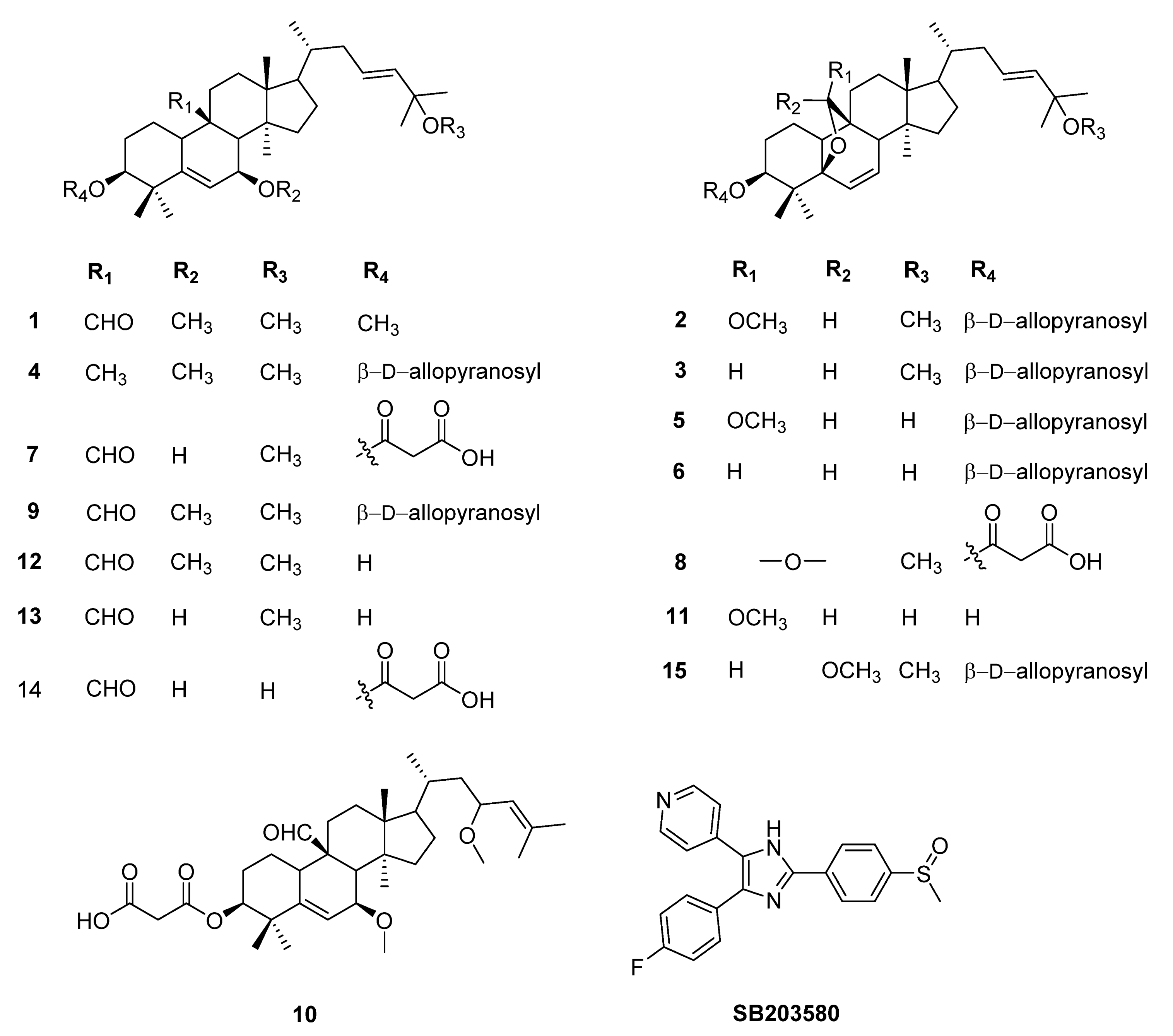
3.3. Extraction and Isolation

The dried fruits of M. charantia (5.0 kg) were cut into pieces and extracted with MeOH (10 L × 3 times) at room temperature. The methanol solution was concentrated using a rotatory evaporator to give a residue of the MeOH extract (360 g), which was further suspended in H2O and successively partitioned with n-hexane (H), dichloromethane (CH2Cl2), and ethyl acetate (EtOAc) to afford H (300 g), D (35 g) EtOAc (E, 40 g), and aqueous extracts (W), respectively. Extract D was separated on silica gel using a mobile phase of CH2Cl2-MeOH (100:1–1:1, v/v) to afford eight fractions D1–D8, respectively. Fraction D3 was further separated into nine subfractions by YMC CC, eluted with a gradient of MeOH-H2O (1:3 to 1:0, v/v) to give four subfractions (D3A–D3D), respectively. Fraction D3C was further purified by YMC CC with MeOH-H2O (2:1–4:1, v/v) to obtain compounds 1 (6.6 mg), 2 (5.8 mg), 3 (9.5 mg), 4 (15.6 mg), and 5 (16.8 mg). Purification of the subfraction D3D via silica gel column eluted with EtOAc-MeOH-H2O (7:1:0.1, v/v/v) was further isolated by RP-C18 silica gel (MeOH-H2O, 3:1, 6:1, v/v) to yield compounds 7 (9.0 mg), 8 (4.9 mg), 11 (26.5 mg), and 15 (6.6 mg). Fraction D6 was isolated by RP-C18 silica gel eluted with (MeOH-H2O, 3:1, 6:1, v/v), and Sephadex™ LH-20 column using MeOH-H2O (5:1. v/v) to afford compounds 9 (7.2 mg), 10 (15.3 mg), 12 (27.8 mg), 13 (315.9 mg), 14 (3.0 mg), and 6 (24.0 mg).

Physical and the Key of Spectroscopic Data of New Natural Compound

Compound 1. White amorphous powder, [α]D20 -77.8 (c = 0.2, MeOH); IR: 1H-NMR (400 MHz, CDCl3) δH 1.07 (s, 3H, H-26), 1.15 (s, 3H, H-26), 1.25 (s, 3H, H-26), 1.43 (s, 3H, H-26), 1.07 (s, 3H, H-26), 1.07 (s, 3H, H-26), 1.07 (s, 3H, H-26), 1.07 (s, 3H, H-26), 1.50 (s, 6H, H-26, and H-28), 5.71, 5.68 (OMe-3) and 13C-NMR (100 MHz, CDCl3): δC 21.7 (C-1), 29.9 (C-2), 80.0 (C-3), 41.7 (C-4), 146.1 (C-5), 121.5 (C-6), 75.1 (C-7), 50.6 (C-8), 36.9 (C-9), 22.7 (C-10), 29.4 (C-11), 29.4 (C-12), 45.7 (C-13), 48.2 (C-14), 34.9 (C-15), 27.7 (C-16), 50.1 (C-17), 15.0 (C-18), 208.0 (C-19), 36.5 (C-20), 18.9 (C-21), 39.5 (C-22), 128.5 (C-23), 137.1 (C-24), 75.6 (C-25), 30.8 (C-26), 30.8 (C-27), 26.2 (C-28), 27.3 (C-29), 18.2 (C-30), 56.1 (3-OMe), 51.2 (7-OMe), 50.3 (25-OMe). HR-ESI-MS: m/z: [M + Na]+ m/z 537.3918.

3.4. Cell Culture and Reagents

BMDCs were grown from wild-type C57BL/6 mice (Orient Bio Inc., Seoul, Korea) [31,32]. All animal procedures were approved by and performed according to the guidelines of the Institutional Animal Care and Use Committee of Jeju National University (#2016-0059). Briefly, bone marrow from the tibia and femur was obtained by flushing with Dulbecco’s Modified Eagle Medium (DMEM; Welgene, Gyeongsan, Korea) and bone marrow cells were cultured in RPMI-1640 medium containing 10% heat-inactivated fetal bovine serum (FBS; Gibco, New York, USA), 50 μM of 2-ME, and 2 mM of glutamine, supplemented with 3% J558L hybridoma cell culture supernatant containing the granulocyte-macrophage colony-stimulating factor (GM-CSF). The culture medium containing GM-CSF was replaced every other day. On day 6 of culturing, non-adherent cells and loosely adherent DC aggregates were harvested, washed, and resuspended in RPMI-1640, supplemented with 5% FBS. DCs were incubated in 48-well plates at a density of 1 × 10⁵ cells/0.5 mL and then treated with the isolated compounds at the indicated concentration for 1 h before stimulation with 10 ng/mL of LPS from Salmonella minnesota (Alexis, New York, NY, USA). Supernatants were harvested 18 h after stimulation. Concentrations of murine IL-12 p40, IL-6, and TNF-α in the culture supernatants were determined by ELISA (BD PharMingen, San Diego, CA, USA) following the manufacturer’s protocol [31,32]. All experiments were carried out at least three times. Data are presented as the mean and the standard deviation (SD) of three independent experiments.

3.5. Cytokine Production Measurements

The BMDCs were incubated in 48-well plates in 0.5 mL containing 1 × 10⁵ cells per well, and then treated with isolated compounds 115 at the indicated concentration for 1 h before stimulation with 10 ng/mL LPS from Salmonella Minnesota (Alexis, New York, NY, USA). Supernatants were collected 18 h after stimulation. Concentrations of murine IL-12 p40, IL-6, and TNF-α in the culture supernatants were identified by ELISA (BD PharMingen, San Diego, CA, USA) according to the manufacturer’s instructions.
The inhibitory activity (I) was expressed as the inhibition rate (%), which was calculated from the following formula:
I = Cdvc Cdcc Cdvc × 100
Cdvc: Cytokine level (ng/mL) in vehicle-treated DCs; Cdcc: Cytokine level (ng/mL) in compound treated DC. The data was obtained by at least three independent experiments performed in triplicate.

3.6. Cell Viability Assay

To identify the effects of isolated compounds on cell viability, the MTT assay was carried out [33]. BMDCs were incubated with 1 to 50 µM of isolated compounds for 18 h. The results demonstrate that tested compounds (115) displayed no notable cytotoxicity against BMDCs.

3.7. Statistical Analysis

All results are presented as the means ± SD. Data were analyzed by one-factor analysis of variance (ANOVA). * p-value < 0.05, and ** p-value < 0.01 were considered statistically significant. All experiments were repeated at least three times independently.

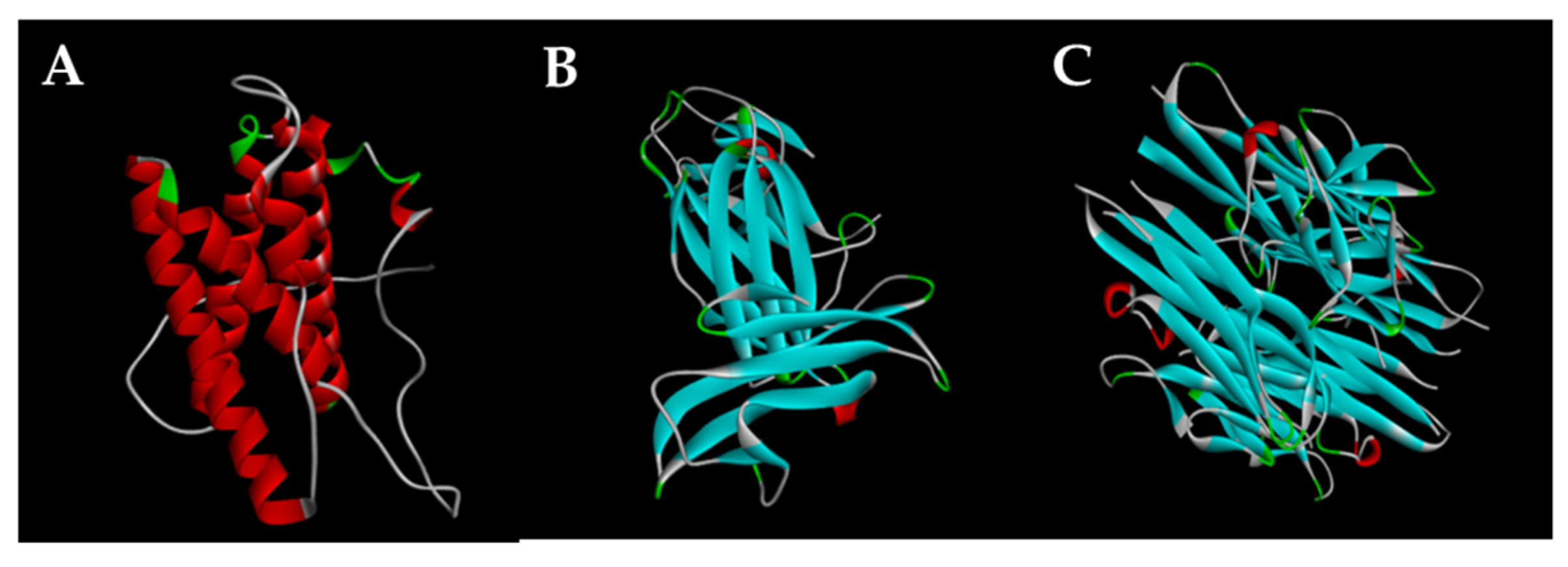
3.8. Preparation of Structures of Proteins Molecular Docking

The structures of IL-6, IL-12 p40, and TNF-α proteins resolved using X-ray diffraction (resolution of 2.16 Å) were downloaded from the Worldwide Protein Data Bank (PDB: 2L3Y, 1F42, and 2AZ5). The crystal structure was prepared for docking using Discovery Studio Client (2020). This step mainly involved the addition of missing disulfide bonds, removal of water molecules, the addition of missing hydrogen atoms, filling of missing amino acids side chains, and optimization of hydrogen bonds. The PyRx 0.9.4 virtual screening software was then used for restrained minimization until the average RMSD of the non-hydrogen atoms converged to 0.30 Å. Thus, this step allowed sufficient movement of heavy atoms to relax strained bonds, angles, and clashes.
The 2D chemical structure (flat structure) of the isolated compounds and positive control were automatically converted to the 3D chemical structure (three-dimensional structure) by the ChemBioOffice 12.0 software. The energy of the generated compounds was then minimized using the PyRx 0.9.4 software.

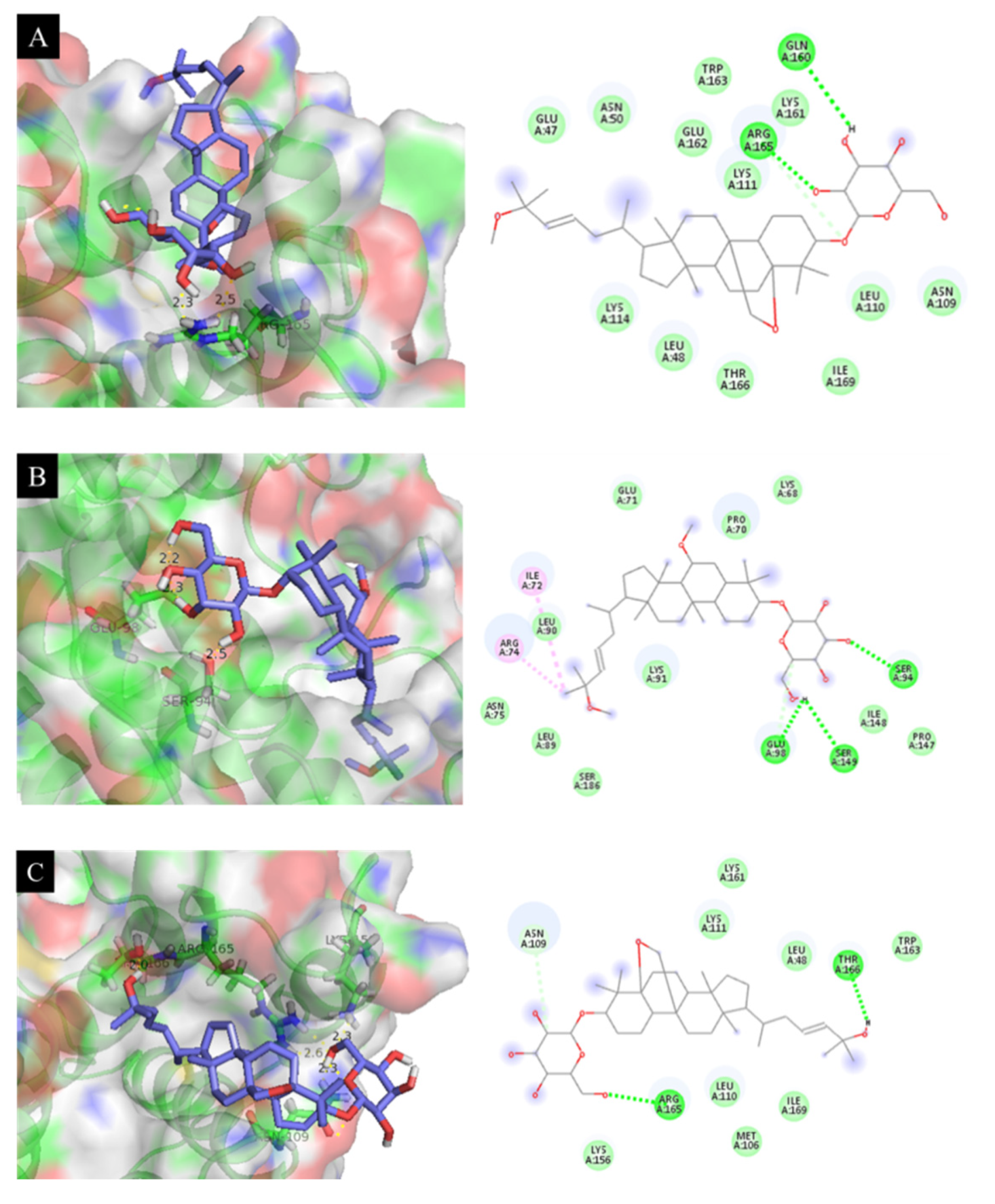
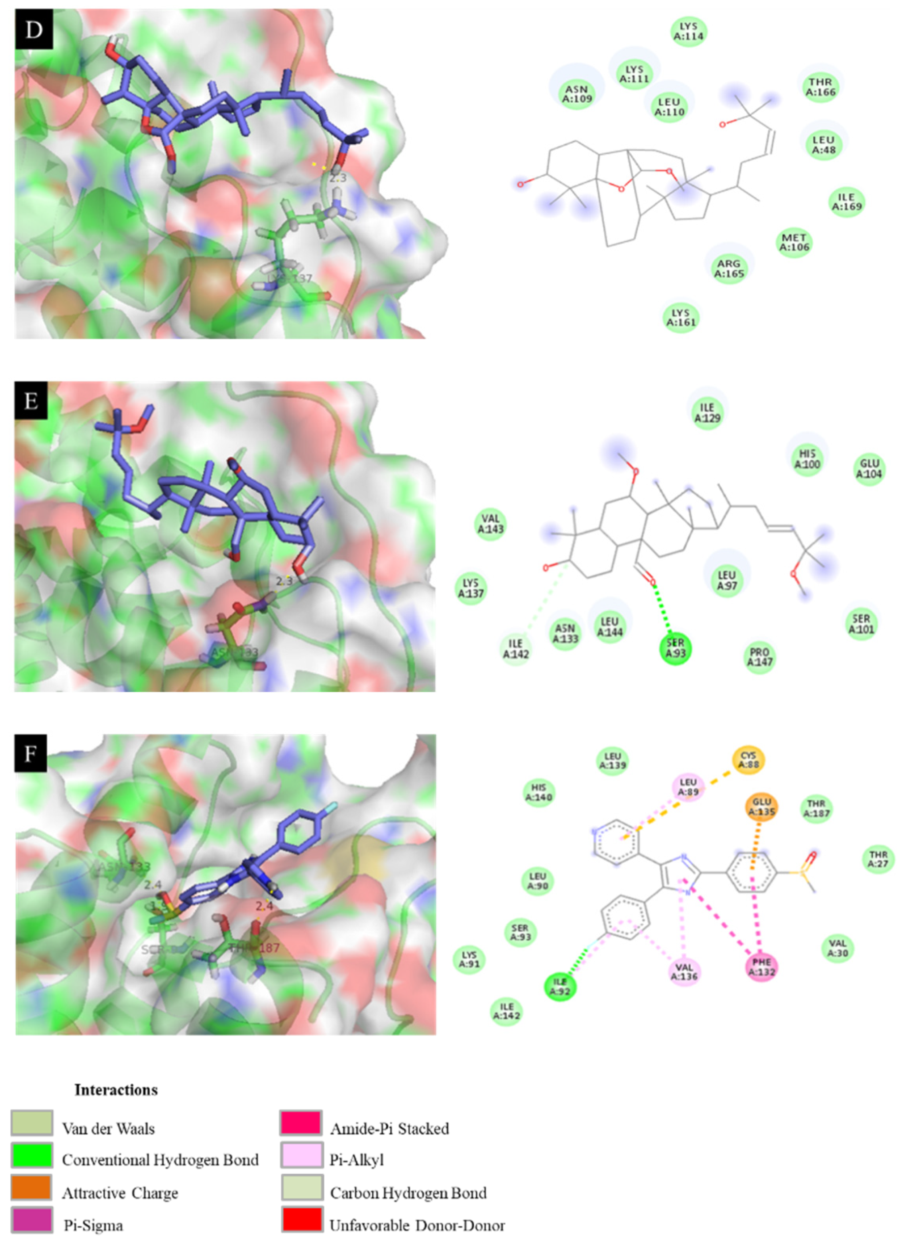
3.9. Molecular Docking of Isolated Compounds with IL-6, IL-12 p40, and TNF-α Proteins

Docking of compounds into prepared protein via conducting the docking process with the PyRx 0.9.4 software was carried out by following the method of placing compound fragments into the triangle matching and retaining the eight best configurations of each compound in the bonding complex for further analysis. The grid for docking studies was generated to enclose all the residues of IL-6, IL-12 p40, and TNF-α proteins making polar interactions with the atoms of the positive control. The best configuration is the one with the lowest docking score (DS) energy (kcal.mol–1). This score is the total energy consumed for the formation of bonding interactions between the compounds and the protein selected.

3.10. Docking Results Analysis

Discovery Studio Client was used for the analysis of the interactions between the compounds and target protein, and the performance of interaction on 2D and 3D planes. Various interactions, such as hydrogen bonds, van der Waals interactions, π–anion bonds, π–π bonds, and the interaction distance between amino acids and the active sites of compounds were plotted. Van der Waals interactions are detected by contact with hydrophilic and hydrophobic surfaces between the compounds and their bonding point.

Author Contributions

Isolation, structure, and identification of metabolites, T.Q.C., N.V.P., D.G. and S.Y.Y.; Bioassay experiments, V.-D.N., L.B.V., H.L.T.A. and Y.S.K. All research and the manuscript were carried out based on the planning of J.H.K. and S.Y.Y. All authors have read and agreed to the published version of the manuscript.

Funding

The research was supported by the Basic Science Research Program through the National Research Foundation of Korea (NRF) funded by the Ministry of Education, Science, and Technology (NRF-2018R1A6A3A11047338), and the basic research project (PJ016127032021) of the National Institute of Horticultural and Herbal Sciences, RDA.

Institutional Review Board Statement

The study was conducted according to the guidelines and approved by the Institutional Animal Care and Use Committee of Jeju National University (#2016-0059).

Informed Consent Statement

Not applicable.

Data Availability Statement

Not applicable.

Acknowledgments

The research was supported by the Basic Science Research Program through the National Research Foundation of Korea (NRF) funded by the Ministry of Education, Science, and Technology (NRF-2018R1A6A3A11047338), and the basic research project (PJ016127032021) of the National Institute of Horticultural and Herbal Sciences, RDA.

Conflicts of Interest

The authors declare no conflict of interest.

Sample Availability

Sample of the isolated compounds (115) available from the authors.

References

  1. Brooks-Worrell, B.; Palmer, J. Immunology in the Clinic Review Series; focus on metabolic diseases: Development of islet autoimmune disease in type 2 diabetes patients: Potential sequelae of chronic inflammation. Clin. Exp. Immunol. 2012, 167, 40–46. [Google Scholar] [CrossRef]
  2. Cao, T.Q.; Tran, M.H.; Kim, J.A.; Tran, P.T.; Lee, J.-H.; Woo, M.H.; Lee, H.-K.; Min, B.S. Inhibitory effects of compounds from Styrax obassia on NO production. Bioorg. Med. Chem. Lett. 2015, 25, 5087–5091. [Google Scholar] [CrossRef]
  3. Wang, R.X.; Zhou, M.; Ma, H.L.; Qiao, Y.B.; Li, Q.S. The Role of Chronic Inflammation in Various Diseases and Anti-inflammatory Therapies Containing Natural Products. ChemMedChem 2021, 16, 1576–1592. [Google Scholar] [CrossRef]
  4. Subramaniam, S.; Selvaduray, K.R.; Radhakrishnan, A.K. Bioactive compounds: Natural defense against cancer? Biomolecules 2019, 9, 758. [Google Scholar] [CrossRef] [PubMed] [Green Version]
  5. Mushtaq, S.; Abbasi, B.H.; Uzair, B.; Abbasi, R. Natural products as reservoirs of novel therapeutic agents. EXCLI J. 2018, 17, 420. [Google Scholar]
  6. Raman, A.; Lau, C. Anti-diabetic properties and phytochemistry of Momordica charantia L. (Cucurbitaceae). Phytomedicine 1996, 2, 349–362. [Google Scholar] [CrossRef]
  7. Perera, W.H.; Shivanagoudra, S.R.; Pérez, J.L.; Kim, D.M.; Sun, Y.; Jayaprakasha, G.K.; Patil, B.S. Anti-Inflammatory, antidiabetic properties and in silico modeling of cucurbitane-type triterpene glycosides from fruits of an indian cultivar of Momordica charantia L. Molecules 2021, 26, 1038. [Google Scholar] [CrossRef]
  8. Kubola, J.; Siriamornpun, S. Phenolic contents and antioxidant activities of bitter gourd (Momordica charantia L.) leaf, stem and fruit fraction extracts in vitro. Food Chem. 2008, 110, 881–890. [Google Scholar] [CrossRef] [PubMed]
  9. Akihisa, T.; Higo, N.; Tokuda, H.; Ukiya, M.; Akazawa, H.; Tochigi, Y.; Kimura, Y.; Suzuki, T.; Nishino, H. Cucurbitane-type triterpenoids from the fruits of Momordica charantia and their cancer chemopreventive effects. J. Nat. Prod. 2007, 70, 1233–1239. [Google Scholar] [CrossRef]
  10. Alam, S.; Asad, M.; Asdaq, S.M.B.; Prasad, V.S. Antiulcer activity of methanolic extract of Momordica charantia L. in rats. J. Ethnopharmacol. 2009, 123, 464–469. [Google Scholar] [CrossRef] [PubMed]
  11. Wang, N.; Yang, L.; Guo, X.-Y.; Zhang, Y.-Y.; Zhang, Y.; Liu, X.-W. Novel cucurbitane-type triterpene saponins from Hemsleya amabilis. J. Asian Nat. Prod. Res. 2020, 22, 30–37. [Google Scholar] [CrossRef] [PubMed]
  12. Sahranavard, S.; Naghibi, F.; Siems, K.; Jenett-Siems, K. New cucurbitane-type triterpenoids from Bryonia aspera. Planta Med. 2010, 76, 1014–1017. [Google Scholar] [CrossRef] [PubMed]
  13. Chen, C.; Qiang, S.; Lou, L.; Zhao, W. Cucurbitane-type triterpenoids from the stems of Cucumis melo. J. Nat. Prod. 2009, 72, 824–829. [Google Scholar] [CrossRef] [PubMed]
  14. Zhu, N.; Sun, Z.; Hu, M.; Li, Y.; Zhang, D.; Wu, H.; Tian, Y.; Li, P.; Yang, J.; Ma, G. Cucurbitane-type triterpenes from the tubers of Hemsleya penxianensis and their bioactive activity. Phytochemistry 2018, 147, 49–56. [Google Scholar] [CrossRef]
  15. Chen, J.-C.; Zhang, G.-H.; Zhang, Z.-Q.; Qiu, M.-H.; Zheng, Y.-T.; Yang, L.-M.; Yu, K.-B. Octanorcucurbitane and cucurbitane triterpenoids from the tubers of Hemsleya endecaphylla with HIV-1 inhibitory activity. J. Nat. Prod. 2008, 71, 153–155. [Google Scholar] [CrossRef]
  16. Huang, H.-T.; Zhang, L.-J.; Huang, H.-C.; Hwang, S.-Y.; Wu, C.-L.; Lin, Y.-C.; Liaw, C.-C.; Cheng, Y.-Y.; Morris-Natschke, S.L.; Huang, C.-Y. Cucurbitane-Type Triterpenoids from the Vines of Momordica charantia and Their Anti-inflammatory Activities. J. Nat. Prod. 2020, 83, 1400–1408. [Google Scholar] [CrossRef] [PubMed]
  17. Harinantenaina, L.; Tanaka, M.; Takaoka, S.; Oda, M.; Mogami, O.; Uchida, M.; Asakawa, Y. Momordica charantia constituents and antidiabetic screening of the isolated major compounds. Chem. Pharm. Bull. 2006, 54, 1017–1021. [Google Scholar] [CrossRef] [Green Version]
  18. Murakami, T.; Emoto, A.; Matsuda, H.; Yoshikawa, M. Medicinal foodstuffs. XXI. Structures of new cucurbitane-type triterpene glycosides, goyaglycosides-a,-b,-c,-d,-e,-f,-g, and-h, and new oleanane-type triterpene saponins, goyasaponins I, II, and III, from the fresh fruit of Japanese Momordica charantia L. Chem. Pharm. Bull. 2001, 49, 54–63. [Google Scholar] [CrossRef] [Green Version]
  19. Okabe, H.; Miyahara, Y.; Yamauchi, T. Studies on the constituents of Momordica charantia L. III. Characterization of new cucurbitacin glycosides of the immature fruits. Structures of momordicosides G, F1, F2 and I. Chem. Pharm. Bull. 1982, 30, 3977–3986. [Google Scholar] [CrossRef] [Green Version]
  20. Liu, J.-Q.; Chen, J.-C.; Wang, C.-F.; Qiu, M.-H. New cucurbitane triterpenoids and steroidal glycoside from Momordica charantia. Molecules 2009, 14, 4804–4813. [Google Scholar] [CrossRef] [PubMed]
  21. Tuan, N.Q.; Lee, D.-H.; Oh, J.; Kim, C.S.; Heo, K.-S.; Myung, C.-S.; Na, M. Inhibition of proliferation of vascular smooth muscle cells by cucurbitanes from Momordica charantia. J. Nat. Prod. 2017, 80, 2018–2025. [Google Scholar] [CrossRef] [PubMed]
  22. Yue, J.; Sun, Y.; Xu, J.; Cao, J.; Chen, G.; Zhang, H.; Zhang, X.; Zhao, Y. Cucurbitane triterpenoids from the fruit of Momordica charantia L. and their anti-hepatic fibrosis and anti-hepatoma activities. Phytochemistry 2019, 157, 21–27. [Google Scholar] [CrossRef] [PubMed]
  23. Yoshizaki, K.; Nishimoto, N.; Mihara, M.; Kishimoto, T. Therapy of Rheumatoid Arthritis by Blocking IL-6 Signal Transduction with a Humanized Anti-IL-6 Receptor Antibody. Springer Semin. Immunopathol. 1998, 20, 247–259. [Google Scholar] [CrossRef] [PubMed]
  24. Hennigan, S.; Kavanaugh, A. Interleukin-6 inhibitors in the treatment of rheumatoid arthritis. Ther. Clin. Risk Manag. 2008, 4, 767. [Google Scholar]
  25. Kudo, O.; Sabokbar, A.; Pocock, A.; Itonaga, I.; Fujikawa, Y.; Athanasou, N. Interleukin-6 and interleukin-11 support human osteoclast formation by a RANKL-independent mechanism. Bone 2003, 32, 1–7. [Google Scholar] [CrossRef]
  26. Zundler, S.; Neurath, M.F. Interleukin-12: Functional activities and implications for disease. Cytokine Growth Factor. Rev. 2015, 26, 559–568. [Google Scholar] [CrossRef]
  27. Trinchieri, G. Interleukin-12 and the regulation of innate resistance and adaptive immunity. Nat. Rev. Immunol. 2003, 3, 133–146. [Google Scholar] [CrossRef]
  28. Kim, T.S.; Kang, B.; Lee, M.; Choe, Y.K.; Hwang, S. Inhibition of interleukin-12 production by auranofin, an anti-rheumatic gold compound, deviates CD4+ T cells from the Th1 to the Th2 pathway. Br. J. Pharmacol. 2001, 134, 571–578. [Google Scholar] [CrossRef]
  29. Romanowska-Próchnicka, K.; Felis-Giemza, A.; Olesińska, M.; Wojdasiewicz, P.; Paradowska-Gorycka, A.; Szukiewicz, D. The Role of TNF-α and Anti-TNF-α Agents during Preconception, Pregnancy, and Breastfeeding. Int. J. Mol. Sci. 2021, 22, 2922. [Google Scholar] [CrossRef]
  30. Ting, H.-K.; Chen, C.-L.; Meng, E.; Cherng, J.-H.; Chang, S.-J.; Kao, C.-C.; Yang, M.-H.; Leung, F.-S.; Wu, S.-T. Inflammatory Regulation by TNF-α-Activated Adipose-Derived Stem Cells in the Human Bladder Cancer Microenvironment. Int. J. Mol. Sci. 2021, 22, 3987. [Google Scholar] [CrossRef]
  31. Vinh, L.B.; Heo, M.; Phong, N.V.; Ali, I.; Koh, Y.S.; Kim, Y.H.; Yang, S.Y. Bioactive Compounds from Polygala tenuifolia and Their Inhibitory Effects on Lipopolysaccharide-Stimulated Pro-inflammatory Cytokine Production in Bone Marrow-Derived Dendritic Cells. Plants 2020, 9, 1240. [Google Scholar] [CrossRef]
  32. Vinh, L.B.; Phong, N.V.; Ali, I.; Dan, G.; Koh, Y.S.; Anh, H.L.T.; Yang, S.Y.; Kim, Y.H. Identification of potential anti-inflammatory and melanoma cytotoxic compounds from Aegiceras corniculatum. Med. Chem. Res. 2020, 29, 2020–2027. [Google Scholar] [CrossRef]
  33. Ali, I.; Manzoor, Z.; Koo, J.-E.; Kim, J.-E.; Byeon, S.-H.; Yoo, E.-S.; Kang, H.-K.; Hyun, J.-W.; Lee, N.-H.; Koh, Y.-S. 3-Hydroxy-4, 7-megastigmadien-9-one, isolated from Ulva pertusa, attenuates TLR9-mediated inflammatory response by down-regulating mitogen-activated protein kinase and NF-κB pathways. Pharm. Biol. 2017, 55, 435–440. [Google Scholar] [CrossRef] [PubMed] [Green Version]
Figure 1. The isolated compounds from M. charantia (115) and SB203580.
Molecules 26 04444 g001
Figure 2. IL-6, IL-12 p40, and TNF-α proteins. (A) 2L3Y, (B) 1F42 and (C) 2AZ5.
Molecules 26 04444 g002
Figure 3. Docking simulation of the interactions between compounds 3 (A), 4 (B), 6 (C), 11 (D), 12 (E), and SB203580 (F), respectively, and the 2L3Y protein of IL-6 expression.
Molecules 26 04444 g003aMolecules 26 04444 g003b
Figure 4. Docking simulation of the interactions between compounds 46 (AC), 8 (D), 9 (E), 11 (F), 12 (G), and SB203580 (H), respectively, and the 1F42 protein of IL-12 p40 expression.
Molecules 26 04444 g004aMolecules 26 04444 g004b
Figure 5. Docking simulation of the interactions between compounds 4 (A), 6 (B), 9 (C), 1114 (DG), and SB203580 (H), respectively, and the 2AZ5 protein of TNF-α expression (AH).
Molecules 26 04444 g005
Table 1. Inhibition of isolated cucurbitane-type triterpenoids (115) on IL-6, IL-12 p40, and TNF-α production in LPS-stimulated bone marrow-derived dendritic cells.
CompoundsIC50 (µM)
IL-6IL-12 p40TNF-α
10.886 ± 0.1181.192 ± 0.1042.809 ± 0.264
21.616 ± 0.2110.733 ± 0.0412.616 ± 0.181
30.245 ± 0.0080.495 ± 0.0034.357 ± 0.037
40.363 ± 0.0150.031 ± 0.0010.810 ± 0.068
50.582 ± 0.0240.063 ± 0.0172.144 ± 0.151
60.381 ± 0.0390.012 ± 0.0000.043 ± 0.006
71.299 ± 0.2220.551 ± 0.0483.888 ± 0.149
81.348 ± 0.0520.085 ± 0.0051.568 ± 0.209
90.568 ± 0.0040.052 ± 0.0050.087 ± 0.033
100.858 ± 0.0020.553 ± 0.0102.143 ± 0.003
110.157 ± 0.0110.073 ± 0.0860.033 ± 0.002
120.028 ± 0.0050.045 ± 0.0260.142 ± 0.009
131.962 ± 0.0960.795 ± 0.0860.388 ± 0.015
140.751 ± 0.0380.299 ± 0.0020.811 ± 0.005
151.553 ± 0.2451.360 ± 0.1013.716 ± 0.011
SB203580a5.000 ± 0.0803.500 ± 0.0807.200 ± 0.060
a: Positive control.
Table 2. Docking simulation results with docking score energy (DS) and root-mean-square deviation (RMSD) between isolated compounds (3, 4, 6, 11, and 12) and the 2L3Y protein.
CompoundsDS (kcal·mol−1)RMSD (Å)Interaction with Amino Acid
3−7.1818.64Arg 165 (2.3 Å)
4−7.4817.07Ser 94 (2.5 Å), Glu 98 (2.2 Å)
6−7.2621.27Lys 156 (2.3 Å), Thr 166 (2.0 Å), Arg 165 (2.6 Å), Asn 109 (2.3 Å)
11−6.9918.85Lys 137 (2.3 Å)
12−6.4111.79Asn 133 (2.3 Å)
SB203580 a−7.967.02Asn 133 (2.4 Å), Ser 93 (1.9 Å), Thr 187 (2.4 Å)
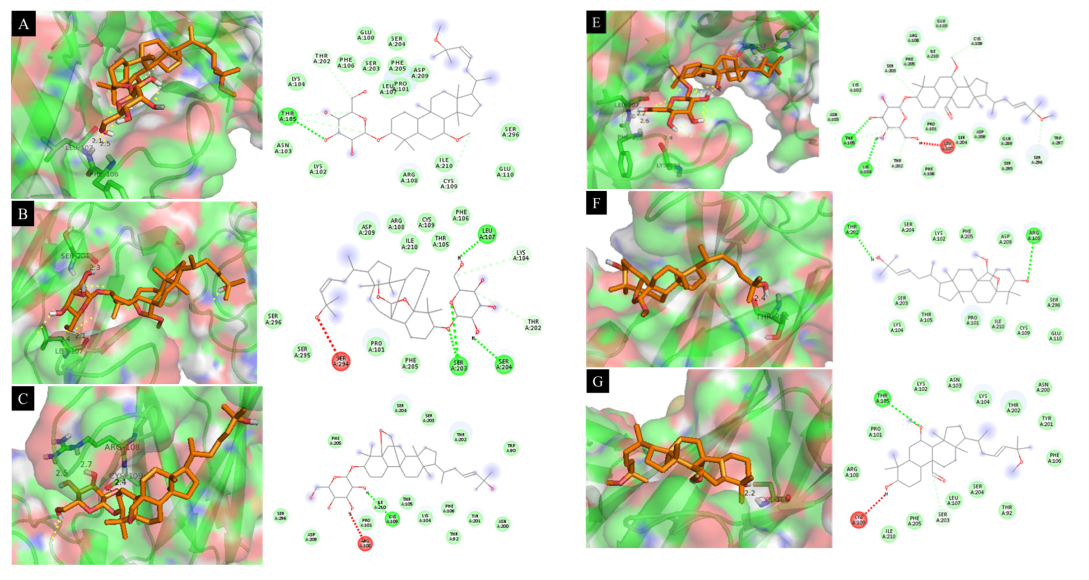
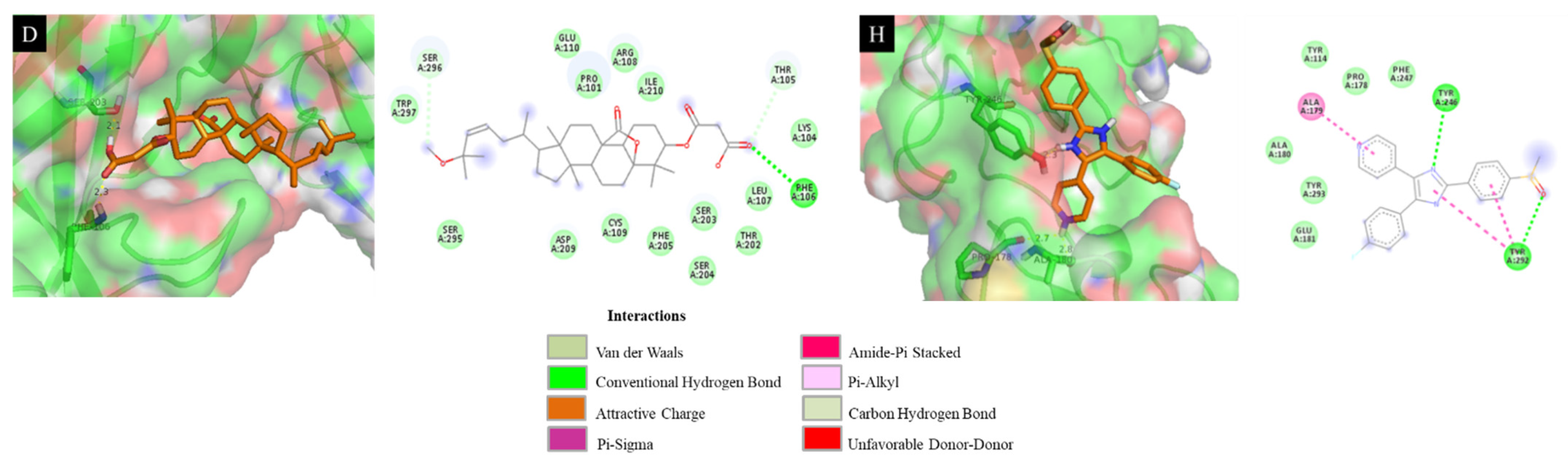
a: Positive control.
Table 3. Docking simulation results with docking score energy (DS) and root-mean-square deviation (RMSD) between isolated compounds (46, 8, 9, 11, and 12) and the 1F42 protein.
CompoundsDS (kcal·mol−1)RMSD (Å)Interaction with Amino Acid
4−6.9312.15Leu 107 (2.1 Å), Phe 106 (2.5 Å)
5−7.285.67Ser 204 (2.3 Å), Leu 107 (2.1 Å)
6−7.3413.21Arg 108 (2.5 Å), Cys 109 (2.4 Å)
8−6.9811.93Ser 203 (2.1 Å), Phe 106 (2.3 Å)
9−7.0213.86Trp 297 (2.3 Å), Leu 107 (2.2 Å), Phe 106 (2.6 Å), Lys 104 (2.4 Å)
11−6.6616.38Thr 202 (2.4 Å)
12−6.3210.39Cys 109 (2.2 Å)
SB203580 a−7.4111.14Tyr 246 (2.3 Å), Pro 178 (2.7 Å), Glu 181 (2.8 Å)
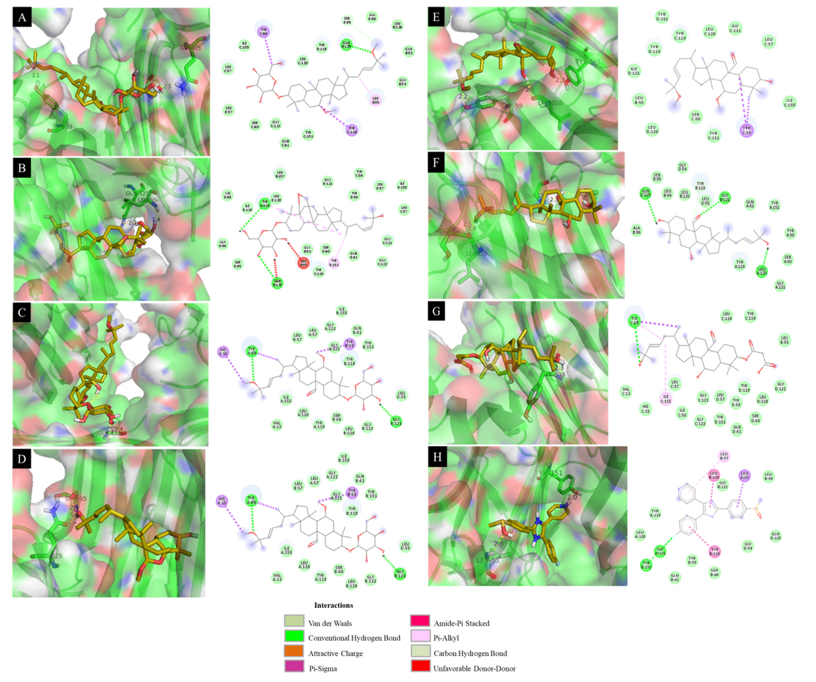
a: Positive control.
Table 4. Docking simulation results with docking score energy (DS) and root-mean-square deviation (RMSD) between isolated compounds (4, 6, 9, and 1114) and the 2AZ5 protein.
CompoundsDS (kcal·mol−1)RMSD (Å)Interaction with Amino Acid
4−8.2310.86Tyr 59 (2.1 Å), Gln 125 (2.5 Å), Gly 121 (2.2 Å)
6−8.4012.39Leu 55 (2.4 Å), Gln 125 (2.7 Å)
9−8.229.15Gly 121 (2.4 Å)
11−8.7215.28Ser 95 (2.6 Å), Gln 125 (2.1 Å)
12−7.8314.34Tyr 151 (2.3 Å), Leu 120 (2.6 Å), Tyr 59 (2.2 Å)
13−8.399.48Gly 121 (2.3 Å), Leu 120 (1.9 Å), Ser 60 (2.5 Å)
14−8.1013.16Tyr 119 (2.3 Å)
SB203580 a−8.2214.53Leu 55 (2.8 Å), Tyr 151 (2.0 Å)
a: Positive control.
Publisher’s Note: MDPI stays neutral with regard to jurisdictional claims in published maps and institutional affiliations.

